Article

Antioxidant Properties and Kidney Cell Protection by the Extracts of Curcuma longa, Artemisia princeps, Salicornia herbacea, and Schisandra chinesis

1
Department of Animal Science and Technology, Konkuk University, 120 Neungdong-ro, Gwangjin-gu, Seoul 05029, Republic of Korea
2
Institute of Animal Resource Center, Konkuk University, Seoul 05029, Republic of Korea
*
Author to whom correspondence should be addressed.
These authors contributed equally to this work.
Fermentation 2022, 8(12), 702; https://doi.org/10.3390/fermentation8120702
Submission received: 2 November 2022 / Revised: 24 November 2022 / Accepted: 30 November 2022 / Published: 2 December 2022
(This article belongs to the Section Microbial Metabolism, Physiology & Genetics)

Abstract

:
This study evaluated the antioxidant properties and kidney cell protection of medicinal plants as natural medicaments. A total of four medicinal plants including turmeric (TM), gangwha mugwort (GM), glasswort (GW), and omija (OM) were selected and fermented. Hot water extracts (HWE) and ethanol extracts (EE) of the plants were prepared before and after fermentation and tested in experiments in vitro and in vivo. Total polyphenol contents (TPC) and total flavonoid content (TFC) of GM were the highest among them. The TPC based HWE decreased after fermentation except OM; in contrast, TFC from HWE increased. The DPPH radical scavenging activity and ABTS value from HWE increased after fermentation, especially OM, which showed significant differences, while DPPH and ABTS from EE were decreased. The cell viability was not changed after addition of these plants extracts below 50 μg/mL; however, TM from HWE significantly decreased. The protective effect on kidney cells against cisplatin showed a 60% range of cell viability in each plant extract. In the in vivo experiment, the protective effect on kidney cells by the supplemented plant extracts was demonstrated by the serum creatinine and BUN level. During experimental periods, the serum creatinine and BUN level of GW and GM-treated mice decreased with significant differences compared to the adenine control group. As a result, these plant extracts had no cytotoxicity and maintained a protective effect as well as antioxidant activity. These results suggest that plants such as gangwha mugwort (GM) and glasswort (GW) may be good extracts for kidney cell protection and antioxidant agents.

1. Introduction

Recently there has be great interest on herbs and natural compounds as complementary and alternative medicines [1]. Various plants species have been utilized for many medical, nutraceutical, flavor, food additive, and cosmetics applications. Medicinal herbs are attractive for healthcare because they have fewer adverse effects, are safer, and are more effective than synthetic medications [2]. Many plants contain secondary metabolic compounds with a wide range of biological activity and pharmaceutical formulations [3]. Nowadays, much attention is focused on antioxidants that can suppress active oxygen radical-mediated peroxidation, which causes fatal diseases such as cancer, cardiovascular disease, and stroke, among many others. Natural antioxidants can be used more safely and effectively than synthetic antioxidants, which have potential toxicity [4]. Natural plants such as turmeric (TM) (Curcuma longa), ganghwa mugwort (GM) (Artemisia princeps), glasswort (GW) (Salicornia herbacea L.), and omija (OM) (Schisandra chinensis) containing physiologically active substances were useful for anti-inflammatory, antioxidant, antibacterial, and antiviral activity.
Turmeric (TM) (Curcuma longa L.), commonly used as a spice in India, is cultivated in a tropical region and has antioxidant, anti-inflammatory, and antibacterial effects [5]. It contains nontoxic polyphenolic derivatives of curcumin, which have biological activities against coughs, colds, dental issues, ingestion, skin infections, blood purification, asthma, piles, bronchitis, tumor, and hepatic disorders [6]. Ganghwa mugwort (GM) (Artemisia princeps Pampanini cv. Sajabal), widely distributed in East Asia, has been used for women’s sickness in folk medicament with therapeutic effects on inflammation, diarrhea, and gastric ulcers [7]. GM contains sesquiterpene, lactone, and flavonoids, which have antimicrobial, insecticidal, and anticancer activities. It can also be used to cure neurological problems, dermal infections, and epileptic disorders, and possess potent immunomodulatory properties [8]. Glasswort (GW) (Salicornia herbacea L.) belongs to the Amaranthaceae family with halophytes containing functional nutrients such as polyphenols, flavonoids, and fibers [9], which have antioxidant, antibacterial, anticancer, and weight loss effects. GW can be used against oxidative stress, inflammation, diabetes, asthma, hepatitis, cancer, and gastroenteritis [10]. Its medicinal function appears to be immunomodulatory, lipid-lowering, antiproliferative, osteoprotective, and hypoglycemic rendering [11]. Omija (OM) (Schisandra chinensis) is grown in Asia, and has been reported for its antioxidant, anticancer, and anti-inflammatory effects [12]. It is well known as a medicinal herb containing terpenoids, polysaccharides, and lignans, and is used to treat diseases such as dyspnea, hepatitis, bronchitis, and coronary heart disease. In particular, lignans in omija are the main bioactive substances with antioxidant, anticancer, and anti-inflammatory, immune-regulatory activity, antimicrobial, and antiviral properties [13]. Fermented mugwort showed a higher anti-inflammatory effect than unfermented mugwort [14]. It is known that fermentation improves in the biological properties, such as the physiological activity, of plants, vegetables, and herbs, and that bioactive compounds like antioxidants [15,16]. This study was performed to investigate the antioxidant effects and protective effects on kidney cell of the above medicinal plants extracts in vivo as well as in vitro to elucidate their potential as a functional food ingredient.

2. Materials and Methods

2.1. Preparation of Medicinal Plants

TM, GW, GM, and OM were purchased from Korean food companies (TM: Tojongherb, Namyangju, Gyeonggi-do, Republic of Korea, GM: Aedam-hyang, Ganghwa-gun, Incheon, Republic of Korea, GW: Suncheon Bay Hamcho Agricultural Cooperative Corporation, Suncheon-si, Jeollanam-do, Republic of Korea, OM: Buan Audinue, Jeollabuk-do, Republic of Korea) in fine powder form.

2.2. Microbes for Fermentation

Fermentation of the medicinal plants was carried out using their indigenous microorganisms. Enterococcus faecium SK4357, E. faecium SK4373, and E. faecium SK4369 were used as a starter culture for the fermentation of TM, GM, and GW, respectively, which had been previously isolated from the respective plant [17]. Yeast, Wickerhamomyces anomalus SK1819, which had been previously isolated from omija byproducts, was used for the fermentation of OM [18].

2.3. Fermentation of Medicinal Plants

Each of the plant powders at a final concentration of 5% (w/v) was added to their respective media. Bacillus minimal medium (BMM) was used for the fermentation of TM, GM, and GW, and one-fifth diluted yeast malt (YM) medium (Becton, Dickinson and company) was used for the fermentation of OM. The media was sterilized after adjusting the pH to 7.0 ± 0.50. Then, 1% (v/v) of the respective starter culture was inoculated and fermentation was carried out using a shaking incubator (BF-60SIRL, Biofree, Seoul, Republic of Korea) at 37 °C (TM, GM, GW) and 30 °C (OM) under shaking (100 rpm) for 48 h. The samples were collected at 0, 4, 8, 12, 16, 24, 36, and 48 h to determine bacterial counts and pH. The pH value was immediately measured using a pH meter (pH/ISE Meter 735P, Istek Co., Ltd., Seoul, Republic of Korea). Viable cell counts were determined by the drop plate method [19]. Three milliliters of fermentation solution was collected at each time point and stored at −20 °C for further analysis.

2.4. Extraction of Fermented Plant Products

To compare the physiological activity of plants before and after fermentation, a sample at 0 h before fermentation and a sample at 16 h, a time set in consideration of the optimal viable count of bacteria and pH, were collected. The samples that were stored at −20 °C were taken out and then thawed at room temperature. Afterward, 7 mL of distilled water or 70% ethanol was added to 3 mL of the sample. In the case of HWE, the samples were boiled in a water bath (Wise Bath, Seoul, Republic of Korea) at 100 °C for 15 min, and then cooled to room temperature. All samples were centrifuged at 14,500 rpm for 10 min (Mega 17R; Hanil, Seoul, Republic of Korea), and the supernatant was collected and frozen in a deep freezer at −80 °C. For EE, samples were sonicated at room temperature (20~30 °C) for 15 min (WUC-D03H, Daihan Scientific, Seoul, Republic of Korea) and incubated overnight in a shaking incubator at 30 °C and 100 rpm. After centrifuging all samples at 14,500 rpm for 10 min (Mega 17R; Hanil, Seoul, Republic of Korea), the supernatant was collected, and the EE sample was concentrated under reduced pressure at 40 °C, using a rotary evaporator (CCA-1100; Eyela, Tokyo, Japan). After freezing the extracts in a deep freezer at −80 °C, the samples were freeze-dried using a freeze-dryer (UniFreeze FD-8, Daihan Scientific Ltd., Seoul, Republic of Korea).

2.5. Estimation of Total Polyphenol Contents

The total phenolic contents (TPC) of fermented and non-fermented samples were measured according to the Folin–Ciocalteu method [20]. Briefly, 20 μL of the sample (5 mg/mL) and 100 μL of 0.2 N Folin-Ciocalteu solution were mixed and allowed to react at room temperature for 5 min; then 80 μL of 7.5% Na2CO3 solution was added and kept in a dark for 60 min. Absorbance was measured using a microplate reader (Synergy 2, BioTek Instruments Inc., Winooski, VT, USA) at 750 nm against a blank (70%, v/v ethanol or hot water) and expressed as mg of gallic acid equivalent per gram of extract (mg GAE/g of extract).

2.6. Estimation of Total Flavonoid Contents

Total flavonoid contents (TFC) of fermented and non-fermented samples were estimated according to a published method. Briefly, 20 μL of the sample (25 mg/mL), 180 μL of 90% diethylene glycol, and 20 μL of 1N NaOH were mixed and allowed to react for 60 min in the darkroom. Absorbance was measured at 405 nm against a blank and expressed as mg of quercetin equivalent per gram of extract (mg QE/g of extract).

2.7. Antioxidant Activity Assays

The antioxidant activities of non-fermented and fermented extracts were determined according to the scavenging abilities against 2,2-diphenyl-1-picrylhydrazyl (DPPH) and 2,2′-azino-bis (3-ethylbenzothiazoline-6-sulphonic acid) (ABTS), as described previously.

2.8. Determination of DPPH Radical Scavenging Activity

For the DPPH radical assay, 20 μL of sample (5 mg/mL) was mixed with 180 μL of 0.2 mM DPPH in a 96 well plate and incubated for 30 min in the darkroom before measuring the DPPH of the EE. The DPPH of the HWE (GM and GW, 5 mg/m; TM and OM, 10 mg/mL) was also measured in the same manner as above except for OM. In case of OM, 5 μL of HWE was mixed with 195 μL of 0.2 mM DPPH. The DPPH radical scavenging activities were expressed as mg of Trolox equivalent per gram of extract (mg TE/g of extract). The constant absorbance readings at 517 nm were recorded using a spectrophotometer (UV-1601, Shimadzu, Kyoto, Japan) at 5 min, and inhibition of DPPH oxidation (%) was calculated as follows:
% Inhibition = (Acontrol − Aextract)/Acontrol × 100
where A is absorbance at 517 nm.

2.9. Determination of ABTS Radical Scavenging Activity

To measure ABTS radical scavenging activity, a stock solution containing 7 mM ABTS radical solution and 2.45 mM potassium persulfate solution was prepared. The working solution was then prepared by mixing the two stock solutions in equal quantities and allowing them to react for 12~16 h at room temperature in the dark. The solution was then diluted to obtain an absorbance of ~0.7 ± 0.02 units at 734 nm using the ELISA microplate reader (Synergy 2, BioTek Instruments Inc., Winooski, VT, USA). Five microliters of EE (5 mg/mL) was reacted with 195 μL of the working solution for 6 min in dark. The ABTS of HWE (5 mg/mL) was also measured in the same manner as above except for OM HWE (10 mg/mL). In the case of OM, 5 μL of HWE was reacted with 195 μL of working solution for 6 min in the dark. The absorbance of all samples for ABTS was then taken at 734 nm for the blank. The ABTS radical scavenging activities were expressed as mg of Trolox equivalent per gram of extract (mg TE/g of extract).

2.10. Cell Culture and Cell Viability of Rat Kidney LLC-PK1

LLC-PK1 cells were cultivated and then incubated for 24 h in DMEM (Dulbecco’s modified eagle medium) supplemented with 10% bovine calf serum (BSA, Gibco, Waltham, MA, USA), and 1% penicillin- streptomycin (Sigma, St. Louis, MO, USA) at 37 °C in a humidified incubator for 24 h. The cell viability was determined using the MTT assay, as a described by Kim et al. [21]. LLC-PK1 cells were transferred to a 96-well plate at a density of 3 × 10³ cells/well and incubated at 37 °C for 24 h. Cells were treated with fermented plant extracts at different concentrations ranging 0.2~50 μg/mL and the cultures were incubated for an additional 24 h. Following the incubations, MTT (5 mg/mL) solution was added to the cultures, which were further incubated for 1 h. The supernatant was removed from each well, and dimethyl sulfoxide (DMSO) was added to dissolve the MTT formazan. The absorbance of each well was measured using a microplate reader at 490 nm in triplicate. Cell viability was calculated using the following equation:
Cell viability (%) = (absorbance of sample/absorbance of control) × 100

2.11. Determination of Serum Creatinine and BUN Levels in Rats

Sprague-Dawley male rats weighting 250 g were purchased from Orient Bio INC. Republic of Korea. Fifty rats were assigned to five groups: one group received a control diet, and the other groups were fed an alanine diet for 3 weeks. The five diet groups were as follows: normal control; adenine (0.75%) + vehicle control; and adenine (0.75%) + 300 mg/5 mL GM, GW, or OM. The rats on adenine-induced kidney injury were supplemented with 0.75% alanine and other groups were additionally fed each fermented plant extract at 300 mg/kg body weight. All treatments were administered orally by feeding for 3 weeks. Body weight was measured at every week with everyday care for food consumption. Following the treatment period, rats were fasted for 12 h and then anesthetized to measure the serum creatinine and urea concentration. Serum creatinine levels were measured by the Jaffe reaction method using a blood chemistry analyzer (AU 480, Beckman coulter, Krefeld, Germany). Blood urea nitrogen (BUN) was measured by the enzyme method (Eiken, Tokyo, Japan) using a biochemical analyzer [22]. All experimental protocols were approved by Institutional Animal Care and Use Committee of NDIC, South Korea (permit No. p222010).

2.12. Statistical Analysis

All data were expressed as mean ± standard deviation of the mean (SEM), and all experiments were performed at least in triplicate. IBM SPSS Statistics 25 Windows software (IBM, New York, NY, USA) was used for all data analysis. Data for total phenol contents and antioxidant activity were statistically evaluated using paired-comparison t-test and Duncan’s multiple range test was performed to compare the differences using one-way ANOVA (p ≤ 0.05).

3. Results and Discussion

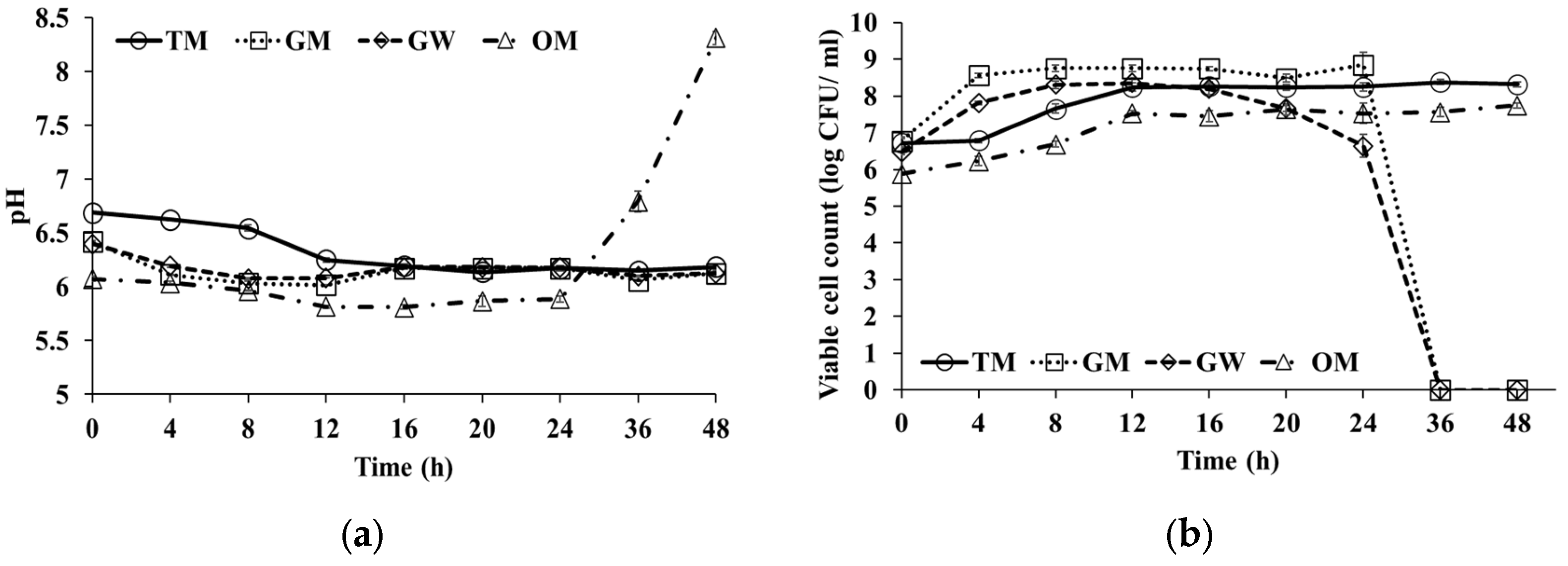
3.1. pH Changes in during Fermentation

Fermentation trends of the four different medicinal plants were examined through measuring changes in pH level (Figure 1a). Prior to the inoculation at 0 h, we recorded that each plant material has an approximate pH of 6.2 ± 0.5. During the fermentation, the pH for TM (turmeric) decreased linearly until 16 h and remained constant thereafter. Similarly, the pH of GM (gangwha mugwort) and GW (glasswort) also decreased until 12 h, and where then maintained during the remaining course of fermentation. Intriguingly, the pH of OM (omija) initially decreased until 12 h and remained unchanged during the following 12 h, and showed a significant increase in pH after 24 h incubation.
pH changes for TM, GM, and GW fermentation were similar to those reported previously by Park et al. [17]. Occasionally, when the lactic acid bacterium (LAB) Enterococcus faecium grows, lactic acid production activity decreases following the fermentative degradation and the release of plant metabolites. Since the glassworts are alkaline [23], they may have compensated the pH decrease with alkaline substrates. However, the pH of the OM substrate decreased during the course of incubation owing to the release of ester compounds through microbial fermentation [24]. However, the pH of the OM fermentation batch increased again after 24 h as its organic acid contents were used as alternative C-source for microbial growth following depletion of the sugars during the later stages of incubation [25].

3.2. Microbial Growth during Fermentation

For all medicinal plant substrates, we observed that the viable cell counts increased linearly from inoculation stage with ~107 CFU/mL to the range of 108~109 CFU/mL (Figure 1b). Viable cell counts for TM fermented product increased significantly up to 12 h and remained constant thereafter during the fermentation. For TM, the cell viability increased to 108.5 CFU/mL during the first 4 h of incubation and remained broadly constant until 48 h. In the GW fermentation batch, the cell viability increased to 107.5 CFU/mL after 4 h of inoculation and was maintained as such until 16 h, and then decreased significantly after 24 h. Similarly for GM fermentation, viable cell counts were linearly increased until 4 h, thereafter remained stationary, and then decreased after 24 h. For the OM fermentation batch, the yeast inoculum Wickerhamomyces anomalus was first increased up to 12 h, and then maintained a constant viable cell counts during the remaining incubation period.
Notably, the observed changes in total microbial counts for TM, GM, and GW fermentation followed the similar trends reported previously by Park et al. [17]. In the case of OM, W. anomalus (optimal pH = 3.5~7.0) used as inoculum has properties that allow its grown and survival under extreme environmental stress conditions including low water activity, high osmotic pressure, and anaerobic conditions, which might have contributed toward its high cell viability during fermentation [26].

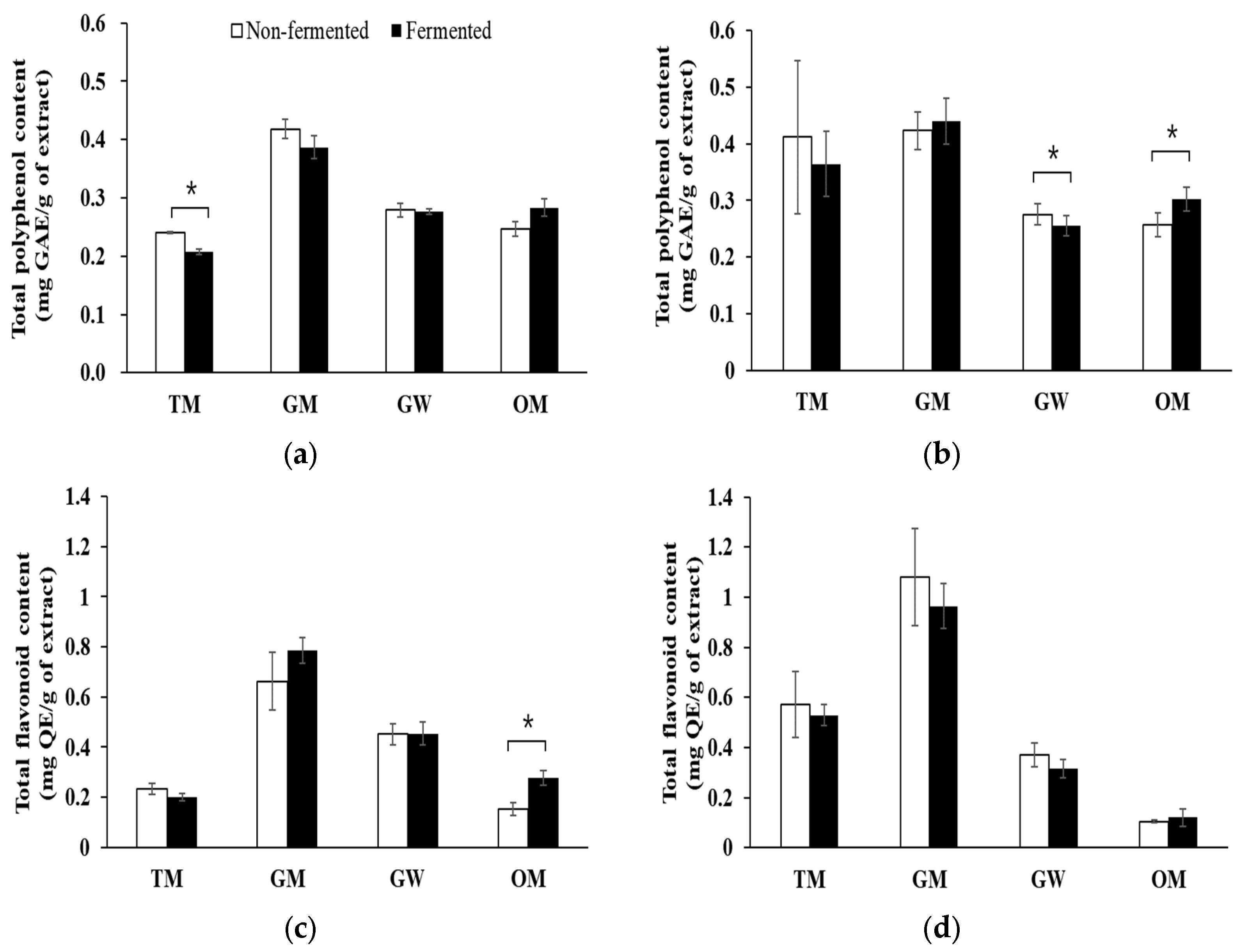
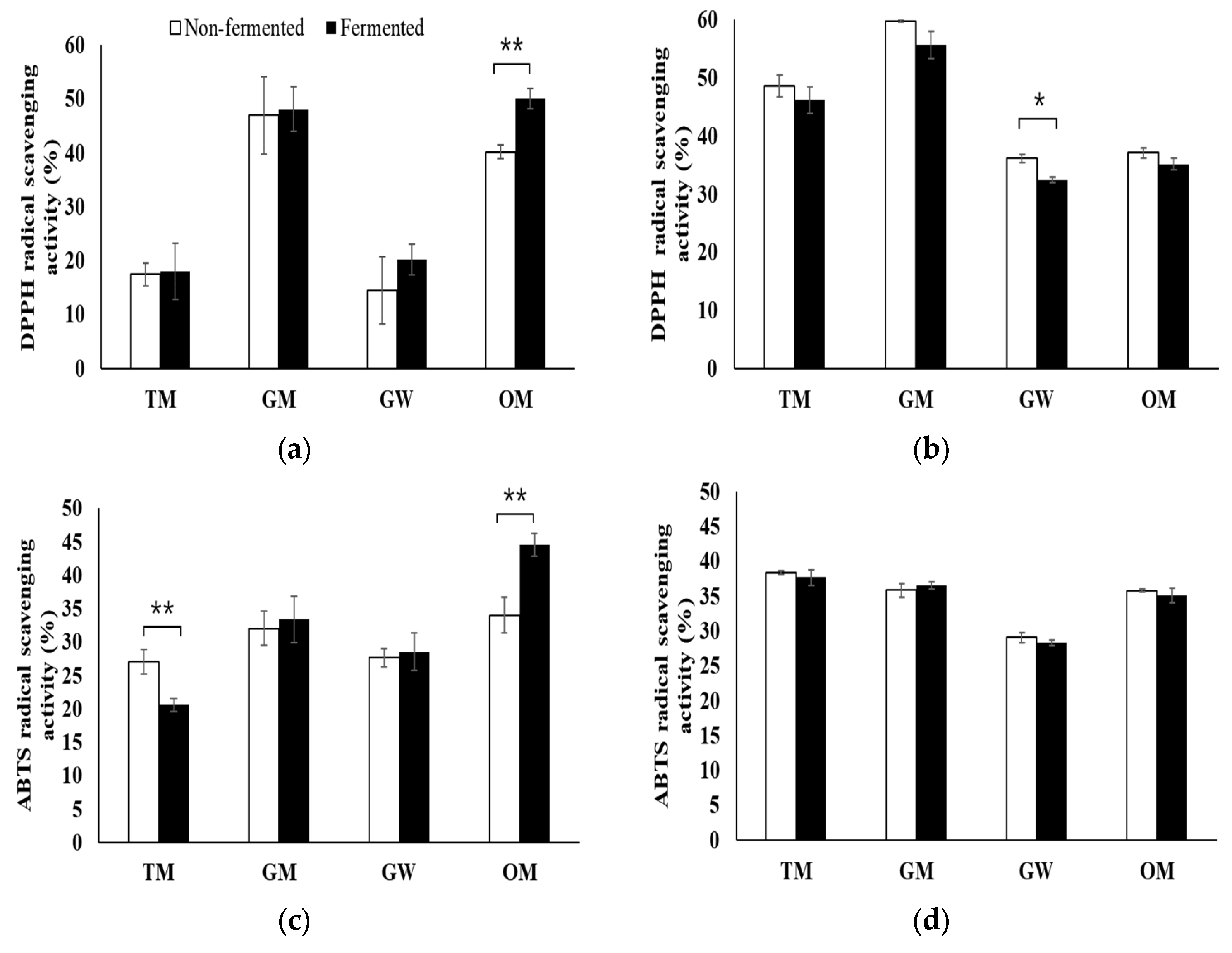
3.3. Phenolic Compound Contents and Antioxidant Activities of Medicinal Plant Extracts

The total polyphenol content (TPC) and TFC from hot water and ethanol extracts of the plants are shown in Figure 2.
The phenolic contents of GM were the highest among the extracts, followed by GW, OM, and TM. TPC from HWE decreased after fermentation, especially TM with significant differences, except for OM. For both the HWE and ethanol extract (EE), fermented OM showed a higher TPC than the non-fermented ones. The elevated TPC in herbs is associated with an increase in the antioxidant activities. Phenolic compounds are one of the most critical ingredients related to free radical scavenging activity in medicinal plants and functional foods [27]. Polyphenols usually detected in plants can be classified into two groups, flavonoids and non-flavonoids, and may be responsible for various pharmacological activities [28]. They exhibit various biological properties, such as strong antioxidant, cardioprotective, antimutagenic, antibacterial, antiviral and anti-inflammatory activities [29].
As shown in Figure 2c,d, total flavonoid content (TFC) from the HWE increased after fermentation, especially for OM with significantly differences compared to the non-fermented one. In contrast, TFC for the EE decreased after fermentation. The health functions of polyphenols, including flavonoids, serve as powerful antioxidants due to their ROS scavenging activities, thereby suppressing the pathogenesis of age-related degenerative diseases, such as diabetes, cardiovascular diseases, cancer, and neuro-degenerative malfunctions [30].
In congruence with the polyphenol contents, all medicinal plant substrates showed varying antioxidant activities following the fermentation (Figure 3).
The DPPH scavenging activity from HWE of all plants substrates increased marginally after fermentation, especially fir OM with significantly differences, while the EE displayed the opposite trend with a decrease in antioxidant bioactivities. DPPH activity for EE for all medicinal plant substrates displayed a 35~60% range. Among the fermented samples, the DPPH bioactivity for the GM based on EE was the highest followed by TM, GW, and OM. We postulate that this decreased antioxidant activity for the medicinal plant extract is affected by the growth and metabolism of Enterococcus faecium on antioxidant metabolites in fermentative substrates [31].
ABTS based on HWE ranged from 20~45% for fermentation plant samples for GM, GW, and OM, which was increased compared to the non-fermented control samples except in TM (Figure 3c). However, the ABTS bioactivity based on the EE marginally decreased for all fermented medicinal plant samples (Figure 3d).
Previously, some studies have reported the increasing antioxidant activities of medicinal plants following fermentation, which can be attributed to the release of antioxidant phenolic compounds or biotransformation of the metabolites from the plant matrix used as a substrate [32]. Antioxidant activity is affected to the presence of several phenolic compounds (gallic acid, coumaric, vanillic, salicylic acid, etc.), and flavonoids, including quercetin and rutin. Furthermore, plant metabolites such as maillard, peptides, amino acids, tocopherol, and carotenoids also function as antioxidant synergistically [33]. Although ethanol was reported as a better solvent than water for extracting polyphenol compounds from GW [34], we observed no significant difference in the bioactivities for the two different extractions used in this study. In congruence to our observations, Kim et al. [35] reported that there was little difference in the bioactivities of grass-based substrates extracted using hot water and ethanol.

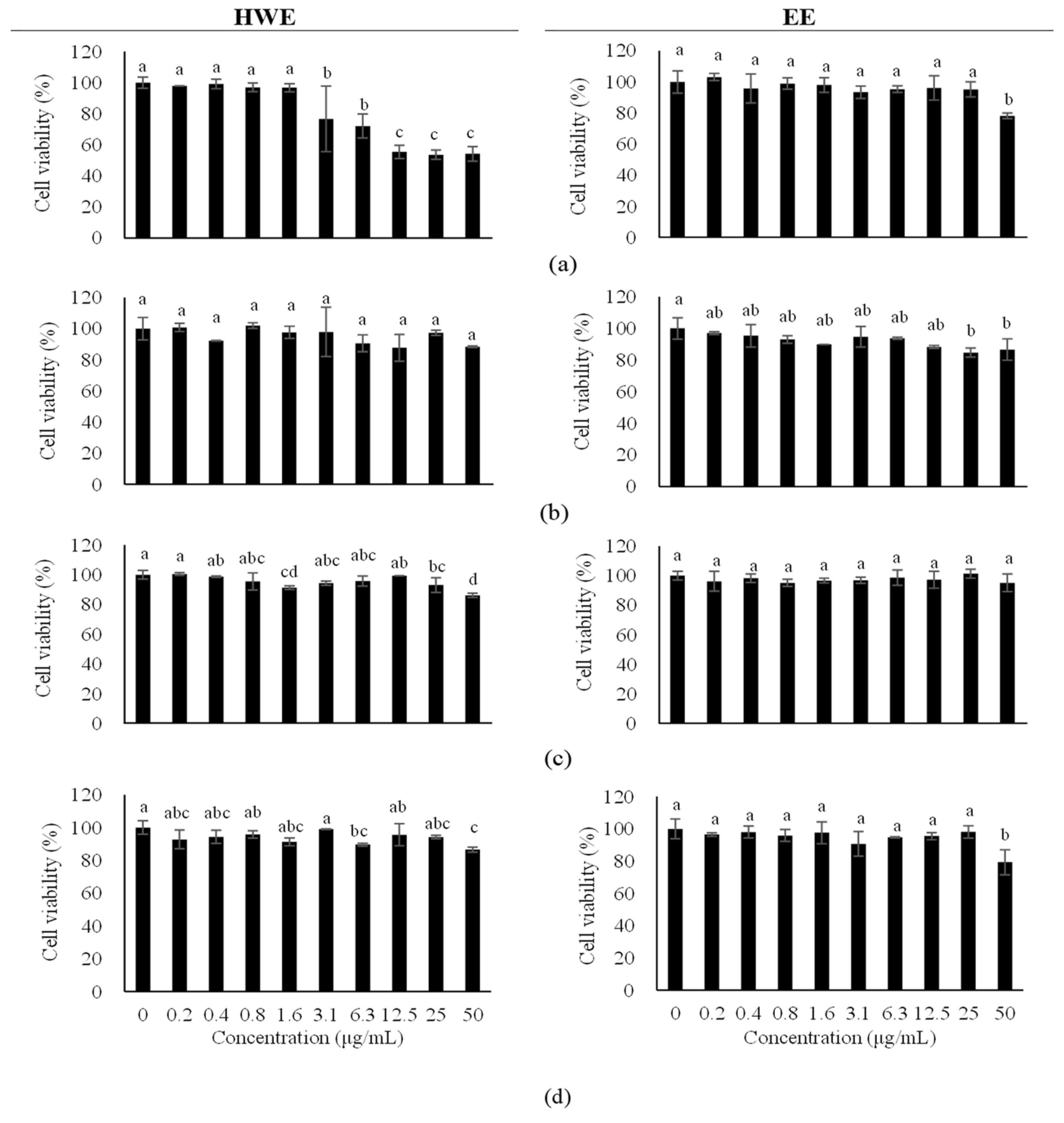
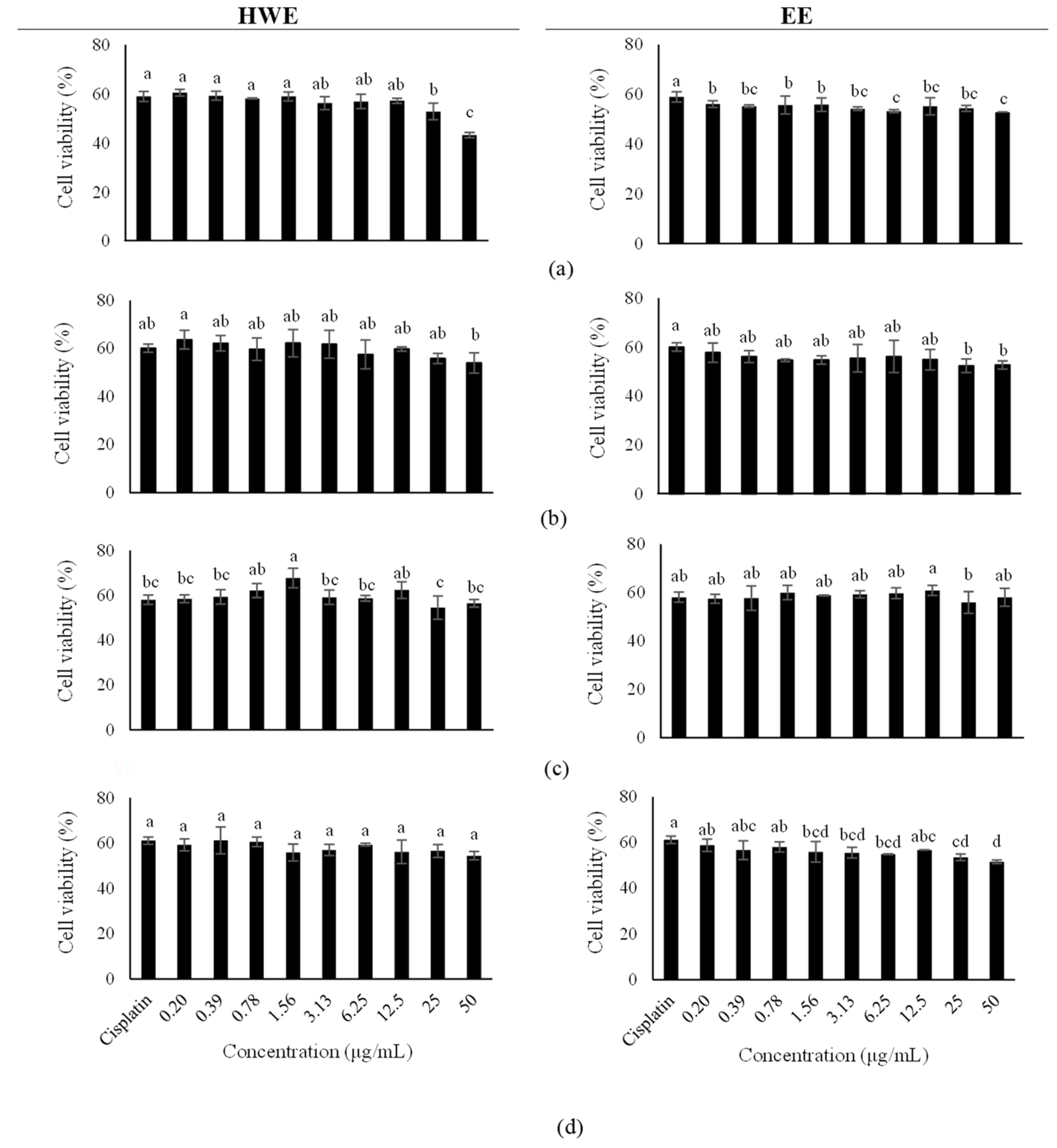
3.4. Cell Viability and Protective Effects of Medicinal Plants on Kidney Cells

Cell viability was assessed to evaluate the cytotoxic effects of fermented medicinal plant extracts on rat kidney LLC-PK1 cells and to examine the non-toxic concentration ranges (Figure 4).
Cell viability after treatment with TM from HWE decreased from 3.1 μg/mL with significant differences, and GM from HWE marginally decreased viability from 0.4 μg/mL. Other fermented plant substrates were observed to be relatively safer below the concentration range of 50 μg/mL. Hence, it can be argued that these plants extracts were relatively safe as observed at a concentration range <50 μg/mL without toxicity. Previously, the omija extracts were reported to have a safe concentration at 50 μg/mL reagrding Haca T-cell toxicity; however, a considerable decrease in cell viability was reported at 100 μg/mL [13]. On the other hand, Artemisia capillaris extracts had prominent cytotoxic effects on the cancer cells [36]. The fraction of aerial parts of glassworts induced toxicity in HCT-cancer cells and inhibited cell viability [37]. This result suggested that these medicinal plants may be a good source that exhibits cell protection without toxicity at an appropriate concentration with possible cancer cell inhibitory properties.
The protective effects of four medicinal plants were estimated against cisplatin-induced kidney oxidative stress. LLC-PK1 cells were exposed at the concentration 0.2~50 μg/mL for 24 h and cell viability was determined using the MTT assay. The cell viability for these concentrations is shown in Figure 5.
The cell viability after treatment with all plant extracts (TM, GM, GW, and OM) was in the range of approximately 60%. All plants from HWE and EE showed protective effects against cisplatin-induced kidney oxidative stress except for TM from HWE at 50 μg/mL. As a result, these plants showed no cytotoxicity and were effective for cell protection. The EE of the Artemisia annua L. had growth inhibition activities against cancer cells at the concentration of 500 μg/mL [38]. Turmeric extract inhibited the cytotoxicity of HT-22 cells and increased bioavailability to treat neuro-generative diseases [39].

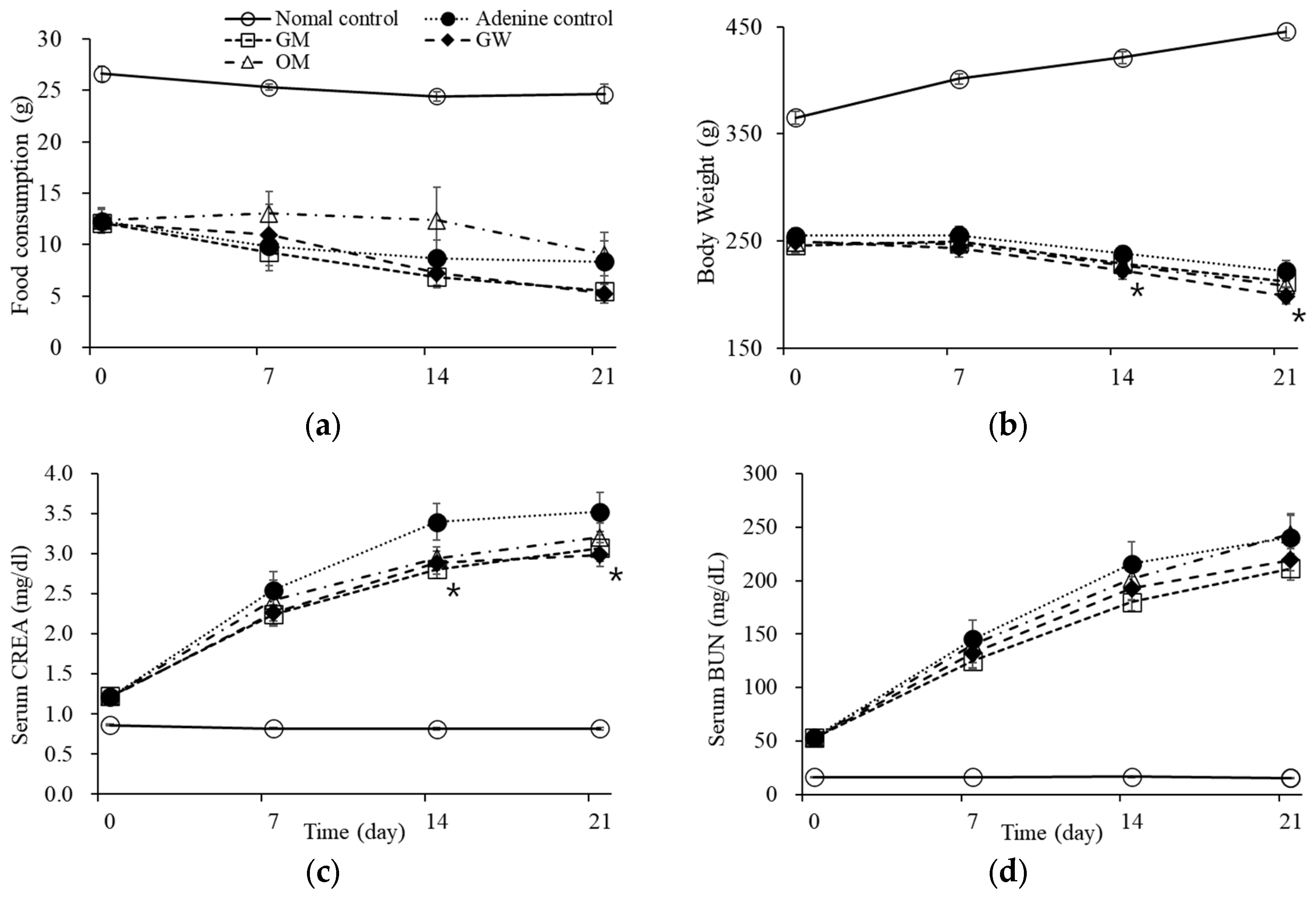
3.5. Serum Creatinine and Blood Urea Level of the Medicinal Plant Extracts in Rats

Creatinine, made from creatine in muscles, is a biomarker of kidney disorder, as is blood urea nitrogen (BUN). To further investigate the protective effects of fermented medicinal plant extracts on kidney cells, SD rats were administrated an adenine-rich diet to induce chronic kidney injury during the experimental period. The levels of serum creatinine and BUN were measured as an indicator of the protective effect of kidney cells (Figure 6).
The body weight of SD rats fed with a supplemented diet decreased during the experimental period and food consumption of all groups decreased for 3 weeks, especially fir the GM group with significant differences. The serum creatinine levels of the rats fed GM, GW, and OM diets were marginally decreased in the first week of the experimental period compared to the vehicle control group; however, the creatinine levels fell significantly during the second week for the GM and GW extract groups. The serum creatinine levels were significantly decreased among the GW group (2.89 ± 0.10 mg/dL) and GM group (2.94 ± 0.08 mg/dL) compared to the adenine control group (3.38 ± 0.15 mg/dL). Similar tendencies were also recorded in third week, with significantly lower levels of serum creatinine observed for the GW group (Figure 6c). In congruence, the serum BUN levels were decreased in treated groups supplemented with fermented medicinal plant extracts as compared to control during the 3-week experimental period (Figure 6d). Notably, we observed a significant decrease in serum creatinine and BUN levels in adenine administered rats supplemented with GW and GM fermented extracts, which demonstrate their potential the protect kidney cells in vivo. Oxidative stress causes to kidney disorders and an imbalance in cell fluid, resulting in a change in BUN and creatinine levels. Previously, it has been reported that turmeric consumption in the diet significantly influence the decrease in serum creatinine and BUN levels [21]. An in vivo experiment on dyslipidemic rats showed that ingestion of turmeric and Haw was effective for kidney and hepatic functional enzyme activities [22,40].

4. Conclusions

This study explored the functional properties of metabolic EE and HWE from four different medicinal plants including turmeric (TM), glasswort (GW), ganghwa mugwort (GM), and omija (OM). All medicinal plants displayed a high phenolic content, especially the TPC of GM, which was higher than the others. Total flavonoid contents (TFC) from HWE fermented substrates were increased following fermentation. DPPH activity based on HWE increased after fermentation, especially for OM with significant differences, in contrast to EE, which decreased. We further observed the protective function of these metabolite extracts in kidney LLC-PK1 cells. The viability of kidney LLC-PK1 cell in response to the plant extracts showed no cytotoxicity at <50 μg/mL, except TM from HWE. In addition, cell viability in cisplatin-treated cells also treated with TM, GM, GW, and OM extracts was in the range of approximately 60%, suggesting their protective effects on cisplatin-induced kidney oxidative stress. This was further substantiated in vivo, where the serum creatinine and BUN levels significantly decreased in extract-treated rat groups compared to the control. Hence, we concluded that controlled fermentation and optimal extraction procedures for the plants might be useful to obtain the functional bioactive compounds from the medicinal plants. The metabolite extracts from the fermented plants have shown promising kidney protective effects as well as antioxidant effects, which need to be substantiated further with various plants species, including physiological and molecular studies.

Author Contributions

S.-K.K., designed this research and acquired fund; J.-Y.O., performed the experiments and data analyzer; J.-M.K., performed data interpretation and analyzer; and prepared the manuscript for publication and revision. D.K., helped the experiments and read the manuscript. All authors have read and agreed to the published version of the manuscript.

Funding

This study was funded by a grant from the Bio Green 21 Program (No. PJ015601), Rural Development Administration, Republic of Korea.

Institutional Review Board Statement

The animal experiment protocol approval No.: N 2021002 from NDIC animal care and use committee.

Informed Consent Statement

Not applicable.

Data Availability Statement

The data presented in this study are available in the article.

Acknowledgments

The authors are thankful to NDIC Corporations for the continuous support.

Conflicts of Interest

The authors declare no conflict of interest.

References

  1. Vlase, L.; Parvu, M.; Parvh, E.A.; Toiu, A. Chemical constituents of three allium species from Romania. Molecules 2013, 18, 114–127. [Google Scholar] [CrossRef] [PubMed] [Green Version]
  2. Yuan, H.; Ma, Q.; Ye, L.; Piao, G. The traditional products. Molecules 2016, 21, 559. [Google Scholar] [CrossRef] [PubMed] [Green Version]
  3. Wink, M. Modes of action of herbal medicines and plant secondary metabolites. Medicines 2015, 2, 251–286. [Google Scholar] [CrossRef] [PubMed]
  4. Oh, J.H.; Lee, S.K.; Choi, S.W. Antioxidant activities of the ethanol extract of hamcho (Salicornia herbacea L.) cake prepared by enzymatic treatment. Food Sci. Biotechnol. 2017, 16, 90–98. [Google Scholar]
  5. Verma, R.K.; Kumari, P.; Verma, R.; Singh, R.K. Medicinal properties of turmeric (Curcuma longa L.): A review. Int. J. Chem. Stud. 2018, 6, 1354–1357. [Google Scholar]
  6. Fuloria, S.F.; Mehta, J.; Chandel, A.; Floria, N.K. A comprehensive review on the therapeutic potential of Curcuma longa Linn in relation to its major active constituents curcuma. Front. Pharmacol. 2022, 13, 1–27. [Google Scholar]
  7. Pal, P.; Ghosh, A.K. Antioxidant, anti-alzheimer and anti-parkinson activity of Artemisia nilagirica leaves with flowering tops. UK J. Pharm. Biosci. 2018, 6, 12–23. [Google Scholar]
  8. Shinde, S.; Sebastian, J.K.; Jain, J.R.; Murthy, H.N. Efficient in vitro propagation of Artemisia nilagirica var. nilagirica and assessment of genetic fidelity of micropropagated plants. Physiol. Mol. Biol. Plants 2016, 2, 595–603. [Google Scholar]
  9. Singh, D.; Buhmann, A.K.; Flowers, T.J. Salicornia as a crop plant in temperate regions: Selection of genetically characterizes ecotypes and optimization of their cultivation conditions. AoB Plants 2014, 6, plu071. [Google Scholar] [CrossRef] [Green Version]
  10. Patel, S. Salicornia: Evaluating the halophytic extremophile as a food and a pharmaceutical candidate. 3 Biotech 2016, 6, 104. [Google Scholar] [CrossRef] [Green Version]
  11. Essaidi, I.; Brahm, Z.; Snoussi, A. Phytochemical investigation of Tunisian Salicornia herbacea L., antioxidant, antimicrobial and cytochrom p450 inhibitory activities of its methanol extract. Food Control 2013, 32, 125–133. [Google Scholar] [CrossRef]
  12. Lee, J.H.; Lee, Y.Y.; Lee, J.; Jang, Y.J.; Jang, H.W. Chemical composition, antioxidant, and anti-inflammatory activity of essential oil from omija (Schisandra chinesis (Turcz.) Baillon.) produced by supercritical fluid extraction using CO2. Foods 2021, 10, 1619. [Google Scholar] [CrossRef] [PubMed]
  13. Kim, M.K.; Lee, Y.Y.; Lee, K.G.; Jung, H.W. Instrumental volatile flavor analyzer of omija using headspace stir-bar sorptive extraction GC/MS and its relationship to human sensory perceptions. Food Res. Int. 2019, 120, 650–655. [Google Scholar] [CrossRef] [PubMed]
  14. Joh, E.-H.; Trinh, H.J.; Kim, D.H.; Han, M.J. Anti-Inflammatory effect of fermented Artemisia princeps Pamp in mice. Biomol. Ther. 2010, 18, 308–315. [Google Scholar] [CrossRef] [Green Version]
  15. Hussein, H.J.; Hameed, I.H.; Hadi, M.Y. Using gas chromatography-mass spectrometry (GC-MS) technique for analysis of bioactive compounds of methanolic leaves extract of Lepidium Sativum. Res. J. Pharm. Technol. 2017, 10, 3981–3989. [Google Scholar] [CrossRef]
  16. Hur, S.J.; Lee, S.Y.; Kim, Y.C.; Choi, I.; Kim, G.B. Effect of fermentation on the antioxidant activity in plant-based foods. Food Chem. 2014, 160, 346–356. [Google Scholar] [CrossRef] [PubMed]
  17. Park, D.H.; Niu, K.M.; Han, S.G.; Yoon, J.E.; Lee, H.G.; Kim, S.K. Effect of fermented medicinal plants as dietary additives on food preference and fecal microbial quality in dogs. Animals 2019, 9, 690. [Google Scholar] [CrossRef] [Green Version]
  18. Chun, H.C. Dietary Effects on Egg Production by Fermented Schisandra chinensis Fruit Byproduct in Laying Hens. Ph.D. Thesis, Konkuk University, Seoul, Republic of Korea, 2011; pp. 1–114. [Google Scholar]
  19. Herigstad, B.; Hamilton, M.; Heersink, J. How to optimize the drop plate method for enumerating bacteria. J. Microbiol. Methods 2001, 44, 121–129. [Google Scholar] [CrossRef]
  20. Dudonne, S.; Vitrac, X.; Coutiere, P.; Woillez, M.; Merillon, J.M. Comparative study of antioxidant properties and total phenolic content of 30 plant extracts of industrial interest using DPPH, ABST, FRAP, SOD and ORA assays. J. Agric. Food Chem. 2009, 57, 1768–1774. [Google Scholar] [CrossRef]
  21. Kim, H.Y.; Jang, S.Y.; Choi, G.I.; Shin, H.C. The effect of patriniae radix on the oxidative stress and NF-KB signaling in mouse LLC-PK1 cell. Korean J. Orient. Med. 2010, 31, 153–165. [Google Scholar]
  22. Oh, D.Y.; Kang, D.S.; Lee, Y.G.; Kim, H.S. Effects of turmeric (Curcuma longa L.) supplementation on blood urea N and enzyme activities in dyslipidemic rats. J. Environ. Sci. Int. 2019, 28, 475–483. [Google Scholar]
  23. Kang, S.; Kim, M.R.; Chiang, M.; Hoyo, J. Evaluation and comparison of functional properties of freshwater-cultivated glasswort (Salicornia herbacea L.) with naturally-grown glasswort. Food Sci. Biotechnol. 2015, 24, 2245–2250. [Google Scholar] [CrossRef]
  24. Min, S.H. Quality characteristics of omija (Schizandra chinesis Baillon) extracts under various conditions for beverage production. J. Korean Soc. Food Cult. 2013, 28, 320–327. [Google Scholar] [CrossRef] [Green Version]
  25. Ye, M.; Yue, T.; Yuan, Y. Effects of sequential mixed cultures of Wickerhamomyces anomalus and Saccharomyces cerevisiae on apple cider fermentation. FEMS Yeast Res. 2014, 14, 873–882. [Google Scholar] [CrossRef] [PubMed] [Green Version]
  26. Stefanello, R.F.; Nabeshima, E.H.; Lamanaka, B.T.; Copetti, M.V. Survival and stability of Lactobacillus fermentum and Wickerhamomyces anomalus strains upon lyophilisation with different cryoprotectant agents. Food Res. Int. 2019, 115, 90–94. [Google Scholar] [CrossRef]
  27. Vlase, L.; Benedec, D.; Hanganu, D.; Damian, G.; Csillag, I.; Sevastre, B.; Mot, A.C.; Silaghi-Dumitrescu, R.; Tilea, I. Evaluation of antioxidant and antimicrobial activities and phenolic profile for Hyssopus offininalis, Ocimum basilicum and Teucrium chamaedrys. Molecules 2014, 19, 5490–5507. [Google Scholar] [CrossRef]
  28. Libro, R.; Fiacoppo, S.; Mazzon, E. Natural phytochemicals in the treatment and prevention of dementia: An overview. Molecules 2016, 21, 518. [Google Scholar] [CrossRef]
  29. Dai, J.; Mumper, R.J. Plant phenolics: Extraction analysis and their antioxidant and anticancer properties. Molecules 2010, 15, 7313–7352. [Google Scholar] [CrossRef]
  30. Lee, C.Y. Challenges in providing credible scientific evidence of health benefits of dietary polyphenols. J. Funct. Foods 2013, 5, 524–526. [Google Scholar] [CrossRef]
  31. Popa, D.S.; Bolfa, P.; Kiss, B.; Vlase, L.; Pop, A.; Loghin, F. Influence of Genista tinctoria L. or methylparaben on subcronic toxicity of bisphenol A in rats. Biomed. Environ. Sci. 2014, 27, 85–96. [Google Scholar]
  32. Shahbazi, H.; Gahruie, H.H.; Gdmakani, M.T.; Eskandari, M.; Movahedi, M. Effect of medicinal plant type and concentration on physicochemical, antioxidant, antimicrobial, and sensorial properties of kombucha. Food Sci. Nutr. 2018, 6, 2568–2577. [Google Scholar] [CrossRef] [PubMed]
  33. Batiha, G.E.S.; Olatunde, A.; El-Mleeh, A.; Hetta, H.T.; Rivero-Perez, N. Bioactive compounds, pharmacological actions, and pharmacokinetics of wormwood (Artemisia absinthium). Antibiotics 2020, 9, 353. [Google Scholar] [CrossRef] [PubMed]
  34. Gouda, M.; Elsebaie, E. Glasswort (Salicornia spp.) as a source of bioactive compounds and its health benefits: A review. J. Food Sci. Technol. 2016, 13, 1–7. [Google Scholar]
  35. Kim, J.K.; Kim, J.H.; Woo, H.J. Antioxidant Activities of Water and Ethanol-Extracts of Glasswort (Salicornia hervacea). Korean Soc. Biotechnol. Bioeng. 2003, 525–525. Available online: http://www.koreascience.or.kr/article/CFKO200336037075463.page (accessed on 1 November 2022).
  36. Kim, H.T.; Kim, J.W.; Lim, M.K.; IM, T.W. Cytotoxic effect of Artemisia capillaris extracts on the cancer cells in vitro. J. Vet. Clin. 2001, 24, 367–371. [Google Scholar]
  37. Kang, S.; Hong, J. Antioxidant activities, production of reactive oxygen species, and cytotoxic properties of fractions from aerial parts of glasswort (Salicornia herbacea L.). Korean J. Food Sci. Technol. 2016, 48, 574–581. [Google Scholar] [CrossRef] [Green Version]
  38. Ryu, J.H.; Lee, S.J.; Kim, M.J.; Shin, J.H.; Sung, N.J. Antioxidant and anticancer activities of Artemisia annua L. and determination of functional compounds. J. Korean Soc. Food Sci. Nutr. 2011, 40, 509–516. [Google Scholar] [CrossRef]
  39. Yoon, N.K.; Kim, B.K.; Ryu, H.Y.; Lee, H.J. In vitro neuroprotective effect of Aricumin (turmeric extract). J. Convergence Culture Technol. 2022, 8, 291–296. [Google Scholar]
  40. Kim, H.S.; Kim, M.A.; Duan, Y.; Jang, S.H.; Cho, H.J.; Kim, S.W. Influences of wild Haw (Crataegus pinnatifida) on lowering BUN and creatinine concentration in dyslipidemia. J. Environ. Sci. Int. 2014, 23, 1029–1035. [Google Scholar] [CrossRef]
Figure 1. Changes in pH (a) and viable cell count (b) before and after fermentation of 5% (w/v) turmeric, ganghwa mugwort, glasswort, and omija. TM: turmeric; GM: ganghwa mugwort; GW: glasswort; OM, omija.
Fermentation 08 00702 g001
Figure 2. TPC and TFC of HWE and EE after fermentation for 5% (w/v) turmeric, ganghwa mugwort, glasswort, and omija. * p < 0.05; (a) TPC-HWE; (b) TPC-EE; (c) TFC-HWE; (d) TFC-EE; TM: turmeric; GM: ganghwa mugwort; GW: glasswort; OM, omija.
Fermentation 08 00702 g002
Figure 3. DPPH radical scavenging activity and ABTS of HWE and EE after fermentation for 5% (w/v) turmeric, ganghwa mugwort, glasswort, and omija. * p < 0.05, ** p < 0.01; (a) DPPH-HWE; (b) DPPH-EE; (c) ABTS-HWE; (d) ABTS- EE; TM: turmeric; GM: ganghwa mugwort; GW: glasswort; OM, omija.
Fermentation 08 00702 g003
Figure 4. Cell viability of kidney LLC-PK1 cells. Cytotoxicity tests of fermented medicinal plants from HWE and EE. The same letters have no significant differences. (a) TM; (b) GM; (c) GW; (d) OM.
Fermentation 08 00702 g004
Figure 5. Cell viability against cisplatin-induced oxidation to assess the protective effects on kidney cells of (a) turmeric; (b) gangwha mugwort; (c) glasswort; and (d) omija. The same letters have no significant differences.
Fermentation 08 00702 g005
Figure 6. Changes in serum creatinine and BUN level of control and adenine-induced chronic kidney injury SD rats treated with test materials for the experimental period. * p < 0.05; (a) Food consumption; (b) body weight; (c) serum creatinine level; (d) serum BUN level; GM: ganghwa mugwort; GW: glasswort; OM, omija.
Fermentation 08 00702 g006
Publisher’s Note: MDPI stays neutral with regard to jurisdictional claims in published maps and institutional affiliations.

