Article

Probiotic Properties of Lactobacillus fermentum InaCC B1295 Encapsulated by Cellulose Microfiber from Oil Palm Empty Fruit Bunches

1
Faculty of Agriculture, Universitas Riau, Pekanbaru 28293, Indonesia
2
Faculty of Nursing, Universitas Riau, Pekanbaru 28127, Indonesia
3
Faculty of Agricultural Technology, Universitas Andalas, Padang 25163, Indonesia
4
Food Technology Department, Faculty of Engineering, Bina Nusantara University, Jakarta 11480, Indonesia
*
Authors to whom correspondence should be addressed.
Fermentation 2022, 8(11), 602; https://doi.org/10.3390/fermentation8110602
Submission received: 22 September 2022 / Revised: 29 October 2022 / Accepted: 31 October 2022 / Published: 3 November 2022
(This article belongs to the Special Issue Recent Trends in Lactobacillus and Fermented Food)

Abstract

:
This study aims at an in vitro characterization of the acid and bile tolerance of Lactobacillus fermentum InaCC B1295 (LFB1295) encapsulated with hydrogel cellulose microfibers (CMF) from oil palm empty fruit bunches (OPEFBs). The viability at different storage temperatures was assessed. The experimental design used in this research was an in vitro trial. The microencapsulated probiotic was stored at 25 °C and 4 °C for 28 days. LFB1295 encapsulated with cellulose microfiber hydrogel from OPEFB showed a stable viability of probiotic bacteria at pH 2 and 0.5% (m/v) oxgall. In addition, the microencapsulation maintained the viability at 25 °C and 4 °C at 0, 14, and 28 days. The characterization of the encapsulant CMF-OPEFB showed that the thickness of CMF was in the range of 5–15 μm, and XRD patterns showed that CMF was of the cellulose I type with a crystallinity index of 77.08%. Based on its resistance to hydrogen peroxide, ability to scavenge DPPH radicals, and activity in scavenging hydroxyl radicals, LFB1295 encapsulated with CMF hydrogel of OPEFB exhibits antioxidant properties as good as the scavenging ability of DPPH radicals with IC50 of 36.880, 188.530, and 195.358 µg/mL, respectively, during storage for 0, 14, and 28 days at room and refrigerated temperature. Furthermore, hydroxyl radicals (HR)-scavenging activity showed an increased inhibition along with the increasing concentration of the Fenton reaction and decreasing concentration of cell-free supernatant (CFS) during storage time. In vitro safety tests, including hemolytic activity, biogenic amines, cytolysin, and gelatinase production, showed that the encapsulated LFB1295 was safe to use as a probiotic. The results of the inhibitory activity against hydrogen peroxide LFB1295 show that the higher the concentration of H2O2, the lower the inhibition value during 28 days of storage. Based on the storage temperature, the inhibition of LAB against H2O2 based on different storage temperatures showed a better level of the inhibition at cold temperatures compared to at room temperature.

1. Introduction

Probiotic bacteria are recognized as promoters of numerous aspects of health and wellbeing, including the modulation of the human gut microbiome through the inhibition of pathogenic bacteria [1] and archaea, preventing the activity of carcinogens, or even the modulation of immune responses [2]. According to Allied Market Research, the probiotic foods market has expanded dramatically, as functional food and beverages, dietary supplements, or animal feeds are included in various markets. The probiotics market will reach USD 73.95 billion by 2030, increasing at an 8.6% compound annual growth rate in the period of 2021–2030 [3]. Hence, maintaining stable viability during processing, storage, and distribution until reaching the consumer is very important [4]. Concurrently, cell encapsulation becomes more prevalent to increase probiotic survival added to various food formulations [5].
Microencapsulation involves increasing the viability of probiotic bacteria during storage conditions at high humidity, particularly in Indonesia, one of the tropical countries [6]. The efficiency of numerous encapsulation materials was necessary for maintaining the stable viability of probiotic cells: (1) cell separation from the products efficiently process, (2) enhanced productivity due to increased cells concentration accumulated, (3) protected bacteria cells against adverse conditions, (4) potentially as packed columns, (5) utilization of immobilized probiotic cells, and the prevention of cell remobilization [7]. In addition, these components should be oxygen-permeable, produce harmful metabolites, and contain nutrients to keep cells alive [8]. Hence, it will manage the release as it passes through the human gastric and intestinal tracts [9].
Probiotic cells have been microencapsulated using a combination of natural water-soluble and synthetic polymers [7]. Producing solid microspheres for bacterial cell immobilization is initiated with selecting an effective encapsulant. For instance, proteins (i.e., casein and whey protein) [10,11], lipids (i.e., cocoa butter, milk fat, and cream) [12,13], and polysaccharides (i.e., chitosan, alginate, gellan gum, and cellulose) [14,15] have been used to immobilize probiotics. Many researchers evaluated the production and identification of cellulose fibers (CF) from oil palm trees and their uses, particularly the encapsulant material of probiotics. One kilogram of dry biomass was extracted from four kilograms of palm oil tree. Indonesia, as the world’s leading producer of palm oil, a result of which a significant output of biomass waste is obtained, which is generally classified into: (1) OPEFBs (oil palm empty fruit bunches), (2) OPFs (oil palm fronds), (3) OPTs (oil palm trunks), (4) PKSs (palm kernel shells), (5) MF (mesocarp fiber), and (6) POME (palm oil mill effluent) [16]. OPEFBs are abundant in lignocellulosic biomass (66.97% holocellulose and 24.45% lignin) [17]. Hence, they have potential as an encapsulant material for probiotics.
The cellulosic microfibers of biomass waste have advantages as encapsulant material. Oil palm waste biomass has low density and is inexpensive, recyclable, and biodegradable [18]. Cellulosic microfiber hydrogels (CMFHs) are natural polymers that create hydrogels, excessively hydrated materials made of cross-linked hydrophilic polymers formed by 3D networks. Furthermore, CMFs are smooth, have very porous structures when implanted, and cause negligible protein adsorption. The extracellular matrix found in human tissues is similar to the structure of hydrogels made of natural materials. These hydrogel polymers are analogous to biological macromolecules that nature has developed to fulfill specific functions in a complex environment [8]. Technologies for encapsulating cells are created to enclose alive, functional cells in a semi-permeable matrix.
Aditiawati et al. [19] studied CF derived from various components of oil palm waste, using, for instance, chemical and mechanical treatments. Based on the study of Galiwango et al. [20], lignocellulosic sources produced cellulose (α-cellulose) isolated and characterized using a diluted acid–alkali treatment, conforming with Fahma et al. [21]. The acid–alkali treatment as the primary method for individual CF isolation from the complex structure of lignocellulose proved to be an effective method. The primary method of producing CF is to eliminate lignin and hemicellulose to obtain microfibers (CMFs) or even nanofibers (CNFs) [22].
A probiotic bacterium called Lactobacillus fermentum InaCC B1295 (LFB1295) was discovered in the fermented buffalo milk known as dadih from the Indonesian provinces of Riau and West Sumatra [23]. Lactobacillus fermentum is a bacterium that has been recognized as having significant potential as probiotic (probiosis and antiviral activity; bio-preservative activity; and immunobiosis) and has been interestingly qualified as a GRAS (Generally Recognized As Safe) microorganism [24]. Numerous research has abundantly proven probiotics’ positive effects on health. However, before using novel strains of probiotics in food for human consumption, especially those without documented safety evidence, the safety of probiotics must be evaluated [25,26]. Essential probiotic safety evaluations include hemolytic activity, biogenic amine, cytolysin, and gelatinase production [27].
Research on lactic acid bacteria (LAB) is ongoing due to growing interest in their antioxidant capacity. For aerobic organisms, producing reactive oxygen species (ROS) and reactive nitrogen species (RNS) is a necessary byproduct of normal metabolic processes. Its concentrations are crucial for the immune response against invasive microorganisms and for controlling intercellular communication. However, excessive ROS/RNS production results in oxidative stress, which damages DNA and causes protein oxidation and lipid peroxidation [28].
This study aims to evaluate the potential of a cellulose microfiber hydrogel (CMFH) from OPEFB waste as an encapsulant for LFB1295 to maintain viability and resistance to acids and bile. The safety evaluation of the probiotic and the DPPH and HR (hydroxyl radicals) scavenging activities of LFB1295 were also evaluated during storage for 28 days at cold and room temperatures.

2. Materials and Methods

2.1. Materials

The OPEFBs from the palm oil variety Tenera was supplied by Pelalawan Regency, Riau Province, Indonesia (PT. Multi Plasma Sejahtera). LFB1295 was obtained from the Indonesian fermented milk called “dadih”.

2.2. Production of Cellulose Microfibers (CMFs)

The production of CMF-OPEFB was prepared as described in detail in the previous research of [29,30]. The dry biomass cellulose of OPEFBs was delivered to Nano Center Indonesia (South Tangerang City, Banten, Indonesia) to be converted into CMF using a planetary ball mill machine.

2.3. Production of Cellulose Microfiber Hydrogels (CMFHs)

A polyvinyl alcohol (PVA) solution was initially prepared by weighing 96 g of PVA, adding 1104 mL of distilled water, and then dissolving at 100 °C. PVA solution cooling at room temperature. PVA (concentration 8% w/v) was combined with CMFs by dissolving CMFs with PVA with a ratio of 1:1 and heating until the CMFs was completely dissolved in the CMFHs. Then, it was autoclaved at 121 °C for 15 min. The sterile CMFHs were chilled at room temperature and was then suitable for the LAB encapsulant [31].

2.4. Preparation of Lactobacillus fermentum InaCC B1295

LFB1295 was prepared as described in previous research [29,30]. The LFB1295 culture was grown on an mRS Broth medium at 37 °C for 24 h. Cells were separated from CFS (cell-free supernatant) by centrifugation at 10,000 rpm for 10 min at 4 °C. After that, sterile distilled water was used to wash the cells twice. The cells were added with phosphate buffer in a 1:1 (w/v) ratio and stored at 4 °C.

2.5. Production CMFH-Encapsulated Lactobacillus fermentum InaCC B1295

The encapsulated LAB was prepared according to Yasim-Anuar et al. [32] with slight adjustments. The biomass of LFB1295 and sterile CMFH with a ratio of 1:1 (40 mL each) was mixed until homogenous. The CMFH-encapsulated LFB1295 was collected and stored based on the treatment. Finally, the CMFH-encapsulated LFB1295 was stored at 25 °C and 4 °C for 28 days for all treatments. The research methodology is thoroughly described in the flowchart in Figure 1.

2.6. X-ray Diffraction Analysis of Cellulose Microfiber (CMF)

An XRD test of CMF-OPEFB was conducted to determine the crystallinity index (CrI). XRD was analyzed using the X’Pert3 MRD Malvern Panalytical’s instrument machine (Malvern, Worcestershire, UK). The calculated CrI is according to the equation:
C r I = I 002 I a m I 002 × 100 %
I002 is a diffraction intensity (crystalline region of the material) of 2θ = 23. Iam denotes the peak at approximately 2θ = 18, representing the material’s amorphous area [32].

2.7. The Total Number of Lactobacillus fermentum InaCC B1295 Encapsulated by CMFH-OPEFB during Storage Time

A total of 1 mL of microfiber cellulose-hydrogel-encapsulated LFB1295 (4 °C and 25 °C at each time point of 14 days interval) was inserted into 5 mL of MRS broth, and then incubated at 37 °C for 18 h. The encapsulated LAB was tested for its viability, according to Pato et al. [29]. The total LAB (Log CFU/mL of LAB control) was enumerated using MRS Agar (MRSA).

2.8. Resistance to Acid

A total of 1 mL of microfiber cellulose-hydrogel-encapsulated LFB1295 at 4 °C and 25 °C at each time point of 14 days interval was inserted into 5 mL of MRS broth modified to pH 2 using 37% hydrogen chloride, and then incubated for 5 h at 37 °C. The total LAB was calculated using the plate count technique on MRSA [33]. The cell viability of resistance to acid from each treatment was calculated using the equation:
Viability   ( % ) = 100 ( log CFU mL   of   LAB   control log CFU mL   of   LAB   at   pH   2 log CFU mL   of   LAB   control × 100 )

2.9. Resistance to Bile

The active culture of LFB1295 was added to an MRSB with 0.5% (m/v) oxbile, and then incubated for 24–48 h at 37 °C. The cell viability of acid tolerance from each treatment was calculated using the equation:
Viability   ( % ) = 100 ( log CFU mL   of   LAB   control log CFU mL   of   LAB   at   0.5 %   oxgall log CFU mL   of   LAB   control × 100 )

2.10. Safety Evaluation of Probiotic

2.10.1. Hemolytic Activity

The hemolytic activity method was performed as described previously [27]. LFB1295 encapsulated by CMF hydrogel etched on a Columbia Blood Agar, which was added with 5% SB Agar and then incubated at 37 °C for 48 h. Hemolytic reactions were noted by simply looking at the colony’s surrounding, clearly visible hydrolysis zone (β-hemolysis), partial and greenish zone (α-hemolysis), or no clear zone (γ-hemolysis).

2.10.2. Production of Biogenic Amine

The method was performed as described previously [34] to test LFB1295 encapsulated by CMF hydrogel to produce biogenic amines. Briefly, the test of LFB1295 encapsulated by hydrogel CMF subcultured twice at 24 h intervals in MRS Broth containing 1% of 4 amino acids (AA), and then 0.005% pyridoxal-5-phosphate was added as a carboxylase code factor. LFB1295 was then streaked in Duplo on Decarboxylases Agar (DA) containing one of the AA as mentioned earlier, along with the color purple bromocresol, and then cultivated for 24 h at 37 °C. DA medium without AA was used as a control. The medium’s color changes from brown to purple, which imply an increase in pH, are thought to be a positive result (probiotics produce biogenic amines).

2.10.3. Cytolysin Activity

Cytolysin production was evaluated using the method of Tan et al. [35]. LFB1295 encapsulated by CMF hydrogel was etched on medium Brain Heart Infusion (BHI) agar, which was added with 5% (v/v) SB Agar. Positive results were defined as a clear zone surrounding the colony, following a 24 h incubation period at 37 °C (probiotics produce cytolysis).

2.10.4. Production of Gelatinase

Gelatinase activity was evaluated using the method of Tan et al. [35]. LFB1295 encapsulated by CMF hydrogel was etched on BHI medium agar and 1.5% skimmed milk was added. The plates were cultivated for 24 h at 37 °C and observed for a clear zone. A clear zone around the colony was a positive result (probiotics produce gelatinase).

2.11. Antioxidant Activity of Probiotic

2.11.1. Resistance to H2O2

Resistance to H2O2 was conducted using the Healthy technique [36]. LFB1295 encapsulated by the CMF hydrogel was cultivated in MRSB at 37 °C for 24 h. Subsequently, this active culture was inoculated as much as 1% (v/v) into MRSB with or without 0.4, 0.7, or 1.0 mM H2O, and then incubated at 37 °C for 8 h. The absorbance was measured to estimate cell growth using a spectrophotometer.

2.11.2. Scavenging of Hydroxyl Radicals

HRS activity was carried out using the Fenton reaction technique [36]. The reaction mixture was created initially by combining Brilliant Green, FeSO4, H2O2, and CFS. LFB1295 encapsulated by CMF hydrogel was cultivated in MRSB at 37 °C for 24 h. LAB cultures were centrifuged to separate the cells and supernatant, and then BAL cells were added with 5 mL of phosphate-buffered solution. After 20 min of incubation at room temperature with various doses of CFS, the absorbance at 624 nm was recorded. The LFB1295’s capacity to scavenge hydroxyl radicals was shown by changes in the reaction mixture’s absorbance. HRS ability was calculated using the following formula:
Scavenging   activity   ( % ) = A s A c A A c
As represents the sample’s absorbance. Ac represents the control’s absorbance. A represents the absorbance without the sample or Fenton reaction.

2.11.3. Scavenging for DPPH Radicals

The scavenging ability of DPPH radicals was carried out using the method of [28,36] with some modifications. DPPH was employed in methanol at a concentration of 0.23 mM. Under dark conditions, 0.1 mL of CFS of LFB1295 and 3.9 mL of DPPH solution were combined, and then homogenized with a Vortex mixer. Using a spectrophotometer, the solution’s absorbance was determined at 517 nm. The following formula was used to determine LFB1295’s DPPH radical scavenging capacity:
Inhibition ( % ) = A b s C o n t r o l A b s S a m p l e A b s C o n t r o l
AbsControl represents the control solution’s absorbance and AbsSample represents the sample’s absorbance.
The IC50 value was determined using an analysis by making a curve of the relationship between the percent inhibition and the concentration of DPPH and then calculated using a linear equation formula as follows:
y = a x + b
where y represents the sample’s absorbance and x represents the sample’s concentration.

2.12. Experimental Design

This study used a completely randomized experimental design. There were two factors considered, i.e., storage temperature (25 °C: room temperature and 4 °C: refrigerated temperature) and storage time (0, 14, and 28 days). Data were obtained in triplicates.

2.13. Data Analysis

ANOVA and DMRT were used at 95% significance with SPSS Version 23, including the total LAB, viability at low pH and bile, and antioxidant activity (DPPH and HR Scavenging). Descriptive analysis was used to characterize CMF-OPEFB and safety assessment of probiotics. The safety assessments included hemolytic activity, biogenic amine, cytolysin, and gelatinase activity.

3. Results

3.1. Evaluation of Cellulose Microfiber-Oil Palm Empty Fruit Bunches

Scanning Electron Microscope of CMF-OPEFB

These parameters were evaluated to determine the shape, size, and X-ray Diffraction of CMF from OPEFBs as a natural encapsulant for the probiotic. CMF can be extracted from various lignocellulosic fibers such as OPEFBs. CMF was purified cellulose fiber consisting of partially disintegrated microfiber aggregates with diameters in the range of 0.02–287 µm and lengths in the range of several micrometers [37,38,39]. In addition, the cellulose fiber could be considered cellulose nanofibers (CNFs) if the size in one dimension is <100 nm [37].
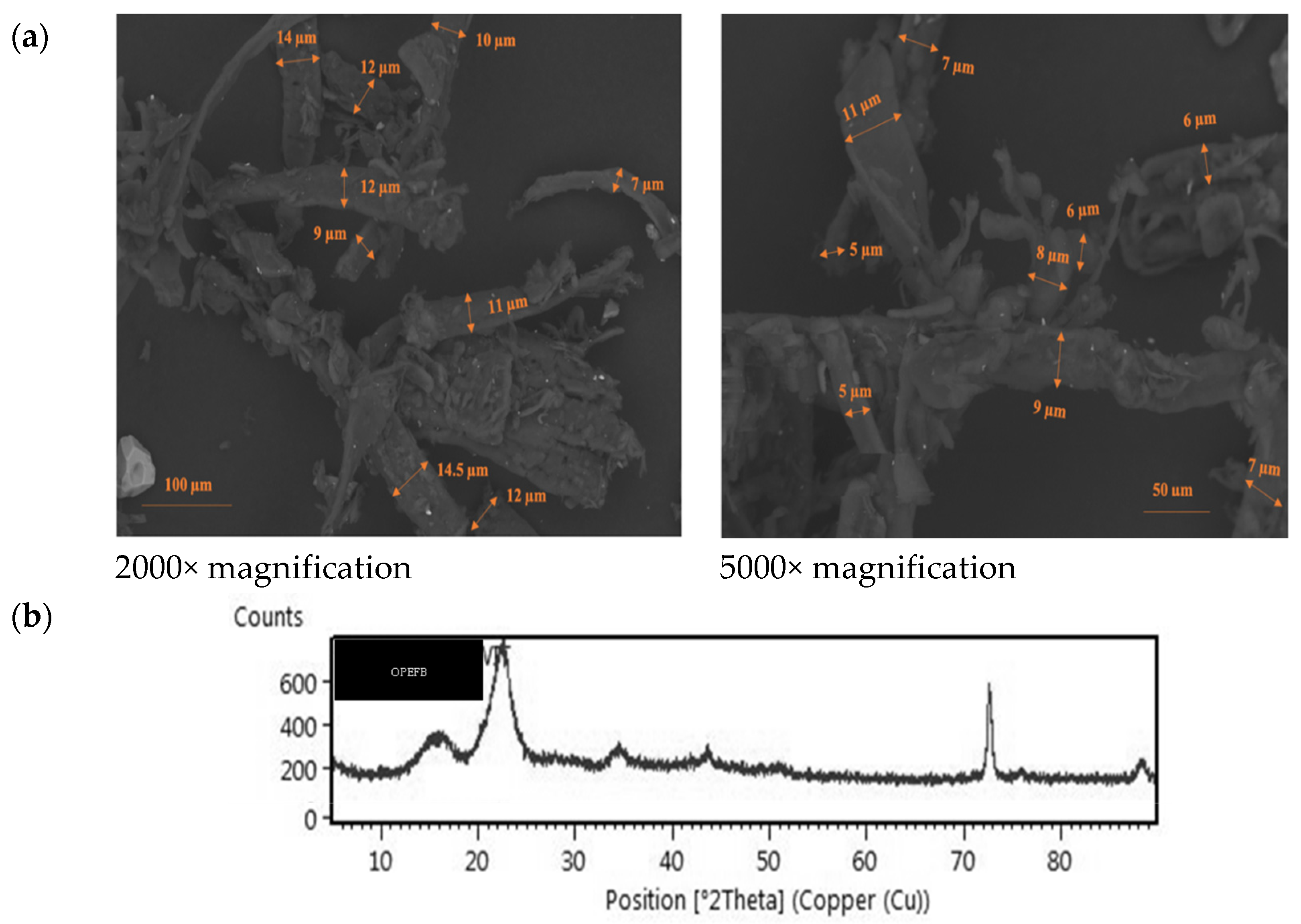
The homogeneity, size, and morphology of CMF are tightly connected to the cellulose source and the technique to extract CF (alkali, acid, and bleaching treatments). The SEM micrographs showed a micrometric scale, rough surface, and adequate homogeneity. The alkali and acid hydrolysis procedures used in this research to extract CMF produced nearly identical microfiber diameters ranging from 5 to 15 µm, indicating that the OPEFB source exhibited the same cellulose fiber quality as other CMFs derived from natural cellulose. Furthermore, the peaks are more amorphous, whereas CMF has a narrower peak due to removing hemicellulose and lignin following the acid hydrolysis treatment (Figure 2).
The components of natural fibers are crystalline, but the non-cellulose polysaccharides lignin and hemicellulose are amorphous. Several procedures exist to eliminate lignin and hemicellulose to increase the CF crystallinity index. The CMF has a higher crystallinity index (77.08%) than CF (36–54%) [40] due to a high amount of lignin and hemicellulose.

3.2. Viability and Acid and Bile Tolerance of CMFH-Encapsulated Probiotics during Storage Time

Measurement of viability and acid and bile tolerance were intended to determine the resistance of the strain LFB1295 during storage at various temperatures and times as a probiotic candidate to be used in the production of certain functional foods. Viability reduction was addressed by the potential use of probiotic cells that were microencapsulated. Hence, the total number of LAB was necessary to conduct this study. The ANOVA analysis revealed that storage temperature did not significantly influence the number of probiotic viability (p < 0.05). Otherwise, there was a noticeable impact of storage time on probiotic persistence and correlation between them (p < 0.05). After 28 days of storage at room and refrigerated temperatures, the number of LFB1295 cells encapsulated in CMFH-OPEFB decreased significantly (Table 1). At refrigerated temperature, the number of LFB1295 cells declined from 9.93 log CFU/g to 9.09 log CFU/g and from 9.80 log CFU/g to 9.11 log CFU/g when stored at room temperature. The LAB reduction was marginally more significant at refrigeration compared to room temperature.
Probiotics are usually encapsulated to increase survival through the gut passage. Low pH and bile salt levels are critical factors for the viability of probiotics. We determined the viability of CMFH-OPEFB as an encapsulant material of probiotic bacteria throughout storage at room and refrigerated temperatures for resistance to acid tolerance and bile salt during 0-, 14-, and 28-day storage. ANOVA was used with a 95% confidence interval (0.05% significant level). Two-way interactions (temperature and time storage) of ANOVA for a 95% confidence interval were conducted in this research, revealing that an F-value was not significant for low pH treatment; otherwise, it was significant for bile salt treatment. The viability of CMFH-encapsulated LFB1295 in acid and bile tolerance was conducted in this research to support the total number of LAB and the potential CMF-OPEFB as an encapsulant. After 28 days’ exposure to acid at pH 2, the viability of encapsulated L. fermentum InaCC B1295 was 98.92–99.71% and 98.08–99.72%, respectively, at 4 °C and 25 °C (Table 1).
The number of LAB was 9.09–9.11 log CFU/g when stored at room or refrigerated temperatures for 28 days. As expected, there was no considerable alteration in the average percent viable count of probiotics in the variety of temperature storage after 28 days under acidic conditions. Even if there is no statistically significant outcome for any treatment, the survivability of encapsulated probiotic bacteria maintained at refrigerated temperature shows higher data, even though about 2%. These data suggest that, even if LFB1295 is administered to achieve its therapeutic effect, the cell concentration of 7.0 log CFU/g is achieved.
Probiotic strains need to endure and flourish in bile salts to endure passage through the small intestine. As a result, while assessing the potential of LAB as an efficient probiotic, the oxgall powder in the criteria mentioned earlier was the most similar to human bile [41]. Table 1 presents the bile tolerance of LFB1295 retained at different temperatures and storage times. The ANOVA analysis revealed that storage temperature does not significantly influence the amount of LFB1295 viability by the treatment bile resistance (p < 0.05). Otherwise, storage conditions significantly influenced probiotic survival, and there was a correlation between them (p < 0.05).
Compared to our earlier findings, LFB1295 demonstrated a slight decrease in cell numbers [23,24]. LFB1295 viability was reduced to 92.55% and 96.59%, respectively, during storage for 28 days at room and cold temperatures. This LAB proved resistant to bile salt throughout storage, more than 9.0 log CFU/g on average. The viability reduction in LFB1295 cells is higher at refrigerated than room temperature, even though the ANOVA analysis is not significant. These data suggest that CMFH-OPEFB was effective in bile to protect probiotic cells.

3.3. Safety Evaluation of CMFH-Encapsulated Probiotic

This parameter was measured to ensure that LFB1295 is safe to use as a probiotic at various temperatures and storage times. The safety test results of LFB1295as a probiotic candidate, including hemolytic activity and production of biogenic amines, cytolysin, and gelatinase during storage time for 28 days at room and refrigerated temperatures, are shown in Table 2.
Based on bacterial safety assessments of hemolytic activity production of biogenic amines, cytolysin and gelatinase showed no clear zone around the colonies on the media, indicating L. fermentum InaCC B1295 was safe as a probiotic.

3.4. Antioxidant Activity of CMFH-Encapsulated Probiotic

3.4.1. Resistance to Hydrogen Peroxide

These parameters were evaluated to determine the resistance of LFB1295 to hydrogen peroxide at various temperatures and storage times. Data on oxidant activity resistance to hydrogen peroxide from LFB1295 encapsulated by OPF CMFH for 28 days of storage at ambient temperature and in a refrigerator are presented in Table 3.
The results of the inhibitory activity against hydrogen peroxide by bacteria B1295 reveal that the higher the concentration of H2O2, the lower the inhibition percentage during 28 days of storage, but based on the storage temperature, the inhibition of LAB against H2O2 based on different storage temperatures showed a better level of inhibition at cold temperatures compared to at room temperature.

3.4.2. Hydroxyl Radical Scavenging Activity

Hydroxyl radical scavenging activity is one of the parameters that needs to be evaluated to determine the potential of LFB1295 to prevent cancer. Data on oxidant activities, including hydroxyl radical scavenging activity from LFB1295 encapsulated by OPF CMFH for 28 days of storage at ambient temperature and in a refrigerator, are presented in Table 4.

3.4.3. DPPH Radical Scavenging Activity

One of the characteristics that needs to be assessed to establish the potential of LFB1295 to prevent cancer is its ability to scavenge DPPH radicals. Data on oxidant activities, including the scavenging ability of DPPH radicals from LFB1295 encapsulated by OPF CMFH for 28 days of storage at ambient temperature and in a refrigerator, are presented in Table 5.
Based on the observations on the IC50 value of LAB B1295, there was a decrease along with storage time, but the storage temperature did not affect the IC50 value.

4. Discussion

The CMF-OPEFB ranges from 5 to 15 µm, showing that the cellulose fiber quality is comparable to that of other CMF generated from natural cellulose. The size of CMF-OPEFB has a lower average diameter than CMF derived from other natural cellulose fibers, such as sisal (287 µm), cotton (131 µm), sugar palm fibers (122 µm), flax (51 µm), tomato plant (20 µm), banana peel (19 µm), and hemp waste (17 µm). In comparison, it is significantly greater than CMF derived from kraft pulp made from softwood (0.1–1 µm), juncus (3 µm), and pineapple leaf (4 µm) [39]. As a result, CMF-OPEFB is a rare natural fiber with potential for further applications.
The yielding CMF-OPEFB had a narrow diameter, making it an excellent contender for developing sophisticated cellulose-based micro products, including those for nanoscale cellulose extraction. CMF-OPEFB was insufficient to generate nanostructured cellulose by the acid hydrolysis process. The CMF-amorphous OPEFBs’ domains are removed using acid hydrolysis. Thus, additional treatment, such as mechanical disintegration, is required to lower the cellulose particle size. H2SO4 hydrolysis typically cuts through the amorphous portion of microfibrils transversely in cellulose fibers, resulting in fibers with sizes between microns and nanometers [42].
Figure 2 shows that the XRD diffractogram of CMF-OPEFB is sharper than the peak of OPEFB cellulose fiber (CF). According to [21], the XRD patterns revealed two peaks of diffraction intensity, indicating that CMF or CNF was I-type cellulose. The patterns of X-ray diffraction for CMF and CF were comparable, according to Fahma et al. [21], indicating that the cellulose structure remained intact after sulfuric acid hydrolysis. Cellulose from palm oil biomass waste showed that the peak at approximately 2θ =23 represents the crystalline area of cellulose (I002), whereas the peak at approximately 2θ =18 represents the amorphous area (Iam) [43]. The diffraction peak for CF-OPEFB was broad at 23°. The peak in CMF-OPEFB, however, was sharper and smaller, indicating that the fibers that were treated had more crystallinity. In this study, the indicator of crystallinity for CMF-OPEFB was 77.08% (Table 1), while Fahma et al. [21] found at 53.83–58.78% (cellulose nanofiber-OPEFB) and 41% (cellulose nanofiber of oil palm mesocarp fiber) was reported by Yasim-Anuar et al. [44].
Cellulosic fibers (CF) have been studied because they have unique biocompatibility, biodegradability, and renewability. Encapsulating probiotics in vegan proteins obtained from plants is an excellent alternative to animal-derived proteins. Encapsulation techniques based on standard biopolymers have certain disadvantages in protecting probiotics from gastrointestinal fluids. Otherwise, cellulose-based hydrogels, either alone or in combination with other biopolymers, have lately demonstrated significant potential for overcoming the constraints of conventional biopolymer-based encapsulating systems.
CMFH is a light, extremely porous polymer with pores with a diameter of 10 μm on average. The procedures used to create CMFH-OPEFB contribute to the materials’ extensive interest and application. Because of the extensive hydrogen bonding and entanglement in the fiber, even in modest doses, CMF can enhance the hydrogel’s functionality and stability. Because of the excellent dispersion of CMF in the hydrogel, the CMF insertion increased the compression resistance, resulting in more effective cross-linking density and interfacial adhesion [16]. Due to their potential to release probiotic bacteria into the stomach, hydrogels based on CMF are intriguing encapsulant materials. Hydroxypropyl methoxy cellulose and CMC provides excellent protection and stability against adverse gastrointestinal conditions [45].
Furthermore, the polyvinyl alcohol (PVA) used in the fabricated CMFH of OPEFBs has a high oxygen barrier when dried. Since it dissolves in water, the efficient recovery of bacteria for viability testing is possible. It is also categorized in GRAS (generally recognized as safe) as encapsulating [46]. In a recent study of probiotics, encapsulated cells with PVA/CA (polyvinyl alcohol/cellulose acetate) could increase the cells’ capacity for survival in simulated gastrointestinal circumstances. PVA/CA hybrid fibers offer superior protection to PVA/PVA fibers (control). Cells in the simulated gastric fluid were better protected because CA slowed down PVA’s degradation during digestive simulation [47].
The present analysis indicated that LFB1295 proves resistant to acidic conditions and bile salt throughout storage with a microencapsulated technique by CMFH-OPEFB as a natural cellulose hydrogel. Mettu et al. [45] described how SA-CNF composite hydrogels preserve probiotic microorganisms. Carboxylic acid group protonation reduces electrostatic repulsion in the chains of CNF with sodium alginate. A protonated carboxyl group forms hydrogen bonds with a carboxylic acid. The hydrogel was made of SA-CNF composite contracts, protecting the probiotic discharged into the hydrogel. In the acidic condition, SA-CNF composite hydrogels shrank less as the cellulose content increased, yet the CNF hydrogel alone failed. Unlike CNF hydrogels, SA-CNF composite hydrogels expanded at a high pH. This backbone stabilized composite hydrogels at both low and high pH values. Consequently, probiotic encapsulation in cellulose hydrogels may be conducted due to protonation and deprotonation under acidic (low pH) and bile salt.
The study showed that the encapsulated LFB1295 with CMFH influences LAB’s survival when preserved in environments under low pH conditions and bile salts. These experiments demonstrate that CMFH affects the survival of LAB when preserved in an extreme environment. Moreover, microencapsulation maintains LAB to low pH and bile salt, more than 9.0 log CFU/g on average, after 28 days of storage at room temperature and in refrigeration. Our prior research with the same LAB demonstrated that the free cells of LFB1295 could survive both acid and bile conditions [48]. This fact suggests that the LFB1295 has a genetic predisposition to acid and bile resistance. This study only established whether LFB1295 can continue to maintain these probiotic qualities when kept both cold and at room temperature. The characterization of CMF-OPEFB results revealed that the typical thickness of cellulose microfiber remained within the range of 5–15 µm. XRD patterns indicated that CMF-OPEFB was obtained from cellulose type I with a 77.08% crystallinity index.
Various microorganisms, including several LAB, synthesize chemical substances known as biogenic amines (BAs). Histamine and tyramine are two BAs that have considerable toxicological issues. In addition, BAs such as putrescine and cadaverine can be synthesized from ornithine and lysine, respectively, by some microbes [34]. The results of this investigation show that LFB1295 did not synthesize BA molecules from the four kinds of amino acids employed. This fact was demonstrated by the colonies on the Decarboxylase Agar medium using purple bromocresol as an indicator not changing color from brown to purple. Several scholars have previously reported findings similar to and divergent from those of this study. Some of the LAB that do not produce BAs were Lb. paracasei, Lb. plantarum, and Lb. brevis 1C3 M [27]. Tyramine was produced by some LAB, specifically enterococci, carnobacteria, and some LAB species, such as Lb. buchneri, Lb. brevis, and Lb. curvatus. The primary producer of BAs such as cadaverine and putrescine was Enterobacteriaceae [34].
Immunosuppression, immunoevasion, and host colonization are some physical disorders that gelatinase can induce. It is a virulence or pathogenic factor. It can be created in the human gut tract by various microorganisms, including E. faecalis. The results of this investigation indicate that LFB1295 did not create gelatinase since there was no clear zone surrounding colonies on the BHI agar medium. Pediococcus pentosaceus, a strain that did not also produce gelatinase, had a comparable result [49].
For example, hemolysin-attacked human red blood cells can be destroyed by cytolysin. According to the study’s findings, LFB1295 could not manufacture cytolysin, as shown by the lack of a defined zone around colonies on the BHI agar medium. Previous research has never revealed that Lb. fermentum is capable of generating cytolysin. Most of the bacteria that produced cytolysin were clinical isolates, constituting 60% of this population [50]. Overall, it demonstrates that the safety of B1295 bacteria is unaffected by storage duration or temperature, making LAB safe to use as probiotic bacteria. The genetic make-up of the bacteria heavily influences the probiotics’ safety attributes. LAB are categorized as GRAS (Generally Recognized As Safe) bacteria [24]. Lb. fermentum is a form of harmless LAB that has been used for a long time as a starter for processing many kinds of fermented foods, such as dadih in Indonesia, which contains LFB1295.
Some bacteria produce a substance called hydrogen peroxide that can prevent the growth of microorganisms, including probiotics [51]. The investigation showed that, as hydrogen peroxide levels in the growth medium grew, the absorbance increased, suggesting that LFB1295 cell growth accelerated. These results demonstrate that LFB1295 is hydrogen-peroxide-resistant up to 1.0 mM. This finding is factual since the LFB1295 strain of the Lb. fermentum bacterium contained actual catalase, which neutralized hydrogen peroxide [52]. Additionally, several Bifidobacterium strains are resistant to hydrogen peroxide up to a concentration of 1.25 mM, including some strains of B. longum and B. lactis [53].
As the most reactive of the ROS, the hydroxyl radical can swiftly cause lipid peroxidation and interact with cellular macromolecules. The research showed that LFB1295 encapsulated with CMFH from OPF had a potent hydroxyl radical scavenging (HRS) activity of 78.43%. Although LGG (Lactobacillus rhamnosus) is the probiotic strain that has been investigated the most, Lb. plantarum Y44 are almost identical, and LFB1295 has a more significant percentage of HRS activity [54].
The free radical DPPH allows for the quick, simple, and inexpensive measurement of probiotic antioxidant capacity. This study demonstrated that LFB1295 encapsulated with OPF CMFH has a 39.78% scavenging efficiency against DPPH radicals. In a prior investigation, the ability of eight LAB species to scavenge DPPH radicals was shown to range from 2.55% to 6.88% [28]. LFB1295 demonstrated a much higher antioxidant activity of 39.78% compared to the LAB reported by Kim et al. [28], which varied from 2.55% to 6.88%. The number of sample concentrations required to capture 50% of free radicals is known as the IC50 value. The study’s findings reveal that the IC50 value for scavenging DPPH free radicals for the LFB1295 sample was 64.13 g/mL. This value, which falls between 50 and 100 g/mL, is a potent antioxidant.

5. Conclusions

A novel probiotic encapsulation material, CMF-hydrogel of OPEFBs, enhances the survivability of loaded cells (Lactobacillus fermentum InaCC B1295) in the simulated gastrointestinal tract (pH 2 and 0.5% oxgall) at different storage times and temperatures. The results show that LFB1295 cells were successfully encapsulated within CMFH-OPEFB without suffering a considerable decline in viability during 28 days of storage time and temperature conditions (4 °C and 25 °C). These experiments demonstrate that CMFH influences the survival of LAB when preserved in an extreme environment. Moreover, microencapsulation maintains LAB at low pH and bile salt, more than 9.0 log cfu/mL on average, after 28 days of storage at room and refrigeration temperatures. Due to its distinct qualities, CMF-OPEFB has seen extensive use in the encapsulations of bioactive compounds. The characterization encapsulant CMF-OPEFB showed that the thickness of CMF was in the range of 5–15 μm, and the XRD patterns showed that CMF was of cellulose I type with a crystallinity index of 77.08%. L. fermentum InaCC B1295 encapsulated with CMF hydrogel of OPEFBs exhibits good antioxidant properties considering the scavenging ability of DPPH radicals with IC50 of 36.880, 188.530, and 195.358 g/mL, respectively, during storage for 0, 14, and 28 days at room temperature and refrigeration. This finding is based on its resistance to hydrogen peroxide and ability to scavenge DPPH and hydroxyl radicals. Additionally, during the storage period, the ability of hydroxyl radicals (HR) to be scavenged increased in inhibition along with the Fenton reaction’s concentration and decreased in the concentration of the CFS. Hemolytic activity, biogenic amines, cytolysin, and gelatinase synthesis were among the in vitro safety tests that demonstrated the safety of using encapsulated L. fermentum InaCC B1295 by CMF hydrogel from OPEFB as a probiotic. The results of the strain B1295’s inhibitory activity against hydrogen peroxide reveal that, during 28 days of storage, the inhibition value decreased with increasing H2O2 concentration. However, based on the storage temperature, the inhibition of LAB against H2O2 showed a higher inhibition level at cold temperatures than at room temperature.

Author Contributions

Conceptualization, U.P. and E.R. (Emma Riftyan); methodology, U.P. and Y.; software, R.H.; validation, U.P., D.S. and I.S.S.; formal analysis, E.R. (Evy Rossi), S.F.A. and N.R.; investigation, I.N.O. and A.; resources, U.P.; data curation, U.P. and E.R. (Emma Riftyan); writing—original draft, E.R. (Emma Riftyan) and U.P.; writing—review and editing, Y. and I.S.S.; visualization, A. and R.H.; supervision, U.P.; project administration, A. and N.R.; funding acquisition, U.P. All authors have read and agreed to the published version of the manuscript.

Funding

Ministry of Education, Culture, Research, and Technology of the Republic of Indonesia with Contract Number: 1638/UN.19.5.1.3/PT.01.03/2022.

Institutional Review Board Statement

Not applicable.

Informed Consent Statement

Not applicable.

Data Availability Statement

Not applicable.

Conflicts of Interest

The authors declare no conflict of interest.

References

  1. Aritonang, S.N.; Roza, E.; Rossi, E.; Purwati, E. Husmaini Isolation and Identification of Lactic Acid Bacteria from Okara and Evaluation of Their Potential as Candidate Probiotics. Pakistan J. Nutr. 2017, 16, 618–628. [Google Scholar] [CrossRef] [Green Version]
  2. Hu, S.; Wang, L.; Jiang, Z. Dietary Additive Probiotics Modulation of the Intestinal Microbiota. Protein Pept. Lett. 2017, 24, 382–387. [Google Scholar] [CrossRef] [PubMed]
  3. Jaya, B.; Roshan, D. Probiotics Market by Ingredient (Bacteria and Yeast), Function (Regular, Preventive Healthcare, and Therapeutic), Application (Food & Beverage, Dietary Supplements, and Animal Feed), and End User (Human and Animal): Global Opportunity Analysis and Industr. Available online: https://www.alliedmarketresearch.com/probiotics-market (accessed on 19 September 2022).
  4. Min, M.; Bunt, C.R.; Mason, S.L.; Hussain, M.A. Non-Dairy Probiotic Food Products: An Emerging Group of Functional Foods. Crit. Rev. Food Sci. Nutr. 2019, 59, 2626–2641. [Google Scholar] [CrossRef] [PubMed]
  5. Rodrigues, F.J.; Cedran, M.F.; Bicas, J.L.; Sato, H.H. Encapsulated Probiotic Cells: Relevant Techniques, Natural Sources as Encapsulating Materials and Food Applications–A Narrative Review. Food Res. Int. 2020, 137, 109682. [Google Scholar] [CrossRef] [PubMed]
  6. Surono, I.; Verhoeven, J.; Verbruggen, S.; Venema, K. Microencapsulation Increases Survival of the Probiotic Lactobacillus plantarum IS-10506, but Not Enterococcus faecium IS-27526 in a Dynamic, Computer-Controlled in Vitro Model of the Upper Gastrointestinal Tract. J. Appl. Microbiol. 2018, 124, 1604–1609. [Google Scholar] [CrossRef] [PubMed]
  7. Rathore, S.; Desai, P.M.; Liew, C.V.; Chan, L.W.; Heng, P.W.S. Microencapsulation of Microbial Cells. J. Food Eng. 2013, 116, 369–381. [Google Scholar] [CrossRef]
  8. Gasperini, L.; Mano, J.F.; Reis, R.L. Natural Polymers for the Microencapsulation of Cells. J. R. Soc. Interface 2014, 11, 20140817. [Google Scholar] [CrossRef] [Green Version]
  9. Chen, J.; Wang, Q.; Liu, C.-M.; Gong, J. Issues Deserve Attention in Encapsulating Probiotics: Critical Review of Existing Literature. Crit. Rev. Food Sci. Nutr. 2017, 57, 1228–1238. [Google Scholar] [CrossRef]
  10. Abd El-Salam, M.H.; El-Shibiny, S. Preparation and Properties of Milk Proteins-Based Encapsulated Probiotics: A Review. Dairy Sci. Technol. 2015, 95, 393–412. [Google Scholar] [CrossRef] [Green Version]
  11. Rossi, E.; Restuhadi, F.; Efendi, R.; Dewi, Y.K. Physicochemical and Microbiological Properties of Yogurt Made with Microencapsulation Probiotic Starter during Cold Storage. Biodiversitas 2021, 22, 2012–2018. [Google Scholar] [CrossRef]
  12. Oberoi, K.; Tolun, A.; Sharma, K.; Sharma, S. Microencapsulation: An Overview for the Survival of Probiotic Bacteria. J. Microbiol. Biotechnol. Food Sci. 2019, 9, 280–287. [Google Scholar] [CrossRef]
  13. Pedroso, D.L.; Dogenski, M.; Thomazini, M.; Heinemann, R.J.B.; Favaro-Trindade, C.S. Microencapsulation of Bifidobacterium animalis subsp. lactis and Lactobacillus acidophilus in Cocoa Butter Using Spray Chilling Technology. Braz. J. Microbiol. 2013, 44, 777–783. [Google Scholar] [CrossRef] [Green Version]
  14. Calinoiu, L.F.; Ştefanescu, B.E.; Pop, I.D.; Muntean, L.; Vodnar, D.C. Chitosan Coating Applications in Probiotic Microencapsulation. Coatings 2019, 9, 194. [Google Scholar] [CrossRef] [Green Version]
  15. Rosas-Flores, W.; Ramos-Ramírez, E.G.; Salazar-Montoya, J.A. Microencapsulation of Lactobacillus helveticus and Lactobacillus delbrueckii Using Alginate and Gellan Gum. Carbohydr. Polym. 2013, 98, 1011–1017. [Google Scholar] [CrossRef] [PubMed]
  16. Padzil, F.N.; Lee, S.H.; Ainun, Z.M.; Lee, C.H.; Abdullah, L.C. Potential of Oil Palm Empty Fruit Bunch Resources in Nanocellulose Hydrogel Production for Versatile Applications: A Review. Materials 2020, 13, 1245. [Google Scholar] [CrossRef] [Green Version]
  17. Sugiwati, S.; Suaidah, S.; Triwahyuni, E.; Muryanto, M.; Andriani, Y.; Abimanyu, H. Hydrolysis of Cellulose from Oil Palm Empty Fruit Bunch Using Aspergillus niger. E3S Web Conf. 2021, 226, 1–7. [Google Scholar] [CrossRef]
  18. Dungani, R.; Aditiawati, P.; Aprilia, S.; Yuniarti, K.; Karliati, T.; Suwandhi, I.; Sumardi, I. Biomaterial from Oil Palm Waste: Properties, Characterization and Applications. In Palm Oil; InTech: Milton, QL, Australia, 2018. [Google Scholar]
  19. Aditiawati, P.; Dungani, R.; Amelia, C. Enzymatic Production of Cellulose Nanofibers from Oil Palm Empty Fruit Bunch (EFB) with Crude Cellulase Of Trichoderma sp. Mater. Res. Express 2018, 5, 34005. [Google Scholar] [CrossRef]
  20. Galiwango, E.; Abdel Rahman, N.S.; Al-Marzouqi, A.H.; Abu-Omar, M.M.; Khaleel, A.A. Isolation and Characterization of Cellulose and α-Cellulose from Date Palm Biomass Waste. Heliyon 2019, 5, e02937. [Google Scholar] [CrossRef] [Green Version]
  21. Fahma, F.; Iwamoto, S.; Hori, N.; Iwata, T.; Takemura, A. Isolation, Preparation, and Characterization of Nanofibers from Oil Palm Empty-Fruit-Bunch (OPEFB). Cellulose 2010, 17, 977–985. [Google Scholar] [CrossRef]
  22. Aditiawati, P.; Dungani, R.; Fikri, R.M.; Hartati, S. Optimization of Cellulose Nanofiber Production from Oil Palm Empty Fruit Bunch Using Trichoderma Sp. with the Solid State Fermentation Method. BioResources 2019, 14, 3688–3700. [Google Scholar] [CrossRef]
  23. Yunaira, R.; Zulfarina; Pato, U. Hyposensitivity Test of Lactobacillus fermentum InaCC B1295 Probiotic Bacteria on the Growth of Mustard Greens (Brassica Junceae L.). J. Phys. Conf. Ser. 2020, 1655, 12–17. [Google Scholar] [CrossRef]
  24. Naghmouchi, K.; Belguesmia, Y.; Bendali, F.; Spano, G.; Seal, B.S.; Drider, D. Lactobacillus fermentum: A Bacterial Species with Potential for Food Preservation and Biomedical Applications. Crit. Rev. Food Sci. Nutr. 2019, 60, 3387–3399. [Google Scholar] [CrossRef] [PubMed]
  25. Pradhan, D.; Mallappa, R.H.; Grover, S. Comprehensive Approaches for Assessing the Safety of Probiotic Bacteria. Food Control 2020, 108, 106872. [Google Scholar] [CrossRef]
  26. Ikhsani, A.Y.; Riftyan, E.; Safitri, R.A.; Marsono, Y.; Utami, T.; Widada, J.; Rahayu, E.S. Safety Assessment of Indigenous Probiotic Strain Lactobacillus plantarum Mut-7 Using Sprague Dawley Rats as a Model. Am. J. Pharmacol. Toxicol. 2020, 15, 7–16. [Google Scholar] [CrossRef]
  27. Pisano, M.B.; Viale, S.; Conti, S.; Fadda, M.E.; Deplano, M.; Melis, M.P.; Deiana, M.; Cosentino, S. Preliminary Evaluation of Probiotic Properties of Lactobacillus Strains Isolated from Sardinian Dairy Products. Biomed Res. Int. 2014, 2014, 286390. [Google Scholar] [CrossRef] [Green Version]
  28. Kim, S.; Lee, J.Y.; Jeong, Y.; Kang, C.H. Antioxidant Activity and Probiotic Properties of Lactic Acid Bacteria. Fermentation 2022, 8, 29. [Google Scholar] [CrossRef]
  29. Pato, U.; Ayu, D.F.; Riftyan, E.; Restuhadi, F.; Pawenang, W.T.; Firdaus, R.; Rahma, A.; Surono, I.S.; Jaswir, I. Physicochemical Property of Oil Palm Leaves and Utilization of Cellulose Microfiber as Probiotic Encapsulant. Biodiversitas J. Biol. Divers. 2021, 22, d220746. [Google Scholar] [CrossRef]
  30. Pato, U.; Ayu, D.F.; Riftyan, E.; Restuhadi, F.; Pawenang, W.T.; Firdaus, R.; Rahma, A.; Jaswir, I. Cellulose Microfiber Encapsulated Probiotic: Viability, Acid and Bile Tolerance during Storage at Different Temperature. Emerg. Sci. J. 2022, 6, 106–117. [Google Scholar] [CrossRef]
  31. Fung, W.-Y.; Yuen, K.-H.; Liong, M.-T. Agrowaste-Based Nanofibers as a Probiotic Encapsulant: Fabrication and Characterization. J. Agric. Food Chem. 2011, 59, 8140–8147. [Google Scholar] [CrossRef]
  32. Yasim-Anuar, T.A.T.; Ariffin, H.; Norrrahim, M.N.F.; Hassan, M.A. Factors Affecting Spinnability of Oil Palm Mesocarp Fiber Cellulose Solution for the Production of Microfiber. BioResources 2017, 12, 715–734. [Google Scholar] [CrossRef]
  33. Nuraida, L.; Winarti, S.; Hana; Prangdimurti, E. In Vitro Evaluation of Cholesterol Assimilation and Bile Salt Deconjugation by Lactic Acid Bacteria Isolated from Breast Milk. J. Teknol. Dan Ind. Pangan 2011, 22, 46–52. [Google Scholar]
  34. Bover-Cid, S.; Holzapfel, W.H. Improved Screening Procedure for Biogenic Amine Production by Lactic Acid Bacteria. Int. J. Food Microbiol. 1999, 53, 33–41. [Google Scholar] [CrossRef]
  35. Tan, Q.; Xu, H.; Aguilar, Z.P.; Peng, S.; Dong, S.; Wang, B.; Li, P.; Chen, T.; Xu, F.; Wei, H. Safety Assessment and Probiotic Evaluation of Enterococcus faecium YF5 Isolated from Sourdough. J. Food Sci. 2013, 78, M587–M593. [Google Scholar] [CrossRef] [PubMed]
  36. Shehata, M.G.; Abu-Serie, M.M.; Abd El-Azi, N.M.; El-Sohaimy, S.A. In Vitro Assessment of Antioxidant, Antimicrobial and Anticancer Properties of Lactic Acid Bacteria. Int. J. Pharmacol. 2019, 15, 651–663. [Google Scholar] [CrossRef] [Green Version]
  37. Muna, N.; Fauzi, A.A.N.; Setyaningsih, D.; Yuliani, S. Isolation of Microfibrilated Cellulose from Oil Palm Empty Fruit Bunches (EFB) Through Peracetic Acid Delignification and Enzyme Hydrolysis. IOP Conf. Ser. Earth Environ. Sci. 2019, 309, 012063. [Google Scholar] [CrossRef]
  38. Fatah, I.Y.A.; Abdul Khalil, H.P.S.; Hossain, M.S.; Aziz, A.A.; Davoudpour, Y.; Dungani, R.; Bhat, A. Exploration of a Chemo-Mechanical Technique for the Isolation of Nanofibrillated Cellulosic Fiber from Oil Palm Empty Fruit Bunch as a Reinforcing Agent in Composites Materials. Polymers 2014, 6, 2611–2624. [Google Scholar] [CrossRef] [Green Version]
  39. Bahloul, A.; Kassab, Z.; Aziz, F.; Hannache, H.; Bouhfid, R.; Qaiss, A.E.K.; Oumam, M.; El Achaby, M. Characteristics of Cellulose Microfibers and Nanocrystals Isolated from Doum Tree (Chamaerops humilis Var. Argentea). Cellulose 2021, 28, 4089–4103. [Google Scholar] [CrossRef]
  40. Deraman, M.; Zakaria, S.; Murshidi, J.A. Estimation of Crystallinity and Crystallite Size of Cellulose in Benzylated Fibres of Oil Palm Empty Fruit Bunches by X-ray Diffraction. Jpn. J. Appl. Physics Part 1 Regul. Pap. Short Notes Rev. Pap. 2001, 40, 3311–3314. [Google Scholar] [CrossRef]
  41. Hu, P.-L.; Yuan, Y.-H.; Yue, T.-L.; Guo, C.-F. Bile Acid Patterns in Commercially Available Oxgall Powders Used for the Evaluation of the Bile Tolerance Ability of Potential Probiotics. PLoS ONE 2018, 13, e0168280. [Google Scholar] [CrossRef] [Green Version]
  42. Goh, K.Y.; Ching, Y.C.; Chuah, C.H.; Abdullah, L.C.; Liou, N.S. Individualization of Microfibrillated Celluloses from Oil Palm Empty Fruit Bunch: Comparative Studies between Acid Hydrolysis and Ammonium Persulfate Oxidation. Cellulose 2016, 23, 379–390. [Google Scholar] [CrossRef]
  43. Nordin, N.I.A.A.; Ariffin, H.; Andou, Y.; Hassan, M.A.; Shirai, Y.; Nishida, H.; Yunus, W.M.Z.W.; Karuppuchamy, S.; Ibrahim, N.A. Modification of Oil Palm Mesocarp Fiber Characteristics Using Superheated Steam Treatment. Molecules 2013, 18, 9132–9146. [Google Scholar] [CrossRef] [PubMed]
  44. Yasim-Anuar, T.A.T.; Ariffin, H.; Hassan, M.A. Characterization of Cellulose Nanofiber from Oil Palm Mesocarp Fiber Produced by Ultrasonication. IOP Conf. Ser. Mater. Sci. Eng. 2018, 368, 012033. [Google Scholar] [CrossRef]
  45. Mettu, S.; Hathi, Z.; Athukoralalage, S.; Priya, A.; Lam, T.N.; Ong, K.L.; Choudhury, R.; Kumar Dutta, N.; Curvello, R.; Garnier, G.; et al. Perspective on Constructing Cellulose-Hydrogel-Based Gut-Like Bioreactors for Growth and Delivery of Multiple-Strain Probiotic Bacteria. J. Agric. Food Chem. 2021, 69, 4959. [Google Scholar] [CrossRef]
  46. López-Rubio, A.; Sanchez, E.; Sanz, Y.; Lagaron, J.M. Encapsulation of Living Bifidobacteria in Ultrathin PVOH Electrospun Fibers. Biomacromolecules 2009, 10, 2823–2829. [Google Scholar] [CrossRef]
  47. Çanga, E.M.; Dudak, F.C. Improved Digestive Stability of Probiotics Encapsulated within Poly(Vinyl Alcohol)/Cellulose Acetate Hybrid Fibers. Carbohydr. Polym. 2021, 264, 117990. [Google Scholar] [CrossRef] [PubMed]
  48. Pato, U. Bile and Acid Tolerance of Lactic Acid Bacteria Isolated from Dadih and Their Antimutagenicity against Mutagenic Heated Tauco. Asian-Australas. J. Anim. Sci. 2003, 16, 1680–1685. [Google Scholar] [CrossRef]
  49. Borges, S.; Barbosa, J.; Silva, J.; Teixeira, P. Evaluation of Characteristics of Pediococcus Spp. to Be Used as a Vaginal Probiotic. J. Appl. Microbiol. 2013, 115, 527–538. [Google Scholar] [CrossRef]
  50. Ness, I.F.; Diep, D.B.; Ike, Y. Enterococcal Bacteriocins and Antimicrobial Proteins That Contribute to Niche Control. In Enterococci: From Commensals to Leading Causes of Drug Resistant Infection [Internet]; Massachusetts Eye and Ear Infirmary: Boston, MA, USA, 2014; pp. 1–24. [Google Scholar]
  51. Villegas, E.; Gilliland, S.E. Hydrogen Peroxide Production by Lactobacillus delbrueckii subsp. lactis I at 5 °C. J. Food Sci. 1998, 63, 1070–1074. [Google Scholar] [CrossRef]
  52. Calasso, M.; Gobbetti, M. Lactic Acid Bacteria|Lactobacillus Spp.: Other Species. In Encyclopedia of Dairy Sciences; Elsevier: Amsterdam, The Netherlands, 2011; pp. 125–131. ISBN 9780123744074. [Google Scholar]
  53. Oberg, T.S.; Steele, J.L.; Ingham, S.C.; Smeianov, V.V.; Briczinski, E.P.; Abdalla, A.; Broadbent, J.R. Intrinsic and Inducible Resistance to Hydrogen Peroxide in Bifidobacterium Species. J. Ind. Microbiol. Biotechnol. 2011, 38, 1947–1953. [Google Scholar] [CrossRef]
  54. Mu, G.; Gao, Y.; Tuo, Y.; Li, H.; Zhang, Y.; Qian, F. Assessing and Comparing Antioxidant Activities of Lactobacilli Strains by Using Different Chemical and Cellular Antioxidant Methods. J. Dairy Sci. 2018, 101, 10792–10806. [Google Scholar] [CrossRef]
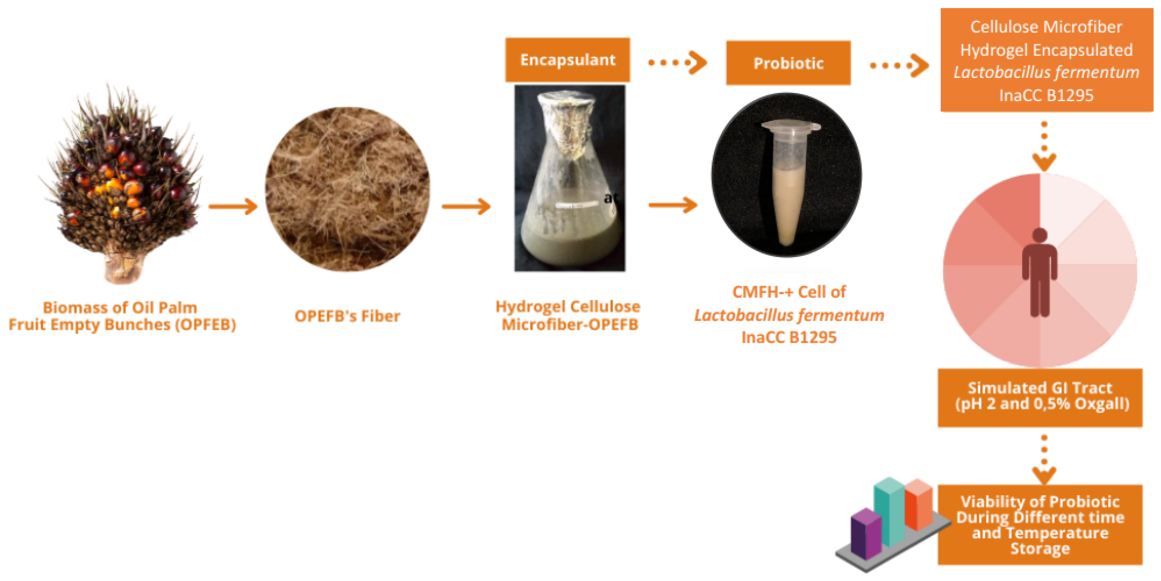
Figure 1. Flowchart of the research methodology.
Fermentation 08 00602 g001
Figure 2. SEM micrograph (a) and X-ray Diffraction (b) of cellulose microfiber from OPEFB. X-ray Diffraction of CMF-OPEFB.
Fermentation 08 00602 g002
Table 1. Viability and bile and acid tolerance of Lactobacillus fermentum InaCC B1295 in CMFH-OPEFB at various temperatures for 28 Days.
Storage Time (Day)Storage Temperature
Refrigerated Temperature (4 °C)Room Temperature (25 °C)
Viability (log CFU/g)
09.93 d9.80 c
149.39 b9.38 b
289.13 a9.17 a
Acid resistance (%)
099.1299.72
1498.9799.08
2898.9298.14
Bile resistance (%)
099.72 b99.25 b
1498.31 b99.13 b
2893.47 a92.07 a
a,b,c,d Significant differences at p-value of 0.05 are denoted by different letters within columns and rows.
Table 2. Safety evaluation of Lactobacillus fermentum InaCC B1295 in CMFH-OPEFB at various temperatures for 28 days.
Probiotic Properties/
Storage Time (Day)
Observation ResultsIndications
Refrigerated Temperature (4 °C)Room
Temperature
(25 °C)
Hemolytic activity
0NoneNoneNo clear zone around colonies on Columbia Blood Agar medium
14NoneNone
28NoneNone
Production of biogenic amines from L-histidine, tyrosine, L-ornithine, Lysine
0NoneNoneNo color change from brown to purple from the colonies on Decarboxylase Agar medium with purple bromocresol as an indicator
14NoneNone
28NoneNone
Production of cytolysin
0NoneNoneNo clear zone around colonies on BHI agar medium
14NoneNone
28NoneNone
Production of gelatinase
0NoneNoneNo clear zone around colonies on BHI agar medium
14NoneNone
28NoneNone
Table 3. Resistance to hydrogen peroxide of Lactobacillus fermentum InaCC B1295 in CMFH-OPEFB at different temperatures for 28 days of storage.
Storage Time (Day)Absorbance at 600 nm
Refrigerated Temperature
(4 °C)
Room Temperature
(25 °C)
Concentration 0.4 mM
01.691 e1.691 e
141.113 c1.091 b
281.139 d0.996 a
Concentration 0.7 mM
01.582 d1.582 d
141.085 c1.034 b
281.034 b0.981 a
Concentration 1.0 mM
01.477 d1.477 d
141.047 c0.981 b
280.837 a0.837 a
a,b,c,d Significant differences at p-value of 0.05 are denoted by different letters within columns and rows.
Table 4. Hydroxyl radical scavenging activity of Lactobacillus fermentum InaCC B1295 in CMFH-OPEFB at different temperatures for 28 days of storage.
Storage Time (Day)Radical Scavenging Activity (µg/mL)
Refrigerated Temperature (4 °C)Room Temperature (25 °C)
Concentration 1:1
068.235 a68.235 a
14105.426 b115.503 b
28111.628 b119.380 c
Concentration 1:2
089.803 a89.803 a
14120.930 b129.457 b
28134.892 b170.542 c
a,b,c Significant differences at p-value of 0.05 are denoted by different letters within columns and rows.
Table 5. The scavenging ability of DPPH radicals of Lactobacillus fermentum InaCC B1295 in CMFH-OPEFB at different temperatures for 28 days of storage.
Storage Time (Day)DPPH Scavenging Activity (µg/mL)
Refrigerated Temperature (4 °C)Room Temperature
(25 °C)
IC50
036.880 a36.880 a
14187.626 b189.435 b
28188.773 b201.943 b
a,b Significant differences at p-value of 0.05 are denoted by different letters within columns and rows.
Publisher’s Note: MDPI stays neutral with regard to jurisdictional claims in published maps and institutional affiliations.