Article

Anti-Influenza Virus Potential of Probiotic Strain Lactoplantibacillus plantarum YML015 Isolated from Korean Fermented Vegetable

by
Rajib Majumder
1,2,3,†,
Md Badrul Alam
4,†,
Keshav Raj Paudel
1,
Khandaker Asif Ahmed
2,5,
Hari Prasad Devkota
6,7,8,
Sang-Han Lee
4,
Philip M. Hansbro
1,* and
Yong-Ha Park
3,*
1
Centre for Inflammation, Centenary Institute and University of Technology Sydney, Faculty of Science, School of Life Sciences, Sydney, NSW 2007, Australia
2
Applied Biosciences, Macquarie University, Sydney, NSW 2109, Australia
3
Department of Applied Microbiology and Biotechnology, Yeungnam University, Gyeongsan 38541, Korea
4
Department of Food Science and Biotechnology, Graduate School, Kyunpook National University, Daegu 37224, Korea
5
Australian Centre for Disease Preparedness (ACDP), CSIRO, Geelong, VIC 3220, Australia
6
Graduate School of Pharmaceutical Sciences, Kumamoto University, 5-1 Oe-honmachi, Chuo-ku, Kumamoto 862-0973, Japan
7
Headquarters for Admissions and Education, Kumamoto University, Kurokami, 2-39-1, Chuo-ku, Kumamoto 860-8555, Japan
8
Pharmacy Program, Gandaki University, Pokhara 33700, Nepal
*
Authors to whom correspondence should be addressed.
These authors contributed equally to this work.
Fermentation 2022, 8(11), 572; https://doi.org/10.3390/fermentation8110572
Submission received: 16 September 2022 / Revised: 17 October 2022 / Accepted: 20 October 2022 / Published: 23 October 2022
(This article belongs to the Special Issue Recent Trends in Lactobacillus and Fermented Food)

Abstract

:
Lactic acid bacteria are one of the potential natural remedies used worldwide, commonly known as probiotics. Here, the aim of this research investigation was to isolate a probiotic Lactobacilli strain, YLM015, from the popular Korean fermented vegetable “Kimchi” and to evaluate its anti-viral potential against influenza virus A (IFVA) H1N1 using the MDCK cell line in vitro, and in embryonated eggs in ovo. The YML015 strain was selected from among the 1200 Lactobacilli isolates for further studies based on its potent anti-viral efficacy. YML015 was identified and characterized as Lactoplantibacillus plantarum YML015 based on the 16S rRNA gene sequencing and biochemically with an API 50 CHL Kit. In ovo assay experienced with embryonated eggs and the hemagglutination inhibition method, as well as cytopathogenic reduction assay, was performed individually to observe anti-influenza viral activity of YML015 against influenza virus A H1N1. Additionally, YML015 was classified for its non-resistance nature as safe for humans and animals as confirmed by the antibiotic susceptibility (MIC) test, cell viability, and hemolysis assay. The heat stability test was also experienced by using different heat-treated cell-free supernatant (CFS) samples of YML015. As a result, YML015 showed highly potent anti-viral activity against influenza virus A H1N1 in vitro in the MDCK cell line. Overall findings suggest that anti-influenza viral activity of L. plantarum YML015 makes it a potential candidate of choice for use as an influential probiotic in pharmacological preparations to protect humans and animals from flu and viral infection.

1. Introduction

Nowadays, probiotics have gained major attention due to their diverse range of therapeutic potential. Probiotics are defined as live microorganisms that, when administered in adequate amounts, confer health benefits on the host (WHO/FAO, 2002). The efficacy of probiotic bacteria is greatly influenced by their functional properties, such as antimicrobial activity, persistence in the gastrointestinal tract (GIT) for intestine-targeted probiotics, and immunomodulatory properties [1,2] In the gut, probiotics affect balancing and restoration of the gut microbiota, protection against pathogens, immunomodulation, and maintenance of intestinal barrier integrity [3]. Studies reported that probiotics are frequently used in dietary supplements, food, infant formula formulations, and medical devices [4,5]. In addition, probiotics have significant potential as therapeutic options for a variety of diseases, mainly gastrointestinal diseases (including acute infectious diarrhea, antibiotic-associated diarrhea, ulcerative colitis, irritable bowel syndrome, functional gastrointestinal disorders, or necrotizing enterocolitis), as well as extra-intestinal disorders, such as hepatic encephalopathy [6,7]. Probiotics not only show the healing effect as live compounds, but they also produce the same pharmacological effect as dead microorganisms, confirming that microbial metabolites present in probiotics have the ability to prevent various types of chronic diseases, including allergy, acute infectious diarrhea, and bowel disease inflammation [8]. Bifidobacterium and Lactobacillus microorganisms are most frequently used as probiotics. Interestingly, previous research has been reported that lactobacillus as a probiotic has potential effect on bacterial infectious diseases caused by Streptococcus pyogenes and Streptococcus pneumoniae [9,10]. In addition, studies found that the early-stage cold has been inhibited using Lactobacillus with yogurt [11]. However, not all probiotics display the same properties; hence, careful selection of specific strains based on their claimed beneficial effects is needed.
Influenza, an infectious disease of birds and mammals, is caused by RNA viruses of the family Orthomyxoviridae (commonly called the influenza viruses). Over the recent pandemic years, between three and five million people have been infected and between 250,000 and 550,000 have died due to the seasonal spread of this deadly influenza virus around the world [12,13,14,15]. Humans, birds, and pigs are the common host for influenza virus. Based on two specific proteins called hemagglutinin (HA) and neuraminidase (NA), there are different subtypes of influenza viruses [16]. Influenza virus A H1N1, which affects humans, has some common symptoms, such as fever, pharyngitis, muscle pain, severe headache, include chills, coughing, and weakness [13,14]. It also causes pneumonia, which might be fatal in more serious cases, especially for young children and elderly people [17]. So far, the management authority of influenza disease has been given optimum priority worldwide to control the spread of influenza viruses. In general, people are vaccinated against influenza as a practical and potentially anti-viral therapy. However, considering the limited efficacy of the current vaccination programs [18] and imposed restrictions on the use of antiviral drugs (those that have severe side effects) [19], the development of probiotic-based natural anti-viral therapies is in high demand. A few studies have demonstrated that probiotics of some lactic acid bacterial strains were able to protect against infectious diseases [20,21] and have the anti-allergic effects on immune diseases in mice [22,23] and humans [24,25]. Yogurt fermented with Lactobacillus was shown to reduce the cases of catching cold in the healthy elderly [11] and to prolong the survival periods of mice with influenza virus infection [24]. Despite this current situation, there is an alarming demand arising for the development of natural antiviral therapy of probiotic supplements against influenza virus, which might be safe and free from any adversary side effects as the lactic acid bacteria (LAB) and/or probiotics are classified as generally safe (GRAS) by US-FDA. Therefore, the primary objective of this research is to isolate, identify, and characterize the novel probiotic strain L. plantarum YML015 and to evaluate its potent antiviral efficacy against influenza virus A H1N1 in vitro and in ovo.

2. Materials and Methods

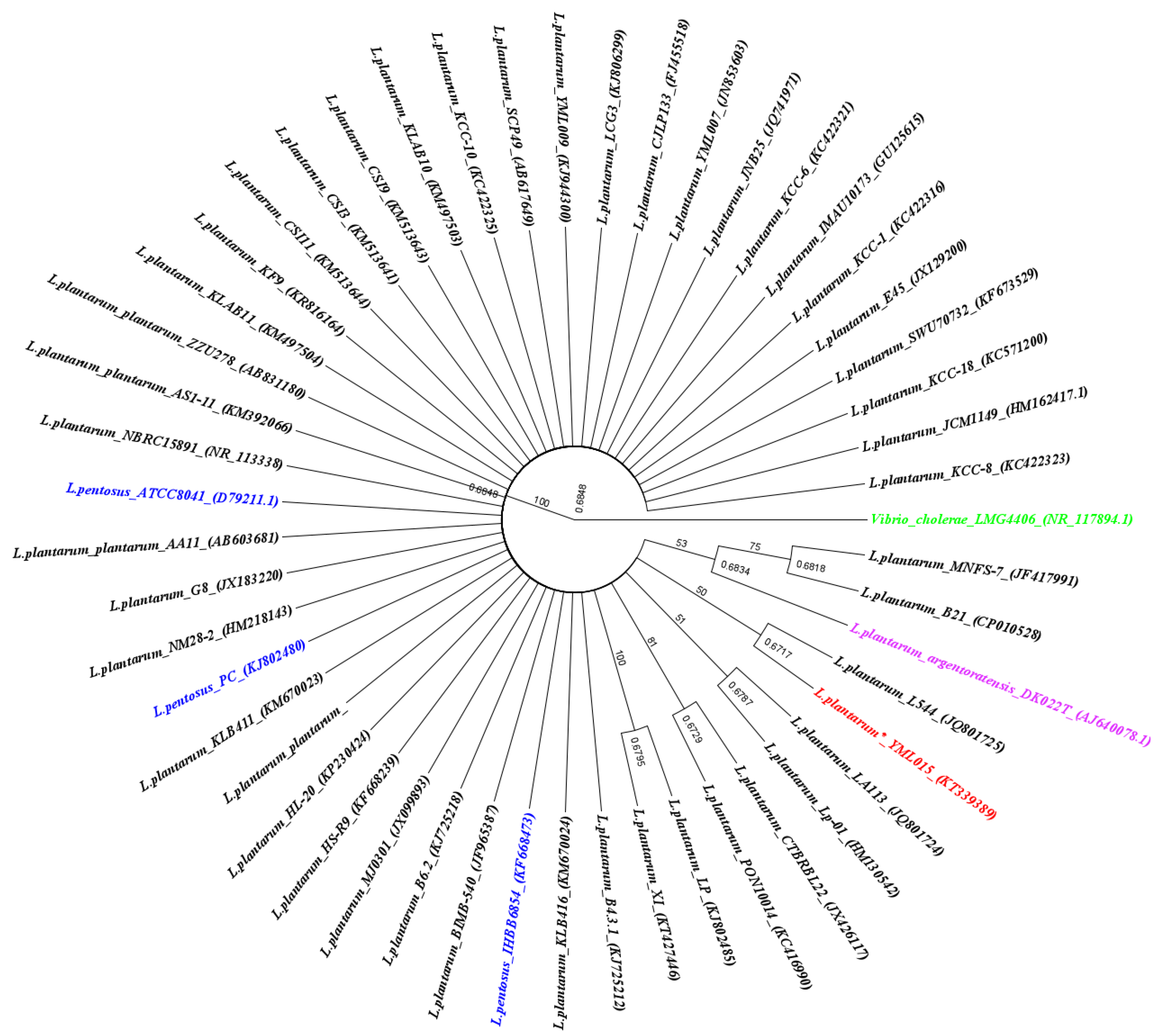
2.1. Isolation and Identification of L. plantarum YML015

2.1.1. Isolation and Stock Preparation Process of L. plantarum YML015

A total of one 1200 Lactic Acid Bacteria (LAB) strains were isolated from various home-made traditional Korean fermented vegetables called “Kimchi” using a serial dilution method of 10-fold dilutions with PBS as described previously with slight modifications [26]. 100 µL of diluted sample was spared and placed on Bromcresol purple agar (BCP agar) medium (Merck, Darmstadt, Germany) for primary identification based on colony morphology and color changes around the single colonies. Prior to final stock preparation, the pure single colony isolated from the BCP agar plates were cultured again on de Man–Rogosa–Sharpe agar medium (MRS agar; Merck, Darmstadt, Germany). The agar plates were incubated at 37 °C inside the anaerobic incubator for 24 h (MRS agar) and 48 h (BCP agar) [27]. Cell-free supernatants (CFS) of bacterial isolates were processed by culturing in MRS broth at 37 °C for 24 h, followed by centrifugation (10,000× g for 10 min) and filtration (0.22 μm filter; Sartorius Stedim Biotech, Goettingen, Germany). Furthermore, bacterial stock was prepared by 30% glycerol and stored as a working cell bank at −80 °C for further use [28,29].

2.1.2. DNA Extraction, PCR, and 16S rRNA Gene Sequencing of L. plantarum YML015

As it flourished on the preliminary antiviral screening against IFVA H1N1, L. plantarum YML015 was selected as the best LAB candidate and further subjected to 16S rRNA gene sequencing to confirm the identification. Before the sample was sent for Sanger sequencing, DNA extraction was performed using a pure single colony of L. plantarum YML015 cultured on MRS agar (Sigma-Aldrich, St. Louis, MO, USA) medium after 24 h of incubation at 37 °C. Whole DNA extraction was carried out using the Isolate II genomic DNA kit from Bioline, USA following the protocol provided by the manufacturer, and extracted DNA were quantified by Nano-drop (Eppendorf, Hamburg, Germany). Standard combination of universal Primers (27f⁄1492r primers) were used to continue initial PCR amplifications [30]. All PCR amplifications in the present study were performed in an Eppendorf thermocycler (Eppendorf, Germany). The PCR profile included an initial denaturing step at 95 °C for 2 min, followed by 35 cycles of 30 s steps at 94 °C, 50 °C, and 72 °C for 90 s, and a final extension step of 72 °C for 5 min. Electrophoresis was performed (110 v–45 min) to analyze the PCR product with 1% agarose gel after amplification, and Easy ladder 1 and loading buffer (Bioline, USA) were used to see the target band. PCR products were sent to Macrogen, South Korea to execute Sanger sequencing and sequence data were analyzed using Geneious R10 software to confirm the identification of L. plantarum YML015, and, finally, submitted to NCBI GenBank. Homology of the selected strain L. plantarum YML015 was compared with other similar strains. A phylogenic tree was constructed using the Geneious version 2021.2 (Biomatters, NZ) software. In brief, a multiple sequence alignment (MSA) was conducted with the MUSCLE algorithm with default settings. The resulting MSA was used to construct a cladogram tree, using Tamura-Nei as the genetic distance model and Neighbor-Joining as the tree-building method. Resampling was conducted using bootstrap methods for random seeding, with 100 replicates, which created a consensus tree with a support threshold of 50%. Various color codes were used to visually present differences. A “similarity matrix” was also added.

2.2. Characterization of L. plantarum YML015

2.2.1. Biochemical Characterization

The strain was characterized biochemically using the API 50 CHL strip and API CHL medium systems, according to the manufacturer’s instructions (API bioMerieux, St. Louis, MO, USA). Briefly, a freshly grown colony of L. plantarum YML015 was harvested and re-suspended in sterile water to achieve a cell density of 108 CFU/mL. A 2 mL aliquot of the cell suspension was inoculated into 10 mL of APL 50 CHL medium and mixed gently by inversion. Further, 120 μL of this suspension was inoculated into API 50 CHL strips that were overlaid with mineral oil and were incubated for 48 h before reading the color changes [31].

2.2.2. Heat Stability Test

In order to confirm stability and antiviral activity of L. plantarum YML015 against IFVA H1N1, 1-fold and 10-fold CFS of L. plantarum YML015 were subjected to different heat treatments through exposure to varying degrees of temperature, such as 30 °C, 45 °C, 60 °C, 90 °C, and 121 °C with different time durations, such as 5 min, 10 min, and 15 min. The antiviral activity of heat-treated 10-fold CFS was determined by cytopathogenic reduction assay.

2.2.3. Antibiotic Susceptibility Assay

The antibiotic susceptibility of L. plantarum YML015 was carried out using the micro-dilution method [32]. Prior to the assay, L. plantarum YML015 was pre-cultured at 37 °C for 24 h and subjected to LAB susceptibility test medium (LSM) formulation consisting of a mixture of IST broth (90%) and MRS broth (10%) and adjusted to pH 6.7. After 24 h of incubation, the inoculum was prepared, and a colony was suspended in a sterile plastic culture tube containing 2 mL of sterile saline until a density corresponding to a McFarland (McF) standard of 1 or a spectrophotometric equivalent (3 × 10⁸ CFU/mL) was obtained. The inoculated saline suspension was diluted at 1:500 to obtain a final concentration of 3 × 10⁵ using LSM broth [33]. According to the European Food Safety Authority (EFSA) guidelines, the following antibiotics were tested at the concentration range (mg/L) given in parenthesis: gentamicin (0.5 to 256 mg/L; Tokyo chemical industry co; Ltd., Tokyo, Japan), kanamycin (2 to 1024 mg/L; Biopure reagent, Seoul Korea), streptomycin (0.5 to 256 mg/L; Tokyo chemical industry co; Ltd., Tokyo, Japan), tetracycline (0.125 to 64 mg/L; Tokyo Chemical Industry Co; Ltd., Tokyo, Japan), erythromycin (0.016 to 8 mg/L; Tokyo Chemical Industry co; Ltd., Tokyo, Japan), clindamycin (0.032 to 16 mg/L; Sigma-Aldrich, St. Louis, MO, USA), chloramphenicol (0.125 to 64 mg/L; Sigma-Aldrich, St. Louis, MO, USA), and ampicillin (0.032 to 16 mg/L; Biopure reagent, Seoul, Korea). Further, 50 μL of 3 × 10⁵ CFU/mL inoculum was added to each well (already containing antibiotics) in a micro-dilution plate within 30 min of the preparation of the standardized inoculum. The well without antibiotics served as positive controls but contained mixture of the test strain and the medium containing solvent, which was used to dissolve the antibiotics at the highest concentration, while wells without the test strain and the antibiotics but with the medium, served as negative controls. The susceptibility panel was incubated anaerobically at 37 °C for between 24 and 48 h after inoculation; growth in the susceptibility panel was evaluated visually by comparing the pellet at the bottom of each well, comparing with the positive and negative controls. The selection of antibiotics and the characterization of sensitivity/resistance were conducted following the guidelines and breakpoints of the European Commission [34] and the European Food Safety Authority [35].

2.2.4. Hemolytic Phenomenon Assay

The hemolytic phenomenon assay was performed according to Maragkoudakis et al., 2009 [36]. L. plantarum YML015 was freshly cultured in MRS broth and then streaked on tryptone soya agar (Oxoid, Basingstoke, UK) containing 5% (v/v) sheep blood. The plates underwent 48 h of micro aerobic incubation (37 °C, 8% CO2) and were examined for hemolytic reaction. Hemolytic activity was confirmed by the presence of a clear zone around the bacterial colony. The experimental strain that produced green-hued zones around the bacterial colonies (α-hemolysis) or did not produce any effect on the blood plates (γ-hemolysis) were considered non-hemolytic. Any strains that displayed blood lysis zones around the bacteria colonies were classified as hemolytic (β-hemolysis).

2.3. Culture of MDCK Cell

Madin–Darby canine kidney (MDCK) cell cultures essential for supporting viral infectivity of the test virus were used in this study. MDCK cells (KTCC® AC30015) were grown with high-glucose Dulbecco’s modified Eagle’s medium (DMEM) (Sigma-Aldrich, St. Louis, MO, USA) supplemented with 10% heat-inactivated standard fetal bovine serum (FBS) (HyClone, Logan, UT, USA) and 1% (v⁄v) 100U/mol penicillin and 100 µg/mL streptomycin solution at room temperature (23 ± 1 °C) under normal atmospheric conditions. Cells were sub-cultured at a 1:3 ratio every 3–4 days. Briefly, media from tissue culture flasks were collected and centrifuged at 1500× g rpm for 5 min. Meanwhile, a 5 mL/flask of a trypsin-versine solution (10 mL 10× trypsin (Sigma-Aldrich, St. Louis, MO, USA) pre-sterilized versine (EDTA)) was added and incubated for 1 min to detach the cell monolayer [37]. Following cell detachment, cleaned medium was added back into the flasks to neutralize the trypsin activity. The contents of the flasks were then removed and centrifuged at 1500× g rpm for 5 min. Following centrifugation, the supernatant was removed, and new growth medium was used to re-suspend the cells. Cells, split 1:3, were placed back into flasks and total media volume was brought up to a 20–22 mL/flask. Flasks were then placed back into their respective incubators and monitored daily.

2.4. MTT Cell Viability Assay

The mitochondrial-dependent reduction of MTT (3-(4,5-Dimethylthiazol-2-Yl)-2,5-Diphenyltetrazolium Bromide) to formazan was used to measure cell respiration as an indicator of cell viability. Briefly, MDCK cells were seeded in 96-well plates at a density of 5 × 10⁴ cells/well. After 24 h of incubation, the adherent cells were treated with various concentrations (CFS 1×, CFS 10×, Heat-Killed (HK) CFS 10×, and CFS 20×) of L. plantarum YML015. Twenty-four hours later, after changing the medium, MTT was added to a final concentration of 0.5 mg/mL, and the cells were incubated for 4 h at 37 °C and 5% CO2. The medium was then removed, and the formazan precipitate was solubilized in DMSO. The absorbance was measured at 570 nm on a microplate reader (Biotek, Winooski, VT, USA) [38].

2.5. Virus Culture

Highly pathogenic Influenza virus (IFV) H1N1 (A/Korea/01/2009) was obtained from the Korea Disease Control and Prevention Agency, Korea. The IFVA H1N1 was incubated in the allantoic cavity of 11-day-old chicken embryos for 3 days. In brief, specific pathogen-free (SPF) chicken eggs were cleaned by 70% (v/v) ethanol and incubated for embrocation in a rotating and static egg incubator at 35 °C with a humidified environment for 11 days. After 11 days, a 100 µL IFVA H1N1 was inoculated in the allantoic cavity by using 1 mL syringes with 27-gauge, 1-inch or 1.5-inch hypodermic or blunt-end needles and the punched holes were sealed with wax followed by incubation at 35 °C in a humidified, static incubator for 2–3 days. At the completion of the incubation period, eggs were prepared for fluid collection by placing them on ice (at 4 °C) overnight. Then, a 3 mL syringe with a 21-gauge, 1-inch hypodermic needle (for collection of allantoic fluids) was used to collect the harvested allantoic fluid (Figure 1). Allantoic fluid was stored at −80 °C as a stock solution of the IFVA H1N1 A virus [39,40]. Virus titers in the allantoic fluid stock solution were determined as embryo infection dose 105.5EID50/0.1 mL by using the 50% end point dilution assay described by Reed and Muench, 1938 [41].

2.6. Antiviral Potential of L. plantarum YML015

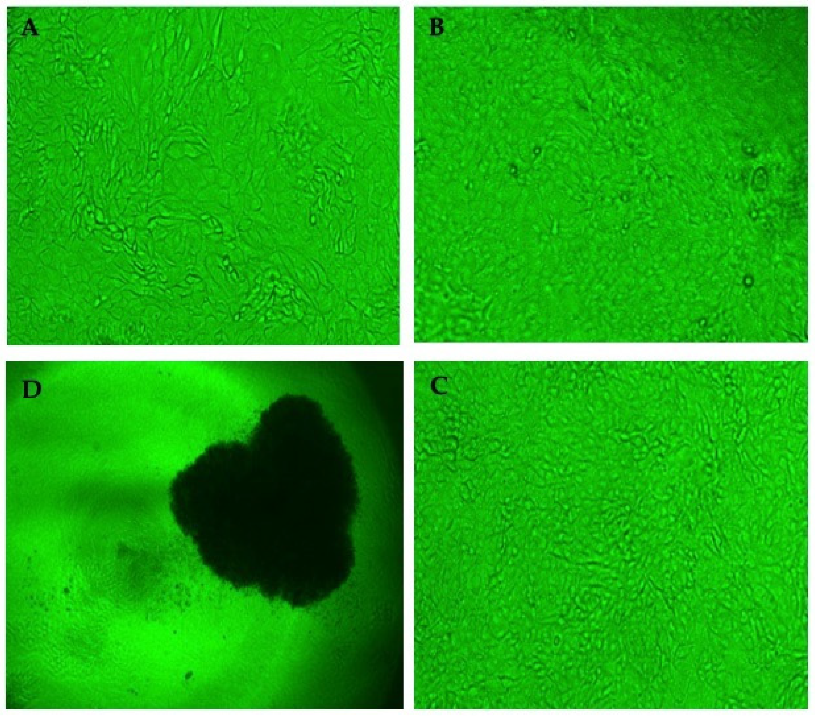
2.6.1. Cytopathogenic Reduction Assay

For assessing the cytopathogenic reduction effect of L. plantarum YML015 in vitro, firstly, the MDCK cell line was cultured in a 96-well microplate using Eagle’s minimal medium (MEM) with 10% (v/v) fetal bovine serum (FBS), 1% (v/v) 100U/mol penicillin, and 100 µg/mL streptomycin solution for 24–36 h at 37 °C in a 5% CO2 cell culture incubator. MDCK cells were seeded onto a 96-well culture plate at a concentration of 2 × 10⁴ cells per well calculated by a hemocytometer [42]. Sterilized cell-free supernatant (CFS) of L. plantarum YML015 prepared by filtration using 0.22 µm cellulose acetate filter was serially diluted two-fold with 2% FBS DMEM solution. IFVA H1N1 was treated with a two-fold dilution of CFS of L. plantarum YML015 (1× and 10×) at 37 °C in a 5% CO2 cell culture incubator for 1 h. This mixture was inoculated into MDCK cells and incubated in DMEM solution with 2% FBS at 37 °C in a humidified chamber with 5% CO2 for 72 h by using a multi-pipette. The plates were observed for cytopathic effect (CPE) after 24 h, 48 h, and 72 h accordingly, and the reduction in CPE was regarded as the presence of antiviral activity [28,39,43].

2.6.2. Hemagglutination Inhibition Assay

The hemagglutination inhibition assay is a widely used, inexpensive, rapid, and standard screening method for detecting activity of the target sample against IFVA H1N1. The CFS (1-fold and 10-fold) and heat-killed CFS (121 °C for 15 min, 10-fold) of L. plantarum YML015 were used for the hemagglutination reduction assay. The mixture of the L. plantarum YML015 supernatant and embryo infection dose 105.5EID50/0.1 mL were incubated for 1 h at 37 °C in a 5% CO2 cell culture incubator and transferred into new tubes for hemagglutination inhibition assay. By using a multi-channel pipette, two-fold dilution of treated samples was made in 50 µL with PBS in a V-form-shaped 96-well microplate. Serially diluted samples were reacted with an equal volume of 1% Specific Pathogen-Free (SPF) chicken red blood cell (RBC) at room temperature for 2–3 h to allow hemagglutination inhibition reaction. The only virus dose was used to check the negative control to compare the activity of the treated sample [28,39,43].

2.6.3. In Ovo Antiviral Assay

Specific pathogen-free embryonated chicken eggs were used to check the in ovo antiviral activity of L. plantarum YML015. The experiment was designed with 6 groups, consisting of group 1: H1N1 + CFS (1-fold and 10-fold), group 2: H1N1 + HKCFS (10-fold), group 3: H1N1 + cell mass (CM), group 4: drug control H1N1 + Tamiflu, and group 5: negative control H1N1 + PBS and group. Each group contained 2 SPF eggs. After dose inoculation, eggs were incubated again for 72–96 h at 37 °C under 80% humidified incubation conditions. A similar dose of IFVA H1N1 105.5EID50/0.1 mL was used as mentioned in previous experiments. The percentage of surviving eggs out of the total was calculated to determine the antiviral activity [44].

2.7. Statistical Analysis

All data were expressed as mean ± standard error of mean (S.E.M.). Statistical significance was analyzed by one-way analysis of variance (ANOVA) followed by Dunnett’s post hoc test of significance. All statistical analyses were performed with Prism 4.0 (GraphPad software Inc., San Diego, CA, USA). p < 0.05 was considered to be significant.

3. Results

3.1. Isolation and Characterization of L. plantarum YMLO15

Among 1200 bacterial strains screened, cell-free supernatant (CFS) of the isolated bacteria Lactobacillus spp., isolated from Kimchi, was found to exert the highest antiviral activity, even at two-fold dilution and selected as a potential probiotic Lactobacilli against highly pathogenic influenza virus A (IFVA) H1N1 for further in vitro experiments. The molecular systematic analysis based on 16S rRNA sequences (Sanger sequencing) isolated from the bacterial strain Lactobacillus spp. displayed 100% similarity with Lactobacillus plantarum spp. Based on these parameters, Lactobacillus plantarum spp. was characterized and named Lactoplantibacillus plantarum YML015 (previously named Lactobacillus plantarum YML015) and the gene sequence was submitted to the GenBank with a nucleotide accession number KT339389. Figure 2 represents the neighbor-joining phylogenetic tree of L. plantarum YML015, using Geneious v10.2.3 (Biomatters, NZ). Sequences were aligned with CLUSTALW and overhanging ends were trimmed off manually.

3.2. Biochemical Characterization of L. plantarum YML015

The analysis of the API 50 CHL Kit for biochemical characterization showed that the strain L. plantarum YML015 was most closely related to Lactoplantibacillus plantarum, as shown in Table 1. According to API web software, the strain L. plantarum YML015 indicated typical carbohydrate utilization that used D-xylose, D-galactose, D-glucose, D-fructose, Dmannose, L-rhamnose, D-mannitol, D-sorbitol, N-acetylglucosamine, amygdalin, arbutin, esculin, salicin, Dcellobiose, D-maltose, D-lactose, D-melibiose, D-saccharose, D-trehalose, D-melezitose, D-raffinose, gentiobiose, D-turanose, D-tagatose, and D-arabitol. The strip capsule changed color from violet to yellow, indicating complete fermentation.

3.3. Antibiotic Susceptibility Assay

The antibiotic susceptibility of L. plantarum YML015 to eight antibiotics is presented in Table 2. L. plantarum YML015 showed sensitivity to all the recommended antibiotics checked. The minimum inhibitory concentration (MIC) values were observed after 24 and 48 h of incubation, which were obtained as follows with green color in the table: gentamicin (0.5 mg/L), kanamycin (4 mg/L), streptomycin (2 mg/L), tetracycline (8 mg/L), erythromycin (0.125 mg/L) clindamycin (0.125 mg/L), chloramphenicol (4 mg/L), and ampicillin (0.25 mg/L). Since L. plantarum YML015 did not show any resistance to the panel of antibiotics, it could be considered as a probiotic bacterium and safe for human consumption.

3.4. Heat Stability Test

The temperature/heat stability of the CFS (10-fold) of L. plantarum YML015 was checked by using the cytopathic reduction assay. The sample treated with 121 °C for 15 min also strongly inhibited the cytopathic effect of IFVA H1N1 on the MDCK cell monolayer in a 96-well microtiter plate. The experiment was continued up to 96 h, and every 24 h later, the activity was checked (Table 3).

3.5. Hemolytic Phenomenon Assay

Lactobacillus plantarum YML015 was streaked on tryptone soy agar supplemented with 5% sheep blood and incubated in a CO2 incubator at 37 °C for 48 h (Figure 3). It was found that no β-hemolysis (clear zones around colonies) was observed on the plate agar. L. plantarum YML015 did not produce any effect or lysis on the blood plates (γ-hemolysis) (Figure 3C). Streptococcus aureus KCTC 1621 was used as a negative control for β-hemolysis (Figure 3B), whereas Lactobacillus sakei probio65 KCTC 10755BP was used as a positive control for γ-hemolysis (Figure 3A).

3.6. MTT Cell Viability Assay

The result of MTT cell viability of the CFS of L. plantarum YML015 significantly showed no evidence of cytotoxicity with cell viability ranging from 80% to 100% at different concentrations (CFS 1×, CFS 10×, HKCFS 10×, and CFS 20×) compared with the standard (Figure 4). However, considering the concentration of 20× (80% cell viability rate), the current experiment shows that the cell viability of L. plantarum YML015 in the concentration of 1× and 10× (both CFS and HKCFS) is 100% without occurring any type of cell damage or depreciation of the cell. Based on the research outcome, L. plantarum YML015 concentrations used for the MTT cell viability assay (cell viability >85%) were selected for the subsequent following antiviral experiments.

3.7. Cytopathic Reduction Assay

In vitro antiviral activity of L. plantarum YML015 was evaluated using MDCK cells by the cytopathic reduction assay. In this assay, L. plantarum YML015 was grown for its antiviral activity against influenza virus A H1N1 on MDCK cells cultured in a 96-well plate. We found that H1N1 inoculation caused a cytopathic effect in MDCK cells when used as negative control (Figure 5). However, the same cytopathic effect was reduced or not observed in MDCK cells treated with cell-free supernatant of L. plantarum YML015 (1-fold and 10-fold v/v) even after 72 h of the viral dose 105.5EID50/0.1 mL (Figure 5B). In addition, microscopic observation results proved that MDCK cells treated with H1N1 and L. plantarum YML015 showed similar morphology as control MDCK cells (without any treatment) as shown in Figure 5C. Therefore, it proved that L. plantarum YML015 could be considered as a potential anti-influenza bacterium able to reduce the cytopathic effect in MDCK cells. In addition, 10-fold (v/v) cell-free supernatant of L. plantarum YML015 showed more specific results. Serial dilutions of 20, 21, and 22 of 10-fold cell-free supernatant also exhibited similar activity to control the CPE in MDCK cells (Table 4).

3.8. Hemagglutination Inhibition Assay

Hemagglutination effect is a common phenomenon of influenza A virus H1N1 on chicken red blood cells. This assay revealed the hemagglutination inhibition potential of the cell-free supernatant of L. plantarum YML 015, which can inhibit the viral HA protein that causes hemagglutination. As described in Table 5, 1-fold, 10-fold, and heat-killed (10-fold) CFS of L. plantarum YML015 showed anti-hemagglutination effect against H1N1. Interestingly, CFS (10-fold) and heat-killed CFS (10-fold) of L. plantarum YML015 showed similar hemagglutination inhibition activity of 2-fold dilution (4 times).

3.9. In Ovo Antiviral Activity

In this assay, influenza A virus H1N1 was treated with 5 groups of treatments prepared form L. plantarum YML015 with 1 group of negative control. As described in Table 6, for this experiment, each group contained 2 embryos obtained from SPF eggs that had been incubated for 11 days. Among 6 groups, the untreated virus control indicated a marked antiviral effect on the test virus. Heat-killed CFS (10-fold) and only CFS (10-fold) treated eggs showed a 100% survival rate. Apart from this, the survival rate was 50% in the case of the positive control treated by Tamiflu (Oseltamivir phosphate). Treatment with cell mass (3 × 10⁸ cfu/mL) and CFS (1-fold) also gave similar results as with the positive control.

4. Discussion

Nowadays, the use of probiotics is in high demand due to their high immunostimulant potency. For example, the probiotic strain Lactobacillus plantarum KLAB21 from Korean kimchi has significant antimutagenic characteristics [45,46]. There is a huge trend among researchers to explore and study different probiotics or Lactic Acid Bacteria (LAB) from Korean fermented food products. Studies have also observed their chemical nature, along with antimicrobial potential against various food spoilage and food-born pathogens, including molds and fungi, for their possible use as food preservatives [29]. In this current study, a total of 1200 LAB strains were isolated from different kinds of Kimchi samples, which were initially screened for their antiviral efficacy. Among them, one of the strains designated as L. plantarum YML015 exhibited remarkable antiviral activity against IFVA H1N1. The 16 s rRNA sequencing confirmed molecular characterization of the YML015 isolate as L. plantarum YML015. The selected strain was also further identified based on the biochemical characterization utilizing carbohydrate interpretation assay (API 50CHL kit). A standard method of carbohydrate utilization according to API web software showed that L. plantarum YML015 used approximately 24 carbohydrates among 50 carbohydrates (Table 1), thus, was characterized as Gram-positive and lactobacilli.
In our study, the probiotic nature of L. plantarum YML015 was confirmed based on the susceptibility test result. However, the European Food Safety Association (EFSA) has already published a new protocol for foods and feeds purposes, which might be helpful to evaluate probiotic strains as a food grade probiotic [47]. Moreover, “Qualified Presumption of Safety” (QPS) also described the importance and procedure of determination of antibiotic resistance for selected probiotic strains. We experienced that the probiotic strain L. plantarum YML015 was very sensitive to clinically essential antibiotics such as ampicillin, gentamycin, kanamycin, streptomycin, erythromycin, clindamycin, and tetracycline. However, phenotypic antibiotic resistance testing in LAB requires standardization as it can be subjected to species specificity but is also dependent on the method and the media used [48]. Eventually, L. plantarum YML015 was found to cause γ-hemolysis (non-hemolysis). Results proved again that L. plantarum YML015 might be a potential probiotic with non-hemolytic characteristics. It is also a common phenomenon of non-hemolytic LAB isolates from food sources [49,50], except some strains of Enterococcus faecalis. Furthermore, Table 2 describes temperature stability of the cell-free supernatant of L. plantarum YML015. Interestingly, at optimum temperature treatment of 121 °C for 15 min, antiviral activity of CFU of L. plantarum YML015 was the same up to 96 h of activity observation. In addition to the MTT cell viability study, it has been significantly demonstrated that L. plantarum YML015 has optimum cell viability percentage with no cell damage or cytotoxic effect (Figure 4). The result supported that, as a probiotic, Lactobacillus plantarum YML015 is highly safe for human and animal consumption. The study also found that heat-killed bacteria, especially heat-treated probiotic strains, showed immunomodulating effects and had antagonizing properties against pathogens [51]. Hence, it was confirmed that L. plantarum YML015, as a strong probiotic, could be used as a heat-stable and effective agent against the H1N1 virus.
Viral invasion causes structural damage or change in the host cells called cytopathic effect (CPE). Two kinds of effects might occur due to CPE, such as the infecting virus leading to lysis of the host cell or because of the inability to reproduce new cells (cell death). It is also called cytopathogenic when morphological changes in the host cell are caused by virus. Moreover, [42,52] described that probiotic Lactobacilli strains exhibited antiviral effect against influenza virus as experienced with the cytopathic reduction assay. In our experiment, we successfully confirmed that newly invented L. plantarum YML015 has significant potential to inhibit the cytopathic effect caused by IFVA H1N1. It was ensured that the cell-free supernatant (1-fold and 10-fold) of L. plantarum YML015 completely suppressed the cytopathic effect of IFVA H1N1 on a monolayer of the MDCK cell line as compared with the negative control as described in Figure 5.
The hemagglutination inhibition assay is a popular screening assay. It allows rapid detection of antiviral activity of any kinds of sample against influenza virus [33,53]. This assay was standardized on the basis of the ability of viral HA protein to bind and to coagulate/agglutinate human or chicken red blood cells (RBC) [39]. Our findings are aligned with the previous observations, and it was ascertained in our research work that CFS of L. plantarum YML015 exhibited most effective potential in the hemagglutination inhibition activity assay (Table 5). This strongly suggests that probiotic dietary uptake of L. plantarum YML015 could be beneficial for direct inhibition of influenza virus A H1N1 infection [42].
In addition, this research also assessed the in ovo potential of the probiotic strain L. plantarum YML015 to reconfirm the antiviral efficacy on influenza A virus H1N1 and was expressed as a percentage of survival from the total number of test samples. Among all treated samples, including the negative control, only heat-killed CFS (10-fold) and CFS (10-fold) showed highest antiviral activity compared to other tested samples, and the survival rate was 100%. However, inoculation of eggs with standard treatment (Tamiflu) showed a 50% survival rate, suggesting that as a probiotic, L. plantarum YML015 could be a good source of natural anti-influenza drugs.

5. Conclusions

In summary, the present research output highlights the significant therapeutic efficiency of the probiotic strain L. plantarum YML015 isolated from the Korean fermented food Kimchi as a successfully competent antiviral agent against extremely pathogenic influenza A virus H1N1. Throughout the in vitro and in ovo experiments, it was clearly evident that the cell-free supernatant (CFS) of L. plantarum YML015 showed remarkable performance to kill or inhibit the activity of influenza A virus H1N1 on the MDCK cell line. The present research findings have further increased the urgency for clinical studies and animal trials to ensure that L. plantarum YML015 could be a safe natural probiotic drug with high therapeutic potential against deadly influenza virus H1N1 as compared with available bioorganic or synthetic drugs.

Author Contributions

Conceptualization, R.M. and Y.-H.P.; methodology, R.M. and M.B.A.; software, R.M.; formal analysis, R.M.; investigation, R.M.; resources, Y.-H.P.; writing—original draft preparation, R.M. and M.B.A.; writing—review and editing, S.-H.L., K.R.P., H.P.D., K.A.A. and P.M.H.; supervision, Y.-H.P.; project administration, Y.-H.P.; funding acquisition, Y.-H.P. All authors have read and agreed to the published version of the manuscript.

Funding

This research received no external funding.

Institutional Review Board Statement

Not applicable.

Informed Consent Statement

Not applicable.

Data Availability Statement

The data presented in this study are available upon request from the corresponding author.

Acknowledgments

This research was supported by the Yeungnam University Research Assistantship (RA) given to Rajib Majumder.

Conflicts of Interest

The authors declare no conflict of interest.

References

  1. Foligne, B.; Nutten, S.; Grangette, C.; Dennin, V.; Goudercourt, D.; Poiret, S.; Dewulf, J.; Brassart, D.; Mercenier, A.; Pot, B. Correlation between in vitro and in vivo immunomodulatory properties of lactic acid bacteria. World J. Gastroenterol. WJG 2007, 13, 236. [Google Scholar] [CrossRef] [PubMed] [Green Version]
  2. Annuk, H.; Shchepetova, J.; Kullisaar, T.; Songisepp, E.; Zilmer, M.; Mikelsaar, M. Characterization of intestinal lactobacilli as putative probiotic candidates. J. Appl. Microbiol. 2003, 94, 403–412. [Google Scholar] [CrossRef] [PubMed] [Green Version]
  3. Zyrek, A.A.; Cichon, C.; Helms, S.; Enders, C.; Sonnenborn, U.; Schmidt, M.A. Molecular mechanisms underlying the probiotic effects of Escherichia coli Nissle 1917 involve ZO-2 and PKCζ redistribution resulting in tight junction and epithelial barrier repair. Cell. Microbiol. 2007, 9, 804–816. [Google Scholar] [CrossRef] [PubMed]
  4. Bermudez-Brito, M.; Plaza-Díaz, J.; Muñoz-Quezada, S.; Gómez-Llorente, C.; Gil, A. Probiotic mechanisms of action. Ann. Nutr. Metab. 2012, 61, 160–174. [Google Scholar] [CrossRef]
  5. Pamer, E.G. Resurrecting the intestinal microbiota to combat antibiotic-resistant pathogens. Science 2016, 352, 535–538. [Google Scholar] [CrossRef] [Green Version]
  6. Draper, K.; Ley, C.; Parsonnet, J. Probiotic guidelines and physician practice: A cross-sectional survey and overview of the literature. Benef. Microbes 2017, 8, 507–519. [Google Scholar] [CrossRef]
  7. Wilkins, T.; Sequoia, J. Probiotics for Gastrointestinal Conditions: A Summary of the Evidence. Am. Fam. Physician 2017, 96, 170–178. [Google Scholar]
  8. Adams, C.A. The probiotic paradox: Live and dead cells are biological response modifiers. Nutr. Res. Rev. 2010, 23, 37–46. [Google Scholar] [CrossRef] [Green Version]
  9. Alexandre, Y.; Le Berre, R.; Barbier, G.; Le Blay, G. Screening of Lactobacillus spp. for the prevention of Pseudomonas aeruginosa pulmonary infections. BMC Microbiol. 2014, 14, 1–10. [Google Scholar] [CrossRef] [Green Version]
  10. Racedo, S.; Villena, J.; Medina, M.; Aguero, G.; Rodriguez, V.; Alvarez, S. Lactobacillus casei administration reduces lung injuries in a Streptococcus pneumoniae infection in mice. Microbes Infect 2006, 8, 2359–2366. [Google Scholar] [CrossRef]
  11. Makino, S.; Ikegami, S.; Kume, A.; Horiuchi, H.; Sasaki, H.; Orii, N. Reducing the risk of infection in the elderly by dietary intake of yoghurt fermented with Lactobacillus delbrueckii ssp. bulgaricus OLL1073R-1. Br. J. Nutr. 2010, 104, 998–1006. [Google Scholar] [CrossRef] [PubMed]
  12. Drouault, S.; Corthier, G. Health effects of lactic acid bacteria ingested in fermented milk. Vet. Res. 2001, 32, 101–117. [Google Scholar] [CrossRef] [PubMed] [Green Version]
  13. Hosoi, T.; Hirose, R.; Saegusa, S.; Ametani, A.; Kiuchi, K.; Kaminogawa, S. Cytokine responses of human intestinal epithelial-like Caco-2 cells to the nonpathogenic bacterium Bacillus subtilis (natto). Int. J. Food Microbiol. 2003, 82, 255–264. [Google Scholar] [CrossRef]
  14. Maeda, N.; Nakamura, R.; Hirose, Y.; Murosaki, S.; Yamamoto, Y.; Kase, T.; Yoshikai, Y. Oral administration of heat-killed Lactobacillus plantarum L-137 enhances protection against influenza virus infection by stimulation of type I interferon production in mice. Int. Immunopharmacol. 2009, 9, 1122–1125. [Google Scholar] [CrossRef] [PubMed]
  15. Kawase, M.; He, F.; Kubota, A.; Yoda, K.; Miyazawa, K.; Hiramatsu, M. Heat-killed Lactobacillus gasseri TMC0356 protects mice against influenza virus infection by stimulating gut and respiratory immune responses. FEMS Immunol. Med. Microbiol. 2012, 64, 280–288. [Google Scholar] [CrossRef] [PubMed]
  16. Jung, Y.-J.; Lee, Y.-T.; Ngo, V.L.; Cho, Y.-H.; Ko, E.-J.; Hong, S.-M.; Kim, K.-H.; Jang, J.-H.; Oh, J.-S.; Park, M.-K. Heat-killed Lactobacillus casei confers broad protection against influenza A virus primary infection and develops heterosubtypic immunity against future secondary infection. Sci. Rep. 2017, 7, 1–12. [Google Scholar] [CrossRef] [Green Version]
  17. Harata, G.; He, F.; Hiruta, N.; Kawase, M.; Kubota, A.; Hiramatsu, M.; Yausi, H. Intranasal administration of Lactobacillus rhamnosus GG protects mice from H1N1 influenza virus infection by regulating respiratory immune responses. Lett. Appl. Microbiol. 2010, 50, 597–602. [Google Scholar] [CrossRef]
  18. Hancock, K.; Veguilla, V.; Lu, X.; Zhong, W.; Butler, E.N.; Sun, H.; Liu, F.; Dong, L.; DeVos, J.R.; Gargiullo, P.M. Cross-reactive antibody responses to the 2009 pandemic H1N1 influenza virus. New Engl. J. Med. 2009, 361, 1945–1952. [Google Scholar] [CrossRef] [Green Version]
  19. Beigel, J.; Bray, M. Current and future antiviral therapy of severe seasonal and avian influenza. Antivir. Res. 2008, 78, 91–102. [Google Scholar] [CrossRef]
  20. Youn, H.-N.; Lee, D.-H.; Lee, Y.-N.; Park, J.-K.; Yuk, S.-S.; Yang, S.-Y.; Lee, H.-J.; Woo, S.-H.; Kim, H.-M.; Lee, J.-B. Intranasal administration of live Lactobacillus species facilitates protection against influenza virus infection in mice. Antivir. Res. 2012, 93, 138–143. [Google Scholar] [CrossRef]
  21. Corr, S.C.; Li, Y.; Riedel, C.U.; O’Toole, P.W.; Hill, C.; Gahan, C.G. Bacteriocin production as a mechanism for the antiinfective activity of Lactobacillus salivarius UCC118. Proc. Natl. Acad. Sci. USA 2007, 104, 7617–7621. [Google Scholar] [CrossRef] [PubMed]
  22. Fujiwara, D.; Inoue, S.; Wakabayashi, H.; Fujii, T. The anti-allergic effects of lactic acid bacteria are strain dependent and mediated by effects on both Th1/Th2 cytokine expression and balance. Int. Arch. Allergy Immunol. 2004, 135, 205–215. [Google Scholar] [CrossRef] [PubMed]
  23. Masuda, S.; Yamaguchi, H.; Kurokawa, T.; Shirakami, T.; Tsuji, R.F.; Nishimura, I. Immunomodulatory effect of halophilic lactic acid bacterium Tetragenococcus halophilus Th221 from soy sauce moromi grown in high-salt medium. Int. J. Food Microbiol. 2008, 121, 245–252. [Google Scholar] [CrossRef] [PubMed]
  24. Nagao, F.; Nakayama, M.; Muto, T.; Okumura, K. Effects of a fermented milk drink containing Lactobacillus casei strain Shirota on the immune system in healthy human subjects. Biosci. Biotechnol. Biochem. 2000, 64, 2706–2708. [Google Scholar] [CrossRef] [PubMed]
  25. Ishida, Y.; Nakamura, F.; Kanzato, H.; Sawada, D.; Hirata, H.; Nishimura, A.; Kajimoto, O.; Fujiwara, S. Clinical effects of Lactobacillus acidophilus strain L-92 on perennial allergic rhinitis: A double-blind, placebo-controlled study. J. Dairy Sci. 2005, 88, 527–533. [Google Scholar] [CrossRef] [Green Version]
  26. Park, C.W.; Youn, M.; Jung, Y.-M.; Kim, H.; Jeong, Y.; Lee, H.-K.; Kim, H.O.; Lee, I.; Lee, S.W.; Kang, K.H. New functional probiotic Lactobacillus sakei probio 65 alleviates atopic symptoms in the mouse. J. Med. Food 2008, 11, 405–412. [Google Scholar] [CrossRef]
  27. Lin, W.-H.; Hwang, C.-F.; Chen, L.-W.; Tsen, H.-Y. Viable counts, characteristic evaluation for commercial lactic acid bacteria products. Food Microbiol. 2006, 23, 74–81. [Google Scholar] [CrossRef]
  28. Seo, B.; Rather, I.; Kumar, V.; Choi, U.; Moon, M.; Lim, J.; Park, Y. Evaluation of Leuconostoc mesenteroides YML003 as a probiotic against low-pathogenic avian influenza (H9N2) virus in chickens. J. Appl. Microbiol. 2012, 113, 163–171. [Google Scholar] [CrossRef] [PubMed]
  29. Rather Ahmad, I.; Seo, B.; Rejish Kumar, V.; Choi, U.H.; Choi, K.H.; Lim, J.; Park, Y.H. Isolation and characterization of a proteinaceous antifungal compound from Lactobacillus plantarum YML007 and its application as a food preservative. Lett. Appl. Microbiol. 2013, 57, 69–76. [Google Scholar] [CrossRef]
  30. Paju, S.; Bernstein, J.M.; Haase, E.M.; Scannapieco, F.A. Molecular analysis of bacterial flora associated with chronically inflamed maxillary sinuses. J. Med. Microbiol. 2003, 52, 591–597. [Google Scholar] [CrossRef]
  31. Rather, I.A.; Choi, K.-H.; Bajpai, V.K.; Park, Y.-H. Antiviral mode of action of Lactobacillus plantarum YML009 on Influenza virus H1N1. Bangladesh J. Pharmacol. 2015, 10, 475–482. [Google Scholar] [CrossRef]
  32. ISO 10932:2010; Milk and Milk Products: Determination of the Minimal Inhibitory Concentration (MIC) of Antibiotics Applicable to Bifidobacteria and Non-Enterococal Lactic Acid Bacteria. ESA: Paris, France, 2012.
  33. Francis, T., Jr.; Pearson, H.E.; Salk, J.E.; Brown, P.N. Immunity in human subjects artificially infected with influenza virus, type B. Am. J. Public Health Nations Health 1944, 34, 317–334. [Google Scholar] [CrossRef] [PubMed] [Green Version]
  34. Chesson, A.; Franklin, A.; Aumaître, A.; Sköld, O.; Leclercq, R.; von Wright, A.; Guillot, J. Opinion of the scientific committee on animal nutrition on the criteria for assessing the safety of microorganisms resistant to antibiotics of human and veterinary importance. In Directorate C—Scientific Opinions. European Commission Health and Consumer Protection Directorate-General, Brussels, Belgium; EFSA: Parma, Italy, 2002. [Google Scholar]
  35. Authority, E.F.S. The EFSA’s 2nd Scientific Colloquium Report-Qps. EFSA Support. Publ. 2005, 2, 109E. [Google Scholar]
  36. Maragkoudakis, P.A.; Mountzouris, K.C.; Psyrras, D.; Cremonese, S.; Fischer, J.; Cantor, M.D.; Tsakalidou, E. Functional properties of novel protective lactic acid bacteria and application in raw chicken meat against Listeria monocytogenes and Salmonella enteritidis. Int. J. Food Microbiol. 2009, 130, 219–226. [Google Scholar] [CrossRef] [PubMed]
  37. Lu, Y.; Aguirre, A.A.; Hamm, C.; Wang, Y.; Yu, Q.; Loh, P.C.; Yanagihara, R. Establishment, cryopreservation, and growth of 11 cell lines prepared from a juvenile Hawaiian monk seal, Monachus schauinslandi. Methods Cell Sci. 2000, 22, 115–124. [Google Scholar] [CrossRef]
  38. Rafiquzzaman, S.; Kim, E.Y.; Lee, J.M.; Mohibbullah, M.; Alam, M.B.; Moon, I.S.; Kim, J.-M.; Kong, I.-S. Anti-Alzheimers and anti-inflammatory activities of a glycoprotein purified from the edible brown alga Undaria pinnatifida. Food Res. Int. 2015, 77, 118–124. [Google Scholar] [CrossRef]
  39. Eisfeld, A.J.; Neumann, G.; Kawaoka, Y. Influenza A virus isolation, culture and identification. Nat. Protoc. 2014, 9, 2663–2681. [Google Scholar] [CrossRef] [Green Version]
  40. Majumder, R.; Rather, I.A.; Bajpai, V.K.; Park, Y.-H. In vitro antiviral activity of Lactobacillus plantarum using SPF embryonated eggs and hemagglutination assay. Bangladesh J. Pharmacol. 2015, 10, 688–691. [Google Scholar] [CrossRef] [Green Version]
  41. Reed, L.J.; Muench, H. A simple method of estimating fifty per cent endpoints. Am. J. Epidemiol. 1938, 27, 493–497. [Google Scholar] [CrossRef]
  42. Choi, H.-J.; Song, J.-H.; Ahn, Y.-J.; Baek, S.-H.; Kwon, D.-H. Antiviral activities of cell-free supernatants of yogurts metabolites against some RNA viruses. Eur. Food Res. Technol. 2009, 228, 945–950. [Google Scholar] [CrossRef]
  43. FAO; WHO. Working Group Report on Drafting Guidelines for the Evaluation of Probiotics in Food London; FAO: Québec, ON, Canada; WHO: Geneva, Switzerland, 2002.
  44. Sood, R.; Swarup, D.; Bhatia, S.; Kulkarni, D.; Dey, S.; Saini, M.; Dubey, S. Antiviral activity of crude extracts of Eugenia jambolana Lam. against highly pathogenic avian influenza (H5N1) virus. Indian J. Exp. Biol. 2012, 50, 179–186. [Google Scholar] [PubMed]
  45. Chang, J.H.; Shim, Y.; Cha, S.K.; Chee, K. Probiotic characteristics of lactic acid bacteria isolated from kimchi. J. Appl. Microbiol. 2010, 109, 220–230. [Google Scholar] [CrossRef] [PubMed]
  46. Park, H.-D.; Rhee, C.-H. Antimutagenic activity of Lactobacillus plantarum KLAB21 isolated from kimchi Korean fermented vegetables. Biotechnol. Lett. 2001, 23, 1583–1589. [Google Scholar] [CrossRef]
  47. EFSA. EFSA Scientific Colloquium. Summary Report. In QPS. Qualified Presumption of Safety of Microorganism in Food and Feed; European Food Safety Authority Brussels: Parma, Italy, 2004. [Google Scholar]
  48. Hummel, A.S.; Hertel, C.; Holzapfel, W.H.; Franz, C.M. Antibiotic resistances of starter and probiotic strains of lactic acid bacteria. Appl. Environ. Microbiol. 2007, 73, 730–739. [Google Scholar] [CrossRef] [PubMed] [Green Version]
  49. Maragkoudakis, P.A.; Zoumpopoulou, G.; Miaris, C.; Kalantzopoulos, G.; Pot, B.; Tsakalidou, E. Probiotic potential of Lactobacillus strains isolated from dairy products. Int. Dairy J. 2006, 16, 189–199. [Google Scholar] [CrossRef]
  50. Zoumpopoulou, G.; Foligne, B.; Christodoulou, K.; Grangette, C.; Pot, B.; Tsakalidou, E. Lactobacillus fermentum ACA-DC 179 displays probiotic potential in vitro and protects against trinitrobenzene sulfonic acid (TNBS)-induced colitis and Salmonella infection in murine models. Int. J. Food Microbiol. 2008, 121, 18–26. [Google Scholar] [CrossRef]
  51. Piqué, N.; Berlanga, M.; Miñana-Galbis, D. Health benefits of heat-killed (Tyndallized) probiotics: An overview. Int. J. Mol. Sci. 2019, 20, 2534. [Google Scholar] [CrossRef] [Green Version]
  52. Suchman, E.; Blair, C. Cytopathic Effects of Viruses Protocols. Microbe Library; American Society for Microbiology: Washington, DC, USA, 2014. [Google Scholar]
  53. Hirst, G.K. The agglutination of red cells by allantoic fluid of chick embryos infected with influenza virus. Science 1941, 94, 22–23. [Google Scholar] [CrossRef]
Figure 1. (A) Determine inoculation punch point; (B) Incubation at 35 °C, 80% humidity in egg incubator; (C) Inoculation of H1N1 virus for culture in allantoic fluid of 11-day-old embryo eggs; (D) Sealed with wax after inoculation; (E) After 2/3 days incubation open the egg shell wall; (F) Collection of allantoic fluid with freshly cultured influenza A virus HN1.
Fermentation 08 00572 g001
Figure 2. Neighbor-joining phylogenetic tree showing the position of L. plantarum YML015 among the different Lactobacillus strains based on 16s rDNA sequence.
Fermentation 08 00572 g002
Figure 3. (A) Hemolytic phenomenon assay. Positive control: Lactobacillus sakei Probio 65 γ hemolysis; (B) Negative control Staphylococcus aureus KCTC 1621 β hemolysis (negative control); (C) Tested strain L. plantarum YML015 γ hemolysis.
Fermentation 08 00572 g003
Figure 4. Effect of L. plantarum YML015 in different concentrations (CFS 1×, CFS 10×, HKCFS 10×, and CFS 20×) on MTT cell viability assay in MDCK cells with LPS induced. Cell viability was measured using the MTT method. Each determination was made in triplicate and less 5% of standard deviation error.
Fermentation 08 00572 g004
Figure 5. Cytopathogenic effects of IFVA H1N1 virus infection in MDCK cells. (A) Control cells without any treatment; (B) MDCK cells treated with L. plantarum YML015 CFS (1-fold) and H1N1l; (C) MDCK cells treated with L. plantarum YML015 CFS (10-fold) and H1N1; (D) cytopathogenic effects in MDCK cells treated with H1N1 (negative control). All pictures were taken by Olympus IX71 Fluorescence Electron Microscope at 10 × 0.30 Megapixel.
Fermentation 08 00572 g005
Table 1. Biochemical test by carbohydrate interpretation assay (API 50 kit).
Biochemical Characterization of YML015 by Carbohydrate Interpretation Assay
Carbohydrates ListResultsCarbohydrates ListResults
ControlEsculin+
GlycerolSalicin+
Erythritold-cellobiose+
d-arabinosed-maltose+
l-arabinose+d-lactose (bovine origin)+
d-ribose+d-melibiose+
d-xylosed-saccharose+
l-xylosed-trehalose
d-adonitolLnulin+
Methyl–β–d- xylopyranoside+d-melezitose
d-galactose+d-raffinose
d-glucose+Amidon (starch)
d-fructose+Glycogen
d-mannoseXylitol+
l-sorboseGentiobiose
l-rhamnosed-turanose
Dulcitold-lyxose+
Inositol+d-tagatose-
d-mannitol+d-fuccose
d-sorbitoll-fuccose
Methyl-a-d-mannopytanoside+d-arabitol
Methyl-a-d-glucopyranoside+l-arabitol+
N-acetylglucosamine+Potassium Gluconate
Amygdalin+Potassium 2-Ketogluconate
ArbutinPotassium 5-Ketogluconate
Note: positive (+) samples were denoted by a change in color to yellow; no change in color indicated negative (−) samples.
Table 2. Antibiotic susceptibility assay (MIC test) of L. plantarum YML015.
=MIC<MIC>MICmg/L
AntibioticsA 1234567891011B 12
AmpicillinP0.0320.0630.1250.250.5124816N
GentamycinP0.51248163264128256N
KanamycinP2481632641282565121024N
StreptomycinP0.51248163264128256N
ErythromycinP0.0160.0320.0630.1250.250.51248N
ClindamycinP0.0320.0630.1250.250.5124816N
TetracyclineP0.1250.250.51248163264N
ChloramphenicolP0.1250.250.51248163264N
Note: Blue marked values are <MIC (mg/L); Green marked values are =MIC (mg/L); Yellow marked values are >MIC (mg/L). A Positive control well without antibiotics, but with the test strain containing solvent, which is used to dissolve the antibiotics at the highest concentration. B Negative control well without the test strain and the antibiotic, but with the medium.
Table 3. Temperature stability test of the cell-free supernatant (CFS) of L. plantarum YML015.
Temperature Stability Test CFS (10-Fold)
TemperatureTime24 h48 h72 h96 h
30 °C5 min++++
10 min++++
15 min++++
45 °C5 min++++
10 min++++
15 min++++
60 °C5 min++++
10 min++++
15 min++++
90 °C5 min++++
10 min++++
15 min++++
121 °C5 min++++
10 min++++
15 min++++
Table 4. Cytopathogenic reduction effect of CFS of L. plantarum YML015 against H1N1.
Cytopathogenic Reduction Effect of CSF of L. Plantarum YML015
Sample NameTwo-Fold Dilution
2021222324
CSF (1-fold)+0000
CSF (10-fold)+++000
Virus titer: 105.5 EID50/0.1 mL; (+) antiviral activity; (++) High antiviral activity; CFS: Cell-free supernatant.
Table 5. Hemagglutination inhibition effect of CFS of L. plantarum YML015 against H1N1.
Hemagglutination Inhibition Effect of CSF of L. plantarum YML015
Sample NameTwo-Fold Dilution
20212223242526
a CSF (10-fold)+++++++++00
b HKCSF (10-fold)+++++++++00
Virus titer: 105.5 EID50/0.1 mL; (++) High hemagglutination inhibition activity; (+) Hemagglutination inhibition activity; (0) Hemagglutination effect of H1N1; a CFS: Cell-free supernatant, b HKCFS: Heat-killed cell-free supernatant.
Table 6. In ovo antiviral activity of L. plantarum YML015 on embryo eggs.
Antiviral Activity of Lactobacillus plantarum YML015 Using Specific Pathogen-Free (SPF) Eggs
GroupDilutionSurvival/Total%
IFVA (H1N1) + CFS a1:1041/250%
IFVA (H1N1) + CFS (10 fold) b1:1042/2100%
IFVA (H1N1) + HKCFS (10 fold) c1:1042/2100%
IFVA (H1N1) + CM d1:104 (3 × 10⁸ CFU/mL)1/250%
IFVA (H1N1) + T e5 mg/0.1 mL1/250%
IFVA (H1N1) + PBS f1:1040/20%
Influenza virus A (IFVA) titer: 105.5 EID50/0.1 mL; a CFS: Cell-free supernatant; b CFS (10-fold): Cell free supernatant (10-fold); c HKCFS (10 fold): heat-killed cell-free supernatant (10-fold); d CM: Cell Mass; e T: Tamiflu (Tamiflu® capsule, containing oseltamivir phosphate 98.5 mg equivalent to oseltamivir 75 mg); f PBS; phosphate buffer solution pH 7.4.
Publisher’s Note: MDPI stays neutral with regard to jurisdictional claims in published maps and institutional affiliations.

