Article

Characterization of Antioxidant Peptides from Thai Traditional Semi-Dried Fermented Catfish

by
Manat Chaijan
1,
Tanutchaporn Rodsamai
1,
Sawanya Charoenlappanit
2,
Sittiruk Roytrakul
2,
Atikorn Panya
3,
Natthaporn Phonsatta
3,
Ling-Zhi Cheong
4 and
Worawan Panpipat
1,*
1
Food Technology and Innovation Research Center of Excellence, School of Agricultural Technology and Food Industry, Walailak University, Nakhon Si Thammarat 80160, Thailand
2
Functional Proteomics Technology Laboratory, Functional Ingredients and Food Innovation Research Group, National Center for Genetic Engineering and Biotechnology, National Science and Technology Development Agency, Bangkok 12120, Thailand
3
Food Biotechnology Research Team, Functional Ingredients and Food Innovation Research Group, National Center for Genetic Engineering and Biotechnology (BIOTEC), National Science and Technology Development Agency, Bangkok 12120, Thailand
4
Zhejiang-Malaysia Joint Research Laboratory for Agricultural Product Processing and Nutrition, College of Food and Pharmaceutical Science, Ningbo University, Ningbo 315211, China
*
Author to whom correspondence should be addressed.
Fermentation 2021, 7(4), 262; https://doi.org/10.3390/fermentation7040262
Submission received: 18 October 2021 / Revised: 10 November 2021 / Accepted: 11 November 2021 / Published: 12 November 2021
(This article belongs to the Section Fermentation for Food and Beverages)

Abstract

:
Herein, the antioxidant peptides from a Thai traditional semi-dried fermented farmed hybrid catfish (Clarias macrocephalus × Clarias gariepinus) catfish, Pla Duk Ra, were characterized. After extraction and deproteinization, Pla Duk Ra crude peptide extract (CPE) was fractioned using 2 connected Hitrap Sephadex-G25 columns, yielding two significant fractions, F1 with higher browning intensity (A420) and F2. CPE, F1, and F2 had different amino acid profiles, contents, and sequences evaluated by LC-MS/MS, which could be responsible for their antioxidant properties. F2 contained the highest numbers of hydrophobic amino acid (HBA) (47.45%) and aromatic amino acid (27.31%), followed by F1, and CPE. The peptides with 8–24 amino acid residues were detected in CPE and its fractions. In CPE, F1, and F2, there were 69, 68, and 85 peptides with varied HBA content, respectively. ARHSYGMLYCSCPPND (50% HBA), ALRKMGRK (37.5% HBA), and ANWMIPLM (87.5% HBA) were the most prevalent peptides found in CPE, F1, and F2. Overall, F2 was the most effective at inhibiting free radicals (DPPH and ABTS●+) and reactive oxygen species (hydroxyl radical, singlet oxygen, and hydrogen peroxide), followed by F1 and CPE. The metal chelation of F1 was, however, superior to that of F2 and CPE. For the stability test, the effects of pH, heating temperature, and in vitro digestion on the DPPH scavenging activity of F2 were investigated. The activity was boosted by lowering the pH and raising the heating temperature. In the gastrointestinal tract model system, however, roughly 50% of DPPH scavenging activity reduced after digesting.

Graphical Abstract

1. Introduction

Pla Duk Ra, a semi-dried fermented catfish, is one of Thailand’s most popular traditional fish products, especially in the south. It is created by salting and fermenting the farmed hybrid catfish (Clarias macrocephalus × Clarias gariepinus), which is Thailand’s most intensively farmed freshwater fish [1,2]. According to the document of Thai Community Product Standard [3] and the observation from local producers in Southern Thailand, fresh fish was cleaned, headed, eviscerated, and drained for 3 h. The fish was then mixed with salt and sugar in a 10:0.4:0.4 (w/w/w) ratio and kept tightly sealed in earthen jar for 1 night at room temperature (27–29 °C) for fermentation. After that, the salted fish was sun-dried for 5 h. Then it was left to ferment for two days under the same conditions. The exudates are allowed to drip off during the fermenting process. Finally, sun drying for 5 h was done till the aw of ≤0.85 was achieved, which was within the range of semi-dried food (aw = 0.6–0.85) [3]. This product with high protein content has unique nutritional benefits for humans, and it also imparts delicacy with a strong excellent flavor developed during fermentation [1].
Fermented fish is a natural source of protein hydrolysate with a wide range of peptides with flavorful taste components and biological activity potential [4,5]. Enzymatic hydrolysis can liberate active peptides that are inactive in their original protein sequence [6]. Peptides’ biological effect is determined by their amino acid profile and sequence [5,6,7]. Bioactive peptides are protein fragments that exhibit specialized functions such as antioxidant, antibacterial, anti-inflammatory, antihypertensive, and anticancer [5,6,7]. To be effective, peptides must reach the target receptors and avoid enzymatic digestion [8]. Bioactive peptides have gotten a lot of attention across the world because of their health-promoting potential as well as their natural and safe features [9]. Peptides generated from protein resources have been extensively explored as functional additives in recent years, owing to the growing need for daily diet supplements to improve human health and wellness [9].
Peptides produced from various fish proteins have been shown to behave as potential antioxidants with various modalities of antioxidant activity, including electron/hydrogen atom donation, scavenging of reactive oxygen species (ROS)/free radicals, and sequestration of pro-oxidants [10,11,12,13,14,15]. One of the most significant antioxidant mechanisms in biological systems is the removal of ROS and other free radicals [16]. Because of their higher activity and stabilities, antioxidant peptides derived from dietary proteins could be used as a potential substitute for synthetic antioxidants [16]. This is in line with the trend of replacing synthetic antioxidants with natural antioxidants derived from dietary origins because of its potential health advantages and devoid of negative effects [10]. Antioxidants are frequently utilized in food products to lower the risk of diseases (e.g., cancer and coronary heart disease) since they can increase the stability of food lipids. By generating secondary oxidation products during processing and storage, oxidative degradation is a serious risk to the quality of lipid-containing foods, producing off-flavors and lowering the nutritional value and safety of the food [17]. Antioxidants also have a variety of industrial applications, such as preservatives in food and cosmetics [10]. Generally, the liquid fraction or sauce contained the majority of the bioactive peptides from fermented fish products [14,18]. Antioxidative peptides from semi-dried fermented fish, such as Pla Duk Ra, a traditional fermented catfish product, have not been evaluated. Therefore, the goals of this work were to fractionate and characterize the antioxidant peptides from semi-dried fermented catfish in vitro.

2. Materials and Methods

2.1. Chemicals

Acetonitrile, 2,2′-azinobis-(3-ethylbenzthiazolin-6-sulfonic acid (ABTS), 2,2-diphenyl-1-picrylhydrazyl (DPPH), ethanol, hydrogen peroxide (H2O2), N-tert-butyldimethylsilyl-N-methyltrifluoroacetamide (MTBSTFA), tert-Butyldimethylchlorosilane, trichloroacetic acid (TCA), thiobarbituric acid (TBA), and thioglycolic acid were purchased from Sigma Aldrich Co. (St. Louis, MO, USA).

2.2. Pla Duk Ra Sample

Freshly produced Pla Duk Ra (10 kg) was purchased from Tha Sak community enterprise, Muang, Nakhon Si Thammarat, Thailand in February 2021. Samples packed in plastic bags were carried to Walailak University’s laboratory in a polystyrene foam box within 1 h. The flesh was manually separated from the bone upon arrival and mashed together using an MK 5087M Panasonic Food Processor (Selangor Darul Ehsan, Malaysia) at speed 1 for 10 min at room temperature to create a composite sample before peptide extraction. All the centesimal composition analysis were performed according to the AOAC guidelines [19]. The Pla Duk Ra sample had 40.16 ± 0.13% moisture, 35.95 ± 0.60% protein, 10.86 ± 0.45% fat, 7.71 ± 0.24% ash, and 5.32 ± 1.19% carbohydrate.

2.3. Peptide Extraction, Deproteinization, and Fractionation

The method of Xiao et al. [9] was used for peptide extraction and deproteinization. Minced Pla Duk Ra (30 g) was homogenized with 120 mL of 0.01 M HCl using an IKA® homogenizer (Staufen, Germany) with an on-off cycle of 20 s each (8 min total homogenization) under the iced bath to prevent overheating. The homogenate was then centrifuged (20 min/4 °C/12,000× g) using an RC-5B plus centrifuge (Sorvall, Norwalk, CT, USA). The supernatant was collected, and 3 vol of absolute ethanol was added after that. To remove the proteins, the mixture was maintained at 4 °C for 20 h. Subsequently, the sample was centrifuged at 12,000× g (4 °C/20 min). The supernatant was filtered with a 0.45 μm Nylon filter after centrifugation under the same conditions, and ethanol was evaporated by an Eyela rotary evaporator (Model N-100, Tokyo, Japan). The residual was referred to as Pla Duk Ra crude peptide extract (CPE). The CPE sample (5 mL) was then fractionated by two jointed HiTrap Sephadex-G25 column (5 mL/bead size 15–70 μm, GE Healthcare, Sweden) according to Hamzeh et al. [14]. The samples were eluted with distilled water at a flow rate of 1 mL/min, and the absorbance of the 1.0-mL fractions was measured at 280, and 420 nm using a UV-Vis spectrophotometer (UV-1900, Shimadzu, Kyoto, Japan) to monitor peptides, and Maillard reaction products (MRPs), respectively. Due to differences in size distribution and browning intensity, two fractions were obtained: fraction 1 (F1) and fraction 2 (F2). Colorimetrically, the peptide content of samples (CPE, F1, and F2) was measured by the Lowry method [20] using L-tyrosine as a standard. The L-tyrosine equivalents were utilized to adjust the peptide concentrations in this study.

2.4. Amino Acid Composition

The amino acid profiles of CPE, F1, and F2 were analyzed using an Agilent 7000D Triple Quadrupole GC/MS (Santa Clara, CA, USA) according to Chinarak et al. [21].

2.5. Peptide Identification Using LC-MS/MS

To examine specific peptide sequences, samples were purified using C18 ZipTip (Merck Millipore, Darmstadt, Germany). The concentration of peptides was measured using the Bradford method [22], which used bovine serum albumin as a standard. Then, peptide samples were examined by LC-MS/MS using an Ultimate3000 Nano/Capillary LC System (Thermo Scientific, Loughborough, UK) linked to a Hybrid quadrupole Q-Tof impact II™ (Bruker Daltonics, MA, USA) equipped with a Nano-captive spray ion source. Specifically, 1 μL of peptide digests was enriched on a µ-Precolumn 300 µm i.d. × 5 mm C18 Pepmap 100, 5 µm, 100 Å (Thermo Scientific, Loughborough, UK), separated on a 75 μm i.d. × 15 cm and packed with Acclaim PepMap RSLC C18, 2 μm, 100 Å, nanoViper (Thermo Scientific, Loughborough, UK). The C18 column was placed in a thermostatic column oven (60 °C). Solvent A (0.1% aqueous formic acid and solvent B (0.1 % formic acid in 80% acetonitrile) were used to elute the peptides by a gradient of 5–55% solvent B for 30 min at a flow rate of 0.3 μL/min. Electrospray ionization was performed at 1.6 kV using the CaptiveSpray. Nitrogen was employed as a drying gas (flow rate ~50 L/h). The product ion mass spectra of collision-induced-dissociation (CID) were obtained using nitrogen gas as the collision gas. Mass spectra (MS) and MS/MS spectra were acquired in the positive-ion mode at 2 Hz over the range (m/z) 150–2200. As a function of the m/z value, the collision energy was modified to 10 eV. The LC-MS analysis of each sample was done in triplicate. The Andromeda search engine was used to link MS/MS spectra to the Uniprot Clarias database, and MaxQuant 1.6.6.0 was used to quantify the proteins in individual samples [23].

2.6. Antioxidant Activities

The antiradical activity (DPPH and ABTS●+), inhibitory activity against ROS (hydroxyl radical (OH), H2O2, and singlet oxygen (1O2)), and metal chelation were determined. For all antioxidant activities, the half maximum inhibitory concentration (IC50; mg/mL) was recorded.
The scavenging activities of DPPH, ABTS●+, OH, and H2O2 were determined using Chaijan and Panpipat’s procedures [24]. The capacity of samples to inhibit 1O2 was measured using the method of Benjakul et al. [25]. Metal chelating activity was analyzed using the method of Chaijan and Panpipat [24].

2.7. Effects of Heating Temperature, pH, and In Vitro Digestion on DPPH Scavenging Activity of Selected Peptide Fraction

Because it was the most effective antioxidant mechanism, the DPPH scavenging activity was chosen to be studied in this section. The impact of heating temperature and pH on the DPPH scavenging activity of a peptide fraction with the strongest antioxidant efficacy was investigated. The selected peptide fraction at the IC50 value was heated for 30 min at 60, 70, 80, 90, and 100 °C, then cooled in ice water for 5 min. The heated samples were then analyzed for DPPH scavenging activity. The control was kept at room temperature for 30 min before being placed in ice water for 5 min. For the influence of pH over the ranges of 1–14, samples were kept at room temperature for 30 min at each pH at the IC50 concentration. After pH treatment, the DPPH scavenging activity was evaluated.
Simulated gastrointestinal digestion was performed using an in vitro pepsin-pancreatin hydrolysis method [26]. The pH of the chosen peptide fraction was brought to 2.0 using 2 M HCl and pepsin (dissolved in 0.1 M HCl) was added to get a final concentration of 40 g pepsin/kg peptide. The reaction mixture was kept at 37 °C for 1 h with constant shaking. Before adding 20 g pancreatin/kg peptide, the pH of the reaction mixture was modified to 5.3 using 1 M NaOH. After that, 1 M NaOH was utilized to bring the pH to 7.5. The mixture was again incubated (37 °C/3 h/constant shaking). The solution was soaked in hot water for 10 min to stop the digestion. During digestion, samples were obtained at random intervals between 0 and 240 min to determine DPPH scavenging activity.

2.8. Statistical Analysis

The data were expressed as means ± standard deviations (SD) of three replications (n = 3) for all analyses. Data analysis was performed by the SPSS 23.0 for Windows (SPSS Inc., Chicago, IL, USA). Significant variations (p < 0.05) across samples were examined using Duncan’s multiple-range test.

3. Results and Discussion

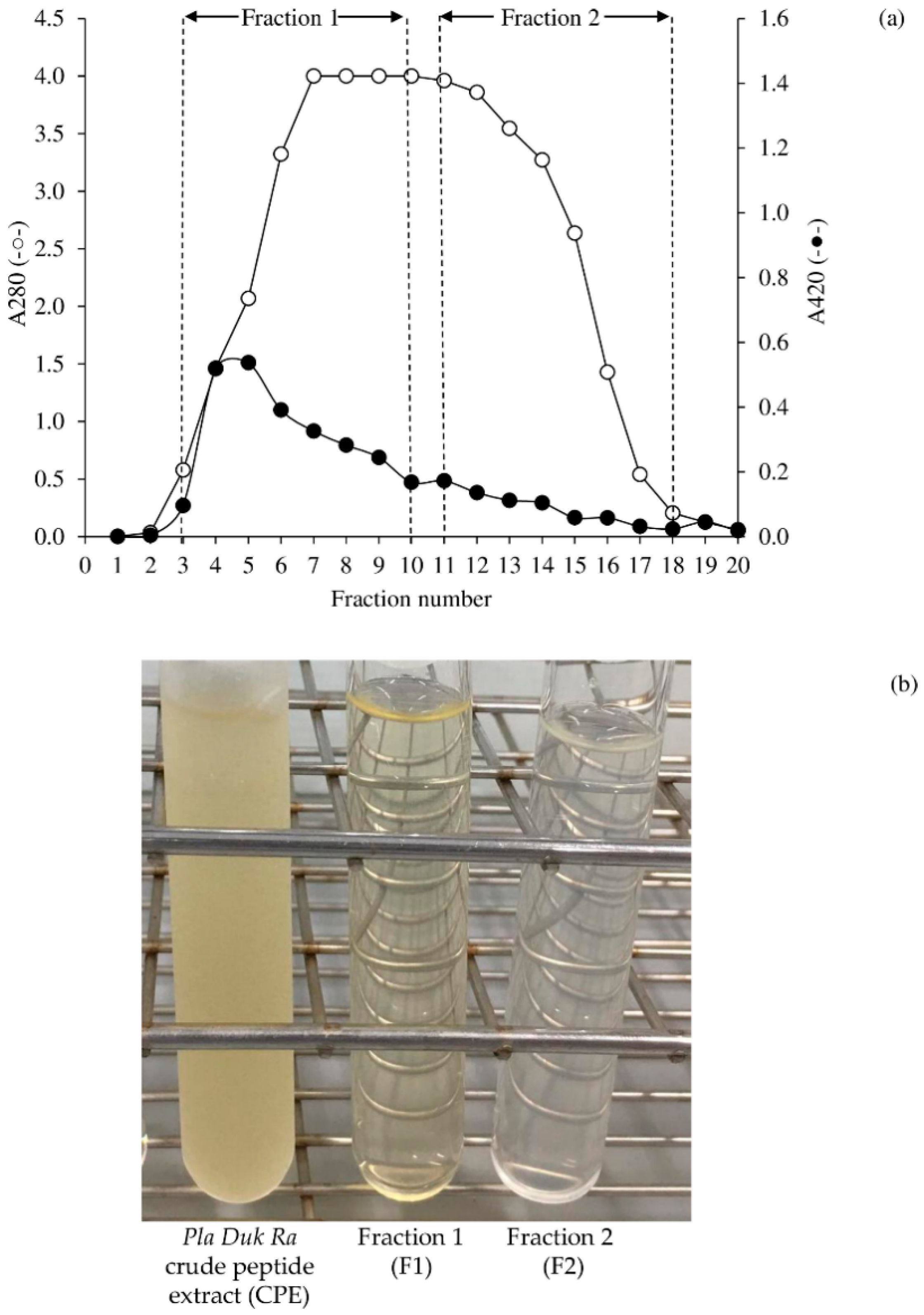
3.1. Peptide Fractionation Using Hitrap Sephadex-G25 Columns

Figure 1a shows chromatograms acquired using Hitrap Sephadex-G25 columns. The peptide distribution was represented by A280, while the browning product was represented by A420. F1 with higher A420 was eluted first, followed by F2. Figure 1b depicts the appearance of CPE, F1, and F2. The A420 of 2.70 ± 0.08, indicated the existence of MRPs in the CPE. Because of the reaction between amine-carbonyl compounds, MRPs can be produced during Pla Duk Ra fermentation. F1 (A420 = 0.07 ± 0.00) was light yellow as well, although with a lesser intensity than CPE. The low intensity of A420 (0.05 ± 0.00) made F2 the clearest. The higher molecular weight MRPs can be isolated sooner using gel filtration. As a result, they were predominantly found in the F1.

3.2. Amino Acid Composition

Table 1 shows the amino acid contents of CPE and its fractions. A variety of amino acid types and concentrations were discovered in the samples. F1 and F2 contained 19 and 14 amino acids, respectively, whereas CPE contained 20 amino acids. Hydrophobic amino acid (HBA) (47.45%) and aromatic amino acid (ARA) (27.31%) were found in the highest concentrations in F2, followed by F1 and CPE. For each individual amino acid, CPE had the highest levels of alanine, valine, leucine, isoleucine, phenylalanine, lysine, and hydroxylysine (p < 0.05). F1 had the greatest levels of arginine and histidine (p < 0.05). The most prevalent amino acids in F2 were tryptophan, glutamic acid, aspartic acid, hydroxyproline, serine, glycine, tyrosine, threonine, proline, and methionine. Their antioxidant action (see Section 3.4 and Section 3.5) may be due to differences in amino acid profile, content, and sequence (see Section 3.3).
According to Zhu et al. [27], amino acid concentration, type, and sequence have a significant effect on peptide antioxidant activity. Some amino acids have been shown to have antioxidant properties. The capacity of amino acid residues to interact with free radicals impacts the peptide’s overall antioxidant power [28]. Cysteine and methionine residues with nucleophilic sulfur side chains, as well as tryptophan, tyrosine, and phenylalanine residues with aromatic side chains can promptly donate hydrogen atoms to stop the radical chain reaction [27]. By transferring a hydrogen from its thiol group, cysteine is supposed to serve as an antioxidant [29]. Methionine is an excellent oxidant scavenger, preventing oxidants from attacking components that are crucial to cellular structure and function [30]. Hernández-Ledesma et al. [31] calculated the oxygen radical absorbance capacity (ORAC) values of different amino acids to evaluate their radical scavenging activity. The antioxidant capacity of tryptophan was found to be the highest, followed by methionine and cysteine.

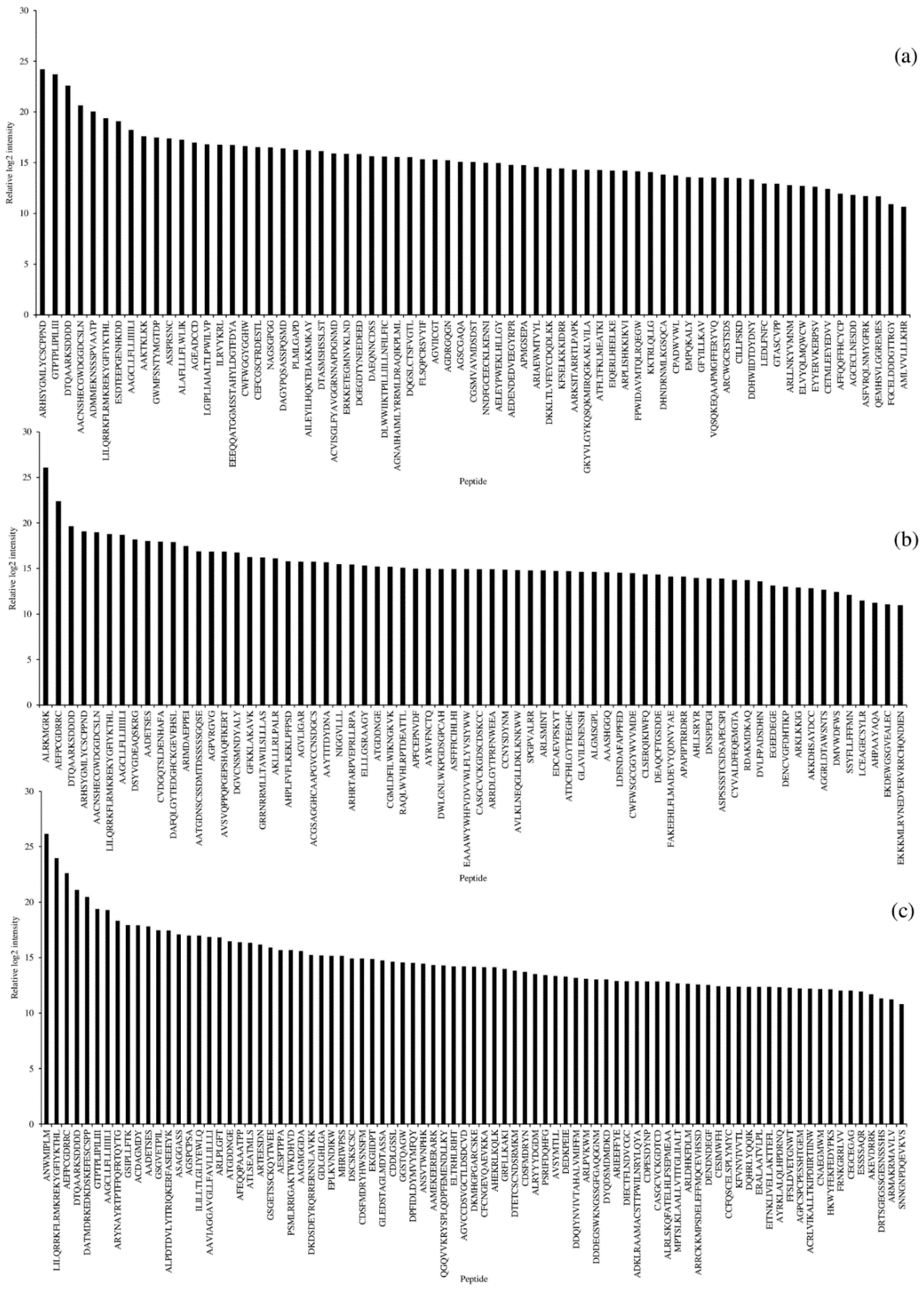
3.3. Peptide Profiles

In CPE and its fractions, peptides with 8–24 amino acid residues were found (Figure 2a–c). Other antioxidant peptides extracted and identified from the muscle protein of diverse aquatic species have distinct sequences [16,32,33]. The peptide length of Pla Duk Ra product, which was fermented and processed in a short period of time (<7 days), was significantly longer than that of other fermented fish products that required long-term fermentation, such as fish sauce (2–6 residues) [14], fish sauce by-products (4–6 residues) [13], Budu (8–11 residues) [18], and Pekasam (8–11 residues) [34]. This was linked to the degree of proteolysis and how fermentation time influenced it. CPE (Figure 2a), F1 (Figure 2b), and F2 (Figure 2c) each had 69, 68, and 85 peptides, respectively. The HBA content of each peptide was varied. ARHSYGMLYCSCPPND (50% HBA), GTPTPLIPILIII (76.9% HBA), DTQAARKSDDDD (20% HBA), AACNSHECGWDGGDCSLN (22.2% HBA), and ADMMEKNSSSPVAATP (50% HBA) were the top five peptides discovered in CPE. ALRKMGRK (37.5% HBA), AEFPCGDRRC (30% HBA), DTQAARKSDDDD (16.7 % HBA), ARHSYGMLYCSCPPND (50% HBA), and AACNSHECGWDGGDCSLN (22.2% HBA) were thein vito top five peptides discovered in F1. The top five peptides found in F2 were ANWMIPLM (87.5% HBA), LILQRRKFLRMKREKYGFIYKTHL (37.5% HBA), AEFPCGDRRC (30% HBA), DTQAARKSDDDD (20% HBA), and DATMDRKEDKDSKEFESCSPP (23.8% HBA). The peptides DTQAARKSDDDD, LILQRRKFLRMKREKYGFIYKTHL, and AAGCLLFLLIIIILI were found in all samples.

3.4. Antiradical Scavenging Activities

Table 2 shows the antiradical scavenging activities of CPE, F1, and F2 as assessed by DPPH and ABTS tests. F2 had the highest DPPH and ABTS●+ scavenging activities, followed by F1 and CPE (p < 0.05). This was in line with the fact that this fraction contained the most hydrophobic and aromatic amino acids (Table 1 and Figure 2c). By interacting with reactive species, the presence of HBA in the peptide increased antioxidant activity [35]. Furthermore, F2 had more short chain peptides (less than 10 residues) than F1. Short chain peptides, according to Hamzeh et al. [14], are thought to have higher radical scavenging action. The lower IC50 value for DPPH scavenging activity (IC50 = 0.23–2.21 mg/mL) compared to ABTS●+ one (IC50 = 0.37–3.45 mg/mL) suggested that all samples had better action against hydrophobic free radicals. This was probably due to the presence of HBA in the peptide mixtures (Table 1 and Figure 2). Hydrophobic free radical scavenging action was aided by peptides containing HBA, such as valine and leucine [27]. In CPE, F1, and F2, for example, valine-containing peptides were determined to be 26, 30, and 26 peptides, respectively. In addition, aromatic amino acids boosted the antioxidant power of peptides by efficiently donating protons to electron-deficient radicals and maintaining their stabilities via resonance structures, as well as increasing radical scavenging activities [16]. In CPE, F1, and F2, phenylalanine-containing peptides were determined to be 22, 25, and 32 peptides. As a result, F1 and F2’s high hydrophobic and aromatic amino acid concentrations may play a role in their strong anti-oxidation. AAVIAGGAVGLLFAVLLILLLI, for example, contained 86.4% HBA in F2, with 3 valine residues, 7 leucine residues, and 1 phenylalanine residue in the sequence, which could be one of the potential candidates. CPE and its fractions appeared to have superior free radical scavenging activity than Budu extract [18]. Budu extract and its fractions had IC50 values for DPPH scavenging activity in the range of 0.76–1.06 mg/mL, and IC50 values for ABTS●+ scavenging activity in the range of 0.54–2.15 mg/mL [18]. Also, CPE and its fractions demonstrated better activity as compared to DPPH scavenging activity of protein hydrolysates from dark muscle of tuna (IC50 = 4.54–9.36 mg/mL) [16]. The results showed that CPE and its fractions contained peptides that scavenge free radicals and stop the radical chain reaction, resulting in stable products. Despite the fact that MRPs have been shown to exhibit anti-oxidative action [36], larger molecular weight MRPs have been shown to have less activity [37]. Thus, rather than MRPs, peptides appear to be significant candidates contributing to Pla Duk Ra’s antioxidant action.

3.5. Inhibitory Activity against ROS (OH, H2O2, and 1O2)

The same trend in inhibitory activity against OH, H2O2, and 1O2 was observed where the lowest IC50 values were found in F2, followed by F1, and CPE (p < 0.05; Table 1), suggesting the most effective in ROS inhibition of F2. As presented in Table 1, the IC50 for OH scavenging activity of CPE, F1, and F2 was 8.27, 4.92, and 0.54 mg/mL, respectively. At the same order, the IC50 for H2O2 was 3.35, 2.12, and 0.35 mg/mL and those for 1O2 was 6.86, 1.87, and 0.32 mg/mL, respectively. Chi et al. [16] reported the IC50 values for OH scavenging activity of protein hydrolysate and its fraction from tuna dark muscle in the ranges of 0.22–4.09 mg/mL, depending on enzyme type and fraction collected.
Inhibitory action against OH, H2O2, and 1O2 followed a similar pattern, with the lowest IC50 values obtained in F2, followed by F1, and CPE (Table 1), implying that F2 is the most effective in ROS inhibition. The IC50 for OH scavenging activity of CPE, F1, and F2 was 8.27, 4.92, and 0.54 mg/mL, respectively, as indicated in Table 2. The IC50 for H2O2 of CPE, F1, and F2 was 3.35, 2.12, and 0.35 mg/mL, respectively, whereas the IC50 for 1O2 was 6.86, 1.87, and 0.32 mg/mL. IC50 values for OH scavenging activity of protein hydrolysate and its fraction from tuna dark muscle were reported by Chi et al. [16] to be in the range of 0.22–4.09 mg/mL, depending on enzyme type and fraction collected. The IC50 for OH scavenging activity was significantly greater in all samples when compared to other ROS and DPPH/ABTS free radicals. This demonstrated that OH has a high oxidation potential. Because of its reactivity with biological macromolecules like protein, lipid, and DNA, OH is the most damaging of the many ROS [27]. As a result, inhibiting OH is likely one of a live body’s most effective defenses against numerous diseases.
Carcinogenesis, aging, and oxygen toxicity appear to be linked to 1O2 interactions with biological molecules [38]. The ability of a vasoactive intestinal peptide, histidine-containing peptides (histidine at C terminal), to quench 1O2 has been documented. All of the samples in this investigation had roughly 20 histidine-containing peptides. In F2, however, only one peptide, HKWYFEKFEDYPKS, began with histidine. As a result, it was assumed to be one of the 1O2 quenchers. Alanine, methionine, tryptophan, and histidine have all been shown to physically quench 1O2 [39]. The five amino acids methionine, cysteine, histidine, tyrosine, and tryptophan exhibit substantial rate constants for 1O2 quenching, according to Sjöberg et al. [40].
Peptides with high effective quantities of oxidatively labile amino acid residues (e.g., tryptophan, methionine, and cysteine) can suppress oxidation reactions [41]. As stated in Table 2, the free radical and ROS scavenging capabilities of F2 with the highest tryptophan and methionine levels (Table 1) were the highest. F2 (Figure 2c) had 17 tryptophan-containing peptides, whereas CPE (Figure 2a) and F1 (Figure 2b) each had 13 peptides (Figure 2b). In F2, F1, and CPE, methionine-containing peptides were detected in 32, 27, and 18 peptides, respectively. ANWMIPLM, a tryptophan and methionine-containing octapeptide, was found to be the most prevalent in F2 and may be responsible for the fraction’s ROS inhibition. Tryptophan has been shown in studies to be effective antioxidants in organisms, eliminating ROS and improving the resistance to free radical damage [42,43,44]. Tryptophan was discovered to be a potent free radical scavenger found in human milk [45,46].

3.6. Metal Chelating Activity

Table 2 presents the metal chelating activities of CPE and its fractions as revealed by IC50 values. CPE, F1, and F2 had IC50 values of 4.74, 2.18, and 3.21 mg/mL, respectively. Metal chelation was ranked in a different order than other antioxidant activities, with F1 having the best metal chelating activity. This was most likely owing to the peptides’ differing amino acid content and sequence. Metal-chelating residues within the sequences of these peptides, such as methionine, glutamic acid, glutamine, arginine, and, lysine have been shown to contribute to the antioxidant peptides’ enhanced Fe2+-chelating capacity as well as their strong radical scavenging capability [47]. F1 had the greatest levels of arginine and histidine (p < 0.05) (Table 1). Due to the metal binding activity of an imidazole moiety, histidine-containing peptides are thought to operate as metal chelators [48]. F1 (Figure 2b) included 23 histidine-containing peptides, whereas F2 (Figure 2c) and CPE (Figure 2a) contained 20 and 17 peptides, respectively. In F1, the possible chelating peptide AVSVQPPQPGEPSHAQFIKERT was discovered, which contains 2 alanine residues, 3 glutamine residues, 2 glutamic acid residues, a histidine residue, a lysine residue, and an arginine residue. ALRKMGRK, the most common peptide in F1, contains 2 arginine residues, a methionine residue, and a lysine residue, which could be one of F1’s active metal chelators. EEEQQATGGMSSTAHYLDGTFDYA, which included 3 glutamic acid residues, 2 glutamine residues, 3 alanine residues, a methionine residue, and a histidine residue, was another probable metal chelating peptide in CPE. AHEKRLKQLK, a peptide with 83.3% probable chelating amino acids, could be one of F2’s active chelating peptides.

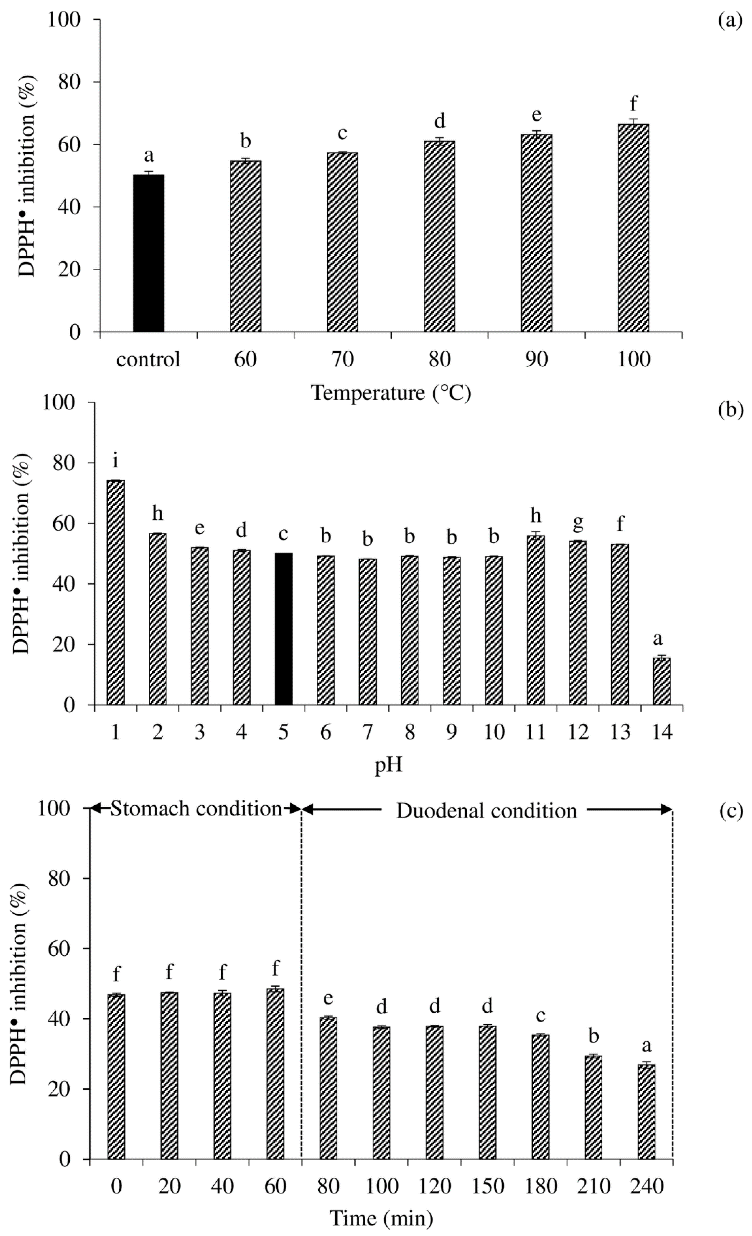
3.7. Effects of Heating Temperature, pH, and In Vitro Digestion on DPPH Scavenging Activity of Selected Peptide Fraction

The heating temperature (Figure 3a) was evaluated at 60–100 °C to mimic the heat treatment of food in comparison to a control that was held at room temperature. The DPPH scavenging activity increased with raising heating temperature (p < 0.05), according to the results. In other peptides, the same pattern has been observed. The trolox equivalent antioxidant capacity (TEAC) of dipeptides (TY and LY) extracted from brown rice hydrolysates tended to increase with increasing heating temperature from 40 to 80 °C, according to Du et al. [49]. The DPPH scavenging activity of a sample heated to 100 °C was enhanced to 66.4% in this study. The peptides from the Pla Duk Ra fraction appeared to be heat-activated peptides. Since the inhibitory activity of DPPH increased as the heating temperature was raised. Heating can break the hydrogen bonds between the side chains of a peptide and the water molecule, exposing the reactive side chains and allowing free radicals to bind. It has been observed that at high temperatures, all water molecules are virtually free and hydrogen bonds between protein residues and water molecules are impossible to form [50]. Furthermore, it has been observed that although short-chain and low-molecular-weight peptides lack tertiary and quaternary structures, they can nevertheless form secondary structures [27,49]. The secondary structure may be broken as the temperature rises, exposing the reactive R-group, which can then be triggered for radical inhibition. As a result, the peptides were shown to be heat stable. As a result, these antioxidant peptides appeared to be promising for usage in the food industry, where heat treatment is used in the majority of food processing methods.
According to the findings, F2 had the most potent antioxidant peptide. As a result, the influence of temperature, pH, and in vitro digestion on F2’s DPPH scavenging activity was investigated. Those circumstances were created to resemble the conditions of food processing and digestion. For the effect of pH (Figure 3b), the DPPH scavenging activity of F2 at the original pH (pH 5) was determined as IC50. The DPPH scavenging activity changed when the pH was shifted away from this pH. The DPPH scavenging activity increased as the pH decreased, reaching its maximum value at pH 1.0. With decreasing pH, the equilibrium protonation of the carbonyl oxygen increases, increasing the electrophilicity of the carbonyl carbon [51]. The occurrence of the deactivating protonated amino group and radical stabilizing clusters on side chains accounted for the positional selectivity and rates of radical assault on peptides [52]. As the pH was raised, the DPPH scavenging activity declined marginally at pH 6–10 and increased at pH 11–13. At pH 14, however, the activity fell dramatically. Over the pH range of 3 to 10, the DPPH scavenging activity appeared to be steady or barely changed in certain situations. In the food processing industry, this was a good connotation. The increase in DPPH scavenging activity at extreme acidic pH may be linked to the stability during digestion. At severe alkaline pH, racemization and deamination of amino acids can occur, resulting in peptides losing their antioxidant activity [27]. As a result of the structural modification of peptides that occurs during pH adjustment, their antioxidant capacity was altered.
To be functional, bioactive peptides must elude enzymatic degradation and reach the intestinal lumen [8]. Measurement of DPPH scavenging activity was used to track F2’s antioxidative activity in a gastrointestinal tract model system (Figure 3c). Under the stomach situation, F2’s DPPH scavenging activity remained consistent (p > 0.05). Despite the fact that pepsin may further cleave peptides under acidic conditions, the peptides’ DPPH scavenging activity remained unaltered. Following that, the DPPH scavenging activity in the duodenal condition fell dramatically (p < 0.05). When compared to the initial activity, the residual activity after 240 min digesting was roughly 54%.
The results showed that both digestion and pH conditions can cause a decrease in DPPH scavenging activity during in vitro digestion. The DPPH scavenging activity was more stable in the acidic condition than in the alkaline condition, according to the pH stability test (Figure 3a,b). Antioxidant peptides from dry-cured ham [53] and Jinhua ham [27] have been shown to reduce DPPH scavenging activity in a simulated gastrointestinal system. The peptides were reported to be more thoroughly degraded during digestion, making them more hydrophilic and thus more difficult to interact with the hydrophobic DPPH [27]. As a result, the DPPH scavenging activity decreased during the digestion in the gastrointestinal tract model system.

4. Conclusions

Pla Duk Ra, a Thai traditional semi-dried fermented catfish, can be characterized as a source of antioxidant peptides. CPE and its fraction with 8–24 amino acid residue had different amino acid profiles and sequences. F2 has the largest concentrations of hydrophobic and aromatic amino acids, which may explain why it was the most effective in inhibiting free radicals (DPPH and ABTS●+) and ROS (OH, H2O2, and 1O2). Lowering the pH and increasing the heating temperature increased F2’s DPPH scavenging activity. However, following digestion, nearly half of the DPPH scavenging activity was lost in the gastrointestinal tract model system. As a result, the antioxidant activity of peptides were influenced by both amino acid content and peptide sequences. The synergist or additive effect of peptide combinations should account for the antioxidant activity of peptides fractionated from Pla Duk Ra.

Author Contributions

Conceptualization, M.C. and W.P.; methodology, M.C., W.P., A.P., L.-Z.C. and S.R.; software, W.P., A.P., N.P., S.C. and S.R.; validation, W.P., A.P., S.R. and M.C.; investigation, T.R., S.C. and N.P.; resources, W.P. and M.C.; data curation, T.R., M.C. and W.P.; writing—original draft preparation, M.C. and W.P.; writing—review and editing, M.C., W.P., L.-Z.C. and S.R.; supervision, M.C. and W.P.; funding acquisition, M.C. and W.P. All authors have read and agreed to the published version of the manuscript.

Funding

This research was supported by THAILAND SCIENCE RESEARCH AND INNOVATION FUND, Contract No. WU-FF64102.

Institutional Review Board Statement

Not applicable.

Informed Consent Statement

Not applicable.

Data Availability Statement

Not applicable.

Acknowledgments

We would like to thank Food Technology and Innovation Research Center of Excellence, Walailak University for facility support.

Conflicts of Interest

The authors declare no conflict of interest.

References

  1. Pongsetkul, J.; Benjakul, S. Development of modified atmosphere packaging (MAP) on shelf-life extension of pla-duk-ra (dried fermented catfish) stored at room temperature. Food Control 2021, 124, 107882. [Google Scholar] [CrossRef]
  2. Phetsang, H.; Panpipat, W.; Panya, A.; Phonsatta, N.; Chaijan, M. Occurrence and development of off-odor compounds in farmed hybrid catfish (Clarias macrocephalus × Clarias gariepinus) muscle during refrigerated storage: Chemical and volatilomic analysis. Foods 2021, 10, 1841. [Google Scholar] [CrossRef] [PubMed]
  3. Thai Community Product Standard. Thai Community Product Standard No. TCPS 1029/2018; Thai Industrial Standards Institute: Bangkok, Thailand, 2018. [Google Scholar]
  4. Zang, J.; Xu, Y.; Xia, W.; Regenstein, J.M. Quality, functionality, and microbiology of fermented fish: A review. Crit. Rev. Food Sci. Nutr. 2019, 60, 1228–1242. [Google Scholar] [CrossRef]
  5. Choksawangkarn, W.; Phiphattananukoon, S.; Jaresitthikunchai, J.; Roytrakul, S. Antioxidative peptides from fish sauce by-product: Isolation and characterization. Agric. Nat. Resour. 2018, 52, 460–466. [Google Scholar] [CrossRef]
  6. Abuine, R.; Rathnayake, A.U.; Byun, H.-G. Biological activity of peptides purified from fish skin hydrolysates. Fish. Aquat. Sci. 2019, 22, 10. [Google Scholar] [CrossRef] [Green Version]
  7. Shahidi, F.; Zhong, Y. Bioactive peptides. J. AOAC Int. 2008, 91, 914–931. [Google Scholar] [CrossRef] [Green Version]
  8. Adessi, C.; Soto, C. Converting a peptide into a drug: Strategies to improve stability and bioavailability. Curr. Med. Chem. 2002, 9, 963–978. [Google Scholar] [CrossRef]
  9. Xiao, C.; Toldrá, F.; Zhou, F.; Gallego, M.; Zhao, M.; Mora, L. Effect of cooking and in vitro digestion on the peptide profile of chicken breast muscle and antioxidant and alcohol dehydrogenase stabilization activity. Food Res. Int. 2020, 136, 109459. [Google Scholar] [CrossRef] [PubMed]
  10. Najafian, L.; Babji, A.S. A review of fish-derived antioxidant and antimicrobial peptides: Their production, assessment, and applications. Peptides 2012, 33, 178–185. [Google Scholar] [CrossRef]
  11. Wiriyaphan, C.; Chitsomboon, B.; Roytrakul, S.; Yongsawadigul, J. Isolation and identification of antioxidative peptides from hydrolysate of threadfin bream surimi processing byproduct. J. Funct. Foods 2013, 5, 1654–1664. [Google Scholar] [CrossRef]
  12. Yin, L.-J.; Tong, Y.-L.; Jiang, S.-T. Improvement of the Functionality of Minced Mackerel by Hydrolysis and Subsequent Lactic Acid Bacterial Fermentation. J. Food Sci. 2006, 70, M172–M178. [Google Scholar] [CrossRef]
  13. Khositanon, P.; Panya, N.; Roytrakul, S.; Krobthong, S.; Chanroj, S.; Choksawangkarn, W. Effects of fermentation periods on antioxidant and angiotensin I-converting enzyme inhibitory activities of peptides from fish sauce by-products. LWT 2020, 135, 110122. [Google Scholar] [CrossRef]
  14. Hamzeh, A.; Noisa, P.; Yongsawatdigul, J. Characterization of the antioxidant and ACE-inhibitory activities of Thai fish sauce at different stages of fermentation. J. Funct. Foods 2019, 64, 103699. [Google Scholar] [CrossRef]
  15. Chen, H.; Wang, S.; Zhou, A.; Miao, J.; Liu, J.; Benjakul, S. A novel antioxidant peptide purified from defatted round scad (Decapterus maruadsi) protein hydrolysate extends lifespan in Caenorhabditis elegans. J. Funct. Foods 2020, 68, 103907. [Google Scholar] [CrossRef]
  16. Chi, C.-F.; Hu, F.-Y.; Wang, B.; Li, Z.-R.; Luo, H.-Y. Influence of Amino Acid Compositions and Peptide Profiles on Antioxidant Capacities of Two Protein Hydrolysates from Skipjack Tuna (Katsuwonus pelamis) Dark Muscle. Mar. Drugs 2015, 13, 2580–2601. [Google Scholar] [CrossRef]
  17. Farvin, K.S.; Andersen, L.L.; Otte, J.; Nielsen, H.H.; Jessen, F.; Jacobsen, C. Antioxidant activity of cod (Gadus morhua) protein hydrolysates: Fractionation and characterisation of peptide fractions. Food Chem. 2016, 204, 409–419. [Google Scholar] [CrossRef] [PubMed] [Green Version]
  18. Najafian, L.; Babji, A.S. Purification and Identification of Antioxidant Peptides from Fermented Fish Sauce (Budu). J. Aquat. Food Prod. Technol. 2018, 28, 14–24. [Google Scholar] [CrossRef]
  19. AOAC. Official Methods of Analysis, 16th ed.; Association of Official Analytical Chemists: Washington, DC, USA, 2000. [Google Scholar]
  20. Lowry, O.H.; Rosebrough, N.J.; Farr, A.L.; Randall, R.J. Protein measurement with the Folin phenol reagent. J. Biol. Chem. 1951, 193, 265–275. [Google Scholar] [CrossRef]
  21. Chinarak, K.; Chaijan, M.; Panpipat, W. Farm-raised sago palm weevil (Rhynchophorus ferrugineus) larvae: Potential and challenges for promising source of nutrients. J. Food Compos. Anal. 2020, 92, 103542. [Google Scholar] [CrossRef]
  22. Bradford, M.M. A rapid and sensitive method for the quantitation of microgram quantities of protein utilizing the principle of protein-dye binding. Anal. Biochem. 1976, 72, 248–254. [Google Scholar] [CrossRef]
  23. Tyanova, S.; Temu, T.; Cox, J. The MaxQuant computational platform for mass spectrometry-based shotgun proteomics. Nat. Protoc. 2016, 11, 2301–2319. [Google Scholar] [CrossRef]
  24. Chaijan, M.; Panpipat, W. Basic composition, antioxidant activity and nanoemulsion behavior of oil from mantis shrimp (Oratosquilla nepa). Food Biosci. 2019, 31, 100448. [Google Scholar] [CrossRef]
  25. Benjakul, S.; Kittiphattanabawon, P.; Shahidi, F.; Maqsood, S. Antioxidant activity and inhibitory effects of lead (Leucaena leucocephala) seed extracts against lipid oxidation in model systems. Food Sci. Technol. Int. 2013, 19, 365–376. [Google Scholar] [CrossRef]
  26. Hamzeh, A.; Benjakul, S.; Senphan, T. Comparative study on antioxidant activity of hydrolysates from splendid squid (Loligo formosana) gelatin and protein isolate prepared using protease from hepatopancreas of Pacific white shrimp (Litopenaeus vannamei). J. Food Sci. Technol. 2016, 53, 3615–3623. [Google Scholar] [CrossRef] [Green Version]
  27. Zhu, C.-Z.; Zhang, W.-G.; Kang, Z.-L.; Zhou, G.-H.; Xu, X.-L. Stability of an antioxidant peptide extracted from Jinhua ham. Meat Sci. 2014, 96, 783–789. [Google Scholar] [CrossRef]
  28. Stadtman, E.R. Oxidation of free amino acids and amino acid residues in proteins by radiolysis and by metal-catalyzed reactions. Annu. ReV. Biochem. 1993, 62, 797–821. [Google Scholar] [CrossRef] [PubMed]
  29. Elias, R.J.; McClements, D.J.; Decker, E.A. Antioxidant activity of cysteine, tryptophan, and methionine residues in continuous phase β-lactoglobulin in oil-in-water emulsions. J. Agric. Food Chem. 2005, 53, 10248–10253. [Google Scholar] [CrossRef]
  30. Levine, R.L.; Mosoni, L.; Berlett, B.S.; Stadtman, E.R. Methionine residues as endogenous antioxidants in proteins. Proc. Natl. Acad. Sci. USA 1996, 93, 15036–15040. [Google Scholar] [CrossRef] [Green Version]
  31. Hernández-Ledesma, B.; Dávalos, A.; Bartolomé, B.; Amigo, L. Preparation of antioxidant enzymatic hydrolysates from α-lactalbumin and β-lactoglobulin. Identification of active peptides by HPLC-MS/MS. J. Agric. Food Chem. 2005, 53, 588–593. [Google Scholar] [CrossRef]
  32. Ko, J.-Y.; Lee, J.-H.; Samarakoon, K.; Kim, J.-S.; Jeon, Y.-J. Purification and determination of two novel antioxidant peptides from flounder fish (Paralichthys olivaceus) using digestive proteases. Food Chem. Toxicol. 2012, 52, 113–120. [Google Scholar] [CrossRef]
  33. Jiang, H.; Tong, T.; Sun, J.; Xu, Y.; Zhao, Z.; Liao, D. Purification and characterization of antioxidative peptides from round scad (Decapterus maruadsi) muscle protein hydrolysate. Food Chem. 2014, 154, 158–163. [Google Scholar] [CrossRef]
  34. Najafian, L.; Babji, A.S. Fractionation and identification of novel antioxidant peptides from fermented fish (pekasam). J. Food Meas. Charact. 2018, 12, 2174–2183. [Google Scholar] [CrossRef]
  35. Mendis, E.; Rajapakse, A.N.; Kim, S.-K. Antioxidant Properties of a Radical-Scavenging Peptide Purified from Enzymatically Prepared Fish Skin Gelatin Hydrolysate. J. Agric. Food Chem. 2004, 53, 581–587. [Google Scholar] [CrossRef]
  36. Langner, E.; Rzeski, W. Biological Properties of Melanoidins: A Review. Int. J. Food Prop. 2013, 17, 344–353. [Google Scholar] [CrossRef]
  37. Nie, X.; Xu, D.; Zhao, L.; Meng, X. Antioxidant activities of chicken bone peptide fractions and their Maillard reaction products: Effects of different molecular weight distributions. Int. J. Food Prop. 2017, 20, S457–S466. [Google Scholar] [CrossRef]
  38. Misra, B.R.; Misra, H.P. Vasoactive intestinal peptide, a singlet oxygen quencher. J. Biol. Chem. 1990, 265, 15371–15374. [Google Scholar] [CrossRef]
  39. Matheson, I.B.C.; Etheridge, R.D.; Kratowich, N.R.; Lee, J. The quenching of singlet oxygen by amino acids and proteins. Photochem. Photobiol. 1975, 21, 165–171. [Google Scholar] [CrossRef]
  40. Sjöberg, B.; Foley, S.; Staicu, A.; Pascu, A.; Pascu, M.; Enescu, M. Protein reactivity with singlet oxygen: Influence of the solvent exposure of the reactive amino acid residues. J. Photochem. Photobiol. B Biol. 2016, 159, 106–110. [Google Scholar] [CrossRef]
  41. Sharp, J.S.; Becker, J.M.; Hettich, R.L. Analysis of Protein Solvent Accessible Surfaces by Photochemical Oxidation and Mass Spectrometry. Anal. Chem. 2003, 76, 672–683. [Google Scholar] [CrossRef]
  42. Xu, K.; Liu, G.; Fu, C. The Tryptophan Pathway Targeting Antioxidant Capacity in the Placenta. Oxidative Med. Cell. Longev. 2018, 2018, 1–8. [Google Scholar] [CrossRef]
  43. Weiss, G.; Diez-Ruiz, A.; Murr, C.; Theur, I.; Fuchs, D. Tryptophan Metabolites as Scavengers of Reactive Oxygen and Chlorine Species. Pteridines 2002, 13, 140–143. [Google Scholar] [CrossRef]
  44. Christen, S.; Peterhans, E.; Stocker, R. Antioxidant activities of some tryptophan metabolites: Possible implication for inflammatory diseases. Proc. Natl. Acad. Sci. USA 1990, 87, 2506–2510. [Google Scholar] [CrossRef] [Green Version]
  45. Tsopmo, A.; Diehl-Jones, B.W.; Aluko, R.E.; Kitts, D.D.; Elisia, I.; Friel, J.K. Tryptophan released from mother’s milk has antioxidant properties. Pediatr. Res. 2009, 66, 614–618. [Google Scholar] [CrossRef] [Green Version]
  46. Nayak, B.N.; Buttar, H.S. Evaluation of the antioxidant properties of tryptophan and its metabolites in in vitro assay. J. Complement. Integr. Med. 2016, 13, 129–136. [Google Scholar] [CrossRef]
  47. Jung, M.; Kim, S.K.; Kim, S.Y. Riboflavin-sensitized photooxidation of ascorbic acid: Kinetics and amino acid effects. Food Chem. 1995, 53, 397–403. [Google Scholar] [CrossRef]
  48. Ihara, H.; Kakihana, Y.; Yamakage, A.; Kai, K.; Shibata, T.; Nishida, M.; Yamada, K.-I.; Uchida, K. 2-Oxo-histidine–containing dipeptides are functional oxidation products. J. Biol. Chem. 2019, 294, 1279–1289. [Google Scholar] [CrossRef] [Green Version]
  49. Du, R.; Liu, K.; Zhao, S.; Chen, F. Changes in antioxidant activity of peptides identified from brown rice hydrolysates under different conditions and their protective effects against AAPH-induced oxidative stress in human erythrocytes. ACS Omega 2020, 5, 12751–12759. [Google Scholar] [CrossRef]
  50. Mallamace, D.; Fazio, E.; Mallamace, F.; Corsaro, C. The Role of Hydrogen Bonding in the Folding/Unfolding Process of Hydrated Lysozyme: A Review of Recent NMR and FTIR Results. Int. J. Mol. Sci. 2018, 19, 3825. [Google Scholar] [CrossRef] [Green Version]
  51. Sun, Y.; Frenkel-Pinter, M.; Liotta, C.L.; Grover, M.A. The pH dependent mechanisms of non-enzymatic peptide bond cleavage reactions. Phys. Chem. Chem. Phys. 2019, 22, 107–113. [Google Scholar] [CrossRef]
  52. Ohashi, Y.; Onuma, R.; Naganuma, T.; Ogawa, T.; Naude, R.; Nokihara, K.; Muramoto, K. Antioxidant Properties of Tripeptides Revealed by a Comparison of Six Different Assays. Food Sci. Technol. Res. 2015, 21, 695–704. [Google Scholar] [CrossRef] [Green Version]
  53. Gallego, M.; Mauri, L.; Aristoy, M.C.; Toldrá, F.; Mora, L. Antioxidant peptides profile in dry-cured ham as affected by gastrointestinal digestion. J. Funct. Foods 2020, 69, 103956. [Google Scholar] [CrossRef]
Figure 1. Representative chromatogram of crude peptide samples extracted from semi-dried fermented catfish (Pla Duk Ra) (CPE) fractionated by two connected Sephadex-G25 desalting columns (5 mL, bead size of 15–70 µm) (a) and appearance of CPE and fraction 1 (F1) and fraction 2 (F2) (b). F1 represented the pooled fractions 3–10, while F2 represented the pooled fractions 11–18. The A420 of CPE, F1, and F2 was 2.70 ± 0.08, 0.07 ± 0.00, and 0.05 ± 0.00, respectively.
Fermentation 07 00262 g001
Figure 2. Peptide profiles of Pla Duk Ra crude peptide extract (CPE) (a), fraction 1 (b), and fraction 2 (c).
Fermentation 07 00262 g002
Figure 3. Effect of heating temperature (a), pH (b), and in vitro digestion (c) on DPPH scavenging activity of fraction 2 fractioned from Pla Duk Ra crude peptide extract (CPE) at the IC50 concentration (0.2 mg/mL). Bars represent standard deviation from triplicate determinations. Different letters indicate significant different (p < 0.05). For effect of heating temperature, samples were heated for 30 min, followed by cooling down in ice water for 5 min. Control (black bar) was kept at room temperature (25–27 °C) for 30 min, and kept in ice water for 5 min. For the effect of pH, samples were incubated at each pH for 30 min at room temperature. The initial pH of the sample was around 5.0 (black bar).
Fermentation 07 00262 g003
Table 1. Amino acid composition of Pla Duk Ra crude peptide extract, fraction 1, and fraction 2.
Amino Acid (%)Crude ExtractFraction 1Fraction 2
Alanine *7.68 ± 0.18 c7.07 ± 0.02 b3.03 ± 0.00 a
Glycine *5.46 ± 0.11 a6.22 ± 0.03 b7.67 ± 0.00 c
Valine *4.59 ± 0.06 c4.01 ± 0.04 b1.45 ± 0.00 a
Leucine *6.80 ± 0.20 b5.24 ± 0.05 aND
Isoleucine *3.79 ± 0.08 c2.83 ± 0.03 b0.23 ± 0.00 a
Proline *3.03 ± 0.09 a3.15 ± 0.03 b3.39 ± 0.00 c
Methionine *2.62 ± 0.04 a3.38 ± 0.00 b4.37 ± 0.00 c
Serine2.66 ± 0.09 a4.34 ± 0.04 b8.30 ± 0.00 c
Threonine2.59 ± 0.05 a3.43 ± 0.02 b5.63 ± 0.00 c
Phenylalanine *,■2.16 ± 0.02 b1.81 ± 0.02 a1.81 ± 0.00 a
Aspartic acid5.66 ± 0.11 a7.66 ± 0.06 b13.15 ± 0.01 c
Hydroxyproline3.39 ± 0.21 a5.12 ± 0.09 b9.05 ± 0.00 c
Cysteine3.19 ± 0.10 a5.31 ± 0.03 bND
Glutamic acid14.78 ± 0.08 a15.36 ± 0.08 b16.38 ± 0.00 c
Lysine13.00 ± 0.86 b0.39 ± 0.29 aND
Arginine1.05 ± 0.02 a1.31 ± 0.02 bND
Histidine7.11 ± 0.21 a10.36 ± 0.07 bND
Tyrosine *,■2.13 ± 0.06 a3.32 ± 0.02 b6.20 ± 0.00 c
Hydroxylysine3.04 ± 0.14NDND
Tryptophan *,■5.25 ± 0.26 a9.69 ± 0.90 b19.30 ± 0.01 c
Total hydrophobic amino acid43.5146.7247.45
Total aromatic amino acid9.5414.8227.31
Values are given as mean ± standard deviation from triplicate determinations. Different letters in the same row indicate significant differences (p < 0.05). ND = non-detectable; * Hydrophobic amino acid; Aromatic amino acid.
Table 2. Antioxidant activities of Pla Duk Ra crude peptide extract, fraction 1, and fraction 2.
Antioxidant Activity (IC50; mg/mL)Crude ExtractFraction 1Fraction 2
Free radical scavenging activity
DPPH radical scavenging activity2.21 ± 0.67 c0.69 ± 0.01 b0.23 ± 0.00 a
ABTS radical scavenging activity3.45 ± 0.78 c1.12 ± 0.08 b0.37 ± 0.06 a
Inhibitory activity against reactive oxygen species
Hydroxyl radical scavenging activity8.27 ± 0.17 c4.92 ± 0.52 b0.54 ± 0.21 a
Hydrogen peroxide scavenging activity3.35 ± 0.05 c2.12 ± 0.20 b0.35 ± 0.10 a
Singlet oxygen scavenging activity6.86 ± 0.87 c1.87 ± 0.07 b0.32 ± 0.11 a
Metal chelating activity4.74 ± 0.07 c2.18 ± 0.04 a3.21 ± 0.32 b
Values are means ± standard deviation from triplicate determinations. Different letters in the same row indicate significant different (p < 0.05).
Publisher’s Note: MDPI stays neutral with regard to jurisdictional claims in published maps and institutional affiliations.

