
Review

Phenyllactic Acid from Lactic Acid Bacteria: A Natural Antimicrobial for Food Biopreservation

by
Emma Mani-López
*,
Beatriz Mejía-Garibay
,
Ricardo H. Hernández-Figueroa
and
Aurelio López-Malo
*
Departamento de Ingeniería Química, Alimentos y Ambiental, Universidad de las Américas Puebla, Santa Catarina Mártir S/N, San Andrés Cholula, Puebla 72810, Mexico
*
Authors to whom correspondence should be addressed.
Fermentation 2026, 12(4), 184; https://doi.org/10.3390/fermentation12040184
Submission received: 24 February 2026 / Revised: 24 March 2026 / Accepted: 27 March 2026 / Published: 2 April 2026
(This article belongs to the Special Issue The Roles of Lactic Acid Bacteria in Food Fermentation)

Abstract

Phenyllactic acid (PLA), a natural antimicrobial metabolite produced by lactic acid bacteria (LAB), has emerged as a key compound for biopreservation in food systems. The aims of this review are to summarize the main findings on LAB-producing strains, the effects of primary PLA precursors, the impacts of culture conditions on PLA production, antimicrobial activity, mechanisms of action, quantification and analysis methods, food applications, regulatory status, and the challenges in PLA production and applications. In this review, the quorum sensing role in PLA production and multi-omics strain improvement was revised. Applications in dairy, bakery, fruits, vegetables, meat, and fish products as well as active packaging are analyzed, demonstrating their effectiveness in controlling microbial spoilage and pathogens while preserving sensory quality. Its broad-spectrum antifungal and antibacterial activities make it particularly attractive as a clean-label alternative to synthetic preservatives, contributing to both food safety and extended shelf life. Finally, current limitations and future research needs are outlined, particularly in optimizing PLA production and establishing its role as a sustainable and effective tool for food safety management.

1. Introduction

3-Phenyllactic acid (2-hydroxy-3-phenylpropionic acid) (PLA) is an organic acid with broad-spectrum antimicrobial activity against bacteria, molds, and yeasts, making it a promising natural preservative and antimicrobial agent [1,2,3]. This acid exists in two chiral isomers called D-PLA and L-PLA, both of which have demonstrated antimicrobial activity. PLA is commonly found in honey and as a metabolite of lactic acid bacteria (LAB). Because of its low cytotoxicity, it is frequently used as a natural preservative and antimicrobial agent in the food processing industry [4]. PLA is an aromatic antimicrobial metabolite derived from phenylalanine metabolism by LAB. Its biosynthesis typically proceeds through transamination of phenylalanine into phenylpyruvic acid (PPA) mediated by aminotransferase, followed by enzymatic reduction, commonly catalyzed by lactate dehydrogenase, producing PLA [2]. Production levels vary substantially among LAB depending on genus, species, strain, and growth conditions, making strain selection critical for food biopreservation applications [5,6]. For instance, 15 strains, including strains from the genera Lactobacillus and Leuconostoc, produced between 0.16 and 0.46 mM PLA (26.58–76.44 mg/L) in de Man–Rogosa–Sharpe (MRS) broth [5]. PLA content in Chinese fermented vegetables ranged from 0.81 to 51.31 mg/kg [7].
Several studies have shown LAB can produce PLA, as can other microorganisms such as propionic acid bacteria and fungi. Among these microorganisms, LAB remain attractive PLA producers because various strains (e.g., Lactococcus lactis, Lactobacillus, Leuconostoc, and related genera) are generally recognized as safe (GRAS) in the U.S. due to historical use and expert consensus [8]. Likewise, most of these species are also included in the EFSA’s Quality Presumption of Safety (QPS) list [9]. LAB are widely used for fermenting foods, whereas their metabolites, as cell-free supernatants (CFSs) or fermentates, are used as antimicrobials and postbiotics [10]. The millennia-long tradition of LAB use supports the safety of LAB and their metabolites in foods and human consumption. Among LAB, genera such as Lactobacillus (including 25 newly described genera, such as Lactiplantibacillus, Lacticaseibacillus, Limosilactobacillus, etc.), Leuconostoc, Pediococcus, Sporolactobacillus, and Weissella are reported to be efficient PLA producers due to their relatively high yields or specific isomer production.
Because LAB are widely used as fermentation starters, PLA has been investigated in food applications during fermentation, as a fermentate, and as a CFS [10]. The PLA minimum inhibitory concentrations (MICs) for bacteria such as Listeria monocytogenes, Escherichia coli, and Enterococcus faecalis ranged from 1.25 to 5 mg/mL [11,12], while for mold they oscillated between 0.5 and 12.5 for Colletotrichum gloeosporioiedes, Botrytis cinerea, Aspergillus flavus, Penicillium expansum, and Penicillium roqueforti [13,14]. Studies have demonstrated PLA’s antimicrobial activity in dairy products, bakery products, fruit juices, beer and wine, meat, fish, and poultry products, as well as in active packaging. Few studies have evaluated physicochemical and sensory properties; however, available studies suggest that PLA generally has little or no detectable impact on these properties while extending shelf life [15].
Recently, researchers have been working intensively to understand the mechanisms of PLA action in fungal and bacterial cells. Therefore, morphological and metabolic activity, cell structural analysis, proteomics, genetic, transcriptomic, and metabolomic studies have partially revealed the primary mechanisms of action of PLA in molds, Gram-positive, and Gram-negative bacteria. The major findings reveal cell membrane disruption, inhibition of ATP biosynthesis, oxidative stress induction, mitochondrial damage, and DNA degradation in molds [14,16], Gram-positive bacteria [11,12,17], and Gram-negative bacteria [18,19,20]. However, further research is required on fungal cells and PLA’s application as a CFS in both fungal and bacterial cells.
Owing to the importance of PLA in food and other industries, some authors have recently summarized the relevant major advances and findings in reviews. For instance, Rajanikar et al. [2] reviewed PLA structure and biosynthesis; LAB associated with PLA production; biotechnological strategies to improve PLA production; and PLA’s antimicrobial effects, food applications, and regulatory and toxicological status. At the same time, Wu et al. [21] summarized information on PLA’s structure, methods of determination, antimicrobial activity, metabolic pathways for biosynthesis, strategies for production, separation and purification processes, and applications. Later, Sun et al. [3] compiled data on PLA’s sources, biosynthesis and production strategies, microbial inhibition, synergy with other antagonistic pathways, applications, and SWOT analyses. These previous reviews highlight the importance of updating the status of PLA in food research, as these authors reviewed its general applications, including medical, cosmetic, plant growth, feed, pharmaceutical, and health benefits. Therefore, this review updates and summarizes current knowledge on PLA (2019–2026), including LAB-producing strains, precursors, culture conditions, and antimicrobial mechanisms. It covers emerging topics like quorum sensing, omics engineering, and PLA in delivery systems. The review assesses methods for PLA quantification, explores its applications in various foods (dairy, bakery, produce, meat/fish), including active packaging, and discusses recent regulations and safety issues. It concludes with challenges and research gaps in PLA production and application, offering perspectives on integrating PLA into sustainable, clean-label food preservation.

2. Genera and Species of LAB Producing Phenyllactic Acid

Lactic acid bacteria (LAB) are Gram-positive microorganisms, comprising approximately 38 genera grouped into six families, that produce lactic acid through central carbon metabolism and can accumulate up to ~1% lactic acid during sugar fermentation [22]. LAB are widely distributed across diverse ecosystems. They exhibit tolerance to a wide range of environmental stresses and naturally inhabit plant and animal environments. In addition, LAB have a long history of use as starter and adjunct cultures in food fermentation and preservation. Their preservative capacity is associated with the production of metabolites such as organic acids (primarily lactic and acetic acids), antimicrobial peptides, biosurfactants, reuterin, and other bioactive compounds. Over the past 25 years, PLA has been identified as a strong antifungal compound produced at variable levels by LAB. The most important LAB involved in food fermentations include members of the Lactobacillus group (recently reclassified into several genera), as well as the genera Lactococcus, Streptococcus, Leuconostoc, Pediococcus, Fructobacillus, Weissella, Carnobacterium, and Enterococcus [22]. These genera play key roles in food fermentation and preservation processes. Because of the importance of LAB in food preservation and the growing interest in natural antimicrobials, researchers continue to investigate PLA-producing LAB from various sources. A summary of selected PLA-producing LAB and their production characteristics is presented in Table 1, highlighting the diversity of LAB genera involved and the range of yields obtained under different conditions (see Appendix A for the list of abbreviations for microbial genera used in the manuscript). Overall, these findings indicate that PLA production varies among LAB strains, depending on their metabolic capacity and environmental factors [2]. While Lactobacillus plantarum and Leuconostoc mesenteroides are among the most extensively studied species, additional genera, such as Pediococcus, Lactobacillus zeae, and Sporolactobacillus inulinus, offer promising opportunities for industrial applications. Leuconostoc mesenteroides synthesizes D-PLA with high stereoselectivity via a specific D-lactate dehydrogenase [23], demonstrating the enzymatic specialization of non-Lactobacillus genera. Sporolactobacillus inulinus has emerged as a highly efficient D-PLA producer [24]; the ATCC 15538 strain achieved conversion yields of 88% and a productivity of 47 mM in whole-cell bioconversion processes of pure D-PLA, outperforming many other LAB [25].
Traditional PLA fermentation processes often rely on refined culture media such as MRS broth, which increases production costs and detracts from sustainability. Most studies reported in Table 1 used MRS broth to assess PLA production by LAB. Few studies attempted to incorporate by-products from food or other industries into culture media formulations. Mu et al. [26] formulated a culture medium using corn steep liquor to optimize PLA production by Lactobacillus sp. SK007. Likewise, Huang et al. [27] added a hydrolysate derived from alga residue (Porphyra sp.). While concentrations from this algal residue were modest (on the order of milligrams per milliliter), the work exemplifies how biomass residues can be integrated into PLA production workflows [27]. More studies are needed to utilize agro-industrial by-products in PLA biosynthesis. Agro-industrial by-products rich in phenylalanine and other PLA precursors, which LAB can convert into PLA under ideal conditions, are especially important to support sustainable PLA production. This process promotes waste-to-value conversion and reduces dependence on refined substrates [28].
Table 1. Selected LAB genera and species reported to produce phenyllactic acid (PLA).
LABSourcePLA ProductionReference
Lactobacillus plantarum ITM21B, ITM21A, ITM20B, ATCC 4008, and ICMP 5826; Lactobacillus alimentarius ATCC 29643, Lactobacillus brevis ATCC 14869, Lactobacillus hilgardii IDM51B, Leuconostoc mesenteroides ITMY30, Leuconostoc citreum ITM22ADifferent culture collections0.34, 0.35, 0.33, 0.26, 0.27, 0.37, 0.46,0.33, 0.57, and 0.43 mM (56.5, 58.1, 54.8, 43.2, 44.8, 61.4, 76.4, 54.8, 94.7, and 71.4 mg/L), respectively[5]
Lb. plantarum 21B, VLT01, SK007, IMAU10124, UM55, WIKIM18Sourdough, DIPROVAL collection, pickled vegetables, koumiss, milk, and kimchi, respectively0.056, 0.083, 0.091, 0.229, 0.0748 g/L and 173 μM (28.7 mg/L), respectively[29,30,31,32,33,34]
Lactobacillus sp. SK007Chinese traditional pickles2.3 g/L in an optimized culture medium (30 g/L glucose, 5 g/L phenylpyruvic acid, 47 g/L corn steep liquor, 3 g/L K2HPO4, 3 g/L CH3COONa, 30 g/L yeast powder and 3 mL/L Tween 80)[26]
Lactobacillus casei 21/1, Lb. casei NRRL B-1922, Lb. casei 12A, Lb. casei DPC3968, Lb. casei ATCC 334, Lactobacillus fermentum NRRL B-1932, Lb. fermentum ATCC 11976, Lactobacillus rhamnosus ATCC 13075, Lb. rhamnosus NRRL B-442, Lactobacillus reuteri NRRL B-14171, Lactobacillus sakei NRRL b-1917Different culture collections0.275, 0.028, 0.131, 0.114, 0.117, 0.120, 0.129, 0.242, 0.086, 0.124, 0.021 mM (45.7, 4.6, 21.7, 18.9, 19.4, 19.9, 21.4, 40.2, 14.3, 20.6, 3.5 mg/L) in MRS broth[35]
Pediococcus pentosaceus SK25Traditional Chinese pickles0.81 mM (135.6 mg/L) in MRS broth and 47.2 mg/L in milk + 2% glucose[36]
Lactobacillus zeae Y44Coffee beans0.81 mM (135.6 mg/L) in MRS broth[37]
Pediococcus acidilactici CRL 1753Silage157.8 mg/L in MRS broth[38]
Lb. plantarum YM-4-3Traditional Chinese fermented soybeans55.63 mg/L in MRS broth after 24 h[39]
Lb. plantarum KP3Commercial fermented foods0.229 mg/mL after 120 h and 0.087 mg/mL after 12 h in medium formulated using 8% Porphyra sp. residues pretreated with cellulase (2000 U) added with 1.36% phenylalanine and 0.5% yeast extract[27]
Lactobacillus crustorum NWAFU 1019, NWAFU 1060, and NWAFU 1078Chinese fermented vegetables1.2 mM (199.4 mg/L) in basal MRS broth[40]
Sporolactobacillus inulinus ATCC 15538----47 mM (7.8 g/L) in glucose–yeast–peptone medium[25]
Lactiplantibacillus plantarum CXG9Chinese fermented vegetable (stinky xiancaigeng)0.6 mM (99.7 mg/L) in MRS broth[7]
Weissella confusa DY2Fish intestines2.45 mg/mL[41]
Co-culture of Lpb. plantarum, Lactobacillus delbrueckii, and Pediococcus pentosaceus at 1:1:1Fresh raw cow milk1225 mg/L in cheese whey: MRS broth (5:2) supplemented with 2.69% phenylalanine, 9.6% glucose, and 5 g/L CaCO3[42]

3. Biosynthesis of Phenyllactic Acid in Lactic Acid Bacteria

3.1. Primary Precursors on PLA

In LAB, PLA can biosynthesize de novo during LAB fermentation from glucose, producing phosphoenolpyruvate and erythrose 4-phosphate. These compounds enter the shikimate pathway to be converted into phenylalanine (Phe) and then into PPA through enzymatic reactions [21]. Phe and PPA are the main precursors of PLA. PLA is produced through amino acid metabolism, primarily involving Phe and α-ketoglutarate [43]. PLA formation begins with the transamination of Phe, in which a non-specific aromatic aminotransferase transfers the α-amino group from Phe to α-ketoglutarate, generating phenylpyruvate and the corresponding amino acid [44]. Phenylpyruvate is subsequently reduced to PLA [45]. This transamination reaction is mediated by aromatic amino acid aminotransferase and represents a key step in PLA biosynthesis. Phenylpyruvate is reduced to PLA by hydroxy acid dehydrogenases, particularly lactate dehydrogenase (LDH) [39]. Therefore, PPA acts as a direct precursor of PLA in LAB and has been shown to enhance PLA production more effectively than Phe, as evidenced by various studies (see Table 2). The conversion of PPA to PLA requires NADH [46], which is primarily generated through glucose metabolism (glycolysis). NADH regeneration can occur through glutamate dehydrogenase activity, which converts glutamate into α-ketoglutarate [47]. Therefore, Phe, PPA, and α-ketoglutarate serve as precursors for PLA biosynthesis [44], with PPA generally considered the most efficient precursor [43]. α-ketoglutarate participates in Phe catabolism and indirectly regulates PLA biosynthesis. Substrates such as glucose, citric acid, and fructose can promote α-ketoglutarate formation and act as electron acceptors, thereby enhancing glutamate dehydrogenase activity and stimulating PLA production [48]. According to the previous information, PPA is a feasible precursor for the large-scale production of PLA due to its one-step involvement in the transformation. However, this conversion requires the coenzyme NADH to make the redox reaction more efficient and continuous during PLA production. NADH is relatively expensive, which limits its application in large-scale PLA production. Therefore, NADH regeneration systems should be considered in PLA bioconversion processes recycling via GlyDH, GDH, or FDH using glycerol, glucose, or formic acid as substrates [1,49,50,51].

3.2. Enzymes Involved (Aromatic Amino Acid Transaminase, Lactate Dehydrogenase)

LDH is the key enzyme in PLA biosynthesis by catalyzing the reduction of PPA to PLA. LAB possess multiple LDH isozyme genes that confer different substrate specificities and lead to the formation of distinct stereoisomers of PLA (D-PLA or L-PLA), with the D-PLA (encoded by d-ldh genes). In many LAB, D-specific dehydrogenases (D-LDH or D-hydroxyisocaproate dehydrogenase) are primarily responsible for PLA production, as reported for enzymes such as D-mandelate dehydrogenase in Enterococcus faecalis and D-hydroxyisocaproate dehydrogenase in Lb. casei, as well as two D-LDH systems in Leuc. mesenteroides [52,53,54]. Some studies suggest D-PLA may have stronger antimicrobial and immunomodulatory effects than L-PLA, but evidence indicates that these differences vary across conditions; both enantiomers can be active against bacteria and fungi, with efficacy affected by pH, organism, and matrix [45]. Although D-PLA is frequently reported as the primary form produced by LAB and may be preferred in specific biotechnological applications, its superior antimicrobial activity should be seen as context-dependent rather than inherent. Optimal pH and temperature for LDH from LAB are between 5.5 and 7.0 and between 30 and 45 °C, respectively; LDH enzymes are thermosensitive at temperatures above 45 °C [55].
In contrast, some strains exhibit strong stereoselectivity. For example, Sp. inulinus ATCC 15538 produces D-PLA (up to 47 mM, 7.8 g/L) predominantly and encodes three D-LDH isoforms with distinct substrate preferences and catalytic roles [25]. Among these, D-LDH1 shows the highest activity toward PPA, and residue Ile307 at the substrate entrance plays a key role in enzymatic function [25]. Likewise, in Leuconostoc mesenteroides ATCC 8293, the D-LDH encoded by the LEUM_1756 gene exhibits higher catalytic activity toward PPA than toward pyruvate, despite lower substrate affinity [23]. Overall, these findings demonstrate that LDH isoform diversity, sequence variation, and structural features collectively govern PLA yield and stereochemistry, and that genome analysis is a powerful tool for predicting production potential. A summary of LDH isoforms, key residues, and substrate preferences across representative strains is provided in Supplementary Table S1.

4. Factors Affecting PLA Production

4.1. Fermentation Conditions (pH, Temperature, Inoculum Size, and Matrix)

Most studies indicate that PLA production primarily occurs between 48 and 72 h of fermentation, corresponding to the stationary phase. For instance, Lb. casei 21/1 began PLA production at the end of the stationary phase and continued to increase thereafter, reaching its highest level after 76 h of incubation [35], whereas Lb. crustorum strains reached their highest PLA yields between 48 and 72 h of fermentation [40]. The composition of the culture media and the supplementation of precursors, particularly PPA, can enhance PLA biosynthesis and reduce fermentation time to ≤24 h (see Table 2). Resting cells may produce PLA more rapidly than actively growing cells, thereby shortening production time; however, the final PLA concentration is often lower. Leuc. mesenteroides ATCC 8293 biosynthesized 24.66 mmol/L (4.09 g/L) PLA from 50 mmol/L (8.3 g/L) of PPA in Tris-HCl buffer (pH 6.0) after 3 h, whereas growing cells required 18 h to achieve comparable production [23].
Temperature influences PLA production in LAB in a manner similar to other metabolites, such as exopolysaccharides [56]. PLA biosynthesis increased by 13–15% at 30 °C compared with 25 °C but remained stable at 35 °C in three strains of Lb. crustorum. Therefore, 30 °C was considered the optimal temperature for production, particularly when energy consumption was taken into account [40]. The best incubation time was 48 h. pH is a key parameter influencing PLA production by LAB; however, variability across studies can mainly be explained by differences in strain-specific LDH isozyme and the buffering capacity of the fermentation medium. Several studies indicate that maintaining the culture pH near the optimal range of enzymes involved in PLA biosynthesis enhances production. Xu et al. [40] evaluated the supplementation of the neutralizing agent CaCO3 (5%) into MRS broth during PLA production and increased PLA yield by 21–40% in three Lb. crustorum strains [40]. CaCO3 maintains pH stability, allowing microbial cells to maintain higher biological activity and synthesize more organic acids. This effect was attributed to improved pH control, as CaCO3 raised the final pH from 3.8 to 5.4, thereby maintaining conditions closer to the optimal pH range (≈6.0–7.0) reported for many LDH enzymes [40]. Similarly, Li et al. [23] found that PLA production by Leuc. mesenteroides ATCC 8293 was maximized at pH 6.0 (tested from 5.0 to 9.0) for growing cells (≈85% conversion) and at pH 7.0 for resting cells (≈92%), highlighting the influence of physiological state on optimal pH conditions. In agreement with this trend, Ped. acidilactici CRL 1753 produces its highest PLA concentration (157.8 mg/L) at pH 7.0, consistent with the reported optimal LDH activity at pH 8.0 [38]. These findings support the concept that aligning process pH with LDH or related optimum dehydrogenases is critical for maximizing PLA yields. However, it is important to note that LDH isozymes differ among LAB species and strains; some exhibit optimal activity at lower pH values (around pH 5.5), reinforcing the need for strain-specific optimization [57]. Conversely, studies reporting limited or no pH effect can often be explained by narrow pH ranges tested or strong medium buffering effects. For instance, initial pH values of 6.25–6.53 did not significantly influence PLA production by Lb. plantarum and Weissella confusa when lactic acid was used for adjustment, although microbial growth was affected [34]. In this case, the relatively small variation in initial pH, together with the buffering capacity of the medium, likely minimized differences in intracellular conditions relevant to enzyme activity. Overall, these findings show that the effect of pH on PLA production depends on the specific context and is influenced by interactions among the enzyme, pH, microbial physiology, and the medium’s buffering capacity. Accordingly, the best pH conditions should be identified separately for each LAB strain and fermentation setup to optimize PLA output.
Furthermore, nutrient availability in the culture medium also influences PLA production. Bustos et al. [38] evaluated the effect of nine components of MRS broth on the Ped. acidilactici CRL 1753 growth and PLA production. The addition of meat peptone, meat extract, and K2HPO4 increased PLA production. At 6 g/L in the culture medium, a maximum PLA value of 133.7 mg/L was observed after 18 h. Optimization is a useful tool for improving metabolite production by microorganisms. Wu et al. [39] evaluated the effects of fermentation conditions on PLA production by Lb. plantarum YM-4-3 grown in MRS broth with varied inoculum size (1–9%), initial pH (4–8), shaking speed (0–200 rpm), temperature (25–40 °C), and incubation time (12–96 h). They found that the best conditions were 4% inoculum size, an initial pH of 6.5, a fermentation temperature of 37 °C, 0 rpm, and 24 h of incubation, yielding ~56 mg/L PLA. Also, growth nutrients affect PLA production; 30 g glucose was identified as the optimal concentration and the most effective carbon source, followed by sucrose, lactose, and fructose. Regarding nitrogen sources, ammonium citrate and peptone yielded the highest PLA yields when used as single sources; 12 g/L of ammonium citrate generated PLA levels (75 mg/L) similar to those with 30 g of glucose and Phe (1 g/L). MRS supplemented with 2 g/L 2-ketoglutarate reached a PLA concentration of 128 mg/L. The highest PLA production (400.7 mg/L) was obtained with 30 g/L glucose + 12 g/L ammonium citrate + 6 mL/L Tween-80 + 2 g/L of 2-ketoglutarate + 1 g/L Phe [39]. This study highlights the importance of investigating the effects of traditional factors (pH, temperature, time) and the substrates involved in PLA production by LAB with potential.
When PLA is produced during food fermentation, lower levels have been reported. For example, PLA content in four commercial kimchi products ranged from 12 to 21.1 μg/mL after one week of fermentation and decreased with fermentation time, reaching levels of 4.8–9.5 μg/mL after four weeks [54]. Zhang et al. [7] analyzed the PLA content of nine fermented vegetables (based on mushroom, bamboo, radish, potherb mustard, amaranth stalk, and bracken) and detected PLA in only three products, Xuecai (0.85 mg/kg), Laoshujiao pickle (1.56 mg/kg), and stinky xiancaigeng (51.31 mg/kg). In another study, PLA in kimchi (homemade or commercial) ranged between 7 and 22 μM (1.16 and 3.65 mg/L) and were predominantly produced by Lb. plantarum, Weisella koreensis, and Weissella viridescens [34]. These findings clearly indicate that PLA production is higher in culture media containing all nutrients under optimal culture conditions than in fermented foods, where not all nutrients may be available, competitive microorganisms are present, and fermented conditions are not optimal for PLA production.

4.2. Substrate Availability (Phenylalanine-Rich Culture Media and PPA Supplementation)

Based on the metabolic pathway of PLA biosynthesis, Phe, PPA, and α-ketoglutaric acid are good precursors. Supplementation with different amounts of Phe improved PLA production by LAB, with strain-dependent effects. Table 2 presents the improvements in PLA production by selected LAB when Phe and PPA are supplemented. According to Table 2, PPA yields higher PLA conversion rates than Phe, independent of the strain. Most studies have used different culture media, primarily MRS broth, and times ranging from 6 to 72 h. The highest production was obtained by Leuc. mesenteroides ATCC 8293 reaching 5.8 g/L when the culture medium was supplemented with 50 mM of PPA. The addition of ingredients, such as CaCO3, could affect PLA yield when Phe and PPA are also supplemented to enhance production. Xu et al. [40] observed a better PPA conversion into PLA when 5% of CaCO3 was added to the culture medium. Without CaCO3, the maximum PLA yield was 32 mM (5.3 g/L), which was obtained at 40 mM PPA, but with CaCO3, it was reached at 80 mM PPA, and the PLA concentration was 52 mM (8.6 g/L). It is important to note that conversion rates decreased with increasing PPA concentration in the presence of CaCO3. Thus, the authors suggested 60 mM PPA and CaCO3 as the optimal conditions for PLA production from Lb. crustorum. In other studies, increasing PPA concentrations did not necessarily result in increased PLA conversion as observed in Leuc. mesenteroides ATCC 8293 at levels > 30 mmol/L PPA [23]. Other supplements, such as tyrosine and 4-hydroxyphenylpyruvic acid, reduced PLA biosynthesis in MRS broth and milk, even when Phe was added to the culture media; this was observed in Lb. zeae Y44 and Ped. pentosaceus SK25 [36,37].
Table 2. Effect of phenyllactic acid (PLA) precursors on the PLA production by lactic acid bacteria (LAB).
LABCulture Medium and Fermentation TimeSubstrateMain FindingsReference
Lactobacillus plantarum CECT-221MRS
31.5 °C, 72 h
Phenylalanine (Phe) 0.1 g/L1.38 mM of PLA was produced (229.3 mg/L).[58]
Leuconostoc mesenteroides ATCC 8293MRS
30 °C, 24 h
Phenylpyruvic acid (PPA) (10, 20, 30, 40, 50, and 60 mmol/L)The best PLA (35 mmol/L, 5.8 g/L) production was obtained at 50 mmol/L, while the best yield conversion (83.3%) was at 20 mmol/L.[23]
Lb. plantarum IMAU10124MRS
48 h
Phe (0, 0.2, 0.5, 1.0, 1.5, 2.0 g/L)
PPA (1.0, 2.0, 2.5, 3.0, 4.0 g/L)
1.5 g/L Phe increased PLA production 2.3-fold (0.518 g/L) with 47.97% of Phe conversion, while 3.0 g/L PPA enhanced PLA production by 12.7-fold (2.9 g/L) with a conversion of 96.05% of PPA; higher PPA concentrations acted as substrate inhibitors.[32]
Pediococcus pentosaceus SK25Milk + 2% glucose
6–12 h
1 mg/mL PhePhe enhanced PLA production (47.2 mg/L) between 1.6- and 1.8-fold (82.5 mg/L) during fermentation time (6 to 12 h).[36]
Lactobacillus zeae Y44MRS
24 h
Phe and PPA 5 mMThe production of PLA was 1.43 mM (237.6 mg/L) and 4.23 mM (702 mg/L) for Phe and PPA, respectively. PLA at 0 mM of supplements was 0.80 mM (132.9 mg/L).[37]
Lb. plantarum 21B, 19A, 5BG; Lactobacillus paracasei IMPC2.1Synthetic medium (SM)
72 h
PPA 6.1 mMPPA enhanced PLA production by 2- or 6-fold compared with SM alone. The improved PLA concentrations were 2.4, 1.1, 1.2, and 1.9 mM (398.8, 182.8, 199.4, and 315.7 mg/L), respectively.[44]
Lactobacillus reuteri R29MRS
0, 24, and 48 h
Phe (0, 0.5, 1.0, 1.5, 2.0 g/L)PLA increased concentration as Phe did. 1.5 and 2.0% of Phe stimulated PLA production by 3- and 4.2-fold (reaching 113.2 and 116.4 mg/L after 24 h and 361.2 and 363.8 mg/L after 48 h, respectively) without a significant difference between both tested concentrations.[59]
Lb. plantarum BLCC2-0069MRS broth
24 h
Phenylpyruvate (3 g/L)The PLA production was 3.96 g/L.[60]
Lactiplantibacillus plantarum WIKIM18 from kimchiMRS broth
6 h
Phe (0, 1, 5, 10 mM)PLA was 173, 207.8, 316.6, and 442.5 μM (28.7, 34.5, 52.6, and 73.5 mg/L).[34]
Additional studies show that the LuxS/AI-2 system, mediated by quorum sensing (QS), might regulate cell growth, glucose metabolism, and LDH activity, which are directly related to PLA production in LAB [61]. The authors quantified the AI-2, cell growth, and PLA production by encapsulated Lb. plantarum AB-1 in MRS broth and found AI-2 accumulation before PLA biosynthesis; thus, AI-2 probably stimulated PLA biosynthesis in encapsulated cells compared with free cells. Also, the relative expression of the LuxS gene in microencapsulated cells was higher than in free cells. Moreover, the supplementation of the precursor of AI-2 (4,5-dihydroxy-2,3-pentanedione, DPD, 24 μM) increased the growth and PLA production significantly from 0.266 g/L to 0.328 g/L [61]. More studies assessing the impact of QS on PLA by LAB are needed to confirm these findings.

4.3. Technological Approaches to Improve PLA Production

As described above, PLA is a useful metabolite and it is therefore desirable to enhance its production in LAB. Various studies have evaluated culture conditions and different LAB strains (see Section 2 and Section 4) to improve and optimize PLA production. Alternative approaches to increase PLA production by LAB include co-culture, immobilized-cell systems, genetic/metabolic engineering, and their combinations. A co-culture of three LAB (Lpb. plantarum, Lb. delbrueckki, and Ped. pentosaceus) at a 1:1:1 ratio increased PLA production 17-fold, from 72 mg/L to 1225 mg/L in an optimized culture medium (cheese whey + MRS + glucose + Phe) [42]. These results confirm the benefits of co-culture in PLA production. A potential careful selection of LAB should be considered before obtaining a pure isomeric form of PLA instead of a mixture.
The use of immobilized cells or polymer-supported systems has been explored to enhance PLA production by LAB, offering advantages in cell stability, reuse, and process control. For example, Lou et al. [62] developed semi-hydrophobic poly(2-hydroxymethyl methacrylate–butyl methacrylate) cryogels loaded with Lb. casei B3, Lb. paracasei 16C3, and their co-culture to evaluate growth and PLA production. Biomass accumulation was higher in immobilized systems (32.7, 38.7, and 30.8 g/L, respectively) compared to free cells (27.6, 28.1, and 25.2 g/L after 48 h). PLA production after 36–48 h reached 60, 75, and 316 mg/L for Lb. casei B3, Lb. paracasei 16C3, and the co-culture, respectively, using phenylalanine (1 mg/mL) as a precursor. Co-culturing improved PLA synthesis through complementary enzymatic activities, achieving a conversion yield of 31.4%, whereas immobilized whole-cell systems reached even higher conversion efficiencies (up to 45.7%) [62]. The co-culture was more efficient for PLA biosynthesis due to the cooperative action of the multienzymes from both strains. From a process engineering perspective, cryogel immobilization offers both benefits and challenges. Its porous structure promotes cell retention and reuse, but mass-transfer limitations—especially for Phe, PPA, and oxygen—affect fluxes and PLA output. Increased viscosity and diffusion resistance may cause gradients in pH, substrate, and metabolite concentrations, thereby impacting enzyme activity and yields. These issues demand careful reactor design, optimizing mixing, flow, and matrix structure to reduce transport problems during scale-up.
Choosing between growing and resting cells is a crucial operational decision. Growing cells regenerate cofactors and support high metabolic activity but may divert carbon to biomass and by-products. Resting cells, often immobilized, convert precursors to PLA more efficiently due to reduced metabolic competition, though they need external cofactor regeneration and are less stable long-term. Immobilized resting cells are reusable and consistent, while growing cells are easier to implement but less efficient at precursor conversion.
Advanced metabolic engineering strategies have further improved PLA yields in LAB. For instance, Lb. plantarum YM-4-3y achieved up to 400.74 mg/L of PLA through multi-omics-guided optimization [39]. Similarly, engineering Lc. lactis F44 with a D-LDH from Lb. pentosus Y52A increased PLA production from 0.943 g/L to 1.344 g/L upon supplementation with PPA (3 g/L) representing a 1.77-fold increase [63]. Although recombinant Escherichia coli systems have also been widely used to overexpress key enzymes (L-amino acid deaminase, NAD-dependent L-lactate dehydrogenase, or NAD-dependent D-lactate dehydrogenase, and formate dehydrogenase), this review focuses on LAB-based systems because of their relevance to food-grade applications.

5. PLA Detection and Quantification (HPLC, LC-MS/MS, and Enzymatic Methods)

Reliable quantification of PLA is essential for comparing production yields across LAB strains, linking metabolite profiles with antimicrobial activity, and supporting translation from laboratory culture media to real food systems. Across the literature, PLA has been quantified mainly by high-performance liquid chromatography (HPLC) using UV detection (HPLC-UV), increasingly complemented by liquid chromatography–mass spectrometry/mass spectrometry (LC-MS/MS) for confirmation/structural elucidation and (far less commonly) enzymatic assays for broader organic-acid profiling. Despite being the most commonly used methods for PLA quantification, there is considerable variability across studies in sample preparation, chromatographic selectivity, calibration strategy, and reporting units, which complicate cross-study comparisons and meta-analysis. Table 3 presents the key sources of variability and analytical method concerns that affect PLA concentrations and comparability across studies. Also, practical recommendations to improve the interpretation and comparability of PLA concentrations are provided.
Beyond the HPLC-UV and LC-MS/MS workflows emphasized below, PLA has been determined using a wider set of separation/detection approaches, including gas chromatography/mass spectrometry (GC-MS), e.g., negative ion chemical ionization–gas chromatography/mass spectrometry (NICI-GC/MS) in biological samples and high-resolution gas chromatography-mass spectrometry (HRGC-MS) in sourdough, capillary electrophoresis with dual electrochemical detection, and ultra-HPLC-MS/MS in fermented foods [54,64,65,66]. Applications outside LAB supernatants include RP-HPLC with diode-array detection in honey and multi-analyte HPLC-UV in vinegar [67,68]. Nevertheless, most reported assays rely on sophisticated instrumentation, and recent reviews highlight the need for future development of lower-cost PLA detection options [21].

5.1. HPLC-UV as the Workhorse Method

Liquid–liquid extraction (LLE) and HPLC-UV quantification remain one of the most widely used workflows for PLA and hydroxy-PLA (OH-PLA) in LAB CFSs. A representative protocol acidifies 10 mL of CFS to pH 2.0 with 10 M formic acid, performs four extractions with ethyl acetate, dries the combined organic phase, reconstitutes the residue in water with 0.05% trifluoroacetic acid (TFA), then filters and injects the filtrate into HPLC. Separation can be achieved on a C2/C18 reversed-phase column (100 × 4.6 mm, 3 µm) using methanol/water systems containing 0.05% TFA, with UV detection at 210 nm (PLA) and 220 nm (OH-PLA). Spiking experiments show matrix-dependent recoveries (e.g., ~97% vs. ~88% for PLA in different culture media) and method detection limits (MDL) defined at a signal-to-noise ratio of 3:1 [5].
Direct injection (filtration only) approaches reduce workload and potential analyte losses. For example, PLA and DL-p-hydroxy-phenyllactic acid (p-OH-PLA) have been quantified after 0.22 µm filtration and direct injection using a Luna Phenyl–Hexyl column (150 × 4.6 mm, 5 µm) and TFA-acidified water/methanol gradients, with UV detection at 210/220 nm and reported detection limits in the low µg range (expressed as DL for the injected amount) [44]. A further simplification for broth samples is a microfiltration-only pretreatment that bypasses extraction/purification while preserving analytical performance (reported recovery ≈ 98.7% with low intra- and inter-day variability). In this approach, PLA was determined by HPLC with UV detection (215–220 nm) using an isocratic mobile phase of methanol and 50 mM sodium acetate buffer (pH 6.5) (8:92, v/v) following sample microfiltration [30].
Food- and complex-matrix samples often require stronger cleanup. In studies targeting multiple LAB organic acids (including PLA), a QuEChERS-style (Quick, Easy, Cheap, Effective, Rugged, Safe) extraction has been applied to CFSs and complex matrices using ethyl acetate acidified with formic acid plus MgSO4/NaCl, followed by solvent collection, evaporation, reconstitution, and HPLC-PDA/UV detection. Such studies typically rely on multi-point calibration curves spanning the expected concentration range (e.g., 0.1–2 mg/mL for PLA in one protocol) and quantify by comparing peak areas to standards [33].

5.2. LC-MS/MS and UPLC-MS for Confirmation and Discovery

While HPLC-UV is cost-effective and accessible, it can suffer from co-elution and mis-assignment in complex mixtures (especially when multiple aromatic/phenolic acids are present). LC-MS/MS provides orthogonal selectivity and enables confirmation through accurate mass/fragmentation patterns. A clear example of the value of LC-MS/MS is the identification of an unknown peak eluting between p-OH-PLA and PLA in HPLC-UV chromatograms; LC-MS/MS revealed a dominant ion at m/z 291.0 consistent with polyporic acid, with supporting MS/MS fragments. The method used a QTrap MS/MS system (Applied Biosystems, Foster City, CA, USA) with ESI (negative mode), defined gas/voltage settings, and full-scan plus product-ion scans (50–1000 a.m.u.) [44].
For fast targeted profiling, ultra-performance liquid chromatography (UPLC) conditions have also been reported for PLA (and related acids such as indole-3-lactic (ILA) and lactic acid), using ammonium acetate in water and methanol gradients at low flow rates with autosampler cooling, illustrating the trend toward higher-throughput separations. High-resolution acquisition was conducted in negative ESI mode, with PDA monitoring across 200–400 nm, and data processing in Thermo Xcalibur (version 4.2); PLA, ILA, and lactic acid standards were used for identification/quantification [69].

5.3. Enzymatic Methods (Current Role and Limitations for PLA)

Enzymatic assays are widely used to quantify major organic acids in fermentation broths (D/L-lactic, acetic, citric, and formic acids), typically through commercial kits and standardized protocols [44]. However, in the studies reviewed here, enzymatic methods have not been commonly used as primary tools for quantifying PLA. This likely reflects the limited availability of validated PLA-specific enzyme kits and the analytical challenges associated with selectively detecting PLA in complex fermentation matrices. Accordingly, while enzymatic assays are useful for monitoring overall acidification profiles, their application to PLA quantification requires caution. In cases where PLA-specific enzymatic methods are used or developed, rigorous validation against chromatographic techniques—such as HPLC or LC–MS—is essential to ensure accuracy and specificity. At present, chromatographic methods remain the reference standard for PLA determination, particularly in complex food and fermentation systems. Therefore, enzymatic approaches should be considered complementary tools unless validated PLA-specific assays become more widely available and standardized.

6. Antimicrobial Activity of PLA and Mechanisms of Action

6.1. Antifungal Activity

Since the report by Lavermicocca et al. [29] describing the antifungal activity of PLA, numerous studies have demonstrated that PLA exhibits stronger antifungal activity than acetic and lactic acids. Researchers have evaluated the antifungal activity of PLA using different criteria for fungal growth inhibition; commonly, the inhibitory concentration to reduce growth by 50% (IC50) or 90% (IC90), and the MIC, are widely used and reported. Antifungal activity is generally classified as fungistatic or fungicidal. Fungistatic activity refers to the inhibition of fungal growth at the MIC, whereas fungicidal activity refers to the minimum fungicidal concentration (MFC), which results in fungal death. Most of the time, MFC is greater than MIC. The antifungal concentration of PLA (MIC or MFC) depends on the fungal genus, species, and even the strain. Table 4 shows selected studies on the antifungal activity of CFS from LAB containing PLA. According to Table 4, the most reported LAB with antifungal activity is Lb. plantarum with variable PLA levels (54.8–2900 mg/L) in the CFS, which is strain-dependent. In general, Aspergillus niger and selected Penicillium species are the most resistant to PLA. Lavermicocca et al. [45] recorded the IC90 between 3.75 and 7.5 mg/mL and MFC (3.75 to >10 mg/mL) for 23 molds (1 Fusarium, 16 Penicillium, and 7 Aspergillus strains) isolated from bread, grain, cereal, wheat flour, or bakery products. The IC50 and IC90 of Aspergillus flavus MUM 17.14 were 4.37 and 11.91 mg/mL, respectively [33]. PLA is one of the compounds that strongly contributes to the inhibition of fungi along with organic acids, bacteriocins and cyclic peptides by LAB and their CFS, since higher concentrations of this acid are associated with greater inhibition of CFSs [33,35]. However, like other acids, its antifungal activity is influenced by the pH of the medium (culture or food), decreasing by up to 50% at pH 5.5 compared with pH 2.6 [45]. Other authors have also observed a loss of antifungal activity when CFS containing PLA was tested at neutral pH. For instance, the CFS (containing PLA from Ped. acidilactici CRL 1753) completely suppressed its antifungal activity at pH 7.0 against five fungi (A. niger CH2, Aspergillus japonicus CH5, P. roqueforti CH4, Penicillium digitatum CH10, Metschnikowia pulcherrima CH7) [38], which is attributed to the antimicrobial activity of the undissociated form of PLA.
In several studies, PLA has been evaluated individually for its antifungal activity. For instance, the MICs for Aspergillus fumigatus and Rhizopus stolonifer were 7 mg/mL; A. niger, Aspergillus terreus, and Penicillium camemberti required 9 mg/mL; Penicillium expansum required 9.5 mg/mL; and P. roqueforti required 12.5 mg/mL [13]. In other studies, the MICs were 3.0 mg/L (P. expansum), 6.0 mg/L (A. flavus), 1.5 mg/L (Botrytis cinerea), 0.5 mg/L (Colletotrichum gloeosporioides) [35]; and 12.5 mmol/L (2.07 g/L) for Mucor racemosus [14].
The growth of Colletotrichum musae was reduced in a PLA-concentration-dependent manner, with a reduction of up to 96.56% at 3 g/L in potato dextrose agar and 74.27% on banana fruit applying 2.5 g/L PLA after 7 days [71]. These results indicated that the MIC of PLA depends on fungal species and strain. A. flavus MUM 17.14 was inhibited by 82% with 8 mg/mL, and its aflatoxin was reduced by more than 99% using 1.5 mg/mL [33]. In another study, the MIC and MFC for PLA against A. flavus CICC 2219 spores were 7.5 and 10 mg/mL, respectively; spore inactivation was also time-dependent, and at higher PLA concentrations, less time was required for inactivation [72]. Using 5 mg/mL PLA delayed aflatoxin B1 (94.3%) production by A. flavus, and this reduction was associated with down-regulation of four key genes involved in aflatoxin B1 biosynthesis [72].

6.2. Antibacterial Activity

PLA has been reported to exhibit antibacterial activity against Listeria species since 1998 [73]. Further studies have confirmed PLA’s antibacterial activity against various bacteria. Table 5 presents selected studies on the antibacterial activity of PLA. Based on data from Table 5, the MIC values varied widely across genera, species, strains, and even within the same strain (e.g., L. monocytogenes 10423S, which required 1.25 mg/L in one study and 6 mg/L in another). In general, Gram-positive bacteria are more sensitive than Gram-negative bacteria due to the outer membrane. For Klebsiella pneumoniae KPLYC2, PLA exhibited similar inhibition activity as kanamycin (19.7–22 mm inhibition zones), obtaining a MIC/MBC of 2.5 mg/mL [74]. The proliferation of Shigella flexneri BDS14 was reduced by 36.3%, while disrupted cells were 56.3%, showing deformed and fractured cells using 2 MIC PLA after 10 min [75]. The MIC for Bacillus cereus spores ATCC 14579 was 1.25 mg/mL, with partial damage observed in the spore coat [15]. The inactivation of selected bacteria improves when PLA is combined with other acids (e.g., lactic acid), antimicrobial peptides, saline solution, and phenolic compounds, among others, thereby reducing antimicrobial levels.
Several studies have shown that PLA can prevent and remove bacterial biofilms. For instance, 1% PLA reduced by 3.94 and 4.07 log10 CFU/mL after 5 min on L. monocytogenes ATCC 5779 biofilms formed at 37 °C for 3 days and 15 °C for 4 days, respectively, while similar reductions (3.99 log10 CFU/mL) were observed with lower PLA concentration (0.5%) using longer treatment (30 min) [76]. The authors reported that higher levels of PLA (3%) reduced the late-matured biofilm of L. monocytogenes (7 days at 37 °C and 15 °C for 7 days, 3.40 and 3.51 log10 CFU/mL) [76]. Likewise, PLA (1.25 and 2.5 mg/mL) reduced biofilm formation from K. pneumoniae CVCC4080, and its bacterial survival decreased with prolonged treatment time (12 to 36 h) [74]. Enterobacter cloacae biofilm treated with 1% PLA for 10 min reached reductions of 2.8 log10 CFU/mL [71]. In contrast, Liu et al. [81] observed decreasing biofilm formation from Ent. feacalis R612-Z1 at subinhibitory PLA treatments 1/2 MIC (2.5 mg/L) and 1/4 MIC (1.25 mg/mL) but CFU/mL counts remained steady (~9 log10 CFU/mL). This behavior was confirmed on L. monocytogenes 14023S biofilms at subinhibitory PLA concentrations (1/4 to 1/10 MIC) that reduced biomass without affecting the cells’ viability; only inhibitory concentrations (1 and 2 MIC) reduced the cells’ viability (32.7–39.9%) significantly [77]. To effectively inactivate Ent. faecalis biofilms, treatments at 10 mg/mL for 30 min were required [81]. Similarly, for Staphylococcus aureus CICC10145, 1 MIC and 2 MIC PLA reduced biofilm density to 62.89% and 51.1% of the control, respectively [79]. In addition, biofilm formation was delayed by 71% and preformed biofilm was degraded by 58% at 2 MIC PLA [78]. To inhibit ~98% of V. parahaemolyticus biofilm, only 1/4 or 1/2 MICs were required [20]. Variable doses of PLA are required for biofilm prevention or removal, but the results are promising.
To understand the mechanisms by which PLA inhibits or removes biofilms, selected studies have investigated how it prevents or destroys bacterial biofilms. PLA interacts with the QS receptors RhlR and PqsR, inhibiting QS in Pseudomonas aeruginosa PAO1 and reducing its virulence factors and biofilm formation [85]. Also, swarming motility was altered by PLA, delaying biofilm formation capability. Motility loss and reduced EPS production are the primary factors affected by PLA, leading to disorganized biofilm structure, reduced thickness, and lower EPS content in the matrix layer of Ent. faecalis R612-Z1 [81]. Moreover, the 2-fold inhibition of Ebp pili gene expression (ebpABC) and 12-fold inhibition of Epa polysaccharide gene expression (epaABE) in Ent. faecalis R612-Z1 during PLA treatment with subinhibitory levels confirmed the observations [81]. Likewise, it was observed in L. monocytogenes 14023S biofilms that extracellular protein content was reduced (66–87.4%) by PLA [77]. Later, Chen et al. [86] revealed that PLA reduced L. monocytogenes’ EGD-e biofilm formation by decreasing the relative expression levels of agr genes and the P2 promoter activity in Brain Heart Infusion and pasteurized milk, indicating that PLA can inhibit the Agr system via the signal molecule autoinducing peptide. Increasing PLA concentration and exposure time are commonly associated with greater biofilm (early-mature) removal, as longer exposure allows PLA to penetrate the exopolysaccharide barrier and inactivate bacterial cells. In mature biofilms, neither antimicrobial agents nor exposure time enhanced biofilm removal because of the thickness of the extrapolymeric matrix surrounding the biofilm, which physically protects cells from surface-active agents.

6.3. Antifungal Mechanisms of Action

The antimicrobial mechanism of action of PLA is similar to that of organic acids. However, Guimarães et al. [33] reported that the pKa of PLA and mold inhibition at pH 4.1 were 4.1 and 45%, respectively. In this regard, selected studies have demonstrated that at neutral pH (6.0–7.0), higher amounts of PLA (28-fold) are required to inhibit microbial growth, or PLA loses its antimicrobial activity [78,87]. The undissociated acid form can cross the microbial cell membrane and enter, and dissociates (because of the cell’s neutral pH), leading to internal cell acidification and intracellular stress. Another study demonstrates that PLA inhibited the nutrient uptake, as weak acids do [88].
PLA at sublethal concentrations delays growth and mold sporulation. Svanström et al. [88] examined the expression of five genes in A. niger involved in asexual sporulation in Aspergilli: phiA (essential for phialide development), tppA (responsible for encoding trehalose phosphate phosphatase), vosA (important for conidial maturation and a negative feedback regulator of sporulation), treB (responsible for trehalose production and related to spore production and viability), and brlA (regulates conidiophore stipe development). The gene brlA was not expressed in 3-day cultures with PLA added, while phiA and vosA were overexpressed after 7 days of PLA exposure. Thus, the sporulation delay results agreed with the brlA suppression [88]. PLA can inhibit phenylalanine dehydrogenase [89], an enzyme involved in the oxidative deamination of phenylalanine. Exposing A. flavus CICC 2219 spores to 5 and 10 mg/mL PLA for 30 min results in spore disruption and damage, leading to abnormal cell morphology and cytoplasmic loss [72].
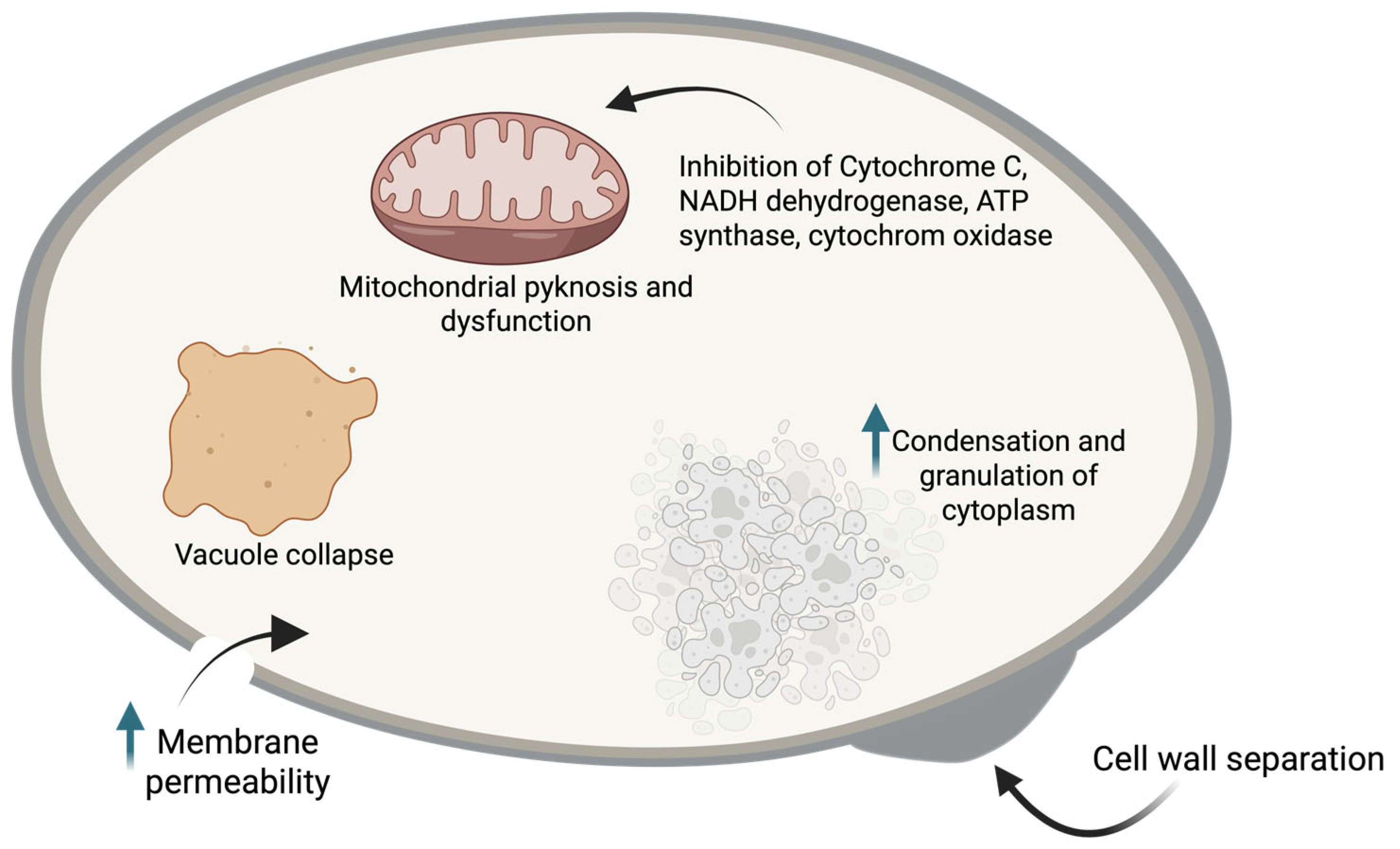
Proteomic analysis of the PLA effect (1/2 MIC, 4 mg/mL) on Rhizopus oryzae revealed 485 differentially expressed proteins between the treated and untreated groups (280 proteins were down-regulated and 405 proteins were up-regulated) [16]. The differentially expressed proteins were involved in translation, ribosomal structure and biogenesis, post-translational modification, protein turnover, chaperones, amino acid transport and metabolism, signal transduction mechanisms, and energy production and conversion, except for those related to energy metabolism which were down-regulated (energy production and conversion (32 proteins) and carbohydrate transport and metabolism (21 proteins)), primarily involved in glycolysis or tricarboxylic acid (TCA) cycle). Moreover, PLA induced ROS accumulation and cytochrome C release in R. oryzae associated with mitochondrial damage and dysfunction, leading to gradual cell apoptosis [16]. The impact of PLA on ATP biosynthesis (energy production) in R. oryzae leads to cell death [16]. Another study on proteomic analysis showed that D- and L-PLA acted similarly on M. racemosus; the cluster analysis evidenced alterations associated with the respiratory chain (6 up-regulated and 14 down-regulated proteins involved in the oxidative phosphorylation-related proteins); thus, key components of the mitochondrial electron transport chain were inhibited (cytochrome C, NADH dehydrogenase, ATP synthase, and cytochrome oxidase) [14]. In addition, significant differences were observed in proteins associated with cell composition, biological processes, and molecular functions, including ATP binding, magnesium ion binding, flavin adenine dinucleotide binding, carbohydrate metabolic process, TCA cycle, and glycolytic process, which could contribute to the inhibition of the metabolic energy process of Mucor [14]. Zhao et al. [72] analyzed the transcriptomic changes in A. flavus NRRL3357 spores after PLA (2.5 mg/mL, 30 min) treatment and identified 980 differentially expressed genes (711 were up-regulated and 269 were down-regulated) grouped into three categories: biological processes, cellular components, and molecular functions. Up-regulated genes were primarily involved in ribosome metabolism, carbon metabolism, RNA polymerase, 2-oxocarboxylic acid metabolism, and the TCA cycle, while the down-regulated genes were associated with proteasome, glycolysis/gluconeogenesis, pyruvate metabolism, and carbon metabolism [72]. The metabolic response of A. flavus detected 59 metabolites, including lipids and lipid-like molecules, organic acids and derivatives, organic oxygen compounds, organoheterocyclic compounds, benzenoids, and organic nitrogen compounds [72]. Figure 1 summarizes the primary PLA mechanisms of action on fungal cells.
At the morphology scale, R. oryzae after 24 h of PLA (1/2MIC, 4 mg/mL) treatment exhibited dense and disorderly mycelium while their tubes were swollen and wrinkled (due to irregular thickness across the wall) without damage or deletion on their surface; thus, PLA did not seriously damage the cell wall [16]. Thus, cell wall composition was not altered by PLA. Cell membrane separation from plasma walls (showing dentate depression), vacuole collapse, and mitochondrial pyknosis were observed after PLA treatment in R. oryzae [16]. PLA at MIC (12.5 mmol/L, 2.07 g/L) after 8 h did not affect the mycelial tips of M. racemosus, slightly disordered the outer surface and collapsed mycelial tubes, without serious damage to the mycelial cell wall [14]. Transmission electron microscopy analysis reveals a separation between the mycelial cell wall and membrane, with ruptured cell membranes leading to disordered, condensed organelles and a granulated cytoplasm [14]. The observed changes were confirmed by fluorescein diacetate and propidium iodide tests; PLA harms Mucor cell membranes and reduces the activity of mycelial cells [14].

6.4. Antibacterial Mechanisms of Action

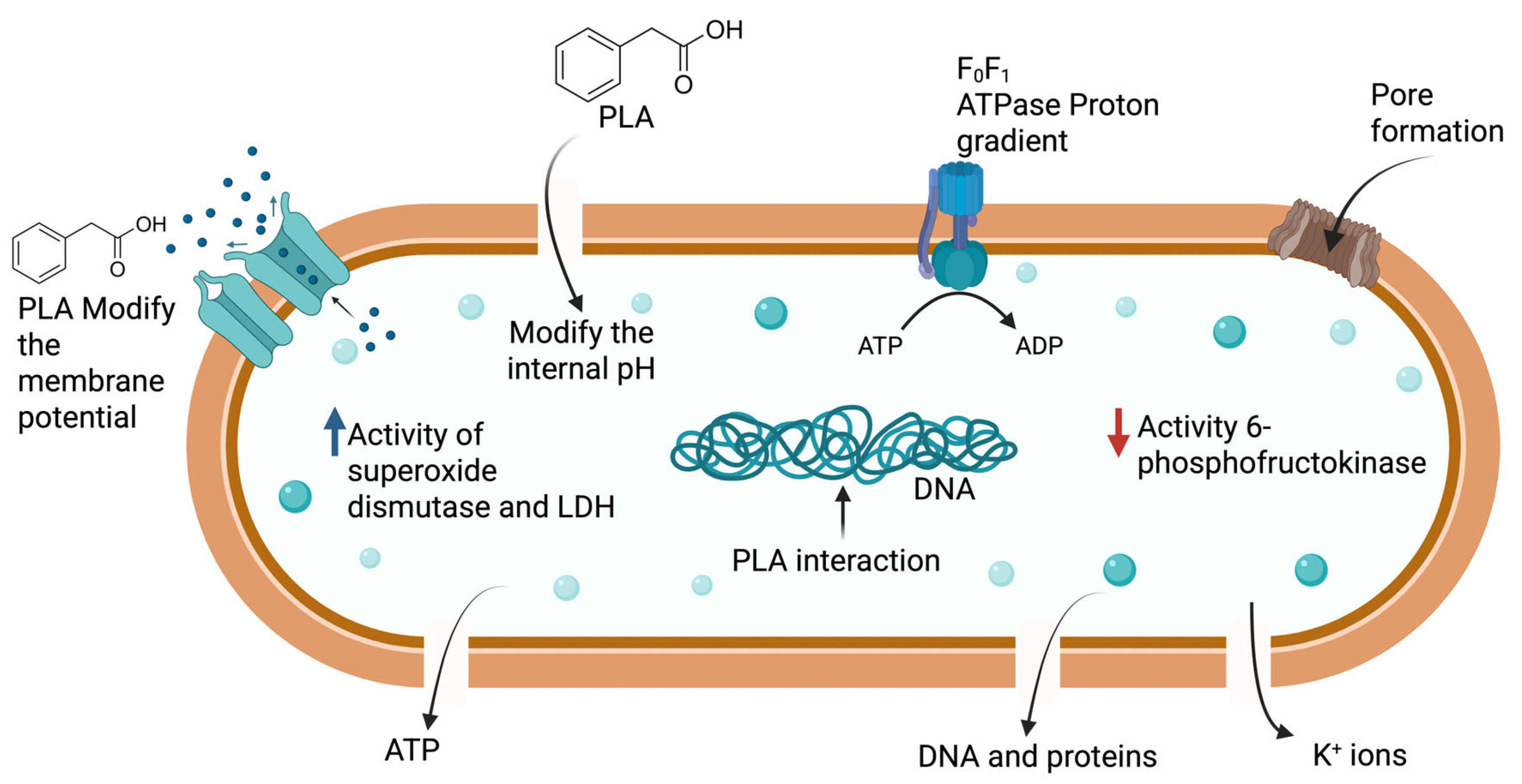
The mechanisms of action of PLA against Gram-positive and Gram-negative bacteria have been explored in various studies, which have reported several target sites. Disruption of membrane integrity (membrane permeability) was observed on L. monocytogenes treated with 0.5, 1, and 2 MIC of PLA. The cytometry analysis revealed that the cells stained with propidium iodide ranged from 75.2% to 94.5% [67]. The amphiphilic nature of PLA (a hydrophobic benzene ring and a hydrophilic carboxy group) facilitates its interaction with cell membrane lipids and proteins, disrupting membrane integrity and increasing permeability [11]. Consequently, pore and local ruptures in cell membranes lead to leakage of materials and the formation of aggregates and adhesions; these changes were more pronounced at higher PLA doses. In another study, the membrane permeability, pore formation, and leakage of intracellular components were confirmed, and additional findings such as losses of cytoplasmic components, vacuolization, protein denaturation, and cell lysis were observed in L. monocytogenes treated with 0.5 or 1 MIC (2.4 mg/mL) PLA [7]. In Listeria innocua, similar outcomes were observed, with Zeta potential becoming significantly more negative (due to cell membrane changes) after PLA exposure, and leakage of cellular content detected at MBC (11.3 mM (1.87 g/L) at pH 5.5) [78]. On Staph. xylosus, 5 mg/mL PLA caused wrinkled surface and split cells [63]. Staph. aureus CICC10145 after PLA treatments (1 MIC and 2 MIC) suffers cell disruption, leakage of cell constituents (DNA, proteins, and ATP), oxidative stress (increases in superoxide dismutase activity and lactate dehydrogenase), and morphological changes (rough cell surface, shrinking, and cell disintegration) [79]. Ent. faecalis R612-Z1 experiences cell surface damage, presenting membrane permeability and depolarization and leakage of ATP, nucleic acids, and proteins after being treated with 1% PLA [12]. The effect of 2 MIC PLA on Lb. bulgaricus CGMCC 1.6970 cells were similar, observing membrane and cell wall disruption, pore formation on the cell surface, cytoplasmic leakage, cell contraction, dissipation of both ΔΨ and ΔpH, inhibition of ATP production, growth, acid metabolism, and disruption of the ion channels and pumps located on the cell membrane [17]. In addition, the enzymatic activity of F0F1-ATPase was reduced by 71%, β-galactosidase by 76%, 6-phosphofructokinase by 59%, and LDH by 71% [17]. In E. coli, PLA does not damage the cell membrane but increases outer membrane permeability by acting on the lipopolysaccharide layer while leaving the inner membrane intact. Externally, bacteria showed slight morphological alterations, including wrinkles, a coarse outer surface, clumped cells, and shrinkage [11]. In contrast, 1% PLA (30 min) completely lysed the cell walls of most Enterobacter cloacae cells, leading to leakage of intracellular components (ATP, proteins, and nucleic acids) and membrane depolarization [18]. S. Typhimurium ATCC 14028 exhibited cell membrane destruction, cellular lysis, dissipation of the ΔΨ, intracellular changes in pH, extracellular electrical conductivity, inhibition of ATP biosynthesis, and DNA interaction with PLA when cells were exposed to 3 MIC (4.5 mg/mL) for 1 h [19]. V. parahaemolyticus presented K+ leakage after 5 min of PLA exposure at ≥1/4 MIC as a result of membrane cell injury and DNA degradation at 1/2 MIC [20]. Likewise, K. pneumoniae loses cell wall integrity with 1 MIC (2.5 mg/mL) PLA for 30 min (observing wrinkles, pores, and local ruptures in the cell surface), 2 MIC PLA after 2 h disrupts the cell membrane, and DNA binds to PLA, leading to its degradation [74]. Similar findings for L-PLA (protein leakage, increases in ROS production, droppings of ATP production, structural damage, changes in fatty acid of cell membrane) were reported for Alicyclobacillus spp. [80]. The PLA interaction and intercalation with DNA base pairs, affecting replication and causing growth inhibition, were also detected in L. monocytogenes, E. coli, and V. parahaemolyticus [11,20]. Other effects of PLA on bacterial cells include reductions in swimming and swarming motilities of Ent. faecalis [81] and V. parahaemolyticus [20]. Figure 2 illustrates the primary mechanisms of action of PLA on bacterial cells.
Genetic analysis revealed that PLA (1 MIC) on Lb. bulgaricus up-regulated 355 genes and down-regulated 351 genes (a total of 706) involved in cellular processes, metabolic processes, localization, biological regulation, reproductive to stimulus and developmental processes, components of the cellular anatomical entity and protein-containing complex, catalytic activity, structural molecule activity, transporter activity, ATP-dependent activity, translation regulator activity, and transcription regulator activity [17].
These modifications result in the suppression of the EMP pathway, blocking the acid-resistant pathway, disrupting amino acid metabolism, and interfering with substance transport by disrupting the ABC transporter-mediated transport process [17]. Complex cellular and metabolic changes were induced by PLA in Gram-positive bacteria; further research across other species and Gram-negative bacteria is needed to better understand its impact on bacterial cells.
For spore-forming bacteria, such as Bacillus cereus, limited information is available on the mechanisms of inactivation. Zhang et al. [15] reported that PLA at 2 MIC disrupted the inner membrane of Bacillus cereus spores (22.6%), showed wrinkles and cracks, and slowed the outgrowth of spores by altering metabolism (inhibition of the oxidative metabolism), inhibiting the outgrowth of vegetative cells. Moreover, PLA degraded DNA, thereby limiting protein biosynthesis. The membrane potential was disturbed in germinated spores. The spore damage level was PLA-concentration-dependent. However, spore germination cannot be inhibited [15].

7. Applications in Food Systems

The acidic nature of PLA influences and reduces food pH more drastically than other acids, such as acetic acid, in a concentration-dependent manner. This pH lowering contributes to improving antimicrobial activity because it is directly related to the undissociated form of PLA at low pH [90]. Therefore, Penicillium paneum inhibition at different pHs requires greater PLA concentrations; for instance, at pH 4, approximately 80 mM (13.29 g/L) PLA is required to delay mold growth by 50%, whereas at pH 6, about 165 mM (27.4 g/L) is required. Other factors, such as temperature and water activity (aw), also affect PLA antimicrobial activity, increasing with higher temperatures and aw [90]. As with other LAB metabolites, PLA can be applied to foods as a partial or purified acid, or as part of a complex mixture in CFS.
Across food matrices, antimicrobial efficacy depends on PLA concentration and matrix composition. Factors like fat, protein, buffering capacity, aw, temperature, and target microorganisms influence outcomes. These variations explain PLA’s synergistic effects with hurdles such as low pH, ethanol, organic acids, phenolics, and refrigeration. In the following sections, the applications of PLA across different food groups are described.

7.1. Dairy Products

PLA is being studied as a “green”, clean-label biopreservative and as a technological metabolite that can affect flavor and shelf life in fermented dairy matrices. Experimental studies using milk and cheese models have suggested that PLA can inhibit pathogenic or spoilage bacteria. Early findings reported by Dieuleveux & Guéguen [73] exhibited a bacteriostatic effect of PLA at 7 mg/mL on L. monocytogenes UCMA L205 (103 CFU/mL) in UHT whole milk over 5 days of culture at 25 °C. However, the use of PLA (20 mg/piece) on Saint-Paulin curd to delay L. monocytogenes growth was insufficient for practical use in manufacturing. PLA concentrations of 3 mg/mL in UHT whole milk and pasteurized milk reduced L. monocytogenes counts by ~1.5 log10 CFU/g after 5 days of storage under refrigerated conditions (4 °C) [75]. However, after 7 days, increasing counts were obtained, exceeding the initial inoculum (5 log10 CFU/g) and reaching ≥6 log10 CFU/g after 14 days [75]. MIC values from model solutions serve as a basis for testing antimicrobials in foods. The growth inhibition of Staph. aureus CICC10145 in skim milk and fresh cheese during storage (4 °C) was delayed in a dose-dependent manner by PLA; thus, at MIC < ½, slight bacterial growth (~0.8 log10 CFU/g) was obtained after 21 days, while at 1/2 MIC, constant counts were registered. For 1 and 2 MIC, reductions of 0.5–1.8 log10 CFU/g were observed [79]. Marginal inhibition of Staph. xylosus were observed in milk treated with 0.1% PLA for 12 h [63]. L. monocytogenes’ EGD-e biofilm biomass was reduced by 35.5%, and the biofilm formation was significantly decreased at 1/4 MIC (1.5 mg/mL) after 7 days at 4 °C [86]. The screening, for starters, was based on isolates of Pediococcus and Lactobacillus that produce higher levels of PLA, thereby enhancing flavor and the cheese’s natural protection [5].
Both the mechanism and application-oriented studies suggest that PLA has the potential to control post-acidification in yogurt by suppressing Lb. delbrueckii subsp. bulgaricus activity after fermentation. Zhou et al. [17] reported that PLA inhibited the growth and lactic acid production of Lb. bulgaricus (MICs ≈ 1.25 mg/mL), thereby reducing post-acidification during storage. This application shows that PLA is a helpful tool beyond controlling spoilage microbes. However, at effective concentrations, PLA can alter aroma and taste (more aromatic acidity). Sensory evaluation is crucial for balancing PLA levels to ensure they control post-acidification without diminishing yogurt’s sensory qualities.

7.2. Fruit and Vegetables

Studies on fruit and vegetables to test the preservative/production effects of PLA use two approaches: first, applying PLA directly as an antimicrobial; and second, using fermentation with LAB. As PLA is derived from amino acid metabolism, it is produced ex novo by selected strains. Lb. plantarum, Lb. casei, Lb. paracasei, and Lb. rhamnosus exhibited the capability to synthesize PLA in sweet cherry juice during fermentation because of amino acid metabolism [91].
Table 6 presents selected studies on PLA applications in fruit and vegetables. According to Table 6, the natural production of PLA has been reported in fruit juices and fermented foods such as kimchi and pickled chili peppers. For instance, Ricci et al. [92,93] reported PLA production in elderberry juice after fermentation (48 h) by Lb. casei 2240 (6 μg/mL) and Lb. plantarum strains (1LE1, 285, C1, POM1) (25 ± 2 μg/mL). In the last study, PLA remained almost constant for 12 days of storage. Recent research by Rizzi et al. [94] demonstrated that Lpb. plantarum KABP051 fermented conventional fruit juices and produced PLA (5.16, 7.57, and 12.04 μM (0.85, 1.25, and 2.0 mg/L) for apple, orange, and peach, respectively). Their metabolomic analyses confirmed that PLA is among the principal antimicrobial compounds generated during fermentation; moreover, fermented juices maintained a similar purchase probability to unfermented juices. Fermented juices retained color and aroma, although consumer acceptability was significantly reduced [94]. This demonstrates that fruit juices serve as feasible carriers for probiotic fermentation and in situ PLA production, thereby facilitating a “natural fermentation” plus preservation process approach. Another interesting insight is the activity or effectiveness of the isomer form of PLA. For example, Cai et al. [80] successfully inactivated Alicyclobacillus spp. using L-PLA in apple juice (see Table 6) without adverse effects on the physicochemical properties, soluble sugars, and volatile aroma components, maintaining the quality of fruit juice. Thus, using PLA-producing starter strains or controlled fermentations is an attractive “clean-label” strategy to improve shelf life and limit undesirable microbial activity in fermented fruit beverages [94]. Fermented juices may have increased microbial stability during storage, reduced spoilage risk, and potentially enhanced functional or health-promoting properties associated with PLA and other metabolites. This is especially relevant given that traditional weak-acid preservatives and sulfites may have undesirable health or sensory effects.
On the other hand, pure PLA has been tested as a post-harvest treatment for various fruits and fresh-cut mushrooms, demonstrating excellent protective properties. The post-harvest effects of PLA on fruits go beyond antimicrobial activity, delaying enzymatic activity and stimulating fruits’ native mechanisms of defense, among others. In some cases, PLA treatment was combined with other preservation methods, such as modified-atmosphere packaging.
Fruit aroma is a very important sensory quality associated with fruit quality. Therefore, studies have assessed the impact of PLA on volatile compounds of fruits such as sweet cherries. Dipping treatment with 12 mmol/L (1994.16 mg/L) PLA and MAP on sweet cherries preserved primary and characteristic aromas (benzaldehyde, hexanal, and (E)-2-hexenal) during refrigerated storage (60 days) [99]. The previous findings reinforce PLA’s potential as a post-harvest preservative for fruits, given its antimicrobial activity while maintaining nutritional, physicochemical, and sensory quality.

7.3. Bakery Products

Bakery products are particularly susceptible to fungal spoilage due to their high water activity and post-baking contamination, with molds such as Penicillium, Aspergillus, and Rhizopus acting as the primary causes of quality loss and waste [10]. Across bakery systems, PLA is a key antifungal compound. Its effectiveness varies based on concentration and preservation methods. In situ PLA production by LAB in sourdough or bio-preserver cultures usually yields sub-millimolar to low-millimolar concentrations. These levels can extend mold-free shelf life by about 2 to 6 days under natural contamination. This is mainly due to synergistic interactions with organic acids and other LAB compounds. Increasing PLA levels, achieved either by optimizing strains or by adding purified PLA, leads to more effective and consistent antifungal results, with shelf-life extensions of up to 7 to 10 days, especially when combined with acetic acid or other clean-label ingredients. The combination of producing PLA cultures in the dough formulation with traditional preservatives (calcium propionate, CP) improved the shelf life of bread.
Table 7 summarizes selected studies on the application of PLA in bakery products. Sourdough fermentation is particularly effective for delivering PLA, highlighting its antifungal effectiveness under both controlled and natural contamination conditions. For example, a fermentate produced by Ped. acidilactici CRL 1753 contained PLA concentrations of 186.5 and 196.4 mg/L after 24 and 48 h of fermentation, respectively, and was used to replace 35% of the water in bread formulations. In this study, a controlled microbial challenge was performed by spraying A. japonicus conidia onto the bread surface after baking. The bread was packaged in polyethylene bags and stored at 30 °C, delaying the onset of visible mold growth by 12–13 days. The final bread pH was approximately 4.9 (dough pH not reported), which likely contributed to the antifungal effect in combination with PLA [38]. Similarly, CFS from Lb. reuteri R29, containing approximately 361.2 ppm PLA, was used as a water replacement in bread formulations. In this case, inhibition was evaluated against natural environmental microbiota (post-baking contamination), with spoilage molds delayed for up to 8 days compared to 4 days in the control bread, corresponding to a 53% increase in mold-free shelf life over 13 days. Although the pH values of dough and bread were not explicitly reported, the combined presence of PLA and other organic acids in the CFS likely contributed to the observed antifungal activity [59]. In another study, par-baked bread with a pH of 4.4 and 100 mM DL-3-PLA was directly incorporated into the dough. Under natural contamination conditions, mold growth during storage at 20 °C in modified-atmosphere packaging was inhibited for up to 9 days, providing protection comparable to that of calcium propionate in bread (control) with pH 5.7 [90].
These studies demonstrate PLA’s effectiveness against key spoilage molds in challenge tests and natural contamination scenarios. PLA, whether added directly or through in situ fermentation, offers a promising clean-label alternative to traditional preservatives such as calcium propionate. From a formulation standpoint, PLA aligns with clean-label trends because it can be produced naturally during fermentation and does not need to be listed as an additive. PLA was identified as a key compound for mold inhibition under natural microbiota conditions. Future research should focus on standardizing effectiveness testing across different types, optimizing in situ PLA production while maintaining sensory quality, and integrating PLA-producing cultures into large-scale baking.

7.4. Meat, Fish, and Poultry Products

In meat, meat-based, fish, and poultry products, the interest in natural preservatives, such as bioprotective microorganisms (mainly LAB) and their metabolites, has increased. PLA shows promise as a natural antimicrobial, enhancing safety and shelf life. Its broad-spectrum antibacterial activity targets spoilage organisms and pathogens in chilled meat products. Pius Bassey et al. [83] demonstrated that PLA could inhibit spoilage bacteria, such as Pseudomonas lundensis and Brochothrix thermosphacta, when applied to fresh pork loins under both air and modified-atmosphere packaging (MAP). The study found that PLA treatment reduced bacterial growth and cell viability. Freezing storage temperatures enhanced PLA antimicrobial activity. For instance, PLA (1.5%) treatment on beef improved the bacterial inactivation when freezing storage (−20 °C) was used, reaching reductions of 1.08–1.06 log10 CFU/g for E. coli O157:H7 after 7 or 14 days, and 1.18 and 1.46 log10 CFU/g for S. Typhimurium DT104 after 7 or 14 days [19]. The incorporation of PLA into hydrogels or other biopolymers acts as a carrier. Liu et al. [102] evaluated an antibacterial hydrogel prepared from electrospun gelatin, chitosan, and PLA nanofibers in chilled chicken meat. The hydrogel effectively inactivated Staph. aureus and E. coli, extending the shelf life of chilled chicken to 4 days. Overall, these studies confirm that PLA, whether used alone or with packaging techniques and other natural antimicrobials, effectively reduces spoilage and pathogenic bacteria in meat and poultry. This method can prolong shelf life, improve safety, and align with clean-label preferences.
Table 8 presents selected studies on PLA applications on meat and fish products. Pork and beef are the most commonly studied products for testing PLA antimicrobial activity, with variable results. The studies show in Table 8 used pure PLA as a direct addition; thus, further research is needed to evaluate PLA from LAB fermentates or as starters during the formulation of fermented meat and fish products.

7.5. Efficacy in Extending Shelf Life and Preserving Sensory Quality

As mentioned in previous sections, PLA exerts antimicrobial activity during contact and during food storage. On the other hand, as a food preservative, PLA’s impact on food sensory properties should be investigated. Few studies have evaluated the effect of PLA on sensory qualities during food storage. In order to reduce 0.9 log10 CFU/mL inoculated Bacillus cereus and extend the shelf life of milk beverages (milk powder, sugar, hydroxymethyl cellulose, ansai honey, aspartame, pH 4.3) for 10 days at 25 °C, 6 MIC (7.5 mg/mL) of PLA was required. Higher PLA concentrations are needed to delay bacterial growth in milk beverages compared with nutrient broth, due to the protective effects of milk components on bacterial spores [15]. Viscosity and pH remained constant during storage. In addition, a milk beverage containing 6 MIC PLA remained stable and maintained relatively good sensory properties after 10 days (color, smell, appearance, texture, and overall preference), confirming the preservative effect of PLA at 6 MIC [15]. Another study reported the inhibition of post-acidification in yogurt for 21 days at 15 °C [17]. The acidity was reduced during storage in a PLA-concentration-dependent manner compared with the control, and the reduction ranged from 3 °T to 6.75 °T for 1/2 MIC-2 MIC. Lb. bulgaricus CGMCC 1.6970 counts maintained levels between 7.65 and 7.05 log10 CFU/mL after 21 days, fulfilling the viable counts of Lb. bulgaricus established by the Codex Alimentarius Commission. Sensory attributes of yogurt were improved by 1 MIC PLA after 21 days of storage. Yogurt + PLA exhibited lower sour smell and better flavor and texture (no particle formation) compared with untreated yogurt. Thus, 1 MIC and PLA effectively controlled yogurt post-acidification while retaining viable counts of Lb. bulgaricus and sensory quality [17].
In pea sprouts, the preservative action of PLA (4 mM, 664.72 mg/L) inhibited softening and lignification by suppressing the activity of enzymes involved in lignin biosynthesis and cell wall degradation, increasing cellulose and hemicellulose content [100]. PLA consistently down-regulated key lignin biosynthesis genes (PsPAL12, PsPAL22, PsCCR1, PsCAD1, PsPER52–1, PER52–2, PsLAC7 and PsLAC11–2) during storage. PLA modulated ROS generation, which mediated the lignification and quality deterioration. In addition, the total polyphenol and flavonoid content was high in PLA pea sprouts after 12 days of storage at 10 °C [98]. Ren et al. [95] investigated and observed the anti-browning effect of PLA (0.05 mol/L, 8.3 g/L) solution on fresh-cut mushrooms (Agaricus bisporus) for 12 days (4 °C). PLA docked into the binding site of polyphenol oxidase (located at the hydrophobic site and surrounded by the residues Phe-264, Met-280, Val-283, and Ala-286, forming a stable hydrophobic binding), reducing its activity and prolonging the shelf life of fresh-cut Agaricus bisporus [95]. In another study, PLA stimulated the expression of genes (involved in disease resistance) and the enzymatic activity of three plant defense responses (β-1, 3-glucanase, and chitinase enzymes), slowing anthracnose disease on banana fruit at the early stages of storage (5 days), contributing to reducing disease severity and prolonging fruit senescence [71]. Similarly, PLA treatment (8 mmol/L, 1.33 g/L) significantly boosted the antioxidant defense system of cherries, modulating the activity of key enzymes (catalase, peroxidase, and phenylalanine ammonia lyase) to prolong their shelf life [97]. In Zaosu pears, PLA treatment increased the relative genes and the activity of phosphate isomerase, hexokinase, glucose-6-phosphate dehydrogenase, succinate dehydrogenase, cytochrome C oxidase, and nicotinamide adenine dinucleotide activity, which elevates the production of glucose-6-phosphate and regulates the biosynthesis and catabolism of fructose-6-phosphate, maintains intracellular redox homeostasis, and provides normal physiological metabolism [98]. In contrast, MDH activity decreased to regulate malate to support adequate respiration and energy in the TCA cycle. In addition, NADP and NADPH content accumulated in pears whilst NAD and NADH were reduced; these changes were associated with reduced respiration, a reduced state that mitigates oxidative damage. All these changes were related to pears’ post-harvest stability. In winter jujube, PLA treatment resulted in higher levels of ascorbic acid and glutathione due to biosynthesis and accumulation via up-regulating the activity and gene transcription levels of L-galactose dehydrogenase, L-galactose-1,4-lactone dehydrogenase, dehydroascorbate reductase, monodehydroascorbate reductase, ascorbate peroxidase, glutathione reductase, and ZjGPI, ZjPMI1-like, ZjGMP, and ZjGGP1 [96]. The previous findings suggest PLA can be used as a regulator for tailored post-harvest management.
In apple juice, the use of PLA as an antimicrobial affects the volatile compound profile associated with aroma, increasing three of the six primary categories of volatile compounds (aldehydes, acids, and ketones), while esters (related to fruity flavor quality) lowered from 29.78 μg/L to 25.39 μg/L [80]. The addition of L-PLA at 8, 16, and 32 mg/mL slightly increased the acidity of apple juice without altering its overall quality, maintaining it within the acceptable range [80].

7.6. Active Packaging

As occurred with other antimicrobials, PLA can be incorporated into active packaging with antimicrobial properties. In this regard, Zhang et al. [104] added three concentrations (1, 1.75, and 2.5%) of PLA into chitosan–PVA films and characterized their physical, optical, barrier, and antimicrobial properties. Incorporation of PLA retained the flexibility, clearness, homogeneity, continuity, and smoothness of films similar to control, demonstrating PLA compatibility with chitosan and PVA. Luminosity, transparency, moisture content, water solubility, ethanol solubility, and water vapor permeability of PLA films were maintained at levels similar to those of the control. Other properties varied with PLA concentration: the UV barrier increased, and thickness and elongation-at-break rose at 2.5% and 1.75% PLA, respectively, while tensile strength reduced using ≥1.75% PLA. The antibacterial activity was PLA-concentration- and species-dependent. The most sensitive bacteria were Sthap. aureus, obtaining similar inhibition (7.5–9 log10 CFU/mL) at the three tested PLA concentrations; the most resistant was L. monocytogenes, inhibited only at 2.5% PLA (9 log10 CFU/mL), whereas E. coli, S. Typhimurium, and Sh. sonnei inhibition depended on PLA concentration, reaching between 9 and 10.5 log10 CFU/mL at 2.5% PLA [7]. Later, an innovative technique was used to design nanofiber films by electrospinning utilizing gelatin (8%), chitosan (1.6%), and different levels of PLA (0, 0.5, 1.0, 1.5%) to preserve chicken breast at 4 °C [102]. An interesting property of these nanofiber films is their water absorption capability, which helps to prevent moisture losses. The hydrogel containing 1% PLA reduced ~4 log10 CFU/mL of Staph. aureus and E. coli in vitro after 12 h. In chicken breast, hydrogel with 1% PLA decreased pH value at the beginning and progressively reached the initial level; total volatile basic nitrogen production was delayed, remaining under the limit (25 mg/100 g) for 8 days, while total viable counts ~reached 6 log10 CFU/g after 6 days. A hydrogel with 1% PLA slowed the spoilage of chicken breast by two days compared with the control [102].
Zhu et al. [105] formulated and optimized a biodegradable composite coating (0.484% chitosan, 0.5% CaCl2, and 0.85% ascorbic acid) containing 0.792% D-PLA and evaluated its preservative effect on Shine-Muscat grapes. Fruits were packaged in a polystyrene box at 24 °C and 75% RH. After 6 days of storage, coated grapes showed reduced decay rate, berry abscission ratio, browning incidence, and weight loss while maintaining their firmness, sensory quality (appearance, color, and freshness), titratable acidity, total soluble solids, ascorbic acid, and total phenols compared with control grapes. This demonstrates the potential preserving effect of PLA in active coating on the sensory and nutritional quality of fruits.

8. Regulatory and Safety Aspects

8.1. GRAS and QPS Status of LAB

The potential application of PLA as a natural antimicrobial in food systems requires careful consideration of regulatory frameworks, toxicological evidence, and consumer-driven expectations such as “clean-label” positioning (Table 9). While many LAB are widely recognized as safe for food fermentations, and many of the metabolites produced in situ may offer benefits in the fermented product [106,107], the regulatory pathway differs when PLA is intentionally used as an added ingredient.
The regulatory standing of LAB plays a central role in the approval of PLA-based biopreservation strategies. In the European Union (EU), the Qualified Presumption of Safety (QPS) list maintained by EFSA includes several LAB taxa (see Table 9), which are considered safe for use in food and feed applications under specified conditions [9]. In the United States, the FDA’s GRAS framework covers many LAB species employed as starter cultures or probiotics [8]. It is important, however, to distinguish between the safety designation of microbial strains and that of individual metabolites [22,106,108]. While LAB used in food fermentations may hold QPS (European Union) or GRAS (United States) status (Table 9), this designation applies to the microorganism itself and not automatically to its metabolites. When PLA is intended for use as a food ingredient, it must be considered separately within the relevant regulatory frameworks. In the European Union, food additives are subject to scientific risk assessment by the European Food Safety Authority, followed by authorization granted by the European Commission under Regulation (EC) No. 1333/2008, before their use in the market. In the United States, substances must either be FDA-approved through a food additive petition or supported by a GRAS determination, which may involve submission of a GRAS notice to the FDA with appropriate safety evidence [109,110]. Consequently, PLA produced in situ during fermentation by QPS/GRAS LAB strains is generally subject to fewer regulatory requirements than purified PLA added as an ingredient. Purified PLA is not currently recognized as an approved food additive in the EU or as self-affirmed GRAS in the U.S. (to our knowledge), so in situ fermentation remains the easier route. This also highlights the need for toxicological dossiers if direct addition is considered.
Table 9. Regulatory and safety considerations for phenyllactic acid (PLA) and lactic acid bacteria (LAB) in food applications.
Entity/AspectScopeStatus/Key PointsImplications for PLA in Foods
EFSA (EU)LAB strains (QPS list)Many LAB taxa (Lactobacillus sensu lato, Leuconostoc, Pediococcus, Sporolactobacillus) included under QPS [9]PLA produced in situ during fermentation by QPS strains is generally acceptable in foods
European Commission Regulation (EC) No 1333/2008Food additivesFood additives must be included in the EU list following a scientific risk assessment by EFSA and authorization granted by the EU [111]Purified PLA, intended as an additive, would require EFSA evaluation and approval
FDA (US)GRAS and food additive frameworkSeveral LAB species are recognized as GRAS for use as starter cultures or probiotics [8]PLA produced in situ during fermentation by GRAS microorganisms is generally allowed within its intended use; however, purified PLA used as an ingredient would need either FDA approval (food additive petition) or support through a GRAS determination or notice submitted to the FDA
Toxicological evidencePLA metaboliteIn vitro studies demonstrate low cytotoxicity at food-relevant concentrations; however, limited subchronic/in vivo data are available [3,21,84]Supports in situ use in fermented foods, but purified PLA as an additive needs a complete toxicological dossier
Clean-label positioningConsumer perception“Clean-label” is not legally defined but is understood to imply natural, minimally processed foods [112,113]In situ PLA aligns with clean-label strategies; exogenous purified PLA may face consumer skepticism

8.2. Toxicological Considerations of PLA

PLA exhibits broad antimicrobial and antifungal activities, with effective concentrations in food systems typically reported in the low millimolar range [35,59,75,114,115,116]. Available in vitro studies suggest that PLA has low cytotoxicity at concentrations relevant to antimicrobial efficacy, although dose-dependent effects have been observed, with higher concentrations causing moderate reductions in cell viability in epithelial cell models [84]. Anand et al. [117] analyzed derivatives of aromatic amino acid metabolites to evaluate their effects on interferon signaling pathways in two human cell lines: A549 (lung epithelial cells) and THP1 (monocytes). They evaluated the effect of PLA at a concentration of 100 μM and controlled viral infections without causing toxicity. Studies using human lung epithelial A549 cells have reported no significant cytotoxic effects within tested ranges, supporting its potential safety at functional levels [85,117]. However, these findings remain limited to short-term assays and specific cell lines.
Currently, no acceptable daily intake (ADI) for PLA has been established by major regulatory agencies due to limited toxicological data required for risk assessment. Information on PLA’s metabolic fate in humans is scarce. Its structural similarity to other aromatic acids suggests it undergoes absorption and metabolic conversion, but direct evidence on its pharmacokinetics, bioavailability, and metabolites is lacking. Long-term and subchronic toxicity data are also missing, impeding regulatory evaluation. Most studies focus on acute or short-term in vitro models, with little data on cumulative effects, allergenicity, or interactions with gut microbiota after repeated intake. While PLA produced in situ by QPS/GRAS microorganisms during fermentation is low-risk in traditional foods, using purified PLA as an ingredient needs more toxicological data, including dose–response studies, ADI derivation, and long-term safety assessments. Overall, the evidence indicates low acute toxicity of PLA at functional concentrations. However, more research on dose–response relationships, metabolic pathways, and long-term effects is needed to support broader use and regulation in food, pharma, and related areas [3,21].

8.3. Clean-Label Applications

The concept of “clean-label” reflects consumer demand for natural, simple, and minimally processed ingredients, though it is not legally defined in most jurisdictions [118]. In practice, the regulatory acceptability of clean-label claims depends on transparent, non-misleading labeling and compliance with local food laws [109,111].
Two main application routes of PLA can be distinguished:
In situ production via fermentation: PLA generated naturally by LAB during food fermentation can be marketed under the existing framework for fermented foods [35], since selected PLA-producing LAB can be used as starters or cultures, allowing their accumulation during fermentation. Because only cultures and raw ingredients are listed on labels, consumers are likely to perceive such products as “natural” and compatible with clean-label positioning [8,9]. In situ production aligns well with clean-label and “natural fermentation” claims. However, PLA yields depend strongly on strain selection, substrate composition, and processing conditions, leading to greater variability in antimicrobial performance [2]. In parallel, optimizing LAB strains capable of high PLA production through improved precursor availability and fermentation optimization remains a priority [2,119].
Exogenous addition of purified PLA: When PLA is added exogenously as a purified compound, it must be clearly declared on the product label and may require authorization in the EU, FDA approval, or GRAS determination in the US, depending on the intended use. Although this approach allows precise control over dosage and functionality, it introduces greater regulatory complexity than in situ production. In addition, the explicit labeling of PLA as an additive may affect consumer perception and potentially conflict with clean-label expectations, which tend to favor ingredients generated through fermentation rather than added as isolated compounds [108,109,110,111].
Overall, the current regulatory and safety landscape favors in situ production of PLA by QPS/GRAS LAB in fermented foods, as this approach aligns with clean-label trends and may involve a more streamlined regulatory pathway [25,26,108,113]. In contrast, the use of purified PLA as a preservative remains constrained by the need for official authorization in the EU, FDA approval in the US, or GRAS validation, as well as the comparatively limited availability of comprehensive toxicological data. Future research integrating robust safety evaluations, regulatory clarification, and consumer perception studies will be essential to support the broader adoption of PLA as a bio-preservative in global food systems [120,121].

9. Future Perspectives and Challenges

The use of PLA, a natural antimicrobial metabolite, presents promising opportunities for food preservation [2,115,122]. However, its wider use depends on progress in strain optimization, combined preservation strategies, industrial practicality, and clear regulations [3,21,121].

9.1. Multi-Omics to Optimize PLA-Producing LAB

Rapid advances in genomics, transcriptomics, proteomics, and metabolomics have transformed the study of PLA biosynthesis in LAB, offering powerful tools to identify key pathways and improve production efficiency [29,123,124,125]. These multi-omics techniques provide a detailed view of microbial physiology and are essential for understanding and improving food fermentation processes [126,127,128]. They help clarify microbial interactions, host–microbe relationships, metabolic activities, and biochemical responses across various conditions, enabling targeted improvements in metabolite production. Metabolomics and related profiling techniques identify metabolic pathways involved in PLA formation and help develop strains with enhanced stress tolerance and higher productivity [129].
Multi-omics studies on LAB have offered valuable insights into the metabolic principles underlying PLA biosynthesis. Wu et al. [39] combined genomic, transcriptomic, and metabolomic analyses to show that some Lb. plantarum strains increase PLA production by strengthening central carbon metabolism and reducing amino acid biosynthesis. This resource reallocation enhances precursor and energy availability, even in the absence of a fully developed de novo PLA pathway. Consequently, engineered strains such as Lpb. plantarum YM-4-3y can produce several times more PLA than their wild-type counterparts [39], highlighting the capacity of biological systems to improve metabolism.
Metabolomics and flux analyses help identify key limiting steps in PLA biosynthesis, such as precursor shortages, NADH/NAD+ imbalances, and competition between pathways [21,124,130]. These findings guide targeted engineering approaches, such as precursor supplementation, redox balance adjustments via enzymes like formate dehydrogenase, and genetic modifications to reroute carbon flow [21]. When combined with process optimization, these omics-driven strategies create a rational framework for designing efficient microbial cell factories [124,130]. For instance, Wu et al. [21] achieved high PLA yields in E. coli by increasing gene copies for phenylpyruvate biosynthesis, knocking out competing tryptophan pathways, and enhancing the supply of PEP and erythrose-4-phosphate. Optimizing fermentation conditions, including dissolved oxygen, yielded 52.89 ± 0.25 g/L PLA in a 6 L fermenter.
Complementary strategies outside strict omics-guided engineering have also advanced PLA production. Screening Lb. crustorum from fermented vegetables identified high-yield strains suitable for whole-cell biocatalysis; repeated cycles in optimized culture media enabled stable and efficient PLA production, highlighting the value of natural strain diversity [40]. Similarly, using Lpb. plantarum with hydrolyzed Porphyra and Phe supplementation boosted PLA production by approximately 2.5-fold compared with unoptimized medium, demonstrating the importance of substrate engineering and the use of non-conventional feedstocks [27]. Enhancing PLA production in Lb. plantarum ITM21B was achieved by increasing Phe concentrations in the culture and maintaining low tyrosine levels. Providing amino acid precursors indicates that higher Phe improves PLA production, particularly when tyrosine remains low, as it limits PLA biosynthesis [5].
Process intensification using fed-batch strategies further improves PLA yields. Maintaining a controlled pH and implementing staged feeding of precursors, such as Phe or phenylpyruvate, has achieved gram-per-liter titers in Lb. plantarum, highlighting the importance of precursor availability [39]. When LAB production is limited or regulatory restrictions are in place, heterologous expression of PLA pathways, particularly in E. coli, combined with fermentation management and redox balancing, has been highly successful [130]. Recent reviews recommend utilizing multi-omics datasets to find regulatory nodes, including transcriptional regulators of aromatic amino acid metabolism, to guide future strain engineering [128,131]. Together, these results show that integrating multi-omics discovery, strategic metabolic engineering, and sophisticated process optimization offers a strong approach for creating high-yield, industrially important PLA-producing LAB strains.

9.2. Combination with Other Natural Preservatives

PLA functions most effectively when applied as part of a multi-hurdle preservation strategy. Its antifungal and antibacterial activities may complement other natural antimicrobials, such as organic acids, bacteriocins (e.g., nisin), plant essential oils, and bioactive peptides [13,19,132,133]. Combining PLA with such agents can produce synergistic inhibitory effects, reduce required dosages, and minimize sensory impacts on foods. This combined effect could be particularly relevant for bakery and dairy applications, where multiple spoilage pathways coexist. Exploring controlled fermentations that favor the co-production of PLA and other natural antimicrobials is an emerging research direction [63].
Fungal spoilage remains a major challenge in bread, dairy, fruit, and vegetable products [134,135]. PLA has demonstrated broad-spectrum antifungal activity against spoilage fungi as described in Section 6.1. The metabolite inhibits both mycelial growth and spore germination, reducing visible spoilage and prolonging shelf life. Additionally, its ability to suppress mycotoxin-producing fungi makes organic acids, including PLA, a promising candidate for mitigating food safety risks [136]. Despite these promising properties, further validation in industrial-scale food systems is required to confirm efficacy under real storage conditions.

9.3. Challenges in Scaling up PLA Production

Despite promising laboratory-scale results, scaling up PLA production remains challenging. Although various PLA concentrations have been obtained under optimized conditions [137], applying these results to industrial processes remains difficult [138]. Most published studies are conducted at laboratory or pilot scale, underscoring the need for industrial-scale validation, extended-shelf-life trials, and consumer-acceptance studies before widespread commercialization.
PLA yields are greatly influenced by precursor availability (such as Phe supplementation), oxygen management, pH control, and strain differences [2,5,7,39,44,139]. Downstream processing presents additional challenges: recovering and purifying PLA from complex fermentation broths is expensive and labor-intensive, limiting overall process viability. Consequently, many potential applications depend on PLA production via LAB fermentation rather than adding purified PLA as an independent preservative. Advances in bioprocess engineering, including fed-batch strategies, metabolic flux analysis, and immobilized-cell systems, provide promising options to address these challenges and boost overall productivity. Table 10 presents selected strategies to optimize PLA production in some LAB.
Several key gaps must be addressed for PLA to be adopted at a commercial scale (Figure 3). Current PLA yields remain too low for large-scale industrial use, underscoring the need for advanced deformation engineering and optimized process conditions to optimize yield [7,137]. Studies vary widely in growth culture media, microbial targets, and PLA concentrations, making comparisons difficult. Harmonized antifungal and antibacterial testing protocols are necessary to ensure consistent results. Although many LAB strains producing PLA are QPS/GRAS, the intentional use of purified PLA requires a comprehensive toxicological assessment and regulatory approval [8,9]. How consumers view PLA, whether explicitly labeled or produced in situ, remains uncertain, particularly within the clean-label movement [112].
Looking ahead, integrating multi-omics tools with optimized strains, synergistic antimicrobial combinations, and scale-up-oriented bioprocess design will be key to advancing PLA from laboratory research to industrial applications. Regulatory approval and consumer acceptance will determine whether PLA can succeed as a natural preservative. Wu et al. [21] emphasized that future studies should include toxicological evaluations and human metabolic assessments to support regulatory dossiers, as well as the development of a food-grade host capable of expressing PLA-producing enzymes under safe, compliant conditions.

10. Concluding Remarks

PLA is an interesting metabolite synthesized by many LAB and has promising applications as an antimicrobial in food preservation. The safe and easy way to use it in foods is through in situ fermentation, or by adding a fermentate to meet regulatory and consumer trends for “clean label” foods. In this regard, several challenges should be overcome before commercial applications are practical, such as strains with adequate PLA production used as starters or probiotics; standardizing culture conditions to ensure consistent PLA production; minimizing interactions and interference with food matrix components; and avoiding impacts on physicochemical and sensory properties, among others. Various strategies drive research on PLA applications in foods, while other investigations focus on optimizing PLA production and targeting microbial targets. While regulatory, toxicological, and human trials are necessary to complete the framework for PLA commercial use, much further investigation is needed into PLA food applications, including studies of sensory and physicochemical properties, industrial-scale production, and toxicological effects on humans.

Supplementary Materials

The following supporting information can be downloaded at https://www.mdpi.com/article/10.3390/fermentation12040184/s1. Table S1: LDH isoforms and key residues associated with PLA production in LAB.

Author Contributions

Conceptualization, R.H.H.-F., E.M.-L. and A.L.-M.; formal analysis, E.M.-L., B.M.-G. and A.L.-M.; investigation, E.M.-L., B.M.-G. and A.L.-M.; writing—original draft preparation, R.H.H.-F., B.M.-G., E.M.-L. and A.L.-M.; writing—review and editing, E.M.-L. and A.L.-M. All authors have read and agreed to the published version of the manuscript.

Funding

This research received no external funding.

Institutional Review Board Statement

Not applicable.

Informed Consent Statement

Not applicable.

Data Availability Statement

No new data were created or analyzed in this study. Data sharing does not apply to this article.

Acknowledgments

The authors thank Universidad de las Américas Puebla for supporting this work.

Conflicts of Interest

The authors declare no conflicts of interest.

Appendix A

Table A1. Abbreviations of the Microbial Genera Cited in This Review.
AbbreviationMicrobial Genus
A.Aspergillus
E.Escherichia
Ent.Enterococcus
F.Fusarium
K.Klebsiella
L.Listeria
Lb.Lactobacillus
Lc.Lactococcus
Leuc.Leuconostoc
Lpb.Lactiplantibacillus
M.Mucor
P.Penicillium
Ped.Pediococcus
R.Rhizopus
S.Salmonella
Sh.Shigella
Sp.Sporolactobacillus
StaphStaphylococcus
V.Vibrio

References

  1. Li, T.; Qin, Z.; Wang, D.; Xia, X.; Zhou, X.; Hu, G. Coenzyme Self-Sufficiency System-Recent Advances in Microbial Production of High-Value Chemical Phenyllactic Acid. World J. Microbiol. Biotechnol. 2023, 39, 36. [Google Scholar] [CrossRef] [Scilit]
  2. Rajanikar, R.V.; Nataraj, B.H.; Naithani, H.; Ali, S.A.; Panjagari, N.R.; Behare, P.V. Phenyllactic Acid: A Green Compound for Food Biopreservation. Food Control 2021, 128, 108184. [Google Scholar] [CrossRef] [Scilit]
  3. Sun, Y.; Zhu, J.; Wang, T.; Liu, Z.; Chu, C.; Yi, J. Recent Advances and Potentiality of Phenyllactic Acid: Source, Antimicrobial Mechanism, and Applications. Trends Food Sci. Technol. 2025, 158, 104926. [Google Scholar] [CrossRef] [Scilit]
  4. Peters, A.; Krumbholz, P.; Jäger, E.; Heintz-Buschart, A.; Çakir, M.V.; Rothemund, S.; Gaudl, A.; Ceglarek, U.; Schöneberg, T.; Stäubert, C. Metabolites of Lactic Acid Bacteria Present in Fermented Foods Are Highly Potent Agonists of Human Hydroxycarboxylic Acid Receptor 3. PLoS Genet. 2019, 15, e1008145. [Google Scholar] [CrossRef] [Scilit]
  5. Valerio, F.; Lavermicocca, P.; Pascale, M.; Visconti, A. Production of Phenyllactic Acid by Lactic Acid Bacteria: An Approach to the Selection of Strains Contributing to Food Quality and Preservation. FEMS Microbiol. Lett. 2004, 233, 289–295. [Google Scholar] [CrossRef]
  6. Arrieta-Echeverri, M.C.; Fernandez, G.J.; Duarte-Riveros, A.; Correa-Álvarez, J.; Bardales, J.A.; Villanueva-Mejía, D.F.; Sierra-Zapata, L. Multi-Omics Characterization of the Microbial Populations and Chemical Space Composition of a Water Kefir Fermentation. Front. Mol. Biosci. 2023, 10, 1223863. [Google Scholar] [CrossRef] [Scilit]
  7. Zhang, J.; Zhang, C.; Lei, P.; Xin, X.; Liu, D.; Yi, H. Isolation, Purification, Identification, and Discovery of the Antibacterial Mechanism of LD-Phenyllactic Acid Produced by Lactiplantibacillus plantarum CXG9 Isolated from a Traditional Chinese Fermented Vegetable. Food Control 2022, 132, 108490. [Google Scholar] [CrossRef] [Scilit]
  8. US Food And Drug Administration (FDA). Recently Published GRAS Notices and FDA Letters; US Food And Drug Administration (FDA): Silver Spring, MD, USA, 2026.
  9. EFSA Panel on Biological Hazards (BIOHAZ); Allende, A.; Alvarez-Ordóñez, A.; Bortolaia, V.; Bover-Cid, S.; De Cesare, A.; Dohmen, W.; Guillier, L.; Jacxsens, L.; Nauta, M.; et al. Update of the List of Qualified Presumption of Safety (QPS) Recommended Microbiological Agents Intentionally Added to Food or Feed as Notified to EFSA 22: Suitability of Taxonomic Units Notified to EFSA until March 2025. EFSA J. 2025, 23, e9510. [Google Scholar] [CrossRef] [Scilit]
  10. Hernández Figueroa, R.H.; López-Malo, A.; Mani-López, E. Antimicrobial Activity and Applications of Fermentates from Lactic Acid Bacteria—A Review. Sustain. Food Technol. 2024, 2, 292–306. [Google Scholar] [CrossRef] [Scilit]
  11. Ning, Y.; Yan, A.; Yang, K.; Wang, Z.; Li, X.; Jia, Y. Antibacterial Activity of Phenyllactic Acid against Listeria Monocytogenes and Escherichia coli by Dual Mechanisms. Food Chem. 2017, 228, 533–540. [Google Scholar] [CrossRef] [Scilit] [PubMed]
  12. Wang, F.; Wu, H.; Jin, P.; Sun, Z.; Liu, F.; Du, L.; Wang, D.; Xu, W. Antimicrobial Activity of Phenyllactic Acid Against Enterococcus faecalis and Its Effect on Cell Membrane. Foodborne Pathog. Dis. 2018, 15, 645–652. [Google Scholar] [CrossRef] [Scilit]
  13. Prema, P.; Smila, D.; Palavesam, A.; Immanuel, G. Production and Characterization of an Antifungal Compound (3-Phenyllactic Acid) Produced by Lactobacillus plantarum Strain. Food Bioprocess Technol. 2010, 3, 379–386. [Google Scholar] [CrossRef] [Scilit]
  14. Zhang, C.; Guo, M.; Kong, Y.; Zhang, J.; Wang, J.; Sun, S.; Li, X.; Zeng, X.; Gong, H.; Fan, X. Antifungal Mechanism of Phenyllactic Acid against Mucor Investigated through Proteomic Analysis. Food Chem. 2024, 452, 139525. [Google Scholar] [CrossRef] [Scilit]
  15. Zhang, D.; Tong, D.; Wang, Z.; Wang, S.; Jia, Y.; Ning, Y. Inactivation Mechanism of Phenyllactic Acid against Bacillus Cereus Spores and Its Application in Milk Beverage. Food Chem. 2024, 453, 139601. [Google Scholar] [CrossRef] [Scilit]
  16. Fan, W.; Li, B.; Du, N.; Hui, T.; Cao, Y.; Li, X.; Ren, H. Energy Metabolism as the Target of 3-Phenyllactic Acid against Rhizopus Oryzae. Int. J. Food Microbiol. 2022, 369, 109606. [Google Scholar] [CrossRef] [Scilit]
  17. Zhou, H.; Pang, X.; Han, P.; Wang, Z.; Wang, S.; Jia, Y.; Ning, Y. Mechanism and Application of Phenyllactic Acid on the Control of Yogurt Post-Acidification Caused by Lactobacillus bulgaricus. Food Res. Int. 2025, 214, 116688. [Google Scholar] [CrossRef] [Scilit]
  18. Liu, F.; Wang, F.; Du, L.; Zhao, T.; Doyle, M.P.; Wang, D.; Zhang, X.; Sun, Z.; Xu, W. Antibacterial and Antibiofilm Activity of Phenyllactic Acid against Enterobacter Cloacae. Food Control 2018, 84, 442–448. [Google Scholar] [CrossRef] [Scilit]
  19. Zhou, Q.; Gu, R.; Li, P.; Lu, Y.; Chen, L.; Gu, Q. Anti-Salmonella Mode of Action of Natural l-Phenyl Lactic Acid Purified from Lactobacillus plantarum ZJ316. Appl. Microbiol. Biotechnol. 2020, 104, 5283–5292. [Google Scholar] [CrossRef] [Scilit] [PubMed]
  20. Fang, M.; Wang, R.; Agyekumwaa, A.K.; Yu, Y.; Xiao, X. Antibacterial Effect of Phenyllactic Acid against Vibrio Parahaemolyticus and Its Application on Raw Salmon Fillets. LWT 2022, 154, 112586. [Google Scholar] [CrossRef] [Scilit]
  21. Wu, H.; Guang, C.; Zhang, W.; Mu, W. Recent Development of Phenyllactic Acid: Physicochemical Properties, Biotechnological Production Strategies and Applications. Crit. Rev. Biotechnol. 2023, 43, 293–308. [Google Scholar] [CrossRef] [Scilit]
  22. Mani-López, E.; Arrioja-Bretón, D.; López-Malo, A. The Impacts of Antimicrobial and Antifungal Activity of Cell-Free Supernatants from Lactic Acid Bacteria in Vitro and Foods. Compr. Rev. Food Sci. Food Saf. 2021, 21, 604–641. [Google Scholar] [CrossRef] [Scilit] [PubMed]
  23. Li, L.; Shin, S.-Y.; Lee, K.W.; Han, N.S. Production of Natural Antimicrobial Compound d-Phenyllactic Acid Using Leuconostoc Mesenteroides ATCC 8293 Whole Cells Involving Highly Active d-Lactate Dehydrogenase. Lett. Appl. Microbiol. 2014, 59, 404–411. [Google Scholar] [CrossRef] [Scilit] [PubMed]
  24. Lee, W.; Park, Y.-T.; Lim, S.; Yeom, S.H.; Jeon, C.; Lee, H.-S.; Yeon, Y.J. Efficient Production of Phenyllactic Acid by Whole-Cell Biocatalysis with Cofactor Regeneration System. Biotechnol. Bioprocess Eng. 2021, 26, 402–407. [Google Scholar] [CrossRef] [Scilit]
  25. Cheng, Y.; Park, T.H.; Seong, H.; Kim, T.; Han, N.S. Biological Characterization of D-lactate Dehydrogenase Responsible for High-yield Production of D-phenyllactic Acid in Sporolactobacillus inulinus. Microb. Biotechnol. 2022, 15, 2717–2729. [Google Scholar] [CrossRef] [Scilit]
  26. Mu, W.; Chen, C.; Li, X.; Zhang, T.; Jiang, B. Optimization of Culture Medium for the Production of Phenyllactic Acid by Lactobacillus sp. SK007. Bioresour. Technol. 2009, 100, 1366–1370. [Google Scholar] [CrossRef] [Scilit]
  27. Huang, C.-H.; Chen, W.-C.; Gao, Y.-H.; Hsiao, H.-I.; Pan, C.-L. Production of Phenyllactic Acid from Porphyra Residues by Lactic Acid Bacterial Fermentation. Processes 2021, 9, 678. [Google Scholar] [CrossRef] [Scilit]
  28. Rahman, N.A.; Ngaini, Z.; Farooq, S.; Ai Ling, S.C.; Shahren, P.N.J. Sustainable Lactic Acid Production from Agricultural Waste: A Review of Current Techniques, Challenges and Future Directions. Bioresour. Bioprocess. 2025, 12, 81. [Google Scholar] [CrossRef] [Scilit]
  29. Lavermicocca, P.; Valerio, F.; Evidente, A.; Lazzaroni, S.; Corsetti, A.; Gobbetti, M. Purification and Characterization of Novel Antifungal Compounds from the Sourdough Lactobacillus plantarum Strain 21B. Appl. Environ. Microbiol. 2000, 66, 4084–4090. [Google Scholar] [CrossRef] [Scilit]
  30. Armaforte, E.; Carri, S.; Ferri, G.; Caboni, M.F. High-Performance Liquid Chromatography Determination of Phenyllactic Acid in MRS Broth. J. Chromatogr. A 2006, 1131, 281–284. [Google Scholar] [CrossRef] [Scilit]
  31. Li, X.; Jiang, B.; Pan, B. Biotransformation of Phenylpyruvic Acid to Phenyllactic Acid by Growing and Resting Cells of a Lactobacillus sp. Biotechnol. Lett. 2007, 29, 593–597. [Google Scholar] [CrossRef] [Scilit]
  32. Zhang, X.; Zhang, S.; Shi, Y.; Shen, F.; Wang, H. A New High Phenyl Lactic Acid-Yielding Lactobacillus plantarum IMAU10124 and a Comparative Analysis of Lactate Dehydrogenase Gene. FEMS Microbiol. Lett. 2014, 356, 89–96. [Google Scholar] [CrossRef] [Scilit]
  33. Guimarães, A.; Santiago, A.; Teixeira, J.A.; Venâncio, A.; Abrunhosa, L. Anti-Aflatoxigenic Effect of Organic Acids Produced by Lactobacillus plantarum. Int. J. Food Microbiol. 2018, 264, 31–38. [Google Scholar] [CrossRef] [Scilit]
  34. Kim, H.-S.; Jung, S.; Kim, M.J.; Jeong, J.Y.; Hwang, I.M.; Lee, J.-H. Comparative Analysis of N -Lactoyl-Phenylalanine and 3-Phenyllactic Acid Production in Lactic Acid Bacteria from Kimchi: Metabolic Insights and Influencing Factors. J. Agric. Food Chem. 2024, 72, 27177–27186. [Google Scholar] [CrossRef] [Scilit] [PubMed]
  35. Cortés-Zavaleta, O.; López-Malo, A.; Hernández-Mendoza, A.; García, H.S. Antifungal Activity of Lactobacilli and Its Relationship with 3-Phenyllactic Acid Production. Int. J. Food Microbiol. 2014, 173, 30–35. [Google Scholar] [CrossRef] [Scilit] [PubMed]
  36. Yu, S.; Zhou, C.; Zhang, T.; Jiang, B.; Mu, W. 3-Phenyllactic Acid Production in Milk by SK25 during Laboratory Fermentation Process. J. Dairy Sci. 2015, 98, 813–817. [Google Scholar] [CrossRef] [Scilit] [PubMed]
  37. Yoo, J.A.; Lim, Y.M.; Yoon, M.H. Production and Antifungal Effect of 3-Phenyllactic Acid (PLA) by Lactic Acid Bacteria. J. Appl. Biol. Chem. 2016, 59, 173–178. [Google Scholar] [CrossRef] [Scilit]
  38. Bustos, A.Y.; Font De Valdez, G.; Gerez, C.L. Optimization of Phenyllactic Acid Production by Pediococcus Acidilactici CRL 1753. Application of the Formulated Bio-Preserver Culture in Bread. Biol. Control 2018, 123, 137–143. [Google Scholar] [CrossRef] [Scilit]
  39. Wu, W.; Deng, G.; Liu, C.; Gong, X.; Ma, G.; Yuan, Q.; Yang, E.; Li, X.; Luo, Y. Optimization and Multiomic Basis of Phenyllactic Acid Overproduction by Lactobacillus plantarum. J. Agric. Food Chem. 2020, 68, 1741–1749. [Google Scholar] [CrossRef] [Scilit]
  40. Xu, J.-J.; Sun, J.-Z.; Si, K.-L.; Guo, C.-F. 3-Phenyllactic Acid Production by Lactobacillus crustorum Strains Isolated from Naturally Fermented Vegetables. LWT 2021, 149, 111780. [Google Scholar] [CrossRef] [Scilit]
  41. Lv, M.; Bai, F.; Cui, F.; Tan, X.; Lv, X.; Li, J.; Guo, X. Screening, Identification of High-Yield Phenyllactic Acid Lactic Bacteria from Fish Intestines and Optimization of Fermentation Conditions. China Brew. 2023, 42, 125–130. [Google Scholar]
  42. Meruvu, H. Enhanced Production of 3-Phenyllactic Acid from Novel Non-Axenic Coculture: Adaptive Evolution and Statistical Fermentation Studies. Biomass Convers. Biorefinery 2024, 14, 31259–31271. [Google Scholar] [CrossRef] [Scilit]
  43. Chaudhari, S.S.; Gokhale, D.V. Phenyllactic Acid: A Potential Antimicrobial Compound in Lactic Acid Bacteria. J. Bacteriol. Mycol. Open Access 2016, 2, 121–125. [Google Scholar] [CrossRef] [Scilit]
  44. Valerio, F.; Di Biase, M.; Lattanzio, V.M.T.; Lavermicocca, P. Improvement of the Antifungal Activity of Lactic Acid Bacteria by Addition to the Growth Medium of Phenylpyruvic Acid, a Precursor of Phenyllactic Acid. Int. J. Food Microbiol. 2016, 222, 1–7. [Google Scholar] [CrossRef] [Scilit] [PubMed]
  45. Lavermicocca, P.; Valerio, F.; Visconti, A. Antifungal Activity of Phenyllactic Acid against Molds Isolated from Bakery Products. Appl. Environ. Microbiol. 2003, 69, 634–640. [Google Scholar] [CrossRef] [Scilit]
  46. Li, X.; Jiang, B.; Pan, B.; Mu, W.; Zhang, T. Purification and Partial Characterization of Lactobacillus Species SK007 Lactate Dehydrogenase (LDH) Catalyzing Phenylpyruvic Acid (PPA) Conversion into Phenyllactic Acid (PLA). J. Agric. Food Chem. 2008, 56, 2392–2399. [Google Scholar] [CrossRef] [Scilit]
  47. Wang, P.; Yang, X.; Lin, B.; Huang, J.; Tao, Y. Cofactor Self-Sufficient Whole-Cell Biocatalysts for the Production of 2-Phenylethanol. Metab. Eng. 2017, 44, 143–149. [Google Scholar] [CrossRef] [Scilit]
  48. Dallagnol, A.M.; Catalán, C.A.N.; Mercado, M.I.; Font De Valdez, G.; Rollán, G.C. Effect of Biosynthetic Intermediates and Citrate on the Phenyllactic and Hydroxyphenyllactic Acids Production by Lactobacillus plantarum CRL 778: Antifungal Compounds from Lactobacilli. J. Appl. Microbiol. 2011, 111, 1447–1455. [Google Scholar] [CrossRef] [Scilit]
  49. Zheng, Z.; Zhao, M.; Zang, Y.; Zhou, Y.; Ouyang, J. Production of Optically Pure L-Phenyllactic Acid by Using Engineered Escherichia coli Coexpressing l-Lactate Dehydrogenase and Formate Dehydrogenase. J. Biotechnol. 2015, 207, 47–51. [Google Scholar] [CrossRef] [Scilit]
  50. Zhu, Y.; Wang, Y.; Xu, J.; Chen, J.; Wang, L.; Qi, B. Enantioselective Biosynthesis of L-Phenyllactic Acid by Whole Cells of Recombinant Escherichia coli. Molecules 2017, 22, 1966. [Google Scholar] [CrossRef] [Scilit]
  51. Qin, Z.; Wang, D.; Luo, R.; Li, T.; Xiong, X.; Chen, P. Using Unnatural Protein Fusions to Engineer a Coenzyme Self-Sufficiency System for D-Phenyllactic Acid Biosynthesis in Escherichia coli. Front. Bioeng. Biotechnol. 2021, 9, 795885. [Google Scholar] [CrossRef] [Scilit] [PubMed]
  52. Wada, Y.; Iwai, S.; Tamura, Y.; Ando, T.; Shinoda, T.; Arai, K.; Taguchi, H. A New Family of D -2-Hydroxyacid Dehydrogenases That Comprises D -Mandelate Dehydrogenases and 2-Ketopantoate Reductases. Biosci. Biotechnol. Biochem. 2008, 72, 1087–1094. [Google Scholar] [CrossRef] [Scilit]
  53. Hummel, W.; Schütte, H.; Kula, M.-R. D-2-Hydroxyisocaproate Dehydrogenase from Lactobacillus casei: A New Enzyme Suitable for Stereospecific Reduction of 2-Ketocarboxylic Acids. Appl. Microbiol. Biotechnol. 1985, 21, 7–15. [Google Scholar] [CrossRef] [Scilit]
  54. Jung, S.; Hwang, H.; Lee, J.-H. Effect of Lactic Acid Bacteria on Phenyllactic Acid Production in Kimchi. Food Control 2019, 106, 106701. [Google Scholar] [CrossRef] [Scilit]
  55. Mu, W.; Yu, S.; Zhu, L.; Zhang, T.; Jiang, B. Recent Research on 3-Phenyllactic Acid, a Broad-Spectrum Antimicrobial Compound. Appl. Microbiol. Biotechnol. 2012, 95, 1155–1163. [Google Scholar] [CrossRef] [Scilit] [PubMed]
  56. Hernández-Figueroa, R.H.; López-Malo, A.; Mani-López, E. Lactic Acid Bacteria-Derived Exopolysaccharides: Dual Roles as Functional Ingredients and Fermentation Agents in Food Applications. Fermentation 2025, 11, 538. [Google Scholar] [CrossRef] [Scilit]
  57. Mu, W.; Yu, S.; Jiang, B.; Li, X. Characterization of D-Lactate Dehydrogenase from Pediococcus Acidilactici That Converts Phenylpyruvic Acid into Phenyllactic Acid. Biotechnol. Lett. 2012, 34, 907–911. [Google Scholar] [CrossRef] [Scilit] [PubMed]
  58. Rodríguez, N.; Salgado, J.M.; Cortés, S.; Domínguez, J.M. Antimicrobial Activity of D-3-Phenyllactic Acid Produced by Fed-Batch Process against Salmonella Enterica. Food Control 2012, 25, 274–284. [Google Scholar] [CrossRef] [Scilit]
  59. Schmidt, M.; Lynch, K.M.; Zannini, E.; Arendt, E.K. Fundamental Study on the Improvement of the Antifungal Activity of Lactobacillus reuteri R29 through Increased Production of Phenyllactic Acid and Reuterin. Food Control 2018, 88, 139–148. [Google Scholar] [CrossRef] [Scilit]
  60. Hou, N.; Xie, Q.; Wang, M.; Lu, Q.; Zhou, H.; Gu, W. Safety Evaluation and Fermentation Process Optimization of a High-Yield Phenyllactic Acid Strain. Mod. Food Sci. Technol. 2020, 37, 67–75. [Google Scholar]
  61. Yang, X.; Li, J.; Shi, G.; Zeng, M.; Liu, Z. Improving 3-Phenyllactic Acid Production of Lactobacillus plantarum AB-1 by Enhancing Its Quorum-Sensing Capacity. J. Food Sci. Technol. 2019, 56, 2605–2610. [Google Scholar] [CrossRef] [Scilit]
  62. Lou, X.; Jiang, Y.; Zhao, F.; Zhang, Y.; Qu, X.; Liu, T.; Zhang, W.; Zhu, L.; Yun, J. Preparation and Characterization of Semi-Hydrophobic Cryogels for Culture of Lactobacillus Strains and Bioconversion towards Phenyllactic Acid Bioproduction. Biochem. Eng. J. 2022, 179, 108312. [Google Scholar] [CrossRef] [Scilit]
  63. Liu, J.; Huang, R.; Song, Q.; Xiong, H.; Ma, J.; Xia, R.; Qiao, J. Combinational Antibacterial Activity of Nisin and 3-Phenyllactic Acid and Their Co-Production by Engineered Lactococcus lactis. Front. Bioeng. Biotechnol. 2021, 9, 612105. [Google Scholar] [CrossRef] [Scilit] [PubMed]
  64. Sarkissian, C.N.; Scriver, C.R.; Mamer, O.A. Measurement of Phenyllactate, Phenylacetate, and Phenylpyruvate by Negative Ion Chemical Ionization–Gas Chromatography/Mass Spectrometry in Brain of Mouse Genetic Models of Phenylketonuria and Non-Phenylketonuria Hyperphenylalaninemia. Anal. Biochem. 2000, 280, 242–249. [Google Scholar] [CrossRef] [Scilit] [PubMed]
  65. Ryan, L.A.M.; Bello, F.D.; Czerny, M.; Koehler, P.; Arendt, E.K. Quantification of Phenyllactic Acid in Wheat Sourdough Using High Resolution Gas Chromatography−Mass Spectrometry. J. Agric. Food Chem. 2009, 57, 1060–1064. [Google Scholar] [CrossRef] [Scilit]
  66. Zhang, D.; Li, W.; Zhang, J.; Tang, W.; Chen, X.; Cao, K.; Chu, Q.; Ye, J. Determination of Unconjugated Aromatic Acids in Urine by Capillary Electrophoresis with Dual Electrochemical Detection—Potential Application in Fast Diagnosis of Phenylketonuria. Electrophoresis 2010, 31, 2989–2996. [Google Scholar] [CrossRef] [Scilit] [PubMed]
  67. Tuberoso, C.I.G.; Bifulco, E.; Caboni, P.; Sarais, G.; Cottiglia, F.; Floris, I. Lumichrome and Phenyllactic Acid as Chemical Markers of Thistle (Galactites tomentosa Moench) Honey. J. Agric. Food Chem. 2011, 59, 364–369. [Google Scholar] [CrossRef] [Scilit]
  68. Cheng, Z.; Ran, Q.; Liu, J.; Deng, X.; Qiu, H.; Jia, Z.; Su, X. Rapid Determination for Benzoic Acid, Sorbic Acid, Phenyllactic Acid, Phenylalanine, and Saccharin Sodium in Vinegar by High-Performance Liquid Chromatography–UV. Food Anal. Methods 2020, 13, 1673–1680. [Google Scholar] [CrossRef] [Scilit]
  69. Lee, M.; Kim, D.; Chang, J.Y. Metabolites of Kimchi Lactic Acid Bacteria, Indole-3-Lactic Acid, Phenyllactic Acid, and Leucic Acid, Inhibit Obesity-Related Inflammation in Human Mesenchymal Stem Cells. J. Microbiol. Biotechnol. 2024, 34, 306–313. [Google Scholar] [CrossRef] [Scilit]
  70. Dopazo, V.; Luz, C.; Quiles, J.M.; Calpe, J.; Romano, R.; Mañes, J.; Meca, G. Potential Application of Lactic Acid Bacteria in the Biopreservation of Red Grape from Mycotoxigenic Fungi. J. Sci. Food Agric. 2022, 102, 898–907. [Google Scholar] [CrossRef] [Scilit]
  71. Gao, Y.; Li, Y.; Li, F.; Zhang, H.; Chen, J.; Yuan, D. Phenyllactic Acid Treatment for Controlling Anthracnose Disease (Colletotrichum musae) and Preserving Banana Fruit Quality during Storage. Physiol. Mol. Plant Pathol. 2024, 129, 102181. [Google Scholar] [CrossRef] [Scilit]
  72. Zhao, C.; Penttinen, P.; Zhang, L.; Dong, L.; Zhang, F.; Li, Z.; Zhang, X. Mechanism of Inhibiting the Growth and Aflatoxin B1 Biosynthesis of Aspergillus flavus by Phenyllactic Acid. Toxins 2023, 15, 370. [Google Scholar] [CrossRef] [Scilit]
  73. Dieuleveux, V.; Guéguen, M. Antimicrobial Effects of D-3-Phenyllactic Acid on Listeria Monocytogenes in TSB-YE Medium, Milk, and Cheese. J. Food Prot. 1998, 61, 1281–1285. [Google Scholar] [CrossRef] [Scilit] [PubMed]
  74. Yu, J.; Hong, C.; Yin, L.; Ping, Q.; Hu, G. Antimicrobial Activity of Phenyllactic Acid against Klebsiella pneumoniae and Its Effect on Cell Wall Membrane and Genomic DNA. Braz. J. Microbiol. 2023, 54, 3245–3255. [Google Scholar] [CrossRef] [Scilit] [PubMed]
  75. Jiang, Y.-H.; Yang, L.-Y.; Xin, W.-G.; Zhang, Q.-L. Combined Antibacterial and Antibiofilm Activity of Phenyllactic Acid and Bacteriocin XJS01 against Shigella Flexneri. Food Biosci. 2022, 45, 101512. [Google Scholar] [CrossRef] [Scilit]
  76. Liu, F.; Du, L.; Zhao, T.; Zhao, P.; Doyle, M.P. Effects of Phenyllactic Acid as Sanitizing Agent for Inactivation of Listeria Monocytogenes Biofilms. Food Control 2017, 78, 72–78. [Google Scholar] [CrossRef] [Scilit]
  77. Jiang, X.; Jiang, C.; Yu, T.; Jiang, X.; Kang, R.; Ren, S.; Chen, H.; Zhang, Y.; Li, Y.; Meng, H.; et al. Phenyllactic Acid Application to Control Listeria Monocytogenes Biofilms and Its Growth in Milk and Spiced Beef. Int. J. Food Microbiol. 2022, 381, 109910. [Google Scholar] [CrossRef] [Scilit]
  78. Sorrentino, E.; Tremonte, P.; Succi, M.; Iorizzo, M.; Pannella, G.; Lombardi, S.J.; Sturchio, M.; Coppola, R. Detection of Antilisterial Activity of 3-Phenyllactic Acid Using Listeria Innocua as a Model. Front. Microbiol. 2018, 9, 1373. [Google Scholar] [CrossRef] [Scilit]
  79. Jiang, Y.-H.; Ying, J.-P.; Xin, W.-G.; Yang, L.-Y.; Li, X.-Z.; Zhang, Q.-L. Antibacterial Activity and Action Target of Phenyllactic Acid against Staphylococcus Aureus and Its Application in Skim Milk and Cheese. J. Dairy Sci. 2022, 105, 9463–9475. [Google Scholar] [CrossRef] [Scilit]
  80. Cai, R.; Gao, L.; Zhang, Y.; Cui, L.; Yuan, Y.; Wang, Z.; Yue, T. Inactivation Activity and Mechanism of Natural Antimicrobial Agents against Alicyclobacillus spp. in Apple Juice. Food Control 2025, 171, 111119. [Google Scholar] [CrossRef] [Scilit]
  81. Liu, F.; Sun, Z.; Wang, F.; Liu, Y.; Zhu, Y.; Du, L.; Wang, D.; Xu, W. Inhibition of Biofilm Formation and Exopolysaccharide Synthesis of Enterococcus faecalis by Phenyllactic Acid. Food Microbiol. 2020, 86, 103344. [Google Scholar] [CrossRef] [Scilit]
  82. Zheng, R.; Zhao, T.; Hung, Y.-C.; Adhikari, K. Evaluation of Bactericidal Effects of Phenyllactic Acid on Escherichia coli O157:H7 and Salmonella Typhimurium on Beef Meat. J. Food Prot. 2019, 82, 2016–2022. [Google Scholar] [CrossRef] [Scilit]
  83. Pius Bassey, A.; Pei Liu, P.; Chen, J.; Kabir Bako, H.; Frimpong Boateng, E.; Isaiah Ibeogu, H.; Ye, K.; Li, C.; Zhou, G. Antibacterial Efficacy of Phenyllactic Acid against Pseudomonas Lundensis and Brochothrix Thermosphacta and Its Synergistic Application on Modified Atmosphere/Air-Packaged Fresh Pork Loins. Food Chem. 2024, 430, 137002. [Google Scholar] [CrossRef] [Scilit]
  84. Maione, A.; Buonanno, A.; Imparato, M.; Maglione, G.; Rossetti, C.; Montone, A.M.I.; Guida, M.; Galdiero, E.; Zinno, P. Antibacterial and Antibiofilm Efficacy of Phenyllactic Acid Against Foodborne Pathogens Salmonella Enterica Serotype Derby and Escherichia coli O26. Molecules 2025, 30, 1738. [Google Scholar] [CrossRef] [Scilit]
  85. Chatterjee, M.; D’Morris, S.; Paul, V.; Warrier, S.; Vasudevan, A.K.; Vanuopadath, M.; Nair, S.S.; Paul-Prasanth, B.; Mohan, C.G.; Biswas, R. Mechanistic Understanding of Phenyllactic Acid Mediated Inhibition of Quorum Sensing and Biofilm Development in Pseudomonas Aeruginosa. Appl. Microbiol. Biotechnol. 2017, 101, 8223–8236. [Google Scholar] [CrossRef] [Scilit]
  86. Chen, H.; Bi, M.; Li, S.; Yang, S.; Zhang, Y.; Zhao, L.; Jiang, X.; Yu, T. Inhibition of Listeria Monocytogenes Biofilm Formation by Phenyllactic Acid in Pasteurized Milk Is Associated with Suppression of the Agr System. Int. J. Food Microbiol. 2026, 444, 111440. [Google Scholar] [CrossRef] [Scilit]
  87. Abdul-Rahim, O.; Wu, Q.; Price, T.K.; Pistone, G.; Diebel, K.; Bugni, T.S.; Wolfe, A.J. Phenyl-Lactic Acid Is an Active Ingredient in Bactericidal Supernatants of Lactobacillus crispatus. J. Bacteriol. 2021, 203, JB0036021. [Google Scholar] [CrossRef] [Scilit]
  88. Svanström, Å.; Boveri, S.; Boström, E.; Melin, P. The Lactic Acid Bacteria Metabolite Phenyllactic Acid Inhibits Both Radial Growth and Sporulation of Filamentous Fungi. BMC Res. Notes 2013, 6, 464. [Google Scholar] [CrossRef] [Scilit] [PubMed]
  89. Hassan, Y.I.; Zhou, T.; Bullerman, L.B. Sourdough Lactic Acid Bacteria as Antifungal and Mycotoxin-Controlling Agents. Food Sci. Technol. Int. 2016, 22, 79–90. [Google Scholar] [CrossRef] [Scilit]
  90. Debonne, E.; Vermeulen, A.; Bouboutiefski, N.; Ruyssen, T.; Van Bockstaele, F.; Eeckhout, M.; Devlieghere, F. Modelling and Validation of the Antifungal Activity of DL-3-Phenyllactic Acid and Acetic Acid on Bread Spoilage Moulds. Food Microbiol. 2020, 88, 103407. [Google Scholar] [CrossRef] [Scilit] [PubMed]
  91. Ricci, A.; Cirlini, M.; Maoloni, A.; Del Rio, D.; Calani, L.; Bernini, V.; Galaverna, G.; Neviani, E.; Lazzi, C. Use of Dairy and Plant-Derived Lactobacilli as Starters for Cherry Juice Fermentation. Nutrients 2019, 11, 213. [Google Scholar] [CrossRef] [Scilit]
  92. Ricci, A.; Cirlini, M.; Calani, L.; Bernini, V.; Neviani, E.; Del Rio, D.; Galaverna, G.; Lazzi, C. In Vitro Metabolism of Elderberry Juice Polyphenols by Lactic Acid Bacteria. Food Chem. 2019, 276, 692–699. [Google Scholar] [CrossRef] [Scilit]
  93. Ricci, A.; Levante, A.; Cirlini, M.; Calani, L.; Bernini, V.; Del Rio, D.; Galaverna, G.; Neviani, E.; Lazzi, C. The Influence of Viable Cells and Cell-Free Extracts of Lactobacillus casei on Volatile Compounds and Polyphenolic Profile of Elderberry Juice. Front. Microbiol. 2018, 9, 2784. [Google Scholar] [CrossRef] [Scilit]
  94. Rizzi, F.; Juan, B.; Espadaler-Mazo, J.; Capellas, M.; Huedo, P. Lactiplantibacillus plantarum KABP051: Stability in Fruit Juices and Production of Bioactive Compounds During Their Fermentation. Foods 2024, 13, 3851. [Google Scholar] [CrossRef] [Scilit]
  95. Ren, H.; Du, N.; Niu, X.; Wang, Y.; Tian, H.; Cao, Y.; Zhang, B.; Fan, W. Inhibitory Effects of L-3-Phenyllacitc Acid on the Activity of Mushnroom Pholyphenol Oxidase. Food Sci. Technol. 2021, 41, 343–351. [Google Scholar] [CrossRef] [Scilit]
  96. Xu, H.; Liu, J.; Jin, Y.; Zhang, F.; Li, C.; Ge, Y. Phenyllactic Acid Maintains the Postharvest Quality of Winter Jujube Fruit via Activating Ascorbic Acid Metabolism. Sci. Hortic. 2023, 322, 112443. [Google Scholar] [CrossRef] [Scilit]
  97. Zhang, C.; Wang, Y.; Wang, M.; Kong, Y.; Li, X.; Song, D.; Zeng, X.; Yang, Y.; Fan, X.; Gong, H. Improvement of Antioxidant Capacity, Aroma Quality, and Antifungal Ability of Cherry by Phenyllactic Acid Treatment during Low Temperature Storage. Front. Plant Sci. 2024, 15, 1529127. [Google Scholar] [CrossRef] [Scilit]
  98. Zhang, S.; Li, C.; Wang, M.; Xu, H.; Guo, Y.; Ge, Y. Phenyllactic Acid Maintains the Storage Quality of ‘Zaosu’ Pears by Regulating Respiration and Energy Metabolism. Postharvest Biol. Technol. 2024, 207, 112607. [Google Scholar] [CrossRef] [Scilit]
  99. Yang, Y.; Li, A.; Guo, M.; Kong, Y.; Zhang, J.; Wang, J.; Sun, S.; Li, X.; Zeng, X.; Gong, H.; et al. Improving the Storage Quality and Aroma Quality of Sweet Cherry by Postharvest 3-Phenyllactic Acid Treatment. Sci. Hortic. 2024, 338, 113661. [Google Scholar] [CrossRef] [Scilit]
  100. Yan, H.; Xia, M.; Liao, Q.; Huang, L.; Li, W.; Yao, T. Phenyllactic Acid Inhibits Softening, Lignification and Quality Deterioration of Postharvest Pea Sprouts. Postharvest Biol. Technol. 2025, 230, 113803. [Google Scholar] [CrossRef] [Scilit]
  101. Xu, J.; Kong, J.; Zheng, X.; Chu, Z.; Osei, P.O.; Wu, P.; Peng, S.; Yang, D.; Wu, S.; Zhao, L.; et al. Discovery of Novel Key Non-Volatile Metabolites Linked to Lactiplantibacillus plantarum in Pickled Chili Peppers Fermentation: DL-p-Hydroxyphenyllactic Acid, 3-Phenyllactic Acid, Catechol, and Acetylcholine. J. Future Foods 2025, S2772566925001582. [Google Scholar] [CrossRef] [Scilit]
  102. Liu, Y.; Wang, R.; Wang, D.; Sun, Z.; Liu, F.; Zhang, D.; Wang, D. Development of a Food Packaging Antibacterial Hydrogel Based on Gelatin, Chitosan, and 3-Phenyllactic Acid for the Shelf-Life Extension of Chilled Chicken. Food Hydrocoll. 2022, 127, 107546. [Google Scholar] [CrossRef] [Scilit]
  103. Huang, Q.; Wang, S.; Hu, C.; Liu, D.; Qiao, Y.; Xiong, G.; Wang, C. Effect of Phenyllactic Acid Treatment on the Quality of Channel Catfish during Low Temperature Storage. Meat Res. 2023, 37, 35–41. [Google Scholar]
  104. Zhang, J.; Chen, J.; Zhang, C.; Yi, H.; Liu, D.; Liu, D. Characterization and Antibacterial Properties of Chitosan–Polyvinyl Alcohol-3-Phenyllactic Acid as a Biodegradable Active Food Packaging. Food Packag. Shelf Life 2022, 34, 100963. [Google Scholar] [CrossRef] [Scilit]
  105. Zhu, D.; Xiong, G.; Tang, W.; Tang, J.; Liu, J.; Chen, Z.; Sun, M.; Fan, X.; Dong, C.; Song, Y.; et al. Preparation of D-Phenyllactic Acid-Chitosan Biodegradable Composite Coating and Its Preservation Effect on Shine-Muscat Grape. Am. J. Biochem. Biotechnol. 2025, 21, 76–89. [Google Scholar] [CrossRef] [Scilit]
  106. Hernández-Figueroa, R.H.; Mani-López, E.; Palou, E.; López-Malo, A. Sourdoughs as Natural Enhancers of Bread Quality and Shelf Life: A Review. Fermentation 2023, 10, 7. [Google Scholar] [CrossRef] [Scilit]
  107. Hernández-Figueroa, R.H.; López-Malo, A.; Mani-López, E. Sourdough Fermentation and Gluten Reduction: A Biotechnological Approach for Gluten-Related Disorders. Microbiol. Res. 2025, 16, 161. [Google Scholar] [CrossRef] [Scilit]
  108. Soto-Reyes, N.; Dávila-Rodríguez, M.; Lorenzo-Leal, A.C.; Reyes-Jurado, F.; Mani-López, E.; Hernández-Figueroa, R.; Morales-Camacho, J.I.; López-Malo, A. Prospects for Food Applications of Products from Microorganisms. In Research and Technological Advances in Food Science; Elsevier: Amsterdam, The Netherlands, 2022; pp. 195–229. ISBN 978-0-12-824369-5. [Google Scholar]
  109. Roberts, A.; Haighton, L.A. A Hard Look at FDA’s Review of GRAS Notices. Regul. Toxicol. Pharmacol. 2016, 79, S124–S128. [Google Scholar] [CrossRef] [Scilit]
  110. US Food And Drug Administration (FDA). Generally Reorganized as Safe (GRAS); US Food And Drug Administration (FDA): Silver Spring, MD, USA, 2023.
  111. European Commission Directorate-General for Health and Food Safety. Food Additives Database. Available online: https://ec.europa.eu/food/food-feed-portal/screen/food-additives/search (accessed on 12 February 2026).
  112. Chauhan, K.; Rao, A. Clean-Label Alternatives for Food Preservation: An Emerging Trend. Heliyon 2024, 10, e35815. [Google Scholar] [CrossRef] [Scilit]
  113. Chen, A.; Kayrala, N.; Trapeau, M.; Aoun, M.; Bordenave, N. The Clean Label Trend: An Ineffective Heuristic That Disserves Both Consumers and the Food Industry? Compr. Rev. Food Sci. Food Saf. 2022, 21, 4921–4938. [Google Scholar] [CrossRef] [Scilit]
  114. Peng, J.-N.; He, H.-R.; Lu, Y.; Lin, L.-B.; Wang, F. Lacticaseibacillus Zeae PW3 Combats Multiple Foodborne Pathogens: Antibacterial Activity and Metabolomics Analysis. LWT 2025, 223, 117666. [Google Scholar] [CrossRef] [Scilit]
  115. Ponzio, A.; Rebecchi, A.; Zivoli, R.; Morelli, L. Reuterin, Phenyllactic Acid, and Exopolysaccharides as Main Antifungal Molecules Produced by Lactic Acid Bacteria: A Scoping Review. Foods 2024, 13, 752. [Google Scholar] [CrossRef] [Scilit]
  116. Zhao, C.J.; Schieber, A.; Gänzle, M.G. Formation of Taste-Active Amino Acids, Amino Acid Derivatives and Peptides in Food Fermentations—A Review. Food Res. Int. 2016, 89, 39–47. [Google Scholar] [CrossRef] [Scilit]
  117. Anand, G.; Clark-Dinovo, C.; Perry, A.M.; Goodwin, V.M.; St. Raymond, E.; Sakleshpur, S.; Steed, A.L. Aromatic Amino Acid Metabolites Alter Interferon Signaling and Influenza Pathogenesis. Front. Mol. Biosci. 2024, 10, 1232573. [Google Scholar] [CrossRef] [Scilit]
  118. Balasubramaniam, V.M.; Lee, J.; Serventi, L. Understanding New Foods: Development of Next Generation of Food Processing, Packaging, and Ingredients Technologies for Clean Label Foods. In Sustainable Food Innovation; Serventi, L., Ed.; Sustainable Development Goals Series; Springer International Publishing: Cham, Switzerland, 2023; pp. 157–167. ISBN 978-3-031-12357-3. [Google Scholar]
  119. Rodríguez-Pazo, N.; Vázquez-Araújo, L.; Pérez-Rodríguez, N.; Cortés-Diéguez, S.; Domínguez, J.M. Cell-Free Supernatants Obtained from Fermentation of Cheese Whey Hydrolyzates and Phenylpyruvic Acid by Lactobacillus plantarum as a Source of Antimicrobial Compounds, Bacteriocins, and Natural Aromas. Appl. Biochem. Biotechnol. 2013, 171, 1042–1060. [Google Scholar] [CrossRef] [Scilit]
  120. Ahansaz, N.; Tarrah, A.; Pakroo, S.; Corich, V.; Giacomini, A. Lactic Acid Bacteria in Dairy Foods: Prime Sources of Antimicrobial Compounds. Fermentation 2023, 9, 964. [Google Scholar] [CrossRef] [Scilit]
  121. Mishra, B.; Mishra, A.K.; Kumar, S.; Mandal, S.K.; Nsv, L.; Kumar, V.; Baek, K.-H.; Mohanta, Y.K. Antifungal Metabolites as Food Bio-Preservative: Innovation, Outlook, and Challenges. Metabolites 2021, 12, 12. [Google Scholar] [CrossRef] [Scilit]
  122. Banicod, R.J.S.; Tabassum, N.; Javaid, A.; Kim, Y.-M.; Khan, F. Lactic Acid Bacteria–Derived Secondary Metabolites: Emerging Natural Alternatives for Food Preservation. Probiotics Antimicrob. Proteins 2025, 18, 3113–3150. [Google Scholar] [CrossRef] [Scilit]
  123. Wang, X.; Hou, Y.; Liu, L.; Li, J.; Du, G.; Chen, J.; Wang, M. A New Approach for Efficient Synthesis of Phenyllactic Acid from L-Phenylalanine: Pathway Design and Cofactor Engineering. J. Food Biochem. 2018, 42, e12584. [Google Scholar] [CrossRef] [Scilit]
  124. Meruvu, H. Redefining Methods for Augmenting Lactic Acid Bacteria Robustness and Phenyllactic Acid Biocatalysis: Integration Valorizes Simplicity. Crit. Rev. Food Sci. Nutr. 2024, 64, 4397–4409. [Google Scholar] [CrossRef] [Scilit] [PubMed]
  125. Zheng, Z.; Xia, M.; Fang, X.; Jiang, T.; Ouyang, J. Enhanced Biosynthesis of Chiral Phenyllactic Acid from L-Phenylalanine through a New Whole-Cell Biocatalyst. Bioprocess Biosyst. Eng. 2018, 41, 1205–1212. [Google Scholar] [CrossRef] [Scilit] [PubMed]
  126. Wen, L.; Yang, L.; Chen, C.; Li, J.; Fu, J.; Liu, G.; Kan, Q.; Ho, C.-T.; Huang, Q.; Lan, Y.; et al. Applications of Multi-Omics Techniques to Unravel the Fermentation Process and the Flavor Formation Mechanism in Fermented Foods. Crit. Rev. Food Sci. Nutr. 2024, 64, 8367–8383. [Google Scholar] [CrossRef] [Scilit]
  127. Sarkar, S.; Sha, S.P.; Ghatani, K. Metabolomics of Ethnic Fermented Foods and Beverages: Understanding New Aspects through Omic Techniques. Front. Sustain. Food Syst. 2023, 7, 1040567. [Google Scholar] [CrossRef] [Scilit]
  128. O’Donnell, S.T.; Ross, R.P.; Stanton, C. The Progress of Multi-Omics Technologies: Determining Function in Lactic Acid Bacteria Using a Systems Level Approach. Front. Microbiol. 2020, 10, 3084. [Google Scholar] [CrossRef] [Scilit]
  129. Tanaka, K.; Bamba, T.; Kondo, A.; Hasunuma, T. Metabolomics-Based Development of Bioproduction Processes toward Industrial-Scale Production. Curr. Opin. Biotechnol. 2024, 85, 103057. [Google Scholar] [CrossRef] [Scilit]
  130. Wu, W.; Chen, M.; Li, C.; Zhong, J.; Xie, R.; Pan, Z.; Lin, J.; Qi, F. Efficient Production of Phenyllactic Acid in Escherichia coli via Metabolic Engineering and Fermentation Optimization Strategies. Front. Microbiol. 2024, 15, 1457628. [Google Scholar] [CrossRef] [Scilit]
  131. Kahraman Ilıkkan, Ö. Lactobacillomics as a New Notion in Lactic Acid Bacteria Research through Omics Integration. World J. Microbiol. Biotechnol. 2025, 41, 68. [Google Scholar] [CrossRef] [Scilit] [PubMed]
  132. Mun, S.Y.; Kim, S.K.; Woo, E.R.; Chang, H.C. Purification and Characterization of an Antimicrobial Compound Produced by Lactobacillus plantarum EM Showing Both Antifungal and Antibacterial Activities. LWT 2019, 114, 108403. [Google Scholar] [CrossRef] [Scilit]
  133. Fugaban, J.I.I.; Jung, E.S.; Todorov, S.D.; Holzapfel, W.H. Evaluation of Antifungal Metabolites Produced by Lactic Acid Bacteria. Probiotics Antimicrob. Proteins 2023, 15, 1447–1463. [Google Scholar] [CrossRef] [Scilit]
  134. Siedler, S.; Balti, R.; Neves, A.R. Bioprotective Mechanisms of Lactic Acid Bacteria against Fungal Spoilage of Food. Curr. Opin. Biotechnol. 2019, 56, 138–146. [Google Scholar] [CrossRef] [Scilit]
  135. Pouris, J.; Kolyva, F.; Bratakou, S.; Vogiatzi, C.A.; Chaniotis, D.; Beloukas, A. The Role of Fungi in Food Production and Processing. Appl. Sci. 2024, 14, 5046. [Google Scholar] [CrossRef] [Scilit]
  136. Sadiq, F.A.; Yan, B.; Tian, F.; Zhao, J.; Zhang, H.; Chen, W. Lactic Acid Bacteria as Antifungal and Anti-Mycotoxigenic Agents: A Comprehensive Review. Compr. Rev. Food Sci. Food Saf. 2019, 18, 1403–1436. [Google Scholar] [CrossRef] [Scilit] [PubMed]
  137. Liu, Y.; Sun, J.; Liu, X.; Zheng, M.; Ma, W.; Jin, G. A Multi-Omics Technology to the Study of Lactic Acid Bacteria Responses to Environmental Stress: The Past, Current and Future Trends. Food Sci. Hum. Wellness 2025, 14, 9250152. [Google Scholar] [CrossRef] [Scilit]
  138. Zang, J.; Lin, T.; Xu, C.; Lin, Y.; Ma, K.; Zhang, C.; Rui, X.; Gan, D.; Li, W. Advances in Lactic Acid Bacteria and Their Metabolites in Fermented Foods: Mechanisms of Food Quality Enhancement, Gut Microbiota Modulation, and Future Prospects. Food Rev. Int. 2025, 1–35. [Google Scholar] [CrossRef] [Scilit]
  139. Sun, D.; Li, H.; Song, D.; Zhang, L.; Zhao, X.; Xu, X. Genome, Transcriptome and Fermentation Analyses of Lactobacillus plantarum LY-78 Provide New Insights into the Mechanism of Phenyllactate Biosynthesis in Lactic Acid Bacteria. Biochem. Biophys. Res. Commun. 2019, 519, 351–357. [Google Scholar] [CrossRef] [Scilit] [PubMed]
Figure 1. Primary mechanisms of action of phenyllactic acid in fungal cells. Blue arrows indicate an increase in specific cellular changes due to PLA interaction.
Fermentation 12 00184 g001
Figure 2. Primary mechanisms of action of phenyllactic acid in bacterial cells. Blue upward arrow indicates an increase in specific enzymatic activity; red downward arrow indicates a decrease in specific enzymatic activity.
Fermentation 12 00184 g002
Figure 3. Prospects and challenges for phenyllactic acid in food biopreservation.
Fermentation 12 00184 g003
Table 3. Key sources of inter-study variability affect reported phenyllactic acid (PLA) concentrations and comparability; considerations are also provided to improve interpretation.
Source of Variability Quantification/ComparabilityConcrete Examples Reported in the LiteraturePractical Standardization Recommendation
Sample preparation strategy
(direct injection vs. extraction/cleanup)
Extraction introduces recovery losses and matrix effects; direct injections can improve throughput, but may increase co-elution risk if chromatography selectivity is insufficientLLE (liquid–liquid extraction) after acidification to pH 2.0 + ethyl acetate (4×) reports matrix-dependent recoveries for PLA (e.g., ~97% in MRS vs. ~88% in synthetic medium) and defines LOD by S/N = 3:1 [5].
Direct microfiltration-only HPLC method reports ~98.7% recovery without extraction [30].
Report recovery (spike) in the same matrix (culture medium/food matrix), and explicitly state whether results are recovery corrected. Include a minimal QC set: blank matrix, spiked matrix, and process replicate.
Matrix composition (culture medium and/or food matrix)PLA recovery, ion suppression (MS), and baseline interferences vary by matrix → studies can report different PLA for the same strain/condition if matrices differRecovery differs between MRS and synthetic medium under the same LLE–HPLC workflow (PLA ~97% vs. ~88%) [5].Specify medium composition and pH at sampling; when comparing strains/studies, normalize to a common matrix or include a matrix-matched calibration/standard addition.
Chromatographic selectivity (column chemistry + gradient program)Changes in the stationary phase/gradient alter resolution (co-elution risk) and retention; can inflate/deflate PLA if nearby peaks overlapLLE workflow separates PLA/OH-PLA on C2/C18 with MeOH/H2O + 0.05% TFA and UV at 210/220 nm. Another workflow uses phenyl–hexyl with direct injection and UV 210/220 nm. Alternative HPLC uses Zorbax SB-C18 with a defined gradient and reported retention times (PLA 6.3 min) [44].Report column type/dimensions, mobile phase additives, and gradient table, and demonstrate peak purity (PDA) or confirmatory MS when matrices are complex.
Detection mode (UV/PDA vs. LC–MS/MS)UV is vulnerable to co-eluting UV-active compounds; MS improves selectivity but introduces ion suppression/enhancement and requires careful internal standardsPDA/UV detection at 210 nm is common across HPLC methods [5].
LC–MS/MS used to identify an unknown co-occurring metabolite (polyporic acid) that could otherwise confound UV quantification [44].
For UV-only methods, add a confirmatory identity step (PDA spectral match + spiking). For MS methods, use isotope-labeled internal standard if available; otherwise, a structurally close surrogate + matrix effect evaluation.
Filtration pore size + injection volumeFiltration affects particulate removal and potential analyte adsorption; injection volume influences sensitivity and peak shape, affecting quant precision at low levelsStudies report 0.22–0.45 μm filtration and injection volumes ranging from 20 to 100 μL, depending on method [5,33,44,59].Standardize filtration (material + pore size) and injection volume, or validate equivalency (recovery/precision) if changing either.
Stereochemistry (D-/L-PLA not always distinguished)Non-chiral methods quantify “total PLA” (racemate) and can mask biologically relevant differences; cross-study comparisons become ambiguous if one study reports total and another the enantiomerEnantiomeric separations reported using cyclodextrin-modified CE or chiral HPLC for PLA [59].State explicitly whether PLA is DL-total or enantiomer-resolved. If not chiral, report as “total PLA (DL)”; for mechanistic work, adopt chiral separation or at least justify non-chiral reporting.
Throughput-oriented UPLC conditions vs. conventional HPLCFaster gradients can improve throughput but may compromise separation unless selectivity is adequate; MS details are sometimes reported elsewhere, limiting reproducibility.Targeted UPLC profiling uses ammonium acetate (A)/methanol (B), low flow, cooled autosampler, and short gradients (high-throughput trend) [30].When adopting fast UPLC, provide the full gradient table + MS acquisition parameters (if MS is used) and demonstrate resolution vs. nearest neighbors (e.g., ILA/OH-PLA)
Table 4. Antifungal activity of cell-free supernatants (CFSs) from lactic acid bacteria (LAB) containing phenyllactic acid (PLA).
LABCulture Medium and PLA ConcentrationMatrix, pH Assay and Antifungal TestTarget Fungi Main FindingsReference
Lactobacillus plantarum ITM21B and ITM20BCell-free supernatant (CFS) from MRS containing 0.34 mM (56.5 mg/L) and 0.33 mM (54.8 mg/L) of PLA, respectivelypH of culture medium not specified. Conidial germination assay.Aspergillus niger, Aspergillus flavus, Eurotium rubrum, Eurotium repens, Endomyces fibuliger, Penicillium corylophilum, Penicillium roqueforti, Monilia sitophilaInhibition of spore germination ranged from 55 to 100%. Most sensitive molds were Eurotium repens, Endomyces fibuliger, P. corylophilum, and Monilia sitophila, and more resistant A. flavus and P. roqueforti.[5]
Lb. plantarum isolated from grass silageCFS from MRS broth after incubation at 30 °C for 48 hMalt extract soft agar
pH of culture medium not specified.
Overlay and well diffusion method.
A. niger, Aspergillus terreus, A. flavus, A. fumigatus, Rhizopus stolonifer, Aspergillus nidulans, Penicillium corylophilum, P. expansum, P. camemberti, and P. roquefortiA. fumigatus and R. stolonifer were the more sensitive molds (inhibition ~100%). A. niger, A. flavus, A. terreus, and A. nidulans were delayed between 86.8 and 89.7%. P. expamsun growth was reduced by 85.8%, P. camemberti by 79%, P. corylophilum by 66.3%, and P. roqueforti by 50%.[13]
Lb. plantarum IMAU10124CFS from MRS broth enriched with 3 g/L of PPA (~2.9 g/L PLA), 20 times concentratedSoft agar.
The pH of the culture medium is not specified.
Agar diffusion assay.
P. roquefortiInhibition zone was 20.6 mm.[32]
Lb. plantarum 21B, 19A, 5BG; Lactobacillus paracasei IMPC2.1CFS containing 2.4 (398 mg/L), 1.1 (182.8 mg/L), 1.2 (199.4 mg/L), and 1.9 mM (315.7 mg/L) PLA, respectivelyCFS from synthetic medium DM.
pH of medium 3.5–3.8.
Microdilution test.
A. niger ITEM 5132, P. roqueforti DTU18687, Endomyces fibuliger DTU605Mold inhibition was PLA-concentration-dependent and ranged from 36 to 68% for A. niger, 35–71% for P. roqueforti, 55–97% for Endomyces fibuliger.[44]
Lb. plantarum UM55CFS from MRS broth containing 74.8 mg/L PLAMalt extract agar.
The pH of the culture medium is not specified.
Poisoned food technique.
A. flavus MUM 17.14Fungal colony diameter was reduced by 50%.[33]
Lactobacillus reuteri R29CFS from MRS broth containing 361 mg/L (ppm) PLACFS from MRS broth.
The pH of the culture medium is not specified.
P. expansum FST 4.22, A. niger FST4.21, and Fusarium culmorum TMW4.2043Fungal growth was reduced by 32.7%, 63.5%, and 84.1% of P. expansum, A. niger, and F. culmorum, respectively, after 120 h.[59]
Lb. plantarum BN16, BN17, E3 and E4CFS from MRS broth contains between 8.15 and 9.55 mg/L PLAPotato dextrose broth.
The pH of the culture medium is not specified.
Minimal inhibitory concentration.
Botrytis cinerea CECT 20973 and Aspergillus (A. carbonarius ISPA 5010, A. niger CECT 2088, A. ochraceus CECT 2093, A. niger CECT 2915,
A. tubingensis CECT 20543 and A. tubingensis CECT 20545)
MIC for Botrytis cinerea was 3.1–6.3 g/L and for Aspergillus 12.5–100 g/L. Minimal fungicidal concentrations for Botrytis cinerea ranged from 6 to 25 g/L.[70]
Table 5. Antibacterial activity of phenyllactic acid (PLA).
Target BacteriaPLA Minimal Inhibitory Concentration (MIC) or Microbial ReductionsReference
Listeria monocytogenes UCMA L205
L. monocytogenes 10403S
L. monocytogenes ATCC 5779
L. monocytogenes
L. monocytogenes 10423S
7 mg/mL reduced by 3 log10 after 15 h and 1 CFU/mL after 27 h;
MIC 1.25 mg/mL;
0.5 and 0.25% PLA inactivated ~7 log10 CFU/mL after 30 and 60 min, respectively;
1% PLA for 1 min reduced to undetectable levels; MIC 2.4 mg/L;
MIC 6 mg/mL
[7,11,73,76,77]
Listeria innocua ATCC 33090MIC ranged from 1.6 mM (265.9 mg/L) to 45 mM (7478.1 mg/L) for pH 4.5–7.0[78]
Enterobacter cloacaeReduced 6 log10 CFU/mL using 1% in 10 min[18]
Staphylococcus xylosusInhibition halo 10.88 mm at 10 mg/mL[63]
Micrococcus luteus ATCC 1024010.12 mm of inhibition at 10 mg/mL[63]
Staphylococcus aureus CICC10145MIC 3.5 mg/mL[79]
Alicyclobacillus spp. (Alicyclobacillus acidoterrestris DSM-3923, Alicyclobacillus acidiphilus DSM-14558, Alicyclobacillus cycloheptanicus DSM-4006, Alicyclobacillus acidocaldarius SD-76 and Alicyclobacillus acidocaldarius SD-94)The Alicyclobacillus spp. MICs were 0.25–0.50 mg/mL, and the minimal bactericidal concentrations (MBCs) were 0.5–16.0 mg/mL[80]
Lactobacillus bulgaricus CGMCC 1.6970MIC 1.25 mg/mL reduced growth in a concentration-dependent manner. 1/2 MIC and 2 MIC reduced growth by 0.55 log10 CFU/mL while 0.19 log10 CFU/mL inactivated it, respectively, compared with the control; falling acid production was also observed[17]
Enterococcus faecalis R612-Z1MIC 5 mg/mL
Reduced by ~6 log10 CFU/mL after 60 and 30 min application of 5 or 10 mg/mL, respectively
[12,81]
Escherichia coli 44752MIC 2.5 mg/mL[11]
E. coli O157:H7 and STEC1.5% PLA killed bacteria (>6 log10 CFU/mL) within 1 min[82]
Pseudomonas lundensis Sneb47 and Brochothrix thermosphacta 24d-s13MIC 10 mg/mL for both bacteria[83]
Salmonella enterica CECT 724At 0.1, 1, and 2 g/L PLA, biomass gradually decreased to 1.6, 1.3, and 1.1 g/L (the corresponding growth inhibitions were 4.4, 23.5, and 38.3%, respectively), which was lower than the control (1.7 g/L)[58]
Salmonella Typhimurium DT104
S. Typhimurium ATCC 14028, S. Arizonae CMCC 47001, and S. Paratyphi-A CMCC 50093
1.5% PLA killed bacteria (>6 log10 CFU/mL) within 1 min
MIC 1.5 mg/mL for S. Typhimurium and Arizonae
MIC 2.5 mg/mL for S. Paratyphi-B
[19,82]
Vibrio parahaemolyticusMIC 3.2 mg/mL for 12 strains, ~6 log10 CFU/mL reduction was obtained after 1 h using 2 MIC[20]
Shigella flexneri BDS14MIC 2.45 mg/mL and 2 MIC reduced to 3.86 log10 CFU/mL after 2 h of treatment[75]
Salmonella enterica Derby, S. Typhimurium DSMZ 18522, and E. coli O26MICs were 2, 2.5, and 2.75 μg/mL, respectively; and the MBCs were 5, 5, and 5.5 μg/mL, respectively[84]
Table 6. Phenyllactic acid (PLA) applications on fruits and vegetables.
Lactic Acid Bacteria/CompoundFood and PLA Production or ApplicationMain FindingsReference
Lactobacillus plantarumKimchiThe supplementation of Lb. plantarum enhanced PLA production 1.7-fold, reaching a maximum after 14 days[54]
Lactobacillus plantarum 1LE1, 285, C1 and POM1Sweet cherry (Prunus avium L.) juicePLA production ranged between 1.21 and 2.09 μg/mL after 48 h of fermentation and remained for 14 days (1.49–2.20 μg/mL) of storage[91]
Phenyllactic acid (PLA)Strawberry treated with 0.1% PLA solutionStrawberry rooting was suppressed by ~45% when the fruit was dipped into PLA solution and stored for 4 days at room temperature, compared with untreated fruits[63]
PLAFresh-cut Agaricus bisporus dipped into PLA (0.03 and 0.05 mol/L, 4.98 and 8.3 g/L), drained, packaged in polypropylene boxes, and stored at 4 °C for 12 days0.05 mol/L (8.3 g/L) PLA inhibited mushroom browning via lowering polyphenol oxidase activity and reducing luminosity losses during storage[95]
Lb. plantarum BN17Cell-free supernatant containing 9.15 mg/L PLA and other acids and phenolic compounds was sprayed on red grapes at 33.3 g/kgContaminated grapes were 40% for Aspergillus niger CECT 2088 and 5% for Botrytis cinerea CECT 20973 compared with 100 and 50% in the control, respectively, after 7 days at room temperature[70]
PLAWinter jujube (Ziziphus jujuba cv. Dongzao) immersed in a PLA (1 g/L) solution for 5 min, packaged in polyethylene bags, and stored at 23 °C, 80% RH for 15 daysThe weight loss was reduced, color change (Luminosity, yellowness, ΔE) increased, and chlorophyll a and b, TSS, and ascorbic acid were maintained during the storage[96]
PLABanana (Musa acuminate L.) dipped into 2.5 g/L PLA for 3 min, inoculated with 20 μL (106 spores Colletotrichum musae), stored at 25 °C and RH 85–90%PLA treatment reduced the anthracnose incidence (5.38%) compared with the control (30.63%) on day 7, while peel color, firmness, and total soluble solids (TSSs) were preserved, and ethylene production and respiration rate were delayed, prolonging fruit senescence[71]
PLAStella sweet cherry (Prunus avium L.) treated with 0, 2, 4, 8, and 16 mmol/L (0, 332.36, 664.72, 1329.44, and 2658.88 mg/L); fruits were punctured and 10 μL PLA and 10 μL Mucor racemosus LD3.0026 (106 spores) were added, and stored at 4 °C for 7 days8 mmol/L (1329.44 mg/L) PLA inhibited mold growth and maintained the appearance of cherries for up to 28 days; PLA (8 mmol/L, 1329.44 mg/L) promoted antioxidant enzyme activity and maintained cherry quality by reducing weight loss and decay of cherries, delaying the decline of firmness, maintaining the sugar content and acidity, slowing down respiration, and preserving the color of fruit skin (delay discoloration and oxidation) while enhancing antioxidant capacity and flavor stability[97]
PLAZaosu pears were immersed in 1 g/L PLA solution for 10 min, drained for 2 h, and stored at 21 °C and 80 RH in semi-sealed PE bags for 8 daysPLA treatment preserved color, flesh firmness, total acidity, TSS, ascorbic acid, soluble sugar content, and chlorophyll, whereas mass loss reduced compared with the control, during storage[98]
PLASweet cherries (Prunus avium L. cv. Youyi and Teiton)
Fruits were immersed for 3 min in PLA solutions (3, 6, 12, and 24 mmol/L, 498.54, 997.08, 1994.16, 3988.32 mg/L), air-dried, packaged in polypropylene trays with a modified atmosphere (3% O2, 10% CO2, 87% N2 for Youyi cherries and 7% O2, 15% CO2, 78% N2 for Tieton) and stored at 1 °C and 80–85% RH
12 mmol/L (1994.16 mg/L) PLA effectively reduced cherry respiration rates, maintained quality attributes (surface color, TSS, total acidity, and firmness, and prevented weight loss) during storage (60 days)[99]
PLAApple juice (12 °Brix) was inoculated with 105 CFU/mL of Alicyclobacillus and incubated at 45 °C for 24 h;
for physicochemical properties, apple juice was mixed with the MBC of L-PLA
The MBC of L-PLA was 0.25 mg/mL for Alicyclobacillus cycloheptanicus DSM 4006 and A. acidocaldarius SD-76 and 16 mg/mL for A. acidoterrestris DSM 3923, A. acidiphilus DSM 14558, and A. acidocaldarius SD-94. pH decreased from 4.05 to 3.20, total acidity rose (6.52 g/L), total phenols increased by 40.82 mg/L, and color change was minimal (ΔE = 1.66)[80]
PLAPea (Pisum sativum L.) sprouts immersed for 5 min in 4 mM (664.72 mg/L) PLA solution and stored at 10 °C for 12 daysCommercial acceptability (sensory test scores) after 12 days was improved (91%) compared with the control (84%); Vitamin C after 9 days was 1.18-fold higher than control; protein content after 6 days was 1.14-fold superior to control; overall odor intensity was significantly higher in treated sprouts, the weight loss was 15% (1% less than control), lower gumminess and chewiness were obtained in sprouts with PLA for 12 days[100]
Lactiplantibacillus plantarum1.54 mg/kg Guizhou traditional pickled chili peppersPLA and six non-volatile metabolites (linoleic acid, phytosphingosine, pyroglutamic acid, DL-p-hydroxyphenyllactic acid, catechol, and acetylcholine) were considered key compounds for the nutritional and taste profile produced during spontaneous pickling[101]
Table 7. Applications of phenyllactic acid (PLA) in bakery products.
Bakery ProductPLA Treatment ConditionMold TargetMain FindingsReference
Bread packaged into polyethylene bags and stored at 30 °CCulture of Pediococcus acidilactici CRL 1753 from an optimized medium (107 CFU/mL) was used to replace 35% of the water formulation of bread.
PLA content after 24 and 48 h was 186.5 and 196.4 mg/L, respectively.
Aspergillus japonicus (1 mL of 103 conidia/mL was sprayed on the bread surface)Cultures of 24 and 48 h delayed mold growth for 12 and 13 days, respectively. The pH of bread was lowered to ~4.9, and the organoleptic characteristics were good.[38]
BreadCell-free supernatant (CFS) from Lactobacillus reuteri R29 (MRS + 1.5% phenylalanine fermented for 48 h) was used to replace water in the bread formulation.
CFS contained 361.2 ppm PLA.
Bread spoilage moldsMolds were inhibited for 8 days compared with 4 days in the control. Moreover, after 13 days, the spread of mold was slowed by 53%.[59]
Part-baked wheat bread stored at 20 °C in modified-atmosphere packaging100 mM (16.61 g/L) DL-3-PLA, which corresponds to 12 mM (1.99 g/L) undissociated PLA in the dough.Molds from natural post-baking contaminationMold growth was inhibited for 9 days of storage, similar to bread with calcium propionate. pH was drastically reduced.[90]
Table 8. Applications of phenyllactic acid (PLA) in meat and fish products.
FoodPLA TreatmentMicrobial TargetMain FindingsReference
Beef pieces (150 g)1 mL of 1.5% PLA sprayedTwo pathogens (Escherichia coli O157:H7 and Salmonella Typhimurium DT104)Marginal inactivation levels (0.34–0.86 log10 CFU/g) were achieved after 24 h of storage at 4 °C on inoculated (2.91 or 3.73 log10 CFU/g, respectively).[82]
Ground pork0.1% of PLANoneTotal mesophilic bacteria counts were slightly delayed during 3 days of storage at 4 °C.[63]
Raw salmon fillets8 MIC (25.6 mg/mL)Vibrio parahaemolyticus was inoculated (~7.7 log10 CFU/g)Reduced the total microbial load to under detectable counts, while 4 MIC to 1 MIC slowed down 99.82–95.72%.
PLA at 2 MIC remained V. parahaemolyticus counts < 5 log10 CFU/g on salmon fillets after 6 days at 4 °C.
[20]
Spiced beefThe application of 6 and 12 mg/mL PLA by dippingListeria monocytogenesInitial counts (5 log10 CFU/g) of L. monocytogenes after 3 days of storage (4 °C) between 1 and 2 log10 CFU/g, then constant increases were recorded up to 15 days. The final counts were 5 and 6.5 log10 CFU/g for the two tested concentrations of PLA.[77]
Channel catfish2.5 g/100 mL PLANoneThe treatment significantly prolonged the shelf life at 4 °C (12 days) with little effect on microbial quality (total bacterial count), physicochemical, and sensory properties.[103]
Fresh pork loinsMeat pieces (15 g) were soaked in PLA solutions (10 or 20 mg/mL, 1 MIC and 2 MIC, respectively) for 30 s, packaged in air or modified-atmosphere packaging (MAP, 70%N2/30%CO2), and stored for 14 days at 4 °CPseudomonas lundensis Sneb47 or Brochothrix thermosphacta 24d-s13, and a mixture of bacteriaLoin pieces treated with 2 MIC PLA + MAP maintained microbial counts between 2 and 3.5 log10 CFU/g in individual or combined bacteria after 14 days. Spoilage indicators (muscle glucose, pH, total volatile basic nitrogen, lipid oxidation) showed minimal changes after 14 days, while sensory attributes (appearance, texture, odor, water-holding capacity) were acceptable.[83]
Minced beefPLA at 1 μg/mL on meat stored at 4 °C for 7 daysSalmonella enterica Derby, S. Typhimurium DSMZ 18522, and E. coli O26 inoculated at approximately 1 × 10⁴ CFU/gE. coli counts dropped ~1.5 log10 CFU/g after 24 h and were maintained (~4 log10 CFU/g) for 7 days; S. Derby reduced ~1 log10 after 48 h and remained at constant levels; S. Typhimurium decreased ~1.5 log10 CFU/g after 24 h and remained steady after 7 days.[84]
Table 10. Representative strategies for optimizing PLA production in LAB and heterologous hosts, illustrating the diversity of approaches and their respective impacts on yield.
Strain/HostApproachKey Intervention(s)Yield/ImpactReference
Lactobacillus crustorum (vegetable isolates)Natural strain screening + whole-cell biocatalysisSelection of high-yielding isolates; repeated batches in optimized culture mediaSignificant PLA increases; stable semi-continuous production[40]
Lactiplantibacillus plantarum (Porphyra residues)Biorefinery + process optimizationEnzymatic hydrolysis of algal residues; strain selection~2.5× higher PLA vs. MRS[27]
Lactiplantibacillus plantarum AB-1Fed-batch + precursor feedingpH-controlled fed-batch with staged phenylalanine/phenylpyruvate additionPLA substantially higher than batch[39]
Disclaimer/Publisher’s Note: The statements, opinions and data contained in all publications are solely those of the individual author(s) and contributor(s) and not of MDPI and/or the editor(s). MDPI and/or the editor(s) disclaim responsibility for any injury to people or property resulting from any ideas, methods, instructions or products referred to in the content.