Article

Collagenase Production from Aspergillus serratalhadensis URM 7866 Using Industrial By-Products: Purification and Characterization

by
Luiz Henrique Svintiskas Lino
1,
Kethylen Barbara Barbosa Cardoso
1,
Pietra Gícia Oliveira Cosmo da Silva
1,
Raphael Luiz Andrade Silva
1,
Maria Eduarda Luiz Coelho de Miranda
2,
Daniel Charles dos Santos Macêdo
1,
Ana Lúcia Figueiredo Porto
2,
Cristina Maria de Souza Motta
3,
Marcia Nieves Carneiro da Cunha
2,
Thiago Pajéu Nascimento
4,
Carolina de Albuquerque Lima Duarte
5,
Romero Marcos Pedrosa Brandão Costa
1 and
Daniela de Araújo Viana Marques
1,*
1
Integrated Laboratory of Applied Biotechnology, Institute of Biological Sciences, University of Pernambuco, Arnóbio Marques, Recife 50100-130, PE, Brazil
2
Department of Animal Morphology and Physiology, Federal Rural University of Pernambuco—UFRPE, Rua Dom Manoel de Medeiros, Recife 52171-900, PE, Brazil
3
Department of Mycology, Federal University of Pernambuco, Av. Nelson Chaves, Recife 50760-420, PE, Brazil
4
Campus Professor Cinobelina Elvas, Federal University of Piaui, Bom Jesus 64900-000, PI, Brazil
5
Multi-User Laboratory of Biotechnology of the Sertão Pernambucano, University of Pernambuco, Arcoverde Campus, Cícero Monteiro de Melo, Arcoverde 56503-146, PE, Brazil
*
Author to whom correspondence should be addressed.
Fermentation 2025, 11(8), 478; https://doi.org/10.3390/fermentation11080478
Submission received: 17 July 2025 / Revised: 8 August 2025 / Accepted: 13 August 2025 / Published: 20 August 2025

Abstract

Collagenases are enzymes with broad biotechnological applications in medicine. This study describes the production and characterization of a collagenase from Aspergillus serratalhadensis URM 7866, isolated from the Caatinga biome. Solid-state fermentations were conducted using wheat bran under varying conditions of pH (6, 7, 8), moisture content (50%, 60%, 70%), and substrate concentration (2.5 g, 5 g, 10 g). The optimal condition—10 g of wheat bran at pH 8 and 70% moisture—yielded the highest collagenolytic activity (177.96 U/mL) and a specific activity of 50.55 U/mg. The enzyme was purified via multiple chromatography, with pre-purification and final purification factors of 18.09 and 20.21, respectively, reaching a specific activity of 1021.86 U/mg. The enzyme showed optimal activity at 50 °C and pH 8, with stability from 20 to 40 °C and pH 7–9. PMSF caused >80% inhibition; EDTA caused ~34% inhibition. Activity increased with Na+ and Ca2+ and was inhibited by Zn2+. The enzyme retained full activity in anionic and non-ionic surfactants (1–10%). FTIR confirmed characteristic amide bands, and kinetic analysis revealed a Km of 1.72 mg/mL and Vmax of 6.89 mg/mL/min. These findings support its potential for alkaline and surfactant-rich industrial processes.

1. Introduction

Microorganisms are widely recognized for their ability to produce proteases and play a fundamental role in various industrial processes, including those in the pharmaceutical and biomedical industries, among others [1]. Although plants and animals can also be sources of proteases, microorganisms offer unique advantages that make them highly desirable for large-scale industrial applications [2]. These advantages include the ability to synthesize proteases under extreme conditions, such as high temperatures, pH variations, or nutrient scarcity [3].
Additionally, microorganisms grow rapidly in compact spaces, possess high genetic diversity, and can be easily genetically manipulated to increase enzyme production or modify their properties to meet specific industrial requirements [4].
Among microorganisms, filamentous fungi, especially species of the genus Aspergillus, have shown considerable potential as producers of proteolytic enzymes, including collagenases [5]. Recent studies have isolated Aspergillus strains from extreme environments, such as the Caatinga biome, a semi-arid region with harsh environmental conditions [5,6]. These strains demonstrated remarkable efficiency in enzyme production, particularly collagenases, which have drawn attention due to their biotechnological relevance [7,8].
Collagenases are highly specific enzymes capable of degrading the triple helix structure of collagen, a key structural protein found in the connective tissues of animals [9]. These enzymes have a wide range of applications, including in food processing, the leather industry, meat tenderization, and the production of pharmaceutical compounds. They are especially important in the medical field, contributing to the treatment of burns, wounds, and scars. They are also employed in organ transplant procedures, management of Parkinson’s disease, and in the treatment of liver cirrhosis. Furthermore, collagenases can be used to improve blood purification techniques, enhancing diagnostic screening processes. Additionally, they help mitigate harmful fibrotic processes. Recently, researchers have also applied collagenases in the modulation of solid tumors [10,11,12].
Given the importance of collagenases, the development of sustainable and efficient methods for enzyme production is essential. In this context, the present study focused on the production, purification, and characterization of collagenase using agro-industrial residues as substrate. Furthermore, the study utilized a novel species of filamentous fungus, A. serratalhadensis URM 7866, isolated from the Brazilian Caatinga biome, a unique and underexplored environment.

2. Materials and Methods

2.1. Microorganism

The microorganism used was A. serratalhadensis, isolated from the soil of the Pernambuco backlands and identified by the Micoteca URM Culture Collection at the Biological Sciences Center of the Federal University of Pernambuco, under registration number 7966. This microorganism was maintained on slant-PDA medium (Potato Dextrose Agar), and for sporulation, the PDA culture medium was also used. After this step, the flask was incubated at 30 °C for 72 h and subsequently used in the fermentation process.

2.2. Solid State Fermentation (SSF)

The production of collagenase by A. serratalhadensis URM 7866 was carried out through solid-state fermentation (SSF) in 125 mL Erlenmeyer flasks containing a concentration of 1 × 10⁷ spores per gram of substrate. To ensure complete drying of the substrate, it was kept in an oven (TECNAL, São Paulo, Brazil) at 50 °C for 72 h. A full 23 factorial design was employed to determine the effects of three factors (Table 1), on collagenase production, which was the response variable analyzed. The fermentation was conducted at a constant temperature of 37 °C for 168 h. In total, nine treatments were performed, corresponding to the eight factorial combinations plus one center point. No replicates were performed for these treatments; therefore, the statistical model was truncated after factor interactions, and the two-factor interaction term was used as an estimate of experimental error. The independent variables were coded according to the equation below [13].
Moisture content was determined according to the following equation:
xi = (Xi − X0)/ΔXi
where Xi is the coded value corresponding to the Xi actual value, X0 the average of the two extreme levels, and ΔXi the range of variation in the i-th factor (1 = substrate concentration, 2 = moisture, 3 = pH). The full 23 design model included a constant term, three main effects, two-factor interactions and one three-factor interactions. Since no replicates had been carried out at this point, the model was truncated after the factor interactions, and the two-factor term was taken as an estimate of the error of an effect. At the end of the fermentation, the fermented material was homogenized in 0.1 M Tris-HCl buffer (pH 8.0), followed by vacuum pump filtration. The resulting enzyme-containing filtrate was then used for subsequent activity assays.

2.3. Determination of Collagenolytic Activity and Total Protein

The collagenolytic activity was determined using a modified method described by Chavira [14], which utilizes azocollagen as the substrate. For the enzymatic reaction, 50 µL of the enzyme-containing liquid and 950 µL of 0.1 M Tris-HCl buffer at pH 7.8 were added to the washed substrate. The reaction occurred at 37 °C for 18 h. After this period, each assay was centrifuged, and 1 mL of the supernatant was taken for spectrophotometric measurement using a Shimadzu UV–Vis spectrometer (UV-1900i, Shimadzu, Kyoto, Japan) at a wavelength of 520 nm. Thus, 1 unit of enzymatic activity (U), under the assay conditions, was defined as an increase of 0.01 in absorbance.
The protein concentration was determined according to the Smith method, using bicinchoninic acid (BCA) [15]. Albumin was used as the standard protein for the protein assay.

2.4. Purification Steps of Collagenolytic Enzymes

2.4.1. Organic Solvent Precipitation

The precipitation process was carried out as described by Nickerson [16], with adaptations, using a 70% concentration of organic solvent (acetone). The mixture was homogenized and maintained at 4–8 °C for 10 min, followed by centrifugation (Centrilab 802BU/CE800U, São Paulo, Brazil) for 10 min at 4 °C and 4000 rpm. After this process, the supernatant was discarded, and the precipitate was resuspended in 1 mL of Tris-HCl buffer pH 8 0.1 M.

2.4.2. Chromatography

For protein purification, multiple chromatographic sessions were performed using the ÄKTA (GE Healthcare, Uppsala, Sweden) coupled with UNICORN 7.0 software. Initially, an ion-exchange chromatography was carried out using a HiTrap ANX FF column, equilibrated with a 20 mM Tris-HCl buffer at pH 8.0, with elution achieved by applying a linear gradient of NaCl up to 1 M. Subsequently, a second round of size-exclusion chromatography on Superdex G-200 was performed to enhance the purification level of the target compound. Throughout the process, eluted fractions were monitored by spectrophotometric readings at 280 nm and 215 nm.

2.4.3. Electrophoresis

For SDS-PAGE, a 10% polyacrylamide gel was used, following the method of Laemmli [17]. The protein bands were visualized by Coomassie blue staining, and destaining was performed using a solution containing water, acetic acid, and methanol (4.5:1:4.5). The protein standard molecular weight markers were phosphorylase b (97.0 kDa), bovine serum albumin (66.0 kDa), ovalbumin (54.0 kDa), carbonic anhydrase (30.0 kDa), trypsin inhibitor (20.1 kDa), and a-lactalbumin (14.4 kDa).

2.5. Effect of pH and Temperature on Enzyme Activity and Stability

The effect of pH on collagenolytic activity was evaluated by incubating the enzyme with its specific substrate, 0.005% (w/v) azocoll, prepared in 0.1 M buffer solutions at different pH levels: sodium citrate (pH 5 and 6); Tris-HCl (pH 7.0, 8.0, and 9.0); and carbonate–bicarbonate (pH 10.0 and 11.0). After incubation, collagenolytic activity was determined. To assess enzyme stability at different pH levels (pH 4–11), the enzyme was incubated with each of the aforementioned buffers in a 1:1 (v/v) ratio at 37 °C for 1 h, followed by the determination of collagenolytic activity. The effect of temperature and thermostability on collagenolytic activity was evaluated at temperatures ranging from 20 °C to 70 °C. For determining the optimum temperature, the assay was performed by incubating the samples with the substrate, azocollagen, in a water bath. In the thermal stability test, the extract was incubated in a water bath for 1 h, and the residual activity was then measured at 37 °C using the method described by Chavira [14]. All reactions were carried out at least in triplicate.

2.6. Effect of Inhibitors on Activity

The effect of substances inhibiting enzymatic activity was assessed using ethylenediaminetetraacetic acid (EDTA) at 1 mM as a metalloprotease inhibitor, phenylmethylsulfonyl fluoride (PMSF) at 1 mM for serine proteases, pepstatin A at 10 mM for aspartic proteases, and iodoacetic acid at 1 mM, which inhibits cysteine proteases. Each inhibitor was maintained in contact with the enzyme for 30 min at 37 °C, after which the collagenolytic activity reaction was carried out according to Chavira [14], comparing the results with the control containing the azocoll degraded by the enzyme without the addition of inhibitor. All reactions were carried out at least in triplicate.

2.7. Effect of Metal Ions on Collagenolytic Activity

The effect of ionic solutions was evaluated at concentrations of 5 mM and 10 mM. The enzyme was exposed to the following ions: zinc sulfate [(ZnSO4) • 7H2O], copper sulfate [CuSO4], calcium chloride [CaCl2], magnesium chloride [(MgCl2) • 4H2O], sodium chloride (NaCl), and potassium chloride [KCl]. The ionic solutions were added, and collagenolytic activity was determined according to Chavira [14]. All reactions were carried out at least in triplicate.

2.8. Effect of Surfactants on Collagenolytic Activity

The enzyme was tested for stability in the presence of surfactant components. The surfactants used were SDS, Triton X-100, Tween-80, and Tween 20, with effects evaluated at concentrations of 1.0% and 10%. Each surfactant component was added to the enzyme diluted in 10 mM Tris-HCl buffer at pH 8.0 in a 1:1 ratio and incubated for 30 min at 25 °C. The residual activity of the protease was assessed as previously described. The control did not receive any surfactants. All reactions were carried out at least in triplicate.

2.9. Fourier Transform Infrared Spectroscopy (FTIR)

The characterization of the peptides and the enzyme was performed using Fourier Transform Infrared Spectroscopy (FTIR) with a Bruker FT-IR VERTEX 80/80 v spectrometer (Boston, MA, USA) in the Attenuated Total Reflectance (ATR) mode, utilizing a diamond crystal accessory. The analyses covered a frequency range of 400 to 4000 cm−1, with 16 scans and a resolution of 4 cm−1. Before measurement, a background spectrum was obtained from a clean surface.

2.10. Data Analysis

The goodness of model fitting was evaluated by the coefficient of determination (R2) and multiple regression in order to determine the validity of the developed model. The statistical analysis of the results was conducted using the Statistica 6.0 statistical software from StatSoft, Inc. Graphs were constructed using Origin 2018 software.

3. Results and Discussion

3.1. Collagenase Production

The Pareto chart (Figure 1) illustrates the standardized effects of the variables and their interactions on collagenase production by A. serratalhadensis URM 7866. The chart provides a visual assessment of which factors significantly influence the response variable, with the reference line corresponding to a significance level of p = 0.05. Factors with bars extending beyond this line are considered statistically significant at the 95% confidence level.
Among the evaluated variables, wheat bran concentration (factor 1) exhibited the most pronounced effect on collagenase production, with a standardized effect estimate of 59.84, indicating its critical role as a carbon source and inducer in the enzymatic biosynthesis process. The interaction between wheat bran and pH (1by3) also had a statistically significant impact (43.23), suggesting a synergistic influence between substrate concentration and environmental pH on fungal metabolic activity. Other significant interactions included wheat bran and moisture (2 × 3, 26.66 of effect estimate) and the triple interaction between wheat bran, moisture, and pH (1 × 2 × 3, 22.95 of effect estimate), reinforcing the complexity of the response surface and the importance of optimizing these parameters simultaneously to enhance enzyme yield. On the other hand, individual factors such as pH (3), moisture content (2), and the interaction between wheat bran and moisture (1 × 2, 13.84 of effect estimete) exhibited lower standardized effect estimates (below the critical threshold), indicating they were not statistically significant under the tested conditions. Notably, although moisture content is known to influence fungal fermentation dynamics, its isolated effect (10.13) did not surpass the significance threshold, implying that its influence may be more relevant in combination with other factors rather than alone.
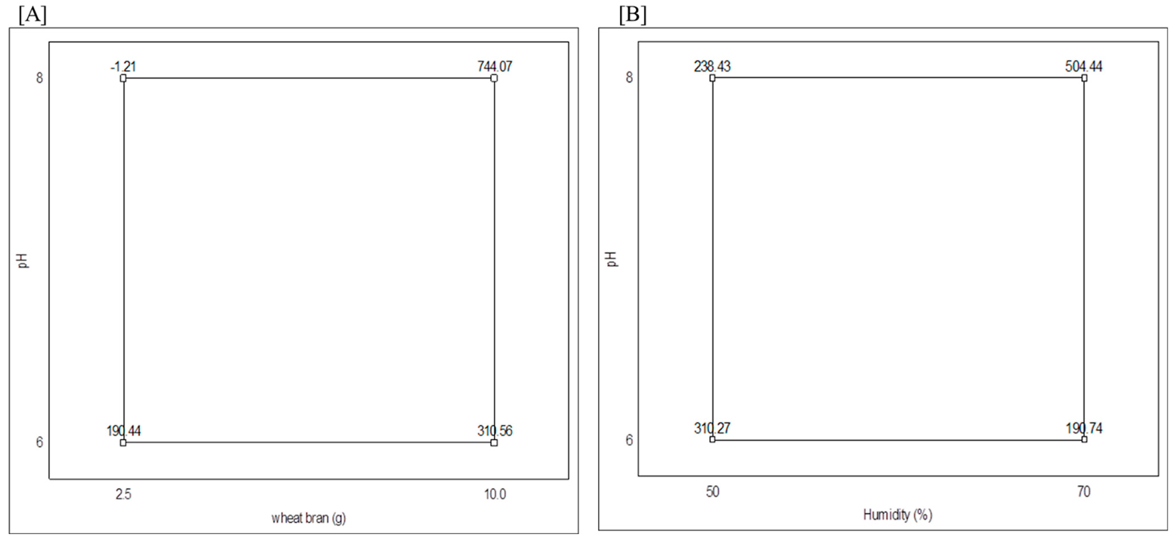
The effects of the evaluated factors on collagenase production by A. serratalhadensis URM 7866 were further explored through interaction plots, which revealed key synergistic relationships among the fermentation parameters. Specifically, the interaction between substrate concentration and pH (Figure 2A) demonstrated that maximal collagenolytic activity (744.07 U/mg) was only achieved when a high wheat bran concentration (10 g) was combined with an alkaline pH (8.0). In contrast, lower or neutral pH levels failed to support enzyme production even at elevated substrate levels, underscoring the dependence of enzyme induction on both nutrient availability and the physicochemical environment. Similarly, the interaction between moisture and pH (Figure 2B) indicated that enzyme production was significantly enhanced (504.44 U/mg) only when high moisture content (70%) was combined with alkaline pH conditions (pH 8.0). Moisture alone, without an optimal pH, did not significantly increase enzymatic activity, suggesting that adequate water availability supports fungal growth and metabolism by facilitating nutrient solubility, oxygen diffusion, and thermal stability, which are crucial for enzyme biosynthesis [18]. These interaction effects emphasize that collagenase biosynthesis in this system is not driven by isolated variables but rather by a finely tuned combination of factors that modulate metabolic pathways conducive to enzyme expression.
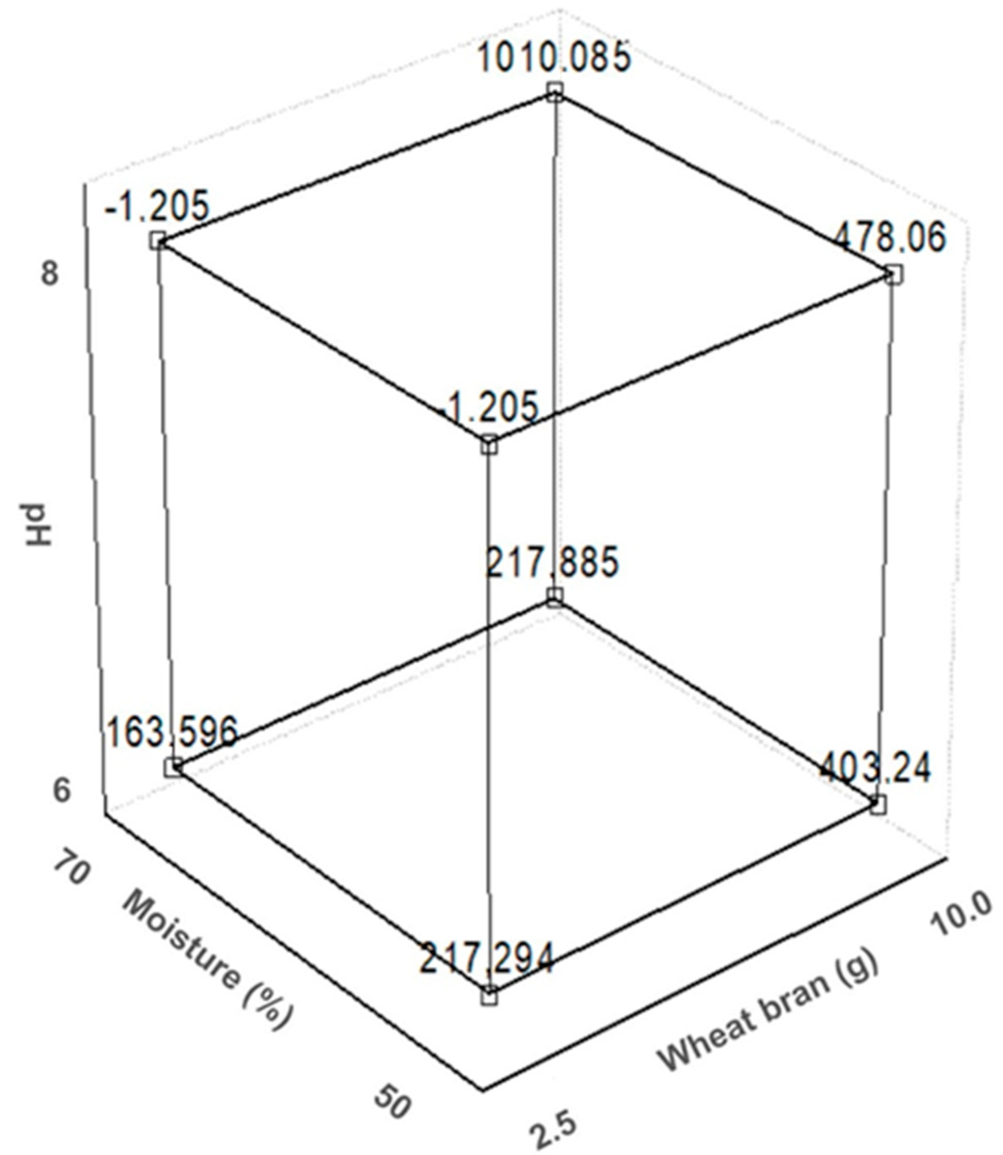
The three-dimensional surface plot (Figure 3) illustrates these synergistic interactions more clearly, confirming that optimal collagenolytic activity is observed only within a narrow combination of high substrate load, alkaline pH, and elevated moisture. Outside this region, enzymatic activity drops drastically, which may be attributed to tight physiological regulation of fungal metabolism and enzyme expression, as well as to the sensitivity of solid-state fermentation systems to environmental conditions [19]. These findings highlight the complexity of optimizing the SSF processes, where multiple factors must be carefully balanced to achieve maximum enzyme production.
The biotechnological potential of filamentous fungi for the production of extracellular proteases, particularly collagenases, has attracted increasing interest due to their relevance in industrial and biomedical applications [19]. In this context, the results obtained for A. serratalhadensis URM 7866 reinforce this potential, especially under optimized conditions of SSF. Similar findings have been reported in the literature, for instance, de Souza et al. [20] demonstrated that Rhizopus microsporus UCP 1296 produced collagenase with an activity of 872 U/mg. Similarly, Silva [21] successfully obtained a collagenolytic enzyme from A. tamarii using SSF, employing a strain also isolated from the Caatinga biome. These reports support the hypothesis that both the ecological origin of the fungal isolate and the assessment of fermentation parameters are decisive factors for maximizing the expression of collagenolytic activity, reinforcing the strategic value of bioprospecting in underexplored biomes such as the Caatinga. Moreover, the observed optimal production of collagenase by A. serratalhadensis suggests that collagenase expression in filamentous fungi may be regulated by similar pH-responsive transcriptional mechanisms. Understanding these regulatory pathways may provide opportunities for genetic or metabolic engineering to further enhance collagenase yields.
The influence of fermentation parameters on collagenase production was further elucidated (Figure 2 and Figure 3), which revealed that the highest enzymatic activity occurred under combined conditions of alkaline pH, elevated moisture (70%), and high wheat bran concentration (10 g). These findings suggest a synergistic effect among the variables, with a clear peak of collagenolytic activity in specific regions of the experimental matrix. Among the parameters evaluated, wheat bran concentration at 10 g proved to be the most favorable for enzyme production, while 70% moisture appeared to maintain conditions conducive to fungal growth and metabolism. Although moisture content does not act directly on enzyme synthesis, it influences the physical state of the fermentation medium, potentially stabilizing temperature and enhancing oxygen diffusion during SSF.
The synergistic interaction among alkaline pH, high moisture, and elevated wheat bran concentration points to a complex physiological regulation of collagenase synthesis in A. serratalhadensis. The collagenolytic activity of fungal enzymes is strongly influenced by the pH conditions of the medium, with different strains exhibiting distinct performance profiles. Studies have shown that the optimal pH for these enzymes is generally within the neutral to alkaline range, varying between pH 7.0 and 9.0. This variation is related to the metabolic diversity among the producing fungi, which can result in enzymes with different structural conformations and stability in response to pH changes [21].
Substrate concentration also proved to be a critical factor. Previous studies demonstrated that an increase in substrate significantly enhanced collagenase yields [22], although excessive levels of substrate can be inhibitory. This phenomenon has been observed in Aspergillus niger, where high cellobiose concentrations negatively affect enzyme productivity [23]. The use of 10 g of wheat bran in our study likely struck a balance between nutrient availability and fungal metabolic demand, resulting in the highest enzymatic output.
Moisture content, while not directly regulating enzyme expression, plays a crucial role in shaping the microenvironment of SSF. Optimal collagenase production has been previously reported at temperatures between 24 °C and 37 °C [21], which are strongly influenced by moisture levels. The observed optimal activity at 70% moisture in this study supports the idea that adequate hydration maintains conditions favorable to fungal physiology and enzyme secretion.
Altogether, these findings underscore the necessity of a multifactorial optimization strategy tailored to the specific fungal strain and fermentation setup. They also highlight the industrial potential of A. serratalhadensis URM 7866 as a source of collagenolytic enzymes, particularly under precisely modulated physicochemical conditions.

3.2. Collagenase Purification

The enzyme purification process involved multiple steps. Initially, the crude extract was pre-purified by acetone precipitation, which resulted in an 18-fold increase in enzymatic activity compared to the crude extract, as shown in Table 2.
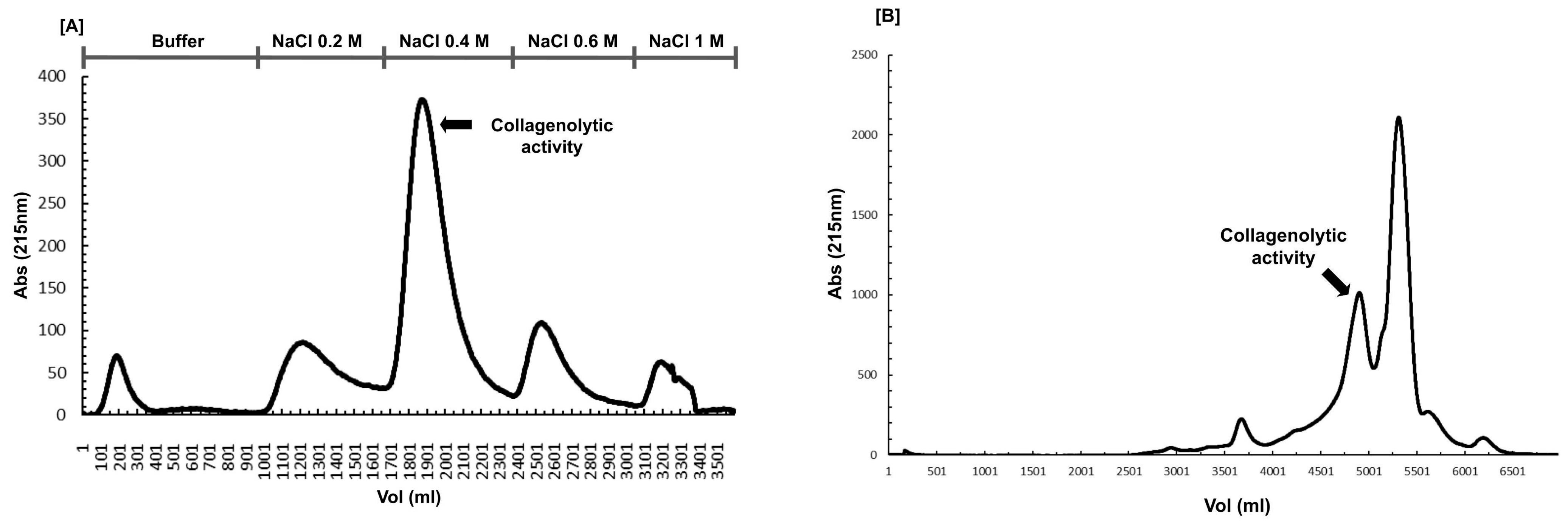
Following the initial extraction, a two-step chromatographic purification strategy was employed to isolate the collagenase produced by A. serratalhadensis URM 7866. The first step involved ion-exchange chromatography using an ANX FF column, pre-equilibrated with a 20 mM Tris-HCl buffer at pH 8.0. Elution was performed through a linear gradient of NaCl at concentrations of 0.2 M, 0.4 M, 0.6 M, and 1 M, reaching a final concentration of 1 M. Fractions were collected and monitored for collagenolytic activity.
Subsequently, a second purification step was carried out by size-exclusion chromatography using a Sephadex G-200 column, also equilibrated with 20 mM Tris-HCl buffer (pH 8.0), aiming to further enhance the purity of the target enzyme. The chromatographic profiles of both purification steps are depicted in Figure 4, where the peak corresponding to collagenolytic activity is highlighted and was selected for subsequent analysis.
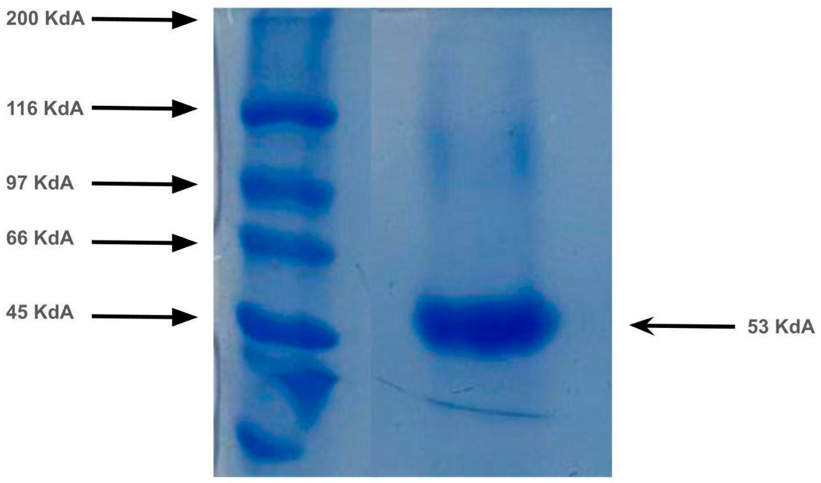
After this purification scheme, the collagenase exhibited a specific activity of 1021.86 U/mg, representing an approximate 20-fold increase compared to the crude extract. SDS-PAGE analysis of the purified fraction revealed a distinct protein band, indicative of a high degree of purity (Figure 5). These results demonstrate the efficiency of the combined ion-exchange and size-exclusion chromatographic approach in obtaining a highly purified collagenolytic enzyme suitable for downstream biochemical characterization.
Additionally, gel chromatography analysis allowed for the estimation of the collagenase’s molecular weight at approximately 53 kDa. This value falls within the typical range reported for fungal collagenases, which generally vary between 30 and 70 kDa. This range is significant for understanding their biochemical properties and potential applications across various fields, including pharmaceuticals and food processing. Specific molecular weights reported in the literature further highlight the diversity of these enzymes among fungal species. For example, a recent study reported a collagenase from Staphylococcus aureus with a molecular weight of 36 kDa [24], while other sources indicate fungal collagenases can reach up to 70 kDa, emphasizing interspecies variability [25].
This improvement aligns with the expected outcome of removing non-target contaminants and potential inhibitors and is consistent with previous studies, such as Novelli et al. [26], who reported protease activities of up to 40 U/mL after purifying enzymes secreted by Aspergillus species and Penicillium roquefortii. Therefore, this multi-step chromatographic approach proved effective for collagenase purification.

3.3. Enzyme Characterization

3.3.1. Effects of Temperature and pH on Enzymatic Activity and Stability

The three-dimensional structure of enzymes, like that of any protein, is sensitive to variations in pH and temperature [27]. After the purification process, the collagenase-containing fraction was subjected to temperature variations to determine the optimal temperature and thermal stability. The enzyme exhibited maximum activity at 50 °C (Figure 6A) and remained stable within temperature range of 20 °C to 40 °C. Beyond this point, a sharp decrease in activity was observed, with the enzyme losing more than 50% of its activity at higher temperatures (Figure 6B), highlighting a shared thermotolerance pattern among Aspergillus-derived proteases.
When compared with data from the literature, the collagenase produced by A. serratalhadensis shows similar optimal temperature values to those reported for other collagenase-producing microorganisms, such as Aspergillus tamarii URM4634, which exhibited an optimal temperature of 50 °C. The enzyme from A. tamarii also showed similar thermal stability, remaining stable between 20 °C and 40 °C [21]. Another study with similar results using a microorganism from the same genus, Aspergillus sp. (UCP 1276), reported an optimal temperature of 40 °C and stability ranging from 25 °C to 60 °C [27]. Similar findings were also observed in the study by Costa et al. [28], in which Streptomyces antibioticus UFPEDA 3421 demonstrated an optimal temperature of 60 °C, with thermal stability between 30 °C and 50 °C, and a strong denaturation effect when the temperature exceeded 55 °C.
The purified enzyme exhibited optimal activity at pH 8.0 (Figure 7A) and remained stable within the pH range of 7 to 9, this stability in alkaline environments is particularly advantageous for industrial applications, such as in detergent formulations or leather processing [28]. When exposed to pH values above 9, the enzyme retained less than 70% of its original activity, as shown in Figure 7B.
Despite subtle variations, the enzyme maintained effective catalytic performance under neutral to alkaline conditions, indicating that the collagenase produced by A. serratalhadensis URM 7866 is more active in mildly alkaline environments. Similar results were reported by Silva et al. [13], who observed that Aspergillus tamarii URM4634 strains produced collagenases with optimal activity at pH 9.0. In the study [28] demonstrated that collagenases from Streptomyces antibioticus UFPEDA 3421 exhibited maximum activity at pH 7.0, with stability between pH 6 and 10. It can be observed that most enzymes have their stability range very close to their optimal activity, and when these conditions are extrapolated or interpolated beyond this critical range, a reduction in enzymatic activity occurs. Overall, the collagenase from A. serratalhadensis shares structural and catalytic resilience observed in other microbial collagenases, supporting its biotechnological relevance.

3.3.2. Effect of Inhibitors and Surfactants on Collagenolytic Activity

To better understand the biochemical characteristics of the collagenase from A. serratalhadensis URM 7866, its sensitivity to various protease inhibitors and surfactants was evaluated. The results are summarized in Table 3.
The enzyme was strongly inhibited by PMSF, a well-known serine protease inhibitor, indicating that a serine residue is likely involved in the active site, classifying it as a serine-type collagenase. In contrast, iodoacetic acid and pepstatin A had minimal effects on enzymatic activity, suggesting that cysteine and aspartic residues do not directly participate in catalysis. EDTA caused moderate inhibition (~34%), indicating that divalent metal ions may play a secondary structural or catalytic role. These results are consistent with findings reported for other serine proteases, such as those produced by Aspergillus terreus [13,22].
Regarding surfactants, the collagenase retained high activity across all tested conditions, including in the presence of anionic (SDS) and non-ionic (Tween 20, Tween 80, Triton X-100) surfactants at both 1% and 10% concentrations. This is a valuable property for potential applications in detergent formulations, as SDS can interact with calcium and magnesium ions in hard water, improving enzyme effectiveness [29]. The enzyme also maintained over 85% of its original activity in the presence of non-ionic surfactants, demonstrating good compatibility and stability. These findings suggest that the A. serratalhadensis URM 7866 collagenase is not only a serine protease but also a promising candidate for biotechnological applications that require tolerance to surfactants and formulation additives.

3.3.3. Effect of Metal Ions on Collagenolytic Activity

Metal ions were tested at concentrations of 5 and 10 mM to assess their influence on the collagenolytic activity of the purified enzyme. Sodium, calcium, and potassium chloride exhibited a positive effect, enhancing enzymatic activity by 34.1%, 18%, and 4%, respectively. In contrast, barium, magnesium, copper, and iron ions reduced enzyme activity by more than 20%, as shown in Table 4.
The positive influence of calcium ions is well documented for serine proteases, as they help stabilize enzyme conformation and enhance thermal resistance [30,31]. In a study by Sattar, Aman, and Qader [30], which investigated the effect of metal ions, solvents, and surfactants on the activity of protease produced by Aspergillus niger KIBGE-IB36, it was observed that iron ions exerted a significant inhibitory effect on enzymatic activity. Similarly, calcium ions were able to enhance protease activity, while other ions, such as magnesium, caused a reduction in activity. These results corroborate the findings of the present study, in which iron demonstrated a strong inhibitory effect on the Aspergillus-derived collagenase, whereas calcium promoted an increase in enzymatic activity, highlighting the importance of these ions in modulating fungal enzyme function.
Additionally, zinc ions caused a substantial inhibitory effect, reducing enzyme activity by up to 80%. This aligns with previous findings reporting the inhibition of serine proteases from Aspergillus sp. by zinc [27]. Although metalloproteases generally require zinc ions for optimal activity and stability, the inhibitory effect observed in this study suggests that the purified collagenase is not a metalloenzyme. Instead, its behavior supports its classification as a serine protease, particularly due to the activation observed in the presence of calcium ions [26,28].
Furthermore, it was found that the presence of the zinc ion resulted in a significant decrease in enzymatic activity, leading to an 80% loss of its activity. This same ion also exerted inhibitory effects on the serine protease present in Aspergillus sp. [27]. Typically, metalloproteinases require the zinc ion specifically to achieve optimal activity and ideal stability. However, it was observed that the purified collagenase had its activity inhibited by the presence of zinc. These findings suggest that the purified collagenase belongs to the serine protease family and is activated in the presence of calcium ions [26,28].

3.4. Fourier Transform Infrared Spectroscopy (FTIR) Analysis

The FTIR spectrum of the purified collagenase is shown in Figure 8. A sharp band is observed at 3181 cm−1, corresponding to N–H stretching vibrations in resonance, and at 2947 cm−1, corresponding to C–H stretching vibrations. Stretching-type deformations are present at 1628 cm−1, attributed to the amide I band, resulting from C=O stretching. Additionally, a peak at 1549 cm−1 corresponds to the amide II band, associated with N–H bending vibrations [32,33]. It is worth noting that these absorbance bands fall within the typical range for proteins. It is worth highlighting the presence of a sharp band observed at 1034 cm-1, characteristic of aromatic compounds, suggesting that aromatic amino acids such as phenylalanine, tryptophan, and tyrosine may be part of the enzyme structure.
Similar spectral features were reported by Jamal et al. [32], in which the collagenase from Clostridium sp. displayed characteristic amide I and amide II bands at 1645 cm−1 and 1554 cm−1, respectively. The authors also highlighted N–H stretching in resonance and C–H vibrations with peaks at 3263 cm−1 and 2947 cm−1, respectively. In a study by Vijay and Kannan [34], the FTIR analysis revealed characteristic bands for amide I at 1635 cm−1, N–H stretching in resonance at 3265 cm−1, and C–H vibrations at 2919 cm−1.

4. Conclusions

The present study successfully demonstrated the production, purification, and biochemical characterization of a collagenase enzyme derived from A. serratalhadensis URM 7866, a filamentous fungus isolated from the Brazilian semi-arid region. The multi-step purification process resulted in a highly active and stable enzyme, with optimal activity at 50 °C and pH 7.0, and remarkable stability across a broad range of temperatures and pH values. The enzymatic activity was strongly inhibited by PMSF, indicating that this collagenase likely belongs to the serine protease family. Furthermore, the enzyme maintained its activity in the presence of various surfactants and demonstrated enhanced performance with calcium ions, highlighting its potential for industrial applications, particularly in detergent formulations. FTIR analysis confirmed the proteinaceous nature of the enzyme and revealed typical structural bands associated with functional proteases. Overall, these findings reinforce the biotechnological relevance of A. serratalhadensis URM 7866 as a novel microbial source of thermostable, alkaline collagenase and open avenues for its application in pharmaceutical, cosmetic, and environmental industries.

Author Contributions

Conceptualization, L.H.S.L., C.d.A.L.D., R.M.P.B.C. and D.d.A.V.M.; methodology, L.H.S.L., K.B.B.C. and R.L.A.S.; software, D.d.A.V.M., T.P.N. and M.N.C.d.C.; validation, C.d.A.L.D., R.M.P.B.C., D.d.A.V.M. and A.L.F.P.; formal analysis, L.H.S.L., P.G.O.C.d.S., M.E.L.C.d.M. and D.C.d.S.M.; investigation, L.H.S.L., P.G.O.C.d.S., M.E.L.C.d.M. and D.C.d.S.M.; resources, C.d.A.L.D., R.M.P.B.C., D.d.A.V.M. and A.L.F.P.; data curation, L.H.S.L., K.B.B.C. and R.L.A.S.; writing—original draft preparation, L.H.S.L., K.B.B.C., R.L.A.S., P.G.O.C.d.S. and M.E.L.C.d.M.; writing—review and editing, L.H.S.L., K.B.B.C., R.L.A.S., P.G.O.C.d.S., M.E.L.C.d.M. and D.d.A.V.M.; visualization, L.H.S.L., K.B.B.C., R.L.A.S., P.G.O.C.d.S. and M.E.L.C.d.M.; supervision, C.d.A.L.D., R.M.P.B.C., C.M.d.S.M., D.d.A.V.M. and A.L.F.P.; project administration, C.d.A.L.D., R.M.P.B.C., C.M.d.S.M., D.d.A.V.M. and A.L.F.P.; funding acquisition, C.d.A.L.D., R.M.P.B.C., C.M.d.S.M., D.d.A.V.M. and A.L.F.P. All authors have read and agreed to the published version of the manuscript.

Funding

This work was funded by CAPES (doctoral scholarship), CNPq, and FACEPE, Brazilian funding institutions, to which we are grateful for the financial support that made this research possible.

Institutional Review Board Statement

Not applicable.

Informed Consent Statement

Not applicable.

Data Availability Statement

The original contributions presented in this study are included in the article. Further inquiries can be directed to the corresponding author.

Acknowledgments

This work was funded by CAPES, CNPq (doctoral scholarship-process number: 88887.646167/2021-00, and FACEPE, Brazilian institutions to which we are grateful for the financial support that made this research possible.

Conflicts of Interest

The authors declare no conflicts of interest.

References

  1. Arasu, V.M.; Choi, K.C.; Sathya, I.; Soundharrajan, I.; Rejiniemon, T.S.; Ponnuswamy, V.R.; Balamuralikrishnan, A.; Pri-yanka, A. Perspectives of microbial enzyme biocatalyst in food industries. In Microbial Oxidative Enzymes: Biotechnological Applications; De Gruyter: Berlin, Germany, 2023; pp. 257–279. [Google Scholar] [CrossRef]
  2. Peng, S.; Xue, Z.; Wang, S.; Xu, W.; Wang, F.; Fu, R.; Wei, F. Microbial proteases and their applications. Front. Microbiol. 2023, 14, 1236368–1236382. [Google Scholar] [CrossRef] [PubMed]
  3. Arya, P.; Jani, S.A.; Rajput, K.N.; Raval, V.H. Thermostable alkaline proteases from bacteria: A review. SSRN Electron. J. 2020. [Google Scholar] [CrossRef]
  4. Mokrani, S.; Nabti, E.H. Recent status in production, biotechnological applications, commercial aspects, and future prospects of microbial enzymes: A comprehensive review. Int. J. Agric. Sci. Food Technol. 2024, 10, 6–20. [Google Scholar] [CrossRef]
  5. Blieva, R.K.; Kalieva, A.K.; Suleimenova, Z.B.; Zhakipbekova, A.S.; Tapenbayeva, I.E. Screening of Aspergillus fungi for extracellular protease and collagenase production. Rep. Natl. Acad. Sci. Kazakhst. 2020, 2, 36–40. [Google Scholar] [CrossRef]
  6. Barbosa, R.N.; Bezerra, J.D.P.; Santos, A.C.S.; Melo, R.F.R.; Houbraken, J.; Oliveira, N.T.; Souza-Motta, C.M. Brazilian tropical dry forest (Caatinga) in the spotlight: An overview of species of Aspergillus, Penicillium and Talaromyces (Eurotiales) and the description of P. vascosobrinhous sp. nov. Acta Bot. Bras. 2020, 34, 409–429. [Google Scholar] [CrossRef]
  7. Bernal, S.P.F.; Gritti, M.A.; Santos, V.P.D.; Ottoni, J.R.; de Oliveira, V.M.; Peichoto, M.E.; Passarini, M.R.Z. Pharmaceutical biotechnological potential of filamentous fungi isolated from textile industry. Arch. Microbiol. 2021, 203, 3933–3944. [Google Scholar] [CrossRef]
  8. Da Rocha, W.R.V.; Agamez-Montalvo, G.S.; Feitosa, V.A.; Machado, S.E.F.; De Souza Lima, G.M.; Pessoa, A., Jr.; Alves, H.S. Screening and optimizing fermentation production of L-asparaginase by Aspergillus terreus strain S-18 isolated from the Brazilian Caatinga Biome. J. Appl. Microbiol. 2019, 126, 1426–1437. [Google Scholar] [CrossRef]
  9. Barzkar, N.; Babich, O.; Sukhikh, S.; Venmathi Maran, B.A.; Tamadoni Jahromi, S.; Luwor, R.B.; Sorsa, T.; Das, R. Exploring the sources and potential applications of marine collagenases. Biocatal. Agric. Biotechnol. 2024, 58, 103150. [Google Scholar] [CrossRef]
  10. Wang, Z.-Z.; Wang, K.; Xu, L.-F.; Su, C.; Gong, J.-S.; Shi, J.-S.; Ma, X.-D.; Xie, N.; Qian, J.-Y. Unlocking the potential of collagenases: Structures, functions, and emerging therapeutic horizons. BioDesign Res. 2024, 6, 0050. [Google Scholar] [CrossRef]
  11. Gao, B.; Tan, C.; Roshani, D.; Yang, R.; Lv, Z.; Li, P.; Shang, N. Microbial collagenases: An updated review on their characterization, degradation mechanisms, and current applications. Crit. Rev. Food Sci. Nutr. 2024, 1–25. [Google Scholar] [CrossRef]
  12. Luo, J.; Zhang, Z.; Zeng, Y.; Dong, Y.; Ma, L. Co-encapsulation of collagenase type I and silibinin in chondroitin sulfate coated multilayered nanoparticles for targeted treatment of liver fibrosis. Carbohydr Polym. 2021, 263, 117964. [Google Scholar] [CrossRef]
  13. Silva, R.L.; de Oliveira, C.M.; Souza-Motta, A.L.F.; Porto, T.S. Novel prote-ase from Aspergillus tamarii URM4634: Produção e caracterização utilizando substratos agroindustriais baratos por fermentação em estado sólido. Adv. Enzima Res. 2016, 4, 125–143. [Google Scholar] [CrossRef]
  14. Chavira, R.; Burnett, T.J.; Hageman, J.H. Ensaio de proteinases com Azocoll. Anal. Bioquímica. 1984, 136, 446–450. [Google Scholar] [CrossRef]
  15. Smith, P.K.; Krohn, R.I.; Hermanson, G.T.; Mallia, A.K.; Gartner, F.H.; Provenzano, M.D.; Fujimoto, E.K.; Goeke, N.M.; Olson, B.J.; Klenk, D.C. Medição de proteína usando ácido bicinconínico. Anal. Biochem. 1985, 150, 76–85. [Google Scholar] [CrossRef]
  16. Nickerson, J.L.; Baghalabadi, V.; Dang, Z.; Miller, V.A.; Little, S.L.; Doucette, A.A. Organic solvent-based protein precipitation for robust proteome purification ahead of mass spectrometry. J. Vis. Exp. 2022, 180, 63503. [Google Scholar] [CrossRef]
  17. Laemmli, U.K. Cleavage of structural proteins during the assembly of the head of bacteriophage T4. Nature 1970, 227, 680–685. [Google Scholar] [CrossRef]
  18. Kango, N.; Jana, U.K.; Choukade, R. Fungal enzymes: Sources and biotechnological applications. In Fungal Enzymes: Sources and Biotechnological Applications; Springer: Singapore, 2019; pp. 515–538. [Google Scholar] [CrossRef]
  19. Jin, G.; Boeschoten, S.; Hageman, J.; Zhu, Y.; Wijffels, R.; Rinzema, A.; Xu, Y. Identifying variables influencing traditional food solid-state fermentation by statistical modeling. Foods 2024, 13, 1317. [Google Scholar] [CrossRef]
  20. de Souza, E.P.; da Silva Araújo, T.; Ramos, D.G.; da Silva Santos, H.L.; de Correia, A.C.C.; Bezerra, R.P.; Porto, A.L.F.; Converti, A.; de Araújo Viana Marques, D.; de Albuquerque, P.B.S.; et al. Production and partial characterization of collagenase from Rhizopus microsporus UCP 1296: Cytotoxic potential and controlled release strategy. J. Polym. Environ. 2025, 1–15. [Google Scholar] [CrossRef]
  21. Silva, O.S.; de Almeida, E.M.; de Melo, A.H.F.; Porto, T.S. Purification and characterization of a novel extracellular serine-protease with collagenolytic activity from Aspergillus tamarii URM4634. Int. J. Biol. Macromol. 2018, 117, 1081–1088. [Google Scholar] [CrossRef]
  22. Mbira, C. Influence of substrate concentration on enzymatic activity in biocatalysis. J. Chem. 2024, 3, 48–58. [Google Scholar] [CrossRef]
  23. Colla, L.M.; Reinehr, C.O.; Manfredini, P.G.; Cavanhi, V.A.F.; Costa, J.A.V. Simultaneous production of proteases and antioxidant biopeptides by solid-state fermentation. Sustain. Food Technol. 2023, 1, 874–885. [Google Scholar] [CrossRef]
  24. Al Shimmery, M.A.A.; Authman, S.H.; Abo Ksou, M.F. Production, purification and determination of molecular weight of coagulase enzyme from Staphylococcus aureus isolated from clinical samples. J. Med. Genet. Clin. Biol. 2024, 1, 359–370. [Google Scholar] [CrossRef]
  25. Sinitsyna, O.A.; Volkov, P.V.; Zorov, I.N.; Rozhkova, A.M.; Emshanov, O.V.; Romanova, Y.M.; Komarova, B.S.; Novikova, N.S.; Nifantiev, N.E.; Sinitsyn, A.P. Physico-chemical properties and substrate specificity of α-(1→3)-d-glucan degrading recombinant mutanase from Trichoderma harzianum expressed in Penicillium verruculosum. Appl. Environ. Microbiol. 2025, 91, e00226-24. [Google Scholar] [CrossRef]
  26. Novelli, P.K.; Barros, M.M.; Fleuri, L.F. Novel inexpensive fungi proteases: Production by solid state fermentation and characterization. Food Chem. 2016, 198, 119–124. [Google Scholar] [CrossRef]
  27. Costa-Junior, R.B.; Brandão-Costa, R.M.P.; Albuquerque, W.W.C.; Batista, J.M.S.; Pedrosa, R.B.; Porto, A.L.F. Collagenase produced from Aspergillus sp. (UCP 1276) using chicken feather industrial residue. Biomed. Chromatogr. 2016, 31, e3863. [Google Scholar] [CrossRef]
  28. Costa, E.P.; Brandão-Costa, R.M.P.; Albuquerque, W.W.C.; Nascimento, T.P.; Conniff, A.E.S.; Cardoso, K.B.B.; Neves, A.G.D.; Batista, J.M.S.; Porto, A.L.F. Extracellular collagenase isolated from Streptomyces antibioticus UFPEDA 3421: Purification and biochemical characterization. Prep. Biochem. Biotechnol. 2023, 54, 260–271. [Google Scholar] [CrossRef]
  29. Li, Y.; Luo, F.; Peng, B.; Xu, B. Impact of typical surfactants on the collagenolytic and elastinolytic activities of proteases. J. Am. Leather Chem. Assoc. 2015, 110, 227–236. [Google Scholar]
  30. Sattar, H.; Aman, A.; Qader, S.A.U. Effect of metal ions, solvents and surfactants on the activity of protease from Aspergillus niger KIBGE-IB36. J. Basic Appl. Sci. 2017, 13, 491–495. [Google Scholar] [CrossRef]
  31. Ye, K.; Xie, T.; Wei, M.; Fan, Y.; Yu, J. Effects of Disulfide Bond and Ca2+ binding Residue on Recombinant Alkaline Protease from Aspergillus Sojae. J. Fujian Agric. For. Univ. 2019, 48, 813–818. [Google Scholar] [CrossRef]
  32. Jamal, H.S.; Raja, R.; Ahmed, S.; Yesiloz, G.; Ali, S.A. Immobilization of collagenase in inorganic hybrid nanoflowers with enhanced stability, proteolytic activity, and their anti- amyloid potential. Int. J. Biol. Macromol. 2024, 274, 133114. [Google Scholar] [CrossRef]
  33. Oh, K.-I.; Baiz, C.R. Empirical S=O stretch vibrational frequency map. J. Chem. Phys. 2019, 151, 234107. [Google Scholar] [CrossRef]
  34. Vijay, L.A.; Kannan, K. Analytical approaches in unveiling the compatibility of collagenase with additives in view of its colloidal delivery. J. Chil. Chem. Soc. 2019, 64, 4558–4564. [Google Scholar] [CrossRef]
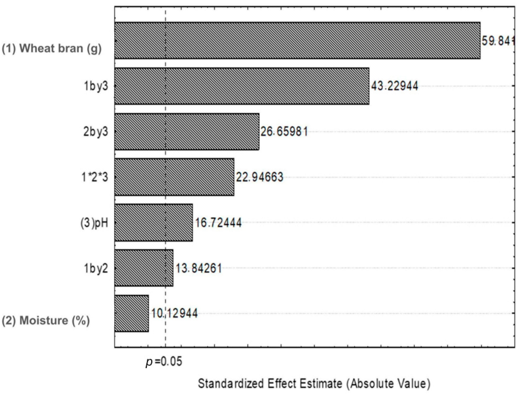
Figure 1. Pareto chart of standardized effects.
Fermentation 11 00478 g001
Figure 2. Effect of interaction between different variables on the collagenolytic activity of Aspergillus serratalhadensis URM 7866 in solid-state fermentation, where (A) shows pH and substrate concentration (wheat bran), and (B) shows pH and moisture (humidity).
Fermentation 11 00478 g002
Figure 3. Three-dimensional interaction between the independent variables pH, moisture (%), and substrate load (g of wheat bran) on the collagenolytic activity (U/mg) produced by A. serratalhadensis URM 7866 in SSF.
Fermentation 11 00478 g003
Figure 4. Resin chromatography for purification of the collagenase. (A) Ion exchange chromatography on ANXFF. (B) Molecular exclusion chromatography on superdex-G200 resin.
Fermentation 11 00478 g004
Figure 5. SDS-PAGE analysis of the purified protease band obtained through the purification methods.
Fermentation 11 00478 g005
Figure 6. Influence of temperature on the collagenolytic activity of A. serratalhadensis URM 7866. (A) optimal temperature; (B) thermal stability.
Fermentation 11 00478 g006
Figure 7. Influence of pH on the collagenolytic activity of A. serratalhadensis URM 7866. (A) optimal pH; (B) pH stability.
Fermentation 11 00478 g007
Figure 8. FTIR spectrum of collagenase produced by A. serratalhadensis URM 7866.
Fermentation 11 00478 g008
Table 1. Factorial design 23 for the production of collagenase by A. serratalhadensis URM 7866.
FactorsCoded Levels
−10+1
(1) Substrate concentration (%)2.55.010.0
(2) Moisture (%)50.060.070.0
(3) pH6.07.08.0
Table 2. Purification summary of a collagenase from A. serratalhadensis URM 7866.
StepProtein Content (mg/mL)Collagenolytic
Activity (U/mL)
Specific
Collagenolytic
Activity (U/mg)
Yield (%)Purification Factor
Crude extract3.52 ± 0.01177.96 ± 0.0950.55100.001.00
Acetone 70% precipitation0.13 ± 0.03118.88 ± 0.07914.523.6918.09
Purification in AKTA Pure0.11 ± 0.01112.40 ± 0.051021.863.1220.21
Table 3. Effect of inhibitors on the collagenolytic activity of A. serratalhadensis URM 7866.
Inhibitor (10 mM)Relative Activity (%)
Control100.00 ± 0.05
EDTA65.9 ± 0.01
PMSF18.1 ± 0.01
Pepstatin A99.9 ± 0.02
Iodoacetic Acid99.1 ± 0.02
Surfactants(1%)(10%)
SDS100.00 ± 0.09100.00 ± 0.08
Tween 8090.25 ± 0.0985.21 ± 0.07
Tween 20100.00 ± 0.191.71 ± 0.09
Triton X-10095.79 ± 0.0689.79 ± 0.10
Table 4. Effect of metal ions on the collagenolytic activity of A. serratalhadensis URM 7866.
Relative Activity (%)
Ion 5 mMIon 10 mM
Control100 ± 0.05100 ± 0.005
K103 ± 0.3098 ± 0.05
NaCl134 ± 0.06139 ± 0.02
Mg100 ± 0.09100 ± 0.10
Cu62 ± 0.0749 ± 0.06
Ca118 ± 0.02125± 0.08
Fe60 ± 0.1094 ± 0.09
Zn52 ± 0.0637 ± 0.09
Disclaimer/Publisher’s Note: The statements, opinions and data contained in all publications are solely those of the individual author(s) and contributor(s) and not of MDPI and/or the editor(s). MDPI and/or the editor(s) disclaim responsibility for any injury to people or property resulting from any ideas, methods, instructions or products referred to in the content.