Article

Immobilized Plant-Based Presumptive Probiotics as Functional Ingredients for Breakfast Cereals

by
Chrysoula Pavlatou
1,
Ioanna Prapa
1,
Electra Stylianopoulou
2,
Gregoria Mitropoulou
1,
George Skavdis
3 and
Yiannis Kourkoutas
1,*
1
Laboratory of Applied Microbiology and Biotechnology, Department of Molecular Biology and Genetics, Democritus University of Thrace, Dragana, 68100 Alexandroupolis, Greece
2
Laboratory of Developmental Biology & Molecular Neurobiology, Department of Molecular Biology Genetics, Democritus University of Thrace, 68100 Alexandroupolis, Greece
3
Laboratory of Molecular Regulation & Diagnostic Technology, Department of Molecular Biology & Genetics, Democritus University of Thrace, 68100 Alexandroupolis, Greece
*
Author to whom correspondence should be addressed.
Fermentation 2025, 11(6), 335; https://doi.org/10.3390/fermentation11060335
Submission received: 28 March 2025 / Revised: 26 May 2025 / Accepted: 29 May 2025 / Published: 10 June 2025

Abstract

Seven wild-type lactic acid bacteria, belonging to Lactiplantibacillus plantarum and Lactococcus cremoris species, were isolated from beetroots and white mushrooms and evaluated for their safety and functional profile. Lc. cremoris isolates were sensitive to all antibiotics tested, while L. plantarum strains exhibited resistance in certain antibiotics. Among them, Lc. cremoris FBMS_5810 showed the highest cholesterol removal ability (51.89%) and adhesion capacity to Caco-2 cell lines (32.14%), while all plant origin strains exhibited strong antagonistic and inhibitory activity against foodborne pathogens, as well as high survival potential during an in vitro digestion model. Subsequently, freeze-dried immobilized Lc. cremoris FBMS_5810 cells on oat flakes were prepared with initial cell loads >8.5 log CFU/g, and the effect of trehalose as a cryoprotectant in cell viability during storage at room and refrigerated temperatures for up to 180 days was studied. A significant reduction in cell loads was observed in all cases studied. However, freeze-dried immobilized Lc. cremoris FBMS_5810 cells on oat flakes prepared using trehalose as a cryoprotectant stored at 4 °C exhibited the highest cell viability (8.75 log CFU/g) after 180 days. In the next step, functional breakfast cereals enriched with freeze-dried immobilized Lc. cremoris FBMS_5810 cells on oat flakes (produced with (MLT) or without (ML) trehalose) were developed and stored at room and refrigerated temperatures for 180 days. The initial cell levels ≥ 9.18 log CFU/g were achieved, while a significant decrease was recorded during storage in all cases. The maintenance of cell loads ≥ 7.75 log CFU/g was documented in the case of both ML and MLT samples stored at 4 °C; however, the presence of trehalose in MLT samples resulted in cell viability 7.52 log CFU/g after 180 days of storage at room temperature. Importantly, the functional breakfast cereals were accepted by the panel during the sensory evaluation.

Graphical Abstract

1. Introduction

One’s diet significantly affects both the quantitative and qualitative composition of gut microbiota, highlighting the importance of consuming foods that confer benefits beyond basic nutrition needs [1]. Functional foods are defined as “natural or processed foods that contain biologically active compounds, which, in defined, effective, non-toxic amounts, provide a clinically proven and documented health benefit utilizing specific biomarkers, to promote optimal health and reduce the risk of chronic/viral diseases and manage their symptoms” [2].
Functional foods enriched with probiotics are considered a very promising area of research. Probiotics have been defined by the FAO/WHO as “microorganisms (bacteria or yeasts) which, when administered in adequate concentrations, provide health benefits to the host” [3]. For the characterization of a wild-type strain as a probiotic, properties regarding safety status, namely their susceptibility to antibiotics and hemolytic activity, their ability to survive during passage through the gastrointestinal (GI) tract, as well as capacity to adhere and colonize the intestinal tract, should be examined [4]. At the same time, several studies have highlighted the role of lactic acid bacteria in reducing cholesterol levels [5,6,7], regulating intestinal microflora, and inhibiting the growth of pathogenic microorganisms [8,9,10].
The maintenance of high cell concentrations (at least 7 log CFU/g at the time of consumption) is a necessary constraint for probiotic foods [11], which is a real bottleneck for the food industry. To fulfill this requirement, cell immobilization is considered a promising approach, as the maintenance of high cell viability has been documented [12,13]. The selection of a suitable food carrier is of great importance, as it affects the adhesion and functionality of microbial cells [14,15]. Plant-based food ingredients, such as cereals, constitute suitable matrixes for cell immobilization, resulting in the production of functional symbiotic components [16]. At the same time, dried products are considered more stable, with a longer shelf life, a low risk of contamination, and greater cost-effectiveness, as they do not necessarily require refrigerated storage conditions [17]. Τhe isolation of novel lactic acid bacteria (LAB) strains from various sources, such as beetroots and mushrooms, is essential to develop functional ingredients suited to plant-based food systems. Such strains may exhibit unique health-promoting properties and align with the growing demand of the food industry for novel beneficial microbial cultures [18]. Hence, the aim of our study was the isolation of new wild-type plant-based LAB strains with promising probiotic potential, in order to be utilized for the production of stable functional food ingredients using oat flakes as the immobilization carrier. The development of novel functional breakfast cereals enriched with the immobilized cells of a selected strain of oat flakes was followed.

2. Materials and Methods

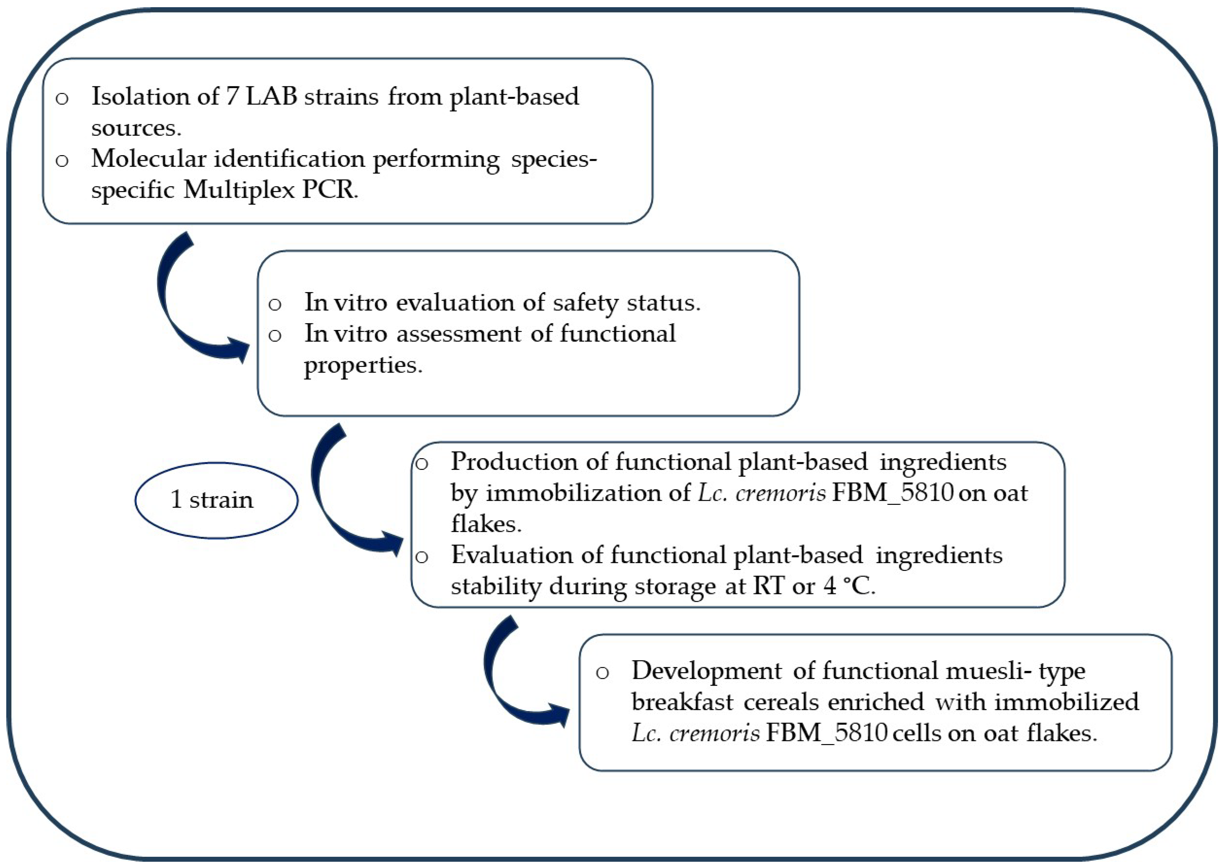
2.1. Isolation of Wild-Type LAB Strains

Fermented beetroot and white mushroom samples were collected from the local market. Ten g from each sample was macerated in 100 mL Man De Rogosa (MRS) broth (Condalab, Madrid, Spain) or M17 broth (Condalab) and incubated at 37 °C for 48 h. Thereafter, 10-fold dilutions and plate counting on MRS agar (Condalab) or M17 agar (Condalab) were carried out, followed by incubation at 37 °C under anaerobic conditions (2.5 L anaerobic jar; Sachets Merck Millipore, Darmstadt, Germany) or at 30 °C for 72 h. Separated colonies were collected, streaked to obtain pure colonies, and colony morphology and microscopic observation were performed. The isolated strains were stored at −80 °C in MRS or M17 broth: glycerol (50:50).

2.2. Molecular Identification of Isolated Strains

For the extraction of bacterial DNA, the NucleoSpin™ Tissue kit (Macherey-Nagel GmbH & Co., KG, Düren, Germany) was used, according to the manufacturer’s guidelines. For lactobacilli identification, species-specific multiplex PCR targeting Lactiplantibacillus spp. group (species Lactiplantibacillus plantarum, Lactiplantibacillus paraplantarum, and Lactiplantibacillus pentosus) [19] was performed, while in the case of lactococci, species-specific multiplex PCR targeting Lactococcus spp. species [20] was used. Each multiplex PCR reaction was carried out in a 20 μL volume and PCR products were analyzed on a 2% w/v agarose gel (Thermo Fisher Scientific, Waltham, MA, USA) stained with GelRed (Biotium, Fremont, CA, USA). The genetic locus recA was targeted for the identification of Lactiplantibacillus strains using the following primers: planF (5′-CCGTTTATGCGGAACACCTA-3′), paraF (5′-GTCACAGGCATTACGAAAAC-3′), and pentF (5′-CAGTGGCGCGGTTGATATC-3′), for L. plantarum, L. pentosus, and L. paraplantarum, respectively, and the reverse primer pREV (5′-TCGGGATTACCAAACATCAC-3′), the combination of which resulted in the following PCR products: 318 bp for L. plantarum, 218 bp for L. pentosus, and 107 bp for L. paraplantarum [19]. The primers LcLspp-F (5′-GTTGTATTAGCTAGTTGGTGAGGTAAA-3′), Lc-R (5′-GTTGAGCCACTGCCTTTTAC-3′), LcCr-F (5′-TGCTTGCACCAATTTGAAGAG-3′), and Lc-R (5′-GTTGAGCCACTGCCTTTTAC-3′) that targeted a specific region of the 16s rRNA gene were used for lactococci identification. Amplification with Lc-R and LcLspp-F yielded a product of 387 bp with genomic DNA templates from both Lactococcus lactis subsp. cremoris and Lactococcus lactis subsp. lactis. However, amplification with Lc-R and LcCr-F yielded an amplification product of 551 bp only with L. lactis subsp. cremoris genomic DNA as a template [20].

2.3. Bacterial Growth Conditions

Lactobacilli strains including the reference strain Lactiplantibacillus plantarum DSM 20174, were cultured in MRS broth at 37 °C for 24 h. Likewise, Lactococcus strains, including the reference Lactococcus subsp. lactis EFSA 20125 strain, were grown in M17 broth at 30 °C for 24 h.
Salmonella enterica subsp. enterica ser. Enteritidis PT4, Listeria monocytogenes NCTC 10527 serotype 4b, Escherichia coli ATCC 25922, and Clostridioides difficile (clinical isolate), were grown in Brain Heart Infusion (BHI) broth (Condalab) at 37 °C for 24 h, except C. difficile, which was incubated at 37 °C for 48 h.

2.4. In Vitro Safety Evaluation

2.4.1. Hemolytic Activity

The hemolytic activity of the isolates was assessed by streaking of cultures on blood agar plates containing 5% (w/v) sheep blood and incubating at 37 (for lactobacilli) or 30 °C (for lactococci) for 48 h. The appearance of greenish (α-hemolysis), clear (β-hemolysis), or no surrounding zones (γ-hemolysis) was documented.

2.4.2. Antibiotic Susceptibility Test

The antibiotic susceptibility test was performed using the broth microdilution method, according to the standard procedure of the International Organization for Standardization (ISO 10932/IDF 223:2010) [21], as described recently [22]. The minimum inhibitory concentration (MIC) was determined according to the cut-off values recommended by the European Food Safety Authority (EFSA) for each species.

2.5. In Vitro Assessment of Functional Properties Linked with Probiotic Potential

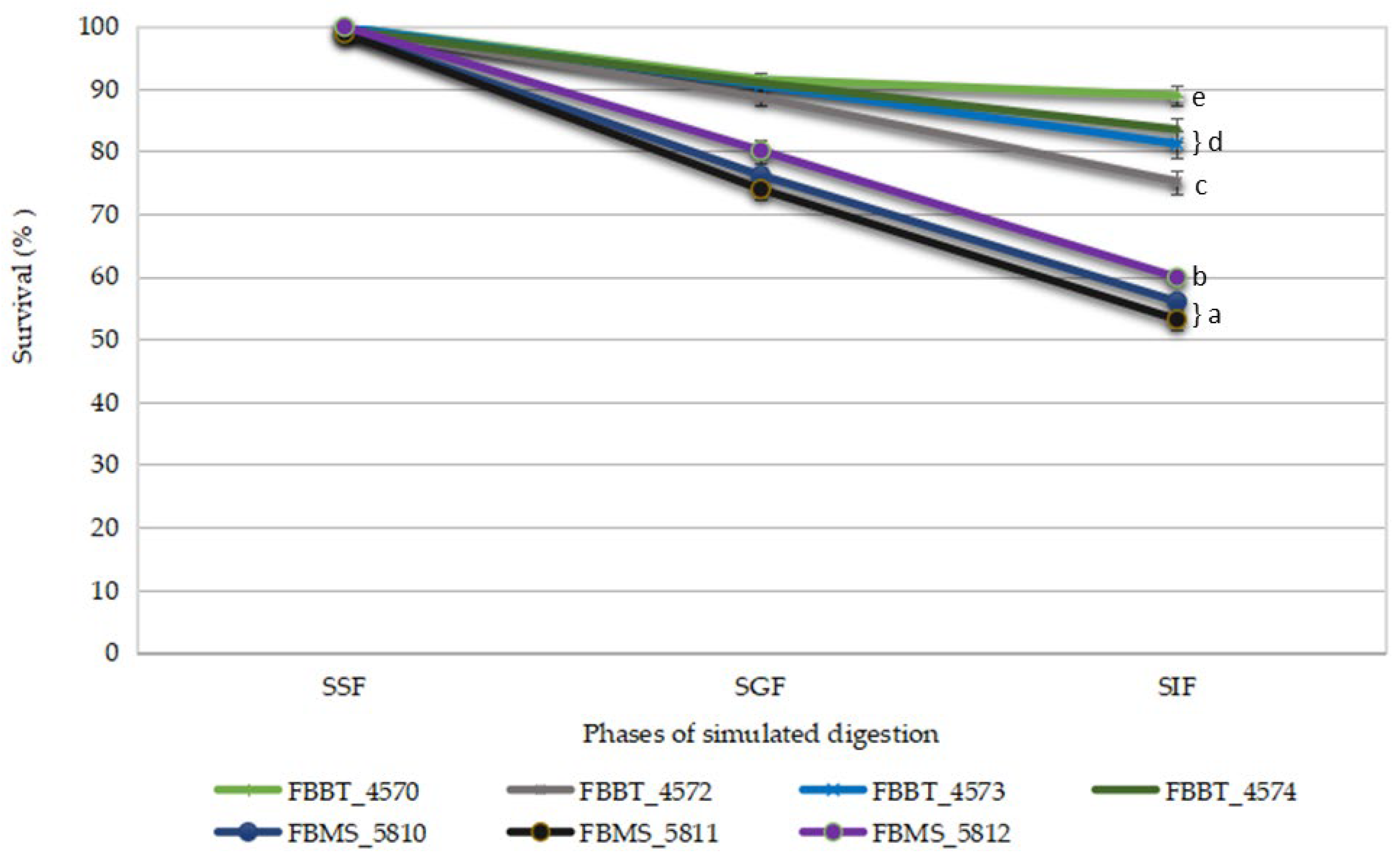
2.5.1. Ability to Survive GI Tract Conditions

The survival of the plant-origin LAB strains under GI-simulating conditions was evaluated in a static in vitro digestion model described by Nelios et al. (2022), which included 3 solutions that mimic the oral, gastric, and intestinal environment [23]. At the beginning of the simulated digestion, as well as at the end of all simulated digestion phases, the samples were collected in order to determine the cell viability via 10-fold serial dilution and plate counting in MRS (for lactobacilli) or M17 agar (for lactococci). The results were expressed as survival rates according to the following formula:
Bacterial survival rate (%) = (logcfu a/logcfu b) × 100,
where a refers to the viable cell counts after each simulated digestion phase and b refers to the counts at the beginning of simulated digestion.

2.5.2. Adhesion Properties of Isolates

Hydrophobicity
The cell hydrophobicity of the LAB strains was evaluated based on the bacterial adhesion to hydrocarbons (BATH) test, described by Prapa et al. (2025), using xylene as an organic solvent [22]. The adhesion capacity of the tested strain was determined as the percentage decrease in the optical density of the bacterial suspension after incubation with xylene due to cell adhesion and was calculated following the formula below:
H (%) = [(A0 − A)/A0] × 100,
where A0 and A refer to the optical density before and after incubation with xylene, respectively.
Auto-Aggregation and Co-Aggregation with Common Foodborne Pathogens
To determine auto-aggregation, the method described by Prapa et al. (2025) was followed [22]. In brief, the bacterial suspensions were incubated at 37 (lactobacilli) or 30 °C (lactococci) and the absorbance at 600 nm was recorded at 0 and 5 h.
The potential of the isolates to co-aggregate with common foodborne pathogens was assessed by following the method described by Li et al. (2014) [24]. Equal volumes of cell suspensions of the isolates and pathogens were mixed and incubated at 37 (lactobacilli) or 30 °C (lactococci), and the absorbance at 600 nm was recorded at 0 and 5 h.
The auto-aggregation and co-aggregation percentages were calculated based on the following formula:
A (%) = [(A0 − A)/A0] × 100,
where A0 and A refer to the optical density before and after the incubation, respectively.
Adhesion to Differentiated Caco-2 Cells
The isolated strains were further evaluated for their adhesion capacity using the Caco-2 cell line (American Type Culture Collection) as the intestinal epithelial cell model. The assay was carried out following the method described by Lappa et al. (2024) [25]. The initial cell loads of isolates, as well as the concentration of cells attached to Caco-2 monolayers, were determined via microbiological analysis. The adhesion percentage (%) of isolates to Caco-2 monolayer was determined by calculating the ratio of the adhered isolate cell load to the initial cell load added [26].

2.5.3. Bile Salt Hydrolase (BSH) Activity

The new isolates were evaluated for their bile salt hydrolase (BSH) activity, according to the method described by Prapa et al. (2025), and the appearance of precipitation zones was assessed [22].

2.5.4. In Vitro Cholesterol Assimilation

The isolates were tested for their ability to assimilate cholesterol, as described by Prapa et al. (2025), using a water-soluble form of polyoxyethylene-cholesteryl sebacate cholesterol (PEG 600, Sigma-Aldrich, St. Louis, MO, USA) [22]. For the determination of cholesterol assimilation, a standard curve was developed based on 50, 100, 125, 166.6, 250, and 500 μg/mL cholesterol in MRS or M17 broth (R2 = 0.99). The absorbance was determined at 570 nm using a SpectraMax ABS Microplate Reader (Molecular Devices, San Jose, CA, USA) and the cholesterol removal ability of the isolates was calculated following this equation:
Cholesterol assimilation (%) = ((C0 − C1)/C0) × 100,
where C0 and C1 represent uninoculated and inoculated MRS or M17 broth–cholesterol–PEG 600, respectively.

2.5.5. Inhibitory Potential of Plant-Origin Isolates Against Foodborne Pathogens

Evaluation of Antagonistic Activity of Isolates Against Foodborne Pathogens
To evaluate the antagonistic activity of the isolates against common foodborne pathogens, a co-culture assay was folllowed as described by Prapa et al. (2025) [22]. After incubation, the cell viability of the pathogens was determined through serial dilutions and plate counting. L. monocytogenes was enumerated on Palcam ISO Agar (Condalab) supplemented with Palcam Listeria Selective Supplement (Condalab). MacConkey Agar EP/USP/ISO (Condalab) was used for E. coli and S. Enteritidis quantification. For C. difficile, cultures were plated on Tryptose Sulfite Cycloserine Agar (Condalab) supplemented with Egg yolk Tellurite (Condalab) and D-cycloserine (Condalab) under anaerobic conditions. The results were expressed as log CFU/mL.
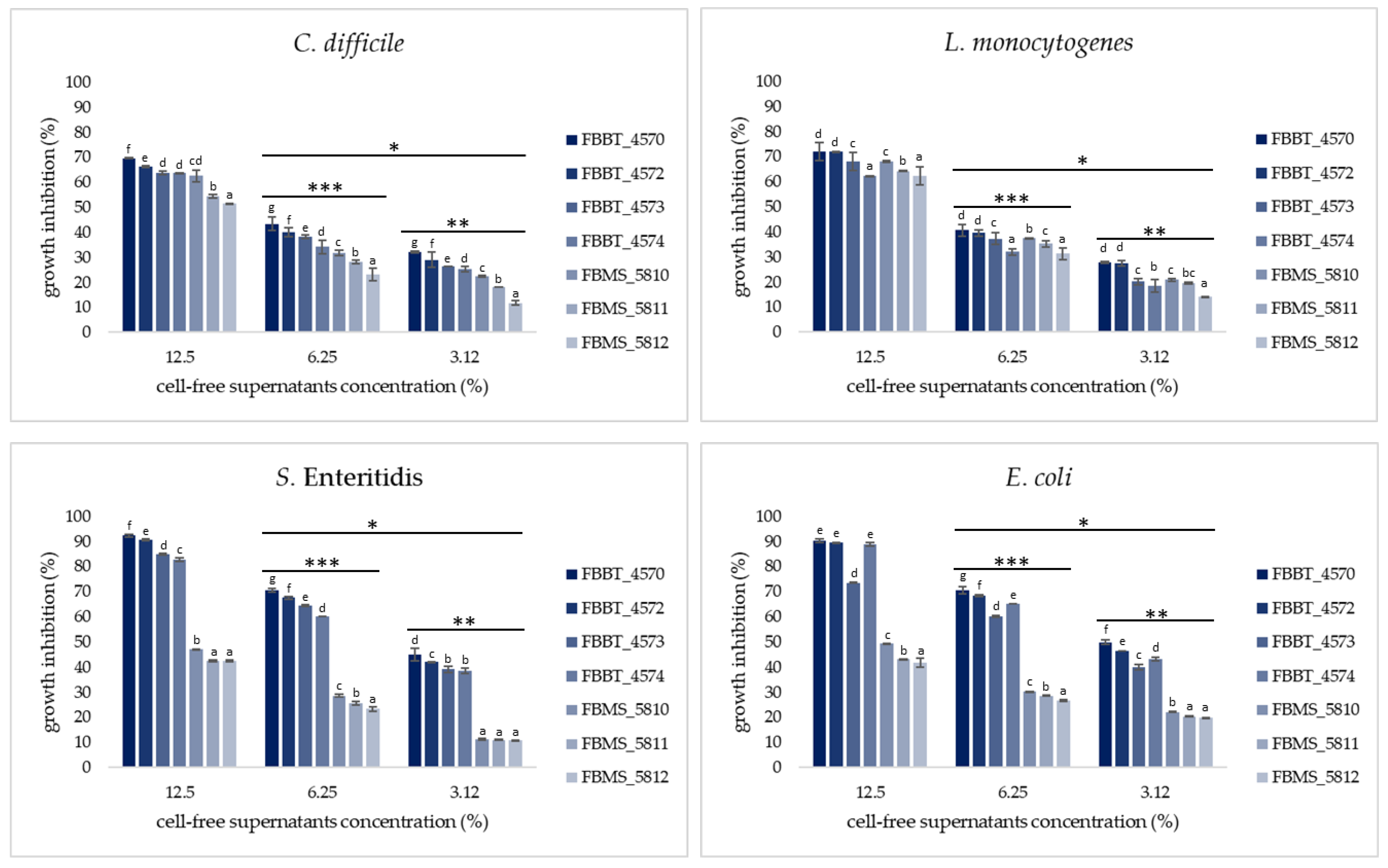
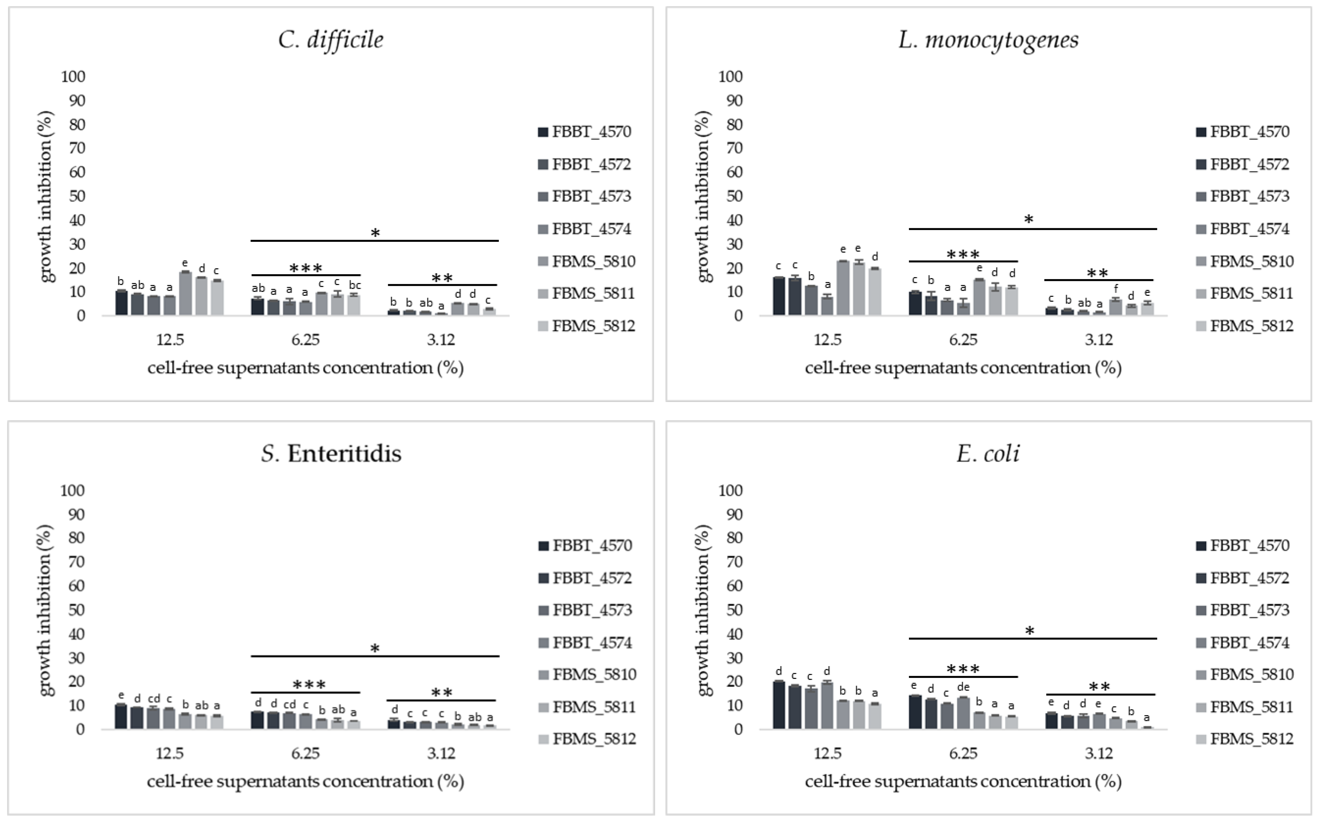
Antimicrobial Activity of CFSs of Isolates Against Common Foodborne Pathogens Using a Broth Microdilution Assay
The cell-free supernatants (CFSs) of the isolates were obtained by centrifugation (8500 rpm, 15 min, 4 °C) freshly grown cultures followed by sterilization through filtration using a 0.22 μm membrane filter (Merck) and the pH value was determined by a WTW pH 330i device (WTW, Weilheim, Germany). The assessment of the inhibitory activity of CFSs was performed following the method described by Somalou et al. (2024) with slight modifications [27]. Both non-neutralized and neutralized (after pH adjustment to 7.0 with sterile 1 M NaOH solution) CFSs were studied at various concentrations (50, 25, 12.5, 6.25, and 3.12%). Thus, 100 μL of supernatant at each concentration and 100 μL of the pathogen culture (concentration 105 cfu/mL) were added to each well. Wells with 100 μL of MRS or M17 broth inoculated with 100 μL of the pathogen culture were considered growth controls, while wells with uninoculated MRS or M17-BHI broth (1:1) were defined as blank controls. The microplates were then incubated at 37 °C for 24 h, except those inoculated with C. difficile, which were incubated at 37 °C for 48 h. Bacterial growth was measured by determining the optical density (endpoint) at 620 nm (SpectraMax ABS Microplate Reader, Molecular Devices, CA, USA), and the percentage of growth inhibition was calculated using the following formula [8]:
% I = [(OD control − OD sample)/OD control] × 100.

2.6. Preparation of Freeze-Dried Immobilized Lc. cremoris FBMS_5810 Cells on Oat Flakes as Functional Plant-Based Ingredients

Freeze-dried immobilized Lc. cremoris FBMS_5810 cells on oat flakes were prepared as previously described (Prapa et al., 2025), with slight modifications [22]. Briefly, Lc. cremoris FBMS_5810 was cultured in sterile food-grade medium (20 g/L Glucose, 25 g/L yeast extract, 2 g/L KH2PO4, 6 g/L CH3COONa, 0.3 g/L MgSO4, and 0.005 g/L MnSO4, pH 6.5) at 30 °C for 24 h. Cell biomass was collected after centrifugation (8500 rpm, 10 min, 4 °C) and rinsed with sterile ¼ Ringer’s solution (VWR International GmbH, Radnor, PA, USA). Pre-heating of oat flakes at 140 °C for 30 min was performed prior to immobilization in order to avoid contamination.
The harvested cell biomass was resuspended in sterile ¼ Ringer’s solution to the initial culture volume and oat flakes were added at 50% w/v. The mixture was allowed to rest at an ambient temperature (18–22 °C) for 30 min and, subsequently, strained and washed with sterile ¼ Ringer’s solution to remove non-immobilized (free) cells. Afterwards, the wet immobilized cells were covered with sterile 10% w/v trehalose solution (at ratio 1:1) used as a cryoprotective agent, and the mixture was incubated at an ambient temperature (18–22 °C) for 30 min. The cryoprotective solution was removed and the wet immobilized cells were transferred to −80 °C overnight. Freeze-drying in a BenchTop Pro (Virtis, SP Scientific, Warminster, PA, USA) freeze-dryer followed, as described by Prapa et al. (2025) [22]. For comparison reasons, freeze-dried immobilized cells with no cryoprotectant, as well as free Lc. cremoris FBMS_5810 cells, were also prepared.
The wet and freeze-dried immobilized or free Lc. cremoris FBMS_5810 cells were stored at room temperature (RT, 18–22 °C) or 4 °C and the cell loads were recorded at various intervals, as described below.

2.7. Production of Functional Muesli-Type Breakfast Cereals Fortified with Immobilized Lc. cremoris FBMS_5810 Cells on Oat Flakes

The muesli-type breakfast cereals were prepared according to a local recipe (Table 1) and were enriched with freeze-dried immobilized Lc. cremoris FBMS_5810 cells on oat flakes (produced with (MLT) or without (ML) trehalose). Breakfast cereals without Lc. cremoris FBMS_5810 cells were also produced (MC). Currants, goji berries, and coconut flakes were roasted at 140 °C for 5 min, while oat flakes, sunflower seeds, almonds, and chia seeds were roasted for 10 min prior to breakfast cereal production. No heat treatment was applied to the chocolate.
The samples were stored at room temperature (RT, 18–22 °C) or 4 °C in sterile containers and the cell loads were recorded at various intervals.

2.8. Analyses

2.8.1. Scanning Electron Microscopy

The immobilization of Lc. cremoris FBM_5810 cells on oat flakes was confirmed by scanning electron microscopy (SEM), according to Prapa et al. (2025) [22].

2.8.2. Physicochemical Analyses

The water activity (aw) was monitored using the HygroLab 3 (Rotronic AG, Basserdorf, Switzerland), according to the manufacturer’s guidelines.
The moisture content was determined as described in ISO:5534 [28].

2.8.3. Microbiological Analyses

The levels of freeze-dried free and immobilized cells were determined before and after rehydration. More specifically, rehydration of freeze-dried immobilized cells was achieved by immersing the cultures in sterile distilled water at a ratio of 1:1 for 30 min at an ambient temperature and then strained. In the case of free cells, the cell biomass was resuspended in sterile distilled water equal to the initial volume (before freeze-drying). For the enumeration of the immobilized cells, 5 g was homogenized with 45 mL of ¼ Ringer’s solution, while 1 g of wet or freeze-dried free cell biomass or 1 mL of dehydrated freeze-dried free cells was added to 9 mL of ¼ Ringer’s solution. Serial dilutions were performed and plating on M17 agar at 30 °C for 72 h was followed.
The enumeration of Lc. cremoris FBM_5810 cells, as well as the assessment of possible microbial contamination in muesli-type breakfast cereals was carried out, as previously described by Prapa et al. (2025) [22]. In brief, samples of 10 g were blended with 90 mL of ¼ Ringer’s solution, and serial dilutions were followed. The cell levels of Lc. cremoris FBM_5810, as well as the presence of spoilage or pathogenic microbes, were evaluated as follows:
(a) total aerobic counts on Plate Count Agar (Condalab) at 30 °C for 72 h, (b) Lactococci on M17 agar at 30 °C for 72 h, (c) staphylococci on Baird Parker Medium Base (Condalab) at 37 °C for 48 h, (d) coliforms on Violet Red Bile Agar (Condalab) at 30 °C for 24 h, (e) Enterobacteriaceae on Violet Red Bile Glucose Agar (Condalab) at 37 °C for 24 h, and (f) yeasts/fungi on Malt Agar (Condalab) at 30 °C for 72 h.
In all cases, the cell viability was expressed as log CFU/g.

2.9. Sensory Evaluation

The sensory evaluation of the functional muesli-type breakfast cereals fortified with freeze-dried immobilized Lc. cremoris FBMS_5810 cells on oat flakes were conducted in accordance with ISO 6658:2017 and ISO 8589:2007 [29,30]. In brief, the sensory analysis took place in separate rooms to minimize distractions for each panelist, with uniform and controllable lighting, as well as comfortable temperature levels. The samples, placed in plastic containers labeled with a random three-digit number, were served alongside strained yogurt (Farm Koukakis SA, Kilkis, Greece). Additionally, still water and unsalted crackers were provided to the assessors to cleanse their palates between samples. The sensory attributes (aroma, texture, taste, and overall quality) were evaluated on a 0–5 scale (0: unacceptable–5: very good). Notably, breakfast cereals without Lc. cremoris FBMS_5810 cells served as the control. The study was conducted in accordance with the Declaration of Helsinki after obtaining verbal informed consent from all participants.

2.10. Experimental Design and Statistical Analysis

The experimental design of the study is presented in a schematic flowchart in Figure 1. All experiments were performed at least in triplicate. Analysis of variance (ANOVA) was applied to the results using Statistica v.10 software (StatSoft, Inc., Tulsa, OK, USA), and Duncan’s multiple range test was used to determine significant differences (p < 0.05).

3. Results and Discussion

3.1. Molecular Identification of LAB Isolates

Seven LAB strains were isolated from beetroots and white mushrooms and were phenotypically identified as Gram-positive and catalase-negative bacteria. Subsequently, the plant origin isolates were identified at the species level using species-specific multiplex PCR of L. plantarum (strains FBBT_4570, FBBT_4572, FBBT_4573, and FBBT_4574) and Lc. cremoris (strains FBMS_5810, FBMS_5811, and FBMS_5812) (Table 2; Supplementary Figure S1).

3.2. Safety Assessment

3.2.1. Hemolytic Activity

Initially, the safety of the isolates was assessed by testing their hemolytic activity. According to the results, none of the tested strains exhibited hemolytic activity (γ-hemolysis).

3.2.2. Antibiotic Susceptibility Test

In the next step, phenotypic antibiotic resistance was investigated using the broth microdilution method, using the microbiological cut-off values for ampicillin, vancomycin, gentamicin, kanamycin, streptomycin, erythromycin, clindamycin, tetracycline, and chloramphenicol, reported in the European Food Safety Authority (EFSA) document for Lactiplantibacillus plantarum and Lactococcus spp. [31]. The results are presented in Table 3. Τhe MIC values obtained indicated that L. cremoris strains were susceptible to all antibiotics tested. On the other hand, all L. plantarum strains exhibited resistance to gentamycin and kanamycin, while L. plantarum FBBT_4570, L. plantarum FBBT_4572, and L. plantarum FBBT_4574 were resistant to erythromycin. Likewise, the isolates L. plantarum FBBT_4573 and L. plantarum FBBT_4574 showed resistance to clindamycin. Resistance to aminoglycosides, such as gentamycin, kanamycin, and streptomycin, is considered innate and is attributed to the lack of cytochrome-mediated electron transfer [22,27,32]. The safety of the newly isolated wild-type strains is the first step in evaluating their suitability in food systems. The existence of resistance genes on plasmids or transposable genetic loci is linked with a high risk of the horizontal spread of resistance genes to normal human microbial flora or potential pathogens [22,27].

3.3. In Vitro Assessment of Functional Properties Linked with Probiotic Potential

3.3.1. Ability to Survive GI Tract Conditions

The enzymes of saliva, the low pH of the stomach, and the secretion of bile salts in the small intestine significantly affect cell viability. Hence, probiotic cell survival in such conditions is an important prerequisite [33]. In this vein, the survival of the new plant-origin isolates was tested in an in vitro digestion assay that incorporated oral, gastric, and intestinal phases consecutively. According to the results (Figure 2), cell viability was not affected (p > 0.05) by exposure to simulated salivary fluid, as survival rates >98.81% were observed. However, subsequent exposure to and residence in simulated gastric fluid resulted in a significant (p < 0.05) decrease in the survival rates that ranged from 73.91 to 91.43%. In the next step, the plant-origin strains were incubated in simulated intestinal fluid, resulting in even lower (p < 0.05) survival percentages (53.12 to 88.19%). During all the simulated digestion phases studied, the highest (p < 0.05) survival rate was observed in L. plantarum FBBT_4570, while the lowest (p < 0.05) was recorded in Lc. cremoris FBMS_5811. The above results are in agreement with corresponding studies evaluating LAB strains regarding their ability to survive in conditions simulating the digestion process [4,27,34]. Indicatively, Feng et al. (2017) reported that exposure to salivary simulation solution had no effect on cell viability, as a survival rate >97% was reported [4]. However, when Lactococcus strains were exposed to a simulated gastric fl uid and, subsequently, to a simulated intestinal fluid, a significant decrease in the survival rates was observed, while L. plantarum strains showed resistance to these conditions [34]. Furthermore, Somalou et al. (2024) also reported that wild-type LAB strains, isolated from olive fruits and raisins, exhibited decreased survival rates after incubation in simulated gastric and intestinal fluids [27].

3.3.2. Adhesion Properties of Isolates

Hydrophobicity and Auto-Aggregation
Both hydrophobicity and auto-aggregation affect the ability of a bacterial strain to adhere to the intestinal epithelium [35]. Hydrophobicity indicates bacterial adhesion to hydrocarbons and serves as a key marker of intestinal adherence [24,36]. Auto-aggregation, driven by interactions between cell surface components like proteins, peptidoglycans, and lipoteichoic acid, reflects a strain’s potential to form clumps and enhance colonization. In this vein, the hydrophobicity percentage of the new plant-origin strains using xylene as a solvent, as well as their auto-aggregation ability, were evaluated (Table 4).
According to the results, the hydrophobic capacity of the isolates varied widely. The highest (p < 0.05) hydrophobicity percentage was exhibited by Lc. cremoris FBMS_5810 strain (55.41 ± 0.05%), while the lowest (p < 0.05) hydrophobicity rate (7.43 ± 0.05%) among the isolates tested was noted in the Lc. cremoris FBMS_5812 strain. Similar hydrophobicity rates were reported in other studies evaluating wild-type bacteria [24,37,38]. For example, quite high hydrophobicity rates (>60%) have been reported for strains of the species L. plantarum [39,40], while hydrophobicity rates >60% have also been reported for Lactococcus species in similar studies [24,41].
Regarding auto-aggregation percentages, high heterogeneity was also noticed among the new plant-origin isolates. More specifically, the auto-aggregation rates ranged from 16.40 to 73.50%, whereas the isolate L. plantarum FBBT_4573 exhibited the highest (p < 0.05) rate. The lowest (p < 0.05) auto-aggregation ability was observed in the Lc. cremoris FBMS_5811 strain. The above results are in line with studies aimed at evaluating strains for their ability to form aggregates [40,42,43]. In fact, Garcia-Cayuela et al. (2014) reported auto-aggregation rates >30% of some L. plantarum isolates after 6 h of incubation at 37 °C, which is similar to the results of the present study [43].
Co-Aggregation with Common Foodborne Pathogens
The ability of the probiotic strains to co-aggregate with pathogens in the GI tract is crucial for pathogen elimination, as it enables them to interfere with the colonization of pathogens by inhibiting the adherence of pathogens to receptors on the epithelial surface and release antimicrobial compounds [44,45].
In this vein, the co-aggregation ability of the new plant-origin isolates with the common foodborne pathogens C. difficile, L. monocytogenes, S. Enteritidis, and E. coli was assessed (Table 4). All strains were able to aggregate with all pathogens studied, as the co-aggregation rate ranged from 34.20 to 70.85%. The highest (p < 0.05) co-aggregation rate was recorded for the Lc. cremoris FBMS_5810 strain with C. difficile (70.85 ± 1.38%), while the lowest (p < 0.05) co-aggregation percentage was noticed after the incubation of the Lc. cremoris FBMS_5812 strain with S. Enteritidis (34.20 ± 1.98%). In agreement with the literature, the co-aggregation ability is a strain-specific trait, which also depends on the pathogen strain [46,47,48]. Furthermore, high co-aggregation rates have also been reported by Bhat et al. (2020) (37–76% for Gram-positive pathogens and 45–63% for Gram-negative pathogens) [49].
Adhesion to Differentiated Caco-2 Cell Lines
A commonly performed in vitro method to evaluate the potential of a bacterial strain to adhere and colonize the intestinal surfaces is the application of Caco-2 cell monolayers as a model surface. Thus, assessment of the adhesion potential of isolates to Caco-2 cell lines was performed and the results are presented in Table 4. The adhesion ability of the new plant- origin isolates ranged from 3.92 to 32.14%, while the highest (p < 0.05) values were observed for the Lc. cremoris FBMS_5810 strain (32.14 ± 3.64%) and the lowest (p < 0.05) for the L. plantarum FBBT_4570 strain (3.92 ± 0.12%). Similar results have been reported in studies regarding the adhesion ability of LAB strains to Caco-2 cell lines [27,50]. Indeed, Somalou et al. (2024) reported that the L. plantarum RS1 strain, isolated from raisins, exhibited a 6.83% adhesion capacity, while Shivani et al. (2024) mentioned that the L. lactis MKL8 strain, isolated from herb Murraya koenigii, showed 13.74% adherence [27,50]. It should also be noted that, according to the literature, the ability to adhere to a surface is a property that may vary among strains of the same genus [25,26,51].
3.3.3. Bile Salt Hydrolase Activity
The ability of probiotics to assimilate and reduce cholesterol levels in intestinal lumen has been linked to the secretion of the BSH enzyme. Hence, the BSH activity of the new plant-origin isolates was evaluated and the results are presented in Table 5. All strains exhibited precipitation zones, indicating their potential to secrete the BSH enzyme.
3.3.4. In Vitro Cholesterol Assimilation
High levels of cholesterol have been linked to the development of cardiovascular disease, and since cholesterol-lowering medications (e.g., statins) can cause various side effects, finding alternative ways to lower cholesterol is essential [52,53]. The ability to assimilate cholesterol is an important characteristic of probiotic microorganisms [54]. According to Table 5, the levels of cholesterol assimilation ranged from 10.22 to 51.89%. The highest (p < 0.05) percentage was observed for the Lc. cremoris FBMS_5810 strain (51.89 ± 1.00%), while the lowest (p < 0.05) cholesterol removal activity was recorded for the L. plantarum FBBT_4572 strain (10.22 ± 0.58%). Several studies have recorded similar results on the in vitro ability of Lactococcus spp. strains to reduce cholesterol levels [55,56,57]. Indeed, Shehata et al. (2019) reported >40% cholesterol removal activity by strains belonging to the genus Lactococcus [56]. Furthermore, Liu et al. (2022) mentioned that L. plantarum strains isolated from pickled Chinese cabbage, carrot, and cowpea showed a cholesterol assimilation ability ranging from 13.59 to 43.06% [58].
3.3.5. Inhibitory Potential of Plant-Origin Isolates Against Common Foodborne Pathogens
Gut dysbiosis and the colonization of intestinal mucosa by pathogens has been linked with the chronic use of antibiotics [59,60,61]. Specifically, it has been reported that long-term antibiotic use increases the risk of C. difficile infection, subsequently increasing the risk of post-infectious irritable bowel syndrome (PI- IBS) [62]. At the same time, pathogens, such as L. monocytogenes, E. coli, and S. Enteritidis, are usually considered responsible for GI infections [63]. The inhibitory activity of LAB is an outcome of their ability to produce antimicrobial compounds combined with their adhesion properties, such as auto-aggregation, co-aggregation, and adhesion to intestinal mucosa [64]. In this vein, the antagonistic activity of the new plant-origin isolates against common foodborne pathogens was evaluated by performing a co-culture assay, while the CFSs of the isolates were further assessed for their antimicrobial effect using a broth microdilution assay.
Evaluation of Antagonistic Activity of Plant-Origin Isolates Against Common Foodborne Pathogens Using a Co-Culture Assay
Co-cultivation of the new plant origin isolates with C. difficile resulted in a significant (p < 0.05) reduction in the cell viability of the pathogen (3.08–8.03 log CFU/mL) (Table 6). The final cell levels of L. monocytogenes were reduced (p < 0.05) in all strains (4.74–6.49 log CFU/mL), except from the Lc. cremoris FBMS_5811 and Lc. cremoris FBMS_5812 strains. Regarding the cell growth of S. Enteritidis and E. coli, no antagonistic potential (p > 0.05) was observed after cultivation with Lactococcus isolates, while the cell levels of S. Enteritidis and E. coli dropped significantly (p < 0.05) to 5.23–6.60 and 5.72–6.66 log CFU/mL, respectively, after incubation with L. plantarum strains. Similar results have been witnessed in other studies [23,65,66,67]. Indeed, Ratsep et al. (2014) studied the antagonistic activity of five L. plantarum strains against six C. difficile strains and observed a significant reduction in the pathogen levels of all L. plantarum strains [65], while Reuben et al. (2019) observed a reduction in the levels of S. Enteritidis and L. monocytogenes by 3 to 6 log CFU/mL in the presence of L. plantarum [66]. At the same time, Maalaoui et al. (2020) and Prapa et al. (2025) reported that strains of the genus Lactococcus exhibited no antagonistic activity against E. coli [22,67].
Antimicrobial Activity of CFSs of Plant-Origin Isolates Against Common Foodborne Pathogens Using a Broth Microdilution Assay
The inhibitory potential of the seven plant-origin isolates against common foodborne pathogens was tested by applying untreated and neutralized CFSs, and the results are presented in Figure 3 and Figure 4.
In general, the untreated CFSs (pH 3.75–5.21) strongly inhibited the growth of all foodborne pathogens by more than 90% in the case of lactobacilli and 80% in the case of lactococci at high concentrations (≥25%), while the inhibition rates dramatically (p < 0.05) decreased when the pathogens were incubated with neutralized CFSs. Moreover, the decrease in the CFS (untreated or neutralized) concentration to ≤12.5% resulted in a significant (p < 0.05) reduction in the inhibitory effect in all cases. Among the strains, L. plantarum FBBT_4570 exhibited the highest growth inhibition activity against all pathogens tested, while the incubation of pathogens with CFSs of the Lc. cremoris FBMS_5812 strain resulted in the lowest inhibition rates.
Regarding the growth inhibition of C. difficile, the untreated CFSs resulted in ≥51% growth inhibition at 12.5%, which dropped to ≥11.85% due to untreated CFSs at 3.12%. The growth inhibition of neutralized CFSs at a 12.5% concentration ranged from 8.33 to 18.41%, significantly (p < 0.05) lower than the corresponding untreated CFSs.
In the presence of untreated CFSs, the percentage of growth inhibition in L. monocytogenes ranged from 62.26 to 72.18% at 12.5% and decreased (p < 0.05) by 14.08–27.87% at 3.12%. After neutralization, the inhibitory activity of CFSs resulted in percentages ≤ 23.02% at 12.5%, while the presence of lower concentrations (≤6.25%) led to growth inhibitory percentages ≤ 15%.
The incubation of S. Enteritidis cells with untreated CFSs resulted in great heterogeneity of the growth inhibition percentages among the new isolated strains. As shown in Figure 3, the new plant-origin Lc. cremoris isolates exhibited significantly (p < 0.05) lower growth inhibitory activity in comparison to L. plantarum strains at all concentrations tested. Specifically, at 12.5%, the growth inhibition ranged from 42.51 to 47.06% and from 82.72 to 92.35% in the cases of lactococci and lactobacilli strains, respectively. Regarding the presence of neutralized CFSs, the growth inhibitory activity dramatically (p < 0.05) dropped to rates ≤ 10.50%.
Likewise, higher (p < 0.05) values of the growth inhibition percentage of E. coli were observed in untreated CFSs of L. plantarum strains (73.56–90.26% at 12.5%) than in Lc. cremoris strains (41.84–49.16% at 12.5% concentration). The growth inhibition percentages decreased (p < 0.05) after neutralization, ranging from 10.89 to 20.35% at 12.5%, regardless of the microbial genus.
All CFSs of the new plant-origin isolates exhibited a significant inhibitory capacity against the common foodborne pathogens tested. However, L. plantarum isolates showed a stronger inhibitory effect against S. Enteritidis and E. coli compared to Lc. cremoris strains. Several studies have investigated the mechanisms of the antimicrobial activity of LAB strains, attributing this property mainly to the action of organic acids, such as lactic and acetic acids, resulting in the lowering of the pH [9,22,26]. CFSs’ pH values of L. plantarum strains ranged from 3.75 to 3.89, while the corresponding pH values of the CFSs of Lc. cremoris strains were significantly (p < 0.05) higher (5.12–5.21) (Table 5). Strains belonging to the genus Lactococcus produce lower concentrations of lactic and acetic acids, while their inhibitory effect has been mainly linked with the production of antimicrobial compounds with a high molecular weight, like bacteriocins [68,69,70,71]. Gram-negative bacteria are more sensitive to the action of organic acids, as these substances can penetrate the cell membrane, resulting in the lowering of the cytoplasm pH and the inhibition of acid-sensitive enzymes [72]. Concurrently, several studies have shown that Gram-negative bacteria are more resistant to bacteriocins than Gram-positive ones. This fact could be attributed to differences in the cell membrane structure between Gram-positive and Gram-negative bacteria. More specifically, bacteriocin-like compounds bind to a bacterial cell wall as a precursor to Gram-positive bacteria, lipid II, forming, pores and resulting in the efflux of cytoplasm contents. On the other hand, the outer membrane barrier structure of Gram- negative bacteria does not allow this binding [70,73].
In general, the results presented above are in agreement with previous studies on the antimicrobial activity of LAB strains. Indeed, Arena et al. (2016) reported that LAB strains exhibited high inhibition rates against pathogenic strains of E. coli, L. monocytogenes, and S. Enteritidis (>70%), while these rates were significantly reduced when neutralized CFSs was applied [9]. Furthermore, Hor et al. (2014) mentioned that the effect of neutralized CFSs of bacteria of the genus Lactobacillus resulted in a lower inhibitory effect against the pathogen S. aureus compared to untreated CFSs [8]. Similar results are also reported by Muñoz et al. (2013), presenting the production of lactic acid and other organic acids as the cause of the antimicrobial effect against enteropathogens [74]. Also, strains of L. lactis had a low inhibitory effect against pathogenic microorganisms, such as E. coli [75].

3.4. Production of Functional Plant-Based Ingredients by Immobilization of Lc. cremoris FBMS_5810 Cells on Oat Flakes

Τo confer the health benefits, it is highly recommended that probiotics maintain high viable cell loads (at least ≥7 logcfu/g) until consumption, posing a challenge to the food industry. Cell immobilization is proposed to enhance cell viability during storage, as the food matrix may protect the cells from bacterial injury and stress [76]. In this vein, Lc. cremoris FBMS_5810 was selected for the production of functional plant-based ingredients, considering the safety and functional criteria. Hence, wet and freeze-dried, free or immobilized cells on oat flakes were produced, while the effect of trehalose used as a cryoprotective agent in order to improve cell survival during freeze-drying and storage was studied. Cell loads >8.50 log CFU/g were achieved in both wet and freeze-dried immobilized cells, while the cell immobilization of Lc. cremoris FBMS_5810 on oat flakes was confirmed via scanning electron microscope (Figure 5).

3.4.1. Effect of Storage on Cell Viability of Freeze-Dried Immobilized Lc. cremoris FBMS_5810 Cells on Oat Flakes

It is well known that the storage temperature significantly affects cell viability [77,78]. Storage at low temperatures (≤4 °C) is highly recommended for long-term preservation; however, the associated maintenance costs are significantly higher. As a result, maintaining high cell concentrations at ambient temperatures remains an industrial challenge [79]. While freeze-dried cells are more stable, freeze-drying may cause loss of viability during the process. To ensure high survival rates, cell immobilization and the use of cryoprotectants solutions, such as trehalose, are usually recommended [80]. In this vein, the effect of a 10% w/v trehalose solution was of interest, and the cell viability of the wet or freeze-dried, free or immobilized cells was monitored during storage at room and refrigerated temperatures for up to 180 days.
Storage at Room Temperature
According to Table 7, the survival rates of freeze-dried free cells dropped (p < 0.05) to <75% at 30 days of storage compared to the initial levels, while freeze-dried immobilized cells exhibited significantly (p < 0.05) higher cell viability (>91% survival). Fungi/molds were detected in wet immobilized cells after 30 days of storage. At the same time, the levels of wet free cells were <3.5 log CFU/g. Viable counts of freeze-dried free cells were only detectable up to 30 days of storage (6.54 ± 0.07 log CFU/g), while the use of trehalose as a cryoprotectant extended the viability of freeze-dried free cells by up to 90 days (5.35 ± 0.04 log CFU/g). Until the 90th day of storage, the cell levels of freeze-dried immobilized cells were monitored >7 log CFU/g, while at day 180, the viable cell loads were 4.55 ± 0.06 log CFU/g. Likewise, trehalose resulted in the maintenance of cell loads > 7.40 log CFU/g in freeze-dried immobilized cells until the 180th day, higher than the minimum recommended concentration.
Storage at Refrigerated Temperature
During storage at 4 °C, the cell viability was significantly (p < 0.05) affected by both the culture type (wet or freeze-dried, free or immobilized) and storage time (Table 8). Freeze-dried immobilized cells exhibited higher (p < 0.05) survival rates (8.65 ± 0.01 log CFU/g) compared to freeze-dried free cells (7.52 ± 0.03 log CFU/g) after 30 days of storage, while trehalose resulted in the maintenance of cell loads at similar (p > 0.05) to the initial levels (9.70 ± 0.01 log CFU/g) in freeze-dried immobilized cells. After 90 days, the cell viability of freeze-dried immobilized cells was recorded at levels 8.31 ± 0.05 log CFU/g. At the same time, freeze-dried immobilized cells produced using trehalose as a cryoprotectant exhibited cell loads of up to 9.31 ± 0.03 log CFU/g. A significant (p < 0.05) reduction was recorded in wet compared to freeze-dried cultures during storage. Specifically, the survival rates of wet free cells were 57.86 ± 0.82% on the 30th day, while after storage for 90 days, no cell viability was recorded. The cell levels of freeze-dried free cells were 5.55 ± 0.05 log CFU/g on the 90th day, whereas no viable counts were recorded after 180 days of storage. On the contrary, freeze-dried immobilized cells exhibited cell levels of 7.32 ± 0.09 log CFU/g after 180 days of storage. It is worth noting that the cell loads were 8.75 ± 0.05 log CFU/g in freeze-dried immobilized cells produced using trehalose as a cryoprotectant on the 180th day.
Indeed, Prapa et al. (2023) mentioned that freeze-dried immobilized Pediococcus acidilactici SK cells on zea flakes and pistachios maintained at levels above 7.5 log CFU/g after storage for 180 days, while, at the same time, the cell loads of freeze-dried free cells were significantly lower [13]. Cell immobilization is a promising technique for maintaining high cell levels in food, as supported by studies showing higher survival rates of immobilized cells in cheese and yogurt samples compared to free cells [20,81]. At the same time, storage at low temperatures is strongly preferred, as it is considered to prolong the survival of cells, a fact that was also confirmed in this study. Although storage at high temperatures could be a more cost-effective solution for industry, it increases the risk of lipid oxidation, the formation of free radicals and, subsequently, cell damage [13,82]. Furthermore, freeze-dried cultures exhibited higher cell survival rates than wet cultures during storage, in accordance with the literature [13,80]. However, freeze-drying is usually accompanied by an initial reduction in cell loads [83]. In this study, the use of a cryoprotective trehalose solution enhanced cell survival during freeze-drying, while it also contributed to the maintenance of high cell doses (>7.40 log CFU/g) even after storage for 180 days at room temperature. Similar results have been documented in previous studies [80,84].

3.4.2. Water Activity and Moisture Content Values of Freeze-Dried Immobilized Lc. cremoris FBMS_5810 Cells on Oat Flakes During Storage

The water activity (aw) and moisture content are considered important factors for the stability of cell cultures, as they affect the cell viability, while they are also linked with the inhibition of the growth of spoilage and pathogenic microorganisms. For dried products, water activity < 0.25 and moisture content < 10% are proposed for prolonging storage [85]. In Table 9, the effect of storage on the water activity and moisture content is presented. The freeze-drying of free and immobilized cells resulted in the lowest (p < 0.05) water activity and moisture content values during storage at both temperatures. However, an increase (p < 0.05) was observed during storage, which was affected (p < 0.05) by the storage temperature and duration. In particular, at room temperature, a greater (p < 0.05) increase the in aw and moisture content values was observed in freeze-dried cultures, which can be attributed to the tendency to balance the difference in moisture content [86].

3.5. Production of Functional Muesli-Type Breakfast Cereals Enriched with Freeze-Dried Immobilized Lc. cremoris FBMS_5810 Cells on Oat Flakes

Cereal-based products, such as granola and muesli, are considered one of the most staple, ready-to-eat foods consumed on a daily basis that are simultaneously rich in dietary fibers, vitamins, minerals, carbohydrates, and polyunsaturated fatty acids [87,88]. Hence, muesli and other cereal-based products could be used as suitable carriers for probiotics. In this vein, breakfast cereals (muesli-type products) enriched with freeze-dried immobilized Lc. cremoris FBMS_5810 cells on oat flakes immerged or not in trehalose solution prior to the freeze-drying process (MLT or ML, respectively), were prepared and stored in sterile containers at room or refrigerated temperatures for up to 180 days, and the cell viability, along with the water activity, moisture content, and sensory attributes, were evaluated. At the same time, muesli-type breakfast cereal products without Lc. cremoris FBMS_5810 cells were produced as controls (MC sample) (Supplementary Figure S2).

3.5.1. Cell Viability of Lc. cremoris FBMS_5810 Cells in Functional Muesli-Type Breakfast Cereals During Storage

The cell loads during the storage of breakfast cereals are shown in Table 10. Of note, the initial levels of Lc. cremoris FBMS_5810 cells in oat flakes were 9.18 ± 0.10 and 9.21 ± 0.01 log CFU/g for ML and MLT samples, respectively.
In general, storage at room temperature had a significant (p < 0.05) effect on cell survival in both samples, as the cell levels were reduced gradually during storage. More specifically, significantly lower cell loads were recorded (p < 0.05) after 30 days of storage in both the ML and MLT samples (8.76 ± 0.12 and 9.11 ± 0.11 log CFU/g, respectively) compared to the initial cell counts. After 90 days of storage at room temperature, the cell loads were 7.18 ± 0.04 and 8.19 ± 0.08 log CFU/g in the ML and MLT samples, while on the 180th day, the concentration only remained above the recommended levels in MLT sample cells (7.75 ± 0.11 log CFU/g).
Refrigerated temperature resulted in the maintenance of high cell loads during storage in both samples. Indeed, cell loads remained >9 log CFU/g after 30 days of storage in both ML and MLT samples. However, in the ML sample, a reduction (p < 0.05) was noted, as cell viability was recorded at 8.74 ± 0.02 log CFU/g after 60 days of storage, while 7.52 ± 0.08 log CFU/g cell loads were observed after 6 months of storage. Regarding the MLT sample, the cell viability remained stable for 60 days of storage, but after 90 days, decreased (p > 0.05) cell levels (9.09 ± 0.11 log CFU/g) were recorded. After 180 days, Lc. cremoris FBMS_5810 was monitored at levels 8.65 ± 0.07 log CFU/g in the MLT sample. Importantly, no spoilage was observed in any case.
Storage at 4 °C resulted in significantly (p < 0.05) higher cell loads compared to room temperature. Similar results have been presented by Monfared et al. (2022), where matcha-enriched muesli products fortified with encapsulated probiotics exhibited higher cell loads during storage at 4 °C for 90 days compared to storage at 25 °C [88]. Cryoprotectants, such as trehalose, sucrose, and skim milk, are usually used to ensure high cell loads [89], in accordance with our results. It is worth noting that even at room temperature, cell viability remained >7.5 log CFU/g after 180 days of storage in the MLT sample, in line with Burca-Busaga et al. (2020), who reported that the addition of 10% w/v trehalose in apple snacks enriched with Lactobacillus salvarius CECT 4063 cells resulted in higher survival rates [90].

3.5.2. Effect of Storage on Water Activity (aw) and Moisture Content in Functional Muesli-Type Breakfast Cereals

The water activity (aw) and moisture content values were also monitored during the storage of MC, ML, and MLT samples (Table 10). The initial aw values of MC, ML, and MLT samples were 0.222 ± 0.05, 0.131 ± 0.07, and 0.134 ± 0.11, respectively, while the corresponding moisture content percentages ranging 8.56 ± 0.11, 5.09 ± 0.05, and 5.16 ± 0.09%. A significant (p < 0.05) increase in both aw and moisture content was observed during storage at both temperatures. This increase could be attributed to a tendency to balance with the conditions prevailing at storage [86]. According to the literature, water activity values <0.3, as well as a moisture content <10%, are more compatible with stable dry food systems stored for long periods [91].

3.5.3. Sensory Evaluation of Functional Muesli-Type Breakfast Cereals Enriched with Freeze-Dried Immobilized Lc. cremoris FBMS_5810 Cells on Oat Flakes

The breakfast cereals were evaluated for their sensory characteristics and the results are presented in Table 10. The aroma of spices and nuts was mainly distinguished in all samples, while, regarding taste, all participants claimed that a sweet taste prevailed, especially in the MLT sample, which could be attributed to the presence of trehalose (Supplementary Table S1). According to the overall sensory evaluation scores, no significant (p > 0.05) difference was observed between the samples.

4. Conclusions

Seven wild-type LAB strains belonging to the genera Lactobacillus and Lactococcus were isolated from plant-origin sources and evaluated for their safety and functional characteristics. All lactococci new isolates were sensitive to all antibiotics tested, whereas Lc. cremoris FBMS_5810 exhibited the highest adhesion ability to Caco-2 cell lines (32.14 ± 3.64%), cholesterol removing capacity (51.89 ± 1.00%), and strong inhibitory and antagonistic activity against pathogens. In the next step, freeze-dried immobilized Lc. cremoris FBMS_5810 cells on oat flakes were prepared, resulting in cell loads >9 log CFU/g, and the effect of storage at room and refrigerated temperatures on cell viability for up to 180 days was monitored. Cell immobilization and freeze-drying, along with the addition of trehalose, produced high cell loads >8.75 log CFU/g during storage at 4 °C. At an ambient temperature, freeze-dried immobilized cells prepared using trehalose as a cryoprotectant exhibited cell loads >7.4 logcfu/g during storage for 180 days. The functional plant-baed food ingredients were then used for the production of muesli-typw breakfast cereals with high initial cell loads (>9.20 log CFU/g). During storage for 180 days at both room and refrigerated temperatures, the maintenance of cell viability at levels above 7.5 log CFU/g was achieved, while the new products were accepted during sensory evaluation.
The production of functional foods enriched with high concentrations of beneficial microorganisms presents a significant challenge for the food industry, particularly in maintaining cell viability during processing and storage. Meanwhile, consumer demand for more plant-based options continues to rise. Fortifying breakfast cereals with immobilized cells of a beneficial plant-derived strain on oat flakes offers strong potential in the growing plant-based functional food market. Future research should focus on developing alternative food systems that more closely resemble the strain’s native environment, as this may enhance cell viability. Nonetheless, the further validation of its functionality through animal models and clinical trials is essential.

Supplementary Materials

The following supporting information can be downloaded at: https://www.mdpi.com/article/10.3390/fermentation11060335/s1. Figure S1: Agarose gel electrophoresis (2% w/v) of the multiplex PCR products: (a) L. plantarum identification (318 bp) by recA primers, (b) Lc. cremoris (387 bp and 551 bp) and Lc. lactis subsp. lactis (387 bp) identification by Lc-R, LcCr-F and LcLspp-F primers; Figure S2: Muesli-type breakfast cereals produced (a) without Lc. cremoris FBMS_5810 cells (control) or (b) fortified with freeze-dried immobilized Lc. cremoris FBMS_5810 cells on oat flakes; Table S1: Sensory evaluation of functional muesli-type breakfast cereals.

Author Contributions

Conceptualization, Y.K.; Data curation, I.P., E.S., G.M., G.S. and Y.K.; Formal analysis, I.P., E.S., G.M., G.S. and Y.K.; Funding acquisition, Y.K.; Investigation, C.P. and E.S.; Methodology, C.P., I.P., E.S., G.M., G.S. and Y.K.; Project administration, Y.K.; Resources, G.M., G.S. and Y.K.; Supervision, G.S. and Y.K.; Validation, I.P., G.M., G.S. and Y.K.; Visualization, Y.K.; Writing—original draft, C.P., I.P. and E.S.; Writing—review and editing, G.M. and Y.K. All authors have read and agreed to the published version of the manuscript.

Funding

We acknowledge support of this work by the project “Infrastructure of Microbiome Applications in Food Systems-FOODBIOMES” (MIS 5047291), which is implemented under the action “Regional Excellence in R&D Infrastructures,” funded by the operational program “Competitiveness, Entrepreneurship and Innovation” (NSRF 2014–2020) and co-financed by Greece and the EU (European Regional Development Fund).

Institutional Review Board Statement

Not applicable.

Informed Consent Statement

Not applicable.

Data Availability Statement

The original contributions presented in the study are included in the article and Supplementary Materials, further inquiries can be directed to the corresponding author.

Acknowledgments

Salmonella enterica subsp. enterica ser. Enteritidis PT4 was kindly provided by G.J. Nychas (Laboratory of Microbiology and Biotechnology, Agricultural University of Athens, Athens, Greece), whereas Listeria monocytogenes NCTC 10527 serotype 4b and Clostridioides difficile clinical isolate were kindly provided by the Laboratory of Clinical Microbiology, Sismanoglio General Hospital, Athens, Greece (Gregoria Mitropoulou).

Conflicts of Interest

The authors declare no conflicts of interest.

References

  1. Laparra, J.M.; Sanz, Y. Interactions of gut microbiota with functional food components and nutraceuticals. Pharmacol. Res. 2010, 61, 219–225. [Google Scholar] [CrossRef] [PubMed]
  2. Danik, M.; Jaishree, S. A New Definition of Functional Food by FFC: What Makes a New Definition Unique? Funct. Foods Health Dis. 2015, 5, 209–223. [Google Scholar] [CrossRef]
  3. FAO; WHO. Guidelines for the Evaluation of Probiotics in Food; Food and Agriculture Organization of the United Nations/World Health Organization: London, UK, 2002; Available online: https://isappscience.org/wp-content/uploads/2019/04/probiotic_guidelines.pdf (accessed on 2 October 2024).
  4. Feng, Y.; Qiao, L.; Liu, R.; Yao, H.; Gao, C. Potential Probiotic Properties of Lactic Acid Bacteria Isolated from the Intestinal Mucosa of Healthy Piglets. Ann. Microbiol. 2017, 67, 239–253. [Google Scholar] [CrossRef]
  5. Miremadi, F.; Ayyash, M.; Sherkat, F.; Stojanovska, L. Cholesterol reduction mechanisms and fatty acid composition of cellular membranes of probiotic Lactobacilli and Bifidobacteria. J. Funct. Foods 2014, 9, 295–305. [Google Scholar] [CrossRef]
  6. Tsai, C.C.; Lin, P.P.; Hsieh, Y.M.; Zhang, Z.Y.; Wu, H.C.; Huang, C.C. Cholesterol-lowering potentials of lactic acid bacteria based on bile-salt hydrolase activity and effect of potent strains on cholesterol metabolism in vitro and in vivo. Sci. World J. 2014, 2014, 690752. [Google Scholar] [CrossRef] [PubMed] [PubMed Central]
  7. Albano, C.; Morandi, S.; Silvetti, T.; Casiraghi, M.C.; Manini, F.; Brasca, M. Lactic acid bacteria with cholesterol-lowering properties for dairy applications: In vitro and in situ activity. J. Dairy Sci. 2018, 101, 10807–10818. [Google Scholar] [CrossRef] [PubMed]
  8. Hor, Y.Y.; Liong, M.T. Use of Extracellular Extracts of Lactic Acid Bacteria and Bifidobacteria for the Inhibition of Dermatological Pathogen Staphylococcus aureus. Dermatol. Sin. 2014, 32, 141–147. [Google Scholar] [CrossRef]
  9. Arena, M.P.; Silvain, A.; Normanno, G.; Grieco, F.; Drider, D.; Spano, G.; Fiocco, D. Use of Lactobacillus plantarum Strains as a Bio-Control Strategy against Food-Borne Pathogenic Microorganisms. Front. Microbiol. 2016, 7, 464. [Google Scholar] [CrossRef] [PubMed] [PubMed Central]
  10. Chen, C.C.; Lai, C.C.; Huang, H.L.; Huang, W.Y.; Toh, H.S.; Weng, T.C.; Chuang, Y.C.; Lu, Y.C.; Tang, H.J. Antimicrobial Activity of Lactobacillus Species Against Carbapenem-Resistant Enterobacteriaceae. Front. Microbiol. 2019, 10, 789. [Google Scholar] [CrossRef] [PubMed] [PubMed Central]
  11. Nguyen, T.H.; Kim, Y.; Kim, J.S.; Kim, J.-E.; Kim, H.; Paek, N.-S.; Kang, C.-H. Evaluating the Cryoprotective Encapsulation of the Lactic Acid Bacteria in Simulated Gastrointestinal Conditions. Biotechnol. Bioprocess Eng. 2020, 25, 287–292. [Google Scholar] [CrossRef]
  12. Mitropoulou, G.; Nedovic, V.; Goyal, A.; Kourkoutas, Y. Immobilization Technologies in Probiotic Food Production. J. Nutr. Metab. 2013, 2013, 716861. [Google Scholar] [CrossRef] [PubMed]
  13. Prapa, I.; Nikolaou, A.; Panas, P.; Tassou, C.; Kourkoutas, Y. Developing Stable Freeze-Dried Functional Ingredients Containing Wild-Type Presumptive Probiotic Strains for Food Systems. Appl. Sci. 2023, 13, 630. [Google Scholar] [CrossRef]
  14. Ouwehand, A.C.; Salminen, S. In vitro adhesion Assays for Probiotics and Their in vivo relevance: A Review. Microb. Ecol. Health Dis. 2003, 15, 175–184. [Google Scholar]
  15. Deepika, G.; Charalampopoulos, D. Surface and Adhesion Properties of Lactobacilli. Adv. Appl. Microbiol. 2010, 70, 127–152. [Google Scholar]
  16. Swanson, K.S.; Gibson, G.R.; Hutkins, R.; Reimer, R.A.; Reid, G.; Verbeke, K.; Scott, K.P.; Holscher, H.D.; Azad, M.B.; Delzenne, N.M.; et al. The International Scientific Association for Probiotics and Prebiotics (ISAPP) Consensus Statement on the Definition and Scope of Synbiotics. Nat. Rev. Gastroenterol. Hepatol. 2020, 17, 687–701. [Google Scholar] [CrossRef]
  17. Nikolaou, A.; Sgouros, G.; Mitropoulou, G.; Santarmaki, V.; Kourkoutas, Y. Freeze-Dried Immobilized Kefir Culture in Low Alcohol Winemaking. Foods 2020, 9, 115. [Google Scholar] [CrossRef]
  18. Mcclements, D.J.; Grossmann, L. Next-Generation Plant-Based Foods Design, Production, and Properties; Springer: Berlin/Heidelberg, Germany, 2022. [Google Scholar]
  19. Torriani, S.; Felis, G.E.; Dellaglio, F. Differentiation of Lactobacillus plantarum, L. pentosus, and L. paraplantarum by recA gene sequence analysis and multiplex PCR assay with recA gene-derived primers. Appl. Environ. Microbiol. 2001, 67, 3450–3454. [Google Scholar] [CrossRef]
  20. Odamaki, T.; Yonezawa, S.; Kitahara, M.; Sugahara, Y.; Xiao, J.Z.; Yaeshima, T.; Iwatsuki, K.; Ohkuma, M. Novel multiplex polymerase chain reaction primer set for identification of Lactococcus species. Lett. Appl. Microbiol. 2011, 52, 491–496. [Google Scholar] [CrossRef]
  21. ISO 10932/IDF 223:2010; Milk and Milk Products—Determination of the Minimal Inhibitory Concentration (MIC) of Antibiotics Applicable to Bifidobacteria and Non-Enterococcal Lactic Acid Bacteria (LAB). ISO: Geneva, Switzerland, 2010.
  22. Prapa, I.; Pavlatou, C.; Kompoura, V.; Nikolaou, A.; Stylianopoulou, E.; Skavdis, G.; Grigoriou, M.E.; Kourkoutas, Y. A Novel Wild- Type Lacticaseibacillus paracasei Strain Suitable for the Production of Functional Yoghurt and Ayran Products. Fermentation 2025, 11, 37. [Google Scholar] [CrossRef]
  23. Nelios, G.; Santarmaki, V.; Pavlatou, C.; Dimitrellou, D.; Kourkoutas, Y. New Wild-Type Lacticaseibacillus rhamnosus Strains as Candidates to Manage Type 1 Diabetes. Microorganisms 2022, 10, 272. [Google Scholar] [CrossRef]
  24. Li, Q.; Liu, X.; Dong, M.; Zhou, J.; Wang, Y. Aggregation and adhesion abilities of 18 lactic acid bacteria strains isolated from traditional fermented food. Int. J. Agric. Policy Res. 2014, 3, 84–92. [Google Scholar]
  25. Lappa, I.K.; Natsia, A.; Alimpoumpa, D.; Stylianopoulou, E.; Prapa, I.; Tegopoulos, K.; Pavlatou, C.; Skavdis, G.; Papadaki, A.; Kopsahelis, N. Novel Probiotic Candidates in Artisanal Feta-Type Kefalonian Cheese: Unveiling a Still-Undisclosed Biodiversity. Probiotics Antimicrob. Proteins 2024, 16. online ahead of print. [Google Scholar] [CrossRef] [PubMed]
  26. Argyri, A.A.; Zoumpopoulou, G.; Karatzas, K.-A.G.; Tsakalidou, E.; Nychas, G.-J.E.; Panagou, E.Z.; Tassou, C.C. Selection of Potential Probiotic Lactic Acid Bacteria from Fermented Olives by in vitro Tests. Food Microbiol. 2013, 33, 282–291. [Google Scholar] [CrossRef]
  27. Somalou, P.; Ieronymaki, E.; Feidaki, K.; Prapa, I.; Stylianopoulou, E.; Spyridopoulou, K.; Skavdis, G.; Grigoriou, M.E.; Panas, P.; Argiriou, A.; et al. Novel Wild-Type Pediococcus and Lactiplantibacillus Strains as Probiotic Candidates to Manage Obesity-Associated Insulin Resistance. Microorganisms 2024, 12, 231. [Google Scholar] [CrossRef]
  28. ISO 5534:2004; Cheese and Processed Cheese. Determination of the Total Solids Content (Reference Method). International Organization for Standardization: Geneva, Switzerland, 2004.
  29. ISO 6658:2017; Sensory Analysis—Methodology—General Guidance. International Organization for Standardization: Geneva, Switzerland, 2017.
  30. ISO 8589:2007; Sensory Analysis—General Guidance for the Design of Test Rooms. International Organization for Standardization: Geneva, Switzerland, 2007.
  31. EFSA Panel on Additives Products or Substances used in Animal Feed (FEEDAP). Guidance on the assessment of bacterial susceptibility to antimicrobials of human veterinary importance. EFSA J. 2012, 10, 2740. [Google Scholar]
  32. Campedelli, I.; Mathur, H.; Salvetti, E.; Clarke, S.; Rea, M.C.; Torriani, S.; Ross, R.P.; Hill, C.; O’Toole, P.W. Genus-Wide Assessment of Antibiotic Resistance in Lactobacillus spp. Appl. Environ. Microbiol. 2019, 85, e01738-18. [Google Scholar] [CrossRef]
  33. Minekus, M.; Alminger, M.; Alvito, P.; Balance, S.; Bohn, T.; Bourlieu, C.; Carriere, F.; Boutrou, R.; Corredig, M.; Dupont, D.; et al. A standarised static in vitro digestion method suitable for food—An international consensus. Food Funct. 2014, 5, 1113–1124. [Google Scholar] [CrossRef]
  34. Grimoud, J.; Durand, H.; Courtin, C.; Monsan, P.; Ouarné, F.; Theodorou, V.; Roques, C. In vitro Screening of Probiotic Lactic Acid Bacteria and Prebiotic Glucooligosaccharides to Select Effective Synbiotics. Anaerobe 2010, 16, 493–500. [Google Scholar] [CrossRef]
  35. Guan, C.; Chen, X.; Jiang, X.; Zhao, R.; Yuan, Y.; Chen, D.; Zhang, C.; Lu, M.; Lu, Z.; Gu, R. In vitro Studies of Adhesion Properties of Six Lactic Acid Bacteria Isolated from the Longevous Population of China. RSC Adv. 2020, 10, 24234–24240. [Google Scholar] [CrossRef]
  36. Pessoa, W.F.B.; Melgaço, A.C.C.; Almeida, M.E.; Ramos, L.P.; Rezende, R.P.; Romano, C.C. In vitro activity of lactobacilli with probiotic potential isolated from cocoa fermentation against Gardnerella vaginalis. J. Appl. Microbiol. 2018, 125, 1074–1084. [Google Scholar] [CrossRef]
  37. Krausova, G.; Hyrslova, I.; Hynstova, I. In vitro Evaluation of Adhesion Capacity, Hydrophobicity, and Auto-Aggregation of Newly Isolated Potential Probiotic Strains. Fermentation 2019, 5, 100. [Google Scholar] [CrossRef]
  38. Collado, M.C.; Meriluoto, J.; Salminen, S. Adhesion and aggregation properties of probiotic and pathogen strains. Eur. Food Res. Technol. 2008, 226, 1065–1073. [Google Scholar] [CrossRef]
  39. Darmastuti, A.; Hasan, P.N.; Wikandari, R.; Utami, T.; Rahayu, E.S.; Suroto, D.A. Adhesion Properties of Lactobacillus plantarum Dad-13 and Lactobacillus plantarum Mut-7 on Sprague Dawley Rat Intestine. J. Pure Appl. Microbiol. 2020, 14, 173–179. [Google Scholar] [CrossRef]
  40. Handa, S.; Sharma, N. In vitro study of probiotic properties of Lactobacillus plantarum F22 isolated from chang—A traditional fermented beverage of Himachal Pradesh, India. J. Funct. Foods 2018, 45, 98–106. [Google Scholar]
  41. Tarazanova, M.; Huppertz, T.; Beerthuyzen, M.; van Schalkwijk, S.; Janssen, P.; Wels, M.; Kok, J.; Bachmann, H. Cell surface properties of Lactococcus lactis reveal milk protein binding specifically evolved in dairy isolates. Front. Microbiol. 2016, 7, 1418. [Google Scholar] [CrossRef]
  42. Zakaria Gomaa, E. Antimicrobial and anti-adhesive properties of biosurfactant produced by lactobacilli isolates, biofilm formation and aggregation ability. J. Gen. Appl. Microbiol. 2013, 59, 425–436. [Google Scholar] [CrossRef]
  43. García-Cayuela, T.; Korany, A.M.; Bustos, I.; Gómez de Cadiñanos, L.P.; Requena, T.; Peláez, C.; Martínez-Cuesta, M.C. Adhesion abilities of dairy Lactobacillus plantarum strains showing an aggregation phenotype. Food Res. Int. 2014, 64, 772–778. [Google Scholar] [CrossRef]
  44. Kaewnopparat, S.; Dangmanee, N.; Kaewnopparat, N.; Srichana, T.; Chulasiri, M.; Settharaksa, S. In vitro probiotic properties of Lactobacillus fermentum SK5 isolated from vagina of a healthy woman. Anaerobe 2013, 22, 6–13. [Google Scholar] [CrossRef]
  45. Sharma, N.; Sharma, K.; Sharma, R. Identification and Evaluation of In vitro Probiotic Attributes of Novel and Potential Strains of Lactic Acid Bacteria Isolated from Traditional Dairy Products of North-West Himalayas. J. Clin. Microbiol. Biochem. Technol. 2016, 2, 018–025. [Google Scholar] [CrossRef]
  46. Tuo, Y.; Yu, H.; Ai, L.; Wu, Z.; Guo, B.; Chen, W. Aggregation and adhesion properties of 22 Lactobacillus strains. J. Dairy Sci. 2013, 96, 4252–4257. [Google Scholar] [CrossRef] [PubMed]
  47. Li, M.; Wang, Y.; Cui, H.; Li, Y.; Sun, Y.; Qiu, H.J. Characterization of Lactic Acid Bacteria Isolated From the Gastrointestinal Tract of a Wild Boar as Potential Probiotics. Front. Vet. Sci. 2020, 7, 49. [Google Scholar] [CrossRef] [PubMed]
  48. Alp, D.; KuleaŞan, H. Determination of competition and adhesion abilities of lactic acid bacteria against gut pathogens in a whole-tissue model. Biosci. Microbiota Food Health 2020, 39, 250–258. [Google Scholar] [CrossRef] [PubMed]
  49. Bhat, B.; Bajaj, B.K. Multifarious cholesterol lowering potential of lactic acid bacteria equipped with desired probiotic functional attributes. 3 Biotech. 2020, 10, 200. [Google Scholar] [CrossRef] [PubMed]
  50. Shivani, T.M.; Sathiavelu, M. Probiotic evaluation, adherence capability and safety assessment of Lactococcus lactis strain isolated from an important herb “Murraya koenigii”. Sci. Rep. 2024, 14, 15565. [Google Scholar] [CrossRef]
  51. Jeong, H.; Kim, S.; Hwang, U.-S.; Choi, H.; Park, Y.-S. Immunostimulatory Activity of Lactococcus lactis subsp. lactis CAB701 Isolated from Jeju Cabbage. Microorganisms 2023, 11, 1718. [Google Scholar]
  52. Cimminiello, C.; Zambon, A.; Polo Friz, H. Hypercholesterolemia and cardiovascular risk: Advantages and limitations of current treatment options. G Ital Cardiol. 2016, 17 (Suppl. 1), 6S–13S. [Google Scholar] [CrossRef]
  53. Ramkumar, S.; Raghunath, A.; Raghunath, S. Statin Therapy: Review of Safety and Potential Side Effects. Acta Cardiol. Sin. 2016, 32, 631–639. [Google Scholar] [CrossRef]
  54. Wang, S.C.; Chang, C.K.; Chan, S.C.; Shieh, J.S.; Chiu, C.K.; Duh, P.-D. Effects of Lactic Acid Bacteria Isolated from Fermented Mustard on Lowering Cholesterol. Asian Pac. J. Trop. Biomed. 2014, 4, 523–528. [Google Scholar] [CrossRef]
  55. Hassanein, W.A.; Awny, N.M.; Ibraheim, S.M. Reduction of cholesterol by Lactococcus lactis KF147. Sch. J. Biol. Sci. 2013, 2, 30–38. [Google Scholar]
  56. Shehata, M.G.; El Sohaimy, S.A.; El-Sahn, M.A.; Youssef, M.M. Screening of Isolated Potential Probiotic Lactic Acid Bacteria for Cholesterol Lowering Property and Bile Salt Hydrolase Activity. Ann. Agric. Sci. 2016, 61, 65–75. [Google Scholar] [CrossRef]
  57. Bandyopadhyay, B.; Das, S.; Mitra, P.K.; Kundu, A.; Mandal, V.; Adhikary, R.; Mandal, N.C. Characterization of two new strains of Lactococcus lactis for their probiotic efficacy over commercial synbiotics consortia. Braz. J. Microbiol. 2022, 53, 903–920. [Google Scholar] [CrossRef] [PubMed]
  58. Liu, C.; Xue, W.-J.; Ding, H.; An, C.; Ma, S.-J.; Liu, Y. Probiotic potential of Lactobacillus strains isolated from fermented vegetables in Shaanxi, China. Front. Microbiol. 2022, 12, 774903. [Google Scholar] [CrossRef]
  59. Patangia, D.V.; Anthony Ryan, C.; Dempsey, E.; Paul Ross, R.; Stanton, C. Impact of antibiotics on the human microbiome and consequences for host health. Microbiologyopen 2022, 11, e1260. [Google Scholar] [CrossRef] [PubMed]
  60. Kesavelu , D.; Jog, P. Current understanding of antibiotic-associated dysbiosis and approaches for its management. Ther. Adv. Infect. Dis. 2023, 10, 20499361231154443. [Google Scholar] [CrossRef] [PubMed] [PubMed Central]
  61. Lathakumari, R.H.; Vajravelu, L.K.; Satheesan, A.; Ravi, S.; Thulukanam, J. Antibiotics and the gut microbiome: Understanding the impact on human health. Med. Microecol. 2024, 20, 100106. [Google Scholar] [CrossRef]
  62. Wadhwa, A.; AlNahhas, M.F.; Dierkhising, R.; Patel, R.; Kashyap, P.; Pardi, D.S.; Khanna, S.; Grover, M. High risk of post-infectious irritable bowel syndrome in patientswith Clostridium difficile infection. Aliment. Pharmacol. Ther. 2016, 44, 576–582. [Google Scholar] [CrossRef]
  63. Alkalbani, N.S.; Turner, M.S.; Ayyash, M.M. Isolation, Identification, and Potential Probiotic Characterization of Isolated Lactic Acid Bacteria and in vitro Investigation of the Cytotoxicity, Antioxidant, and Antidiabetic Activities in Fermented Sausage. Microb. Cell Factories 2019, 18, 188. [Google Scholar] [CrossRef]
  64. Choi, A.R.; Patra, J.K.; Kim, W.J.; Kang, S.S. Antagonistic activities and probiotic potential of lactic acid bacteria derived from a plant-based fermented food. Front. Microbiol. 2018, 9, 1963. [Google Scholar] [CrossRef]
  65. Ratsep, M.; Naaber, P.; Kõljalg, S.; Smidt, I.; Shkut, E.; Sepp, E. Effect of Lactobacillus plantarum strains on clinical isolates of Clostridium difficile in vitro. J. Probiotics Health 2014, 2, 1000119. [Google Scholar] [CrossRef]
  66. Reuben, R.C.; Roy, P.C.; Sarkar, S.L.; Alam, R.-U.; Jahid, I.K. Isolation, characterization, and assessment of lactic acid bacteria toward their selection as poultry probiotics. BMC Microbiol. 2019, 19, 253. [Google Scholar] [CrossRef]
  67. Maalaoui, A.; Trimeche, A.; Marnet, P.; Demarigny, Y. Use of Lactococcus lactis subsp. lactis strains to inhibit the development of pathogens. Food Nutr. Sci. 2020, 11, 98–112. [Google Scholar] [CrossRef]
  68. Tejero-Sariñena, S.; Barlow, J.; Costabile, A.; Gibson, G.R.; Rowland, I. In vitro evaluation of the antimicrobial activity of a range of probiotics against pathogens: Evidence for the effects of organic acids. Anaerobe 2012, 18, 530–538. [Google Scholar] [CrossRef] [PubMed]
  69. Enan, G.; Abdel-Shafi, S.; Ouda, S.; Negm, S. Novel antibacterial activity of Lactococcus lactis subspecies lactis z11 isolated from zabady. Int. J. Biomed. Sci. 2013, 9, 174–180. [Google Scholar] [CrossRef] [PubMed] [PubMed Central]
  70. Suzuki, A.; Suzuki, M. Antimicrobial Activity of Lactococcus lactis subsp. lactis Isolated from a Stranded Cuvier’s Beaked Whale (Ziphius cavirostris) against Gram-Positive and -Negative Bacteria. Microorganisms 2021, 9, 243. [Google Scholar] [CrossRef]
  71. Sanca, F.M.M.; Blanco, I.R.; Dias, M.; Moreno, A.M.; Martins, S.M.M.K.; Stephano, M.A.; Mendes, M.A.; Mendonça, C.M.N.; Pereira, W.A.; Azevedo, P.O.S.; et al. Antimicrobial Activity of Peptides Produced by Lactococcus lactis subsp. lactis on Swine Pathogens. Animals 2023, 13, 2442. [Google Scholar] [CrossRef]
  72. Alakomi, H.L.; Skyttä, E.; Saarela, M.; Mattila-Sandholm, T.; Latva-Kala, K.; Helander, I.M. Lactic acid permeabilizes gram-negative bacteria by disrupting the outer membrane. Appl. Environ. Microbiol. 2000, 66, 2001–2005. [Google Scholar] [CrossRef] [PubMed] [PubMed Central]
  73. Sukrita, P.; Phumkhachorn, P.; Rattanachaikunsopon, P. Nisin: Production and mechanism of antimicrobial action. Int. J. Curr. Res. Rev. 2015, 7, 47. [Google Scholar]
  74. Muñoz-Quezada, S.; Bermudez-Brito, M.; Chenoll, E.; Genov’cs, S.; Gomez-Llorente, C.; Plaza-Diaz, J.; Matencio, E.; Gil, A.; Omero, F.; Ramon, D.; et al. Competitive inhibition of three novel bacteria isolated from faeces of breast milk-fed infants against selected enteropathogens. Br. J. Nutr. 2013, 109, S63–S69. [Google Scholar] [CrossRef]
  75. Hladíková, Z.; Smetanková, J.; Greif, G.; Greifová, M. Antimicrobial activity of selected lactic acid cocci and production of organic acids. Acta Chim. Slovaca 2012, 5, 80–85. [Google Scholar] [CrossRef]
  76. Ribeiro, L.L.S.M.; Araújo, G.P.; de Oliveira Ribeiro, K.; Torres, I.M.S.; De Martinis, E.C.P.; Marreto, R.N.; Alves, V.F. Use of Encapsulated Lactic Acid Bacteria as Bioprotective Cultures in Fresh Brazilian Cheese. Braz. J. Microbiol. 2021, 52, 2247–2256. [Google Scholar] [CrossRef]
  77. Tripathi, M.K.; Giri, S.K. Probiotic Functional Foods: Survival of Probiotics during Processing and Storage. J. Funct. Foods 2014, 9, 225–241. [Google Scholar] [CrossRef]
  78. Savedboworn, W.; Teawsomboonkit, K.; Surichay, S.; Riansa-Ngawong, W.; Rittisak, S.; Charoen, R.; Phattayakorn, K. Impact of protectants on the storage stability of freeze-dried probiotic Lactobacillus plantarum. Food Sci. Biotechnol. 2018, 28, 795–805. [Google Scholar] [CrossRef] [PubMed] [PubMed Central]
  79. Mercier, S.; Villeneuve, S.; Mondor, M.; Uysal, I. Time-Temperature Management along the Food Cold Chain: A Review of Recent Developments: Food Preservation along the Cold Chain: A Review of Recent Developments. Compr. Rev. Food Sci. Food Saf. 2017, 16, 647–667. [Google Scholar] [CrossRef]
  80. Dimitrellou, D.; Kandylis, P.; Kourkoutas, Y. Effect of Cooling Rate, Freeze-Drying, and Storage on Survival of Free and Immobilized Lactobacillus casei ATCC 393. Lebenson. Wiss. Technol. 2016, 69, 468–473. [Google Scholar] [CrossRef]
  81. Pavlatou, C.; Nikolaou, A.; Prapa, I.; Tegopoulos, K.; Plesssas, S.; Grigoriou, M.E.; Bezirtzoglou, E.; Kourkoutas, Y. Effect of Immobilized Pediococcus acidilactici ORE5 Cells on Pistachio Nuts on the Functional Regulation of the Novel Katiki Domokou-Type Cheese Microbiome. Appl. Sci. 2023, 13, 8047. [Google Scholar] [CrossRef]
  82. Mani-López, E.; Palou, E.; López-Malo, A. Probiotic Viability and Storage Stability of Yogurts and Fermented Milks Prepared with Several Mixtures of Lactic Acid Bacteria. J. Dairy Sci. 2014, 97, 2578–2590. [Google Scholar] [CrossRef]
  83. Hoobin, P.; Burgar, I.; Zhu, S.; Ying, D.; Sanguansri, L.; Augustin, M.A. Water Sorption Properties, Molecular Mobility and Probiotic Survival in Freeze Dried Protein-Carbohydrate Matrices. Food Funct. 2013, 4, 1376–1386. [Google Scholar] [CrossRef]
  84. Jofré, A.; Aymerich, T.; Garriga, M. Impact of different cryoprotectants on the survival of freeze-dried Lactobacillus rhamnosus and Lactobacillus casei/paracasei during long-term storage. Benef. Microbes. 2015, 6, 381–386. [Google Scholar] [CrossRef]
  85. Dianawati, D.; Mishra, V.; Shah, N.P. Survival of Microencapsulated Probiotic Bacteria after Processing and during Storage: A Review. Crit. Rev. Food Sci. Nutr. 2016, 56, 1685–1716. [Google Scholar] [CrossRef]
  86. Poddar, D.; Das, S.; Jones, G.; Palmer, J.; Jameson, G.B.; Haverkamp, R.G.; Singh, H. Stability of probiotic Lactobacillus paracasei during storage as affected by the drying method. Int. Dairy J. 2014, 39, 1–7. [Google Scholar] [CrossRef]
  87. Aspri, M.; Papademas, P.; Tsaltas, D. Review on Non-Dairy Probiotics and Their Use in Non-Dairy Based Products. Fermentation 2020, 6, 30. [Google Scholar] [CrossRef]
  88. Monfared, K.E.; Gharachorloo, M.; Jafarpour, A.; Varvani, J. Production feasibility of functional probiotic muesli containing matcha and investigation of its physicochemical, microbial, and sensory properties. J. Food Meas. Charact. 2022, 16, 975–986. [Google Scholar] [CrossRef]
  89. Saarela, M.; Virkajärvi, I.; Nohynek, L.; Vaari, A.; Mättö, J. Fibres as carriers for Lactobacillus rhamnosus during freeze-drying and storage in apple juice and chocolate-coated breakfast cereals. Int. J. Food Microbiol. 2006, 112, 171–178. [Google Scholar] [CrossRef] [PubMed]
  90. Burca-Busaga, C.G.; Betoret, N.; Seguí, L.; Betoret, E.; Barrera, C. Survival of Lactobacillus salivarius CECT 4063 and Stability of Antioxidant Compounds in Dried Apple Snacks as Affected by the Water Activity, the Addition of Trehalose and High Pressure Homogenization. Microorganisms 2020, 8, 1095. [Google Scholar] [CrossRef]
  91. Albadran, H.A.; Chatzifragkou, A.; Khutoryanskiy, V.V.; Charalampopoulos, D. Stability of Probiotic Lactobacillus plantarum in Dry Microcapsules under Accelerated Storage Conditions. Food Res. Int. 2015, 74, 208–216. [Google Scholar] [CrossRef]
Figure 1. Schematic flowchart of study design.
Fermentation 11 00335 g001
Figure 2. Survival rates (%) of the new plant-origin isolates in simulated digestion tract. SSF, simulated salivary fluid; SGF, simulated gastric fluid; and SIF, simulated intestinal fluid. Significant differences (p < 0.05) are shown with different letters.
Fermentation 11 00335 g002
Figure 3. Growth inhibitory activity of untreated CFSs of new plant-origin isolates against common foodborne pathogens. The values are expressed as percentage of growth inhibition compared to the control. *: p < 0.05 vs. 12.5% concentration, **: p < 0.05 vs. 6.25% concentration, and ***: p < 0.05 vs. 3.12% concentration. Significant differences (p < 0.05) among isolates within the same concentration are shown with different letters in superscript.
Fermentation 11 00335 g003
Figure 4. Growth inhibitory activity of neutralized CFSs of new plant-origin isolates against common foodborne pathogens. The values are expressed as percentage of growth inhibition compared to the control. *: p < 0.05 vs. 12.5% concentration, **: p < 0.05 vs. 6.25% concentration, and ***: p < 0.05 vs. 3.12% concentration. Significant differences (p < 0.05) among isolates within the same concentration are shown with different letters in superscript.
Fermentation 11 00335 g004
Figure 5. Scanning electron microscope photographs of immobilized Lc. cremoris FBMS_5810 cells on oat flakes at (a) 50 μm and (b) 20 μm magnification.
Fermentation 11 00335 g005
Table 1. Composition of breakfast cereals.
Ingredientsg/100 g of Product
Oat flakes40
Sunflower seeds20
Almonds10
Dark chocolate (60% cocoa content)10
Corinthian currants5
Goji berries5
Coconut flakes5
Chia seeds5
Table 2. Source of isolation and species identification of the new plant origin isolates.
Source of IsolationMicrobial SpeciesStrain Code
BeetrootLactiplantibacillus plantarumFBBT_4570
BeetrootLactiplantibacillus plantarumFBBT_4572
BeetrootLactiplantibacillus plantarumFBBT_4573
BeetrootLactiplantibacillus plantarumFBBT_4574
White MushroomLactococcus cremorisFBMS_5810
White MushroomLactococcus cremorisFBMS_5811
White MushroomLactococcus cremorisFBMS_5812
Table 3. Minimum inhibitory concentration (MIC) values of antibiotics (mg/L) against the new plant-origin isolates.
Isolated StrainAMCChEGSTVK
Cut-off values for L. plantarum (mg/L) (EFSA, 2012)228116n. r. 132n. r. 164
L. plantarum FBBT_45700.25282 R64 Rn. r. 116n. r. 1512 R
L. plantarum FBBT_45720.50.542 R64 Rn. r. 116n. r. 1512 R
L. plantarum FBBT_45730.254 R4132 Rn. r. 116n. r. 1128 R
L. plantarum FBBT_45740.1254 R82 R32 Rn. r. 116n. r. 1128 R
Cut-off values for Lactococcus spp. (mg/L) (EFSA, 2012)218132324464
Lc. cremoris FBMS_58100.50.2540.58160.5132
Lc. cremoris FBMS_58110.50.540.2532320.5116
Lc. cremoris FBMS_58120.50.2540.2516160.25132
1: Not required (EFSA, 2012); AM: ampicillin; C: clindamycin; Ch: chloramphenicol; E: erythromycin; G: gentamycin, S: streptomycin; T: tetracycline; V: vancomycin; K: kanamycin; and R: resistant according to cut-off values by EFSA [31].
Table 4. Evaluation of the adhesion properties (hydrophobicity, auto-aggregation, adhesion to Caco-2 cell lines, and co-aggregation with pathogens) of the new plant-origin isolates.
Isolates Hydrophobicity (%)Auto-Aggregation (%)Adhesion to Caco-2 Cell Lines (%)Co-Aggregation (%)
C. difficileL. monocytogenesS. EnteritidisE. coli
L. plantarum FBBT_457023.30 ± 0.09 d47.70 ± 0.20 e3.92 ± 0.12 b68.89 ± 0.76 e67.87 ± 0.51 d69.62 ± 0.36 g68.08 ± 0.47 d
L. plantarum FBBT_457214.80 ± 0.10 b26.70 ± 0.21 b5.80 ± 0.17 a68.72 ± 0.74 d67.76 ± 0.28 c69.14 ± 0.86 f69.20 ± 0.07 f
L. plantarum FBBT_457331.30 ± 0.03 f73.50 ± 0.07 g5.83 ± 0.15 a69.34 ± 0.18 f69.03 ± 0.72 f68.55 ± 0.27 d69.09 ± 0.20 e
L. plantarum FBBT_457427.90 ± 0.05 e35.20 ± 0.04 c8.95 ± 0.28 c67.91 ± 0.46 c67.96 ± 0.54 e68.93 ± 0.25 e69.39 ± 0.22 g
Lc. cremoris FBMS_581055.41 ± 0.05 g55.40 ± 0.05 f32.14 ± 3.64 g70.85 ± 1.38 g58.45 ± 0.33 a41.90 ± 1.96 c65.43 ± 2.86 c
Lc. cremoris FBMS_581117.96 ± 0.02 c16.40 ± 0.10 a10.35 ± 0.21 d61.67 ± 0.62 b62.22 ± 2.74 b41.76 ± 1.71 b60.42 ± 5.83 a
Lc. cremoris FBMS_58127.43 ± 0.05 a38.40 ± 0.20 d15.21 ± 0.29 e58.13 ± 0.33 a58.48 ± 0.52 a34.20 ± 1.98 a60.61 ± 0.74 b
Data are expressed as mean values ± standard deviation (STDEV). Significant differences (p < 0.05) are shown with different letters in superscript.
Table 5. CFS pH values, BSH, and cholesterol assimilation activity of the new plant-origin isolates.
IsolatesCFS pHBSH ActivityCholesterol
Assimilation (%)
L. plantarum FBBT_45703.75 ± 0.02+36.00 ± 1.41 b
L. plantarum FBBT_45723.76 ±0.01+10.22 ± 0.58 c
L. plantarum FBBT_45733.78 ± 0.01+29.05 ± 0.98 d
L. plantarum FBBT_45743.89 ± 0.01+26.92 ± 0.62 a
Lc. cremoris FBMS_58105.17 ± 0.02+51.89 ± 1.00 e
Lc. cremoris FBMS_58115.12 ± 0.01+37.16 ± 1.05 b
Lc. cremoris FBMS_58125.21 ± 0.04+26.08 ± 1.12 a
Data are expressed as mean values ± standard deviation (STDEV). “+” indicates the presence of precipitation zones. CFS: cell-free supernatant. Significant differences (p < 0.05) are shown with different letters in superscript.
Table 6. Evaluation of antagonistic activity of plant-origin isolates against pathogens following a co-culture assay.
Isolates C. difficileL. monocytogenesS. EnteritidisE. coli
L. plantarum FBBT_45703.71 ± 0.08 b*5.69 ± 0.11 b*6.20 ± 0.04 c*5.72 ± 0.19 b*
L. plantarum FBBT_45725.57 ± 0.14 d*5.79 ± 0.18 b*6.60 ± 0.08 c*6.39 ± 0.20 a*
L. plantarum FBBT_45734.46 ± 0.04 c*4.74 ± 0.11 a*5.42 ± 0.68 b*6.66 ± 0.10 a*
L. plantarum FBBT_45743.08 ± 0.17 a*4.83 ± 0.10 a*5.23 ± 0.96 b*6.38 ± 0.09 a*
Lc. cremoris FBMS_58103.94 ± 0.35 b*6.49 ± 0.66 c*8.82 ± 0.07 a9.09 ± 0.25 c
Lc. cremoris FBMS_58117.53 ± 0.04 e*8.58 ± 0.12 d9.10 ± 0.01 a9.14 ± 0.05 c
Lc. cremoris FBMS_58128.03 ± 0.09 e*8.62 ± 0.37 d8.72 ± 0.03 a9.22 ± 0.02 c
Growth control9.09 ± 0.019.08 ± 0.018.79 ± 0.049.05 ± 0.11
Data are expressed as mean values ± standard deviation (STDEV). *: p < 0.05 vs. growth control. Significant differences (p < 0.05) are shown with different letters in superscript.
Table 7. Effect of storage at room temperature on cell loads (log CFU/g) and survival rates (%) of immobilized Lc. cremoris FBMS_5810 cells on oat flakes.
Logcfu/g% Survival
d0d30d90d180d0d30d90d180
Oat flakes W9.01 ± 0.03 c0 *0 *0 *100NDNDND
Oat flakes FD8.59 ± 0.03 a,c7.52 ± 0.01 c,d,f,g6.35 ± 0.01 c,d,e,g 2.22 ± 0.02 c,d,e,f10087.54 ± 1.01 c,d,f,g73.92 ± 1.15 c,d,e,g25.84 ± 0.31 c,d,e,f
Oat flakes FD (DW)9.00 ± 0.06 a,c8.23 ± 0.01 c,d,f,g7.15 ± 0.01 c,d,e,g 4.55 ± 0.06 c,d,e,f10091.44 ± 1.18 c,d,f,g79.44 ± 1.85 c,d,e,g50.56 ± 0.87 c,d,e,f
Oat flakes TR W9.15 ± 0.05 b,cND *ND *ND *100NDNDND
Oat flakes TR FD9.10 ± 0.01 a,b,c8.25 ± 0.03 b,c,d,f,g7.82 ± 0.05 b,c,d,e,g6.55 ± 0.07 b,c,d,e,f10090.66 ± 1.46 b,c,d,f,g85.93 ± 1.68 b,c,d,e,g71.98 ± 0.96 b,c,d,e,f
Oat flakes TR FD (DW)9.79 ± 0.08 a,b,c9.01 ± 0.01 b,c,d,f,g8.55 ± 0.03 b,c,d,e,g7.42 ± 0.05 b,c,d,e,f10092.03 ± 1.24 b,c,d,f,g87.33 ± 1.97 b,c,d,e,g75.59 ± 1.28 b,c,d,e,f
Free cells W9.42 ± 0.063.12 ± 0.01 d<1<110033.12 ± 1.45 dNDND
Free cells FD8.94 ± 0.05 a5.22 ± 0.03 a,d<1<110058.39 ± 0.77 a,dNDND
Free cells FD (DW)10.47 ± 0.08 a6.54 ± 0.07 a,d<1<110062.46 ± 1.75 a,dNDND
Free cells TR W9.52 ± 0.06 b3.45 ± 0.01 b,d<1<110036.24 ± 2.22 b,dNDND
Free cells TR FD9.21 ± 0.02 a,b7.11 ± 0.05 a,b,d,f3.24 ± 0.02 b,d,e<110077.20 ± 1.72 a,b,d,f35.18 ± 1.92 b,d,eND
Free cells TR FD (DW)11.56 ± 0.05 a,b8.34 ± 0.01 a,b,d,f5.35 ± 0.04 b,d,e<110072.15 ± 1.28 a,b,d,f46.28 ± 1.48 b,d,eND
Data are expressed as mean values ± standard deviation (STDEV). W: wet cultures, FD: freeze-dried cultures after rehydration, FD (DW): freeze-dried cultures before rehydration, TR: 10% w/v trehalose solution used as cryoprotectant during freeze-drying, ND: not detected, *: presence of fungi/molds, NC: not determined, a: p < 0.05 vs. W, b: p < 0.05 vs. without trehalose, c: p < 0.05 vs. free cells, d: p < 0.05 vs. d0, e: p < 0.05 vs. d30, f: p < 0.05 vs. day 90, and g: p < 0.05 vs. day 180. Significant differences (p < 0.05) are shown with different letters in superscript.
Table 8. Effect of storage at refrigerated temperature on cell loads (log CFU/g) and survival rates (%) of immobilized Lc. cremoris FBMS_5810 cells on oat flakes.
Logcfu% Survival
d0d30d90d180d0d30d90d180
Oat flakes W9.01 ± 0.03 c8.22 ± 0.02 c,d0 *0 *10091.23 ± 1.11 c,dNDND
Oat flakes FD8.59 ± 0.03 a,c8.11 ± 0.01 a,c,d,f,g7.74 ± 0.05 c,d,e,g6.67 ± 0.08 d,e,f10094.41 ± 0.67 a,c,d,f,g90.10 ± 1.58 c,d,e,g77.65 ± 2.55 d,e,f
Oat flakes FD (DW)9.00 ± 0.06 a,c8.65 ± 0.01 a,c,d,f,g8.31 ± 0.05 c,d,e,g7.32 ± 0.09 d,e,f10096.11 ± 0.88 a,c,d,f,g92.33 ± 1.74 c,d,e,g81.33 ± 2.02 d,e,f
Oat flakes TR W9.15 ± 0.05 b,c8.74 ± 0.04 b,c,dND *ND *10095.52 ± 1.12 b,c,dNDND
Oat flakes TR FD9.10 ± 0.01 a,b,c9.05 ± 0.03 a,b,c,d,f,g8.75 ± 0.05 b,c,d,e,g8.19 ± 0.07 b,c,d,e,f10099.45 ± 1.58 a,b,c,d,f,g96.15 ± 0.75 b,c,d,e,g90.00 ± 1.88 b,c,d,e,f
Oat flakes TR FD (DW)9.79 ± 0.08 a,b,c9.70 ± 0.01 a,b,c,d,f,g9.31 ± 0.03 b,c,d,e,g8.75 ± 0.05 b,c,d,e,f10099.08 ± 1.77 a,b,c,d,f,g95.10 ± 0.88 b,c,d,e,g89.38 ± 1.44 b,c,d,e,f
Free cells W9.42 ± 0.065.45 ± 0.01 d<1<110057.86 ± 0.82 dNDND
Free cells FD8.94 ± 0.05 a7.52 ± 0.03 a,d,f5.55 ± 0.05 d,e<110084.12 ± 1.15 a,d,f62.08 ± 1.74 d,eND
Free cells FD (DW)10.47 ± 0.08 a9.45 ± 0.07 a,d,f7.69 ± 0.08 d,e<110090.26 ± 2.17 a,d,f73.45 ± 2.38 d,eND
Free cells TR W9.52 ± 0.06 b7.22 ± 0.01 b,d,f3.51 ± 0.07 d,e<110075.84 ± 1.23 b,d,f36.87 ± 0.88 d,eND
Free cells TR FD9.21 ± 0.02 a,b8.75 ± 0.07 a,b,d,f,g7.12 ± 0.02 a,b,d,e,g3.08 ± 0.0 d,e,f10095.01 ± 1.66 a,b,d,f,g77.31 ± 2.05 a,b,d,e,g33.44 ± 1.08 d,e,f
Free cells TR FD (DW)11.56 ± 0.05 a,b10.47 ± 0.01 a,b,d,f,g9.21 ± 0.04 a,b,d,e,g5.33 ± 0.05 d,e,f10090.57 ± 1.48 a,b,d,f,g79.67 ± 1.18 a,b,d,e,g46.11 ± 1.85 d,e,f
Data are expressed as mean values ± standard deviation (STDEV). W: wet cultures, FD: freeze-dried cultures after rehydration, FD (DW): freeze-dried cultures before rehydration, TR: 10% w/v trehalose solution used as cryoprotectant during freeze-drying, ND: not detected, *: presence of fungi/molds, ND: not determined, a: p < 0.05 vs. W, b: p < 0.05 vs. without trehalose, c: p < 0.05 vs. free cells, d: p < 0.05 vs. d0, e: p < 0.05 vs. d30, f: p < 0.05 vs. day 90, and g: p < 0.05 vs. day 180. Significant differences (p < 0.05) are shown with different letters in superscript.
Table 9. Effect of storage on (a) water activity (aw) and (b) moisture content (%) of immobilized Lc. cremoris FBMS_5810 cells on oat flakes.
Room TemperatureRefrigerated Temperature
d0d30d90d180d0d30d90d180
(a) Water activity (aw)
Oat flakes W0.957 ± 0.01 bNDNDND0.957 ± 0.01 b0.931 ± 0.01 a,b,cNDND
Oat flakes FD0.085 ± 0.02 b0.091 ± 0.01 b,c,e0.125 ± 0.03 c,dND0.085 ± 0.02 b0.091 ± 0.02 c,e,f0.099 ± 0.02 a,b,c,d,f0.117 ±0.01 c,d,e
Oat flakes TR W0.984 ± 0.03 bNDNDND0.984 ± 0.03 b0.936 ± 0.01 b,cNDND
Oat flakes TR FD0.089 ± 0.01 b0.095 ± 0.02 c,e,f0.131 ± 0.01 b,c,d,f0.202 ± 0.02 c,d,e0.089 ± 0.01 b0.096 ± 0.01 c,e,f0.105 ± 0.02 a,b,c,d,f0.122 ± 0.01 a,b,c,d,e
Free cells W0.915 ± 0.020.897 ± 0.01 cNDND0.915 ± 0.020.899 ± 0.03 cNDND
Free cells FD0.072 ± 0.010.098 ± 0.02 cNDND0.072 ± 0.010.091 ± 0.02 a,c,e0.107 ± 0.01 c,dND
Free cells TR W0.956 ± 0.010.931 ± 0.02 cNDND0.956 ± 0.010.901 ± 0.01 a,c,e0.895 ± 0.03 c,dND
Free cells TR FD0.084 ± 0.020.095 ± 0.01 c,e0.123 ± 0.02 c,dND0.084 ± 0.020.096 ± 0.04 c,e,f0.117 ± 0.01 a,c,d,f0.151 ± 0.02 c,d,e
(b) Moisture content (%)
Oat flakes W62.48 ± 0.06 bNDNDND62.48 ± 0.13 b69.15 ± 0.09 b,cNDND
Oat flakes FD3.61 ± 0.11 b4.08 ± 0.05 b,c,e6.28 ± 0.08 c,dND3.61 ± 0.02 b3.81 ± 0.07 a,b,c,e,f4.79 ± 0.02 a,b,c,d,f7.61 ± 0.11 c,d,e
Oat flakes TR W65.28 ± 0.22 bNDNDND65.28 ± 0.05 b72.22 ± 0.15 b,cNDND
Oat flakes TR FD3.65 ± 0.04 b4.11 ± 0.03 b,c,e6.24 ± 0.12 b,c,d,f9.81 ± 0.02 b,c,d,e3.67 ± 0.08 b3.92 ± 0.02 a,b,c,e,f4.83 ± 0.09 a,b,c,d,f7.77 ± 0.05 a,b,c,d,e
Free cells W52.23 ± 0.09 66.74 ± 0.05 cNDND52.23 ± 0.1159.24 ± 0.04 a,cNDND
Free cells FD2.55 ± 0.113.16 ± 0.01 cNDND2.85 ± 0.043.04 ± 0.02 a,c,e4.11 ± 0.22 c,dND
Free cells TR W55.42 ± 0.0864.15 ± 0.09 cNDND55.42 ± 0.0260.87 ± 0.11 a,c,e71.55 ± 0.08 c,eND
Free cells TR FD2.88 ± 0.033.21 ± 0.05 c,e,f4.51 ± 0.08 c,d,f6.11 ± 0.11 c,d,e2.88 ± 0.063.08 ± 0.03 a,c,e,f4.15 ± 0.08 a,c,d,f5.87 ± 0.04 a,c,d,e
Data are expressed as mean values ± standard deviation (STDEV). W: wet cultures, FD: freeze-dried cultures, TR: 10% w/v trehalose solution used as cryoprotectant during freeze-drying, a: p < 0.05 vs. RT (Room Temperature), ND: not determined, b: p < 0.05 vs. free cells, c: p < 0.05 vs. d0, d: p < 0.05 vs. d30, e: p < 0.05 vs. d90, and f: p < 0.05 vs. day 180. Significant differences (p < 0.05) are shown with different letters in superscript.
Table 10. Evaluation of cell levels (log CFU/g), water activity, moisture content (%), and overall sensory attributes of functional muesli-type breakfast cereals fortified with immobilized Lc. cremoris FBMS_5810 cells on oat flakes.
Storage Time (Days)MCMLMLT
RT4 °CRT4 °CRT4 °C
log CFU/g
0NAΝA 9.18 ± 0.109.18 ± 0.109.21 ± 0.019.21 ± 0.01
30NA ΝA 8.76 ± 0.12 c,e,f,g9.12 ± 0.04 a,c,e,f,g9.11 ± 0.11 b,e,f,g9.18 ± 0.04 b,e.f.g
60NA ΝA 7.54 ± 0.02 c,d,f,g8.74 ± 0.02 a,c,d,f,g8.61 ± 0.06 b,c,d,f,g9.14 ± 0.09 a,b,c,f,g
90NA ΝA 7.18 ± 0.04 c,d,e,g8.35 ± 0.05 a,c,d,e,g8.19 ± 0.08 b,c,d,e9.09 ± 0.11 a,b,c,d,e,g
180NA ΝA 6.52 ± 0.02 c,d,e,f7.52 ± 0.08 a,c,d,e,f7.75 ± 0.11 b,c,d,e8.65 ± 0.07 a,b,c,d,e,f
Water activity (aw)
00.222 ± 0.050.222 ± 0.050.131 ± 0.07 h0.131 ± 0.07 h0.134 ± 0.11 h0.134 ± 0.11 h
300.254 ± 0.07 c,e,f,g0.242 ± 0.13 a,c,e,f,g0.177 ± 0.12 c,e,f,g,h0.153 ± 0.03 a,c,e,f,g,h0.189 ± 0.05 b,c,e,f,g,h0.163 ± 0.08 a,b,c,e,f,g,h
600.323 ± 0.11 c,d,f,g0.287 ± 0.02 a,c,d,f,g0.224 ± 0.08 c,d,f,g,h0.174 ± 0.05 a,c,d,f,g,h0.242 ± 0.03 b,c,d,f,g,h0.179 ± 0.09 a,b,c,d,f,g,h
900.382 ± 0.08 c,d,e,g0.311 ± 0.05 a,c,d,e,g0.271 ± 0.09 c,d,e,g,h0.184 ± 0.11 a,c,d,e,g,h0.283 ± 0.10 b,c,d,e,g,h0.188 ± 0.02 a,b,c,d,e,g,h
1800.446 ± 0.12 c,d,e,f0.413 ± 0.07 a,c,d,e,f0.358 ± 0.05 c,d,e,f,h0.265 ± 0.02 a,c,d,e,f,h0.365 ± 0.09 b,c,d,e,f,h0.273 ± 0.06 a,b,c,d,e,f,h
Moisture content (%)
08.56 ± 0.118.56 ± 0.115.09 ± 0.05 h5.09 ± 0.05 h5.16 ± 0.09 b,h5.16 ± 0.09 b,h
3010.05 ± 0.08 c,e,f,g8.73 ± 0.04 a,c,e,f,g5.77 ± 0.04 c,e,f,g,h5.32 ± 0.07 a,c,e,f,g,h5.88 ± 0.02 b,c,e,f,g,h5.40 ± 0.04 a,b,c,e,f,g,h
6010.84 ± 0.02 c,d,f,g9.03 ± 0.07 a,c,d,f,g6.10 ± 0.09 c,d,f,g,h5.50 ± 0.01 a,c,d,f,g,h6.21 ± 0.07 b,c,d,f,g,h5.57 ± 0.11 a,b,c,d,f,g,h
9011.32 ± 0.06 c,d,e,g10.60 ± 0.12 a,c,d,e,g6.96 ± 0.11 c,d,e,g,h5.75 ± 0.03 a,c,d,e,g,h7.05 ± 0.05 b,c,d,e,g,h5.73 ± 0.08 a,c,d,e,g,h
18013.35 ± 0.07 c,d,e,f12.54 ± 0.03 a,c,d,e,f8.00 ± 0.05 c,d,e,f,h7.12 ± 0.07 a,c,d,e,f,h8.03 ± 0.08 c,d,e,f,h7.15 ± 0.05 a,c,d,e,f,h
Overall sensory evaluation
4.08 ± 0.904.08 ± 0.884.75 ± 0.45
Data are expressed as mean values ± standard deviation (STDEV). MC: muesli-type breakfast cereals without Lc. cremoris FBMS_5810 cells (control sample), ML: muesli-type breakfast cereals fortified with immobilized Lc. cremoris FBMS_5810 cells on oat flakes, MLT: muesli-type breakfast cereals fortified with immobilized Lc. cremoris FBMS_5810 cells on oat flakes prepared using 10% w/v trehalose solution as cryoprotectant, RT: room temperature, NA: not applicable a: p < 0.05 vs. RT, b: p < 0.05 vs. ML, c: p < 0.05 vs. day 0, d: p < 0.05 vs. day 30, e: p < 0.05 vs. day 60, f: p < 0.05 vs. day 90, g: p < 0.05 vs. day 180, and h: p < 0.05 vs. MC. Significant differences (p < 0.05) are shown with different letters in superscript.
Disclaimer/Publisher’s Note: The statements, opinions and data contained in all publications are solely those of the individual author(s) and contributor(s) and not of MDPI and/or the editor(s). MDPI and/or the editor(s) disclaim responsibility for any injury to people or property resulting from any ideas, methods, instructions or products referred to in the content.

