Article

Enhanced Antioxidant Properties of Saccharomyces-Fermented Defatted Tenebrio molitor Larvae Extract: A Sustainable Alternative Protein Source

R&D Team, Suncheon Research Center for Bio Health Care, Suncheon-si 57962, Republic of Korea
*
Author to whom correspondence should be addressed.
Fermentation 2025, 11(5), 272; https://doi.org/10.3390/fermentation11050272
Submission received: 31 March 2025 / Revised: 29 April 2025 / Accepted: 7 May 2025 / Published: 9 May 2025
(This article belongs to the Section Fermentation for Food and Beverages)

Abstract

:
The study aims to evaluate the feasibility of using defatted Tenebrio molitor larvae proteins as substrates in microbial fermentation, targeting the development of high-value products to combat age-related health issues. Amid increasing demand for sustainable and nutrient-dense food sources, this study investigates the potential of extracts from Tenebrio molitor larvae as functional bioactive materials. Specifically, it compares the biological antioxidant activities of defatted Tenebrio molitor larvae extracts before (SMNFE) and after (SMFE) fermentation with Saccharomyces cerevisiae. Defatting removes lipids, while fermentation enhances the extract’s bioactivity. Biochemical analyses showed that SMFE had significantly higher total polyphenol (36.04 ± 1.04 μg GAE/mg) and flavonoid (12.69 ± 0.76 μg QE/mg) contents—over three times those of SMNFE. In addition, SMFE exhibited superior radical scavenging activity, reducing power, and electron-donating ability. The total amino acid content also increased by about 18% post-fermentation, with all measured amino acids except arginine showing elevated concentrations. These enhancements demonstrate the effectiveness of yeast fermentation in improving the nutritional and functional properties of insect-derived materials. The findings support the application of fermented insect extracts in medical foods and functional cosmetics, offering a promising direction for future bioconversion technologies.

1. Introduction

Antioxidant enzymes play a critical role in cellular defense mechanisms, providing a vital barrier against oxidative damage. Key enzymes such as superoxide dismutase, glutathione peroxidase, catalase, glutathione reductase, and glutathione S-transferase are integral to this protective network. These enzymes are essential in neutralizing reactive oxygen species and in regulating crucial cellular processes, including proliferation, apoptosis, and cell survival [1,2,3]. Alongside various antioxidant compounds, these enzymes contribute significantly to the mitigation of oxidative stress [4,5,6]. Through interactions with glutathione, these enzymes facilitate the formation of glutathione adducts, which are pivotal for neutralizing free radicals and preventing cellular damage [4,7,8]. Despite the efficacy of these antioxidant systems, their capacity to combat oxidative stress can become overwhelming, leading to protein degradation and DNA damage [1,2]. This concern has driven the development and use of synthetic antioxidants such as butylated hydroxytoluene (BHT), butylated hydroxyanisole (BHA), and tert-butylhydroquinone (TBHQ). However, their application is often limited due to potential adverse effects, including cytotoxicity, carcinogenicity, and endocrine disruption, as reported in various studies [9,10]. Consequently, there is growing interest in natural antioxidants as safer and more sustainable alternatives. Tenebrio molitor larvae, commonly known as mealworms, have garnered increasing attention as a promising natural source of antioxidants due to their rich nutritional composition and diverse bioactive compounds. Recent studies have highlighted their antioxidant potential, supporting their application in functional foods and nutraceuticals [11,12]. This beetle species is widely distributed across the globe, thriving in nocturnal environments, particularly in grain-based habitats. Its ability to reproduce prolifically and consume a variety of organic matter makes it an ideal candidate for large-scale industrial applications. Mealworms possess a short and manageable breeding cycle, which supports year-round cultivation [13,14]. Their high reproductive rate positions them as a valuable resource for both animal feed and human consumption, with notable usage in countries such as China and the Netherlands. In particular, the Netherlands has been a leader in edible insect research and commercialization, supported by government-backed initiatives and growing consumer acceptance driven by sustainability and health trends [15,16,17,18]. Nutritionally, mealworms are rich in essential amino acids and short-chain unsaturated fatty acids, which contribute to their efficient fatty acid metabolism and potential health benefits. While early studies have reported favorable effects of mealworm-derived components on metabolic disorders such as diabetes and neurodegenerative conditions like dementia [19], more recent research has demonstrated a broader spectrum of biological activities. These include antioxidant, anti-inflammatory, antimicrobial, immunomodulatory, and hepatoprotective effects, underscoring the functional potential of mealworm-based products beyond just two disease categories [11,20,21,22]. Therefore, ongoing investigations into processing techniques and bioactive compound profiling are essential to fully realize the health-promoting capabilities of mealworms.
Emerging research has explored the fermentation of Tenebrio molitor larvae as an effective strategy to enhance their antioxidant properties and overall bioactivity. Fermentation, a widely used technique in food processing, plays a crucial role in improving the nutritional and functional profiles of various substrates by increasing the bioavailability of phenolic compounds, peptides, and other bioactives [23,24]. In the case of edible insects, microbial fermentation—particularly using lactic acid bacteria—has been shown to enhance antioxidant capacity, reduce anti-nutritional factors, and improve sensory characteristics [25,26]. When applied to Tenebrio molitor larvae, fermentation has demonstrated potential not only for increasing radical scavenging activity but also for producing bioactive peptides with health-promoting effects, thereby expanding their applicability in the development of nutraceuticals and functional food products [20,27]. These findings highlight the significance of fermentation as a value-adding step in the processing of insect-based ingredients aimed at functional health markets. Therefore, we considered it necessary to evaluate the antioxidant potential of a culture extract produced using edible insect-derived protein as a microbial substrate in comparison with its non-fermented counterpart. The primary objective of this study was to evaluate the potential of protein components derived from edible insects as a microbial culture medium and to explore their application as high-value-added materials targeting antioxidants, which have recently gained increasing attention. Based on the findings, the fermented materials derived from edible insects exhibited significant antioxidant and metal ion-chelating activities, suggesting their potential application as active ingredients in medical foods targeting oxidative stress-related conditions, as well as in functional cosmetics for skin protection and anti-aging.

2. Materials and Methods

2.1. Chemicals and Reagents

The following reagents were used in this study: ascorbic acid, 1,1-diphenyl-2-picrylhydrazyl (DPPH), gallic acid, (+)-catechin, nitro blue tetrazolium (NBT), 2,2′-azino-bis (3-ethylbenzothiazoline-6-sulfonic acid) diammonium salt (ABTS), potassium ferricyanide (K3[Fe(CN)6]), ferrous sulfate (FeSO4), ferric chloride (FeCl3), copper(II) chloride (CuCl2), iron(II) chloride (FeCl2), Folin–Ciocalteu phenol reagent, ethylenediaminetetraacetic acid (EDTA), and dimethyl sulfoxide (DMSO). All chemicals were obtained from Sigma-Aldrich (St. Louis, MO, USA), except for potassium persulfate, which was purchased from J.T. Baker (Phillipsburg, NJ, USA), and HPLC-grade methanol and ethanol, which were also sourced from Junsei (Tokyo, Japan). RPMI 1640 medium and fetal bovine serum (FBS) were acquired from Hyclone (Logan, UT, USA), while the Cell Counting Kit-8 (CCK-8) was purchased from Dojindo Laboratories (Kumamoto, Japan).

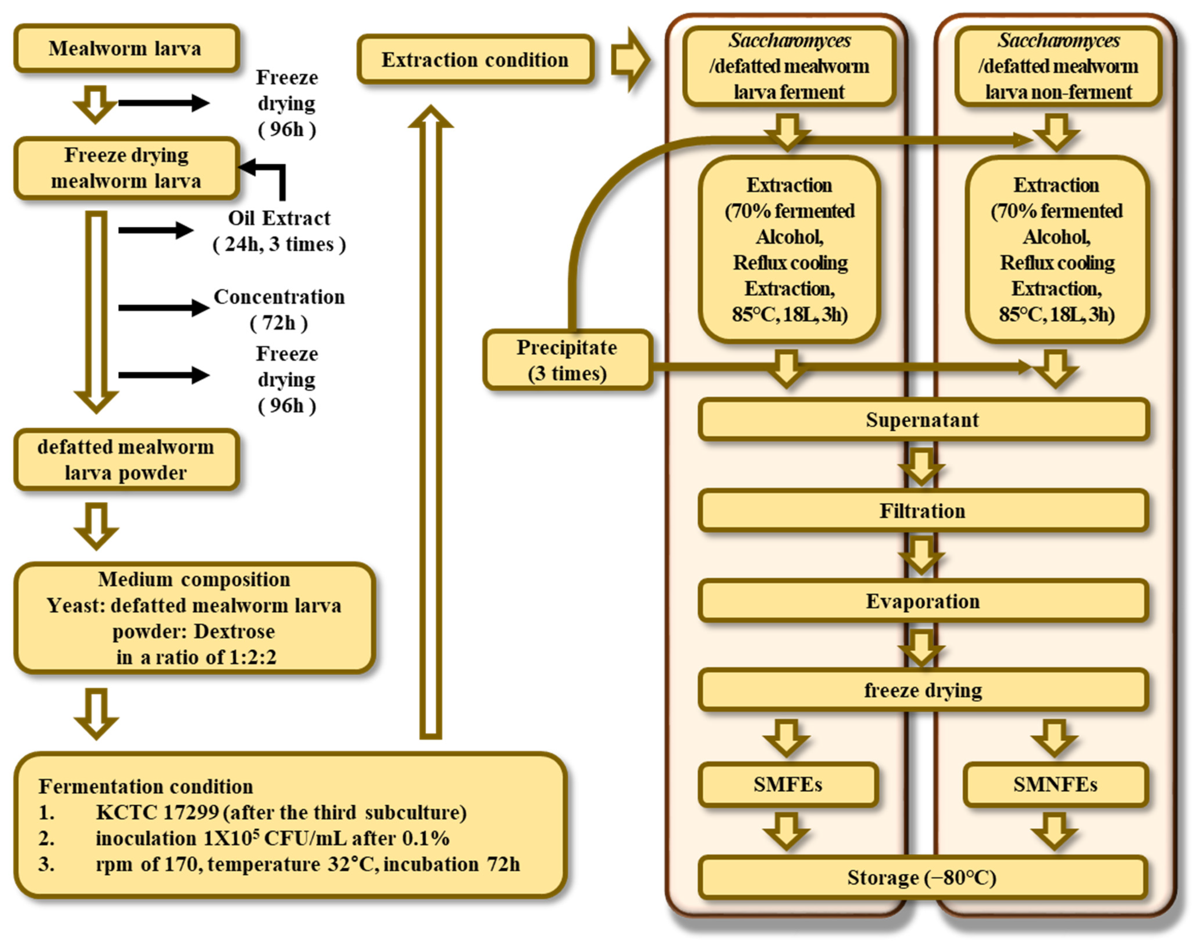
2.2. Pretreatment of Samples

Live Tenebrio molitor larvae (Myeongpum Enterprise, Jangseong, Republic of Korea) were purchased after fasting for 3 days to remove any remaining excrement before use. After, the Tenebrio molitor larvae underwent the pretreatment process of removing moisture through freeze-drying. The freeze-dried larvae were degreased using edible hexane (Dongsung Chemical Ltd., Busan, Republic of Korea). The solvent was added at a ratio of 1:5 (Freeze-dried Tenebrio molitor larvae– food-grade hexane) to the weight of the freeze-dried larvae, and the extraction was repeated three times with continuous stirring at room temperature for 24 h. The same sample was repeatedly extracted under identical conditions during each extraction cycle. The larvae were not pulverized prior to defatting to facilitate subsequent concentration and drying steps, thereby improving the recovery of defatted larvae for use as a medium component. After oil extraction, the defatted mealworm larvae were filtered, and the resulting percolates were evaporated by a rotary vacuum concentrator, which was subsequently lyophilized to remove residual hexane completely. The pretreated, defatted mealworm larvae were ground three times at 10,000 rpm for 1 min each using an IKA MultiDrive basic (IKA-Werke, Staufen im Breisgau, Germany). After grinding, particles passing through a 25-mesh sieve were separated using a sieve shaker (Cheonggye Sieve, Gunpo, Republic of Korea) and subsequently incorporated into the fermentation medium, as outlined in Figure 1.

2.3. Fermentation and Extraction of Samples

The Saccharomyces cerevisiae strain (KCTC 17299) was obtained from the Korean Cell Line Bank for fermentation of defatted mealworm larvae. The fermentation medium was composed of yeast, defatted mealworm larva powder, and dextrose at a ratio of 1:2:2. In this medium composition, defatted mealworm larva powder served as a protein source, replacing peptone. A mixture of 5 L of water, 50 g of powdered yeast, 100 g of powdered dextrose, and 100 g of defatted mealworm larva powder was prepared, sterilized using an autoclave, and subsequently used for the experiment. Fermentation was conducted in a jar fermenter, with S. cerevisiae (KCTC 17299) used after the third subculture. The strain was inoculated at a concentration of 1 × 10⁵ CFU/mL (0.1% inoculation), followed by incubation under shaking conditions at 170 rpm and 32 °C for 72 h under anaerobic conditions.
After fermentation, the fermented medium was subjected to extraction in triplicate using fermented ethanol (Ethanol Supplies World Co., Ltd., Jeonju, Republic of Korea) through exhaustive maceration (fermented medium:fermented ethanol = 3:7) at 85 °C for 3 h via the reflux extraction method. The extracts were filtered using Whatman No. 2 paper filters. The filtered extracts were concentrated using a rotary vacuum concentrator and then freeze-dried to obtain powdered samples. The concentrated and powdered extracts were stored at −80 °C until further use in experiments, as shown in Figure 1. The two samples that were finally created through the process were, firstly, Saccharomyces/defatted Tenebrio molitor (mealworm) larva ferment extract (SMFE) and, secondly, Saccharomyces/defatted Tenebrio molitor (mealworm) larva non-ferment extracts (SMNFE). For the SMNFE group, the medium composition and water volume were identical to those used for the SMFE group. Following autoclave sterilization, incubation was carried out under the same culture conditions as SMFE but without the addition of Saccharomyces cerevisiae strain (KCTC 17299). The extraction procedure was also conducted in the same manner. The key distinction between the two samples lies in the presence or absence of microbial fermentation.

2.4. Antioxidant Activity

2.4.1. Estimation of Total Phenolics, Tannins, and Flavonoids Content

For the analysis of chemical composition, including total phenolics, tannins, and flavonoids, experiments were conducted using sample concentrations of 50, 100, 500, and 1000 μg/mL. The condensed tannin content was determined based on the reactivity of catechins and proanthocyanidins with vanillin, as described by the vanillin method [28] with slight modifications. Each extract and fraction (1 mL) was transferred into a test tube, followed by the addition of 2 mL of vanillin solution (1% vanillin in 7 M H2SO4). The mixture was incubated in an ice bath and subsequently held at 25 °C for 15 min. Absorbance was measured at 500 nm using a spectrophotometer. Tannin concentrations were calculated as catechin equivalents (CE) per kilogram of dry mass from a calibration curve, where tannin content was expressed as μg CE/mg. Total flavonoid content was measured using the aluminum colorimetric method with minor modifications [29,30]. To each well of a 96-well microplate, 100 µL of distilled water was added, followed by 10 µL of 50 g/L NaNO2 and 25 µL of either standard or sample solution. After 5 min of incubation, 15 µL of 100 g/L AlCl3 was added. Six minutes later, 50 µL of 1 M NaOH and 50 µL of distilled water were added. The plate was shaken for 30 s using a microplate reader prior to measuring absorbance at 510 nm. A calibration curve was generated using quercetin as a standard at concentrations ranging from 1 to 500 μg/mL, with total flavonoid concentration expressed as μg quercetin equivalents (QE)/mg. Total phenolic content was determined using the Folin–Denis method with slight modifications [31]. In each well of a 96-well plate, 75 µL of distilled water was added, followed by 25 µL of either the sample or standard solution and 25 µL of Folin–Ciocalteu reagent (diluted 1:1, v/v, with distilled water). All reagents, except the sample and standard solutions, were dispensed using a repeating pipette. After mixing and incubating the reaction for 6 min, 100 µL of 75 g/L Na2CO3 was added to each well. The solutions were mixed again, the plate was covered, and samples were incubated in the dark for 90 min. Absorbance was then measured at 765 nm. Gallic acid was used as the standard, with concentrations ranging from 1 to 500 μg/mL to generate a calibration curve. The total phenolic content was expressed as μg gallic acid equivalents (GAE)/mg.

2.4.2. Radical and Anion Scavenging Activity

Antioxidant activity was evaluated using the DPPH assay, following the method outlined by Blois [32] with slight modifications [29]. The DPPH (2,2-diphenyl-1-picrylhydrazyl) radical scavenging assay was employed to assess the antioxidant capacity of the samples. 0.45 mM DPPH solution was freshly prepared each day in a 50 mL conical tube using 70% ethanol and stored in the dark at 4 °C until use. For the assay, 120 μL of DPPH solution was mixed with 60 μL of the test sample, control, or standard solution (ascorbic acid) at various concentrations, also prepared in 70% ethanol. The reaction mixtures were gently mixed, covered to prevent light exposure, and incubated in the dark at room temperature for 15 min. Following incubation, the absorbance was measured at 517 nm using a spectrophotometer. An ABTS cation radical decolorization assay was performed to assess the ABTS cation radical scavenging activity of the extracts, as described in the spectroscopic method by Re et al. [33]. The ABTS radical cation (ABTS•⁺) was generated by mixing a 7 mM aqueous solution of ABTS (2,2′-azino-bis(3-ethylbenzothiazoline-6-sulfonic acid)) with 2.45 mM potassium persulfate. The mixture was allowed to react in the dark at room temperature for 12–16 h to form the stable ABTS•⁺ radical. Prior to the assay, the ABTS•⁺ solution was diluted with ethanol to achieve an absorbance of 0.750 ± 0.05 at 734 nm. For the assay, 100 μL of the ABTS•⁺ solution was added to 100 μL of the test sample, control, or standard solution (ascorbic acid) prepared in 70% ethanol at various concentrations. The mixtures were gently mixed, protected from light, and incubated at room temperature for 7 min. After incubation, the absorbance was measured at 734 nm using a spectrophotometer. The superoxide radical scavenging activity was determined using the protocol provided by Zhishen et al. [34], with slight modifications. Each sample, standard, and control solutions (10 µL) were added to the 96 wells, followed by 40 µL of 0.1 M phosphate buffer (pH 8.0) and 100 µL of xanthine oxidase (sol. in buffer, 0.05 unit/mL). Next, the 100 µL of substrate [sol. in buffer, xanthine (0.4 mM) + NBT (0.24 mM nitroblue tetrazolium)] was added to the mixture. This was covered and allowed to react in the dark at 37 °C for 20 min. The plate was read at 510 nm. Ascorbic acid was used as the standard. Radical and anion scavenging activities were evaluated across a broader concentration range of 1, 3, 10, 30, 100, 300, and 1000 μg/mL.
The scavenging activity of each extract was calculated as percent inhibition according to the following equation:
Scavenging rate (%) = [A (blank) − A (sample)]/A (blank) × 100
The blank used for analysis refers to the absorbance of a solution containing the substrate and the solvent used to elute the sample, whereas the sample absorbance represents the reaction between the substrate and the test material.

2.4.3. Fenton Reaction and Reducing Power Activity

The metal chelating ability of the extracts was assessed using the Dinis method [35] with slight modifications [36]. For the assay, 30 μL of 0.6 mM iron(II) chloride (FeCl2) solution was added to 30 μL of the test sample, control, or standard solution (ascorbic acid), each prepared in methanol at various concentrations. The reaction was initiated by adding 30 μL of 5 mM 2,2′-bipyridyl solution. The mixtures were gently mixed, protected from light, and incubated at room temperature for 10 min. Following incubation, the absorbance was measured at 562 nm using a spectrophotometer. The metal chelating ability of each extract was calculated as percent inhibition using the following equation: scavenging rate (%) = [A (blank) − A (sample)]/A (blank) × 100].
The Cu2+ reducing power was determined using the method described by Apak et al. [37] and Gülçin [38]. For the assay, 25 μL of 0.01 M copper(II) chloride (CuCl2) solution, 25 μL of 0.0075 M ethanolic neocuproine solution, and 25 μL of 1.0 M ammonium acetate (NH4Ac) buffer solution were added to 25 μL of the test sample, control, or standard solution (ascorbic acid), each prepared in distilled water at various concentrations. The mixtures were gently mixed, protected from light, and incubated at room temperature for 30 min. Following incubation, the absorbance was measured at 450 nm using a spectrophotometer. The ferric-reducing antioxidant power (FRAP) of the extracts was evaluated according to the method of Benzie and Strain [39] and Yanping et al. [40] with slight modifications. For the assay, a TPTZ solution (2.25 mL; 10 mM TPTZ in 40 mM HCl, dissolved in water at 50 °C) was freshly prepared and mixed with 25 mL of 0.3 M acetate buffer (pH 3.6) and 2.25 mL of 20 mM FeCl3 solution to prepare the FRAP working reagent (cocktail solution). The mixture was incubated in a water bath at 37 °C for 10 min prior to use.
For the reaction, 25 μL of the test sample, control, or standard solution (ascorbic acid), each prepared in acetate buffer at various concentrations, was added to each well, followed by 175 μL of the freshly prepared cocktail solution. The mixtures were incubated at 37 °C for 10 min, and the absorbance was subsequently measured at 593 nm using a spectrophotometer [41]. The Fe3+ reduction method, which measures the conversion of Fe3+(CN6) to Fe2+(CN6) as described by Gülçin et al. [42] and Gülçin [43], was used to perform the reduction assay with slight modifications. For the assay, 30 μL of the test sample, control, or standard solution (ascorbic acid) prepared in deionized water was added to 50 μL of 0.2 M phosphate buffer (pH 6.6) and 50 μL of 1% potassium ferricyanide [K3Fe(CN)6]. The mixture was protected from light and incubated at 50 °C for 20 min. Following incubation, 50 μL of 10% trichloroacetic acid (TCA) was added. Then, 20 μL of 0.1% FeCl3 solution was added to the mixture. The absorbance was measured at 700 nm using a spectrophotometer. An increase in absorbance corresponds to an increase in reduction capability [44,45].

2.5. Cell and Culture

RAW 264.7 cells were obtained from the Korean Cell Line Bank. The cells were cultured in RPMI 1640 medium supplemented with 10% fetal bovine serum (FBS) and 1% penicillin-streptomycin and maintained at 37 °C in a humidified incubator with 5% CO2.

2.6. Cell Viability

Cell viability assays were performed using two concentrations, 300 and 1000 μg/mL, selected based on preliminary cytotoxicity screening. Cell viability was assessed using the Cell Counting Kit-8 (CCK-8) assay. Briefly, RAW 264.7 cells were seeded at a density of 2.5 × 10⁵ cells per well in 96-well plates. Test samples (100 μL) were then added to each well, and the cells were incubated at 37 °C for 24 h. After incubation, 10 μL of CCK-8 solution was added to each well, and the plates were further incubated at 37 °C for 3 h. The absorbance was measured at 450 nm using a microplate reader (VersaMax, Molecular Devices, Sunnyvale, CA, USA). Cell viability was expressed as a percentage of the control cell viability, calculated using the following formula: Inhibition (%) = 1 − (OD drug/OD control) × 100%.

2.7. Total Glutathione Quantification Assay

RAW 264.7 macrophages were treated as previously described, then collected and washed with phosphate-buffered saline (PBS). The cells were lysed in 10 mM HCl by freezing and thawing twice, followed by centrifugation at 8000× g for 10 min. Total glutathione levels were quantified using a total glutathione quantification kit (Dojindo, Kumamoto, Japan) according to the manufacturer’s instructions. For the total glutathione quantification assay, sample concentrations of 300 and 1000 μg/mL were also employed, as these doses did not exhibit cytotoxic effects in the cell viability assay.

2.8. Analysis of Free Amino Acids

The sample preparation method was adapted from Marino et al. [46] with some modifications. To determine the free amino acid composition, 1 g of sample was combined with 50 mL of pure 0.02 N HCl in volumetric flasks and sonicated for 30 min. The resulting mixtures were filtered through 0.2 µm syringe filters and transferred into glass vials for analysis.
The free amino acid composition was analyzed using high-performance liquid chromatography (HPLC, Agilent 1220, Agilent Technologies, Santa Clara, CA, USA). The analytical column used was a Proshell HPH C18 (150 × 4.6 mm, 4 µm; Agilent). A variable wavelength detector was set to 338 nm. The column temperature was maintained at 40 °C. Mobile phase A consisted of 10 mM sodium phosphate dibasic and 10 mM sodium tetraborate decahydrate (1:1, v/v, pH 8.2), and mobile phase B consisted of a mixture of acetonitrile, methanol, and water (4.5:4.5:1, v/v/v). Solvents A and B were run at a flow rate of 1.5 mL/min using the following gradient: 98% A (2% B) at 0 min, steady at 98% A for 1.9 min, decreasing to 43% A over 16.2 min, decreasing to 20% A over 0.5 min, holding steady at 20% A for 3.7 min, and then increasing to 98% A over 0.9 min. The column was equilibrated with 98% A for 3.7 min before the next injection. For derivatization, O-phthalaldehyde (Sigma-Aldrich, St. Louis, MO, USA) and 3-mercaptopropionic acid (Sigma-Aldrich) in borate buffer (Agilent) were used. Individual amino acids were identified by comparing the retention times of the sample peaks with those of known amino acid standards obtained from Agilent Technologies. This comparative approach ensured accurate peak identification by matching both retention times and chromatographic profiles under identical analytical conditions.

2.9. Statistical Analysis

For the purpose of statistical analysis, all experiments were repeated in at least three independent trials. Statistical analyses were conducted using one-way analysis of variance (ANOVA) with the Statistical Analysis System (SAS) version 9.4 (SAS Institute Inc., Cary, NC, USA). Differences between means were determined using the Duncan multiple-range test, with significance set at p < 0.05.

3. Results and Discussion

3.1. Chemical Composition: Phenolics, Tannins, and Flavonoids

The total phenolic compounds, flavonoid content, and condensed tannin content of Saccharomyces/defatted mealworm larva ferment (SMFE) and non-ferment (SMNFE) extracts were quantified using calibration curves for gallic acid, quercetin, and catechin. The concentrations of these compounds are presented in Table 1.
In the SMFE, the total phenolic content was 36.04 ± 1.04 μg GAE/mg, the total flavonoid content was 12.69 ± 0.76 μg QE/mg, and the condensed tannin content was 0.55 ± 0.11 μg CE/mg. In contrast, the SMNFE exhibited a total phenolic content of 11.11 ± 0.13 μg GAE/mg and a flavonoid content of 3.61 ± 0.00 μg QE/mg. No condensed tannins were detected in the SMNFE.
These results indicate a significant increase in beneficial compounds following the fermentation of defatted mealworm larva. Specifically, the total phenolic compounds in the SMFE increased by 3.2 times, the total flavonoid content rose by 3.5 times, and the presence of condensed tannins was also enhanced due to the fermentation process.
Previous studies have demonstrated similar results. Kim and Yook [47] reported that after fermenting yams with five different strains (Bacillus subtilis KACC 14549, Saccharomyces cerevisiae KACC 48234, Levilactobacillus brevis KACC 18270, Lacticaseibacillus casei KACC 12413, and Lactiplantibacillus plantarum KACC 18510), the total polyphenol content increased from 3.66 mg per g in the non-fermented group to 6.82–9.42 mg per g in the fermented group. Similarly, Kim et al. [48] observed that the fermentation of barley sprouts using Lactobacillus plantarum KCL005 and Leuconostoc mesenteroides KCL007 strains resulted in an increase in the content of beneficial components over fermentation time. These findings corroborate the results observed in this study, demonstrating that fermentation enhances the levels of beneficial compounds in the product.
The results from this study further support that fermentation significantly elevates the levels of phenolics and flavonoids in the SMFE when compared to the SMNFE. Moreover, the presence of condensed tannins, detected only after fermentation, suggests that these compounds are formed through metabolic processes during fermentation [47,48]. These findings indicate that the flavonoids and other phenolic compounds of natural products play a key role in enhancing antioxidant activities, likely due to their electron-donating ability in proportion to their phenolic content.

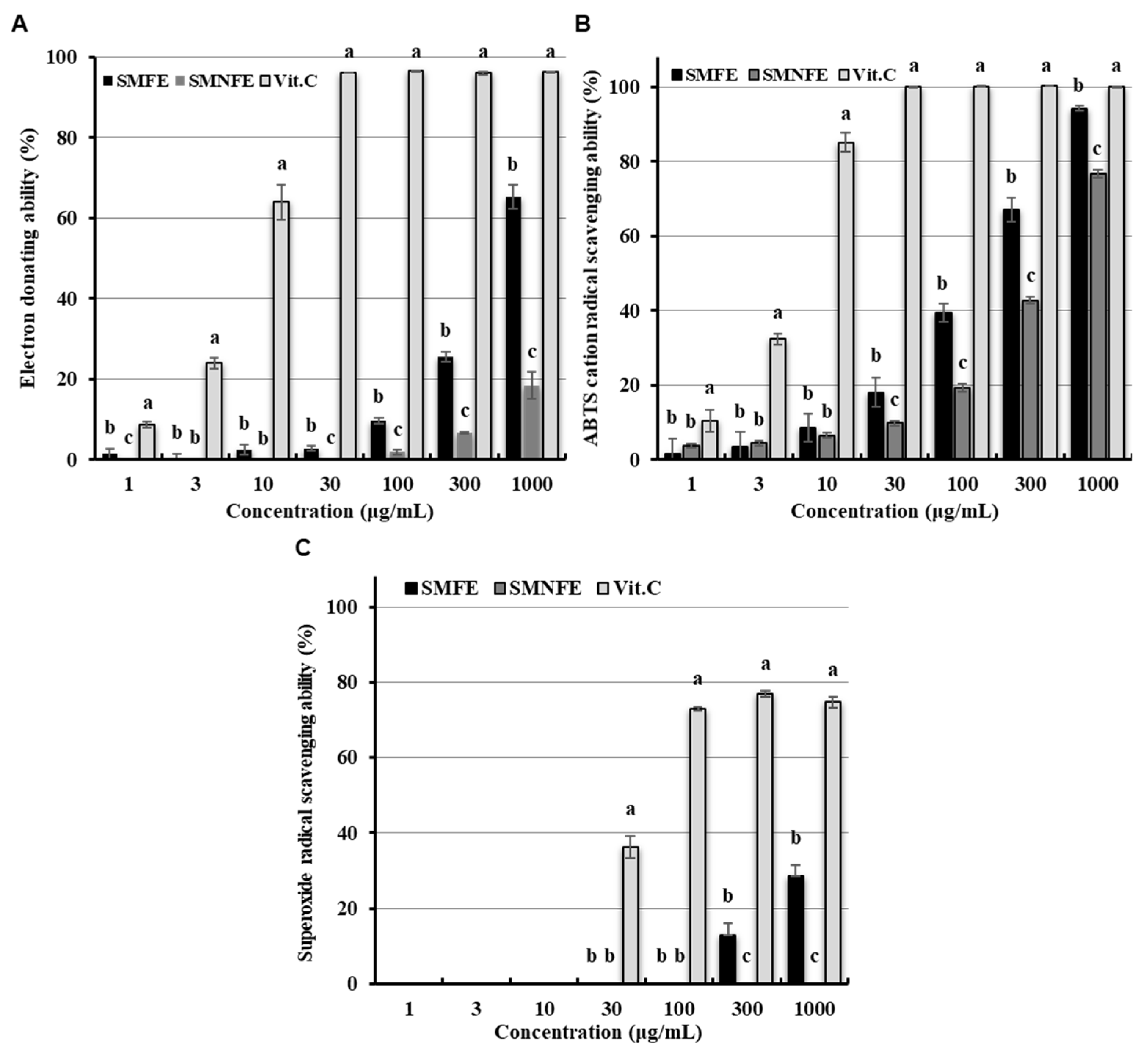
3.2. Determination of Antioxidant Activities: Radical and Anion Scavenging Activities

Figure 2 illustrates the concentrations of the electron-donating ability, ABTS cation radical scavenging activity, and superoxide anion radical scavenging activity in both the Saccharomyces/defatted mealworm larva ferment (SMFE) and non-ferment (SMNFE) extracts. The electron-donating ability assay revealed that the SMFE exhibited an activity level of 65.25% at a concentration of 1000 μg/mL, while the SMNFE demonstrated a significantly lower activity level of 18.44% at the same concentration (Figure 2A). These findings indicate a substantial enhancement in electron-donating activity following fermentation. No significant changes were observed in the activity of superoxide dismutase, an antioxidant enzyme responsible for neutralizing reactive oxygen species. However, the ABTS cation radical scavenging activities were notably higher in the SMFE (94.23%) compared to the SMNFE (76.69%) at a concentration of 1000 μg/mL (Figure 2B), reinforcing the results observed for electron-donating abilities. The NBT assay, which measures superoxide anion radical scavenging activity through color change (purple at 570 nm), showed that at the same concentration, the SMFE exhibited approximately 30% scavenging activity, while no significant scavenging activity was detected in the SMNFE (Figure 2C). This suggests that condensed tannins formed during fermentation may contribute to the enhanced scavenging activity of SMFE. These findings suggest that after fermentation of Tenebrio molitor, the electron-donating ability increased 3.5 times, ABTS cation radical scavenging activity increased by 1.2 times, and superoxide scavenging activity (as measured by the NBT assay) was significantly elevated, indicating a substantial increase in the antioxidant potential due to fermentation.
The observed increase in antioxidant activity is consistent with previous studies, which showed that the electron-donating ability improves when the total polyphenol content is higher. Specifically, studies have demonstrated that higher polyphenol and flavonoid contents correlate with enhanced antioxidant activities in fermented products [49]. In a study by Kim et al. [48], the electron-donating ability increased from 36.63% at the start of fermentation to 52.93% and 47.09% after fermentation, and ABTS scavenging activity rose from 44.63% to 65.70% and 57.09% as fermentation progressed. These results align with the findings of the present study, supporting the hypothesis that the radical scavenging activities of SMFE increased as a result of metabolic processes during microbial fermentation.

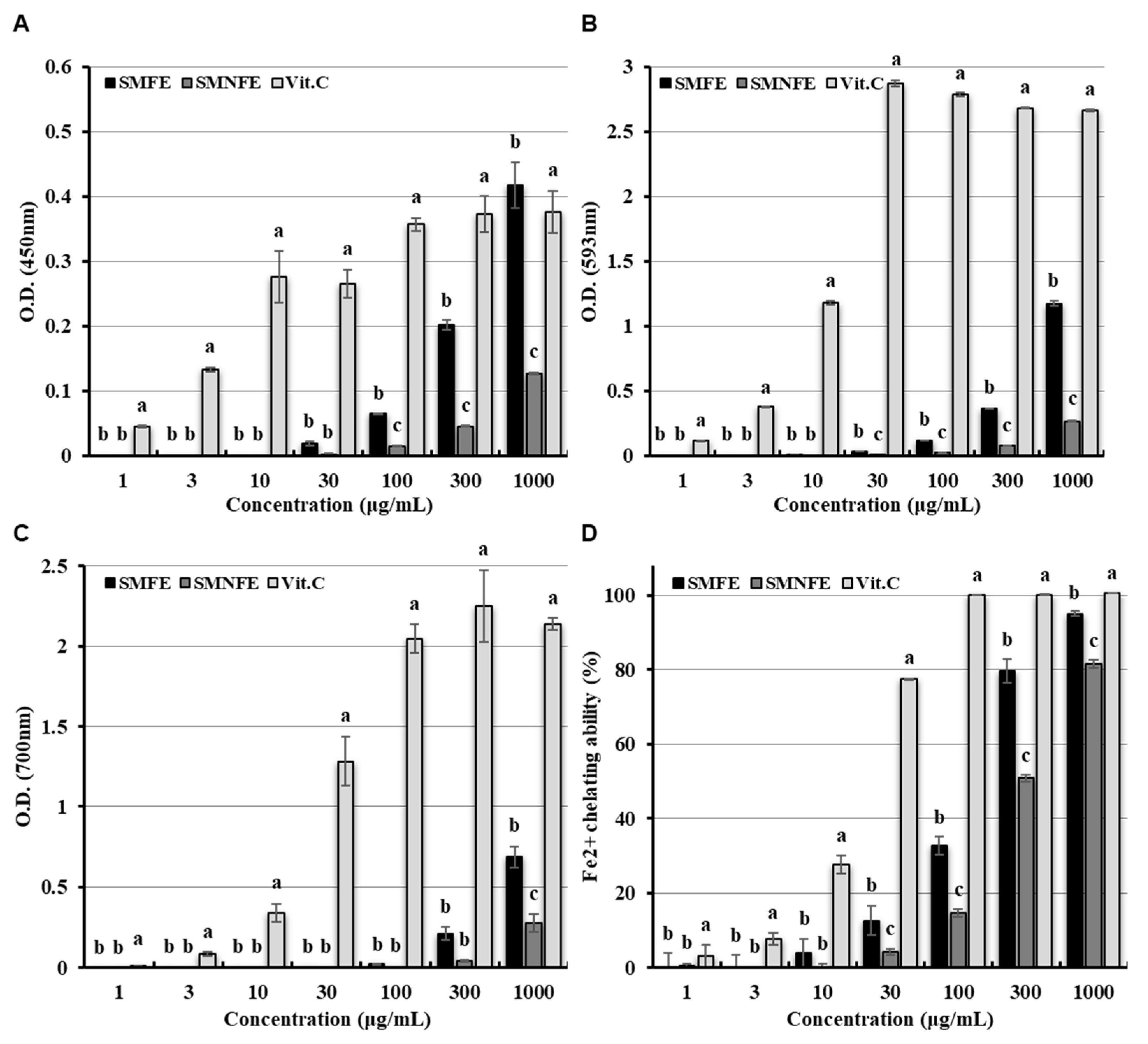
3.3. Determination of Antioxidant Activities: Fenton Reaction and Reducing Power Activities

Figure 3 displays the concentrations of the Fenton reaction and reducing power activities in both the Saccharomyces/defatted mealworm larva ferment (SMFE) and non-ferment (SMNFE) extracts.
In the Fenton oxidation reaction, hydrogen peroxide (H2O2) and ferrous iron (Fe2+) react to form hydroxyl radicals (OH), which can interact with organic compounds. The reducing power in this oxidation reaction was evaluated by measuring the reducing abilities of Fe2+ and Cu2+, as well as the antioxidant activities of chelation reactions that inhibit the formation of the Fe2+-ferrozine complex, which confirms the presence of this activity. The reducing power of Cu2+ was notably higher in the SMFE (0.43 ± 0.03) than in the SMNFE (0.14 ± 0.00) at a concentration of 1000 μg/mL (Figure 3A), exhibiting a pattern consistent with the observed phenolic contents and radical scavenging activities. The Ferric Reducing Antioxidant Power (FRAP) assay is based on the reduction of ferric tripyridyl triazine (Fe3+-TPTZ) to ferrous tripyridyl triazine (Fe2+-TPTZ) by a reducing agent at low pH. The FRAP value for the SMFE was 1.24 ± 0.02 (OD) at a concentration of 1000 μg/mL, while the SMNFE displayed a significantly lower FRAP value of 0.34 ± 0.00 (OD) (Figure 3B). Additionally, the reducing power of ferrous-ferricyanide (Fe3+), which stabilizes free radicals by donating hydrogen to ferric-ferricyanide, was 0.69 ± 0.07 (OD) for SMFE and 0.28 ± 0.05 (OD) for SMNFE at a concentration of 1000 μg/mL (Figure 3C). The chelating activities of the SMFE and SMNFE at a concentration of 1000 μg/mL were 95.08% ± 1.86% and 81.59% ± 0.81%, respectively (Figure 3D), indicating that the reducing powers and chelating effects were significantly enhanced in the SMFE due to various Fenton reactions.
In this study, Cu2+ reducing ability, FRAP, Fe3+ to Fe2+ reductive potential, and Fe2+ chelation were employed as indicators to evaluate the antioxidant activities of the SMFE and SMNFE [36,37,38,39,40,41,42,43,44,45]. The significantly higher values observed in the SMFE compared to the SMNFE suggest that fermentation with Saccharomyces cerevisiae enhances the antioxidant capacity of the extract. Consistent with these findings, previous studies have shown that fermentation improves the antioxidant properties of edible insects. For example, Seong et al. [50] reported that the supernatant of a 50% methanol extract derived from brown rice supplemented with field cricket powder, which fermented with four probiotic strains (Leuconostoc mesenteroides, Lactobacillus plantarum, Bacillus amyloliquefaciens or Lactobacillus rhamnosus) exhibited significantly greater antioxidant activities including reducing power, DPPH and ABTS+ radical scavenging activities, and SOD-like activity compared to that of fermented brown rice alone. Similarly, Jang et al. [51] reported that the supernatant of a heat-reflux extract from Tenebrio molitor fermented with Lactobacillus plantarum, Aspergillus kawachii, or Bacillus subtilis exhibited strong DPPH radical scavenging activity and Cu²⁺ reducing capacity. These effects were attributed to the increased levels of phenolic compounds and flavonoids resulting from the fermentation process. Therefore, these results collectively indicate that SMFE has the potential to serve as an effective antioxidant additive, especially when compared to its non-fermented counterpart.

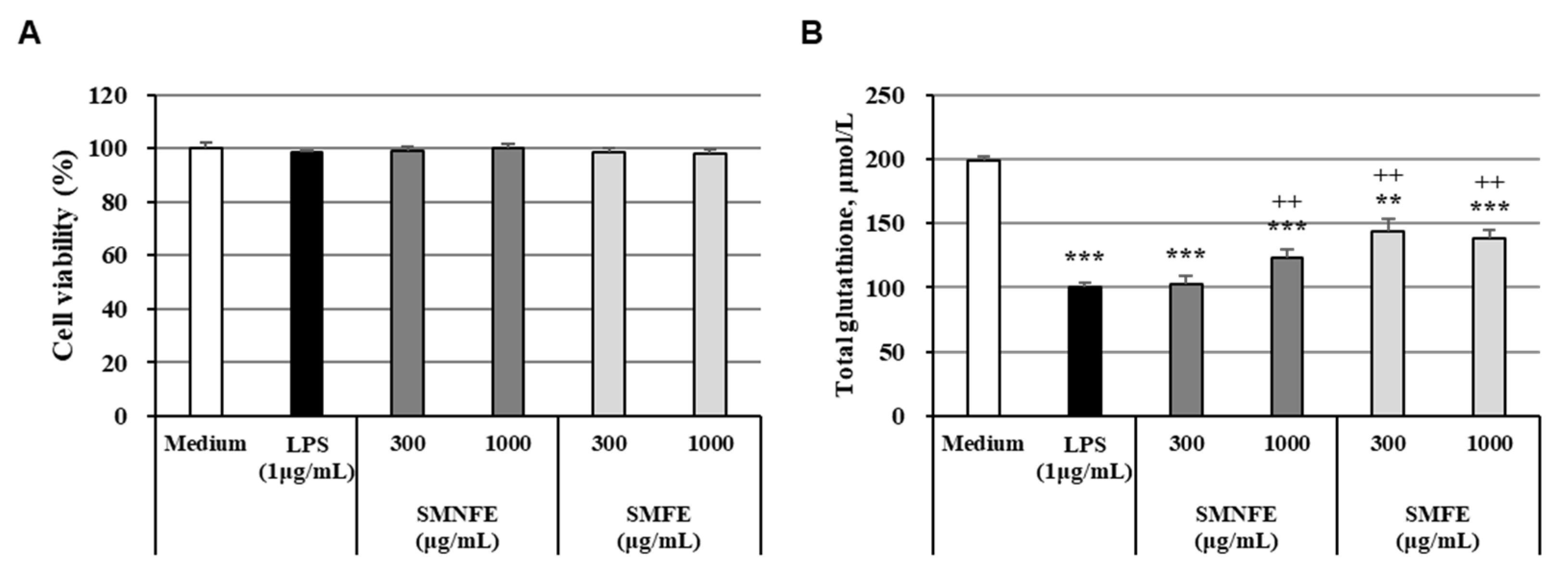
3.4. Activity of Antioxidant Enzymes

The cytotoxic effects of the SMFE and SMNFE on RAW 264.7 cells were evaluated by exposing the cells to varying concentrations (300 and 1000 μg/mL) of SMFE and SMNFE for 24 h. Our results indicated no cytotoxic effects at the tested concentrations, as shown in Figure 4A. Therefore, SMFE and SMNFE at concentrations of 300 and 1000 μg/mL were selected for subsequent experiments.
We further examined the effects of SMFE and SMNFE on total glutathione levels, which are crucial components of the antioxidative defense system. Our results demonstrated that SMFE significantly enhanced total glutathione activity compared to the LPS-induced control group at both 300 and 1000 μg/mL (Figure 4B). Oxidative stress, typically characterized by an overproduction of reactive oxygen species (ROS) or a deficiency in the antioxidant defense mechanisms, was alleviated by SMFE treatment. While LPS exposure led to a reduction in the expression of antioxidant-related enzymes, the SMFE group showed a substantial increase in the levels of these enzymes, suggesting an enhancement of the antioxidative defense system.
Glutathione, a tripeptide composed of glutamic acid, cysteine, and glycine, plays a pivotal role in antioxidant defense by scavenging free radicals during the reductive detoxification of hydrogen peroxide and lipid peroxides [52,53]. Previous studies have shown that glutathione production by Saccharomyces cerevisiae can be enhanced during the fermentation process. For instance, in a study examining glutathione production under different culture conditions (static vs. shaking culture), it was found that shaking culture resulted in higher glutathione production than static culture. Furthermore, glutathione production was inversely correlated with glucose concentration, with the highest production occurring when 0.04% of the three amino acids (glutamate, cysteine, and glycine) were added [54].
The effects on antioxidant enzymes observed in this study following the administration of fermented edible insect extracts are consistent with findings from previous research. Lee et al. [55] reported that supplementation with Bacillus subtilis-fermented Protaetia brevitarsis powder (100 mg/mL) in a high-fat diet-induced diabetic mouse model significantly reduced malondialdehyde levels, while simultaneously enhancing superoxide dismutase activity and increasing the levels of reduced glutathione compared to the unfermented control. These findings suggest that the fermentation process may contribute to the production of bioactive compounds that enhance antioxidant defense mechanisms and alleviate oxidative stress. Similarly, Sim et al. [56] demonstrated that dietary supplementation with 5% air-dried powder of solid-state fermented Tenebrio molitor led to a significant reduction in lipid peroxidation and an increase in glutathione content in both liver and serum in orotic acid-induced non-alcoholic fatty liver disease rat models. Taken together, the increase in glutathione content observed in SMFE, as compared to SMNFE, can be attributed to the fermentation process. Therefore, this elevation in glutathione levels is thought to contribute significantly to the enhanced antioxidant activity of SMFE observed in the present study.

3.5. Determination of Free Amino Acids

In our study, the total amino acid content increased by approximately 18% following fermentation compared to the unfermented control (Table 2). Notably, concentrations of all analyzed amino acids except for arginine increased after fermentation. These findings are consistent with a recent study by Cao et al. [57], which demonstrated that yeast fermentation significantly improves the amino acid profile of soybean meal. This study emphasizes the efficacy of yeast-based bioconversion in improving nutritional value by increasing amino acid content, which supports the validity of the results of this study. The content of free amino acids increased by 6.39 to 73.26 percent following fermentation, compared to pre-fermentation levels. SMFE concentrations increased in aspartic acid, glutamic acid, serine, histidine, glycine, threonine, alanine, tyrosine, valine, methionine, phenylalanine, isoleucine, leucine, and lysine of 40.34%, 40.20%, 6.39%, 19.01%, 32.47%, 16.02%, 28.85%, 47.28%, 36.69%, 73.26%, 30.12%, 24.42%, 16.70%, 22.50%, and 22.49%, respectively, relative to the SMNFE. However, the decrease in arginine content observed after fermentation is likely due to its utilization by Saccharomyces cerevisiae as a nitrogen source. During fermentation, arginine is catabolized via the arginase pathway into ornithine and urea or redirected toward polyamine biosynthesis, both of which support microbial growth and stress response. This metabolic shift is a well-documented phenomenon in yeast fermentation systems [58,59].
Amino acids play a crucial role as precursors for compounds with antioxidant properties [60]. Among these, histidine, tyrosine, methionine, and cysteine are known to exhibit antioxidant activity. Histidine, in particular, demonstrates strong radical scavenging ability due to the cleavage of the imidazole ring [61,62,63]. The current study observed that histidine, tyrosine, and methionine levels in free amino acids increased by approximately 1.2 to 1.7 times post-fermentation.
These results indicate that the fermentation process mediated by Saccharomyces cerevisiae leads to an increased abundance of free amino acids, including histidine, tyrosine, and methionine, which are associated with antioxidant activity. The observed enhancement in amino acid content is presumed to result from a combination of proteolytic activities during fermentation and the accumulation of yeast biomass. Although differentiating amino acids originating from yeast cell lysis versus fermentation remains a limitation, this study underscores the potential of microbial fermentation to enhance the nutritional and functional value of protein hydrolysates for applications in food, nutraceuticals, and related industries.

4. Conclusions

This study demonstrates that Saccharomyces-fermented defatted Tenebrio molitor larva extract (SMFE) exhibits significantly enhanced antioxidant activity, along with increased levels of total phenolic and flavonoid compounds. These findings suggest that fermentation may serve as an effective strategy for enhancing the antioxidant potential of insect-derived extracts. The observed increase in free amino acid content following fermentation also indicates potential nutritional value, although further validation is needed.
It is important to note that this study focused solely on evaluating changes in antioxidant activity and free amino acid content before and after fermentation, using insect-derived protein components as a nutrient source for microbial growth. Therefore, additional research is required to assess other potential bioactivities, verify the bioavailability of active compounds, and evaluate the safety and efficacy of SMFE in practical applications.
Despite these limitations, the current findings support the potential of fermented insect extracts as novel and sustainable ingredients for the development of functional foods and antioxidant formulations.

Author Contributions

Conceptualization, K.-W.P. and K.-Y.K.; Data curation, K.-Y.K., K.-W.P., J.-H.K. and B.-G.J.; Formal analysis, K.-Y.K. and J.-H.K.; Investigation, J.-H.K., B.-G.J. and K.-Y.K.; Methodology, K.-W.P., J.-H.K. and K.-Y.K.; Project administration, K.-W.P.; Supervision, K.-W.P. and K.-Y.K.; Validation, K.-W.P.; Writing—original draft, K.-Y.K.; Writing—review and editing, K.-W.P. and K.-Y.K. All authors have read and agreed to the published version of the manuscript.

Funding

This research received no external funding.

Institutional Review Board Statement

Not applicable.

Informed Consent Statement

Not applicable.

Data Availability Statement

The original contributions presented in the study are included in the article. Further inquiries can be directed to the corresponding author.

Acknowledgments

The authors sincerely appreciate the financial/administrative support from Suncheon-si.

Conflicts of Interest

The authors declare no conflicts of interest.

References

  1. Bors, W.; Saran, M. Radical scavenging by flavonoid antioxidants. Free Radic. Res. Commun. 1987, 2, 289–294. [Google Scholar] [CrossRef]
  2. Chang, S.S.; Ostric-Matijasevice, B.; Hsieh, O.A.L.; Huang, C. Natural antioxidants from rosemary and sage. J. Food Sci. 1977, 42, 1102–1110. [Google Scholar] [CrossRef]
  3. Kang, K.Y.; Hwang, Y.H.; Lee, S.J.; Jang, H.Y.; Hong, S.G.; Mun, S.K.; Kim, S.J.; Kim, J.J.; Park, K.W.; Seo, K.S.; et al. Verification of the functional antioxidant activity and antimelanogenic properties of extracts of Poria cocos Mycelium fermented with freeze-dried plum powder. Int. J. Biomater. 2019, 2019, 9283207. [Google Scholar] [CrossRef] [PubMed]
  4. Kwon, D.H.; Cha, H.J.; Lee, H.; Hong, S.H.; Park, C.; Park, S.H.; Kim, G.Y.; Kim, S.; Kim, H.S.; Hwang, H.J.; et al. Protective effect of glutathione against oxidative stress-induced cytotoxicity in RAW 264.7 macrophages through activating the nuclear factor erythroid 2-related factor-2/heme oxygenase-1 pathway. Antioxidants 2019, 8, 82. [Google Scholar] [CrossRef] [PubMed]
  5. Hou, Y.; Wang, L.; Yi, D.; Wu, G. N-acetylcysteine and intestinal health a focus on its mechanism of action. Front. Biosci. 2015, 20, 872–891. [Google Scholar] [CrossRef]
  6. Deponte, M. The incomplete glutathione puzzle: Just guessing at numbers and figures? Antioxid. Redox Signal. 2017, 27, 1130–1161. [Google Scholar] [CrossRef]
  7. Gomex-Cabrera, M.C.; Salvador-Pascual, A.; Cabo, H.; Ferrando, B.; Viña, J. Redox modulation of mitochondriogenesis in exercise. Does antioxidant supplementation blunt the benefits of exercise training? Free Radic. Biol. Med. 2015, 86, 37–46. [Google Scholar] [CrossRef]
  8. Purohit, V.; Abdelmalek, M.F.; Barve, S.; Benevenga, N.J.; Halsted, C.H.; Kaplowitz, N.; Kharbanda, K.K.; Liu, Q.Y.; Lu, S.C.; McClain, C.J.; et al. Role of S-adenosylmethionine, folate, and betaine in the treatment of alcoholic liver disease: Summary of a symposium. Am. J. Clin. Nutr. 2007, 86, 14–24. [Google Scholar] [CrossRef]
  9. Williams, G.M.; Iatropoulos, M.J.; Whysner, J. Safety assessment of butylated hydroxyanisole and butylated hydroxytoluene as antioxidant food additives. Food Chem. Toxicol. 1999, 37, 1027–1038. [Google Scholar] [CrossRef]
  10. Shahidi, F.; Zhong, Y. Novel antioxidants in food quality preservation and health promotion. Eur. J. Lipid Sci. Technol. 2010, 112, 930–940. [Google Scholar] [CrossRef]
  11. Zielińska, E.; Baraniak, B.; Karaś, M.; Rybczyńska, K.; Jakubczyk, A. Selected species of edible insects as a source of nutrient composition. Food Res. Int. 2015, 77, 460–466. [Google Scholar] [CrossRef]
  12. Sogari, G.; Amato, M.; Biasato, I.; Chiesa, S.; Gasco, L. The potential role of insects as feed: A multi-perspective review. Animals 2019, 9, 119. [Google Scholar] [CrossRef] [PubMed]
  13. Lee, J.H.; Kim, T.; Choi, B.D.; Kim, G.E.; Lee, K.H. Effects of containing pigments of dried grasshopper on the lipid deterioration. J. Korean Soc. Food Sci. Nutr. 1987, 16, 300–305. [Google Scholar]
  14. Yu, T.J.; Lee, K.Y.; Lee, S.K. Studies on the development of cocoon pupas for food materials. Korean J. Nutr. 1978, 11, 39–43. [Google Scholar]
  15. Van Huis, A. Potential of insects as food and feed in assuring food security. Annu. Rev. Entomol. 2013, 58, 563–583. [Google Scholar] [CrossRef]
  16. Lensvelt, E.J.S.; Steenbekkers, L.P.A. Exploring consumer acceptance of entomophagy: Determinants in a cross-cultural study among the young Dutch and Chinese. Appetite 2014, 83, 152–161. [Google Scholar]
  17. Netherlands Enterprise Agency. Insect Sector Development Plan—Towards a Circular Agriculture; Netherlands Enterprise Agency: Den Haag, The Netherlands, 2021. [Google Scholar]
  18. Chung, M.Y.; Kwon, E.Y.; Hwang, J.S.; Goo, T.W.; Yun, E.Y. Pre-treatment conditions on the powder of Tenebrio molitor for using as a novel food ingredient. J. Seric. Entomol. Sci. 2013, 51, 9–14. [Google Scholar]
  19. Chung, S.J.; Lee, Y.H.; Chung, J.H.; Lee, B.R.; Han, D.M. Antifungal effect and activity spectrum of crude antifungal proteins from hemolymph of larvae of Tenebrio molitor in Korea. Kor. J. Mycol. 1995, 23, 232–237. [Google Scholar]
  20. Jang, Y.S.; Park, M.W.; Kim, J.H.; Lee, Y.S. Anti-inflammatory and antioxidant effects of Tenebrio molitor larvae in vitro and in vivo. Food Sci. Biotechnol. 2021, 30, 293–301. [Google Scholar]
  21. Kim, T.K.; Yong, H.I.; Kim, Y.B.; Kim, H.W.; Choi, Y.S. Functional properties of edible insects and their applications in food: A review. Food Sci. Anim. Resour. 2020, 40, 863–880. [Google Scholar]
  22. Yi, L.; Lakemond, C.M.M.; Sagis, L.M.C.; Eisner-Schadler, V.; van Huis, A.; van Boekel, M.A.J.S. Extraction and characterisation of protein fractions from five insect species. Food Chem. 2013, 141, 3341–3348. [Google Scholar] [CrossRef] [PubMed]
  23. Marco, M.L.; Heeney, D.; Binda, S.; Cifelli, C.J.; Cotter, P.D.; Foligné, B.; Gänzle, M.; Kort, R.; Pasin, G.; Pihlanto, A.; et al. Health benefits of fermented foods: Microbiota and beyond. Curr. Opin. Biotechnol. 2017, 44, 94–102. [Google Scholar] [CrossRef] [PubMed]
  24. Swieca, M.; Gawlik-Dziki, U.; Dziki, D.; Baraniak, B.; Czyz, J. Antioxidant and nutritional evaluation of fermented wheat sprouts. Food Chem. 2014, 164, 101–107. [Google Scholar]
  25. van Huis, A.; Tomberlin, J.K.; dos Santos Oliveira, J.V. Edible Insects: Future Prospects for Food and Feed Security, 2nd ed.; FAO: Rome, Italy; Springer: Berlin/Heidelberg, Germany, 2021. [Google Scholar]
  26. Zielińska, E.; Pankiewicz, U. Fermentation as a tool for improving the quality of edible insect-based foods. Innov. Food Sci. Emerg. Technol. 2020, 67, 102510. [Google Scholar]
  27. Baek, M.; Kim, S.; Cho, J.; Lee, S.; Yoon, S. Effect of fermentation on antioxidant activity and peptide profile of Tenebrio molitor larvae protein hydrolysate. Korean J. Food Sci. Technol. 2019, 51, 543–550. [Google Scholar]
  28. Broadhurst, R.B.; Jones, W.T. Analysis of condensed tannins using acidified vanillin. J. Sci. Food Agric. 1978, 29, 788–794. [Google Scholar] [CrossRef]
  29. Herald, T.J.; Gadgil, P.; Tilley, M. High-throughput micro plate assays for screening flavonoid content and DPPH-scavenging activity in sorghum bran and flour. J. Sci. Food Agric. 2012, 92, 2326–2331. [Google Scholar] [CrossRef]
  30. Pękal, A.; Pyrzynska, K. Evaluation of aluminium complexation reaction for flavonoid content assay. Food Anal. Methods 2014, 7, 1776–1782. [Google Scholar] [CrossRef]
  31. Zhang, W.; Zhang, J.; Shen, J.; Silva, A.; Dennis, D.A.; Barrow, C.J. A simple 96-well microplate method for estimation of total polyphenol content in seaweeds. J. Appl. Phycol. 2006, 18, 445–450. [Google Scholar] [CrossRef]
  32. Blois, M.S. Antioxidant determinations by the use of a stable free radical. Nature 1958, 181, 1199–1200. [Google Scholar] [CrossRef]
  33. Re, E.; Pellegrini, N.; Proteggente, A.; Pannala, A.; Yang, M.; Rice-Evans, C. Antioxidant activity applying an improved ABTS radical cation decolorization assay. Free Radic. Biol. Med. 1999, 26, 1231–1237. [Google Scholar] [CrossRef]
  34. Zhishen, J.; Mengcheng, T.; Jianming, W. The determination of flavonoid contents in mulberry and their scavenging effects on superoxide radicals. Food Chem. 1999, 64, 555–560. [Google Scholar] [CrossRef]
  35. Dinis, T.C.P.; Madeira, V.M.C.; Almeida, L.M. Action of phenolic derivatives (acetaminophen, salicylate, and 5-aminosalicylate) as inhibitors of membrane lipid peroxidation and as peroxyl radical scavengers. Arch. Biochem. Biophys. 1994, 315, 161–169. [Google Scholar] [CrossRef] [PubMed]
  36. Gülçin, İ. Comparison of in vitro antioxidant and antiradical activities of L-tyrosine and L-Dopa. Amino Acids 2007, 32, 431–438. [Google Scholar] [CrossRef]
  37. Apak, R.; Gulcu, K.; Ozyurek, M.; Karademir, S.E.; Ercag, E. The cupric ion reducing antioxidant capacity and polyphenolic content of some herbal teas. Int. J. Food Sci. Nutr. 2006, 57, 292–304. [Google Scholar] [CrossRef]
  38. Gülçin, İ. In vitro prooxidant effect of caffeine. J. Enzyme Inhib. Med. Chem. 2008, 23, 149–152. [Google Scholar] [CrossRef] [PubMed]
  39. Benzie, I.F.F.; Strain, J.J. The ferric reducing ability of plasma (FRAP) as a measure of “antioxidant power”: The FRAP assay. Anal. Biochem. 1996, 239, 70–76. [Google Scholar] [CrossRef]
  40. Zou, Y.; Chang, S.K.C.; Gu, Y.; Qian, S.Y. Antioxidant activity and phenolic compositions of lentil (Lens culinaris var. Morton) extract and its fractions. J. Agric. Food Chem. 2011, 59, 2268–2344. [Google Scholar] [CrossRef]
  41. Göçer, H.; Gülçin, İ. Caffeic acid phenethyl ester (CAPE): Correlation of structure and antioxidant properties. Int. J. Food Sci. Nutr. 2011, 62, 821–825. [Google Scholar] [CrossRef]
  42. Gülçin, İ.; Elmastaş, M.; Aboul-Enein, H. Determination of antioxidant and radical scavenging activity of Basil (Ocimum basilicum L. Family Lamiaceae) assayed by different methodologies. Phytother. Res. 2007, 21, 354–361. [Google Scholar] [CrossRef]
  43. Gülçin, İ. Antioxidant properties of resveratrol: A structure–activity insight. Innov. Food Sci. Emerg. Technol. 2010, 11, 210–218. [Google Scholar] [CrossRef]
  44. Elmastaş, M.; Turkekul, I.; Ozturk, L.; Gülçin, İ.; Isildak, O.; Aboul-Enein, H. Antioxidant activity of two wild edible mushrooms (Morchella vulgaris and Morchella esculanta) from North Turkey. Comb. Chem. High Throughput Screen 2006, 9, 443–448. [Google Scholar] [CrossRef] [PubMed]
  45. Bursal, E.; Koksal, E.; Gülçin, İ.; Bilsel, G.; Goren, A.C. Antioxidant activity and polyphenol content of cherry stem (Cerasus avium L.) determined by LC–MS/MS. Food Res. Int. 2013, 51, 66–74. [Google Scholar] [CrossRef]
  46. Marino, R.; Iammarino, M.; Santillo, A.; Muscarella, M.; Caropresse, M.; Albenzio, M. Rapid method for determination of amino acids in milk. J. Dairy Sci. 2010, 93, 2367–2370. [Google Scholar] [CrossRef]
  47. Kin, Y.H.; Yook, H.S. Antioxidant activites of Yam (Dioscorea japonica Thunb.) fermented with various microorganisms. J. Korean Soc. Food Sci. Nutr. 2024, 53, 965–972. [Google Scholar]
  48. Kim, G.S.; Yang, K.H.; Kim, H.K.; Kim, J.E.; Yun, H.N.; Yu, J.W.; Kim, B.S. Antioxidant and immunomodulatory effect of lactic acid bacteria fermented barley sprout hot water extract. J. Korean Soc. Food Sci. Nutr. 2022, 51, 1027–1035. [Google Scholar] [CrossRef]
  49. Anagnostopoulou, M.A.; Kefalas, P.; Papageorgiou, V.P.; Assimopoulou, A.N.; Boskou, D. Radical scavenging activity of various extracts and fractions of sweet orange peel (Citrus sinensis). Food Chem. 2006, 94, 19–25. [Google Scholar] [CrossRef]
  50. Seong, H.Y.; Kim, M.S. Enhanced protein quality and antioxidant activity of fermented brown rice with Gryllus bimaculatus. LWT 2021, 150, 111948. [Google Scholar] [CrossRef]
  51. Jang, S.H.; Sim, S.Y.; Ahn, H.Y.; Seo, K.W.; Cho, Y.S. Physicochemical properties and biological activities of Tenebrio molitor fermented by several kinds of micro-organisms. J. Life Sci. 2018, 28, 923–930. [Google Scholar]
  52. Foyer, C.H.; Lopez-Delgado, H.; Dat, J.F.; Scott, I.M. Hydrogen peroxide- and glutathione-associated mechanisms of acclimatory stress tolerance and signalling. Physiol. Plant. 1997, 100, 241–254. [Google Scholar] [CrossRef]
  53. Lu, S.C. Regulation of glutathione synthesis. Mol. Aspects Med. 2009, 30, 42–59. [Google Scholar] [CrossRef] [PubMed]
  54. Jang, H.T.; Oh, C.H.; Oh, N.S. Production of glutathione by the yeast mutant Saccharomyces cerevisiae Sa59. Korean J. Food Sci. Technol. 2013, 45, 801–804. [Google Scholar] [CrossRef]
  55. Lee, H.L.; Kim, J.M.; Go, M.J.; Lee, H.S.; Kim, J.H.; Kim, I.Y.; Seong, G.S.; Heo, H.J. Fermented Protaetia brevitarsis larvae alleviates high-fat diet-induced non-alcoholic fatty liver disease in C57BL/6 mice via regulation of lipid accumulation and inflammation. J. Microbiol. Biotechnol. 2025, 35, e2409025. [Google Scholar] [CrossRef]
  56. Sim, S.Y.; Jang, S.H.; Cho, Y.S.; Ahn, H.Y. Effect of Tenebrio molitor larvae fermented with Saccharomyces cerevisiae M1 (KACC 93023) on non-alcoholic fatty liver-induced rats. J. Life Sci. 2020, 30, 434–442. [Google Scholar]
  57. Cao, Y.; Lu, J.; Cai, G. Quality improvement of soybean meal by yeast fermentation based on the degradation of anti-nutritional factors and accumulation of beneficial metabolites. J. Sci. Food Agric. 2024, 104, 1441–1449. [Google Scholar] [CrossRef]
  58. Cooper, T.G. Nitrogen metabolism in Saccharomyces cerevisiae. Annu. Rev. Microbiol. 1982, 36, 1–32. [Google Scholar]
  59. Zaman, S.; Lippman, S.I.; Zhao, X.; Broach, J.R. How Saccharomyces responds to nutrients. Annu. Rev. Genet. 2008, 42, 27–81. [Google Scholar] [CrossRef]
  60. Jung, S.H. Effect of amino acids on anoxia-induced cell injury. J. Biomed. Lab. Sci. 2001, 7, 127–131. [Google Scholar]
  61. Bellia, F.; Amorini, A.M.; Mendola, D.L.; Vecchio, G.; Tavazzi, B.; Giardina, B.; Pietro, V.D.; Lazzarino, G.; Rizzarelli, E. New glycosidic derivatives of histidine-containing dipeptides with antioxidant properties and resistant to carnosinase activity. Eur. J. Med. Chem. 2008, 43, 373–380. [Google Scholar] [CrossRef]
  62. Udenigwe, C.C.; Aluko, R.E. Chemometric analysis of the amino acid requirements of antioxidant food protein hydrolysates. Int. J. Mol. Sci. 2011, 12, 3148–3161. [Google Scholar] [CrossRef]
  63. Xu, N.; Chen, G.; Liu, H. Antioxidative categorization of twenty amino acids based on experimental evaluation. Molecules 2017, 22, 2066. [Google Scholar] [CrossRef] [PubMed]
Figure 1. The process of Saccharomyces/defatted Tenebrio molitor (mealworm) larva ferment and non-ferment extracts (SMFE and SMNFE, respectively).
Fermentation 11 00272 g001
Figure 2. The 2-picrylhydrazyl (DPPH) radical scavenging activity of SMFE and SMNFE (A). The ABTS+ radical scavenging activity of SMFE and SMNFE (B). The Superoxide radical scavenging activity of SMFE and SMNFE (C). Results are the mean ± SD of triplicated data. Vit.C: ascorbic acid. Data are the mean ± SD of three independent measurements. Different letters indicate statistically significant differences between samples at the same concentration, as determined by Duncan’s multiple range test (p < 0.05).
Fermentation 11 00272 g002
Figure 3. The Cu2+ reducing ability of SMFE, SMNFE, and reference antioxidants (A). The ability of SMFE, SMNFE, and reference antioxidants to reduce Fe3+–TPTZ to Fe2+–TPTZ (B). The Fe3+ → Fe2+ reductive potential differences between SMFE, SMNFE, and reference antioxidants (C). The Fe2+ chelating of different of SMFE, SMNFE, and reference antioxidants (D). Results are the mean ± SD of triplicated data. Vit.C: ascorbic acid; EDTA: ethylenediaminetetraacetic acid. Data are the mean ± SD of three independent measurements. Different letters indicate statistically significant differences between samples at the same concentration, as determined by Duncan’s multiple range test (p < 0.05).
Fermentation 11 00272 g003
Figure 4. Oxidant enzyme activity induced by SMFE and SMNFE treatment of RAW 264.7 cells. Cells were cultured in the presence of SMFE and SMNFE at concentrations of 300 and 1000 μg/mL. (A) Cell viability. (B) Oxidant enzyme activity due to SMFE and SMNFE based on total glutathione. The results are presented as the mean ± SD of experiments performed in triplicate. Differences in data between groups are presented as the mean ± SD of 3 replicates. Statistical differences were analyzed using Student’s t-test. p values less than 0.05 were considered significant (p values ** <0.01, *** <0.001 vs. Medium, ++ <0.01 vs. LPS).
Fermentation 11 00272 g004
Table 1. The chemical composition of Saccharomyces/defatted mealworm larva ferment and non-ferment extracts (SMFE and SMNFE).
Type of SamplePolyphenols (GAE) aFlavonoids (QE) bTannins (CE) c
SMFE36.04 ± 1.04 a12.69 ± 0.76 a0.55 ± 0.11 a
SMNFE11.11 ± 0.13 b3.61 ± 0.00 bN.D. b
Data are the mean ± SD of three independent measurements. Different letters show the differences in Duncan’s multiple range tests (p < 0.05) between the rows. N.D.: not detected. a microgram of Gallic acid equivalents per milligram. b microgram Quercetin equivalents per milligram. c microgram Catechin equivalents per milligram.
Table 2. The free amino acids of Saccharomyces/defatted mealworm larva ferment and non-ferment extracts (SMFE and SMNFE).
Contents (mg/g)SMFESMNFE
Aspartic Acid5.22 ± 0.09 a3.72 ± 0.09 b
Glutamic Acid11.25 ± 0.25 a8.02 ± 0.11 b
Serine5.60 ± 0.17 a5.27 ± 0.05 b
Histidine4.99 ± 0.05 a4.19 ± 0.04 b
Glycine4.04 ± 0.16 a3.05 ± 0.09 b
Threonine4.84 ± 0.06 a4.17 ± 0.10 b
Arginine4.41 ± 0.09 b7.62 ± 0.08 a
Alanine14.12 ± 0.38 a10.96 ± 0.09 b
Tyrosine7.72 ± 0.19 a5.24 ± 0.07 b
Valine10.50 ± 0.14 a7.68 ± 0.11 b
Methionine2.52 ± 0.08 a1.46 ± 0.02 b
Phenyalanine7.8 ± 0.16 a6.02 ± 0.06 b
Isoleucine7.66 ± 0.15 a6.16 ± 0.10 b
Leucine12.20 ± 0.37 a10.45 ± 0.11 b
Lysine9.69 ± 0.20 a7.91 ± 0.05 b
Total112.60 ± 2.36 a91.93 ± 0.75 b
Data are the mean ± SD of three independent measurements. Different letters show the differences in Duncan’s multiple range tests (p < 0.05) between the rows.
Disclaimer/Publisher’s Note: The statements, opinions and data contained in all publications are solely those of the individual author(s) and contributor(s) and not of MDPI and/or the editor(s). MDPI and/or the editor(s) disclaim responsibility for any injury to people or property resulting from any ideas, methods, instructions or products referred to in the content.

