Article

Evaluation of In Vitro Digested Mulberry Leaf Tea Kombucha: A Functional Fermented Beverage with Antioxidant, Anti-Inflammatory, Antihyperglycemic, and Antihypertensive Potentials

1
Department of Food Technology, Faculty of Agricultural Technology, Kalasin University, Kalasin 46000, Thailand
2
Department of Mechanical Engineering, Faculty of Agriculture and Technology, Rajamangala University of Technology Isan, Surin Campus, Surin 32000, Thailand
3
Public Health and Environmental Policy in Southeast Asia Research Cluster (PHEP-SEA), Mahasarakham University, Maha Sarakham 44150, Thailand
4
Faculty of Public Health, Mahasarakham University, Maha Sarakham 44150, Thailand
5
Vinh Long Department of Health, Vĩnh Long 85000, Vietnam
*
Author to whom correspondence should be addressed.
Essentially intellectual contributor.
Fermentation 2025, 11(5), 258; https://doi.org/10.3390/fermentation11050258
Submission received: 2 April 2025 / Revised: 1 May 2025 / Accepted: 3 May 2025 / Published: 5 May 2025

Abstract

:
Oxidative stress and inflammation are critical factors in hypertension and type 2 diabetes mellitus (T2DM). Kombucha, a fermented tea beverage, is enriched with bioactive compounds during fermentation. This study evaluated the antihypertensive, antihyperglycemic, antioxidant, and anti-inflammatory activities of kombucha made from mulberry leaf green tea (MLGT) and black tea (MLBT) during in vitro digestion. The bioaccessibility of 1-deoxynojirimycin (DNJ), γ-aminobutyric acid (GABA), phenolics, and flavonoids was assessed through simulated oral, gastric, and intestinal phases. MLGT kombucha exhibited higher initial antioxidant activity, while MLBT showed greater compound stability and ACE inhibitory activity during digestion. Notably, α-glucosidase inhibition declined significantly in the intestinal phase, in parallel with reduced DNJ and flavonoid content. Strong correlations were observed between specific phenolic acids and bioactivity profiles, highlighting ρ-coumaric and sinapic acids in ACE inhibition and DNJ in antiglycemic activity. These findings demonstrate the functional potential of mulberry leaf kombucha as a beverage to support metabolic health, pending confirmation through in vivo studies.

1. Introduction

Hypertension and type 2 diabetes mellitus (T2DM) are the most common non-communicable diseases (NCDs) worldwide [1]. Oxidative stress and inflammation play central roles in hypertensive and hyperglycemic development [2,3]. Reactive oxygen species (ROS) initiate an inflammatory response by stimulating the synthesis and secretion of proinflammatory cytokines, further impeding insulin signal transduction in adipose tissue, skeletal muscle, and the liver, resulting in insulin resistance and type 2 diabetes mellitus (T2DM) [4]. Angiotensin II enhances proinflammatory cytokine production in hypertensive patients, exacerbating endothelial dysfunctions and higher blood pressure [5]. T2DM is a predisposing factor to hypertension through inducing sympathetic outflow and renin-angiotensin-aldosterone systems [6]. T2DM–hypertension comorbidity is very predominant and commonly linked to poor clinical outcomes and quality of life [7]. On top of antiglycemic and antihypertensive drugs, bioactive molecules have demonstrated favorable outcomes and have been under research pipelines as therapies for both diseases [8].
Growing interests in functional foods and beverages are driven by novel discoveries on their advantages over basic nutrition, such as enhancing immunity, improving digestive health, and lowering the risk of chronic diseases [9]. Functional food markets have expanded due to this trend, especially in the beverage industry, where products serve as adaptable and easy delivery systems for beneficial compounds such as probiotics, vitamins, minerals, and antioxidants [10]. The production of functional foods has been the main focus of recent innovations in the food sector [11]. Targeting various demographics, such as children, the elderly, athletes, and people with risks of NCDs, the functional beverage industry is one of the most rapidly growing sectors [12].
Kombucha, a fermented tea beverage produced from SCOBY, has become a functional drink because of its many health benefits [13]. The microbial community in kombucha typically includes acetic acid bacteria (e.g., Gluconacetobacter xylinus) and osmophilic yeasts (e.g., Saccharomyces spp., Zygosaccharomyces spp.), which work synergistically to convert sucrose into organic acids (acetic, gluconic, glucuronic acids), ethanol, vitamins, and carbon dioxide [14]. Fermentation adds organic acids, polyphenols, and other metabolites to kombucha, enhancing its antibacterial, antioxidant, and anti-inflammatory properties [15]. Kombucha has gained attention as a functional beverage due to its potential to support gastrointestinal health, modulate oxidative stress, and improve metabolic profiles [16]. While traditionally prepared from black or green tea, recent innovations have explored alternative substrates such as herbal infusions and medicinal plants, offering a new route to enhance kombucha products’ nutritional and therapeutic value [17]. Types of tea used as a base and fermentation conditions significantly impact the final profile of bioactive compounds and their health benefits [18]. Mulberry (Morus alba L.) leaf tea is a promising base for kombucha production due to its high content of polyphenols, flavonoids, and other bioactive compounds like 1-deoxynojirimycin (DNJ), and γ-aminobutyric acid (GABA) [19]. These compounds have been related to anti-inflammatory and antioxidant actions [20]. Previous studies showed that mulberry leaf extracts have multiple mechanisms that help control lipid metabolism, reduce insulin resistance, and prevent metabolic syndrome [21,22,23]. However, mulberry leaf tea kombucha’s antihypertensive and antiglycemic properties have not been investigated.
Therefore, the primary purpose of the present study was to evaluate the antihypertensive and antiglycemic properties of mulberry leaf green tea (MLGT) and black tea (MLBT) kombucha along the in vitro digestion phases. Antioxidant and anti-inflammatory properties were also examined. Additionally, the correlation between bioactive properties and bioavailability was analyzed. We hypothesized that the bioactive substances with antihypertensive, antiglycemic properties, antioxidant, and anti-inflammatory properties were present and stable in these beverages throughout the digestion.

2. Materials and Methods

2.1. Materials and Analysis Instruments

Mulberry (Morus alba L.) leaves were sourced from the Dacha Som-ngam herbal products community enterprise in Kalasin Province, Thailand, in November 2020. The kombucha starter cultures (SCOBY, containing Acetobacter xylinum, Gluconobacter, and Saccharomyces cerevisiae) were purchased from a commercial supplier in Thailand. The compounds 1-deoxynojirimycin (DNJ), γ-aminobutyric acid, standard phenolic acids (gallic, protocatechuic, ρ-hydroxybenzoic, vanillic, chlorogenic, caffeic, syringic, ρ-coumaric, ferulic, and sinapic acids), and flavonoids (rutin, myricetin, quercetin, apigenin, and kaempferol) were procured from Sigma-Aldrich (St. Louis, MO, USA). Other reagents and enzymes, such as α-amylase, pepsin, pancreatin, lipase, and angiotensin I-converting enzyme (ACE), were also from Sigma-Aldrich. Other analytical chemicals were used, while HPLC-grade acetonitrile and 9-fluorenyl methyl chloroformate (FMOC-CL) were used for HPLC analysis. Absorbance was measured using a Biochrom Libra S22 UV/Visible spectrophotometer. Reverse-phase high-performance liquid chromatography (RP-HPLC) was conducted using Shimadzu LC-20AC pumps and SPD-M20A diode array detection on an Inertsil ODS-3 C18 column (4.6 mm × 250 mm, 5 μm) from Hichrom Limited, Reading, UK.

2.2. Sample Preparation

The mulberry leaves were made into green and black tea using traditional tea processing techniques [24]. Ultrasound pretreatment was applied to enhance bioactive compounds in mulberry leaves. The process of green tea includes withering, fixation, rolling, and drying, while the black tea process follows withering, rolling, fermentation, and drying. Fresh mulberry leaves were removed from impurities and petioles, washed, and drained. The clean leaves were withered for two hours and cut into 1 cm wide strips. Ultrasound pretreatment was applied in an ultrasonic bath (Powersonic 405, 10 L, frequency of 40 kHz, power 400 Watts). A 100 g strip was soaked in 500 mL distilled water and sonicated for 10 min at ambient temperature conditions during ultrasound, which did not exceed 40 °C. They were separated into two groups to make mulberry leaf tea. For MLGT preparation, the pretreated strips were fixed using a steaming pot for 1 min, rolling in the pan at 40–45 °C for 30 min, then dried under 80 °C for 3 h into green tea. In the preparation of MLBT, the pretreated strips were rolled in the pan at 40–45 °C for 30 min, fermented at 45 °C for 6 h, and dried under 80 °C into black tea. The dehydration was carried out to obtain a sample of mulberry leaf teas with a moisture content of less than 7%. All samples were packaged in an aluminum seal bag and stored at −20 °C. The infusions prepared from the mulberry leaf teas were made by steeping the 8 g sample in 1 L of boiled water for 5 min. The mixtures were filtered through filter paper (Whatman No. 1), and the filtrate was then filtered for further analysis. All analyses were performed in triplicate.
The kombucha beverages were divided into kombucha based on MLGT and kombucha based on MLBT. The starter culture used in the present article was stored in a refrigerator (4 °C) and consisted of sour broth and a cellulosic layer (SCOBY floating on the liquid surface). The kombucha starter (SCOBY) was obtained from a previous fermentation cycle and stored under sterile conditions at 4 °C. Prior to use, the viable bacterial count of the starter culture was determined by plate count on nutrient agar and found to be (1.2 ± 0.3) × 106 CFU/mL, consistent with previous reports for active kombucha cultures [14]. In preparing kombucha beverages, 8 g of mulberry leaf tea samples, 100 g of sugar, and 1 L of hot, distilled water (90 °C) were mixed. The solution was infused for 5 min in a sterile conical flask. After cooling to ambient temperature, the infusion was obtained by filtering the mixture through a sieve for fermentation. Then, 100 mL of kombucha starter (liquid and SCOBY) was added to each flask. Fermentation was carried out by incubating the kombucha culture at ambient temperature (28 ± 2 °C) for 14 days. The collected samples were filtered through nylon filters (0.45 μm) into clean glass bottles for subsequent experiments. The fermentation protocol was adapted and modified from established kombucha production methods using herbal teas [14,25].

2.3. Determination of In Vitro Bioactivity

2.3.1. Antihypertensive Activity

An antihypertensive effect of mulberry tea beverages was determined based on the inhibition rate of angiotensin-converting enzyme (ACE). The ACE-inhibitory activity was analyzed according to a modification of the method described by Wu and colleagues [26]. Briefly, 15 µL of the sample solution was added to 50 µL of 8 mM HHL as substrate and 10 µL of ACE solution (0.25 U/mL). The mixtures were mixed and incubated for 1 h at 37 °C. The reaction mixtures were stopped by adding 62.5 µL HCl 1 M. The hippuric acid formed was extracted with 375 µL of ethyl acetate. Subsequently, the solution was added to 4 mL of distilled water, and the hippuric acid’s absorbance was measured at 228 nm. The ACE inhibitor was calculated based on inhibition percentage versus ACE activity by using the formula:
A C E i n h i b i t o r y   a c t i v i t y % = A B A C × 100
where A is the absorbance of ACE and substrate mixture solution, B is the absorbance of sample, ACE, and substrate mixture solution, and C is a substrate and sample mixture solution.

2.3.2. Antiglycemic Activity

The antiglycemic activity of kombucha beverages was determined based on their inhibitory effect against carbohydrate digestive enzymes (α-amylase and α-glucosidase). The α-amylase inhibition assay was performed according to the method of Kwon and colleagues with some modifications [27]. The sample (50 µL) was mixed with 50 µL of α-amylase (13 U/mL) in sodium phosphate buffer (0.02 M, pH 6.9, containing 6 mM NaCl) and incubated at 25 °C for 10 min. Then, 50 µL of 1% soluble starch (prepared in 0.02 M sodium phosphate buffer, pH 6.9, containing 6 mM NaCl, and boiled for 15 min) was added, and the mixture was incubated again at 25 °C for 10 min. The reaction was stopped by adding 100 µL of DNS reagent. Immediately, the mixture was heated in a boiling water bath for 5 min and then cooled in an ice bath. Finally, the reaction mixture was diluted by adding 1 mL of distilled water, and the absorbance was measured at 540 nm. Acarbose 1 mM was used as a positive control, whereas sodium phosphate buffer was used as a negative control. The α-amylase inhibitory activity was calculated as the percentage of inhibition utilizing the following equation.
α - a m y l a s e   i n h i b i t i o n ( % ) = ( A C A S   A C ) × 100
where AC is the absorbance of the control, and AS is the absorbance of samples.
The α-glucosidase inhibition assay was performed according to the method of Kim et al. with some modifications [28]. The enzyme solution included 125 μL of 0.1 M phosphate buffer (pH 6.9) and 5 μL of α-glucosidase (25 U/mL). In addition, 11 mM of P-nitrophenyl-α-D-glucopyranoside dissolved in the phosphate buffer (pH 6.9) was applied as a substrate solution. Then, 20 μL of the sample at various concentrations was mixed with the enzyme solution and incubated for 15 min at 37 °C. The reaction was initiated by adding 20 μL of the substrate solution and incubating for an extra 15 min. The addition of 0.2 M sodium carbonate (80 μL) retarded the reaction. The absorbances of the samples were determined at 405 nm, while the control did not contain the sample. The blank did not contain α-glucosidase, and acarbose was used as the standard sample. Finally, the enzyme inhibition rates of the samples were determined via the following equation:
α - g l u c o s i d a s e   i n h i b i t i o n % = ( A C A S   A C ) × 100
where AC is the absorbance of the control and AS is the absorbance of samples.

2.3.3. Antioxidant Activity

The in vitro antioxidant activity of kombucha beverages was determined using the 2,2-diphenyl-1-picrylhydrazyl (DPPH) radical scavenging activity assays and ferric-reducing antioxidant power (FRAP), as previously described by Wanyo et al. [29]. For the DPPH assay, 100 μL of the kombucha sample was mixed with 2.9 mL of a 0.1 mM ethanolic DPPH solution. The reaction mixture was incubated in the dark at room temperature for 30 min. Absorbance was then measured at 517 nm using a UV-Vis spectrophotometer. An ethanol–DPPH solution served as the control and ethanol alone was used as the blank. The percentage of DPPH radical scavenging activity was calculated using the following formula:
D P P H   i n h i b i t i o n % = ( A C A S   A C ) × 100
where AC is the absorbance of the control (DPPH solution) and AS is the absorbance of samples.
For the FRAP assay, the FRAP reagent was prepared fresh by mixing 300 mM acetate buffer (pH 3.6), 10 mM TPTZ solution (in 40 mM HCl), and 20 mM FeCl3·6H2O in a 10:1:1 ratio. A 60 μL aliquot of kombucha sample was mixed with 1.8 mL of FRAP reagent and incubated at 37 °C for 4 min. Absorbance was measured at 593 nm. The antioxidant power was expressed as millimoles of FeSO4 equivalents per 100 mL sample (mM FeSO4/100 mL) using a standard calibration curve prepared with ferrous sulfate.

2.3.4. Anti-Inflammatory Activity

Anti-inflammatory activity was determined based on albumin denaturation inhibition (ADI) and lipoxygenase (LOX) inhibition activities. ADI activity was determined by a few modifications [30]. Phosphate buffered saline (PBS, pH 6.4), 0.2 mL of 1% bovine albumin, and 0.02 mL of extract made up the reaction mixture (5 mL). The mixture was then incubated for 15 min at 37 °C in a water bath. For 5 min, the mixture was heated to 70 °C. The turbidity at 660 nm was measured after the mixture cooled. A phosphate-buffered solution was used as a control. The percentage inhibition of BSA denaturation was calculated as follows:
A D I   a c t i v i t y % = ( A C A S   A C ) × 100
where AC is the absorbance of the control (phosphate-buffered solution) and AS is the absorbance of samples.
The inhibition of LOX activity was determined by a spectrophotometric method reported by Yawer and co-workers [31]. The reaction mixture, containing test compound solution (inhibition solution) and lipoxygenase solution in 0.1 M phosphate buffer (pH 8.0), was incubated for 10 min at ambient temperature. Then, the reaction was initiated by the addition of a solution substrate. After 6 min, the absorbance value was measured at 234 nm. Quercetin was used as a standard inhibitor. The percent inhibition of lipoxygenase activity was calculated as follows:
L O X   i n h i b i t i o n   a c t i v i t y % = 1 A   B × 100
A is the enzyme activity without an inhibitor and B is the activity in the presence of an inhibitor.

2.3.5. Color Interference Correction in Spectrophotometric Assays

To address potential absorbance interference caused by the inherent color of kombucha samples—particularly due to phenolics and flavonoids that may shift chromogenic response through pH interactions—blank correction procedures were implemented across all colorimetric assays (DPPH, FRAP, ACE inhibition, α-amylase, α-glucosidase, ADI, and LOX assays). A sample blank was prepared for each assay by mixing the kombucha sample with all reagents except the chromogenic or enzymatic reagent (e.g., DPPH, DNS, or enzyme substrate). This blank was run under identical assay conditions, and its absorbance was subtracted from that of the full reaction mixture to eliminate the influence of background color or turbidity. All results were thus corrected for sample-specific optical properties, ensuring a more accurate calculation of percent inhibition and antioxidant activity.

2.4. Determination of Bioactive Compounds

2.4.1. Determination of 1-Deoxynojirimycin (DNJ) Content

The DNJ was quantified through derivatization with 9-fluorenylmethyl chloroformate, followed by reversed-phase high-performance liquid chromatography (RP-HPLC), as previously reported [32]. The sample solution (30 μL) was combined with 30 μL of 0.4 mol/L potassium borate buffer (pH 8.5). Then, it reacted with 60 μL of 5 mmol/L FMOC-Cl in acetonitrile at 25 °C for 20 min. The reaction was quenched with 30 μL of 1 mol/L glycine, stabilized with 0.1% aqueous acetic acid, and diluted with distilled water before filtering. The RP-HPLC analysis was performed with a mobile phase of acetonitrile and 0.1% aqueous acetic acid (55:45 v/v). The operational parameters were a column temperature of 25 °C, an injection volume of 50 µL, and UV-diode array detection at 254 nm. The calibration curve of the DNJ standard was used to determine the DNJ concentration. DNJ concentration was calculated from the calibration curve of the DNJ standard, and the results were expressed as milligrams per milliliter (mg/mL).

2.4.2. Determination of γ-Aminobutyric Acid (GABA) Content

GABA content was measured following Jin et al. [33]. A 1.0 mL sample was mixed with 1.0 mL of 0.1 mol/L sodium tetraborate, 1.2 mL of 6% redistilled phenol solution, and 0.6 mL of 7% sodium hypochlorite. The combination was heated in a water bath at 95 °C for 10 min before cooling in an ice bath for 5 min. The absorbance was measured at 360 nm with GABA as the reference standard.

2.4.3. Total Phenolic and Total Flavonoid Contents

Total phenolic content (TPC) and total flavonoid content (TFC) were determined as described by Wanyo et al. [29]. The sample (300 µL) was combined with 2.25 mL of 10% Folin–Ciocalteu reagent for the TPC assay, and it was then allowed to stand at room temperature for 5 min. After adding 2.25 mL of a 6% Na2CO3 solution, the mixture was allowed to stand at room temperature for 90 min. The absorbance was measured at 725 nm and reported as milligrams of gallic acid equivalents per milliliter (mg GAE/mL). For the TFC assay, the sample (0.5 mL) was mixed with distilled water (2.25 mL) and a 5% NaNO2 solution (0.15 mL) in a test tube. After 6 min, a 10% AlCl3·6H2O solution (0.3 mL) was added and left to stand at room temperature for 5 min. Finally, 1 M NaOH (1.0 mL) was added and vortexed before the absorbance was read at 510 nm. The results were presented as milligrams of rutin equivalents per milliliter (mg RE/mL).

2.4.4. Identification and Quantification of Phenolic Compounds

Individual phenolic acids and flavonoids were characterized using RP-HPLC, as described by Wanyo et al. [29]. The mobile phase consisted of purified water with acetic acid (solvent A) and acetonitrile (solvent B) at a flow rate of 0.8 mL/min. A 38 °C column temperature, a 20 µL injection volume, and UV-diode array detection at specific wavelengths for hydroxybenzoic acids (280 nm), hydroxycinnamic acids (320 nm), and flavonoids (370 nm) were the operational parameters. Phenolic compounds were identified by comparing retention times and UV spectra with standard compounds.

2.5. Bioaccessibility During In Vitro Digestion

The in vitro digestion procedure followed the INFOGEST protocol by Brodkorb et al. [32], with modifications. The process simulated oral, gastric, and intestinal digestion stages. A solution of simulated saliva fluid (SSF) was made by dissolving 0.24 g Na2HPO4, 0.02 g KH2PO4, and 0.80 g NaCl in 100 mL distilled water, adjusting pH to 6.8 and adding α-amylase for enzyme activity of 200 U. A solution like stomach acid, simulated gastric fluid (SGF), was made by mixing 0.32 g pepsin with 0.6 mL HCl and 100 mL 0.03 M NaCl, then adjusting pH to 1.2 with 1 M HCl. Intestinal digestion, 0.14 g of pancreatin, and 0.86 g of porcine bile extract were dissolved in 100 mL of 0.1 M NaHCO3 to create simulated intestinal fluid (SIF), with the pH adjusted to 7.4 using 0.1 M NaHCO3. Digestion occurred in three stages: mouth (mixture of kombucha beverage samples and SSF incubated for 2 min at 37 °C), stomach (oral phase sample at pH 2.0, mixed with SGF, incubated for 2 h at 37 °C), and intestines (gastric phase sample at pH 7.0, mixed with SIF, incubated for 2 h at 37 °C). The digested products were centrifuged for 10 min at 2500 rpm after the enzyme processes were stopped using ice water. For further analysis, supernatants were separated and kept at −20 °C. In triplicate, the bioaccessibility index method was utilized to obtain the bioaccessibility percentages following the simulation of digestion [34].
B i o a c c e s s i b i l i t y   i n d e x   % = C D C U × 100
The CD is the total bioactive compound (mg/mL) content in the soluble fractions after in vitro gastrointestinal digestion and CU is the total bioactive compound (mg/mL) content in the samples before digestion (Supplementary Data S1 and S2).

2.6. Statistical Analysis

Statistical analyses were evaluated using IBM SPSS Statistics 19.0. All determinations were carried out at least in triplicate, and the results were expressed as mean ± standard deviation. Analysis of variance (ANOVA) in a completely randomized design, Duncan’s multiple range test, and Pearson’s correlation coefficients were performed to compare the data. The confidence limits used in this study were based on 95% (p < 0.05).

3. Results

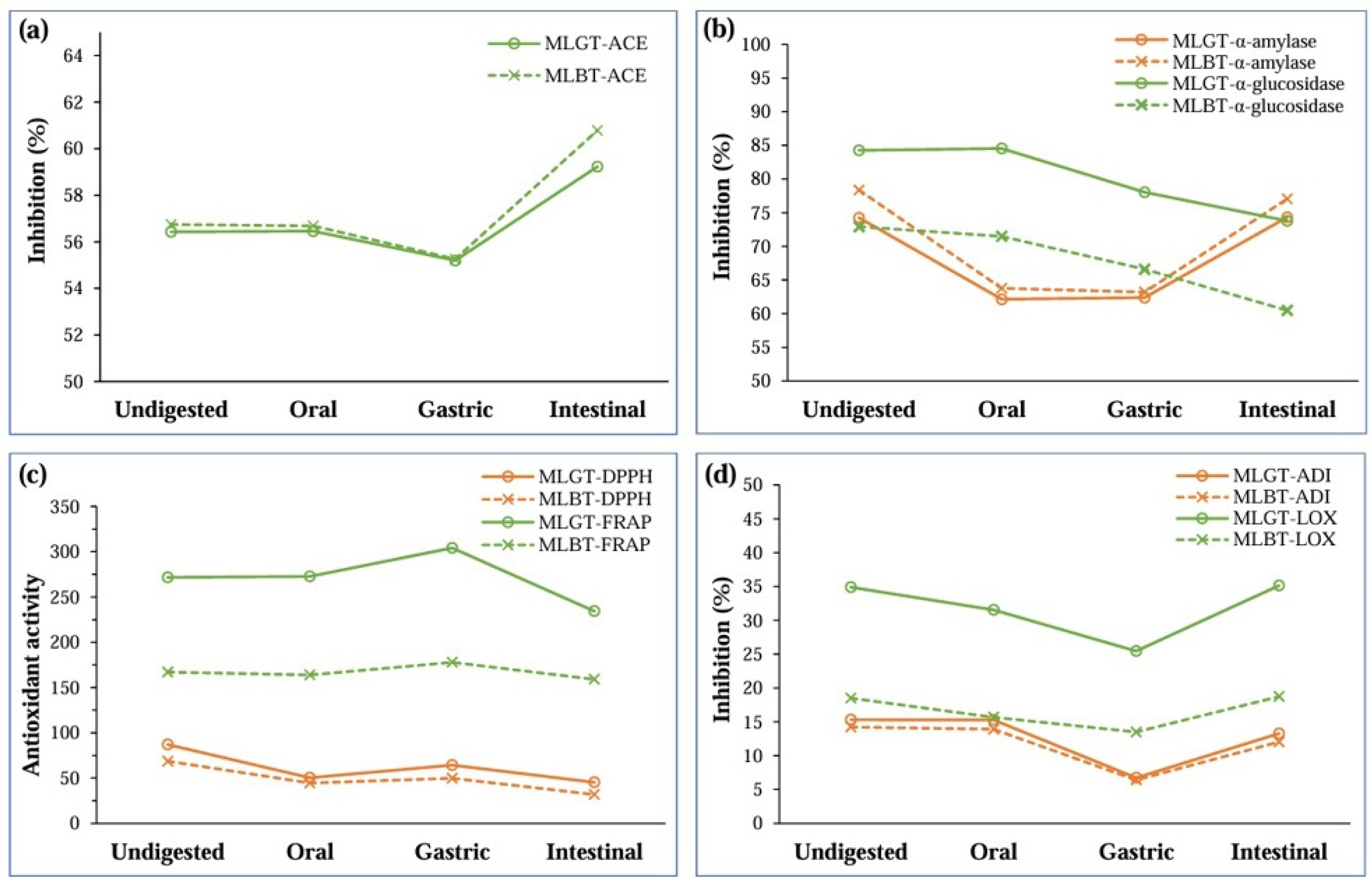
3.1. Changes in Bioactivity During Digestion

The bioactivities of mulberry leaf tea kombucha, including antihypertensive, antiglycemic, antioxidant, and anti-inflammatory activities, were assessed during different stages of simulated in vitro digestion (oral, gastric, and intestinal phases) compared to the undigested beverages, as shown in Figure 1.
Results showed that MLGT and MLBT kombucha maintained reasonably consistent ACE inhibition during different stages of in vitro digestion (Figure 1a). The ACE inhibition for MLGT was 56.43%, while MLBT was 56.75%. MLGT (56.46%) and MLBT (56.68%) retained their ACE inhibition values during the oral phase. However, both kombuchas showed a slight decrease in ACE inhibition in the stomach phase, with MLGT at 55.20% and MLBT at 55.26%. In the intestinal phase, MLGT and MLBT ACE inhibition increased to 59.23% and 60.78%, respectively.
The inhibition of α-amylase and α-glucosidase revealed distinct patterns (Figure 1b). The undigested MLGT and MLBT kombucha samples had the highest level of α-amylase inhibition, which remained relatively constant throughout the digestion process. In contrast, α-glucosidase inhibition significantly decreased in the intestinal phase.
There were notable variations in the antioxidant activity as determined by FRAP and DPPH radical scavenging investigations (Figure 1c). For MLGT kombucha, DPPH inhibition decreased from 87.06% (undigested) to 45.31% in the intestinal phase, while the FRAP value peaked in the gastric phase (304.10 mM FeSO4/100 mL) and was lowest in the intestinal phase (234.28 mM FeSO4/100 mL). Similar trends were observed in the MLBT kombucha, where the intestinal phase’s DPPH inhibition decreased from 68.71% (undigested) to 31.85% (intestinal phase). The FRAP values were highest in the gastric phase (177.80 mM FeSO4/100 mL) and decreased in the intestinal phase (159.07 mM FeSO4/100 mL). The reduction in DPPH inhibition following digestion in kombucha indicates the breakdown of polyphenols in the beverage.
The anti-inflammatory activity showed significant variation during the digestion phases as determined by ADI and LOX inhibition (Figure 1d). The undigested sample of MLGT kombucha had the highest ADI (15.31%), which declined throughout the gastric phase (6.73%) and then gained once more in the intestinal phase (13.27%). LOX inhibition decreased in the gastric phase (25.45%) while increasing in the intestinal phase (35.12%). ADI was highest in undigested samples (14.23%), considerably decreased in the gastric phase (6.41%), and increased in the intestinal phase (12.04%) for MLBT kombucha, which exhibited a similar trend.

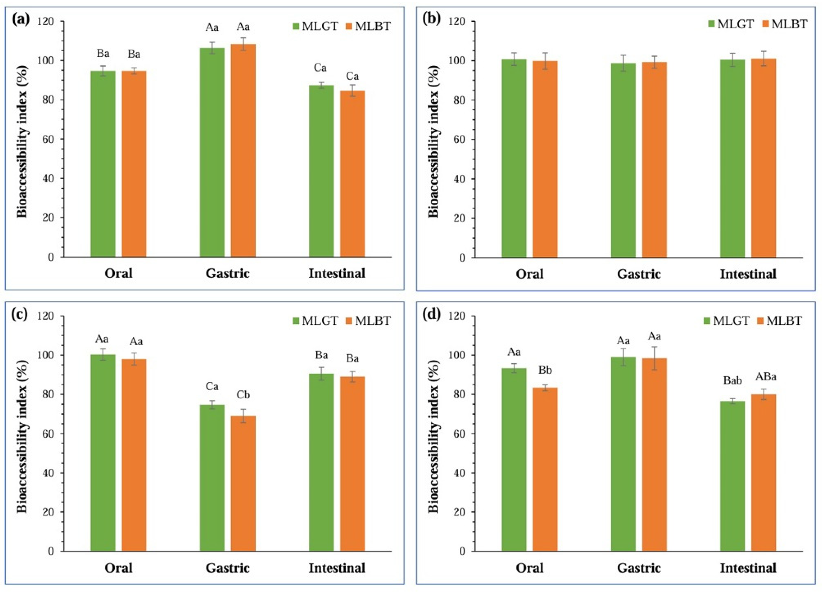
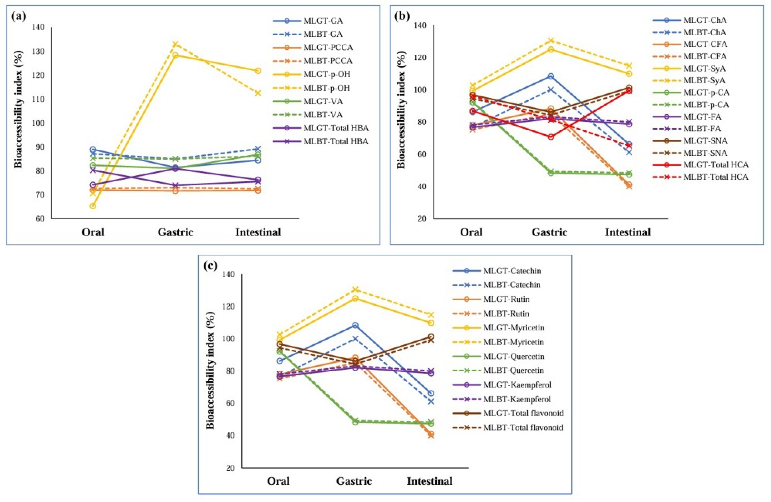
3.2. Bioaccessibility

Bioaccessibility refers to the proportion of a compound released from its food matrix during digestion and available for absorption in the gastrointestinal tract. Simulated in vitro digestion was used to assess the bioaccessibility of bioactive components from kombucha made from MLGT and MLBT, including DNJ, GABA, phenolic compounds, and flavonoids. During digestion, the bioaccessibility indices of several bioactive compounds, such as DNJ, GABA, TPC, and TFC, in various mulberry leaf tea kombucha samples were evaluated. Throughout digestion, there was a significant difference in the bioaccessibility of DNJ, phenolic compounds, and flavonoids, but not GABA (Figure 2).
The bioaccessibility index of phenolic compounds, such as hydroxybenzoic acids, hydroxycinnamic acids, and flavonoids, in various mulberry leaf tea kombucha during digestion is shown in Figure 3. During digestion, there was a significant difference in the bioaccessibility of phenolic acids, including hydroxybenzoic and hydroxycinnamic acids. For instance, ρ-hydroxybenzoic acid showed increased bioaccessibility in the gastric phase (128.31% for MLGT and 132.98% for MLBT) while decreased in the intestinal phase (121.83% for MLGT and 112.50% for MLBT).
In contrast, the bioaccessibility of hydroxycinnamic acids such as chlorogenic acid was higher in the gastric phase (108.33% for MLGT and 100.03% for MLBT) while decreasing significantly in the intestinal phase (66.17% for MLGT and 61.18% for MLBT). Flavonoids exhibited differential bioaccessibility during digestion. For instance, the highest values for catechin bioaccessibility were observed in the gastric phase for both MLGT (81.02%) and MLBT (77.34%). In contrast, values in the intestinal phase were decreased (79.99% for MLGT and 64.50% for MLBT). Similar trends were observed for rutin, a glycosylated flavonoid; bioaccessibility was highest in the gastric phase (73.67% for MLGT and 75.98% for MLBT) and dramatically decreased in the intestinal phase (53.83% for MLBT and 49.17% for MLGT).

3.3. Pearson’s Correlation Analysis

Pearson’s correlation analysis provides insights into the bioactive compounds in mulberry leaf kombucha, which are primarily responsible for their biological activities. Table 1 demonstrates Pearson’s correlation between bioactivity and bioactive compound concentrations (Supplementary Data S2). DPPH radical scavenging activity, there were significant positive correlations observed with caffeic acid (R2 = 0.872, p < 0.01), catechin (R2 = 0.829, p < 0.01), and ρ-coumaric acid (R2 = 0.663, p < 0.01), indicating that these compounds contribute to the antioxidant activity of the kombucha. On the other hand, TFC (R2 = −0.502, p < 0.05) was negatively correlated, indicating that not all flavonoids in the kombucha contribute equally to antioxidant potential. Strong positive correlations were found in the FRAP assay with DNJ (R2 = 0.949, p < 0.01) and ferulic acid (R2 = 0.932, p < 0.01), while a significant negative correlation was found with TPC (R2 = −0.892, p < 0.01) and several hydroxybenzoic acids.
Regarding α-amylase inhibition, non-significant correlations were observed for most bioactive compounds, indicating that the presence of these specific compounds in kombucha may not be the main reason for the inhibition. In terms of α-glucosidase inhibition, strong positive correlations with DNJ (R2 = 0.771, p < 0.01) and ρ-coumaric acid (R2 = 0.895, p < 0.01) suggest these compounds are potent inhibitors of α-glucosidase, supporting their role in antiglycemic effects.
For anti-inflammatory activity, albumin denaturation inhibition (ADI) was positively correlated with ρ-coumaric acid (ρ-CA, R2 = 0.540) and sinapic acid (SNA, R2 = 0.452). In contrast, another proxy of the anti-inflammatory activity, lipoxygenase (LOX) inhibition, was positively correlated with 1-deoxynojirimycin (DNJ), caffeic acid (CFA), syringic acid (SyA), ρ-CA, ferulic acid (FA), SNA, and catechin.
ACE inhibition indicates that these substances could play a role in modulating blood pressure, as they positively correlate with sinapic acid (R2 = 0.149) and GABA (R2 = 0.294). The negative correlations with caffeic acid (R2 = −0.478, p < 0.05) indicate that various compounds influence differently the inhibition of ACE.

4. Discussion

Hypertension and T2DM share common pathophysiological mechanisms, such as oxidative stress and vascular inflammation [7]. Therefore, phytochemicals that can scavenge free radicals and combat inflammation might be beneficial for preventing hypertension and T2DM [35,36]. The present study proved that fermented MLGT and MLBT kombucha beverages contain stable phytoactive compounds working against hypertension and diabetes based on two mechanisms: directly via antihypertensive and antiglycemic of the bioactive compounds and indirectly via antioxidant and anti-inflammatory properties.
Regarding antihypertensive activity, the remarkably continuous ACE inhibition observed throughout the various stages of digestion demonstrated that the kombucha fermentation process retained or even improved the bioavailability of ACE-inhibitory compounds. Human angiotensin-converting enzyme (ACE) is a crucial zinc-dependent enzyme in controlling blood pressure and fluid balance [37]. It works by regulating the renin-angiotensin-aldosterone system (RAAS). Blocking ACE activity is a fundamental approach to treating high blood pressure. Kombucha contains phenolic compounds that are known to suppress ACE [38]. In this study, we found that ACE inhibition was correlated with sinapic acid and GABA concentration. Previous studies showed that phenolic acids, including sinapic acid, inhibited ACE activity [39]. Mechanistically, sinapic acid directly binds with the ACE binding site with hydrophobic interactions and hydrogen bonding [40]. In contrast, GABA from purple-colored leaf tea can form hydrogen bonds with the ACE residues Tyr523 and Glu411 and tunnels inside to chelate directly with the zinc ion, reducing the ACE activity and thus inhibiting angiotensin I conversion to angiotensin II [41]. Our findings align with the abovementioned analysis and prompted further in vivo investigations.
Fermentation can enhance the concentration and bioavailability of these polyphenols in kombuchas prepared from various teas [42]. The presence of different microbial species in kombucha, which convert complex polyphenols into more straightforward, more accessible forms, is likely to explain the observed increase in antihypertensive activity [43]. Gaggía and colleagues suggest that the recovery could be attributed to the release of peptide fragments from protein digestion that inhibit ACE [44]. The positive correlation with ACE inhibition supports GABA’s ability to regulate blood pressure and its potential for bioactivity in fermented beverages [43]. However, a negative correlation with caffeic acid indicates that its hypotensive action could not be straightforward and could be influenced by other interacting factors. The study demonstrates that mulberry leaf kombucha exhibits diverse bioactivities, primarily driven by specific bioactive compounds such as DNJ, catechin, caffeic acid, and ρ-coumaric acid. These findings provide valuable insights into the potential health benefits of kombucha and underscore the importance of targeted research on the specific roles of different bioactive compounds in fermented beverages.
The increasing prevalence of type 2 diabetes and the unsatisfactory side effects of commercially available antidiabetic drugs led to the development of novel therapeutic strategies for controlling postprandial glucose levels [45]. With fewer adverse effects than manufactured medications, natural enzyme inhibitors of the digestion of carbohydrates may be an effective option for preventing the absorption of carbohydrates from food. A metalloenzyme enzyme α-amylase breaks down polysaccharides into smaller molecules like maltose and maltotriose, which α-glucosidase further digests [46]. Thus, these enzymes contribute to postprandial hyperglycemia and high blood glucose levels. To reduce postprandial plasma glucose levels, scientists develop drugs that target α-amylase and α-glucosidase inhibition that can slow down the release of glucose from carbohydrate chains and prolong its absorption.
We found a relationship between the fermented MLGT and MLBT kombucha beverages’ antiglycemic properties and the bioaccessibility and bioactivity of their active ingredients. The reduced bioaccessibility of 1-deoxynojirimycin (DNJ), total phenolic compounds (TPCs), and total flavonoid compounds (TFCs), but not γ-aminobutyric acid (GABA) in the intestinal phase. Accordingly, the bioactivity of MLGT and MLBT kombucha showed stable α-amylase inhibition, but α-glucosidase inhibition significantly decreased in the intestinal phase. As the post-α-amylase dextrins were thought to be converted to glucose by intestinal mucosal α-glucosidase, starches can be digested differently by the mucosal α-glucosidase [47]. Thus, it can be implied that the reduced α-glucosidase inhibition was due to reduced DNJ, TPCs, and TFCs. By blocking α-glucosidase activity, DNJ lowers blood insulin and glucose levels and enhances the metabolism of carbohydrates [48]. Recently, dose–response inhibition of the α-glucosidase activity of phenolic compounds and flavonoids [49,50]. However, phenolic compounds are more stable in an acidic pH milieu like the stomach (pH 2.0) than in an alkaline condition, explaining why the α-glucosidase inhibition in the intestinal phase (pH 7.0) declined [51].
Additionally, a recent study reported that flavonoids recovered in acidic pH ranges (2.5–3.5) and fell in higher pH ranges [52]. Developing digestive matrices that shield them from enzymatic hydrolysis may also increase their stability [53]. Nevertheless, the reduction in α-glucosidase inhibition in MLBT indicates the partial decomposition or alteration of active substances, which correlates with studies about the digestion of flavonoid-rich foods [54]. The vital role that DNJ plays in lowering postprandial glucose levels is highlighted by its considerable positive correlation with α-glucosidase inhibition, which supports its usage as a functional component in diabetes management [53]. It is worth noting that enzyme inhibition data were obtained using a single concentration of kombucha. While useful for comparing digestion-phase trends, this approach limits the ability to determine maximal inhibitory potential. Future studies should include dose–response evaluations to determine IC50 values for key enzyme targets to substantiate bioactivity claims and optimize dosage recommendations.
As oxidative stress and low-grade inflammation are hallmarks of hypertension and T2DM [55], we also evaluated the anti-inflammatory and antioxidant properties of the fermented MLGT and MLBT kombucha beverages. MLBT retained better bioaccessibility and antihypertensive activity through digestion, but MLGT kombucha displayed higher initial phenolic content and antioxidant activity. These results are consistent with earlier research demonstrating that phenolic compounds in kombucha degrade during digestion, although they can also increase due to bound phenolics released by enzymatic hydrolysis [13]. Higher TPCs and TFCs values in MLGT at the initial stages correlate with the higher antioxidant activities of the undigested kombucha beverages; however, these levels decreased during digestion. Its relatively consistent FRAP values suggest that TFC’s stability in MLBT during the gastric phase contributes to its persistent antioxidant capability. Antolak et al. stated that the fermentation process and the phenolic content from the tea base have an essential effect on the antioxidant properties of kombucha [13]. Based on its higher TPC and TFC, MLGT has a more profound initial antioxidant activity. This finding is consistent with Wanyo et al., who found that higher phenolic contents contribute to increased antioxidant capacities [29].
During in vitro digestion, the bioaccessibility of several bioactive compounds was influenced by physicochemical and biochemical factors, particularly pH changes, enzymatic hydrolysis, and compound–matrix interactions. The gastric phase (pH ~2.0) often promotes the hydrolysis of polyphenolic glycosides, leading to an increase in the detectable levels of free phenolic acids such as ρ-hydroxybenzoic acid and chlorogenic acid, consistent with the observed rise in their bioaccessibility [56]. The acidic gastric environment may also cause the protein to unfold, thereby releasing bound phenolics and flavonoids from the food matrix or SCOBY-derived metabolites [57].
Conversely, during the intestinal phase (pH ~7.0), many acid-sensitive compounds (e.g., catechins, rutin) undergo oxidation, deconjugation, or degradation, resulting in a notable decline in their measured bioaccessibility. Enzymes such as pancreatin and bile salts facilitate the emulsification and breakdown of phenolic–macromolecule complexes, which may further expose them to degradation or microbial transformation. DNJ and GABA’s fluctuations across phases may relate to enzyme-mediated liberation or reabsorption dynamics, where compounds temporarily bind to proteins or fibers and are re-released under different digestive conditions [19]. These phase-dependent patterns underscore the complexity of compound stability during gastrointestinal transit and suggest the need for encapsulation or matrix shielding strategies to protect labile phytochemicals during digestion [58].
The observed nonlinear release patterns of DNJ and GABA during digestion reflect complex biochemical and physicochemical transformations. The initial decline of DNJ in the oral phase may result from temporary pH-induced aggregation or matrix interaction that reduces solubility or detectability rather than actual salivary protein binding. However, in the gastric phase, enzymatic proteolysis and acidic pH conditions (pH 2.0) can enhance matrix breakdown, thereby releasing DNJ that was previously bound or encapsulated within cellular structures of the tea matrix or SCOBY metabolites [19,58].
A similar dynamic applies to GABA: its initial increase in the oral phase may be due to rapid diffusion from the aqueous kombucha matrix. In the gastric phase, partial adsorption or complexation with precipitated proteins may reduce its free concentration. However, during the intestinal phase, the solubilizing effects of bile salts and enzymatic hydrolysis by pancreatin facilitate the re-release of GABA, resulting in an observed rebound in its concentration [59].
These observations are consistent with the matrix–compound interaction model, where phytochemicals may shift between bound, partially solubilized, and freely bioaccessible forms depending on pH, ionic strength, and enzyme-mediated hydrolysis during digestion [60].
The anti-inflammatory activity of MLGT and MLBT kombucha was reduced in the gastric but recovered in the intestinal phase. These variations indicate that some bioactive compounds are degraded in the stomach. However, others may be released or reactivated within the intestine, such as DNJ, CFA, SyA, ρ-CA, FA, SNA, and catechin, indicating that these compounds might mitigate inflammation [61]. Not all compounds contribute positively to anti-inflammatory action, reflected by significant negative correlations for hydroxycinnamic acids, myricetin, quercetin, and kaempferol. According to the correlation data, by inhibiting lipoxygenase and preventing albumin denaturation, catechin, sinapic acid, and ρ-coumaric acid could have an essential role in the anti-inflammatory effects. Reducing the inflammatory response suggests a protective function against inflammation-associated diseases [62].
Traditional kombucha beverages, primarily prepared from black and green teas, have been extensively studied for their antioxidant and antimicrobial properties. However, studies focusing on their antihypertensive and antiglycemic capacities are comparatively limited [14,44]. In comparison, mulberry leaf tea kombucha investigated in this study demonstrated superior retention of bioactive compounds across the gastrointestinal phases, attributed largely to its unique content of 1-deoxynojirimycin (DNJ) and γ-aminobutyric acid (GABA). Unlike traditional tea kombucha, DNJ provides potent α-glucosidase inhibitory activity, which could support blood glucose regulation [14,25]. Additionally, mulberry leaf kombucha maintained higher ACE inhibition after intestinal digestion, suggesting stronger resilience of antihypertensive compounds [44]. These comparative advantages position mulberry leaf tea kombucha as a promising functional beverage candidate for supporting metabolic health management beyond the traditional antioxidant roles emphasized in classic tea-based kombuchas.
The results of this in vitro study highlight the phase-dependent release and activity of key bioactives in mulberry leaf kombucha. However, further research is necessary to validate these findings and maximize their translational value. In vivo studies using animal models or human volunteers are essential to confirm the physiological relevance of the observed antihypertensive and antiglycemic effects. Furthermore, strategies such as microencapsulation, co-fermentation with probiotic strains, or matrix modification using dietary fibers or emulsifiers should be explored to improve bioavailability and compound retention. Such approaches may protect unstable compounds like flavonoids and DNJ from degradation in the gastrointestinal tract and support their sustained release. Additionally, metabolomic profiling and gut microbiota analysis could provide deeper insight into kombucha-derived phytochemicals’ metabolic fate and bioconversion. These strategies would contribute to developing next-generation functional kombucha products with enhanced health benefits.

5. Conclusions

In conclusion, kombucha prepared from mulberry leaf green and black teas exhibits promising in vitro antihypertensive, antiglycemic, antioxidant, and anti-inflammatory properties. These effects are closely linked to the bioaccessibility and digestion-phase stability of phenolic acids, DNJ, and GABA. Notably, MLBT kombucha demonstrated greater resistance to compound degradation, suggesting its potential for enhanced functionality in physiological settings. However, these findings are based on a static in vitro gastrointestinal digestion model, and the observed disease-inhibitory activities should be interpreted as preliminary indications of potential efficacy. In vivo validation, including human or animal studies, is essential to substantiate these effects and to assess bioavailability, absorption, metabolism, and clinical relevance.

Supplementary Materials

The following supporting information can be downloaded at: https://www.mdpi.com/article/10.3390/fermentation11050258/s1. Data S1. Changes of bioactivity in mulberry leaf tea kombucha during simulated in vitro digestion; Data S2: Bioactive compounds in mulberry leaf tea kombucha during in vitro digestion.

Author Contributions

Conceptualization, P.W. and N.T.; methodology, P.W.; software, T.C.; validation, K.T., P.W. and N.T.; formal analysis, P.W.; investigation, P.W. and T.C.; resources, N.T. and L.K.N.; data curation, P.W. and L.K.N.; writing—original draft preparation, P.W.; writing—review and editing, T.C., L.K.N., N.T. and K.T.; visualization, P.W.; supervision, K.T., L.K.N. and N.T.; project administration, T.C.; funding acquisition, P.W., N.T. and K.T. All authors have read and agreed to the published version of the manuscript.

Funding

This research is financially supported by Mahasarakham University, Thailand. The funding number is PHEP-SEA 2025.

Institutional Review Board Statement

Not applicable.

Informed Consent Statement

Not applicable.

Data Availability Statement

The raw data supporting the conclusions of this article will be made available by the authors on request.

Acknowledgments

The authors acknowledge Kalasin University for partial support of the investigations.

Conflicts of Interest

The authors declare no conflicts of interest.

References

  1. Ortega-Montiel, J.; Montoya, A.; Soria-Saucedo, R.; Gallegos-Carrillo, K.; Ramirez-Palacios, P.; Salmeron, J.; Salazar-Martinez, E. Trends of antihypertensive, antidiabetic, and nonsteroidal anti-inflammatory drugs use among the health workers cohort study, Mexico 2004 to 2018. Adv. Pharmacol. Pharm. Sci. 2023, 2023, 5555274. [Google Scholar] [CrossRef] [PubMed]
  2. Mlynarska, E.; Biskup, L.; Mozdzan, M.; Grygorcewicz, O.; Mozdzan, Z.; Semeradt, J.; Uramowski, M.; Rysz, J.; Franczyk, B. The role of oxidative stress in hypertension: The insight into antihypertensive properties of vitamins A, C and E. Antioxidants 2024, 13, 848. [Google Scholar] [CrossRef] [PubMed]
  3. Gonzalez, P.; Lozano, P.; Ros, G.; Solano, F. Hyperglycemia and oxidative stress: An integral, updated and critical overview of their metabolic interconnections. Int. J. Mol. Sci. 2023, 24, 9352. [Google Scholar] [CrossRef] [PubMed]
  4. Puschel, G.P.; Klauder, J.; Henkel, J. Macrophages, low-grade inflammation, insulin resistance and hyperinsulinemia: A mutual ambiguous relationship in the development of metabolic diseases. J. Clin. Med. 2022, 11, 4358. [Google Scholar] [CrossRef]
  5. Maranduca, M.A.; Tanase, D.M.; Branisteanu, D.C.; Serban, D.N.; Branisteanu, D.E.; Serban, I.L. Involvement of proinflammatory cytokines in angiotensin II-induced hypertension in rat. Exp. Ther. Med. 2020, 20, 3541–3545. [Google Scholar] [CrossRef]
  6. Moke, E.G.; Omogbai, E.K.I.; Osagie-Eweka, S.E.; Uchendu, A.P.; Obayuwana, O.M.; Okoro-Akpandu, E.; Ben-Azu, B. Antihypertensive and antihyperglycemic effects of combinations of losartan with metformin and/or glibenclamide in desoxycorticosterone acetate and streptozotocin-induced hypertensive diabetic rats. Lab. Anim. Res. 2023, 39, 7. [Google Scholar] [CrossRef]
  7. Petrie, J.R.; Guzik, T.J.; Touyz, R.M. Diabetes, hypertension, and cardiovascular disease: Clinical insights and vascular mechanisms. Can. J. Cardiol. 2018, 34, 575–584. [Google Scholar] [CrossRef]
  8. Nugraha, R.; Kurniawan, F.; Abdullah, A.; Lopata, A.L.; Ruethers, T. Antihypertensive and antidiabetic drug candidates from milkfish (chanos chanos)-identification and characterization through an integrated bioinformatic approach. Foods 2024, 13, 2594. [Google Scholar] [CrossRef]
  9. Vignesh, A.; Amal, T.C.; Sarvalingam, A.; Vasanth, K. A Review on the influence of nutraceuticals and functional foods on health. Food Chem. Adv. 2024, 2024, 100749. [Google Scholar] [CrossRef]
  10. Corbo, M.R.; Bevilacqua, A.; Petruzzi, L.; Casanova, F.P.; Sinigaglia, M. Functional beverages: The emerging side of functional foods. Compr. Rev. Food Sci. Food Saf. 2014, 13, 1192–1206. [Google Scholar] [CrossRef]
  11. Misra, S.; Pandey, P.; Mishra, H.N. Novel approaches for co-encapsulation of probiotic bacteria with bioactive compounds, their health benefits and functional food product development: A review. Trends Food Sci. Technol. 2021, 109, 340–351. [Google Scholar] [CrossRef]
  12. Gupta, A.; Sanwal, N.; Bareen, M.A.; Barua, S.; Sharma, N.; Joshua Olatunji, O.; Prakash Nirmal, N.; Sahu, J.K. Trends in functional beverages: Functional ingredients, processing technologies, stability, health benefits, and consumer perspective. Food Res. Int. 2023, 170, 113046. [Google Scholar] [CrossRef] [PubMed]
  13. Antolak, H.; Piechota, D.; Kucharska, A. Kombucha Tea—A double power of bioactive compounds from tea and symbiotic culture of bacteria and yeasts (SCOBY). Antioxidants 2021, 10, 1541. [Google Scholar] [CrossRef]
  14. Jayabalan, R.; Malbasa, R.V.; Loncar, E.S.; Vitas, J.S.; Sathishkumar, M. A Review on kombucha tea-microbiology, composition, fermentation, beneficial effects, toxicity, and tea fungus. Compr. Rev. Food Sci. Food Saf. 2014, 13, 538–550. [Google Scholar] [CrossRef] [PubMed]
  15. Su, J.; Tan, Q.; Wu, S.; Abbas, B.; Yang, M. Application of kombucha fermentation broth for antibacterial, antioxidant, and anti-inflammatory processes. Int. J. Mol. Sci. 2023, 24, 13948. [Google Scholar] [CrossRef]
  16. Greenwalt, C.J.; Steinkraus, K.H.; Ledford, R.A. Kombucha, the fermented tea: Microbiology, composition, and claimed health effects. J. Food Prot. 2000, 63, 976–981. [Google Scholar] [CrossRef] [PubMed]
  17. Batista, P.; Rodrigues Penas, M.; Vila-Real, C.; Pintado, M.; Oliveira-Silva, P. Kombucha: Challenges for health and mental health. Foods 2023, 12, 3378. [Google Scholar] [CrossRef]
  18. Chakravorty, S.; Bhattacharya, S.; Chatzinotas, A.; Chakraborty, W.; Bhattacharya, D.; Gachhui, R. Kombucha tea fermentation: Microbial and biochemical dynamics. Int. J. Food Microbiol. 2016, 220, 63–72. [Google Scholar] [CrossRef]
  19. Tu, J.; Liu, G.; Jin, Y.; Tang, C.; Yao, T.; Zhuo, J.; Li, Q.; Liu, L.; Wang, J. Enrichment of γ-aminobutyric acid in mulberry leaves and the inhibitory effects of the water extract on ACE and α-glucosidase activity. Ind. Crops Prod. 2022, 177, 114485. [Google Scholar] [CrossRef]
  20. Marchetti, L.; Truzzi, E.; Frosi, I.; Papetti, A.; Cappellozza, S.; Saviane, A.; Pellati, F.; Bertelli, D. In vitro bioactivity evaluation of mulberry leaf extracts as nutraceuticals for the management of diabetes mellitus. Food Funct. 2022, 13, 4344–4359. [Google Scholar] [CrossRef]
  21. Fongsodsri, K.; Thaipitakwong, T.; Rujimongkon, K.; Kanjanapruthipong, T.; Ampawong, S.; Reamtong, O.; Aramwit, P. Mulberry-derived 1-deoxynojirimycin prevents type 2 diabetes mellitus progression via modulation of retinol-binding protein 4 and haptoglobin. Nutrients 2022, 14, 4538. [Google Scholar] [CrossRef] [PubMed]
  22. He, L.; Xing, Y.; Ren, X.; Zheng, M.; Yu, S.; Wang, Y.; Xiu, Z.; Dong, Y. Mulberry leaf extract improves metabolic syndrome by alleviating lipid accumulation in vitro and in vivo. Molecules 2022, 27, 5111. [Google Scholar] [CrossRef]
  23. Li, R.; Zhu, Q.; Wang, X.; Wang, H. Mulberry leaf polyphenols alleviated high-fat diet-induced obesity in mice. Front. Nutr. 2022, 9, 979058. [Google Scholar] [CrossRef] [PubMed]
  24. Cabrera, C.; Artacho, R.; Giménez, R. Beneficial effects of green tea—A review. J. Am. Coll. Nutr. 2006, 25, 79–99. [Google Scholar] [CrossRef] [PubMed]
  25. Villarreal-Soto, S.A.; Beaufort, S.; Bouajila, J.; Souchard, J.P.; Taillandier, P. Understanding kombucha tea fermentation: A review. J. Food Sci. 2018, 83, 580–588. [Google Scholar] [CrossRef]
  26. Wu, Q.; Du, J.; Jia, J.; Kuang, C. Production of ACE inhibitory peptides from sweet sorghum grain protein using alcalase: Hydrolysis kinetic, purification and molecular docking study. Food Chem. 2016, 199, 140–149. [Google Scholar] [CrossRef]
  27. Kwon, Y.-I.; Apostolidis, E.; Shetty, K. Inhibitory potential of wine and tea against α-amylase and α-glucosidase for management of hyperglycemia linked to type 2 diabetes. J. Food Biochem. 2008, 32, 15–31. [Google Scholar] [CrossRef]
  28. Kim, Y.M.; Jeong, Y.K.; Wang, M.H.; Lee, W.Y.; Rhee, H.I. Inhibitory effect of pine extract on alpha-glucosidase activity and postprandial hyperglycemia. Nutrition 2005, 21, 756–761. [Google Scholar] [CrossRef]
  29. Wanyo, P.; Kaewseejan, N.; Meeso, N.; Siriamornpun, S. Bioactive compounds and antioxidant properties of different solvent extracts derived from Thai rice by-products. Appl. Biol. Chem. 2016, 59, 373–384. [Google Scholar] [CrossRef]
  30. Williams, L.A.; O’Connar, A.; Latore, L.; Dennis, O.; Ringer, S.; Whittaker, J.A.; Conrad, J.; Vogler, B.; Rosner, H.; Kraus, W. The in vitro anti-denaturation effects induced by natural products and non-steroidal compounds in heat treated (immunogenic) bovine serum albumin is proposed as a screening assay for the detection of anti-inflammatory compounds, without the use of animals, in the early stages of the drug discovery process. West Indian Med. J. 2008, 57, 327–331. [Google Scholar]
  31. Yawer, M.A.; Ahmed, E.; Malik, A.; Ashraf, M.; Rasool, M.A.; Afza, N. New Lipoxygenase-inhibiting constituents from calligonum polygonoides. Chem. Biodivers. 2007, 4, 1578–1585. [Google Scholar] [CrossRef] [PubMed]
  32. Kim, J.W.; Kim, S.U.; Lee, H.S.; Kim, I.; Ahn, M.Y.; Ryu, K.S. Determination of 1-deoxynojirimycin in Morus alba L. leaves by derivatization with 9-fluorenylmethyl chloroformate followed by reversed-phase high-performance liquid chromatography. J. Chromatogr. A 2003, 1002, 93–99. [Google Scholar] [CrossRef] [PubMed]
  33. Jin, Y.; Tu, J.; Han, X.; Zhuo, J.; Liu, G.; Han, Y.; Du, H.; Wang, J.; Xiao, H. Characteristics of mulberry leaf powder enriched with gamma-aminobutyric acid and its antioxidant capacity as a potential functional food ingredient. Front. Nutr. 2022, 9, 900718. [Google Scholar] [CrossRef] [PubMed]
  34. Santana Andrade, J.K.; Chagas Barros, R.G.; Pereira, U.C.; Nogueira, J.P.; Gualberto, N.C.; Santos de Oliveira, C.; Shanmugam, S.; Narain, N. Bioaccessibility of bioactive compounds after in vitro gastrointestinal digestion and probiotics fermentation of Brazilian fruits residues with antioxidant and antidiabetic potential. LWT 2022, 153, 112469. [Google Scholar] [CrossRef]
  35. Prado, N.J.; Ramirez, D.; Mazzei, L.; Parra, M.; Casarotto, M.; Calvo, J.P.; Cuello Carrion, D.; Ponce Zumino, A.Z.; Diez, E.R.; Camargo, A.; et al. Anti-inflammatory, antioxidant, antihypertensive, and antiarrhythmic effect of indole-3-carbinol, a phytochemical derived from cruciferous vegetables. Heliyon 2022, 8, e08989. [Google Scholar] [CrossRef]
  36. Arabshomali, A.; Bazzazzadehgan, S.; Mahdi, F.; Shariat-Madar, Z. Potential benefits of antioxidant phytochemicals in type 2 diabetes. Molecules 2023, 28, 7209. [Google Scholar] [CrossRef]
  37. Acharya, K.R.; Gregory, K.S.; Sturrock, E.D. Advances in the structural basis for angiotensin-1 converting enzyme (ACE) inhibitors. Biosci. Rep. 2024, 44, BSR20240130. [Google Scholar] [CrossRef]
  38. Anantachoke, N.; Duangrat, R.; Sutthiphatkul, T.; Ochaikul, D.; Mangmool, S. Kombucha beverages produced from fruits, vegetables, and plants: A review on their pharmacological activities and health benefits. Foods 2023, 12, 1818. [Google Scholar] [CrossRef]
  39. Yu, M.; Kim, H.J.; Heo, H.; Kim, M.; Jeon, Y.; Lee, H.; Lee, J. Comparison of the antihypertensive activity of phenolic acids. Molecules 2022, 27, 6185. [Google Scholar] [CrossRef]
  40. Abdullah, R.; Arshad, H.; Kaleem, A.; Iqtedar, M.; Aftab, M.; Saleem, F. Assessment of angiotensin converting enzyme inhibitory activity and quality attributes of yoghurt enriched with Cinnamomum verum, Elettaria cardamomum, Beta vulgaris and Brassica oleracea. Saudi J. Biol. Sci. 2023, 30, 103556. [Google Scholar] [CrossRef]
  41. Yang, G.; Zhu, Y.; Shi, J.; Peng, Q.; Lin, Z.; Lv, H. Effects of anaerobic treatment on the non-volatile components and angiotensin-converting enzyme (ACE) inhibitory activity of purple-colored leaf tea. Food Chem. X 2024, 23, 101649. [Google Scholar] [CrossRef] [PubMed]
  42. Kapp, J.M.; Sumner, W. Kombucha: A systematic review of the empirical evidence of human health benefit. Ann. Epidemiol. 2019, 30, 66–70. [Google Scholar] [CrossRef]
  43. Cardoso, R.R.; Neto, R.O.; dos Santos D’Almeida, C.T.; do Nascimento, T.P.; Pressete, C.G.; Azevedo, L.; Martino, H.S.D.; Cameron, L.C.; Ferreira, M.S.L.; Barros, F.A.R.d. Kombuchas from green and black teas have different phenolic profile, which impacts their antioxidant capacities, antibacterial and antiproliferative activities. Food Res. Int. 2020, 128, 108782. [Google Scholar] [CrossRef] [PubMed]
  44. Gaggìa, F.; Baffoni, L.; Galiano, M.; Nielsen, D.S.; Jakobsen, R.R.; Castro-Mejía, J.L.; Bosi, S.; Truzzi, F.; Musumeci, F.; Dinelli, G.; et al. Kombucha beverage from green, black and rooibos teas: A comparative study looking at microbiology, chemistry and antioxidant activity. Nutrients 2019, 11, 1. [Google Scholar] [CrossRef] [PubMed]
  45. Padhi, S.; Nayak, A.K.; Behera, A. Type II diabetes mellitus: A review on recent drug based therapeutics. Biomed. Pharmacother. 2020, 131, 110708. [Google Scholar] [CrossRef]
  46. Lee, B.H.; Eskandari, R.; Jones, K.; Reddy, K.R.; Quezada-Calvillo, R.; Nichols, B.L.; Rose, D.R.; Hamaker, B.R.; Pinto, B.M. Modulation of starch digestion for slow glucose release through “toggling” of activities of mucosal alpha-glucosidases. J. Biol. Chem. 2012, 287, 31929–31938. [Google Scholar] [CrossRef]
  47. Lin, A.H.; Lee, B.H.; Nichols, B.L.; Quezada-Calvillo, R.; Rose, D.R.; Naim, H.Y.; Hamaker, B.R. Starch source influences dietary glucose generation at the mucosal alpha-glucosidase level. J. Biol. Chem. 2012, 287, 36917–36921. [Google Scholar] [CrossRef]
  48. Zhang, R.; Zhang, Y.; Huang, G.; Xin, X.; Tang, L.; Li, H.; Lee, K.S.; Jin, B.R.; Gui, Z. Chemical synthesis, inhibitory activity and molecular mechanism of 1-deoxynojirimycin-chrysin as a potent alpha-glucosidase inhibitor. RSC Adv. 2021, 11, 38703–38711. [Google Scholar] [CrossRef]
  49. An, H.; Thanh, L.N.; Khanh, L.Q.; Ryu, S.H.; Lee, S.; Yeon, S.W.; Lee, H.H.; Turk, A.; Lee, K.Y.; Hwang, B.Y.; et al. Characterization of antioxidant and alpha-glucosidase inhibitory compounds of Cratoxylum formosum ssp. pruniflorum and optimization of extraction condition. Antioxidants 2023, 12, 511. [Google Scholar] [CrossRef]
  50. Barber, E.; Houghton, M.J.; Williamson, G. Flavonoids as human intestinal alpha-glucosidase inhibitors. Foods 2021, 10, 1939. [Google Scholar] [CrossRef]
  51. Saarniit, K.; Lang, H.; Kuldjarv, R.; Laaksonen, O.; Rosenvald, S. The stability of phenolic compounds in fruit, berry, and vegetable purees based on accelerated shelf-life testing methodology. Foods 2023, 12, 1777. [Google Scholar] [CrossRef]
  52. Chaves, J.O.; de Souza, M.C.; da Silva, L.C.; Lachos-Perez, D.; Torres-Mayanga, P.C.; Machado, A.; Forster-Carneiro, T.; Vazquez-Espinosa, M.; Gonzalez-de-Peredo, A.V.; Barbero, G.F.; et al. Extraction of flavonoids from natural sources using modern techniques. Front. Chem. 2020, 8, 507887. [Google Scholar] [CrossRef] [PubMed]
  53. Kluz, M.I.; Pietrzyk, K.; Pastuszczak, M.; Kacaniova, M.; Kita, A.; Kapusta, I.; Zaguła, G.; Zagrobelna, E.; Struś, K.; Marciniak-Lukasiak, K.; et al. Microbiological and physicochemical composition of various types of homemade kombucha beverages using alternative kinds of sugars. Foods 2022, 11, 1523. [Google Scholar] [CrossRef]
  54. Wu, P.; Deng, R.; Wu, X.; Wang, Y.; Dong, Z.; Dhital, S.; Chen, X.D. In vitro gastric digestion of cooked white and brown rice using a dynamic rat stomach model. Food Chem. 2017, 237, 1065–1072. [Google Scholar] [CrossRef] [PubMed]
  55. Pouvreau, C.; Dayre, A.; Butkowski, E.G.; de Jong, B.; Jelinek, H.F. Inflammation and oxidative stress markers in diabetes and hypertension. J. Inflamm. Res. 2018, 11, 61–68. [Google Scholar] [CrossRef] [PubMed]
  56. Odriozola-Serrano, I.; Nogueira, D.P.; Esparza, I.; Vaz, A.A.; Jimenez-Moreno, N.; Martin-Belloso, O.; Ancin-Azpilicueta, C. Stability and bioaccessibility of phenolic compounds in rosehip extracts during in vitro digestion. Antioxidants 2023, 12, 1035. [Google Scholar] [CrossRef]
  57. Guan, H.; Zhang, W.; Sun-Waterhouse, D.; Jiang, Y.; Li, F.; Waterhouse, G.I.; Li, D. Phenolic-protein interactions in foods and post ingestion: Switches empowering health outcomes. Trends Food Sci. Technol. 2021, 118, 71–86. [Google Scholar] [CrossRef]
  58. Minekus, M.; Alminger, M.; Alvito, P.; Ballance, S.; Bohn, T.; Bourlieu, C.; Carriere, F.; Boutrou, R.; Corredig, M.; Dupont, D.; et al. A standardised static in vitro digestion method suitable for food—An international consensus. Food Funct. 2014, 5, 1113–1124. [Google Scholar] [CrossRef]
  59. Bouayed, J.; Bohn, T. Exogenous antioxidants—Double-edged swords in cellular redox state: Health beneficial effects at physiologic doses versus deleterious effects at high doses. Oxid. Med. Cell Longev. 2010, 3, 228–237. [Google Scholar] [CrossRef]
  60. Macagnan, F.T.; da Silva, L.P.; Hecktheuer, L.H. Dietary fibre: The scientific search for an ideal definition and methodology of analysis, and its physiological importance as a carrier of bioactive compounds. Food Res. Int. 2016, 85, 144–154. [Google Scholar] [CrossRef]
  61. De Filippis, F.; Troise, A.D.; Vitaglione, P.; Ercolini, D. Different temperatures select distinctive acetic acid bacteria species and promotes organic acids production during kombucha tea fermentation. Food Microbiol. 2018, 73, 11–16. [Google Scholar] [CrossRef] [PubMed]
  62. Morales, D. Biological activities of kombucha beverages: The need of clinical evidence. Trends Food Sci. Technol. 2020, 105, 323–333. [Google Scholar] [CrossRef]
Figure 1. Changes in bioactivities of mulberry leaf tea kombucha during in vitro digestion. (a) ACE-inhibitory activity (%); (b) α-amylase and α-glucosidase inhibition (%); (c) antioxidant activity measured by DPPH scavenging (%) and FRAP (mM FeSO4/100 mL); (d) anti-inflammatory activity assessed by albumin denaturation inhibition (ADI, %) and lipoxygenase (LOX, %) inhibition. MLGT = mulberry leaf green tea kombucha; MLBT = mulberry leaf black tea kombucha. Different lines represent the digestion phases: Undigested, Oral, Gastric, and Intestinal. Data are presented as mean values (n = 3).
Fermentation 11 00258 g001
Figure 2. Bioaccessibility index of bioactive compounds in mulberry leaf tea kombucha during in vitro digestion: (a) 1-deoxynojirimycin (DNJ), (b) γ-aminobutyric acid (GABA), (c) total phenolic content (TPC), and (d) total flavonoid content (TFC). Green bars represent MLGT kombucha; orange bars represent MLBT kombucha. Values are expressed as mean ± standard deviation (n = 3). Different lowercase letters (a, b) within the same tea type indicate significant differences between digestion phases (p < 0.05). Different uppercase letters (A–C) indicate significant differences between MLGT and MLBT in the same digestion phase (p < 0.05).
Fermentation 11 00258 g002
Figure 3. Bioaccessibility index of individual phenolic acids and flavonoids in mulberry leaf tea kombucha during in vitro digestion. (a) hydroxybenzoic acids: GA = gallic acid, PCCA = protocatechuic acid, ρ-OH = ρ-hydroxybenzoic acid, VA = vanillic acid, and Total HBA = total hydroxybenzoic acids. (b) hydroxycinnamic acids: ChA = chlorogenic acid, CFA = caffeic acid, SyA = syringic acid, ρ-CA = ρ-coumaric acid, FA = ferulic acid, SNA = sinapic acid, and total HCA = total hydroxycinnamic acids. (c) flavonoids: catechin, rutin, myricetin, quercetin, kaempferol, and total flavonoid content. MLGT = mulberry leaf green tea kombucha; MLBT = mulberry leaf black tea kombucha. Values are expressed as mean ± standard deviation (n = 3). These line plots illustrate the dynamic bioaccessibility patterns of individual and total phenolic subgroups through oral, gastric, and intestinal digestion phases.
Fermentation 11 00258 g003
Table 1. Pearson’s correlation between bioactivity and bioactive compounds.
VariablesPearson’s Correlation Coefficient (R2)
DPPHFRAPα-Amylase Inhibitionα-Glucosidase InhibitionADILOX InhibitionACE Inhibition
DNJ0.583 **0.949 **−0.2270.771 **−0.0300.772 **−0.280
GABA−0.092−0.0140.0800.0290.1660.0370.294
TPC−0.336−0.892 **0.300−0.584 **0.220−0.751 **0.183
TFC−0.502 *−0.096−0.1060.042−0.271−0.449 *−0.464 *
GA−0.380−0.925 **0.232−0.736 **−0.084−0.887 **0.125
PCCA−0.302−0.904 **0.270−0.684 **−0.068−0.869 **0.064
ρ-OH−0.399−0.776 **0.185−0.837 **−0.477 *−0.818 **0.092
VA−0.193−0.855 **0.345−0.639 **0.038−0.760 **0.087
HBAs−0.314−0.908 **0.266−0.698 **−0.083−0.874 **0.071
ChA−0.073−0.731 **0.018−0.505 *−0.288−0.880 **−0.211
CFA 0.872 **0.806 **−0.2660.842 **0.0660.523 **−0.478 *
SyA0.2310.838 **−0.3210.448 *−0.3840.604 **−0.137
ρ-CA0.663 **0.738 **−0.0930.895 **0.540 **0.802 **−0.189
FA0.555 **0.932 **−0.940.790 **0.1810.923 **−0.132
SNA0.3250.513 *0.1440.587 **0.452 *0.752 **0.149
HCAs0.382−0.341−0.103−0.080−0.276−0.612 **−0.469 *
Catechin0.829 **0.590 **0.2510.623 **0.2230.626 **−0.307
Rutin0.702−0.1060.1490.3190.291−0.176−0.357
Myricetin0.122−0.660 **0.211−0.385−0.087−0.722 **−0.144
Quercetin−0.275−0.748 **0.302−0.689 **−0.124−0.727 **0.120
Kaempferol−0.354−0.450 *−0.097−0.371−0.236−0.543 **−0.003
TFs−0.725 **−0.1070.1980.2960.266−0.169−0.365
TPC: total phenolic content; TFC: total flavonoid content; DNJ: 1-deoxynojirimycin; GABA: γ-aminobutyric acid GA: gallic acid; PCCA: protocatechuic acid; ρ-OH: ρ-hydroxybenzoic acid; VA: vanillic acid; HBAs: hydroxybenzoic acids; chlorogenic acid: ChA; CFA: caffeic acid; SyA: syringic acid; ρ-CA: ρ-coumaric acid; FA: ferulic acid; SNA: sinapic acid; HCAs: hydroxycinnamic acids; TFs: total flavonoids. Significantly correlated at ** p < 0.01, * p < 0.05, and N = 24.
Disclaimer/Publisher’s Note: The statements, opinions and data contained in all publications are solely those of the individual author(s) and contributor(s) and not of MDPI and/or the editor(s). MDPI and/or the editor(s) disclaim responsibility for any injury to people or property resulting from any ideas, methods, instructions or products referred to in the content.

