Article

Enhancing Antioxidant Activity and Modulating Gut Microbiota Through Lactiplantibacillus plantarum-Fermented Processing Wastewater of Yuba (FPWY)

1
School of Public Health, Qingdao University, Qingdao 266071, China
2
Hubei Key Laboratory of Fruit & Vegetable Processing & Quality Control, Huazhong Agricultural University, Wuhan 430070, China
3
College of Food Science and Technology, Huazhong Agricultural University, Wuhan 430070, China
*
Authors to whom correspondence should be addressed.
Fermentation 2025, 11(4), 212; https://doi.org/10.3390/fermentation11040212
Submission received: 13 March 2025 / Revised: 2 April 2025 / Accepted: 8 April 2025 / Published: 12 April 2025
(This article belongs to the Special Issue The Health-Boosting Power of Fermented Foods and Their By-Products)

Abstract

:
Processing wastewater of yuba (PWY), a by-product of yuba production, contains valuable bioactive compounds such as soy isoflavones. However, its utilization remains limited. This study investigated the effects of Lactiplantibacillus plantarum fermentation on the bioactivity of PWY, focusing on its antioxidant properties and gut microbiota modulation. The fermentation resulted in a significantly increased amount of free flavonoids from 63.62 μg/mL to 145.91 μg/mL, and transformed glycosylated isoflavones into their more bioavailable aglycone forms. FPWY exhibited stronger antioxidant activity than non-fermented PWY (NFPWY) as indicated by DPPH, ABTS, and FRAP assays. Furthermore, FPWY promoted the growth of beneficial gut bacteria, including Bifidobacterium, Akkermansia, Ruminococcus, and butyrate bacteria, while inhibiting Escherichia coli. FPWY also enhanced the production of short-chain fatty acids (SCFAs), with propionic acid increasing from 5.01 to 9.30 mmol/L and butyric acid increasing from 0.11 to 2.54 mmol/L. These findings suggest that FPWY has a beneficial effect in relation to gut health and oxidative stress.

1. Introduction

During the production of yuba, a traditional soybean product, substantial quantities of processing wastewater of yuba (PWY) are generated. Despite being considered a by-product, PWY retains a high concentration of bioactive components, such as soy isoflavones, soy oligosaccharides, and soy peptides. These components exhibit significant functional properties, including antioxidant activity [1,2,3], prebiotic potential [4], and health-promoting effects against non-communicable diseases [5], making PWY a valuable resource for further utilization. Current utilization of PWY has primarily concentrated on the isolation of bioactive components, the application as a texturiser and food ingredient, and the isolation and screening of lactic acid bacteria (LAB) [6,7]. However, the majority of PWY is currently discarded, leading to both resource waste and environmental pollution.
LAB fermentation has been widely applied as an effective strategy to improve nutritional value or to synthesize biologically active compounds in various agricultural by-products [8,9]. Studies have demonstrated that LAB fermentation can release free flavonoids and aglycone isoflavones from soybean products and residues by breaking down macromolecular complexes, thus improving their bioavailability, antioxidant activity, and other functional properties [10,11,12]. Additionally, researchers have reported that LAB-fermented soy milk can improve fecal enzyme activity and keep the balance of the gut microbiome [13], or attenuate intestinal inflammation and modulate the SCFA-producing bacteria growth [14], indicating that LAB-fermented soy product could be a dietary intervention strategy for gut health. However, despite promising research outcomes, the industrial-scale application of PWY fermentation remains in its infancy, requiring further optimization for economic viability and process scalability.
Our previous study found that fermentation of polyphenol-rich mulberry pomace with Lactiplantibacillus plantarum could enhance its antioxidant activity and ability to regulate intestinal microbiota [15]. Therefore, we hypothesize that Lactiplantibacillus plantarum fermentation may similarly enable isoflavone-rich PWY to exert comparable bioactive effects. In this study, the effects of Lactiplantibacillus plantarum fermentation on PWY to enhance its content of free flavonoids and soy isoflavones were investigated. Additionally, the antioxidant properties and gut microbiota modulation effects of fermented PWY (FPWY) were evaluated using in vitro simulated digestion and colonic fermentation. The findings are expected to provide insights into the valorization of PWY.

2. Materials and Methods

2.1. Materials

Soybeans were purchased from Tmall Mart (Gaia Farm, China). Lactiplantibacillus plantarum (CICC 20265) was stored at Huazhong Agricultural University (Wuhan, China) and reactivated twice (37 °C, 12 h) in MRS broth before use. All reference standards used in this work were purchased from Shanghai Yuanye Biotechnology Co., Ltd., Shanghai, China. All culture media were purchased from Qingdao High-Tech Industrial Park Haibo Biotechnology Co., Ltd., Qingdao, China. Pepsin, trypsin, and bile salts were purchased from Sigma-Aldrich (Shanghai) Trading Co., Ltd., Shanghai, China. Other chemical reagents were purchased from Sinopharm Chemical Reagent Co., Ltd., Shanghai, China.

2.2. Preparation of PWY

The PWY was prepared following the method by Zhang et al. [16] with minor modifications. Soybeans were washed 3–4 times with distilled water after removing impurities. They were soaked at a soybean-to-water ratio of 1:4 at room temperature for 12 h, reaching a water absorption rate of about 120%. The soaked soybeans were then blended with distilled water at a ratio of 1:10, and the slurry was filtered using a double-layer nylon sieve (100 mesh) to remove the residue. The obtained soy milk was boiled and transferred to an 80 °C water bath for 4 h, with the skin formed on the surface removed every 15 min. The final PWY product was stored at −80 °C.

2.3. Preparation and Fermentation of FPWY

The frozen PWY was thawed under 4 °C and sterilized using an autoclave (YXQ-LS-18SI, Shanghai Boxun Industrial Co., Ltd., Shanghai, China) at 121 °C for 20 min under standard pressure. The reactivated Lactiplantibacillus plantarum was inoculated at 2%, 4%, 6%, and 10% (v/v) into PWY and the same dose of MRS broth into NFPWY. All the conical bottles were sealed and stationarily fermented at a constant temperature of 37 °C for 1 day (1 d), 2 days (2 d), 3 days (3 d), and 5 days (5 d). Bacterial counts were determined by serial dilution in 0.9% physiological saline and plating on MRS agar at 37 °C for 48 h.

2.4. Comparison of FPWY and NFPWY Flavonoid Components

2.4.1. Determination of Free Flavonoid Content

The free flavonoid content in the samples was determined using the aluminum nitrate method with slight modifications [17]. FPWY/NFPWY sample solution (250 μL) was mixed with 75 μL of 5% sodium nitrite solution, then left to stand in the dark for 6 min. Subsequently, 150 μL of 10% aluminium chloride hexahydrate solution was added, and left in the dark for 5 min. Finally, 500 μL of sodium hydroxide solution was added, and the absorbance was measured at 510 nm using a multimode reader (MULTISKAN GO, Thermo Fisher, Waltham, MA, USA). Rutin was used as a standard. The free flavonoid content in the samples was expressed in terms of rutin equivalent (μg/mL). Three independent batches of FPWY and NFPWY (n = 3 per group) were analyzed for free flavonoid content.

2.4.2. Determination of Soy Isoflavone Content

The soy isoflavone content was determined using High-Performance Liquid Chromatography (HPLC) (E2695, Waters, Milford, MA, USA). FPWY/NFPWY samples (5 mL) were diluted with 80% methanol to 50 mL and ultrasonicated for 20 min. The samples were centrifuged at 10,000 rpm for 15 min, and the supernatant was filtered through a 0.22 µm organic filter for HPLC analysis. The HPLC column used was an Agilent Zorbax SB-C18 column (250 × 4.6 mm, 5 µm, Agilent Technologies, Santa Clara, CA, USA), with a flow rate of 1 mL/min, column temperature of 25 °C, and detection wavelength of 260 nm. The mobile phase was acetonitrile (A) and 0.1% (v/v) formic acid in water (B), with a gradient elution program of 0–5 min, 15% A; 5–28 min, 35% A; 28–42 min, 45% A; 42–47 min, 90% A; 47–59 min, 15% A. Soy aglycone, daidzin, glycitin, genistin, genistein, and daidzein were used as standards. Three independent batches of FPWY and NFPWY (n = 3 per group) were analyzed for soy isoflavone content.

2.5. Comparison of Antioxidant Activities of FPWY and NFPWY

2.5.1. Determination of DPPH Radical Scavenging Activity

The DPPH radical scavenging activity was measured based on the method by Tang et al. [15] with slight modifications. Briefly, 8 mg of DPPH reagent was dissolved in 100 mL of 80% methanol solution. Then, 0.2 mL of the FPWY/NFPWY sample solution and 1.8 mL of the DPPH solution were mixed and left to react in the dark at room temperature for 20 min. The absorbance was measured at 517 nm using a multimode reader (MULTISKAN GO, Thermo Fisher, Waltham, MA, USA). All assays were performed in triplicate (n = 3 per group). The DPPH scavenging capacity of the sample was expressed in terms of Trolox equivalent antioxidant capacity (TEAC, (Tokyo, Japan) mmol/L Trolox).

2.5.2. Determination of ABTS Radical Scavenging Activity

The ABTS radical scavenging activity was determined following the method by Tang et al. [15] with slight modifications. Briefly, ABTS+ solution (7.4 mmol/L) and K2S2O8 solution (2.6 mmol/L) were mixed (1 mL each) and reacted at room temperature in the dark for 14 h. To prepare the working solution, the mixture was diluted 25 times with 80% ethanol until the absorbance was within 0.7 ± 0.02 at 734 nm. Then, 0.8 mL of the working solution was mixed with 0.2 mL of the FPWY/NFPWY sample solution and reacted in the dark at room temperature for 6 min. The absorbance was measured at 734 nm using a multimode reader (MULTISKAN GO, Thermo Fisher, Waltham, MA, USA). All assays were performed in triplicate (n = 3 per group). The ABTS radical scavenging activity of the sample was expressed as Trolox equivalent antioxidant capacity (TEAC, mmol/L Trolox).

2.5.3. Determination of Ferric Reducing Antioxidant Power (FRAP)

The FRAP assay was performed according to the method of Tang et al. [15] with slight modifications. To prepare the FRAP working solution, NaCl (178 mmol/L) dissolved in acetic acid, 10 mmol/L 2,4,6-tris (2-pyridyl)-s-triazine (TPTZ) dissolved in 40 mmol/L HCl, and 34 mmol/L FeCl3 were mixed at a ratio of 10:1:1, incubating in water bath at 37 °C for 10 min. Then, 0.2 mL of the FPWY/NFPWY sample solution was added to 1.8 mL of the FRAP working solution, and reacted in the dark at room temperature for 20 min. The absorbance was measured at 593 nm using a multimode reader (MULTISKAN GO, Thermo Fisher, Waltham, MA, USA). All assays were performed in triplicate (n = 3 per group). The FRAP value of the sample was expressed as Trolox equivalent antioxidant capacity (TEAC, μmol/L Trolox).

2.6. In Vitro Digestion Fermentation

2.6.1. The Digestion in the Stomach and Intestine

The method of Tuncil et al. [18] with some modifications was used as a guide. Sampling 25 mL of FWPY/NFPWY and L. plantarum (1.0 × 10⁸ cfu/L) solution in a tube. Then 1 mol/L of the hydrochloric acid solution was added to adjust the pH to 2.0–3.0. And 350 μL of simulated gastric solution (72 mg/mL pepsin) was added, and the pH was adjusted to 2.0 with the hydrochloric acid solution. The mixture was digested for 120 min in a shaking water bath (150 r/min) at 37 °C. The samples were taken at 60 min and 120 min for further measure.
The method was adapted from Li et al. [19] with modifications. After the gastric digestion, the pH was adjusted to 6.5 with 1 mol/L of NaHCO3. Immediately, 1.5 mL of 2 mg/mL of bile salt solution and 4.5 mL of 10 mg/mL of trypsase were added and the pH was adjusted to 7.4 with 1 mol/L of NaOH solution. The mixture was then reacted for 120 min in a shaking water bath (ZWE-110X50, Shanghai Zhicheng Analytical Instrument Manufacturing Co., Ltd., Shanghai, China) at 37 °C (150 r/min). The samples were taken at 60 min and 120 min for further measure.

2.6.2. The Fermentation in the Colon

The method was adapted from Wu et al. [20] with some modifications. Fecal samples from three healthy donors (aged 22–35 years, non-smokers, on a balanced diet, and had abstained from antibiotics for 3 months) were pooled, homogenized in phosphate buffer saline (1:5 w/v), and filtered through a sterile sieve to obtain a uniform slurry. The pH of the culture medium (L-Cysteine 0.1 g, bile salt 0.1 g, 0.025 g/100 mL, resazurin 0.8 mL, Tween-80 0.4 mL, yeast extract 0.4 g, peptone 0.4 g, NaCl 0.02 g, K2HPO4 0.008 g, KH2PO4, 0.008 g, MgSO4 • 7H2O 0.002 g, CaCl2 • 2H2O 0.002 g and NaHCO3 0.4 g) was adjusted to 7.0. Two aliquots (8.35 mL each) of the gastric and intestinal digesta were separately mixed with 5.15 mL of the culture medium and 1.5 mL of fecal slurry. The final mixture was then subjected to anaerobic fermentation. The compositions of the blank group were 13.5 mL of the culture medium and 1.5 mL of fecal slurry. The mixture was then flushed with N2 for 10 s and incubated anaerobically (90% N2, 5% CO2 and 5% H2) at 37 °C for 48 h. Then the samples were centrifuged (8000× g) at 4 °C for 20 min. The supernatant was collected for measuring the content of SCFA and lactic acid. The precipitate was collected for measuring the gut microbiota. Three technical replicates per treatment group (NFPWY, FPWY, L. plantarum, and blank) were analyzed.

2.7. Analysis of Bioactive Components During Digestion

2.7.1. Changes in Free Flavonoid Content

The samples were taken from each digestive juice in the stomach and intestine and were measured by the same method in Section 2.4.1.

2.7.2. Changes in Isoflavone Content

The samples were taken from each digestive juice in the stomach and intestine and the supernatant in Section 2.6.2 and were measured by the same method in Section 2.4.2.

2.8. The Effect of In Vitro Digestion on Antioxidant Activity

2.8.1. Determination of DPPH Radical Scavenging Activity

The samples were taken from each digestive juice in the stomach and intestine and were measured by the same method in Section 2.5.1.

2.8.2. Determination of ABTS Radical Scavenging Activity

The samples were taken from each digestive juice in the stomach and intestine and were measured by the same method in Section 2.5.2.

2.8.3. Determination of Ferric Reducing Antioxidant Power (FRAP)

The samples were taken from each digestive juice in the stomach and intestine and were measured by the same method in Section 2.5.3.

2.9. Quantification of the Gut Microbiota by qPCR

The DNA of the fecal microbe from Section 2.6.2 was extracted according to the instruction of Omega Stool DNA Kit (Omega Bio-Tek, Norcross, GA, USA). The quantification of gut microbiota was measured according to the method of Cheng et al. [21] with slight modifications. Specific primers (Table 1) of Bifidobacterium, Ruminococcus, butyrate-producing bacteria, Lactobacillus, Akkermansia, Escherichia coli, and Bacteroides were synthesized by TsingKe Biological Technology Co., Ltd. (Beijing, China). The qPCR amplification reaction was carried out using a PCR System (qTOWER 2.2, Analytikjena, Jena, Germany). The program used for amplification was as follows: initial denaturation at 95 °C for 30 s, followed by 40 cycles at 95 °C for 5 s, 53 °C for 30 s, and 72 °C for 30 s. The relative expression of each microbiota was calculated according to the 2−ΔΔCT method [22].
The purity and integrity of fecal microbial genetic DNA are shown in Figure 1. The A260/A280 ratio ranged from 1.8 to 2.0, indicating that the genomic DNA was not contaminated by proteins or other impurities during the extraction process. The A260/A230 ratio was greater than 2.0, suggesting that the DNA was free from contamination by carbohydrates, organic solvents, or salts, thus exhibiting high purity. Based on the electrophoresis results, the DNA fragments were relatively intact, making them suitable for PCR amplification.

2.10. Determination of SCFAs and Lactic Acid

The SCFA concentration was measured according to the method of Tang et al. [15] with some modifications. The colonic fermented supernatant (400 μL) was sampled from Section 2.6.2 and mixed with 80 μL of 50% H2SO4 and 40 μL of 2-ethyl butyric acid (780 μmol/L). The mixture was then vortexed for 10 min and acidified at 4 °C for 1 h. Then, 400 μL of ethyl acetate was added and vortexed for 5 min. Then the mixture was incubated at 4 °C for 10 min and centrifuged (15,000× g) for 5 min. The organic phase was collected and injected into a gas chromatography (Agilent 6890N, Santa Clara, CA, USA) equipped with a DB-FFAP chromatographic capillary column (30 m × 0.25 mm × 0.50 μm, Agilent, Santa Clara, CA, USA). The injection volume was 1 μL, and the detector was a FID detector. As the carrier gas, nitrogen had a constant flow rate of 1 mL/min. The program was as follows: initial temperature at 105 °C for 3 min, then ramp up to 170 °C at a rate of 10 °C/min, followed by a rapid increase to 240 °C at 70 °C/min and hold for 1 min. The total duration of the program is 11.5 min. The signal was detected at 250 °C. Acetic acid, propionic acid, isobutyric acid, butyrate, isovaleric acid, valeric acid, and 2-methyl butyric acid were used as standards.
The lactic acid concentration was measured according to the manufacturer’s instructions of the lactic acid assay kit (A019-2-1, Nanjing Jiancheng, Nanjing, China).

2.11. Statistical Analysis

Statistical analyses were performed using SPSS Statistics 26 (IBM, Armonk, NY, USA) and presented as the mean ± standard deviation. Significance analysis was determined by one-way ANOVA with Tukey’s post hoc test and Pearson’s correlation analysis. The p < 0.05 was considered as significant. Graphical plotting was performed using GraphPad Prism 8 (GraphPad Software, San Diego, CA, USA).

3. Results and Discussion

3.1. Effect of Fermentation on the Bacterial Count of L. plantarum in PWY

The bacterial counts of L. plantarum in PWY with different inoculation levels and fermentation times are shown in Figure 2. The highest bacterial count was observed in PWY fermented with a 6% inoculation for 3 days. When L. plantarum was added to PWY and fermented for 5 days, the bacterial counts for all inoculations initially increased with the extension of fermentation time, then decreased, and eventually stabilized. Specifically, with a 2% inoculation, the maximum bacterial count was 1.38 × 10⁸ cfu/mL after 4 days of fermentation; with a 6% inoculation, the maximum reached 1.66 × 10⁸ cfu/mL after 3 days of fermentation; and with a 10% inoculation, the maximum was 1.22 × 10⁸ cfu/mL after 2 days of fermentation.
These results indicated that increasing the inoculation level within a certain range facilitates the adaptation of the strain to fermentation conditions and accelerates its entry into the logarithmic growth phase. However, when the inoculation level is excessively high, the nutrient supply in PWY, as the sole fermentation substrate, becomes insufficient to support the growth of the strain, leading to a slower growth rate. These findings suggested that PWY has the capacity to promote the growth of L. plantarum, with the highest viable cell count observed in PWY fermented with a 6% inoculation for 3 days. It was also reported that the demineralized skimmed tofu whey could be a low-cost growth medium for L. plantarum LB17 [23].

3.2. Effect of Fermentation on Main Components of PWY

3.2.1. Content of Free Flavonoids in NFPWY and FPWY

The results of free flavonoid content determination are shown in Figure 3. The free flavonoid content in FPWY was significantly higher (p < 0.05) than that in NFPWY. The increase in free flavonoid content followed a trend similar to the total flavonoid changes observed during the fermentation of soy whey using water kefir grains [24]. This increase in free flavonoid content may be attributed to the release of phenolic and flavonoid compounds bound to macromolecules such as proteins and lipids during fermentation [25,26], or the production of new phenolic or flavonoid compounds by the metabolic activity of fermenting strains [27]. In conclusion, fermentation of PWY with L. plantarum effectively increased the total flavonoid content.

3.2.2. Content of Soy Isoflavones in NFPWY and FPWY

The results of soy isoflavone determination are shown in Table 2. The sum of individual isoflavones in NFPWY and FPWY were 943.91 μmoL/L and 645.99 μmoL/L, respectively. Additionally, the compound glycitin was not detected in FPWY. After fermentation, the content of glycosylated soy isoflavones, including daidzin, glycitin, and genistin, significantly decreased. In contrast, the content of aglycone soy isoflavones, including daidzein, glycitein, and genistein, significantly increased. These results are consistent with the findings of Tu et al. [24], who reported a similar trend that the isoflavones aglycones were significantly improved after fermentation of soy whey. It was found that β-glucosidase secreted by lactic acid bacteria can hydrolyze glycosylated isoflavones into their aglycone forms [28]. The results of this study align with those of Otieno et al. [29], who reported that fermentation of soy milk with Lactobacillus acidophilus, Lactococcus lactis, and Bifidobacterium resulted in significant increases in aglycone isoflavone concentration and decreases in glycosylated isoflavone concentration (p < 0.05).

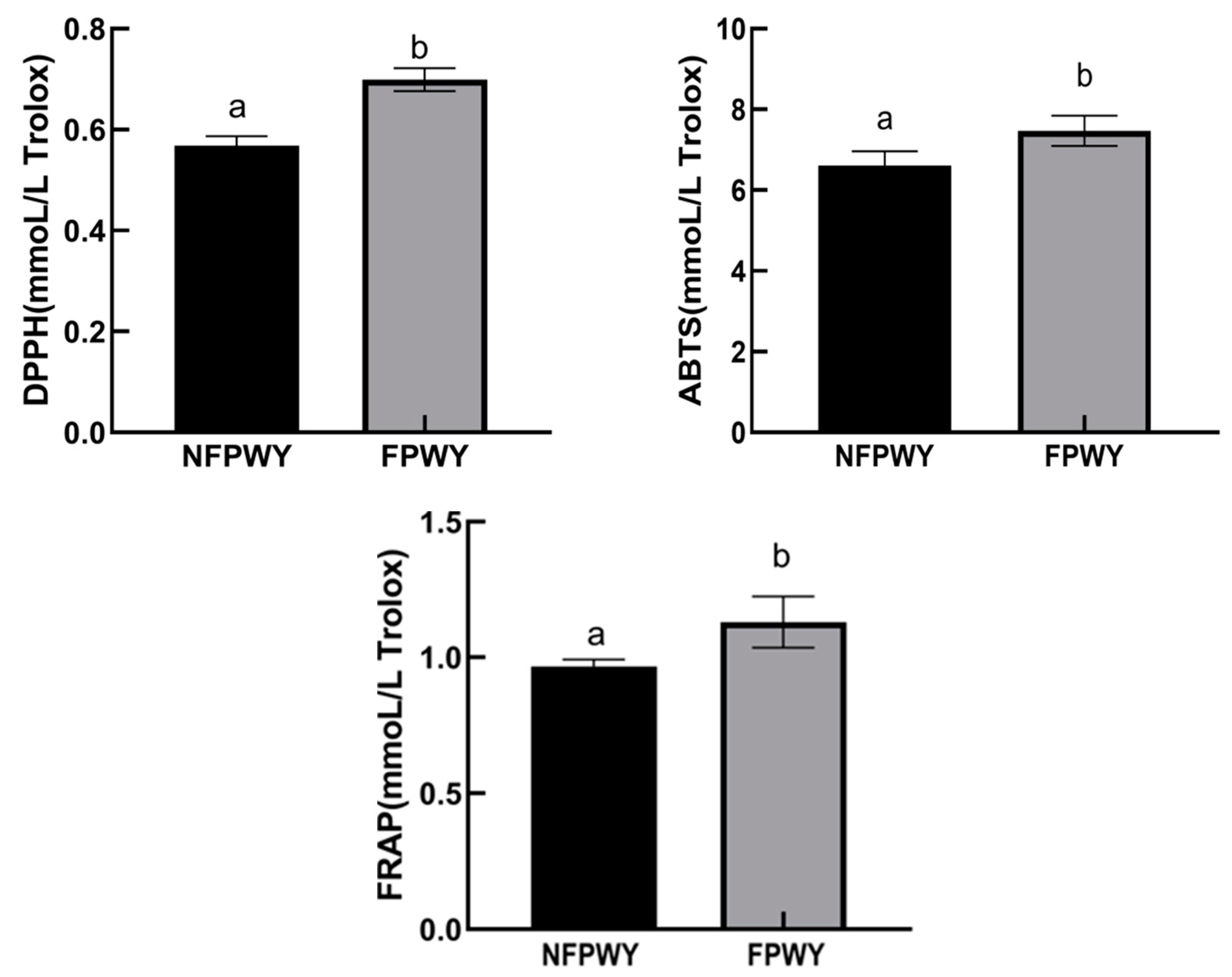
3.3. Comparison of Antioxidant Activity Between NFPWY and FPWY

The antioxidant activities of FPWY and NFPWY are shown in Figure 4. All three antioxidant evaluations exhibited the same trend: the three antioxidant activity indices of FPWY were statistically significant higher than those of NFPWY (p < 0.05), by ABTS activity, 1.13-fold increase, DPPH activity, 1.23-fold increase, and FRAP activity, 1.17-fold increase, respectively. While the absolute percentage increase in antioxidant activity may appear modest, the consistent enhancement across all three assays underscores the biological relevance of fermentation to a certain extent. Similarly, Xiao et al. [30] found that compared to unfermented soy whey, L. plantarum-fermented soy whey had significantly lower values of EC50 for the ABTS radical scavenging activity, reducing power, hydroxyl radical scavenging activity, and superoxide anion scavenging activity. These indicated that fermentation enhances the antioxidant activity of PWY, which correlates with changes in total flavonoid content and the aglycone isoflavone content.
During fermentation, active components such as enzymes naturally present in lactic acid bacteria can react with substrates to generate bioactive compounds such as polyphenols and amino acids, potentially increasing the antioxidant activity of the fermented product [31]. Xiao et al. [30] reported that during probiotic fermentation, β-glucosidase secreted by probiotics hydrolyzes glycosylated soy isoflavones into aglycones, which exhibit higher antioxidant activity and improved in vitro antioxidant capacity.

3.4. Changes in Main Components During In Vitro Simulated Digestion

3.4.1. Changes in Free Flavonoids

As shown in Figure 5, the changes in NFPWY and FPWY during the in vitro simulated gastric digestion process (0–120 min) and intestinal digestion process (120–240 min) followed similar trends. After gastric digestion, the flavonoid content increased, while during intestinal digestion, the content slightly decreased before increasing again. Overall, the free flavonoid content significantly increased after digestion. Specifically, the free flavonoid content in NFPWY (expressed as rutin equivalent) increased from 38.15 μg/mL to 97.77 μg/mL, while in FPWY, it increased from 63.62 μg/mL to 145.91 μg/mL.
These results indicated that both gastric and intestinal digestion promote the release of free flavonoids from the matrix. The increase and relative stability of flavonoid content in the gastric environment may be attributed to the low pH, where flavonoids are stable and less prone to degradation. During intestinal digestion, the initial decrease followed by an increase in flavonoid content might be due to pH changes, as well as the action of bile salts and trypsin in the digestive fluid, which may facilitate the release of flavonoid compounds. Ma et al., 2014 [32] also reported that digestion significantly influenced the contents of total flavonoids of heat-treated soy milks (4.48–31.10%).

3.4.2. Changes in Soy Isoflavones

As shown in Table 3, the changes in NFPWY and FPWY during in vitro simulated gastric and intestinal digestion followed similar trends, with the contents of six soy isoflavones listed in the table decreasing during both gastric and intestinal digestion. After digestion, the contents of all soy isoflavones were significantly reduced compared to their pre-digestion levels, which is consistent with the findings of Rodríguez-Roque et al. [33]. Rodríguez-Roque et al. observed a significant reduction in soy isoflavone content after in vitro gastrointestinal digestion of soy milk, likely due to increased absorption of bioactive isoflavones.

3.5. Changes in Antioxidant Activity During In Vitro Simulated Digestion

From Figure 6, it was shown that, for DPPH and ABTS capacities, both NFPWY and FPWY exerted a significant increase during digestion, while for FRAP, NFPWY had a slight decrease and FPWY remained stable during digestion. This may be explained that antioxidant compounds act through different mechanisms against oxidizing agents. However, three antioxidant assays all showed that during both the gastric and intestinal digestion phases, the antioxidant activity of FPWY was superior to that of NFPWY. This may be due to the increase of free flavonoids as reported in Section 3.4.1, or also be attributed to fermentation breaking down macromolecular polysaccharides into smaller sugars, which possess higher antioxidant activity [34]. Huo et al. [35] also found that fermented soy milk had significantly higher antioxidant activity than soy milk after intestinal digestion.

3.6. The Relative Abundances of Fecal Microbiota

The results of the relative abundance analysis of major gut microbiota after simulated colonic fermentation are shown in Table 4. Compared to the blank, the L. plantarum group did not affect the abundance of fecal microbiota except of dramatically inhibiting the pathogenic bacteria E. coli, while both NFPWY and FPWY significantly promoted the growth of Bifidobacterium, butyrate bacteria, and Bacteroides and inhibited the growth of E. coli. These results indicated that the microbiota modulation effects were mainly due to the bioactive components from NFPWY and FPWY. In the review of Huang et al. [36], efforts have also been made to elucidate that soy protein, oligosaccharides, and isoflavones may contribute to the modulation of gut microbiota. In the intestinal system, Bifidobacterium and Lactobacillus are key probiotics with major beneficial effects [37]. Butyrate bacteria contribute to maintaining gut health by producing butyrate [38]. Bacteroides are the main protein-metabolizing bacteria in the gut [39]. Thus, NFPWY, FPWY, and L. plantarum all benefit the gut microbiota to varying degrees.
Compared with NFPWY, FPWY significantly increased the abundance of Ruminococcus and Akkermansia. Ruminococcus is associated with intestinal barrier integrity and short-chain fatty acid production [40]. It was found that Ruminococcus was promoted in mice fed soy isoflavones [41] as well as in mice fed fresh soybean meal [42]. Akkermansia can reduce obesity, prevent diabetes, and alleviate symptoms such as insulin resistance [43]. It was also reported that the abundance of Akkermansia increased by 42.31, 1.83, and 20.51 times from 0 day to 28 days in different groups of mice fed fermented soy whey [44]. The abundance of Lactobacillus was significantly higher in NFPWY compared to the FPWY and L. plantarum groups, which may be attributed to the exogenous L. plantarum in the FPWY and L. plantarum groups suppressing the growth of endogenous Lactobacillus, resulting in a decrease in the abundance of this genus in those groups. Among these, FPWY exhibited the most significant effects. In summary, FPWY demonstrated better effects in promoting beneficial bacteria and inhibiting harmful bacteria compared to the NFPWY and L. plantarum groups, likely due to the synergistic effects of L. plantarum and PWY.

3.7. The Contents of SCFAs and Lactic Acid

The effects of NFPWY, FPWY, and L. plantarum on SCFAs and lactic acid content are shown in Table 5. For the production of SCFAs, L. plantarum treatment significantly increased the acetic acid production and decreased the production of isovaleric and valeric acids. It was reported that oral administration of L. plantarum Y44 enhanced acetic acid production in mice feces [45]. Another study showed that L. plantarum HJZW08 supplementation in broilers significantly enhanced the concentrations of isobutyric acid and isovaleric acid, with no obvious change of other SCFAs [46]. These inconsistent results may be attributed to variations in strains, differences in inoculum amount, and the use of different host fecal samples. Both NFPWY and FPWY significantly increased the production of propionic and butyric acids. NFPWY inhibited the production of isovaleric acid, while FPWY had a promotion effect. Changes in the gut microbiota led to differences in SCFAs [47]. As from Section 3.6, NFPWY and FPWY improved the growth of butyrate bacteria to 6.52- and 2.50-fold, respectively, which leads to the increase of butyric acids. However, the content of butyric acids in these two groups was almost at the same level. This may be because Ruminococcus, another butyrate-producing bacteria, was inhibited by NFPWY while promoted by FPWY. As Ruminococcus was reported to be able to ferment complex sugars to produce propionic acid [48], it could partly explain why the FPWY group had a higher propionic acid production than NFPWY.
For the lactic acid production, NFPWY and FPWY significantly enhanced lactic acid levels, while the lactic acid produced by L. plantarum alone showed no significant difference compared to the blank. This may be because microorganisms such as Lactobacillus and Bifidobacterium can decompose substrates to produce lactic acid. However, unlike SCFAs, lactic acid is less readily absorbed and utilized and is further decomposed by microorganisms like Clostridium cluster into smaller acids such as butyrate, which is subsequently converted into propionate, acetate, and other substances [49]. Combining this with the results from Section 3.6, where NFPWY increased Lactobacillus abundance more effectively than FPWY, the higher lactic acid content in NFPWY compared to FPWY can be explained. SCFAs and lactic acid play crucial roles in intestinal health and gut microbiome balance [50,51]. The regulatory effects of FPWY on SCFAs and lactic acid are consistent with its influence on fecal microbiota, indicating its potential in maintaining gut health.

4. Conclusions

This study underscores the potential of Lactobacillus plantarum fermentation as a biotechnological intervention to unlock the bioactive potential of yuba processing wastewater (PWY). By converting glycosylated isoflavones into bioavailable aglycones and enhancing free flavonoid content, the fermentation improves the intrinsic antioxidant capacity of PWY. The observed modulation of gut microbiota, particularly the enrichment of Bifidobacterium and Akkermansia, alongside elevated SCFA production, highlights FPWY’s dual role as a prebiotic and postbiotic agent. These effects suggest its applicability in dietary strategies targeting gut dysbiosis and oxidative stress-related disorders. Furthermore, the synergy between L. plantarum and PWY-derived compounds underscores the importance of strain–substrate interactions in optimizing functional outcomes.
However, this study primarily focused on the effects of free flavonoids and isoflavones on the bioactivity of PWY. Other bioactive components present in PWY, such as soybean oligosaccharides and peptides, may also contribute to its biological activity. Further research should explore their potential roles and interactions with isoflavones, as well as conduct in vivo studies to validate the health benefits of FPWY in the host. Another limitation is that the in vitro colonic fermentation model may not fully replicate human gut conditions, and fecal donor variability (e.g., diet, age) was not explored. Future work should validate these findings in vivo. Additionally, human trials are needed to confirm its efficacy in improving gut health.

Author Contributions

T.W.: Conceptualization, formal analysis, writing—original draft preparation. F.H.: Methodology, software and formal analysis. S.T.: Methodology and formal analysis. X.X.: Resources and supervision. D.L.: Writing—review and editing, supervision. All authors have read and agreed to the published version of the manuscript.

Funding

This work was supported by the Knowledge Innovation Program of Wuhan-Basic Research 2023020201010107 and Project 2662023PY020 from the Fundamental Research Funds for the Central Universities.

Institutional Review Board Statement

Not applicable.

Informed Consent Statement

Informed consent was obtained from all subjects involved in the study.

Data Availability Statement

The datasets presented in this article are not readily available due to time limitations. Requests to access the datasets should be directed to all authors.

Conflicts of Interest

The authors declare no conflicts of interest.

References

  1. Kalita, C.; Mehta, U.; Aayush, K.; Sawant, P.; Chavan, P.; Rasane, P.; Sharma, S.; Singh, G.P.; Nawghare, G.K.; Dhruv; et al. Recent trends in antioxidative peptides derived from soybean and other soy-based products: A comprehensive review. Process Biochem. 2024, 136, 311–323. [Google Scholar] [CrossRef]
  2. Lee, C.H.; Yang, L.; Xu, J.Z.; Yeung, S.Y.V.; Huang, Y.; Chen, Z.-Y. Relative antioxidant activity of soybean isoflavones and their glycosides. Food Chem. 2005, 90, 735–741. [Google Scholar] [CrossRef]
  3. Mateos-Aparicio, I.; Mateos-Peinado, C.; Jiménez-Escrig, A.; Rupérez, P. Multifunctional antioxidant activity of polysaccharide fractions from the soybean byproduct okara. Carbohydr. Polym. 2010, 82, 245–250. [Google Scholar] [CrossRef]
  4. Swallah, M.S.; Fan, H.; Wang, S.; Yu, H.; Piao, C. Prebiotic Impacts of Soybean Residue (Okara) on Eubiosis/Dysbiosis Condition of the Gut and the Possible Effects on Liver and Kidney Functions. Molecules 2021, 26, 326. [Google Scholar] [CrossRef] [PubMed]
  5. Tan, S.T.; Tan, S.S.; Tan, C.X. Soy protein, bioactive peptides, and isoflavones: A review of their safety and health benefits. PharmaNutrition 2023, 25, 100352. [Google Scholar] [CrossRef]
  6. Chua, J.-Y.; Liu, S.-Q. Soy whey: More than just wastewater from tofu and soy protein isolate industry. Trends Food Sci. Technol. 2019, 91, 24–32. [Google Scholar] [CrossRef]
  7. Wang, Y.; Serventi, L. Sustainability of dairy and soy processing: A review on wastewater recycling. J. Clean. Prod. 2019, 237, 117821. [Google Scholar] [CrossRef]
  8. Khubber, S.; Marti-Quijal, F.J.; Tomasevic, I.; Remize, F.; Barba, F.J. Lactic acid fermentation as a useful strategy to recover antimicrobial and antioxidant compounds from food and by-products. Curr. Opin. Food Sci. 2022, 43, 189–198. [Google Scholar] [CrossRef]
  9. Sabater, C.; Ruiz, L.; Delgado, S.; Ruas-Madiedo, P.; Margolles, A. Valorization of Vegetable Food Waste and By-Products Through Fermentation Processes. Front. Microbiol. 2020, 11, 581997. [Google Scholar] [CrossRef]
  10. Phomkaivon, N.; Pan-Utai, W.; Surojanametakul, V.; Varichanan, P.; Kaewtathip, T.; Kanyakam, K.; Klinsoda, J. Isoflavone aglycone-rich powder from soybean residue submerged fermentation using Lactobacillus fermentum 44197. NFS J. 2023, 33, 100157. [Google Scholar] [CrossRef]
  11. Langa, S.; Peirotén, Á.; Curiel, J.A.; de la Bastida, A.R.; Landete, J.M. Isoflavone Metabolism by Lactic Acid Bacteria and Its Application in the Development of Fermented Soy Food with Beneficial Effects on Human Health. Foods 2023, 12, 1293. [Google Scholar] [CrossRef]
  12. Pyo, Y.-H.; Lee, T.-C.; Lee, Y.C. Effect of Lactic Acid Fermentation on Enrichment of Antioxidant Properties and Bioactive Isoflavones in Soybean. J. Food Sci. 2005, 70, S215–S220. [Google Scholar] [CrossRef]
  13. Dai, S.; Pan, M.; El-Nezami, H.S.; Wan, J.M.; Wang, M.; Habimana, O.; Louie, J.C.; Shah, N.P. Effects of Lactic Acid Bacteria-Fermented Soymilk on Isoflavone Metabolites and Short-Chain Fatty Acids Excretion and Their Modulating Effects on Gut Microbiota. J. Food Sci. 2019, 84, 1854–1863. [Google Scholar] [CrossRef] [PubMed]
  14. Madjirebaye, P.; Peng, F.; Mueed, A.; Huang, T.; Mahamat, B.; Pahane, M.M.; Xi, Q.; Chen, X.; Moussa, K.; Kadebe, Z.T.; et al. Exploring Impact of Probiotic-Fermented Soymilk on Dextran-Sulfate-Sodium-Induced Ulcerative Colitis via Modulating Inflammation and Gut Microbiota Profile. Mol. Nutr. Food Res. 2024, 68, 2300586. [Google Scholar] [CrossRef] [PubMed]
  15. Tang, S.; Cheng, Y.; Wu, T.; Hu, F.; Pan, S.; Xu, X. Effect of Lactobacillus plantarum-fermented mulberry pomace on antioxidant properties and fecal microbial community. LWT 2021, 147, 111651. [Google Scholar] [CrossRef]
  16. Zhang, S.; Kim, N.; Yokoyama, W.; Kim, Y. Effects of moisture content on mechanical properties, transparency, and thermal stability of yuba film. Food Chem. 2018, 243, 202–207. [Google Scholar] [CrossRef] [PubMed]
  17. Shraim, A.M.; Ahmed, T.A.; Rahman, M.M.; Hijji, Y.M. Determination of total flavonoid content by aluminum chloride assay: A critical evaluation. LWT 2021, 150, 111932. [Google Scholar] [CrossRef]
  18. Tuncil, Y.E.; Thakkar, R.D.; Arioglu-Tuncil, S.; Hamaker, B.R.; Lindemann, S.R. Fecal Microbiota Responses to Bran Particles Are Specific to Cereal Type and In Vitro Digestion Methods That Mimic Upper Gastrointestinal Tract Passage. J. Agric. Food Chem. 2018, 66, 12580–12593. [Google Scholar] [CrossRef]
  19. Li, E.; Yang, H.; Zou, Y.; Wang, H.; Hu, T.; Li, Q.; Liao, S. In-vitro digestion by simulated gastrointestinal juices of Lactobacillus rhamnosus cultured with mulberry oligosaccharides and subsequent fermentation with human fecal inocula. LWT-Food Sci. Technol. 2019, 101, 61–68. [Google Scholar] [CrossRef]
  20. Wu, T.; Chu, X.; Cheng, Y.; Tang, S.; Zogona, D.; Pan, S.; Xu, X. Modulation of Gut Microbiota by Lactobacillus casei Fermented Raspberry Juice In Vitro and In Vivo. Foods 2021, 10, 3055. [Google Scholar] [CrossRef]
  21. Cheng, Y.; Wu, T.; Chu, X.; Tang, S.; Cao, W.; Liang, F.; Fang, Y.; Pan, S.; Xu, X. Fermented blueberry pomace with antioxidant properties improves fecal microbiota community structure and short chain fatty acids production in an in vitro mode. LWT 2020, 125, 109260. [Google Scholar] [CrossRef]
  22. Vendrame, S.; Guglielmetti, S.; Riso, P.; Arioli, S.; Klimis-Zacas, D.; Porrini, M. Six-Week Consumption of a Wild Blueberry Powder Drink Increases Bifidobacteria in the Human Gut. J. Agric. Food Chem. 2011, 59, 12815–12820. [Google Scholar] [CrossRef] [PubMed]
  23. Ben Ounis, W.; Champagne, C.; Makhlouf, J.; Bazinet, L. Utilization of tofu whey pre-treated by electromembrane process as a growth medium for Lactobacillus plantarum LB17. Desalination 2008, 229, 192–203. [Google Scholar] [CrossRef]
  24. Tu, C.; Azi, F.; Huang, J.; Xu, X.; Xing, G.; Dong, M. Quality and metagenomic evaluation of a novel functional beverage produced from soy whey using water kefir grains. LWT 2019, 113, 108258. [Google Scholar] [CrossRef]
  25. Bhanja, T.; Kumari, A.; Banerjee, R. Enrichment of phenolics and free radical scavenging property of wheat koji prepared with two filamentous fungi. Bioresour. Technol. 2009, 100, 2861–2866. [Google Scholar] [CrossRef]
  26. Cheng, K.-C.; Wu, J.-Y.; Lin, J.-T.; Liu, W.-H. Enhancements of isoflavone aglycones, total phenolic content, and antioxidant activity of black soybean by solid-state fermentation with Rhizopus spp. Eur. Food Res. Technol. 2013, 236, 1107–1113. [Google Scholar] [CrossRef]
  27. Hur, S.J.; Lee, S.Y.; Kim, Y.-C.; Choi, I.; Kim, G.-B. Effect of fermentation on the antioxidant activity in plant-based foods. Food Chem. 2014, 160, 346–356. [Google Scholar] [CrossRef]
  28. Lee, J.H.; Hwang, C.E.; Son, K.S.; Cho, K.M. Comparisons of nutritional constituents in soybeans during solid state fermentation times and screening for their glucosidase enzymes and antioxidant properties. Food Chem. 2019, 272, 362–371. [Google Scholar] [CrossRef]
  29. Otieno, D.O.; Ashton, J.F.; Shah, N.P. Evaluation of enzymic potential for biotransformation of isoflavone phytoestrogen in soymilk by Bifidobacterium animalis, Lactobacillus acidophilus and Lactobacillus casei. Food Res. Int. 2006, 39, 394–407. [Google Scholar] [CrossRef]
  30. Xiao, Y.; Wang, L.; Rui, X.; Li, W.; Chen, X.; Jiang, M.; Dong, M. Enhancement of the antioxidant capacity of soy whey by fermentation with Lactobacillus plantarum B1–6. J. Funct. Foods 2015, 12, 33–44. [Google Scholar] [CrossRef]
  31. Sarıtaş, S.; Portocarrero, A.C.M.; López, J.M.M.; Lombardo, M.; Koch, W.; Raposo, A.; El-Seedi, H.R.; Alves, J.L.d.B.; Esatbeyoglu, T.; Karav, S.; et al. The Impact of Fermentation on the Antioxidant Activity of Food Products. Molecules 2024, 29, 3941. [Google Scholar] [CrossRef]
  32. Ma, Y.; Zhou, M.; Huang, H. Changes of heat-treated soymilks in bioactive compounds and their antioxidant activities under in vitro gastrointestinal digestion. Eur. Food Res. Technol. 2014, 239, 637–652. [Google Scholar] [CrossRef]
  33. Rodríguez-Roque, M.J.; Rojas-Graü, M.A.; Elez-Martínez, P.; Martín-Belloso, O. Soymilk phenolic compounds, isoflavones and antioxidant activity as affected by in vitro gastrointestinal digestion. Food Chem. 2013, 136, 206–212. [Google Scholar] [CrossRef] [PubMed]
  34. Tian, J.; Mao, Q.; Dong, M.; Wang, X.; Rui, X.; Zhang, Q.; Chen, X.; Li, W. Structural Characterization and Antioxidant Activity of Exopolysaccharide from Soybean Whey Fermented by Lacticaseibacillus plantarum 70810. Foods 2021, 10, 2780. [Google Scholar] [CrossRef]
  35. Huo, C.; Yang, X.; Li, L. Non-beany flavor soymilk fermented by lactic acid bacteria: Characterization, stability, antioxidant capacity and in vitro digestion. Food Chem. X 2023, 17, 100578. [Google Scholar] [CrossRef] [PubMed]
  36. Huang, H.; Krishnan, H.B.; Pham, Q.; Yu, L.L.; Wang, T.T.Y. Soy and Gut Microbiota: Interaction and Implication for Human Health. J. Agric. Food Chem. 2016, 64, 8695–8709. [Google Scholar] [CrossRef]
  37. Braga, A.R.C.; Mesquita, L.M.D.S.; Martins, P.L.G.; Habu, S.; de Rosso, V.V. Lactobacillus fermentation of jussara pulp leads to the enzymatic conversion of anthocyanins increasing antioxidant activity. J. Food Compos. Anal. 2017, 69, 162–170. [Google Scholar] [CrossRef]
  38. Louis, P.; Young, P.; Holtrop, G.; Flint, H.J. Diversity of human colonic butyrate-producing bacteria revealed by analysis of the butyryl-CoA:acetate CoA-transferase gene. Environ. Microbiol. 2010, 12, 304–314. [Google Scholar] [CrossRef]
  39. Rist, V.T.S.; Weiss, E.; Sauer, N.; Mosenthin, R.; Eklund, M. Effect of dietary protein supply originating from soybean meal or casein on the intestinal microbiota of piglets. Anaerobe 2014, 25, 72–79. [Google Scholar] [CrossRef]
  40. Bajaj, J.S.; Fagan, A.; White, M.B.; Wade, J.B.; Hylemon, P.B.; Heuman, D.M.; Fuchs, M.; John, B.; Acharya, C.; Sikaroodi, M.; et al. Specific Gut and Salivary Microbiota Patterns Are Linked with Different Cognitive Testing Strategies in Minimal Hepatic Encephalopathy. Am. J. Gastroenterol. 2019, 114, 1080–1090. [Google Scholar] [CrossRef]
  41. Huang, L.; Zheng, T.; Hui, H.; Xie, G. Soybean isoflavones modulate gut microbiota to benefit the health weight and metabolism. Front. Cell. Infect. Microbiol. 2022, 12, 1004765. [Google Scholar] [CrossRef] [PubMed]
  42. Wang, Z.; Wang, P.; Zhou, Y.; Zhuang, S. Quercetin Supplementation Improves Intestinal Digestive and Absorptive Functions and Microbiota in Rats Fed Protein-Oxidized Soybean Meal: Transcriptomics and Microbiomics Insights. Animals 2024, 14, 2326. [Google Scholar] [CrossRef]
  43. Han, F.; Wang, Y.; Han, Y.; Zhao, J.; Han, F.; Song, G.; Jiang, P.; Miao, H. Effects of Whole-Grain Rice and Wheat on Composition of Gut Microbiota and Short-Chain Fatty Acids in Rats. J. Agric. Food Chem. 2018, 66, 6326–6335. [Google Scholar] [CrossRef] [PubMed]
  44. Han, W.; Zhuang, X.; Liu, Q.; Sun, B.; Miao, H.; Zhang, X. Fermented soy whey induced changes on intestinal microbiota and metabolic influence in mice. Food Sci. Hum. Wellness 2022, 11, 41–48. [Google Scholar] [CrossRef]
  45. Liu, Y.; Gao, Y.; Ma, F.; Sun, M.; Mu, G.; Tuo, Y. The ameliorative effect of Lactobacillus plantarum Y44 oral administration on inflammation and lipid metabolism in obese mice fed with a high fat diet. Food Funct. 2020, 11, 5024–5039. [Google Scholar] [CrossRef]
  46. Xiao, X.; Cui, T.; Qin, S.; Wang, T.; Liu, J.; Sa, L.; Wu, Y.; Zhong, Y.; Yang, C. Beneficial effects of Lactobacillus plantarum on growth performance, immune status, antioxidant function and intestinal microbiota in broilers. Poult. Sci. 2024, 103, 104280. [Google Scholar] [CrossRef]
  47. Morrison, D.J.; Preston, T. Formation of short chain fatty acids by the gut microbiota and their impact on human metabolism. Gut Microbes 2016, 7, 189–200. [Google Scholar] [CrossRef]
  48. Lin, W.; Wen, L.; Wen, J.; Xiang, G. Effects of Sleeve Gastrectomy on Fecal Gut Microbiota and Short-Chain Fatty Acid Content in a Rat Model of Polycystic Ovary Syndrome. Front. Endocrinol. 2021, 12. [Google Scholar] [CrossRef]
  49. Louis, P.; Flint, H.J. Diversity, metabolism and microbial ecology of butyrate-producing bacteria from the human large intestine. FEMS Microbiol. Lett. 2009, 294, 1–8. [Google Scholar] [CrossRef]
  50. Tan, J.K.; Macia, L.; Mackay, C.R. Dietary fiber and SCFAs in the regulation of mucosal immunity. J. Allergy Clin. Immunol. 2023, 151, 361–370. [Google Scholar] [CrossRef]
  51. Aldunate, M.; Srbinovski, D.; Hearps, A.C.; Latham, C.F.; Ramsland, P.A.; Gugasyan, R.; Cone, R.A.; Tachedjian, G. Antimicrobial and immune modulatory effects of lactic acid and short chain fatty acids produced by vaginal microbiota associated with eubiosis and bacterial vaginosis. Front. Physiol. 2015, 6, 164. [Google Scholar] [CrossRef] [PubMed]
Figure 1. Purity and integrity of fecal microbial genetic DNA. 1–3 represented the Control; 4–7 represented the NFPWY; 8–10 represented the FPWY; 11–15 represented the L. plantarum.
Fermentation 11 00212 g001
Figure 2. The bacterial counts of L. plantarum in PWY with different inoculation levels and fermentation days. Data were presented as mean ± standard deviation. Different lowercase letters indicated significant differences among fermentation times within the same inoculation level (p < 0.05). Different uppercase letters indicated significant differences among inoculation levels within the same fermentation time (p < 0.05).
Fermentation 11 00212 g002
Figure 3. Contents of free flavonoid in FPWY and NFPWY. NFPWY: Non-fermented processing wastewater of yuba; FPWY: Fermented processing wastewater of yuba. Different superscript letters indicated significant differences (p < 0.05).
Fermentation 11 00212 g003
Figure 4. Comparison of antioxidant activities between NFPWY and FPWY. NFPWY: Non-fermented processing wastewater of yuba; FPWY: Fermented processing wastewater of yuba. Different letters indicated significant differences (p < 0.05).
Fermentation 11 00212 g004
Figure 5. Changes of free flavonoids during in vitro simulated digestion. NFPWY: Non-fermented processing wastewater of yuba; FPWY: Fermented processing wastewater of yuba. Different lowercase letters indicated significant differences among NFPWY groups (p < 0.05), while different uppercase letters indicated significant differences among FPWY groups (p < 0.05).
Fermentation 11 00212 g005
Figure 6. Antioxidant activities of NFPWY and FPWY during in vitro simulated digestion. NFPWY: Non-fermented processing wastewater of yuba; FPWY: Fermented processing wastewater of yuba. Different lowercase letters indicated significant differences among NFPWY groups (p < 0.05), while different uppercase letters indicated significant differences among FPWY groups (p < 0.05).
Fermentation 11 00212 g006
Table 1. Primers of intestinal flora-related genes.
NamePrimers
BifidobacteriumF: GGGTGGTAATGCCGGATG
R: TAAGCGATGGACTTTCACACC
RuminococcusF: TTAACACAATAAGTWATCCACCTGG
R: ACCTTCCTCCGTTTTGTCAAC
Butyrate bacteriaF: GCIGAICATTTCACITGGAAYWSITGGCAYATG
R: CCTGCCTTTGCAATRTCIACRAANGC
LactobacillusF: AGCAGTAGGGAATCTTCCA
R: CACCGCTACACATGGAG
AkkermansiaF: CAGCACGTGAAGGTGGGGAC
R: CCTTGCGGTTGGCTTCAGAT
Escherichia coliF: GTTAATACCTTTGCTCATTA
R: ACCAGGGTATCTTAATCCTGTT
BacteroidesF: ATAGCCTTTCGAAAGRAAGAT
R: CCAGTATCAACTGCAATTTTA
Table 2. Content of soy isoflavones in NFPWY and FPWY (μmoL/L).
NFPWYFPWY
Daidzein116.85 ± 8.52 a312.77 ± 15.24 b
Daidzin371.06 ± 6.14 a77.51 ± 19.68 b
Glycitein3.67 ± 0.65 a46.82 ± 4.12 b
Glycitin30.03 ± 2.97 an.d.
Genistein22.66 ± 1.76 a93.52 ± 2.77 b
Genistin399.64 ± 29.86 a115.37 ± 20.43 b
NFPWY: Non-fermented processing wastewater of yuba; FPWY: Fermented processing wastewater of yuba. Different superscript letters indicated significant differences (p < 0.05). n.d.: Not detected.
Table 3. Changes of soybean isoflavone content during in vitro simulated digestion (μmoL/L).
Soy IsoflavoneUndigestedStomach Digestion
at 60 min
Stomach Digestion
at 120 min
Intestine Digestion
at 60 min
Intestine Digestion
at 120 min
NFPWYFPWYNFPWYFPWYNFPWYFPWYNFPWYFPWYNFPWYFPWY
Daidzein116.85
± 8.52 c
312.77
± 15.24 B
79.34
± 4.51 b
303.96
± 5.32 B
80.24
± 9.37 b
298.33
± 10.33 B
59.70
± 0.38 a
215.57
± 18.50 A
59.39
± 1.27 a
218.85
± 10.82 C
Daidzin371.06
± 6.14 c
77.51
± 19.68 B
222.09
± 10.87 b
55.23
± 5.06 A
212.84
± 19.78 b
56.36
± 9.75 A
162.00
± 3.13 a
38.97
± 3.47 A
171.78
± 1.36 a
42.80
± 1.64 A
Glycitein3.67
± 0.65 a
46.82
± 4.12 C
2.94
± 0.34 a
36.48
± 0.42 B
3.37
± 1.17 a
43.25
± 2.64 C
6.22
± 0.32 b
30.49
± 2.64 A
5.70
± 0.25 b
30.87
± 1.13 A
Glycitin30.03
± 2.97 d
n.d.20.10
± 0.93 c
n.d.17.80
± 1.59 bc
n.d.13.70
± 0.09 a
n.d.14.71
± 0.19 ab
n.d.
Genistein22.66
± 1.76 c
93.52
± 2.77 B
15.44
± 0.76 b
96.00
± 1.49 B
14.91
± 1.50 b
92.26
± 3.06 B
10.31
± 0.54 a
64.92
± 6.39 A
11.59
± 0.44 a
67.70
± 3.36 A
Genistin399.64
± 29.86 c
115.37
± 20.43 C
263.48
± 12.17 b
90.53
± 7.85 B
266.32
± 15.35 b
95.43
± 26.12 BC
189.90
± 6.23 a
63.37
± 5.80 A
184.39
± 6.06 a
67.79
± 2.00 A
NFPWY: Non-fermented processing wastewater of yuba; FPWY: Fermented processing wastewater of yuba. n.d.: Not detected. Different lowercase letters indicated significant differences among NFPWY groups (p < 0.05), while different uppercase letters indicated significant differences among FPWY groups (p < 0.05).
Table 4. Relative abundances of fecal microbiota with different treatment groups.
BlankNFPWYFPWYL. plantarum
Bifidobacterium1.00 ± 0.00 a2.17 ± 0.66 b4.24 ± 0.19 c1.22 ± 0.3 ab
Ruminococcus1.00 ± 0.00 b0.55 ± 0.04 a1.32 ± 0.10 c1.13 ± 0.18 bc
Butyrate bacteria1.00 ± 0.00 a6.52 ± 0.16 c2.50 ± 0.10 b1.06 ± 0.03 a
Lactobacillus1.00 ± 0.00 a2.70 ± 0.16 c1.26 ± 0.04 ab1.03 ± 0.01 ab
Akkermansia1.00 ± 0.00 ab1.13 ± 0.22 bc1.50 ± 0.21 c0.68 ± 0.00 a
E. coli1.00 ± 0.00 d0.46 ± 0.03 c0.27 ± 0.00 b0.01 ± 0.00 a
Bacteroides1.00 ± 0.00 a6.78 ± 0.83 c3.94 ± 0.75 b1.44 ± 0.04 a
NFPWY: Non-fermented processing wastewater of yuba; FPWY: Fermented processing wastewater of yuba. Means greater or less than 1.00 indicated that the treatment either increased or decreased the abundance of specific fecal microbiota, respectively. Data in the same row with different superscript letters indicated significant differences (p < 0.05).
Table 5. The contents of SCFA and lactic acid with different treatment groups (mmol/L).
BlankNFPWYFPWYL. plantarum
Acetic acid 52.672 ± 3.337 a57.541 ± 4.341 a59.420 ± 1.688 a70.010 ± 4.628 b
Propionic acid 5.006 ± 0.080 a8.151 ± 0.270 b9.299 ± 0.219 c0.305 ± 0.015 a
Isobutyric acid0.173 ± 0.074 b0.032 ± 0.001 a0.098 ± 0.001 ab0.119 ± 0.003 ab
Butyric acid0.107 ± 0.017 a2.428 ± 0.695 b2.536 ± 0.232 b0.038 ± 0.001 a
Isovaleric acid0.322 ± 0.031 c0.177 ± 0.010 b0.425 ± 0.002 d0.104 ± 0.013 a
Valeric acid0.029 ± 0.006 b0.022 ± 0.001 ab0.032 ± 0.006 b0.016 ± 0.004 a
Lactic acid0.344 ± 0.018 a3.193 ± 0.251 c1.122 ± 0.239 b0.533 ± 0.070 a
NFPWY: Non-fermented processing wastewater of yuba; FPWY: Fermented processing wastewater of yuba. Different letters in the same row indicated significant differences (p < 0.05).
Disclaimer/Publisher’s Note: The statements, opinions and data contained in all publications are solely those of the individual author(s) and contributor(s) and not of MDPI and/or the editor(s). MDPI and/or the editor(s) disclaim responsibility for any injury to people or property resulting from any ideas, methods, instructions or products referred to in the content.

