Article

Optimizing Fermentation of Morus nigra L. Residues with Schizophyllum commune to Enhance Anthocyanin Release and Anti-Inflammatory Activity via Pyroptosis Pathway Modulation

1
School of Bioscience and Bioengineering, South China University of Technology, Guangzhou 510006, China
2
Guangdong Marubi Biotechnology Co., Ltd., Guangzhou 510700, China
3
Development and Research Center for Biological Marine Resources, Southern Marine Science and Engineering Guangdong Laboratory (Zhanjiang), Zhanjiang 524000, China
*
Authors to whom correspondence should be addressed.
Fermentation 2025, 11(3), 145; https://doi.org/10.3390/fermentation11030145
Submission received: 5 February 2025 / Revised: 24 February 2025 / Accepted: 12 March 2025 / Published: 14 March 2025
(This article belongs to the Section Industrial Fermentation)

Abstract

:
Morus nigra L. is rich in anthocyanins and other active ingredients, but its extraction residues pose a burden on the environment. In the present study, Morus nigra L. extraction residue resource utilization was achieved through liquid fermentation of Schizophyllum commune, with the aim of enhancing anthocyanin solubilization and evaluating anti-inflammatory activity. Response surface methodology was used to optimize fermentation parameters and quantify anthocyanin fractions by HPLC. The anti-inflammatory effect was evaluated using the lipopolysaccharide-induced inflammation model of human foreskin fibroblast (BJ cell), and the interaction of cyanidin-3-O-glucoside (C3G) with NLRP3, a key target of the pyroptosis pathway, was resolved by molecular docking. Our results indicated that the optimal conditions (substrate 3.4%, inoculum 9%, time 50 h) enabled the total anthocyanin to reach 85.1 μg/mL, of which the C3G content was elevated to 66.7 μg/mL (release efficiency of 83.9%). The fermented filtrate effectively promoted BJ cell proliferation and inhibited the lipopolysaccharide-induced inflammatory response, with the pyroptosis signaling pathway playing a significant role. Molecular docking confirmed that C3G binds strongly to the NLRP3 protein. This technology provides a new strategy for high-value utilization of Morus nigra L. residues and the development of natural anti-inflammatory drugs.

1. Introduction

Morus nigra L., belonging to the Moraceae family and the Morus genus, is a plant that serves as both food and medicine [1]. Studies indicate that Morus nigra L. is rich in physiologically active components such as anthocyanosides, polysaccharides, alkaloids, resveratrol, and proanthocyanidins. A variety of physiologically active effects such as immune enhancement, free radical scavenging, weight loss, anti-tumor and anti-inflammatory effects, and lowering of blood glucose and blood lipids have also been identified [2]. Given their relatively simple molecular structure and ease of absorption, anthocyanosides have received considerable attention from researchers in recent years [3]. The most predominant anthocyanin in black mulberries (Morus nigra L.) is cyanidin-3-O-glucoside (C3G), followed by cyanidin-3-rutinoside (C3R). Kongtawelert et al. found that C3G significantly inhibits the expression of the pro-inflammatory cytokines, interleukin-1β (IL-1β), and IL-6 in lipopolysaccharide (LPS)-stimulated BV2 microglial cells, demonstrating a significant anti-inflammatory effect [4].
As the physiological activities of anthocyanins continue to be explored, black mulberries, which are rich in anthocyanins, are considered a high-quality raw material for the development of functional foods and cosmetics. Anthocyanins are primarily located within the sap of the fruit cells of black mulberries. Conventional industrial extraction employs physical methods such as crushing and pressing to rupture the fruit cell walls, thereby facilitating the release of anthocyanins from the sap [5]. Due to the adsorption of anthocyanosides by the cavity structure of the cell wall, a significant portion of anthocyanosides remain present in the Morus nigra L. extraction residue (MR) [6]. Therefore, there is an urgent need to develop effective re-extraction technologies to reduce resource waste and environmental pollution, as well as to increase the application value of MR in the fields of foods, nutraceuticals, and cosmetics [7]. Studies have indicated that fermentation can significantly enhance the total anthocyanin content in black mulberries, which augments their antioxidant activity. As such, biotechnological fermentation could serve as a potentially effective extraction method for promoting the release of anthocyanins from black mulberries [8].
Bidirectional fermentation is a technology that utilizes a microorganism as the fermenting strain and a plant as the substrate. This method provides essential nutrients for microorganisms while enhancing substrate utilization and the nutritional value of the substrate through the transformative action of the microorganism. This method has shown great potential in the fields of medicine and health [9]. Schizophyllum commune (SC) belongs to the phylum Fungi, family Schizophyllaceae, genus Schizophyllum, and is widely distributed in many provinces of China [10]. Because its genome contains 240 glycoside hydrolase candidate genes, of which 89 are involved in the degradation of plant polysaccharides, SC has the potential to degrade all components of lignocellulosic biomass. This suggests that SC can utilize most plants as its sole carbon source for growth [11]. Recent studies have also shown the potential of SC in the fermentation of plant residues to enhance the production of bioactive compounds [12]. Ji et al. found that the fermentation of young apples with SC DS1 could produce mycoprotein with a protein content of 33.56 ± 0.82%, demonstrating the feasibility of using plant residues for valuable product generation through fungal fermentation [13]. SC has been shown to utilize Citrus unshiu peel to release a large amount of flavanone aglycones such as naringin and hesperidin from the peel [14]. These flavanone aglycones have demonstrated protective effects against UVA-induced phototoxicity in vitro and have shown inhibitory effects on the activity of inflammation-related enzymes such as COX-2 and 5-LOX. This suggests that during the process of utilizing the cell walls of these plants for growth and metabolism, active components such as flavonoids and anthocyanins are prompted to flow from the cell sap into the extracellular environment. SC, which can gently induce the release of active components and enhance their antioxidant and anti-inflammatory activities, serves as an efficient fungus for the decomposition of plant cell walls [15]. For SC, the carbohydrate-rich components (such as cell walls) that are present in MR serve as an excellent carbon source. Consequently, SC can secrete polysaccharide hydrolases to degrade the cell walls within MR, thereby releasing active constituents like C3G. Simultaneously, during this process, SC also produces β-glucans, which potentially offer protective effects against oxidation and discoloration of C3G [16]. Therefore, co-fermentation utilizing SC and MR not only facilitates the extraction of active compounds from plant residues but also yields metabolites that confer protective benefits to these active constituents. This characteristic has significant implications for both the pharmaceutical and food industries. Current research on the fermentation of MR by SC is limited, and the impact of fermentation parameters on the production and activity of anthocyanins remains unclear.
The objective of this study was to optimize the fermentation conditions for anthocyanin production via SC using response surface methodology (RSM) and to assess anti-inflammatory activity using cellular assays. Molecular docking analysis was employed to investigate the interaction between C3G and NLRP3. We also evaluated the effects on NLRP3, a key upstream inflammatory pathway protein, to preliminarily elucidate the anti-inflammatory mechanism of C3G. This study provides significant insight into the reuse of MR and the preparation of anthocyanin materials with potent anti-inflammatory activity.

2. Materials and Methods

2.1. Materials

The SC standard strain was provided by Guangdong Institute of Microbiology, Guangzhou, China (GDMCC 5.43). Morus nigra L. extract residue was purchased from Best (Guangzhou, China) Pharmaceutical Co. The C3G standard (purity ~99%) was purchased from Sigma (St. Louis, MO, USA). The C3G stock solution (1 mg/mL) was prepared in methanol (containing 2% hydrochloric acid) using the C3G standard (~99% purity) and stored at −20 °C.

2.2. Preparation and Optimization of the SC–Morus nigra L. Extract Residue Through Bidirectional Fermentation of the Bacterial Mass

The mycelium of Schizophyllum commune was subcultured on a slant agar medium and incubated at 28 °C for 5–7 days until the mycelium covered the entire slant surface. Subsequently, the slant culture was transferred to a liquid medium (30 g/L glucose, 3 g/L yeast extract, 1 g/L potassium dihydrogen phosphate, 0.5 g/L magnesium sulfate) and incubated in a shaking incubator (28 °C, 160 rpm) for 72 h to obtain the seed culture for MR fermentation.
The response surface methodology and the Box–Behnken central composite design principle were employed to optimize the bidirectional fermentation conditions of Schizophyllum commune with the Morus nigra L. extraction residue. The variables included the addition of MR (A), the addition of Schizophyllum commune seed culture (B), and fermentation time (C). A three-factor, three-level response surface design was conducted (Table 1). Experiments were carried out according to the factor level combinations provided by Design-Expert (Version 13.0). The experimental results were fitted with equations and subjected to an analysis of variance to select the optimal design parameters. The aforementioned optimal parameters were applied to the MR bidirectional fermentation of Schizophyllum commune, and the total anthocyanin content was measured to verify the goodness of fit of the response surface.

2.3. Detection of Total Anthocyanins

Centrifugation of the fermentation broth was performed at 14,000× g for 15 min at 4 °C, and the supernatant containing the fermentation product of SC–M was collected. The anthocyanin content was determined under various fermentation conditions according to the Plant Anthocyanin Content Assay Kit instructions (Beijing Boxbio Company, Beijing, China).

2.4. High-Performance Liquid Chromatography Detection of C3G Concentration

Because C3G is the most abundant anthocyanin in Morus nigra L., high-performance liquid chromatography (HPLC) was employed to detect C3G in the SC–M fermentation product. Briefly, 1.0 g of SC–M sample was taken and mixed with 25 mL of 2% hydrochloric acid–methanol solution in a 50 mL centrifuge tube. The sample was vortexed at 2000 rpm for 3 min, followed by ultrasonication for 5 min. The resulting mixture was centrifuged at 12,000× g for 5 min at 4 °C, and the supernatant was collected and filtered through a 0.22 μm membrane.
The anthocyanin content was analyzed as described by Zhang et al. [17] via HPLC using a Thermo Scientific Surveyor HPLC system (Waltham, MA, USA). The analytes were separated on a Zorbax Extend C18 column (5 μm, 250 × 4.6 mm, Agilent Technologies Corporation, Santa Clara, CA, USA). A binary elution system was slightly modified, consisting of 10% formic acid in water (solvent A) and a solution composed of 24% acetonitrile and methanol in 10% formic acid water (solvent B). The elution program was as follows: 0–0.1 min, 93% A; 0.1–35 min, 75% A; 35–45 min, 35% A; 45–46 min, 0% A. The flow rate was set at 600 μL/mL. The injection volume was 20 μL. Detection was carried out using a single-wavelength UV detector (Thermo Fisher Scientific, Waltham, MA, USA) set at 535 nm. Standard solutions of C3G with concentrations of 0, 50, 100, 200, 300, and 400 μg/mL were prepared and analyzed concurrently with the samples by HPLC. A standard curve was plotted with the peak area on the y-axis and the concentrations of different C3G standard solutions on the x-axis, and a linear equation was fitted. The content of C3G in SC–M was calculated based on the fitted formula.
In the present extraction and determination procedures, when 1 mg of C3G was added to the SC fermentation product of the Morus nigra L. residue-untreated control (10 mL) and the extract analyzed by HPLC, 84.6 ± 0.7 μg/mL C3G (mean ± SD) were confirmed to be recovered, indicating that the recovery was 84.6 ± 0.7% for C3G.

2.5. Cell Culture

Human foreskin fibroblasts (BJ cells) were seeded into a 96-well plate at a density of 40,000 cells per well and cultured for 24 h. Subsequently, DMEM complete medium containing various concentrations of SC–M (50, 40, 30, 20, 10, 5, 2.5, 1.25, 0.6125, and 0.306%) was added; the control wells received an equivalent volume of DMEM complete medium. Six replicates were set for each concentration. After a further 24 h of incubation, the cell viability was assessed using the Cell Counting Kit-8, and the cell survival rate was calculated.
BJ cells were seeded into a 6-well plate at a density of 200,000 cells per well and cultured for 24 h, after which the culture medium was removed. Then, DMEM complete medium containing 2 μg/mL of LPS and varying concentrations of SC–M (5, 1, and 0.2%) was added; control wells received an equivalent volume of DMEM complete medium. After 24 h of incubation, the cell culture supernatant and cells were collected for subsequent analysis.

2.6. Scratch Assay

Quadruple wound healing inserts were inserted into a 12-well plate, and BJ cells in the logarithmic growth phase were seeded into the 12-well plate at a density of 2 × 10⁴ cells/well. After 24 h of culture, the inserts were removed and washed with PBS, and serum-free DMEM containing different concentrations of SC–M (5, 1, and 0.2%) was added. Photographs were then taken (T0 time). The control wells received an equivalent volume of serum-free DMEM culture medium, and the positive control group was treated with DMEM culture medium containing 10% serum. After 24 h, the migration distance was measured using a fluorescence microscope (Zeiss, Axio Imager D2, Oberkochen, Germany). The DMEM culture medium containing 10% serum served as the positive control group.

2.7. Biochemical Analysis

IL-1β and IL-18 in the cell culture fluid were measured by ELISA (Nanjing Jiancheng Bioengineering Institute, Nanjing, China) according to the manufacturer’s instructions.

2.8. RNA Isolation and Real-Time Quantitative PCR

The relative mRNA expression levels of cellular focal death signaling pathway proteins (TLR4, NLRP3, caspase-1, GSDMD, IL-1β, and IL-18) in BJ cells were detected using real-time quantitative PCR, as described by Ye et al. [18]. The transcript expressions of genes were calculated using ABI Quantity Studio 3 (Applied Biosystems, Foster City, CA, USA). The primer sequences were as follows: GAPDH: 5′-ATC ACT GCC ACC CAG AAG AC-3′ and 5′-TTT CTA GAC GGC AGG TCA GG-3′; TLR4: 5′-GAC GGT GAT AGC GAG CCA C-3′ and 5′-TTA GGA ACC ACC TCC ACG CAG-3′; NLRP3: 5′-CCA CAA GAT CGT GAG AAA ACC C-3′ and 5′-CGG TCC TAT GTG CTC GTC A-3′; caspase-1: 5′-GCT TTC TGC TCT TCC ACA CC-3′ and 5′-CAT CTG GCT GCT CAA ATG AA-3′; GSDMD: 5′-GTG TGT CAA CCT GTC TAT CAA GG-3′ and 5′-CAT GGC ATC GTA GAA GTG GAA G-3′; IL-1β: 5′-ATG ATG GCT TAT TAC AGT GGC AA-3′ and 5′-GTC GGA GAT TCG TAG CTG GA-3′; IL-18: 5′-TCT TCA TTG ACC AAG GAA ATC GG-3′ and 5′-TCC GGG GTG CAT TAT CTC TAC-3′; COL1A2: 5′-GTT GCT GCT TGC AGT AAC CTT-3′ and 5′-AGG GCC AAG TCC AAC TCC TT-3′; and COL3A1: 5′-GGA GCTG GCT ACT TCT CGC-3′ and 5′-GGG AAC ATC CTC CTT CAA CAG-3′.

2.9. Western Blotting

The relative levels of key pyroptosis signaling proteins (NLRP3 and mature caspase-1) in BJ cells were determined using immunoblotting, as described by Ye et al. [19]. Primary anti-NLRP3 was purchased from Cell Signaling Technology (Danvers, MA, USA); primary anti-matured caspase-1 and primary anti-GAPDH were acquired from Affinity Biosciences. Bands were detected using an ECL chemiluminescence kit (Biosharp, Beijing, China). Chemiluminescence was recorded with an Amersham Imager 600 (GE Healthcare, Wuxi, China). The band results were analyzed using Image J software (v1.52a, National Institutes of Health, Bethesda, MD, USA).

2.10. Molecular Docking of C3G with NLRP3

The 3D structure of NLRP3 (PDB ID: 6NPY) was downloaded from the Protein Data Bank (PDB, https://www.rcsb.org, accessed on 20 November 2024). The 3D molecular structures of C3G and C3R were obtained from the PubChem Compound Database (https://pubchem.ncbi.nlm.nih.gov, accessed on 20 November 2024). Molecular docking studies were conducted using LeadIT software (v2.18) to evaluate the binding affinity between NLRP3 and different anthocyanins, as well as the receptor–ligand interaction sites and interaction patterns. Water molecules from NLRP3 were removed, and hydrogen atoms were added to the protein to stabilize the target protein. Chain A (CD180) and Chain B (NEK7) were selected for docking studies. The grid box was centered to cover the structural domains of each protein and to accommodate free molecular movement. The docking pocket was set as a 30 × 30 × 30 Å cubic pocket with a grid spacing of 0.05 nm. Interaction patterns were analyzed using LeadIT (v2.18). The conformation with the lowest energy was selected for analysis, and PyMOL software (v2.6.0a0) was used to visualize the data.

2.11. Statistical Analysis

Data processing and graphing were conducted using SPSS Statistics 21 (v21.0.0.0) and Origin 2022 software (v2022.SR1). Statistical significance was assessed via one-way ANOVA, followed by Tukey’s post hoc test for multiple comparisons. Data normality and homogeneity of variance were verified using Shapiro–Wilk and Levene’s tests, respectively.

3. Results

3.1. Response Surface Analysis and Optimization of Morus nigra L. Residue Following Fermentation with L. Schizophyllum commune

This experiment was conducted based on the Box–Behnken design principle and included 17 runs, resulting in a regression equation which took the total anthocyanin content in the fermentation product as the response value. The equation was as follows: Total anthocyanin content = 66.21 + 10.17 × A + 0.4520 × B − 8.92 × C+ 0.9324 × AB − 2.89 × AC − 13.88 × BC − 8.98 × A2 − 12.88 × B2 − 0.2366 × C2. The correlation coefficient R2 of the regression equation was 0.9849, indicating good credibility of the equation and accurate analysis and prediction of the total anthocyanin production. The F-value of the model was 50.82, and the p value was less than 0.0001, indicating that the model was significant. The first-order terms A and B, as well as the second-order terms BC, A2, and B2, were highly significant, suggesting that they had a substantial impact on the response value and were not linearly related in a simple manner. According to the F-values (Table 2), the order of importance of factors affecting the total anthocyanin content in the fermentation residue of SC and Morus nigra L. was as follows: plant substrate addition amount > fermentation days > mycelial liquid addition amount.

3.2. Analysis of Factor Interactions

After processing with Design Expert software (v13.0.1.0), the response surface and contour plots for the interactive effects of MR addition amount (A), SC mycelial liquid addition amount (B), and fermentation time (C) were obtained. As shown in Figure 1, the interaction between the plant substrate addition amount and mycelial liquid addition amount significantly affected the total anthocyanin content. The effects of plant substrate addition amount and fermentation time, as well as mycelial liquid addition amount and fermentation time, on the total anthocyanin content were relatively significant. Through software simulation and prediction, the optimal parameters for total anthocyanin content were as follows: Morus nigra L. addition amount of 3.4%; mycelial inoculation amount of 9.0%; and fermentation time of 2.05 days (50 h). These led to a predicted total anthocyanin content of 83.1 μg/mL. Experimental validation showed that the actual value reached 85.1 ± 2.3 μg/mL, with a difference of only 2.4%. This indicated that these conditions significantly promoted the preparation of anthocyanins via SC fermentation of MR.
Given that C3G is the most abundant anthocyanin in Morus nigra L. [2], this study used HPLC to detect the content of C3G in MR and the Schizophyllum communeMorus nigra L. fermentation filtrate (SC–M). The total anthocyanin content in the Morus nigra L. residue was 119.5 μg/mL, and the C3G content was 79.5 μg/mL. After fermentation with SC, the total anthocyanin content in the filtrate was 85.1 μg/mL, and the C3G content was 66.7 μg/mL (Figure 2). Approximately 83.9% of C3G was released into the fermentation liquid, demonstrating that Schizophyllum commune effectively initiated the release of C3G from the Morus nigra L. residue into the fermentation liquid.

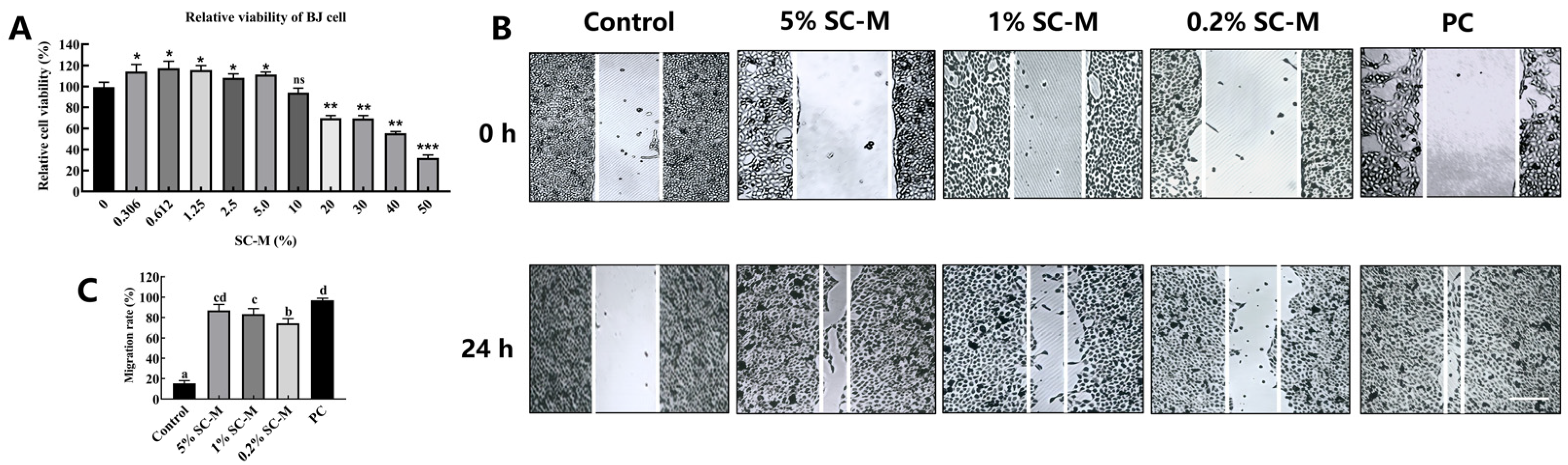
3.3. Effect of SC–M on BJ Cell Proliferation

BJ cells are a unique human skin fibroblast cell line that possesses physiological characteristics and metabolic pathways that are similar to those of human cells. This makes them an important tool for studying cellular activities, mechanisms of disease occurrence, and mechanisms of drug action [20]. In this study, we investigated the effects of varying concentrations of SC–M on the growth of BJ cells. The experimental results indicated that within the concentration range of 0.3–5%, SC–M significantly promoted the proliferation of BJ cells (p < 0.05). This may be attributed to the positive effects of the C3G that was released during the fermentation process on cell growth. However, when the concentration of the SC–M exceeded 10%, cell growth significantly decreased to 38%, compared to 76% in the control group (p < 0.01). This suggests that high concentrations of the SC–M have a distinctly toxic effect on BJ cells.
Given that SC–M at concentrations between 0.3 and 5% had significant proliferative effects on BJ cells, we further assessed its impact on cell migration via wound healing assay. As shown in Figure 3B, compared to the blank control group, a low concentration (0.2%) of the SC–M did not significantly affect the migration of BJ cells. A medium concentration (1%) and high concentration (5%) of SC–M significantly enhanced the migration rate of BJ cells by 64 and 83%, respectively (p < 0.01). These data indicate that medium-to-high concentrations of SC–M not only promoted the proliferation of BJ cells but also significantly enhanced their migration.

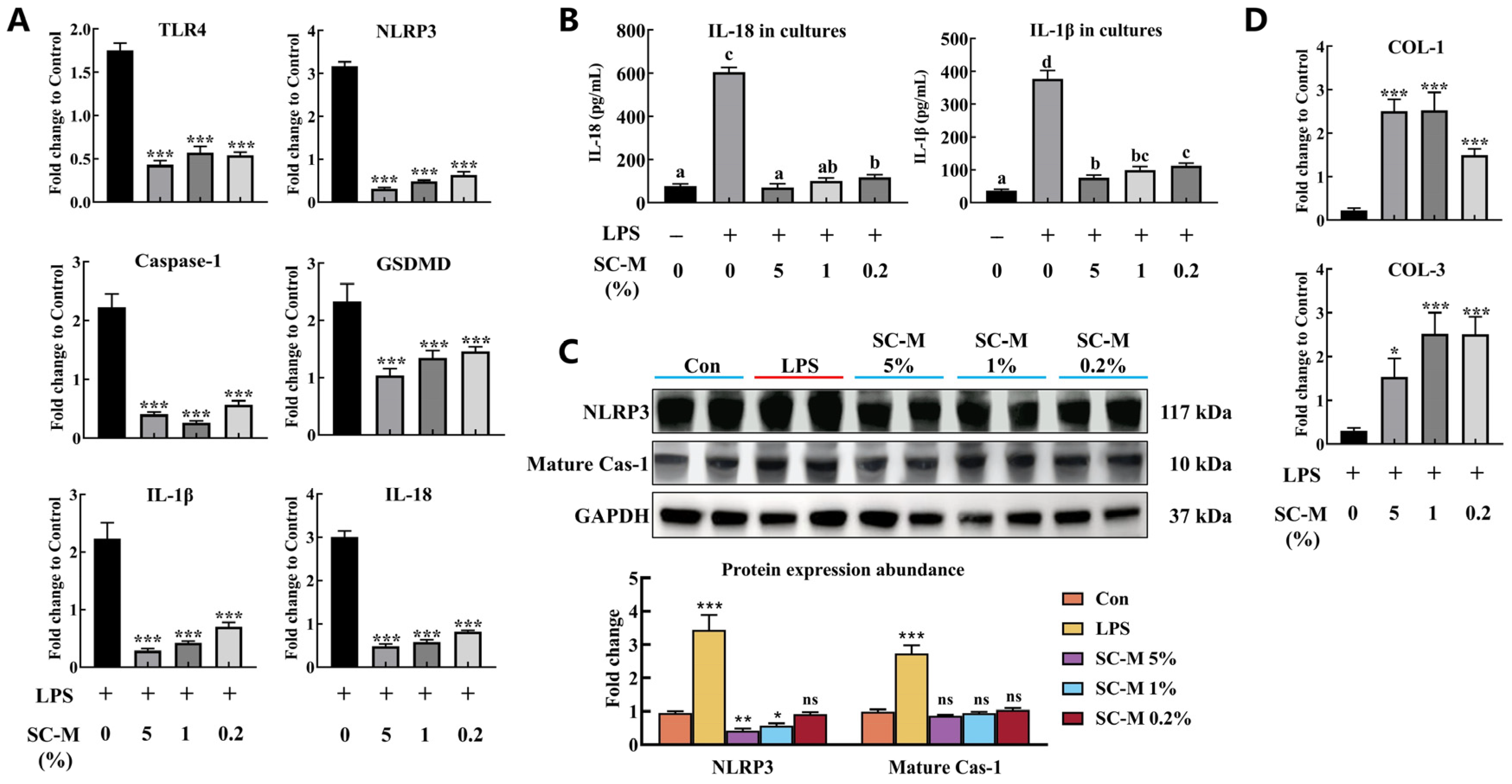
3.4. Anti-Inflammatory Effect of SC–M

Studies have indicated that mulberry anthocyanins can reduce serum glucose and leptin levels; decrease the production of malondialdehyde; enhance the activities of superoxide dismutase and glutathione peroxidase; downregulate the expression of genes for tumor necrosis factor-alpha, interleukin-6, inducible nitric oxide synthase, and nuclear factor kappa B; and inhibit diet-induced oxidative stress and inflammation [21]. Therefore, we employed quantitative polymerase chain reaction (q-PCR) to investigate the anti-inflammatory effects of SC–M. Through these experiments, we assessed the impact of the filtrate on the expression levels of the pyroptosis signaling pathway and senescence-associated proteins in LPS-induced BJ cells. Our results showed that compared to the control group cells, the relative mRNA expression levels of pyroptosis pathway-related proteins (TLR4, NLRP3, caspase-1, GSDMD, IL-1β, and IL-18) in BJ cells significantly increased after the addition of 2 μg/mL LPS, to approximately 1.6–3.2 times that of the control cells (p < 0.001, Figure 4A). The contents of IL-1β and IL-18 in the cell culture supernatant of the LPS group also significantly increased (p < 0.05, Figure 4B). This may be due to LPS activation of NLRP3, which is an upstream protein in the pyroptosis pathway. The activation of NLRP3 led to a significant increase in its content and induction of the maturation of caspase-1 (p < 0.001, Figure 4C), promoting the release of a large amount of activated IL-1β and IL-18 into the extracellular environment. The relative mRNA expression levels of type I collagen and type III collagen were also significantly reduced to 10 and 12% of the control cells, respectively (p < 0.001, Figure 4D). This indicates that LPS significantly activated the pyroptosis signaling pathway, leading to the release of a large number of pro-inflammatory factors into the extracellular environment. This significantly inhibited the synthesis of type I and type III collagen, inducing an inflammatory response.
After the addition of low (0.2%), medium (1%), and high (5%) concentrations of SC–M, the relative mRNA expression levels of TLR4, NLRP3, caspase-1, GSDMD, IL-1β, and IL-18 were significantly reduced to 25–60% of the relative mRNA expression levels in the control group cells (p < 0.001, Figure 4A). Notably, the contents of IL-1β and IL-18 in the cell culture supernatant of the SC–M groups were significantly decreased (p < 0.05, Figure 4B). This result can be attributed to the significant inhibition of LPS-induced activation of NLRP3 and maturation of caspase-1 by the SC–M. This was evidenced by a significant reduction in NLRP3 and matured caspase-1 protein contents (p < 0.05, Figure 4C), along with a significant increase in the relative type I and type III collagen mRNA expression levels. These results indicate that the SC–M significantly suppressed the release of pro-inflammatory factors and promoted the synthesis of type I and type III collagen. Overall, these results demonstrate that SC–M significantly inhibited the LPS-induced inflammatory response in BJ cells.

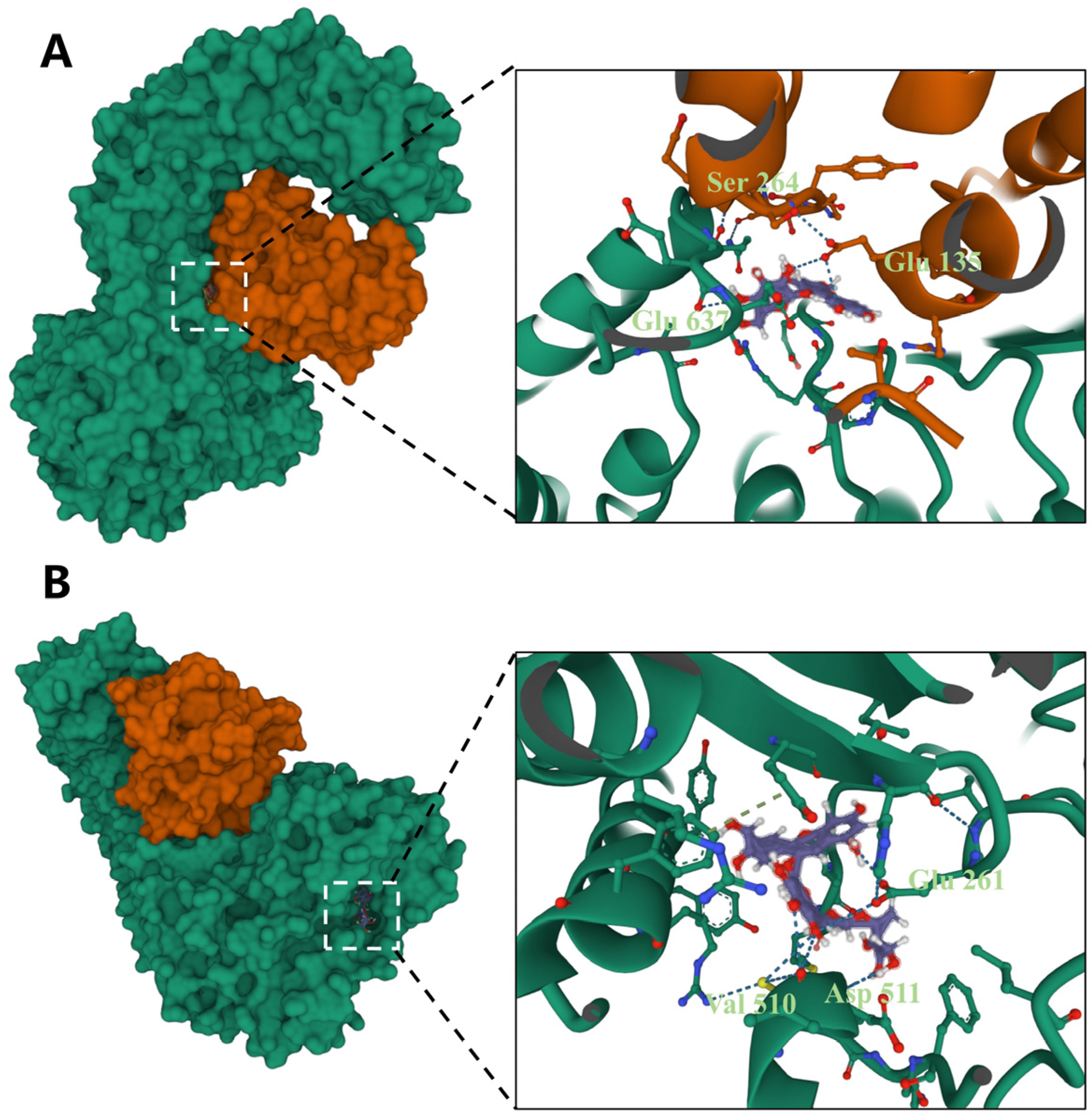
3.5. Docking of Different Anthocyanins with the NLRP3 Protein

To elucidate the mechanisms by which SC–M inhibited LPS-induced inflammation in BJ cells, we utilized LeadIT software to analyze the molecular docking of C3G with NLRP3. The results indicated that C3G bound favorably with NLRP3, with docking scores of −11.013 (Table 3). A docking score of less than −10 kcal/mol suggests stable binding. Notably, C3R, the second most abundant anthocyanin in Morus nigra L., had a docking score with NLRP3 of −9.746, which was lower than that of C3G. This indicated a more stable binding of C3G with NLRP3. C3G formed several crucial hydrogen bonds (Ser264, Glu637, and Glu135) with the carbonyl main chain and side chains of NLRP3 (Figure 5A). These results suggest that C3G in the SC–M has a high affinity for NLRP3, thereby inhibiting the activation of the LPS-induced pyroptosis signaling pathway.

4. Discussion

In this study, the optimized fermentation conditions, achieved using the response surface methodology, significantly enhanced the yield of anthocyanins from Morus nigra L. In addition, the metabolic activity of SC promoted the release of anthocyanins from Morus nigra L., which may have been related to the production of enzymes such as pectinases and β-glucosidases by SC [22]. These enzymes are involved in the breakdown of Morus nigra L. cell walls, resulting in greater anthocyanin release and increased production of C3G [23]. These results are consistent with previous studies, where the presence of glycosidases facilitated the release of C3G from Morus nigra L. by SC. Although it is known that SC possesses a strong ability to decompose lignin, its application in promoting the release of residual anthocyanins in MR had not been reported. Therefore, this study identified key fermentation factors involved in SC fermentation of extraction residue of Morus nigra L., providing an important basis for the reuse of Morus nigra L. extraction residue. It is worth noting that co-fermentation of SC with Morus nigra L. extraction residue is a two-way fermentation technology. Existing research has shown that bidirectional fermentation technologies can accelerate the acquisition of bioactive products with high yields and high activity levels [24]. Zhou et al. found that treatment of barley with edible mushroom fermentation increased flavonoid and triterpenoid production and enhanced antioxidant activity [25]. Modified fermentation with Lactobacillus sp. enhanced the structure, physicochemical properties, and antioxidant activity of linseed gum, improving its extraction rate and resulting in a high content of high-quality linseed gum [26]. Further analysis of the synergistic effects of SC metabolites (such as SC polysaccharides) with C3G is warranted.
The SC–M promoted the proliferation and migration of BJ cells within a specific concentration range. Higher concentrations of fermentation filtrates had an inhibitory effect on BJ cell proliferation, which may be due to the toxicity of anthocyanins at high concentrations [27]. Inflammation is a complex process involving multiple cell types. It is a fundamental defense mechanism that protects against and repairs damage caused by external inflammatory agents. This study found that the SC–M significantly inhibited the LPS-induced inflammatory response in BJ cells. This effect may be related to the anti-inflammatory action of anthocyanins in the fermentation filtrate. Beyond anthocyanins, SC-produced β-glucans may synergistically enhance anti-inflammatory activity by suppressing NF-κB signaling [28]. Additionally, polysaccharides could stabilize C3G via non-covalent interactions [29], potentially prolonging its bioactivity in inflammatory microenvironments.
Recent research has shown that anthocyanins exert their anti-inflammatory effects by inhibiting activation of the NLRP3 inflammasome, reducing the expression of TLR4, and suppressing activation of the NF-κB and MAPK signaling pathways [30]. In addition, anthocyanins activate Bim through the mitochondrial pathway, promoting apoptosis and exerting anticancer effects [31]. C3G exhibits strong antioxidant and anti-inflammatory activities and is the most abundant anthocyanin in mulberries [32]. Despite extensive research highlighting the anti-inflammatory activity of C3G, its mechanism of action in regulating pyroptosis pathways remains poorly understood. Our molecular docking results revealed the potential binding modes between C3G and TLR4 and NLRP3. The interaction of C3G with these proteins may block LPS-induced signal transduction, thereby inhibiting the inflammatory response. This finding provides a molecular-level explanation for the anti-inflammatory mechanism of anthocyanins and provides a basis for novel anti-inflammatory therapies. While the molecular docking predicted stable C3G-NLRP3 binding, this static model overlooks dynamic interactions in vivo. For instance, post-translational modifications of NLRP3 (e.g., phosphorylation) or competitive binding with endogenous ligands may alter the actual efficacy of C3G, and experimental validation is still required to confirm the actual binding affinity and functional consequences.
Due to their sensitivity to light, anthocyanosides are prone to degradation and discoloration during food processing and storage. This limits their use in functional foods [33]. However, the SC polysaccharides that were produced during the fermentation of Morus nigra L. residues exhibited significant color-preserving effects on anthocyanins. SC polysaccharides serve as a color protectant for anthocyanins through multiple potential mechanisms [16]. First, SC polysaccharides form a physical protective layer around anthocyanins, reducing their direct exposure to light and oxygen and effectively preventing photodegradation. Second, SC polysaccharides bind to anthocyanins through non-covalent bonds (such as hydrogen bonds), enhancing the chemical stability of anthocyanins and reducing the degradation caused by pH changes and temperature fluctuations. Finally, the antioxidant properties of SC polysaccharides aid in free radical scavenging. This reduces anthocyanin destruction from oxidative stress, preserves their color and bioactivity, and improves their stability and bioavailability in food systems [34]. These potential color-preserving mechanisms offer new directions for subsequent research. Future studies should investigate the specific molecular mechanisms behind the color-preserving effects of SC polysaccharides on anthocyanins. In addition, biological variability during fermentation, such as Schizophyllum commune-specific enzyme expression (e.g., an unexploited source for lignocellulose-degrading enzymes) and seasonal variations in the composition of Morus nigra L. residues, may significantly affect the release of anthocyanins. Future research should incorporate multi-batch fermentation trials and strain screening to address these factors and utilize bidirectional fermentation to enhance the color preservation and activity of anthocyanins, thereby improving their stability and potential applications in the food industry.

5. Conclusions

This study established a two-way fermentation method for the production of anthocyanins using SC and MR. The optimal conditions for this process were as follows: the addition of MR at 3.4%; SC inoculation at 9.0%; and a fermentation time of 50 h. Under the optimal fermentation conditions, the total anthocyanin and C3G contents were 85.1 and 66.7 μg/mL, respectively. During the fermentation process, SC promoted the release of 83.9% of C3G from Morus nigra L. These results indicated the bidirectional fermentation of SC and Morus nigra L. as an effective approach for anthocyanin production. In addition, the SC–M significantly promoted the proliferation of BJ cells, regulated pyroptosis pathways, inhibited the release of pro-inflammatory factors, and delayed LPS-induced inflammatory responses in BJ cells. These data provided valuable insight for the utilization of SC–M as a medicinal and food resource.

Author Contributions

Conceptualization, L.Y.; methodology, L.Y; software, Q.H.; validation, L.Y. and Q.H.; formal analysis, L.Y.; investigation, C.G.; resources, Y.L.; data curation, C.G. and Q.H.; writing—original draft preparation, L.Y.; writing—review and editing, Q.H. and C.G.; visualization, L.Y. and Q.H.; supervision, C.G. and Q.H.; project administration, C.G. and Q.H.; funding acquisition, C.G. and Q.H. All authors have read and agreed to the published version of the manuscript.

Funding

This research was funded by the Key-Area Research and Development Program of Guangdong Province, grant number 21202107201900003, 21202107201900005, and Zhanjiang Science and Technology Plan Project, grant number 2024R1010.

Institutional Review Board Statement

Not applicable.

Informed Consent Statement

Not applicable.

Data Availability Statement

The original contributions presented in this study are included in the article. Further inquiries can be directed to the corresponding author.

Conflicts of Interest

Authors Lin Ye and Chaowan Guo are employed by the Guangdong Marubi Biotechnology Co., Ltd. The remaining authors declare that the research was conducted in the absence of any commercial or financial relationships that could be construed as a potential conflict of interest.

Abbreviations

The following abbreviations are used in this manuscript:
C3GCyanidin-3-O-glucoside
C3RCyanidin-3-rutinoside
IL-1βInterleukin-1β
LPSLipopolysaccharide
MRMorus nigra L. extraction residue
SCSchizophyllum commune
RSMResponse surface methodology
SC–MSchizophyllum communeMorus nigra L. fermentation filtrate
q-PCR Quantitative polymerase chain reaction

References

  1. Choi, K.H.; Lee, H.A.; Park, M.H.; Han, J.-S. Mulberry (Morus alba L.) Fruit Extract Containing Anthocyanins Improves Glycemic Control and Insulin Sensitivity via Activation of AMP-Activated Protein Kinase in Diabetic C57BL/Ksj-db/db Mice. J. Med. Food 2016, 19, 737–745. [Google Scholar] [CrossRef] [PubMed]
  2. Hao, J.; Gao, Y.; Xue, J.; Yang, Y.; Yin, J.; Wu, T.; Zhang, M. Phytochemicals, Pharmacological Effects and Molecular Mechanisms of Mulberry. Foods 2022, 11, 1170. [Google Scholar] [CrossRef]
  3. Guo, S.; Bai, L.; Ho, C.-T.; Bai, N. Characteristic Components, Biological Activities and Future Prospective of Fructus Mori: A Review. Curr. Pharmacol. Rep. 2018, 4, 210–219. [Google Scholar] [CrossRef]
  4. Kaewmool, C.; Udomruk, S.; Phitak, T.; Pothacharoen, P.; Kongtawelert, P. Cyanidin-3-O-Glucoside Protects PC12 Cells Against Neuronal Apoptosis Mediated by LPS-Stimulated BV2 Microglial Activation. Neurotox. Res. 2020, 37, 111–125. [Google Scholar] [CrossRef] [PubMed]
  5. Shi, D.; Xu, J.; Sheng, L.; Song, K. Comprehensive Utilization Technology of Aronia melanocarpa. Molecules 2024, 29, 1388. [Google Scholar] [CrossRef]
  6. Xue, H.; Zha, M.; Tang, Y.; Zhao, J.; Du, X.; Wang, Y. Research Progress on the Extraction and Purification of Anthocyanins and Their Interactions with Proteins. Molecules 2024, 29, 2815. [Google Scholar] [CrossRef] [PubMed]
  7. Tena, N.; Asuero, A.G. Up-To-Date Analysis of the Extraction Methods for Anthocyanins: Principles of the Techniques, Optimization, Technical Progress, and Industrial Application. Antioxidants 2022, 11, 286. [Google Scholar] [CrossRef]
  8. Kwaw, E.; Ma, Y.; Tchabo, W.; Apaliya, M.T.; Wu, M.; Sackey, A.S.; Xiao, L.; Tahir, H.E. Effect of lactobacillus strains on phenolic profile, color attributes and antioxidant activities of lactic-acid-fermented mulberry juice. Food Chem. 2018, 250, 148–154. [Google Scholar] [CrossRef]
  9. Dutta, R.; Ghosal, S.; Bhattacharyya, D.K.; Bhowal, J. Effect of Fungal Fermentation on Enhancement of Nutritional Value and Antioxidant Activity of Defatted Oilseed Meals. Appl. Biochem. Biotechnol. 2023, 195, 2172–2195. [Google Scholar] [CrossRef]
  10. Abd Razak, D.L.; Abd Ghani, A.; Mohd Lazim, M.I.; Khulidin, K.A.; Shahidi, F.; Ismail, A. Schizophyllum commune (Fries) mushroom: A review on its nutritional components, antioxidative, and anti-inflammatory properties. Curr. Opin. Food Sci. 2024, 56, 101129. [Google Scholar] [CrossRef]
  11. Ohm, R.A.; de Jong, J.F.; Lugones, L.G.; Aerts, A.; Kothe, E.; Stajich, J.E.; de Vries, R.P.; Record, E.; Levasseur, A.; Baker, S.E.; et al. Genome sequence of the model mushroom Schizophyllum commune. Nat. Biotechnol. 2010, 28, 957–963. [Google Scholar] [CrossRef]
  12. Deng, Y.; Huang, Q.; Hu, L.; Liu, T.; Zheng, B.; Lu, D.; Guo, C.; Zhou, L. Enhanced exopolysaccharide yield and antioxidant activities of Schizophyllum commune fermented products by the addition of Radix Puerariae. RSC Adv. 2021, 11, 38219–38234. [Google Scholar] [CrossRef] [PubMed]
  13. Ji, Z.; Ma, W.; Liang, P.; Wang, X.; Zhang, S.; Han, Y.; Guo, Y. Anti-inflammatory potential of mycoprotein peptides obtained from fermentation of Schizophyllum commune DS1 with young apples. Int. J. Biol. Macromol. 2024, 281, 136638. [Google Scholar] [CrossRef] [PubMed]
  14. Song, M.H.; Bae, J.T.; Ko, H.; Jang, Y.M.; Lee, J.D.; Lee, G.S.; Pyo, H.B. Anti-Oxidant Effect and Anti-Inflammatory of Fermented Citrus unshiu Peel Extract by using Schizophyllum commune. J. Soc. Cosmet. Sci. Korea 2011, 37, 351–356. [Google Scholar]
  15. Wang, K.; Guan, L.; Gao, Y.; Yan, S.; Li, J.; Ji, N.; Li, B.; Zhou, Y. Optimization of Fermentation Process of Panax quinquefolius L. by Schizophyllum commune and Its Antioxidant Capacity in vitro. Sci. Technol. Food Ind. 2024, 45, 142–151. [Google Scholar] [CrossRef]
  16. Guo, W.; Mehrparvar, S.; Hou, W.; Pan, J.; Aghbashlo, M.; Tabatabaei, M.; Rajaei, A. Unveiling the impact of high-pressure processing on anthocyanin-protein/polysaccharide interactions: A comprehensive review. Int. J. Biol. Macromol. 2024, 6, 132042. [Google Scholar] [CrossRef]
  17. Zhang, J.; Wu, J.; Liu, F.; Tong, L.; Chen, Z.; Chen, J.; He, H.; Xu, R.; Ma, Y.; Huang, C. Neuroprotective effects of anthocyanins and its major component cyanidin-3-O-glucoside (C3G) in the central nervous system: An outlined review. Eur. J. Pharmacol. 2019, 858, 172500. [Google Scholar] [CrossRef]
  18. Ye, L.; Chen, H.; Wang, J.; Tsim, K.W.K.; Wang, Y.; Shen, X.; Lei, H.; Liu, Y. Aflatoxin B1-induced liver pyroptosis is mediated by disturbing the gut microbial metabolites: The roles of pipecolic acid and norepinephrine. J. Hazard. Mater. 2024, 474, 134822. [Google Scholar] [CrossRef]
  19. Ye, L.; Chen, H.; Tsim, K.W.K.; Shen, X.; Li, X.; Li, X.; Lei, H.; Liu, Y. Aflatoxin B1 Induces Inflammatory Liver Injury via Gut Microbiota in Mice. J. Agric. Food Chem. 2023, 71, 10787–10797. [Google Scholar] [CrossRef]
  20. Plikus, M.V.; Wang, X.; Sinha, S.; Forte, E.; Thompson, S.M.; Herzog, E.L.; Driskell, R.R.; Rosenthal, N.; Biernaskie, J.; Horsley, V. Fibroblasts: Origins, definitions, and functions in health and disease. Cell 2021, 184, 3852–3872. [Google Scholar] [CrossRef]
  21. Wu, T.; Yin, J.; Zhang, G.; Long, H.; Zheng, X. Mulberry and cherry anthocyanin consumption prevents oxidative stress and inflammation in diet-induced obese mice. Mol. Nutr. Food Res. 2016, 60, 687–694. [Google Scholar] [CrossRef] [PubMed]
  22. Tovar-Herrera, O.E.; Martha-Paz, A.M.; Pérez-Llano, Y.; Aranda, E.; Tacoronte-Morales, J.E.; Pedroso-Cabrera, M.T.; Arévalo-Niño, K.; Folch-Mallol, J.L.; Batista-García, R.A. Schizophyllum commune: An unexploited source for lignocellulose degrading enzymes. Microbiol. Open 2018, 7, e00637. [Google Scholar] [CrossRef]
  23. Wang, B.; Wang, Q.; Yang, Y.; Zhang, X.; Wang, J.; Jia, J.; Wu, Q. Bidirectional fermentation of Monascus and Mulberry leaves enhances GABA and pigment contents: Establishment of strategy, studies of bioactivity and mechanistic. Prep. Biochem. Biotechnol. 2024, 54, 73–85. [Google Scholar] [CrossRef]
  24. Song, Z.; Geng, J.; Wang, D.; Fang, J.; Wang, Z.; Wang, C.; Li, M. Reparative effects of Schizophyllum commune oat bran fermentation broth on UVB-induced skin inflammation via the JAK/STAT pathway. Bioresour. Bioprocess. 2024, 11, 73. [Google Scholar] [CrossRef] [PubMed]
  25. Zhou, X.; Wang, J.; Shao, G.; Chang, X.; Liu, Y.; Xiang, T.; Zhu, Q.; Ren, A.; Jiang, A.; He, Q. Bidirectional Solid-State Fermentation of Highland Barley by Edible Fungi to Improve Its Functional Components, Antioxidant Activity and Texture Characteristics. Plant Foods Hum. Nutr. 2024, 79, 308–315. [Google Scholar] [CrossRef] [PubMed]
  26. Wang, S.; Zhang, M.; Liu, Y.; Liu, J.; Zheng, T.; Li, Y.; He, S.; Jiang, M.; Wu, L.; Liu, F. Influence of fermentation with lactic bacteria on the structure, functional properties and antioxidant activity of flaxseed gum. Int. J. Biol. Macromol. 2024, 281, 136133. [Google Scholar] [CrossRef]
  27. Chen, J.; Xu, B.; Sun, J.; Jiang, X.; Bai, W. Anthocyanin supplement as a dietary strategy in cancer prevention and management: A comprehensive review. Crit. Rev. Food Sci. Nutr. 2022, 62, 7242–7254. [Google Scholar] [CrossRef]
  28. Zhong, X.; Wang, G.; Li, F.; Fang, S.; Zhou, S.; Ishiwata, A.; Tonevitsky, A.G.; Shkurnikov, M.; Cai, H.; Ding, F. Immunomodulatory effect and biological significance of β-glucans. Pharmaceutics 2023, 4, 1615. [Google Scholar] [CrossRef]
  29. Luo, F.; Zhang, Z.; Lu, F.; Li, D.; Zhou, C.; Li, Y.; Niu, L.; Xu, Y.; Feng, L.; Dai, Z.; et al. Ultrasound modification of pectin and the mechanism of its interaction with cyanidin-3-O-glucoside. Food Hydrocoll. 2024, 152, 109898. [Google Scholar] [CrossRef]
  30. Aboonabi, A.; Aboonabi, A. Anthocyanins reduce inflammation and improve glucose and lipid metabolism associated with inhibiting nuclear factor-kappaB activation and increasing PPAR-γgene expression in metabolic syndrome subjects. Free Radic. Biol. Med. 2020, 150, 30–39. [Google Scholar] [CrossRef]
  31. Ma, Z.; Du, B.; Li, J.; Yang, Y.; Zhu, F. An Insight into Anti-Inflammatory Activities and Inflammation Related Diseases of Anthocyanins: A Review of Both In Vivo and In Vitro Investigations. Int. J. Mol. Sci. 2021, 22, 11076. [Google Scholar] [CrossRef] [PubMed]
  32. Hao, X.; Guan, R.; Huang, H.; Yang, K.; Wang, L.; Wu, Y. Anti-inflammatory activity of cyanidin-3-O-glucoside and cyanidin-3-O-glucoside liposomes in THP-1 macrophages. Food Sci. Nutr. 2021, 9, 6480–6491. [Google Scholar] [CrossRef] [PubMed]
  33. Oliveira, H.; Correia, P.; Pereira, A.R.; Araújo, P.; Mateus, N.; de Freitas, V.; Oliveira, J.; Fernandes, I. Exploring the Applications of the Photoprotective Properties of Anthocyanins in Biological Systems. Int. J. Mol. Sci. 2020, 21, 7464. [Google Scholar] [CrossRef] [PubMed]
  34. Zang, Z.; Tang, S.; Li, Z.; Chou, S.; Shu, C.; Chen, Y.; Chen, W.; Yang, S.; Yang, Y.; Tian, J.; et al. An updated review on the stability of anthocyanins regarding the interaction with food proteins and polysaccharides. Compr. Rev. Food Sci. Food Saf. 2022, 21, 4378–4401. [Google Scholar] [CrossRef]
Figure 1. Three-dimensional plots and corresponding contour plots of the three variables, (A) Morus nigra L. (MNL) contents; (B) inoculum size of Schizophyllum commune (SC); and (C) fermentation period on the response of the total anthocyanin contents.
Fermentation 11 00145 g001
Figure 2. The concentrations of total anthocyanin and cyanidin-3-O-glucoside (C3G) in the Morus nigra L. extraction residue (M-R) and Schizophyllum commune–Morus nigra L. fermentation filtrate (SC–M) were detected using HPLC. (A) Total ion chromatography of cyanidin-3-O-glucoside in the standard solution and sample. (B) The standard curve of cyanidin-3-O-glucoside. (C) The concentration of total anthocyanin and cyanidin-3-O-glucoside (C3G).
Fermentation 11 00145 g002
Figure 3. Schizophyllum communeMorus nigra L. fermentation filtrate (SC–M) can enhance the migration of BJ cells. (A) Cell survival rate based on CKK-8 assay. (B) The wound healing scratch images of BJ cells treated with SC–M. After 24 h of treatment, the cells were photographed under a microscope at 10× magnification. Scale bar: 100 μm. (C) The histogram represents the relative percentage of cut closure, calculated by image analysis using ImageJ software. Values are means ± SD. *, **, and *** indicate significant difference from control at p < 0.05, p < 0.01, and p < 0.001, respectively. Different lowercase letters indicate significant differences (p < 0.05) between different groups by one-way ANOVA.
Fermentation 11 00145 g003
Figure 4. SC–M can significantly inhibit LPS-induced pyroptosis in BJ cells. (A) The relative mRNA expressions of TLR4, NLRP3, caspase-1, GSDMD, IL-1β, and IL-18 in BJ cells were determined by RT-PCR. (B) The levels of IL-1β and IL-18 in the cell cultures of different groups were measured by ELISA. (C) The relative protein abundance levels of NLRP3 and mature caspase-1were measured. (D) The relative mRNA expressions of COL I and COL III in BJ cells were determined by RT-PCR. Values are means ± SD. *, **, and *** indicate significant difference from control or LPS group at p < 0.05, p < 0.01, and p < 0.001, respectively. Different lowercase letters indicate significant differences (p < 0.05) between different groups by one-way ANOVA.
Fermentation 11 00145 g004
Figure 5. Molecular docking analysis of cyanidin-3-O-glucoside (A) and cyanidin-3-O-rutinoside chloride (B) with NLRP3 protein by LeadIT software.
Fermentation 11 00145 g005
Table 1. Factors and levels of response surface design. A, addition of Morus nigra L. residue; B, addition of Schizophyllum commune mycelial liquid; C, fermentation time.
FactorsLevels
A: Addition of Morus nigra L. residue (g/L)253035
B: Addition of Schizophyllum commune (%)5.07.510.0
C: Fermentation time (day)234
Table 2. ANOVA analysis of total anthocyanin content using response surface methodology. A: addition of Morus nigra L. residue; B: addition of Schizophyllum commune seed culture; C: fermentation time.
SourceSum of SquaresdfMean SquareF-Valuep-Value
Model3374.159374.9150.82<0.0001
A826.691826.69112.05<0.0001
B1.6311.630.22150.6522
C636.971636.9786.34<0.0001
AB3.4813.480.47130.5145
AC33.51133.514.540.0705
BC770.191770.19104.4<0.0001
A2339.891339.8946.070.0003
B2698.551698.5594.68<0.0001
C20.235710.23570.03190.8632
Residual51.6477.38--
Lack of Fit36.14312.053.110.151
Pure Error15.543.88--
Cor Total3425.7916---
Table 3. Molecular docking fraction of NLRP3 and microbial metabolites.
AnthocyaninBest Docking Score (kcal/mol)Number of Hydrogen BondsInteracting
Amino Acids
Cyanidin-3-O-glucoside−11.0135Ser264, Glu637, Glu135
Cyanidin-3-O-rutinoside−9.7466Glu261, Val510, Asp511
Disclaimer/Publisher’s Note: The statements, opinions and data contained in all publications are solely those of the individual author(s) and contributor(s) and not of MDPI and/or the editor(s). MDPI and/or the editor(s) disclaim responsibility for any injury to people or property resulting from any ideas, methods, instructions or products referred to in the content.

