Review

Secondary Metabolites from Actinokineospora spp.: Insights into a Sparsely Studied Genus of Actinomycetes

by
Oleksandr Yushchuk
Department of Genetics and Biotechnology, Ivan Franko National University of Lviv, 79005 Lviv, Ukraine
Fermentation 2025, 11(12), 663; https://doi.org/10.3390/fermentation11120663 (registering DOI)
Submission received: 4 October 2025 / Revised: 20 November 2025 / Accepted: 25 November 2025 / Published: 27 November 2025

Abstract

The genus Actinokineospora (family Pseudonocardiaceae) has recently emerged as a prolific source of structurally diverse, biologically active specialized metabolites. Actinokineospora spp. are filamentous actinomycetes isolated from various terrestrial biotopes. The genus is still sparsely represented taxonomically, with only 19 species holding validly published names and genome sequences available for an additional six strains. Nevertheless, Actinokineospora appears to have one of the highest biosynthetic novelty index values among actinomycetes, making it a prime candidate for the discovery of new specialized metabolites. To date, several Actinokineospora strains have shown antimicrobial activity, including Actinokineospora acnipugnans R434T, Actinokineospora alba 03-9939T, Actinokineospora fastidiosa NRRL B-16697T, Actinokineospora riparia C-39162T, Actinokineospora sp. G85, and Actinokineospora sp. PR83; the active compounds from these strains remain to be identified and characterized. By contrast, detailed chemical characterization has been achieved for several producers: Actinokineospora spheciospongiae EG49T (polyketides actinospene and actinosporins; the lasso peptide actinokineosin), Actinokineospora bangkokensis 44EHWT (polyene thailandins), Actinokineospora fastidiosa ATCC 202099 (nocathiacin thiopeptides), Actinokineospora sp. UTMC 2448 (persiathiacin thiopeptides), and Actinokineospora auranticolor DSM 44650T (kineomіcin glycopeptides). Collectively, these findings establish Actinokineospora as a promising yet underexplored genus for antibiotic discovery and biosynthetic engineering. In this review, we summarize current knowledge on Actinokineospora spp. and provide an in-depth account of specialized metabolite production for those compounds whose structures have been elucidated.

1. Introduction

Since the discoveries of Selman Waksman [1], mycelial bacteria of the phylum Actinomycetota—collectively termed actinomycetes—have remained the most prolific source of bioactive specialized metabolites, including clinically relevant antibiotics, antiprotozoals, and antifungals [2]. Among them, Streptomyces spp. are unrivaled producers of bioactive compounds [3], a status likely influenced by isolation and handling bias: Streptomyces are broadly distributed across diverse biotopes (facilitating isolation) and are generally easier to cultivate and manipulate than many other actinomycetes (facilitating drug discovery) [3]. Nevertheless, the success of streptomycetes has been constrained by the frequent rediscovery of known molecules [4]. Consequently, under the pressure of persistent multidrug resistance, research has increasingly turned to “rare” actinomycetes—operationally defined as non-Streptomyces genera [5]—to discover novel metabolites [4].
In the post-genomic era, the biosynthetic potential of actinomycetes can be rapidly assessed via genome sequence analysis because pathways for specialized metabolites are encoded in co-localized, co-expressed, and co-regulated biosynthetic gene clusters (BGCs) [6]. A recent study introduced the biosynthetic novelty index (BiNI) to prioritize strains by the predicted novelty of their BGCs [7]; applying this metric to high-quality genomes from varied taxa and niches highlighted Streptosporangium, Lentzea, Actinokineospora, and Saccharothrix as top candidates for the discovery of specialized metabolites, having the highest novelty indices [7].
In this review, we survey Actinokineospora (family Pseudonocardiaceae) as a source of structurally diverse, biologically active specialized metabolites. We start with the brief overview of the genus Actinokineospora, discussing its content and taxonomic status, as well as the ecology and isolation approaches. Next, we synthesize current knowledge on chemical diversity, bioactivities, production, and BGCs/biosynthetic pathways. Here, emphasis is made on the strains with the chemical structure of active compound elucidated, i.e., Actinokineospora spheciospongiae EG49T (multiple bioactive metabolites) [8,9,10], Actinokineospora bangkokensis 44EHWT (polyene antifungals, thailandins) [11], and Actinokineospora fastidiosa ATCC 202099 and Actinokineospora sp. UTMC 2448 (glycosylated thiopeptides) [12,13], as well as Actinokineospora auranticolor DSM 44650T, recently reported to produce novel glycopeptide antibiotic (GPA) complex [14].

2. Overview of the Genus Actinokineospora

The genus Actinokineospora was first described in 1988 and belongs to the family Pseudonocardiaceae (phylum Actinomycetota) [15]. Actinokineospora spp. are filamentous, sporulating actinomycetes that often produce zoospores [15]. They have been isolated primarily from terrestrial biotopes, including soil, plant debris, the rhizosphere, and endophytic environments (Table 1). At present, 19 species within the genus have validly published names (according to the List of Prokaryotic names with Standing in Nomenclature, LPSN), while genome sequences are available for an additional six strains that most likely also belong to Actinokineospora (Table 1).
Although only 19 species are currently recognized, Actinokineospora spp. are clearly present in diverse biotopes [29,30,31], and the number of isolated strains is expected to increase in the coming years. In the MicrobeAtlas database [32], 10 Actinokineospora operational taxonomic units (OTUs; species level) were identified in metagenomic datasets from 3471 samples (≈347 samples per OTU). For comparison, 24 OTUs of another “rare” genus in Pseudonocardiaceae, Amycolatopsis, were found in datasets from 38,608 samples (≈1609 per OTU), whereas 115 OTUs of Streptomyces were detected in 310,633 samples (≈2701 per OTU). A range of isolation methods and media has been used to obtain Actinokineospora spp. (Table 1), and the isolation of Actinokineospora does not appear to differ substantially from that of other Pseudonocardiales spp.; for example, species of this genus are often recovered without sample pretreatment by direct inoculation onto common solid media (Table 1). Thus, Actinokineospora spp. appear to be genuinely rare, although more systematic analyses are needed to normalize abundance data and improve comparability with other genera.
Several Actinokineospora spp. (discussed below) produce well-characterized bioactive secondary metabolites, including polyketides and ribosomally synthesized and post-translationally modified peptides (RiPPs). Nevertheless, the chemical diversity within the genus is likely to expand, as additional strains exhibit antimicrobial activity but remain chemically uncharacterized (Table 2, Table 3). Examples include Actinokineospora acnipugnans R434T (active against Propionibacterium acnes and Staphylococcus epidermidis [16]), Actinokineospora alba 03-9939T (active against Staphylococcus aureus and Pseudomonas aeruginosa [17]), and Actinokineospora fastidiosa NRRL B-16697T (broad activity against Gram-positive bacteria [22]); notably, Actinokineospora riparia C-39162T displays rare antimycoplasmal activity [15]. These strains warrant further investigation to identify and characterize their active metabolites.
Table 3. Number of BGCs found in the genomes of Actinokineospora spp. using AntiSMASH 8.0 in a “relaxed” mode.
SpeciesStrainAnalyzed AssemblyPutative BGC Similarity to Known Ones from MiBIG Database *
HighMediumLowNoTotal
Actinokineospora albaDSM 45114GCF_0043625151071523
Actinokineospora auranticolorDSM 44650GCF_04189780544101836
Actinokineospora baliensisDSM 45656GCF_0169076953682441
Actinokineospora cianjurensisDSM 45657GCF_00366379543131636
Actinokineospora bangkokensis44EHWGCF_00194045532121431
Actinokineospora diospyrosaDSM 44255GCF_02417192534152143
Actinokineospora globicatenaDSM 44256GCF_02417194534111937
Actinokineospora inagensisDSM 44258GCF_00048286534112240
Actinokineospora terraeDSM 44260GCF_90011117542151637
Actinokineospora enzanensisDSM 44649GCF_00037444555101838
Actinokineospora fastidiosaJCM 3276GCF_0146484153292438
Actinokineospora guangxiensisCGMCC 4.7154GCF_0426578452381629
Actinokineospora peganiTRM 65233GCF_00974597542163355
Actinokineospora soliJCM 17695JBHTEY0100000001491630
Actinokineospora spheciospongiaeCECT 8578GCF_00318241562111938
Actinokineospora xionganensisHBU206404GCF_0143237251181121
Actinokineospora sp.G85GCF_04967288553131233
Actinokineospora sp.HUAS TT18GCF_0513646351282233
Actinokineospora sp.NBRC 105648GCF_03026964544192552
Actinokineospora sp.PR83GCF_02105630544183965
Actinokineospora sp.UTMC 2448GCF_02476056532102439
* “High”: BGC similarity to known ones ≥75%, “Medium”: BGC similarity to known ones 75–50%, “Low”: BGC similarity to known ones 50–15%, “No”: BGC similarity to known ones <15% [33].
Within the genus Actinokineospora, available genomes harbor dozens of putative BGCs, contributing to substantial potential diversity of specialized metabolites, with most predicted BGCs showing no close similarity to characterized BGCs (Table 3). Although some exceptionally high BGC counts (e.g., 52, 55, and 65 in Actinokineospora sp. NBRC 105648, A. pegani TRM 65233T, and Actinokineospora sp. PR83, respectively; Table 3) are likely inflated by assembly fragmentation and associated prediction bias, it is reasonable to estimate that Actinokineospora genomes on average carry ~20–40 BGCs for specialized metabolites—comparable to other genera within the order Pseudonocardiales [34]. Overall, the breadth and novelty of the putative BGC repertoire in Actinokineospora clearly warrant detailed investigation in silico.

3. Actinokineospora spheciospongiae EG49T: One Strain—Many Compounds

A. spheciospongiae EG49T (=DSM 45935T) was isolated from the marine sponge Spheciospongia vagabunda (Ridley, 1884) [27]. Upon isolation, A. spheciospongiae EG49T was found to inhibit the growth of Candida albicans S314 and trypanosomatids (Leishmania major and Trypanosoma brucei brucei TC221, the causative agents of leishmaniasis and African trypanosomiasis, respectively) [35]. Subsequent investigations revealed EG49T to be a prolific source of novel bioactive compounds.
Actinosporins. Metabolomic profiling of ethyl acetate extracts from EG49T cultures (ISP2 solid medium, g/L: 4 yeast extract, 10 malt extract, 4 dextrose, 20 agar; 168 h of cultivation) revealed the production of diverse anthraquinones—known inhibitors of trypanothione reductase, catalyzing the reduction of trypanothione disulfide to trypanothione, a key process in oxidative stress defense [36]. Generally, anthraquinones of various structures are produced in different organisms, including plants, lichen-forming fungi, or insects [37,38,39]. However, actinomycetes (e.g., Streptomyces spp.) are another prolific source of such compounds [40].
Among the detected compounds were several known metabolites—atramycin A [41], elloramycin [42], BE-12406A [43] (all three produced by Streptomyces spp.), and galtamycinone (produced by Micromonospora sp.) [44]—as well as compounds with unprecedented molecular masses. Two of the latter were designated actinosporins A and B, and subjected to detailed characterization [8].
To optimize actinosporins production, several cultivation approaches were tested in addition to ISP2 agar cultivation: (a) ISP2 liquid culture; (b) ISP2 liquid culture supplemented with 20 g/L XAD-16 resin; and (c) ISP2 liquid culture with bacterial cells immobilized on calcium alginate beads [8]. In all cases, 120-h precultures (presumably grown in ISP2 liquid medium) were used to inoculate main cultures, fermented for 168 h. In all cases, supernatants were extracted with ethyl acetate, while XAD-16 resin with bacterial cells was extracted with acetone, and free cells or cells on calcium alginate beads were extracted with methanol [8].
The strongest antitrypanosomal activity was observed in extracts from ISP2 liquid culture supernatants. Two active fractions (corresponding to actinosporins A and B) were purified by silica gel chromatography, Sephadex LH-20 gel filtration, and reversed-phase HPLC. Their structures were elucidated using 1D/2D NMR and high-resolution tandem mass spectrometry (MS/MS) (Figure 1a,b). Actinosporins A and B possess modified benz(α)anthraquinone cores bearing two or one l-rhamnose residues, respectively (Figure 1a). Thus, they represent rhamnosylated analogues of the known anthraquinone antibiotic tetrangulol, previously isolated from Streptomyces rimosus (Figure 1a,b) [45]. Interestingly, tetrangulol was later detected in EG49T co-cultures with Rhodococcus sp. UR59 (ISP2 liquid medium) [46]. Two additional congeners, actinosporins C and D (Figure 1a) were later identified from calcium alginate bead cultures and differed from actinosporin A in glycosylation pattern [47].
Another set of angucycline-type secondary metabolites was obtained from EG49T cultures grown in ISP2 liquid medium prepared with artificial seawater and supplemented with 50 μM N-acetyl-d-glucosamine (GlcNAc) [48]. GlcNAc, a known signaling molecule in actinomycetes, often induces secondary metabolite production [49]. In this case, it stimulated the biosynthesis of actinosporins F, G, and H (analogues of actinosporin A, Figure 1a) and actinosporin E (an analogue of actinosporin B, Figure 1b) [48]. Finally, 168-h-old ISP2 agar cultures supplemented with 50 μM GlcNAc yielded another distinct metabolite profile [50], containing actinosporins C, D, and G, together with several additional anthraquinones. The latter included known compounds G-2N (from Frankia sp. G-2 [51]), fridamycin E (produced in Streptomyces sp. Tü 1989 mutant [52]), saptomycin F (from Streptomyces sp. HP530 [53]), and two new fridamycin derivatives—fridamycins H and I (Figure 1c) [50].
Pure actinosporin A exhibited activity against L. major and T. brucei brucei TC221 without cytotoxicity toward J774.1 macrophages, whereas actinosporin B was inactive against T. brucei brucei TC221 [8]. Actinosporins C and D were inactive against trypanosomatids but demonstrated strong antioxidant activity, mitigating H2O2-induced cytotoxicity and DNA damage in HL-60 cells [47]. Actinosporins E, G, and H were active against Plasmodium falciparum 3D7, with IC50 values of 11–14 μg/mL—significantly weaker than chloroquine (IC50 = 0.022 μg/mL) [48]. Fridamycin H exhibited potent activity against T. brucei brucei TC221 without macrophage cytotoxicity, while fridamycin I was inactive [50].
In summary, strain EG49T represents an exceptionally prolific producer of microheterogeneous angucyclines. Although its genome sequence is available [54], the genetic basis of actinosporins biosynthesis remains to be elucidated.
Actinospene. In addition to anthraquinones, strain DSM 45935T was shown to produce another bioactive specialized metabolite—a polyene macrolide designated actinospene [9]. The discovery of actinospene involved a two-step approach [9]. First, the aa sequence of NysDIII, a GDP-mannose-4,6-dehydratase from the nystatin producer Streptomyces noursei ATCC 11455, was used as a probe to screen publicly available genome libraries. Since GDP-mannose-4,6-dehydratases are commonly encoded in multiple polyene BGCs [55], identification of homologues was considered indicative of the presence of a polyene BGC. As a result, numerous NysDIII orthologues were detected in Streptomyces spp., and only a few in representatives of less-studied genera, including DSM 45935T [9].
Subsequently, DSM 45935T was cultivated under various conditions (>20 media tested), and antifungal activity was observed exclusively in one of them—the SFM medium [9]. For actinospene production, DSM 45935T was first grown in GYM preculture medium (g/L: 4 glucose, 4 yeast extract, 10 malt extract, 2 CaCO3). After 168 h of incubation, precultures were used to inoculate SFM production medium (g/L: 20 soya flour, 20 d-mannitol), which was fermented for 360 h [9].
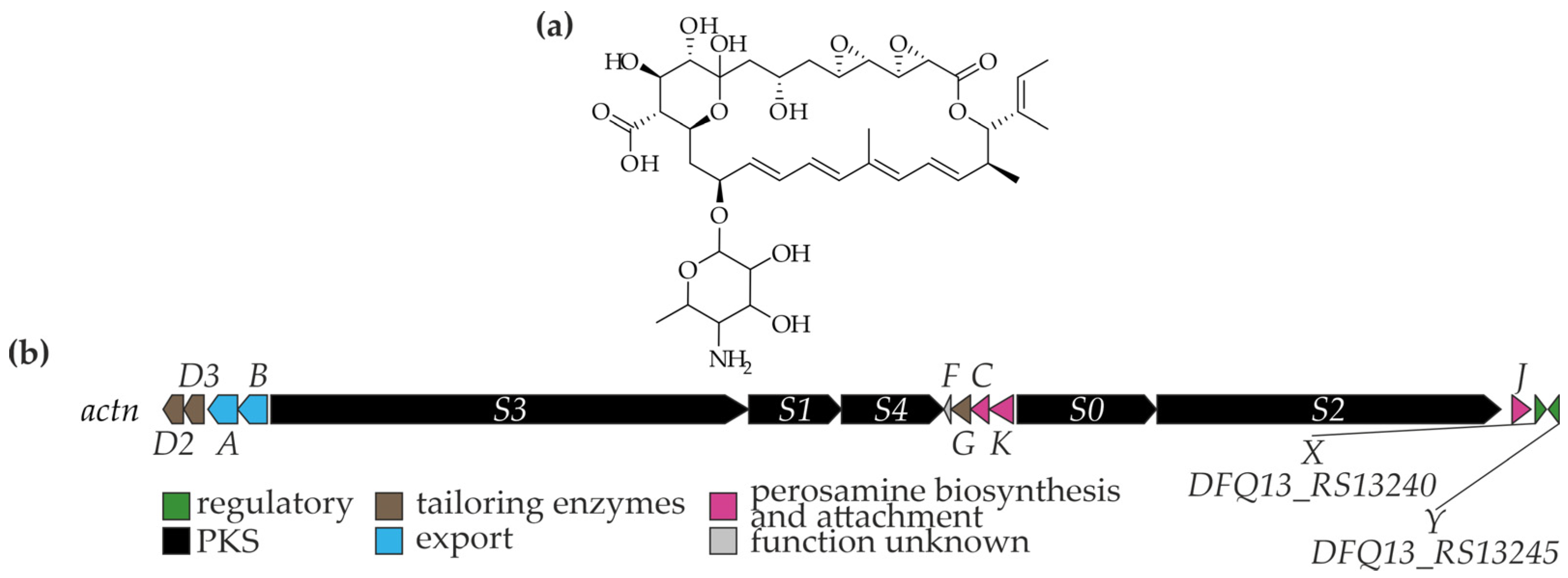
Actinospene was purified by sequential silica gel chromatography, Sephadex LH-20 gel filtration, and semipreparative HPLC. Structural elucidation was performed using HPLC-HRMS and NMR spectroscopy, revealing actinospene to be an unusual polyene macrolide bearing two epoxy groups and an isobutenyl side chain, with an aglycone glycosylated by d-perosamine (Figure 2a) [9].
Actinospene exhibited antifungal activity against representatives of Ascomycota, Basidiomycota, and Oomycota, including Saccharomyces cerevisiae, Candida albicans, Cryptococcus neoformans, Fusarium oxysporum, Fusarium graminearum, Sclerotium rolfsii, Colletotrichum capsici, Alternaria alternata, and Phytophthora capsici, with minimum inhibitory concentrations (MICs) ranging from 2 to 50 µg/mL. However, its activity was markedly lower compared to amphotericin B (0.16–5 µg/mL) [9].
Genome analysis of strain DSM 45935T enabled identification of the BGC responsible for actinospene production, designated actn [9]. The highly fragmented draft genome available at that time [54] hindered accurate reconstruction of the actn BGC, resulting in several misinterpretations. For instance, the nysDIII orthologue, actnJ, was incorrectly proposed to reside outside the main BGC. In the draft assembly, most actn genes were located on contig NZ_AYXG01000206, while actnJ appeared at the terminus of another contig (NZ_AYXG01000229). Moreover, one of the polyketide synthase genes, actnS2, was positioned at the contig boundary, yielding a truncated sequence and leading to the erroneous assumption that certain actn PKS modules operated iteratively [9].
Fortunately, a more contiguous genome assembly for A. spheciospongiae CECT 8578 (=DSM 45935T) has since become available, with the actn cluster located centrally within a single large contig (NZ_QHCP01000004), thereby allowing its reanalysis in the present review. Analysis of this sequence using antiSMASH 8.0.1 [33] revealed a complete actn BGC, displaying high similarity and near-colinearity with the lcm BGC encoding lucensomycin biosynthesis in Streptomyces cyanogenus S136 [56].
Thus, the actn BGC most likely comprises 16 genes, 14 of which were previously described [9] and two newly assigned to the BGC in this work (Figure 2b). The genes actnS3–S1–S4 and actnS0–S2 encode a 14-modular PKS. actnG (orthologous to lcm6) encodes a cytochrome P450 monooxygenase presumably involved in aglycone carboxylation, whereas actnD2 (orthologue of tetrK from the tetramycin B BGC [57]) and actnD3 (orthologue of lcm10) encode additional P450 monooxygenases likely participating in hydroxylation and epoxidation reactions [9,56].
A triad of genes—actnJ, actnC, and actnK (orthologues of lcm2, lcm5, and lcm4, respectively)—encode a GDP-d-mannose 4,6-dehydratase, a GDP-3-keto-6-deoxy-d-mannose C-3 aminotransferase, and a glycosyltransferase responsible for the biosynthesis and attachment of the d-perosamine residue [9,56]. In addition, actn harbors two ABC transporter genes (actnA and actnB, orthologues of lcm8 and lcm9) and two genes for transcriptional regulators (actnX and actnY, orthologues of lcmRII and lcmRI), which are likely involved in compound export and biosynthesis regulation [9,56]. Finally, actnF (orthologue of lcm7) has no clearly defined role in actinospene biosynthesis [9,56].
Consequently, the biosynthetic model originally proposed for the actinospene PKS [37] requires substantial revision based on the now-available contiguous actn sequence.
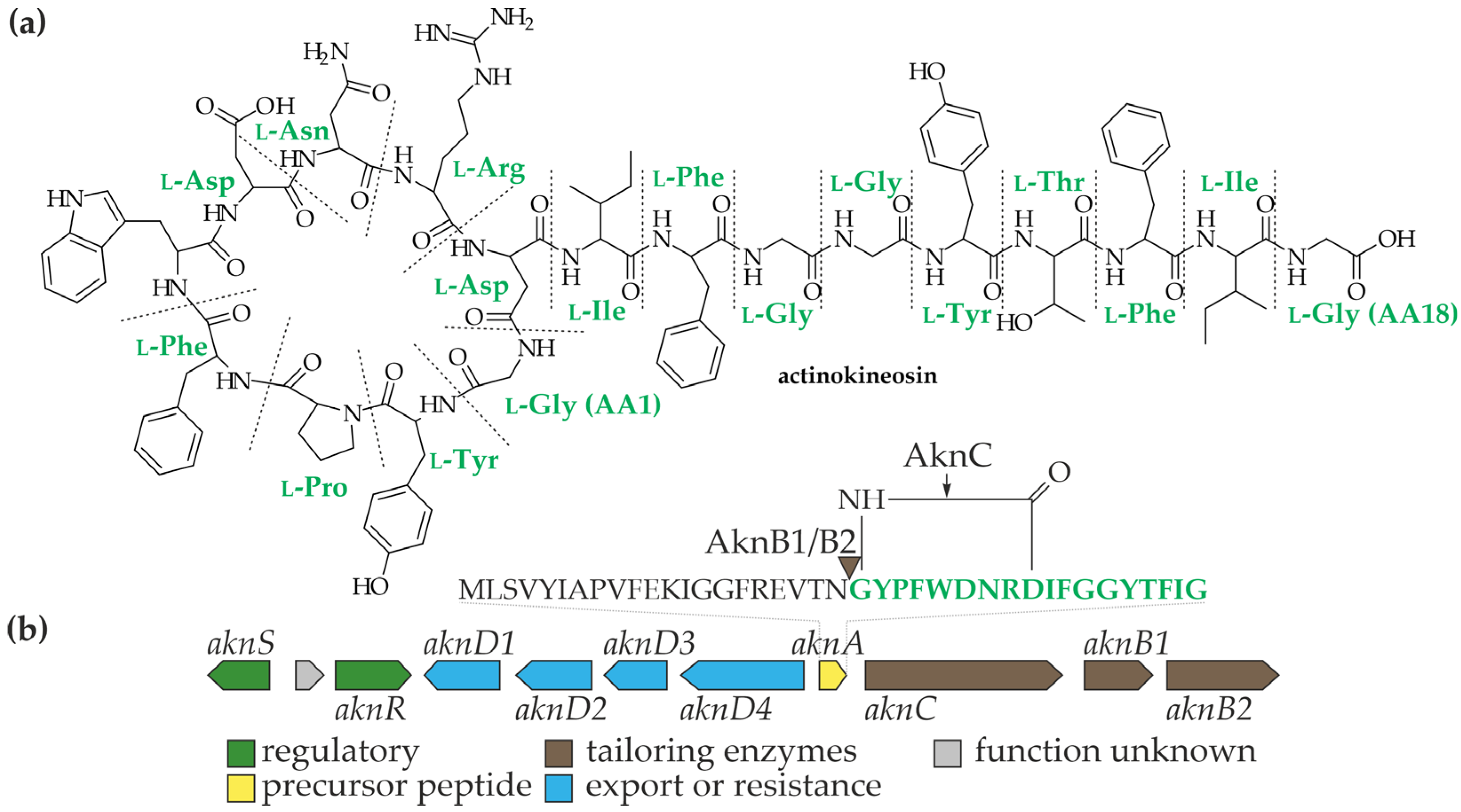
Actinokineosin. A RiPP—named actinokineosin—was another active compound, discovered in the cultivation broth of DSM 45935. This was a genomics-guided discovery: DSM 45935 was chosen for the research as its genome [54] carried a homologue for the precursor peptide biosynthesis gene of other RiPP—propeptin from Microbispora sp. SNA-115 [58]. Actinokineosin was extracted from the cells of DSM 45935 cultivated on ISP2 agar medium with methanol and further purified using CHP-20P resin and preparative HPLC [10]. Structure of actinokineosin, resolved with mass spectrometry on chemically and enzymatically degraded derivative, resembled the one of propeptin (which also was evident from the precursor peptides amino acid sequence comparison) and consisted of 18 amino acids (aa): GYPFWDNRDIFGGYTFIG [10] (Figure 3a).
BGC for actinokineosin (named akn) consisted of 11 genes, including 10 putatively involved in the biosynthesis, export, self-resistance or regulation, and one gene coding for a protein of unknown function [10] (Figure 3b). Among those, aknA coded for the precursor peptide, while aknC and aknB1/B2 code for a lasso cyclase (responsible for the of the Gly1-Glu9 amide bond) and RiPP recognition element (RRE) containing protein/endopeptidase (responsible for the recognition and cleavage of the precursor peptide), respectively [10]. By turn, aknS and aknR code for a sensory histidine kinase and a response transcriptional regulator, respectively, which are likely involved in the transcriptional regulation of actinokineosin production. Finally, genes aknD1-D4 code for the components of the ABC-transporter probably responsible for the export of actinokineosin (Figure 3b) [10].
When tested against a set of Gram-positive, Gram-negative bacteria, and ascomycetes in a disc diffusion assay, actinokineosin appeared to be active only against Micrococcus luteus, although exact MIC was not measured [10].
Discovery of actinokineosin serves as an important example of a knowledge-guided genome mining of an uncommon actinomycete, moving in a “genes to compound” direction.

4. Thailandins: Polyene Antifungals from Actinokineospora bangkokensis 44EHWT

A. bangkokensis 44EHWT was isolated from the rhizospheric soil of taro (Colocasia esculenta (L.) Schott) in Bangkok, Thailand [20].
To test the antagonistic properties, 44EHWT was cultivated in 4 liquid media, including: HA liquid medium (g/L: 4 yeast extract, 10 malt extract, 4 glucose); NL5 (g/L: 1 NaCl, 1 KH2PO4, 0.5 MgSO4 × 7H2O, 2.5% (v/v) glycerol, 5.84 l-glutamine, 0.2% (v/v) trace elements solution); MS (g/L: 20 soya flour, 20 d-mannitol), SG (g/L: 20 glucose, 10 soya flour, 2 CaCO3, 1 CoCl2, 2 l-valine) [11]. 48-h old TSB cultures were used to inoculate abovementioned fermentation media [11].
Preliminary bioactivity-guided screening of strain 44EHWT cultivated in HA, MS, and SG media revealed strong inhibition of phytopathogenic Colletotrichum spp. (Ascomycota), while NL5 cultures demonstrated antibacterial activity against Bacillus subtilis ATCC 6051 [11]. Further LC-MS analysis suggested that the antifungal activity was attributable to the production of a putative polyene compound with an unprecedented molecular mass [11].
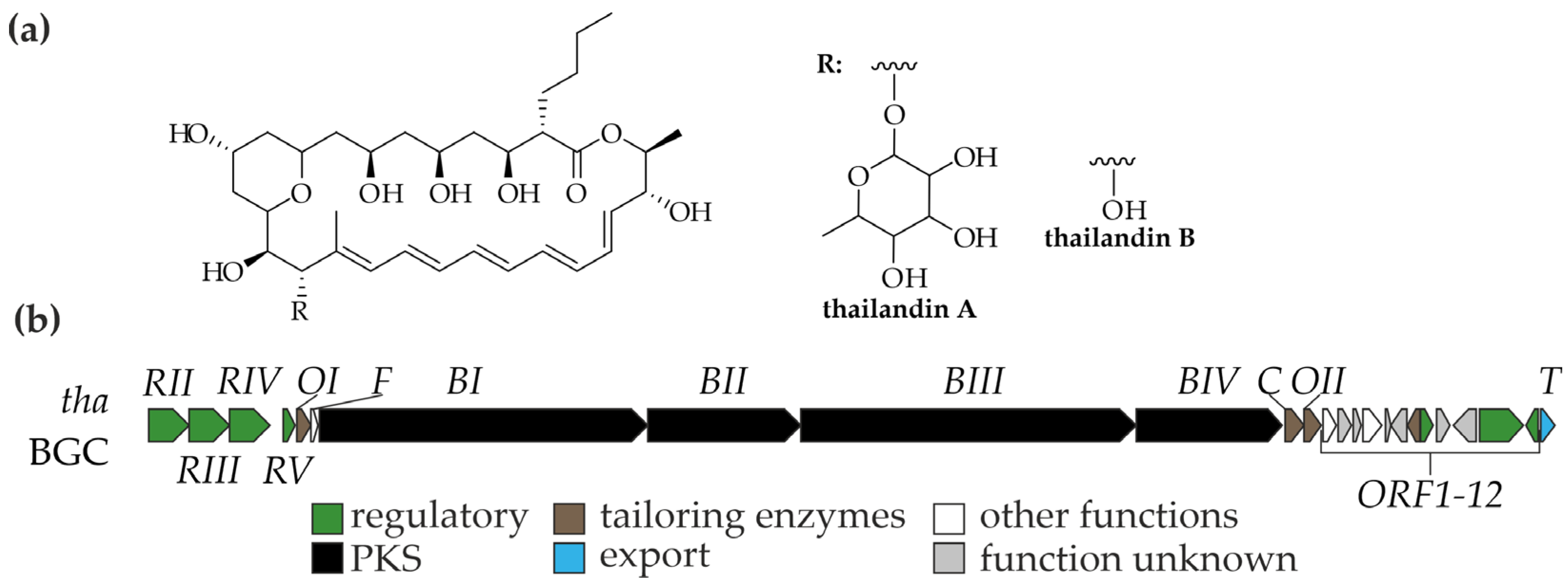
Two active compounds were extracted from 44EHWT cultures grown in HA liquid medium with ethyl acetate or ethyl acetate and acetone, followed with a bioactivity-guided purification using semi-preparative HPLC and Sephadex-LH20 column chromatography. Obtained compounds were designated thailandins A and B. Structure elucidation, performed using MS, 1D, and 2D NMR data, revealed that they are 28-membered macrocyclic lactones containing two methyl groups and seven free hydroxyl groups. Thailandin A was identified as a rhamnosylated congener of thailandin B (Figure 4a) [11].
Both thailandins A and B inhibited the growth of various Colletotrichum spp., Candida spp. (including C. albicans MT 2013/1), Cryptococcus neoformans MT 2013/2, and Saccharomyces cerevisiae IFO 10217. Although thailandin B (MICs in the range of 8–16 µg/mL) appeared more active than thailandin A (MICs in the range of 16–32 µg/mL), both compounds exhibited substantially lower activity compared to amphotericin B (MIC 0.25 µg/mL against all tested strains) [11]. It was proposed that thailandins, like other polyene antifungals, target ergosterol in the fungal cell membrane [11].
Furthermore, sequencing of the 44EHWT genome (using 454 and Illumina HiSeq 1000 technologies) enabled identification of the corresponding BGC (tha, verified with the insertional knockout of the PKS gene, Figure 4b) and the proposal of a biosynthetic model for thailandins [59]. The tha BGC consisted of 25 genes (Figure 4b). Among these, thaBI–BIV encode a type I 14-modular PKS, in which the last module is predicted to have ethylmalonyl-CoA specificity, while thailandins incorporate a butylmalonyl-CoA unit instead. It was predicted that ThaC, a crotonyl-CoA carboxylase, is likely involved in the biosynthesis of this butylmalonyl-CoA extender unit. Release of the linear precursor from the PKS is most likely catalyzed by the C-terminal thioesterase domain of ThaBIV, followed by modification through two P450 monooxygenases, ThaO1 and ThaO2, which mediate cyclization and hydroxylation, leading to thailandin B [59]. Although thailandin A is a rhamnosylated derivative of thailandin B, no glycosyltransferase genes were found within tha [59]. Finally, the tha BGC contains genes for four LuxR-like transcriptional regulators (thaRI–IV) and carries a gene for a transmembrane transporter (thaT), likely responsible for the export of the compounds (Figure 4b) [59].
Overall, thailandins are an interesting example of polyketides carrying an unusual butylmalonyl extender unit and have a striking resemblance with other polyene antifungal—chainin (produced by Chainia sp.) [60]. Genus Chainia is currently obsolete and former members were reclassified as Streptomyces spp. instead [61]; however, it cannot be excluded that chainin was isolated from some Actinokineospora sp., misclassified at that time.

5. Glycosylated Thiopeptides from Actinokineospora spp.

Thiopeptide antibiotics are a distinctive class of RiPPs with macrocyclic scaffolds enriched in thiazole/oxazole rings and a central nitrogen heteroaromatic core, features that underlie their exceptional potency against Gram-positive pathogens and mark them as structurally more complex than most other RiPP families [62]. Nevertheless, their clinical development has stalled due to poor aqueous solubility and gastrointestinal absorption, problems typical of peptide antibiotics. Glycosylation can enhance solubility, making the discovery of naturally glycosylated thiopeptides particularly significant [63]. In this context, two structurally related glycosylated thiopeptide complexes—nocathiacins and persiatiacins—have been isolated and extensively studied.
Persiatiacins are produced by Actinokineospora sp. UTMC 2448, whereas the taxonomy of the nocathiacins producer has been more ambiguous. The strain was initially reported as Nocardia sp. ATCC 202099 [12], but later reclassified as Amycolatopsis fastidiosa ATCC 202099 based on a polyphasic approach [64]. Phylogenetic reconstruction of the 16S rRNA gene sequence from ATCC 202099 (EU072442) placed it in the same clade as Am. fastidiosa ATCC 31181T [64], which itself was subsequently reassigned to A. fastidiosa ATCC 31181T [22]. To further clarify the placement of ATCC 202099, analysis of EU072442 using the EzBioCloud platform [65] identified its closest matches as A. fastidiosa IMSNU 20054T (99.72% identity) and A. acnipugnans R434T (97.91% identity). Taken together, these data strongly support that the nocathiacin producer ATCC 202099 belongs to the genus Actinokineospora.
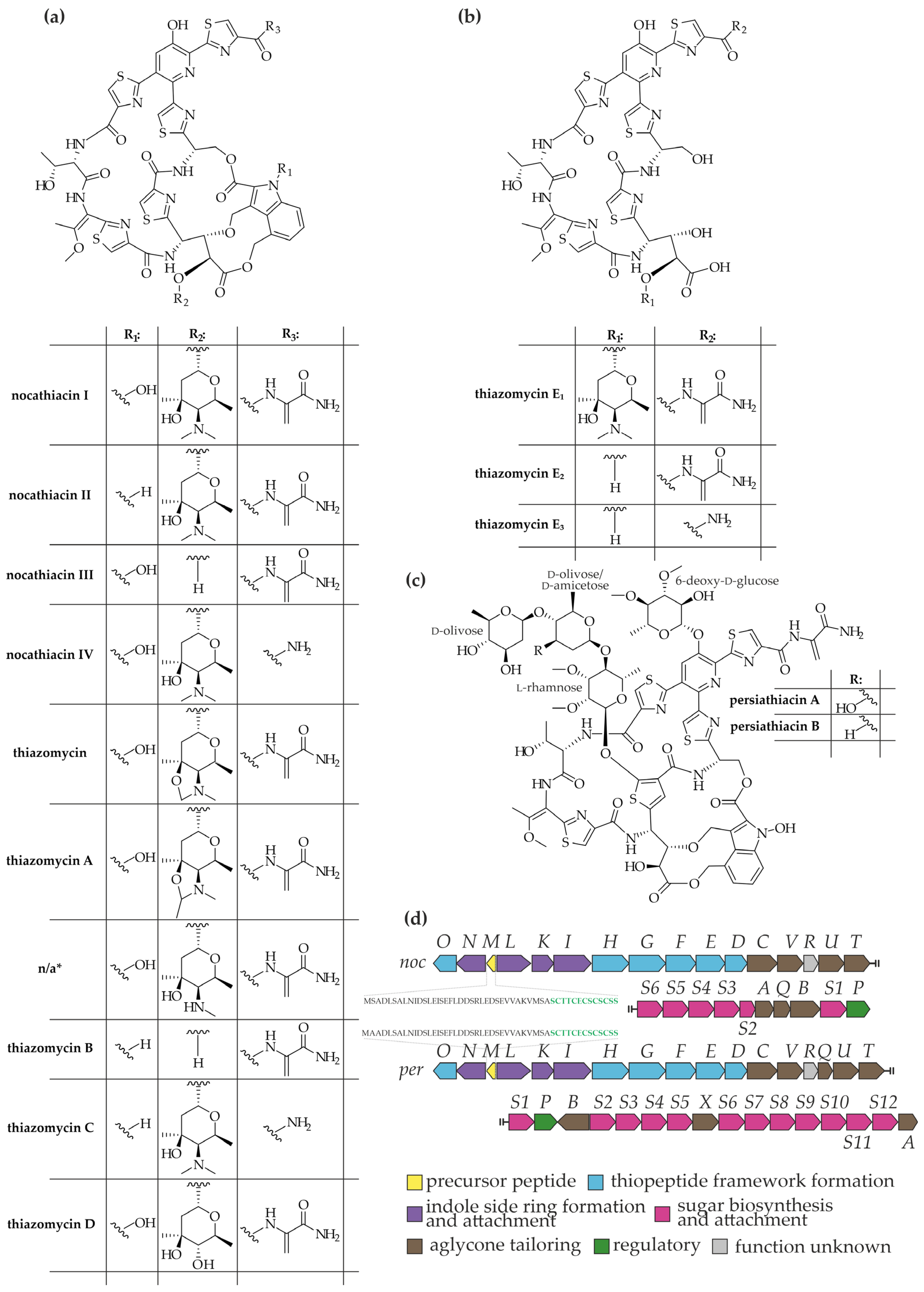
Nocathiacin family of thiopeptides: structure and biosynthesis. A. fastidiosa ATCC 202099 was originally isolated from a soil sample collected in New Mexico, USA [12]. Fermentation of this strain in seed medium (g/L: 20 soluble starch, 5 dextrose, 3 N-Z-case, 2 yeast extract, 5 fish meat extract, 3 CaCO3) for 72 h, followed by cultivation in HYDN production medium (g/L: 10 Hy-Yest 412, 20 dextrose, 1 Nutrisoy) for 96–120 h, yielded three thiopeptide congeners, nocathiacins I–III (Figure 5a) [12]. These compounds were purified from culture broth by sequential chromatographic separation on silica gel and Sephadex LH-20, and their structures were elucidated by 1D/2D NMR, FAB-MS, CD spectroscopy, and chemical degradation studies [66]. The peptide cores of nocathiacins I–III were nearly identical to that of nosiheptide (from Streptomyces actuosus 40037 [67]), with nocathiacins I and II further modified by a 2,4,6-trideoxy-3-methyl-4-N,N-dimethylamino-l-hexose residue (Figure 5a) [66]. Subsequent studies expanded this family of metabolites from ATCC 202099 to include nocathiacins I–IV [67], thiazomycin [64], thiazomycins A–D [68], and thiazomycins E1–E3 [69], which differ in glycosylation, hydroxylation, and variations in the peptide core (Figure 5b).
BGC for nocathiacins production was identified in ATCC 202099 and comprises 26 genes [12] (Figure 5d). Within this cluster, nocM encodes a 49-aa precursor peptide consisting of a 36-aa leader and a 13-aa structural region (SCTTCECSCSCSS), fully consistent with the peptide backbone of nocathiacin I (Figure 5d) [12].
The noc BGC encodes the full enzymatic machinery required for nocathiacin biosynthesis. Core formation is directed by nocO and nocHGFED (Figure 5d), which encode a cyclodehydratase/dehydrogenase pair (NocG/NocF), dehydratases (NocE/NocD), and two putative proteins (NocH/NocO). Together, NocG and NocF catalyze cyclodehydration and dehydrogenation to form thiazoles, NocE and NocD dehydrate most Ser/Thr residues (except Thr39 and Ser44), and NocH/NocO mediate formation of the nitrogen heterocycle [12].
A second gene set—nocLKI and nocN (Figure 5d)—is implicated in indole side-ring synthesis and attachment. The radical SAM enzymes NocL and NocN likely convert l-tryptophan into a 3,4-dimethylindole intermediate [70], which is then 4-hydroxylated and attached to Ser44 and Glu42 of the peptide scaffold via the acyltransferase NocK and the CoA ligase NocI [12].
Additional tailoring is performed by seven genes (nocCV, nocUT, and nocAQB, Figure 5d). These include five cytochrome P450 enzymes (NocB, NocC, NocT, NocU, and NocV) responsible for hydroxylations, a SAM-dependent methyltransferase (NocQ), and a hypothetical protein (NocA) proposed to generate the terminal amide moiety by removing the Ser-derived acrylic acid group [12].
Genes nocS6–S2 and nocS1 (Figure 5d) encode enzymes for biosynthesis and attachment of the characteristic 2,4,6-trideoxy-3-methyl-4-N,N-dimethylamino-l-hexose residue. The aminosugar is obtained from 6-deoxy-4-keto-d-glucose through the sequential action of sugar 2,3-dehydratase (NocS5), 3-keto-reductase (NocS4), C-methyltransferase (NocS3), aminotransferase (NocS6), and N-dimethyltransferase (NocS2), and subsequently transferred to the peptide backbone by the glycosyltransferase NocS1 [12].
Beyond biosynthetic functions, the BGC also carries a regulatory gene (nocP) and an additional gene of unknown function (nocR) [12].
Persiathiacins: structure and biosynthesis. Actinokineospora sp. UTMC 2448 was isolated from a mud sample collected in Bushehr, Iran [13]. Screening for antibiotic activity revealed that ethyl acetate extracts from ISP2 solid cultures inhibited the growth of methicillin-resistant Staphylococcus aureus (MRSA). Purification and structural elucidation by MS and NMR identified the active compound, persiathiacin A, as a thiopeptide antibiotic structurally related to nocathiacins [13] (Figure 5c). Its key distinguishing feature is an extensive glycosylation pattern, with four sugars attached to the peptide core: doubly methylated l-rhamnose, doubly methylated 6-deoxy-d-glucose, and two exotic 6-deoxysugars: d-olivose and d-amicetose (Figure 5c). A minor congener, persiathiacin B, was also characterized, differing by the replacement of d-amicetose with a second d-olivose [13] (Figure 5c).
BGC for persiathiacins was identified in the genome of Actinokineospora sp. UTMC 2448 and designated per (Figure 5d) [13]. This cluster shows strong similarity to noc, enabling functional assignment of most per genes. perM encodes a precursor peptide differing from nocM by a single amino acid in the leader region (Figure 5d); perO and perHGFED are orthologues of nocO and nocHGFED and likely direct thiopeptide core assembly; while perLKI and perN, orthologous to nocLKI and nocN, are implicated in indole side-ring biosynthesis and attachment. Tailoring steps are mediated by perCV, perQUT, perA, perB, and perX, most of which (except perX) have counterparts in noc [13].
The major distinction between per and noc lies in the repertoire of genes for 6-deoxysugar biosynthesis, methylation, and glycosylation (Figure 5d) [13]. Enzymes encoded by perS1, perS2, perS10, perS11, and perS12 (coding for NDP-hexose-3-ketoreductase, UDP-glucose 4-epimerase, NDP-hexose 2,3-dehydratase, NDP-hexose 4-ketoreductase, and NDP-hexose 3,4-dehydratase, respectively) catalyze the conversion of d-glucose into 6-deoxy-d-glucose (PerS2, PerS11), d-olivose (PerS2, PerS10, PerS1, PerS11), or d-amicetose (PerS2, PerS10, PerS1, PerS12, PerS11). Three O-methyltransferases (PerS3, PerS5, PerS7) are proposed to methylate l-rhamnose and 6-deoxy-d-glucose residues, while four glycosyltransferases (PerS4, PerS6, PerS8, PerS9) mediate the incorporation of all four sugar units [13]. Further work is required to clarify the substrate specificity of the methyltransferases and glycosyltransferases, as well as the timing of these biosynthetic steps.
Antimicrobial properties of nocathiacin family thiopeptides and persiathiacins. Similarly to other thiopeptides, nocathiacins and persiathiacins exhibit remarkable activity against Gram-positive bacteria. Nocathiacins I–III display MICs in the nanogram range against Klebsiella pneumoniae strains (including penicillin-resistant isolates), Streptococcus pyogenes A9604, enterococci, staphylococci, various strains of Moraxella catarrhalis, Chlamydia trachomatis, Clostridium perfringens, Clostridioides difficile, Peptostreptococcus spp., and Eubacterium lentum [12,71]. Notably, nocathiacin I also demonstrated potent activity against Mycobacterium tuberculosis and Mycobacterium avium strains, including multiple drug-resistant clinical isolates (MICs in range of 0.008–0.25 µg/mL) [71,72].
In vivo, nocathiacins I–III showed superior efficacy compared to vancomycin in a systemic St. aureus infection mouse model, having PD50 in range 0.62–0.89 mg/kg/day comparing to the 1.3 mg/kg/day for vancomycin [12]. Thiazomycin exhibited comparable (MICs in range of 0.004–0.064 µg/mL) in vitro and in vivo antibacterial potency to nocathiacins I–III, whereas thiazomycin A was slightly less active in vitro (MICs in range 0.002–0.25 µg/mL) [68,69,73]. Moreover, thiazomycin inhibited the growth of numerous St. aureus strains resistant to clinically relevant antibiotics [68,69]. Similar activity profiles were observed for thiazomycins B, C, and D, while loss of the indole moiety in thiazomycins E1–E3 resulted in complete loss of antibacterial activity against Gram-positive bacteria [69]. Persiathiacin A displayed a comparable antimicrobial spectrum, showing strong activity against methicillin-resistant St. aureus (MRSA, 0.025 μg/mL MIC) and M. tuberculosis clinical isolates (MICs in range of 1.6–3.9 µg/mL), including rifampicin- and isoniazid-resistant strains [13].
Like other thiopeptides of similar structure, nocathiacin family compounds and persiathiacin A were demonstrated to inhibit translation elongation by targeting ribosomal protein L11 [71,73,74].
Although nocathiacins and persiathiacins share highly similar biosynthetic pathways and antimicrobial profiles, differences in their tailoring and glycosylation machineries suggest that Actinokineospora spp. may harbor additional, yet-undiscovered glycosylated thiopeptides.

6. Kineomicins: Novel Glycopeptide Antibiotic Complex from Actinokineospora auranticolor DSM 44650T

GPAs are an important class of lipid II binders that inhibit cell-wall biosynthesis in Gram-positive bacteria, including MDR staphylococci and enterococci [75,76,77]. As antimicrobial resistance further constrains the efficacy of clinically used GPAs (e.g., teicoplanin and vancomycin), the search for novel, naturally occurring GPAs remains a viable strategy to counter rising GPA resistance.
Using a comparative-genomics approach focused on the order Pseudonocardiales, BGCs with atypical nonribosomal peptide synthetase (NRPS) features were identified, including ambiguous substrate predictions for module-3 adenylation (A-) domains. This triage flagged Actinokineospora auranticolor DSM 44650T as a candidate producer of GPAs with unconventional peptide core [14]. Subsequent experiments confirmed that A. auranticolor DSM 44650T produces a complex mixture of bioactive GPAs—termed kineomicins—at strikingly high titres (0.7–1.3 g/L in standard R5 medium [78]), enabling isolation and structural analysis of the dominant congener, kineomicin B [14]. NMR elucidation revealed an unprecedented aglycone architecture that combines an aromatic aa at position 1 with an aliphatic aa at position 3—an arrangement that does not conform to classical GPA Types I–IV [79] and appears to bridge structural features of Types I and II. Phylogenetic analysis showed that the module-3 A-domain of the kineomicin NRPS is closely related to counterparts from Type II GPA NRPSs. Accordingly, it was proposed subdividing Type II GPAs into Type IIa (including kineomicins) and Type IIb (encompassing previously known Type II GPAs). Despite their structural novelty, the antibacterial activity of the kineomicin complex against diverse Gram-positive bacteria (MICs 0.5–16 µg/mL) was comparable to that of vancomycin (MICs 0.5–8 µg/mL) [14].
Finally, integrating NMR, LC-MS, and MS/MS data across multiple kineomicin congeners with bioinformatic analysis of the corresponding BGC (knm) enabled the proposal of a biosynthetic model for kineomicin production [14].
Summarizing, A. auranticolor DSM 44650T was shown to produce a novel GPA complex and constitutes a naturally robust GPA overproducer suitable as a host for heterologous expression of GPA BGCs.

7. Conclusions and Outlook

The genus Actinokineospora has emerged as an unexpectedly rich source of chemically and structurally diverse specialized metabolites, including ribosomally synthesized and post-translationally modified peptides (RiPPs) such as actinokineosin and persithiacin, polyene macrolides such as actinospene and thailandins, as well as anthraquinones collectively referred to as actinosporins.
From a broader perspective, findings discussed in the current review illustrate several important trends. First, the continuous discovery of RiPPs and polyketide-derived metabolites from Actinokineospora spp. underscores the value of genome-guided mining in rare actinomycetes. Second, the diversity of enzymatic tailoring reactions (e.g., unusual extender units in thailandins or glycosylation pattern of persiathiacins) provides new opportunities for biosynthetic engineering and synthetic biology of novel compounds. Third, despite their promising novelty, the majority of Actinokineospora metabolites remain only partially characterized in terms of pharmacological potential, biosynthetic regulation, and ecological role.
In conclusion, Actinokineospora spp. represent an underutilized but promising reservoir of bioactive natural products. Future integrative studies that combine classical microbiology, advanced analytical chemistry, and genome-based approaches are expected to unlock the full biosynthetic potential of this genus. Comprehensive metabolomic and transcriptomic analyses under varied cultivation conditions, coupled with genome mining and heterologous expression, will likely reveal additional cryptic BGCs and novel scaffolds.

Funding

This work was funded by the Ministry of Education and Science of Ukraine under BG-14E grant (to O.Y.) and by the Parliament of Ukraine through a personal stipendium (to O.Y.).

Institutional Review Board Statement

Not applicable.

Informed Consent Statement

Not applicable.

Data Availability Statement

No new data were created or analyzed in this study. Data sharing is not applicable to this article.

Conflicts of Interest

The author declares no conflicts of interest.

References

  1. Waksman, S.A.; Woodruff, H.B. The soil as a source of microorganisms antagonistic to disease-producing bacteria. J. Bacteriol. 1940, 40, 581–600. [Google Scholar] [CrossRef] [PubMed]
  2. Hutchings, M.; Truman, A.; Wilkinson, B. Antibiotics: Past, present and future. Curr. Opin. Microbiol. 2019, 51, 72–80. [Google Scholar] [CrossRef] [PubMed]
  3. Donald, L.; Pipite, A.; Subramani, R.; Owen, J.; Keyzers, R.A.; Taufa, T. Streptomyces: Still the biggest producer of new natural secondary metabolites, a current perspective. Microbiol. Res. 2022, 13, 418–465. [Google Scholar] [CrossRef]
  4. Parra, J.; Beaton, A.; Seipke, R.F.; Wilkinson, B.; Hutchings, M.I.; Duncan, K.R. Antibiotics from rare actinomycetes, beyond the genus Streptomyces. Curr. Opin. Microbiol. 2023, 76, 102385. [Google Scholar] [CrossRef]
  5. Tiwari, K.; Gupta, R.K. Rare actinomycetes: A potential storehouse for novel antibiotics. Crit. Rev. Biotechnol. 2012, 32, 108–132. [Google Scholar] [CrossRef]
  6. Zdouc, M.M.; Blin, K.; Louwen, N.L.L.; Navarro, J.; Loureiro, C.; Bader, C.D.; Bailey, C.B.; Barra, L.; Booth, T.J.; Bozhüyük, K.A.J.; et al. MIBiG 4.0: Advancing biosynthetic gene cluster curation through global collaboration. Nucleic Acids Res. 2025, 53, D678–D690. [Google Scholar] [CrossRef]
  7. González-Salazar, L.A.; Quezada, M.; Rodríguez-Orduña, L.; Ramos-Aboites, H.; Capon, R.J.; Souza-Saldívar, V.; Barona-Gomez, F.; Licona-Cassani, C. Biosynthetic novelty index reveals the metabolic potential of rare actinobacteria isolated from highly oligotrophic sediments. Microb. Genom. 2023, 9, mgen000921. [Google Scholar] [CrossRef]
  8. Abdelmohsen, U.R.; Cheng, C.; Viegelmann, C.; Zhang, T.; Grkovic, T.; Ahmed, S.; Quinn, R.J.; Hentschel, U.; Edrada-Ebel, R.A. Dereplication strategies for targeted isolation of new antitrypanosomal actinosporins A and B from a marine sponge associated-Actinokineospora sp. EG49. Mar. Drugs 2014, 12, 1220–1244. [Google Scholar] [CrossRef]
  9. Tang, Y.; Zhang, C.; Cui, T.; Lei, P.; Guo, Z.; Wang, H.; Liu, Q. The discovery of actinospene, a new polyene macrolide with broad activity against plant fungal pathogens and pathogenic yeasts. Molecules 2021, 26, 7020. [Google Scholar] [CrossRef]
  10. Takasaka, N.; Kaweewan, I.; Ohnishi-Kameyama, M.; Kodani, S. Isolation of a new antibacterial peptide actinokineosin from Actinokineospora spheciospongiae based on genome mining. Lett. Appl. Microbiol. 2017, 64, 150–157. [Google Scholar] [CrossRef]
  11. Intra, B.; Greule, A.; Bechthold, A.; Euanorasetr, J.; Paululat, T.; Panbangred, W. Thailandins A and B, New polyene macrolactone compounds isolated from Actinokineospora bangkokensis strain 44EHWT, possessing antifungal activity against anthracnose fungi and pathogenic yeasts. J. Agric. Food Chem. 2016, 64, 5171–5179. [Google Scholar] [CrossRef]
  12. Li, W.; Leet, J.E.; Ax, H.A.; Gustavson, D.R.; Brown, D.M.; Turner, L.; Brown, K.; Clark, J.; Yang, H.; Fung-Tomc, J.; et al. Nocathiacins, new thiazolyl peptide antibiotics from Nocardia sp. I. Taxonomy, fermentation and biological activities. J. Antibiot. 2003, 56, 226–231. [Google Scholar] [CrossRef] [PubMed]
  13. Dashti, Y.; Mohammadipanah, F.; Zhang, Y.; Cerqueira Diaz, P.M.; Vocat, A.; Zabala, D.; Fage, C.D.; Romero-Canelon, I.; Bunk, B.; Spröer, C.; et al. Discovery and biosynthesis of persiathiacins: Unusual polyglycosylated thiopeptides active against multidrug resistant tuberculosis. ACS Infect. Dis. 2024, 10, 3378–3391. [Google Scholar] [CrossRef] [PubMed]
  14. Yushchuk, O.; Berini, F.; Zhong, L.; Rückert-Reed, C.; Bernasconi, E.; Bartolone, L.; Busche, T.; Kalinowski, J.; Süssmuth, R.D.; Marinelli, F. A rare peptide scaffold in kineomicins, the glycopeptide antibiotics produced by Actinokineospora auranticolor DSM 44650. Commun. Chem. 2025, 8, 134. [Google Scholar] [CrossRef] [PubMed]
  15. Hasegawa, T. Actinokineospora: A new genus of the Actinomycetales. Actinomycetologica 1988, 2, 31–45. [Google Scholar] [CrossRef]
  16. Dahal, R.H.; Shim, D.S.; Kim, J. Description of Actinokineospora acnipugnans sp. nov., an actinomycete isolated from soil, showing potential uses in cosmetics. Int. J. Syst. Evol. Microbiol. 2017, 67, 3043–3049. [Google Scholar] [CrossRef]
  17. Yuan, L.J.; Zhang, Y.Q.; Yu, L.Y.; Liu, H.Y.; Guan, Y.; Lee, J.C.; Kim, C.J.; Zhang, Y.Q. Alloactinosynnema album gen. nov., sp. nov., a member of the family Actinosynnemataceae isolated from soil. Int. J. Syst. Evol. Microbiol. 2010, 60, 39–43. [Google Scholar] [CrossRef]
  18. Otoguro, M.; Hayakawa, M.; Yamazaki, T.; Tamura, T.; Hatano, K.; Iimura, Y. Numerical phenetic and phylogenetic analyses of Actinokineospora isolates, with a description of Actinokineospora auranticolor sp. nov. and Actinokineospora enzanensis sp. nov. Actinomycetologica 2001, 15, 30–39. [Google Scholar] [CrossRef]
  19. Lisdiyanti, P.; Otoguro, M.; Ratnakomala, S.; Lestari, Y.; Hastuti, R.D.; Triana, E.; Katsuhiko, A.; Widyastuti, Y. Actinokineospora baliensis sp. nov., Actinokineospora cibodasensis sp. nov. and Actinokineospora cianjurensis sp. nov., isolated from soil and plant litter. Int. J. Syst. Evol. Microbiol. 2010, 60, 2331–2335. [Google Scholar] [CrossRef]
  20. Intra, B.; Matsumoto, A.; Inahashi, Y.; Omura, S.; Takahashi, Y.; Panbangred, W. Actinokineospora bangkokensis sp. nov., isolated from rhizospheric soil. Int. J. Syst. Evol. Microbiol. 2013, 63, 2655–2660. [Google Scholar] [CrossRef]
  21. Tamura, T.; Hayakawa, M.; Nonomura, H.; Yokota, A.; Hatano, K. Four new species of the genus Actinokineospora: Actinokineospora inagensis sp. nov., Actinokineospora globicatena sp. nov., Actinokineospora terrae sp. nov., and Actinokineospora diospyrosa sp. nov. Int. J. Syst. Bacteriol. 1995, 45, 371–378. [Google Scholar] [CrossRef]
  22. Labeda, D.P.; Price, N.P.; Tan, G.Y.A.; Goodfellow, M.; Klenk, H.P. Emended description of the genus Actinokineospora Hasegawa 1988 and transfer of Amycolatopsis fastidiosa Henssen et al. 1987 as Actinokineospora fastidiosa comb. nov. Int. J. Syst. Evol. Microbiol. 2010, 60, 1444–1449. [Google Scholar] [CrossRef] [PubMed]
  23. Wu, H.; Liu, B. Actinokineospora guangxiensis sp. nov isolated from soil. Int. J. Syst. Evol. Microbiol. 2015, 65, 4650–4654. [Google Scholar] [CrossRef] [PubMed]
  24. Aouiche, A.; Bouras, N.; Mokrane, S.; Zitouni, A.; Schumann, P.; Spröer, C.; Sabaou, N.; Klenk, H.P. Actinokineospora mzabensis sp. nov., a novel actinomycete isolated from Saharan soil. Antonie Van Leeuwenhoek Int. J. Gen. Mol. Microbiol. 2015, 107, 291–296. [Google Scholar] [CrossRef]
  25. Lei, Y.J.; Xia, Z.F.; Luo, X.X.; Zhang, L.L. Actinokineospora pegani sp. nov., an endophytic actinomycete isolated from the surface-sterilized root of Peganum harmala L. Int. J. Syst. Evol. Microbiol. 2020, 70, 4358–4363. [Google Scholar] [CrossRef]
  26. Tang, X.; Zhou, Y.; Zhang, J.; Ming, H.; Nie, G.X.; Yang, L.L.; Tang, S.K.; Li, W.J. Actinokineospora soli sp. nov., a thermotolerant actinomycete isolated from soil, and emended description of the genus Actinokineospora. Int. J. Syst. Evol. Microbiol. 2012, 62, 1845–1849. [Google Scholar] [CrossRef]
  27. Kämpfer, P.; Glaeser, S.P.; Busse, H.J.; Abdelmohsen, U.R.; Ahmed, S.; Hentschel, U. Actinokineospora spheciospongiae sp. nov., isolated from the marine sponge Spheciospongia vagabunda. Int. J. Syst. Evol. Microbiol. 2015, 65, 879–884. [Google Scholar] [CrossRef]
  28. Wang, Y.; Shi, J.; Liu, T.; Zhang, Y.; Zhang, L.; Zhang, X. Actinokineospora xionganensis sp. nov., a filamentous actinomycete isolated from the lakeside soil of Baiyangdian. Antonie Van Leeuwenhoek Int. J. Gen. Mol. Microbiol. 2021, 114, 487–496. [Google Scholar] [CrossRef]
  29. Meena, B.; Rajan, L.A.; Vinithkumar, N.V.; Kirubagaran, R. Novel marine actinobacteria from emerald Andaman & Nicobar Islands: A prospective source for industrial and pharmaceutical byproducts. BMC Microbiol. 2013, 13, 145. [Google Scholar] [CrossRef]
  30. Arn, F.; Frasson, D.; Kroslakova, I.; Rezzonico, F.; Pothie, J.F.; Riedl, R.; Sievers, M. Isolation and identification of actinomycetes strains from Switzerland and their biotechnological potential. Chimia 2020, 74, 382–390. [Google Scholar] [CrossRef]
  31. Oberhofer, M.; Hess, J.; Leutgeb, M.; Gössnitzer, F.; Rattei, T.; Wawrosch, C.; Zotchev, S.B. Exploring Actinobacteria associated with rhizosphere and endosphere of the native alpine medicinal plant Leontopodium nivale subspecies alpinum. Front. Microbiol. 2019, 10, 2531. [Google Scholar] [CrossRef]
  32. Frederico, J.; Rodrigues, M.; Tackmann, J.; Malfertheiner, L.; Patsch, D.; Perez-Molphe-Montoya, E.; Näpflin, N.; Gaio, D.; Rot, G.; Danaila, M.; et al. The MicrobeAtlas database: Global trends and insights into Earth’s microbial ecosystems. bioRxiv 2025. [Google Scholar] [CrossRef]
  33. Blin, K.; Shaw, S.; Vader, L.; Szenei, J.; Reitz, Z.L.; Augustijn, H.E.; Cediel-Becerra, J.D.D.; De Crécy-Lagard, V.; Koetsier, R.A.; Williams, S.E.; et al. AntiSMASH 8.0: Extended gene cluster detection capabilities and analyses of chemistry, enzymology, and regulation. Nucleic Acids Res. 2025, 53, W32–W38. [Google Scholar] [CrossRef]
  34. Morin, L.M.C.; Dekoninck, K.; Sridhar, V.; Disney-McKeethen, S.; Proctor, T.; Eng, A.Y.; Traxler, M.F. Why do filamentous Actinomycetota produce such a vast array of specialized metabolites? Annu. Rev. Microbiol. 2025, 79, 753–772. [Google Scholar] [CrossRef] [PubMed]
  35. Abdelmohsen, U.R.; Pimentel-Elardo, S.M.; Hanora, A.; Radwan, M.; Abou-El-Ela, S.H.; Ahmed, S.; Hentschel, U. Isolation, phylogenetic analysis and anti-infective activity screening of marine sponge-associated actinomycetes. Mar. Drugs 2010, 8, 399–412. [Google Scholar] [CrossRef] [PubMed]
  36. Hoet, S.; Opperdoes, F.; Brun, R.; Quetin-Leclercq, J. Natural products active against African trypanosomes: A step towards new drugs. Nat. Prod. Rep. 2004, 21, 353–364. [Google Scholar] [CrossRef] [PubMed]
  37. Llewellyn, T.; Nowell, R.W.; Aptroot, A.; Temina, M.; Prescott, T.A.K.; Barraclough, T.G.; Gaya, E. Metagenomics shines light on the evolution of “sunscreen” pigment metabolism in the Teloschistales (lichen-forming Ascomycota). Genome Biol. Evol. 2023, 15, evad002. [Google Scholar] [CrossRef]
  38. Frandsen, R.J.N.; Khorsand-Jamal, P.; Kongstad, K.T.; Nafisi, M.; Kannangara, R.M.; Staerk, D.; Okkels, F.T.; Binderup, K.; Madsen, B.; Møller, B.L.; et al. Heterologous production of the widely used natural food colorant carminic acid in Aspergillus nidulans. Sci. Rep. 2018, 8, 12853. [Google Scholar] [CrossRef]
  39. Stalman, M.; Koskamp, A.M.; Luderer, R.; Vernooy, J.H.J.; Wind, J.C.; Wullems, G.J.; Croes, A.F. Regulation of anthraquinone biosynthesis in cell cultures of Morinda citrifolia. J. Plant Physiol. 2003, 160, 607–614. [Google Scholar] [CrossRef]
  40. Cheemalamarri, C.; Batchu, U.R.; Thallamapuram, N.P.; Katragadda, S.B.; Reddy Shetty, P. A review on hydroxy anthraquinones from bacteria: Crosstalk’s of structures and biological activities. Nat. Prod. Res. 2022, 36, 6186–6205. [Google Scholar] [CrossRef]
  41. Fujioka, K.; Shimazu, A.; Hayakawa, Y.; Seto, H.; Furihata, K. Isolation and characterization of atramycin A and atramycin B, new isotetracenone type antitumor antibiotics. J. Antibiot. 1991, 44, 1025–1028. [Google Scholar] [CrossRef]
  42. Ramos, A.; Lombó, F.; Braña, A.F.; Rohr, J.; Méndez, C.; Salas, J.A. Biosynthesis of elloramycin in Streptomyces olivaceus requires glycosylation by enzymes encoded outside the aglycon cluster. Microbiology 2008, 154, 781–788. [Google Scholar] [CrossRef] [PubMed]
  43. Kojiri, K.; Arakawa, H.; Okura, A.; Suda, H.; Okanishi, M.; Satoh, F.; Kawamura, K. New antitumor substances, Be-12406A and Be-12406B, produced by a streptomycete I. Taxonomy, fermentation, isolation, physico-chemical and biological properties. J. Antibiot. 1991, 44, 1054–1060. [Google Scholar] [CrossRef] [PubMed]
  44. Murenets, N.V.; Kudinova, M.K.; Klyev, N.A. Galtamycin: Galtamycinone structure. Antibiot. I Med. Biotechnol. 1986, 31, 431–434. [Google Scholar]
  45. Kuntsmann, M.P.; Mitscher, L.A. The structural characterization of tetrangomycin and tetrangulol. J. Org. Chem. 1966, 31, 2920–2925. [Google Scholar] [CrossRef]
  46. Alhadrami, H.A.; Thissera, B.; Hassan, M.H.A.; Behery, F.A.; Ngwa, C.J.; Hassan, H.M.; Pradel, G.; Abdelmohsen, U.R.; Rateb, M.E. Bio-guided isolation of antimalarial metabolites from the coculture of two red sea sponge-derived Actinokineospora and Rhodococcus spp. Mar. Drugs 2021, 19, 109. [Google Scholar] [CrossRef]
  47. Grkovic, T.; Abdelmohsen, U.R.; Othman, E.M.; Stopper, H.; Edrada-Ebel, R.; Hentschel, U.; Quinn, R.J. Two new antioxidant actinosporin analogues from the calcium alginate beads culture of sponge-associated Actinokineospora sp. strain EG49. Bioorganic Med. Chem. Lett. 2014, 24, 5089–5092. [Google Scholar] [CrossRef]
  48. Dashti, Y.; Grkovic, T.; Abdelmohsen, U.R.; Hentschel, U.; Quinn, R.J. Actinomycete metabolome induction/suppression with N-acetylglucosamine. J. Nat. Prod. 2017, 80, 828–836. [Google Scholar] [CrossRef]
  49. Liu, G.; Chater, K.F.; Chandra, G.; Niu, G.; Tan, H. Molecular regulation of antibiotic biosynthesis in Streptomyces. Microbiol. Mol. Biol. Rev. 2013, 77, 112–143. [Google Scholar] [CrossRef]
  50. Tawfike, A.; Attia, E.Z.; Desoukey, S.Y.; Hajjar, D.; Makki, A.A.; Schupp, P.J.; Edrada-Ebel, R.A.; Abdelmohsen, U.R. New bioactive metabolites from the elicited marine sponge-derived bacterium Actinokineospora spheciospongiae sp. nov. AMB Express 2019, 9, 12. [Google Scholar] [CrossRef]
  51. Gerber, N.N.; Lechevalier, M.P. Novel benzo[α]naphthacene quinones from an actinomycete, Frankia G-2 (ORS 020604). Can. J. Chem. 1984, 62, 2818–2821. [Google Scholar] [CrossRef]
  52. Künzel, E.; Faust, B.; Oelkers, C.; Weissbach, U.; Bearden, D.W.; Weitnauer, G.; Westrich, L.; Bechthold, A.; Rohr, J. Inactivation of the urdGT2 gene, which encodes a glycosyltransferase responsible for the C-glycosyltransfer of activated d-olivose, leads to formation of the novel urdamycins I, J, and K. J. Am. Chem. Soc. 1999, 121, 11058–11062. [Google Scholar] [CrossRef]
  53. Naoki, A.; Yasukazu, N.; Takehiko, N.; Nobuyasu, E.; Hideaki, U.; Masanobu, M. Novel antitumor antibiotics, saptomycins I. Taxonomy of the producing organism, fermentation, HPLC analysis and biological activities. J. Antibiot. 1993, 46, 1530–1535. [Google Scholar] [CrossRef] [PubMed]
  54. Harjes, J.; Ryu, T.; Abdelmohsen, U.R.; Moitinho-Silva, L.; Horn, H.; Ravasi, T.; Hentschel, U. Draft genome sequence of the antitrypanosomally active sponge-associated bacterium Actinokineospora sp. strain EG49. Genome Announc. 2014, 2, e00160-14. [Google Scholar] [CrossRef] [PubMed]
  55. Aparicio, J.F.; Caffrey, P.; Gil, J.A.; Zotchev, S.B. Polyene antibiotic biosynthesis gene clusters. Appl. Microbiol. Biotechnol. 2003, 61, 179–188. [Google Scholar] [CrossRef]
  56. Yushchuk, O.; Ostash, I.; Mösker, E.; Vlasiuk, I.; Deneka, M.; Rückert, C.; Busche, T.; Fedorenko, V.; Kalinowski, J.; Süssmuth, R.D.; et al. Eliciting the silent lucensomycin biosynthetic pathway in Streptomyces cyanogenus S136 via manipulation of the global regulatory gene adpA. Sci. Rep. 2021, 11, 3507. [Google Scholar] [CrossRef]
  57. Sheng, Y.; Ou, Y.; Hu, X.; Deng, Z.; Bai, L.; Kang, Q. Generation of tetramycin B derivative with improved pharmacological property based on pathway engineering. Appl. Microbiol. Biotechnol. 2020, 104, 2561–2573. [Google Scholar] [CrossRef]
  58. Kimura, K.I.; Kanou, F.; Takahashi, H.; Esumi, Y.; Uramoto, M.; Yoshihama, M. Propeptin, a new inhibitor of prolyl endopeptidase produced by Microbispora. I. Fermentation, isolation and biological properties. J. Antibiot. 1997, 50, 373–378. [Google Scholar] [CrossRef]
  59. Greule, A.; Intra, B.; Flemming, S.; Rommel, M.G.E.; Panbangred, W.; Bechthold, A. The draft genome sequence of Actinokineospora bangkokensis 44EHWT reveals the biosynthetic pathway of the antifungal thailandin compounds with unusual butylmalonyl-CoA extender units. Molecules 2016, 21, 1607. [Google Scholar] [CrossRef]
  60. Pandey, R.C.; Narasimhachari, N.; Rinehart, K.L.; Millington, D.S. Polyene antibiotics. IV. Structure of chainin. J. Am. Chem. Soc. 1972, 94, 4306–4310. [Google Scholar] [CrossRef]
  61. Goodfellow, M.; Williams, S.T.; Alderson, G. Transfer of Chainia species to the genus Streptomyces with emended description of species. Syst. Appl. Microbiol. 1986, 8, 55–60. [Google Scholar] [CrossRef]
  62. Just-Baringo, X.; Albericio, F.; Álvarez, M. Thiopeptide antibiotics: Retrospective and recent advances. Mar. Drugs 2014, 12, 317–351. [Google Scholar] [CrossRef] [PubMed]
  63. Just-Baringo, X.; Albericio, F.; Álvarez, M. Thiopeptide engineering: A multidisciplinary effort towards future drugs. Angew. Chem. Int. Ed. 2014, 53, 6602–6616. [Google Scholar] [CrossRef] [PubMed]
  64. Jayasuriya, H.; Herath, K.; Ondeyka, J.G.; Zhang, C.; Zink, D.L.; Brower, M.; Gailliot, F.P.; Greene, J.; Birdsall, G.; Venugopal, J.; et al. Isolation and structure elucidation of thiazomycin—A potent thiazolyl peptide antibiotic from Amycolatopsis fastidiosa. J. Antibiot. 2007, 60, 554–564. [Google Scholar] [CrossRef]
  65. Yoon, S.H.; Ha, S.M.; Kwon, S.; Lim, J.; Kim, Y.; Seo, H.; Chun, J. Introducing EzBioCloud: A taxonomically united database of 16S rRNA gene sequences and whole-genome assemblies. Int. J. Syst. Evol. Microbiol. 2017, 67, 1613–1617. [Google Scholar] [CrossRef]
  66. Leet, J.E.; Li, W.; Ax, H.A.; Matson, J.A.; Huang, S.; Huang, R.; Cantone, J.L.; Drexler, D.; Dalterio, R.A.; Lam, K.S. Nocathiacins, new thiazolyl peptide antibiotics from Nocardia sp. II. Isolation, characterization, and structure determination. J. Antibiot. 2003, 56, 232–242. [Google Scholar] [CrossRef]
  67. Benazet, F.; Cartier, M.; Florent, J.; Godard, C.; Jung, G.; Lunel, J.; Mancy, D.; Pascal, C.; Renaut, J.; Tarridec, P.; et al. Nosiheptide, a sulfur-containing peptide antibiotic isolated from Streptomyces actuosus 40037. Experientia 1980, 36, 414–416. [Google Scholar] [CrossRef]
  68. Zhang, C.; Zink, D.L.; Ushio, M.; Burgess, B.; Onishi, R.; Masurekar, P.; Barrett, J.F.; Singh, S.B. Isolation, structure, and antibacterial activity of thiazomycin A, a potent thiazolyl peptide antibiotic from Amycolatopsis fastidiosa. Bioorganic Med. Chem. 2008, 16, 8818–8823. [Google Scholar] [CrossRef]
  69. Zhang, C.; Herath, K.; Jayasuriya, H.; Ondeyka, J.G.; Zink, D.L.; Occi, J.; Birdsall, G.; Venugopal, J.; Ushio, M.; Burgess, B.; et al. Thiazomycins, thiazolyl peptide antibiotics from Amycolatopsis fastidiosa. J. Nat. Prod. 2009, 72, 841–847. [Google Scholar] [CrossRef]
  70. Zhang, Q.; Chen, D.; Lin, J.; Liao, R.; Tong, W.; Xu, Z.; Liu, W. Characterization of NocL involved in thiopeptide nocathiacin I biosynthesis: A [4Fe-4S] cluster and the catalysis of a radical S-adenosylmethionine enzyme. J. Biol. Chem. 2011, 286, 21287–21294. [Google Scholar] [CrossRef]
  71. Pucci, M.J.; Bronson, J.J.; Barrett, J.F.; Denbleyker, K.L.; Discotto, L.F.; Fung-Tomc, J.C.; Ueda, Y. Antimicrobial evaluation of nocathiacins, a thiazole peptide class of antibiotics. Antimicrob. Agents Chemother. 2004, 48, 3697–3701. [Google Scholar] [CrossRef]
  72. Singh, S.B.; Xu, L.; Meinke, P.T.; Kurepina, N.; Kreiswirth, B.N.; Olsen, D.B.; Young, K. Thiazomycin, nocathiacin and analogs show strong activity against clinical strains of drug-resistant Mycobacterium tuberculosis. J. Antibiot. 2017, 70, 671–674. [Google Scholar] [CrossRef] [PubMed]
  73. Singh, S.B.; Occi, J.; Jayasuriya, H.; Herath, K.; Motyl, M.; Dorso, K.; Gill, C.; Hickey, E.; Overbye, K.M.; Barrett, J.F.; et al. Antibacterial evaluations of thiazomycin—A potent thiazolyl peptide antibiotic from Amycolatopsis fastidiosa. J. Antibiot. 2007, 60, 565–571. [Google Scholar] [CrossRef] [PubMed]
  74. Woodgate, J.; Sumang, F.A.; Salliss, M.E.; Belousoff, M.; Ward, A.C.; Challis, G.L.; Zenkin, N.; Errington, J.; Dashti, Y. Mode of action and mechanisms of resistance to the unusual polyglycosylated thiopeptide antibiotic persiathiacin A. ACS Infect. Dis. 2025, 11, 155–163. [Google Scholar] [CrossRef] [PubMed]
  75. Yushchuk, O.; Binda, E.; Marinelli, F. Glycopeptide antibiotic resistance genes: Distribution and function in the producer actinomycetes. Front. Microbiol. 2020, 11, 1173. [Google Scholar] [CrossRef]
  76. Yushchuk, O.; Ostash, B. Glycopeptide antibiotics: Genetics, chemistry, and new screening approaches. In Natural Products from Actinomycetes: Diversity, Ecology and Drug Discovery; Springer: Singapore, 2022; pp. 411–444. [Google Scholar] [CrossRef]
  77. Yushchuk, O.; Homoniuk, V.; Ostash, B.; Marinelli, F.; Fedorenko, V. Genetic insights into the mechanism of teicoplanin self-resistance in Actinoplanes teichomyceticus. J. Antibiot. 2020, 73, 255–259. [Google Scholar] [CrossRef]
  78. Kieser, T.; Bibb, M.J.; Buttner, M.J.; Chater, K.F.; Hopwood, D.A. Practical Streptomyces Genetics; John Innes Foundation: Norwich, UK, 2000. [Google Scholar]
  79. Nicolaou, K.C.; Boddy, C.N.C.; Bräse, S.; Winssinger, N. Chemistry, biology, and medicine of the glycopeptide antibiotics. Angew. Chem. Int. Ed. 1999, 38, 2096–2152. [Google Scholar] [CrossRef]
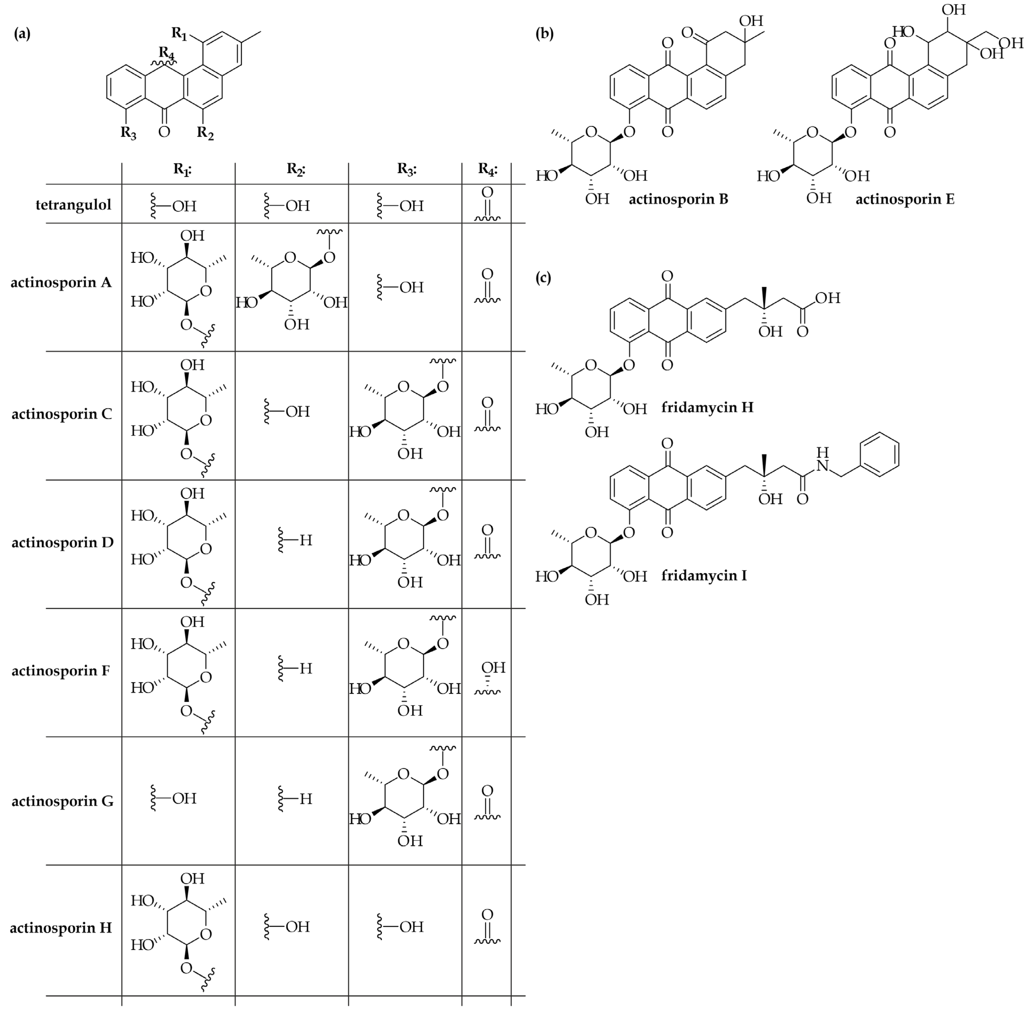
Figure 1. Structures of the actinosporin family of angucyclines (a,b), as well as fridamycins H and I (c), discovered through the cultivation of A. spheciospongiae EG49T.
Fermentation 11 00663 g001
Figure 2. Structure of actinospene, obtained through the cultivation of A. spheciospongiae EG49T (a) and the genetic organization of the corresponding BGC (named actn, (b)).
Fermentation 11 00663 g002
Figure 3. Structure of actinokineosin, obtained through the cultivation of A. spheciospongiae EG49T (a) and the genetic organization of the corresponding BGC (named akn, (b)).
Fermentation 11 00663 g003
Figure 4. Structure of thailandins A and B discovered from A. bangkokensis 44EHWT (a) and the genetic organization of the corresponding BGC (tha, (b)).
Fermentation 11 00663 g004
Figure 5. Structures of nocathiacin family of thiopeptides (isolated from A. fastidiosa ATCC 202099, (a,b)) and related persiathiacins (Actinokineospora sp. UTMC 2448) (c), as well as the genetic organizations of the corresponding BGCs (noc for nocathiacins and per for persiathiacins, (d)). *n/a—not available.
Fermentation 11 00663 g005
Table 1. List of the Actinokinespora spp. with validly published and correct names, as well as other putative Actinokineospora spp. for which genomic records are available. Table includes information about the biotope of the isolation and isolation media and methods.
SpeciesStrainValidly PublishedGenome SequenceBiotope of IsolationIsolation ApproachReference
Actinokineospora acnipugnansR434yesn/a *Reclaimed grassland soil, South KoreaSoil sample inoculated on Reasoner’s 2A medium (R2A), no pretreatment[16]
Actinokineospora alba03-9939yesGCA_004362515 (DSM 45114)
GCA_900099755 (CPCC 201030)
GCA_900104505
(IBRC-M 10655)
Forest soil sample, ChinaCzapek’s agar[17]
Actinokineospora auranticolorYU 961-1yesGCA_002934265 (YU 961-1)
GCA_041897805 (DSM 44650)
Fallen leaves in a level-land forest, JapanDry CaCO3 enrichment treatment, Humic acid-vitamin (HV) agar, 10 mg/L nalidixic acid, 20 mg/L trimethoprim[18]
Actinokineospora baliensisID03-0561yesGCA_016907695 (DSM 45656)Soil, IndonesiaDifferential centrifugation for selective isolation of motile actinomycetes, HV agar[19]
Actinokineospora cianjurensisID03-0810yes(GCA_003663795) DSM 45657Leaf-litter of the botanical garden, IndonesiaDifferential centrifugation for selective isolation of motile actinomycetes, HV agar[19]
Actinokineospora cibodasensisID03-0748yesn/aLitter of the botanical garden, IndonesiaDifferential centrifugation for selective isolation of motile actinomycetes, HV agar[19]
Actinokineospora bangkokensis44EHWyesGCA_001940455 (4EHW)Rhizospheric soil under Colocasia esculenta, ThailandDry heat pretreatment at 120 °C for 1 h, water-proline agar, 25 µg/mL nalidixic acid, 50 µg/mL cycloheximide[20]
Actinokineospora diospyrosaYU8-1yesGCA_024171925
(DSM 44255)
GCA_039534305
(JCM 9921)
Fallen persimmon leaves, JapanDessication/rehydration pretreatment, HV agar[21]
Actinokineospora globicatenaYU6-1yesGCA_024171945
(DSM 44256)
GCA_039534325
(JCM 9922)
Soil around a pond, JapanDessication/rehydration pretreatment, HV agar[21]
Actinokineospora inagensisYU4-1yesGCA_00048286
(DSM 44258)
Fallen leaves of a lakeside, JapanDessication/rehydration pretreatment, HV agar[21]
Actinokineospora terraeYU6-3yesGCA_900111175
(DSM 44260)
Soil around a pond, JapanDessication/rehydration pretreatment, HV agar[21]
Actinokineospora enzanensisYU 924-101yesGCA_000374445
(DSM 44649)
Level-land forest soil, JapanDry CaCO3 enrichment treatment, Humic acid-vitamin (HV) agar[18]
Actinokineospora fastidiosaNRRL B-16697yesGCA_014648415 (JCM 3276)Soil, Egyptn/a[22]
Actinokineospora guangxiensisGk-6yesGCA_042657845
(CGMCC 4.7154)
Soil, ChinaSoil sample inoculated on HV agar, no pretreatment[23]
Actinokineospora mzabensisPAL84yes (heterotypic synonym of Actinokineospora spheciospongiae)n/aSaharan soil from a palm grove, AlgeriaChitin-vitamin agar, 80 µg/mL cycloheximide, 25 µg/mL rifampicin, no pretreatment[24]
Actinokineospora peganiTRM 65233yesGCA_009745975 (TRM 65233)Surface-sterilized root of Peganum harmala, ChinaStarch casein agar at pH 9.0 supplemented with K2Cr2O7, 50 µg/mL nystatin, 50 µg/mL cycloheximide, 80 µg/mL nalidixic acid[25]
Actinokineospora ripariaC-39162yesn/aSoil, the Ado River, Japann/a[15]
Actinokineospora soliYIM 75948yesGCA_042666155
(JCM 17695)
Soil, ChinaSoil sample inoculated on ISP2 agar, no pretreatment[26]
Actinokineospora spheciospongiaeEG49yesGCA_000564855
(EG49)
GCA_003182415 (CECT 8578)
Marine sponge Spheciospongia vagabunda from the Red Sea, EgyptM1, ISP2, Oligotrophic medium, M1 plus, Actinomycete Isolation Agar, Marine Agar, Glycerol Asparagine Agar, R2A Agar, 100 μg/mL cycloheximide, 25 μg/mL nystatin, 25 μg/mL nalidixic acid, no pretreatment[27]
Actinokineospora xionganensisHBU206404noGCA_014323725
(HBU206404)
Lakeside soil, ChinaSoil extract medium, 1% (v/v) recombinant resuscitation-promoting factor, no pretreatment[28]
Actinokineospora sp.G85noGCA_049672885
(G85)
Rock surface, Tunisian/an/a
Actinokineospora sp.HUAS TT18noGCA_051364635
(HUAS TT18)
Soil, Chinan/an/a
Actinokineospora sp.NBRC 105648noGCA_030269645 (NBRC 105648)Soil, Japann/an/a
Actinokineospora sp.PR83noGCA_021056305
(PR83)
Sediment, Mexicon/a[7]
Actinokineospora sp.UTMC 2448noGCA_024760565
(UTMC 2448)
Coastal area, Irann/a[13]
* n/a—not available.
Table 2. List of Actinokinespora spp. where antagonistic properties were observed.
SpeciesStrainBioactivities Identified upon Identification AgainstIdentified Specialized MetabolitesReference
Actinokineospora acnipugnansR434Propionibacterium acnes, Staphylococcus epidermidisn/a *[16]
Actinokineospora alba03-9939Staphylococcus aureus CPCC 100051
Pseudomonas aeruginosa CPCC 100109
n/a[17]
Actinokineospora auranticolorYU 961-1n/aKineomicin[18]
Actinokineospora bangkokensis44EHWn/aThailandin[20]
Actinokineospora fastidiosaNRRL B-16697Various Gram-positive bacterian/a[22]
Actinokineospora guangxiensisGk-6No antibacterial activity was observed against Staphylococcus aureus and Pseudomonas aeruginosan/a[23]
Actinokineospora ripariaC-39162Antimycoplasmal activityn/a[15]
Actinokineospora spheciospongiaeEG49n/aActinosporins, actinospene, actinokineosin[27]
Actinokineospora sp.G85Broad-spectrum antimicrobial activityn/an/a
Actinokineospora sp.PR83Bacillus subtilis, carcinoma cells (NCIH460)n/a[7]
Actinokineospora sp.UTMC 2448Staphylococcus aureusPersiathiacin A and B[13]
* n/a—not available.
Disclaimer/Publisher’s Note: The statements, opinions and data contained in all publications are solely those of the individual author(s) and contributor(s) and not of MDPI and/or the editor(s). MDPI and/or the editor(s) disclaim responsibility for any injury to people or property resulting from any ideas, methods, instructions or products referred to in the content.