Article

Gut-Derived Lactic Acid Bacteria from Cotton Bollworm Exhibit Efficient Gossypol Degradation and Probiotic Potential During Solid-State Fermentation of Cottonseed Meal

College of Animal Science and Technology, Shihezi University, Shihezi 832000, China
*
Authors to whom correspondence should be addressed.
Fermentation 2025, 11(10), 598; https://doi.org/10.3390/fermentation11100598
Submission received: 14 September 2025 / Revised: 8 October 2025 / Accepted: 16 October 2025 / Published: 19 October 2025
(This article belongs to the Topic News and Updates on Probiotics)

Abstract

Cottonseed meal (CSM), an important protein-rich feed ingredient, faces limited utilization in livestock diets due to the presence of free gossypol (FG)—a potent antinutritional toxin. This study aimed to isolate FG-degrading bacteria from the cotton bollworm, Helicoverpa armigera, and to evaluate their potential as probiotics in vitro. Eleven gossypol-tolerant strains were isolated from the gut of Helicoverpa armigera larvae using a screening medium containing gossypol as the sole carbon source. Among these, four lactic acid bacteria strains—Pediococcus acidilactici GM-NP, Pediococcus acidilactici GM-P, Enterococcus faecalis GM-6, and Weissella confusa GM-2—were selected for further investigation of their gossypol degradation capacity and probiotic potential. Probiotic characterization revealed that all strains exhibited tolerance to gastrointestinal fluids and bile salts, safe γ-hemolysis, and strong auto-aggregation, cell surface hydrophobicity, and antimicrobial activity. Solid-state fermentation of CSM with these strains reduced FG content by more than 50%, increased crude protein by over 6%, and elevated acid-soluble protein content by more than 70%, thereby effectively enhancing the nutritional quality of CSM. This study is the first to demonstrate that bacterial isolates from the gut of Helicoverpa armigera possess concurrent high-efficiency gossypol degradation and probiotic properties, providing a theoretical foundation for developing novel probiotic resources and promoting the safe utilization of CSM.

1. Introduction

The rising global consumption of animal products has heightened the demand for meat, dairy, and other animal-derived goods, consequently increasing the necessity for feed to sustain growing livestock populations [1]. Cottonseed meal (CSM), a globally significant agricultural by-product with an annual production exceeding 10 million metric tons, exhibits a well-balanced amino acid profile comparable to soybean meal and is free of antigenic proteins and undesirable oligosaccharides. Thus, CSM represents a viable alternative protein source for animal feed [2]. However, the presence of gossypol, a characteristic secondary metabolite in cottonseeds, significantly limits the utilization of CSM in animal feed due to its well-documented toxicity [3]. Gossypol exists in both free (FG) and bound (BG) states. Free gossypol (FG) exhibits high reactivity due to its accessible aldehyde (–CHO) and phenolic hydroxyl (–OH) groups. Its toxicity in animals manifests through multiple mechanisms: inhibition of digestive enzyme activity, impairment of reproductive function, and bioaccumulation within the food chain that ultimately poses risks to human health [4,5,6].
The predominant techniques for detoxifying CSM encompass physical, chemical, and microbiological treatments. However, physical and chemical methods are often associated with high costs, significant nutrient loss, and potential solvent residues due to inherent methodological limitations [7,8]. In contrast, microbial fermentation is regarded as a cost-effective, efficient, and safe detoxification process [9,10], making it the preferred approach for FG removal from CSM. Current research on gossypol-degrading microbes primarily focuses on two sources: rumen microbiota and environmental isolates. Studies utilising rumen-derived microbes include: LV et al. identified a Lactobacillus mucosae strain from sheep rumen, demonstrating notable probiotic characteristics and achieving a 69.5% breakdown rate of FG when cultivated in MRS broth [11]. Wang et al. obtained a Lactobacillus rhamnosus strain from the bovine rumen, which accomplished 83% breakdown of FG during a 24 h solid-state fermentation (SSF) of CSM [12]. Research based on environmental isolates includes: Wang et al. isolated a strain of Meyerozyma guilliermondii WST-M1 from naturally mouldy CSM, which achieved a degradation of 74.70% of FG after 144 h of solid-state fermentation [13]. This research illustrates significant advancements in the detoxification of CSM through the use of rumen and ambient bacteria, highlighting the efficacy of microbial methodologies. Therefore, the discovery of novel microbial strains with superior detoxification capacity and enhanced host adaptation potential necessitates the targeted exploration of host systems that have evolved under long-term exposure to specific toxic compounds [14]. The insect gut microbiome constitutes a valuable repository of functional resources with considerable biotechnological potential [15,16]. The specialist herbivore Helicoverpa armigera is persistently exposed to high levels of gossypol through its consumption of cotton tissues. Consequently, its gut microbiota may have evolved specialized mechanisms for gossypol degradation or detoxification through long-term co-evolution. For instance, the consumption of feed containing FG triggers glycosylation activities in the larval midgut, indicating a potential endogenous detoxifying mechanism [17]. Moreover, increasing evidence suggests that bacterial symbionts or their metabolites in the gut may aid the host in alleviating gossypol toxicity [18,19]. Given the remarkable environmental adaptability and biotechnological potential of the cotton bollworm, together with the functional interdependence between insects and their microbiota, the study of its gut microbiome and associated detoxification mechanisms offers considerable promise for the development of novel microbial agents for gossypol degradation.
Therefore, strains with gossypol degradation activity were isolated from the guts of Helicoverpa armigera larvae fed a CSM-containing diet. The gossypol degradation rate in liquid culture medium was determined, and their probiotic properties—including tolerance to simulated gastrointestinal fluids, bile salt resistance, auto-aggregation, cell surface hydrophobicity, antioxidant activity, antimicrobial capacity, hemolytic activity, and antibiotic susceptibility—were systematically evaluated. Finally, solid-state fermentation of CSM was conducted to validate its gossypol detoxification effect. This study provides an effective biological detoxification strategy for CSM, offering applied support for developing CSM as an animal feed resource.

2. Materials and Methods

2.1. Materials and Reagents

Eggs of Helicoverpa armigera were purchased from Henan Jiyuan Baiyun Industrial Co., Ltd. (Jiyuan, Henan, China). Gossypol standard (≥98% purity) was obtained from Yuanye Bio-Technology Co., Ltd. (Shanghai, China). The D1600 Bacterial DNA Extraction Kit was acquired from Solarbio Science & Technology Co., Ltd. (Beijing, China). Bacterial strains were cultured in gossypol-supplemented liquid media; the compositions of these media are detailed in Table 1. All media were sterilised by autoclaving at 121 °C for 15 min. FG was supplemented to the media after the autoclaving process. All other chemicals were procured from Sinopharm Chemical Reagent Co., Ltd. (Shanghai, China).

2.2. Isolation of Gossypol-Degrading Bacteria from Helicoverpa Armigera Larval Gut

Helicoverpa armigera larvae were reared on an artificial diet containing CSM until the sixth instar (mature stage) under controlled conditions: temperature 27–28 °C, relative humidity 50%, and a photoperiod of 14L:10D. The larvae were then individually transferred to 2 mL tubes and starved overnight. Subsequently, they were subjected to surface sterilisation in a sterile Petri dish by sequential immersion in 75% ethanol for 30 s, 1% sodium hypochlorite for 5 min, followed by ten times with sterile water. The surface-sterilised larvae were dissected under aseptic conditions to collect gut contents. The gut contents were then serially diluted in PBS over a range of 10−1 to 10−6. Aliquots from the 10−4 and 10−6 dilutions were spread onto G-MRSA agar medium [20], with three replicates per dilution. To verify the efficacy of the surface sterilisation process, the final rinse water was plated onto LB agar and incubated at 37 °C for 48 h [21]. All inoculated plates were incubated at 37 °C for 48 h.

2.3. Identification of Gossypol-Degrading Bacteria from Helicoverpa armigera Gut

For initial characterization, bacterial isolates were subjected to Gram staining using a commercial kit according to the manufacturer’s instructions, wherein a bacterial suspension aliquot was applied to sterile glass slides. Genomic DNA was extracted using the Bacterial DNA Extraction Kit (D1600; Solarbio Co., Ltd., Beijing, China). The 16S rDNA gene was amplified by PCR with universal bacterial primers 27F (5′-CAGAGTTTGATCCTGGCT-3′) and 1492R (5′-AGGAGGTGATCCAGCCGCA-3′). The amplified products were purified and sequenced by RuiBoXingKe Biotechnology Co., Ltd. (Beijing, China). The resulting sequences were compared against available 16S rDNA sequences in the NCBI database to determine taxonomic identity. Phylogenetic analysis was performed using MEGA version 7.0 software.
For physiological and biochemical characterization, bacterial isolates were characterized using SHBG13 micro-biochemical test tubes (Qingdao Hope Biotechnology Co., Ltd., Qingdao, China) according to the manufacturer’s instructions.

2.4. Determination of the Growth Curve and Free Gossypol Degradation Rate for the Target Strain

Target strain stored in glycerol at −80 °C was thawed and subcultured twice in MRS broth. A 1 mL aliquot of the activated culture was inoculated into 100 mL of MRS broth and incubated at 37 °C for 36 h. (9 incubation times × 3 replicates). The optical density at 600 nm (OD600) and the pH of the culture were measured at predetermined time intervals using a UV-2000 spectrophotometer (UNIAC Instrument Co., Ltd., Shanghai, China) and a P302 pH meter (Shanghai Youke Instrument Co., Ltd., Shanghai, China), respectively.
The gossypol degradation rate was determined according to a previously described method with minor modifications [12]. The target strain was cultured to the logarithmic growth phase and then inoculated into 100 mL of GMRS medium containing 0.1 g/L FG as the sole carbon source. The culture was incubated at 37 °C with shaking at 180 rpm for 24 h to allow for degradation. After incubation, 1.0 mL of the culture broth was mixed with 3.0 mL of acetone. The mixture was filtered through a 0.22 μm organic solvent-compatible membrane filter, and the resulting filtrate was collected as the test sample. Subsequently, 1.0 mL of this sample was combined with 2.0 mL of background solution and reacted in a 55 °C water bath for 5 min. The gossypol content in the reaction mixture was quantified spectrophotometrically at 550 nm based on a standard curve of gossypol.

2.5. Artificial Gastric Juice and Bile Salt Tolerance

The gastric juice tolerance was assessed according to a previously described method with minor modifications [22]. Activated cultures were inoculated into MRS broth (pH 2.5) containing 0.3% (w/v) pepsin and incubated at 37 °C for 2 h.
For the bile salt tolerance assay, overnight cultures were harvested, washed, and resuspended in MRS broth supplemented with 0.3% (w/v) ox bile salts (Solarbio Science & Technology Co., Ltd., Beijing, China). MRS broth without bile salts was used as the control. Samples were collected at 0 and 240 min, subjected to serial dilution, and plated onto MRS agar. Survival rates were calculated using the following formula:
Survival rate (%) = (B/A) × 100
where A represents the initial bacterial count (Log10 CFU/mL) and B represents the count after incubation (Log10 CFU/mL).

2.6. Temperature Tolerance Assessment

Overnight target strain cultures were subjected to heat treatment in a water bath at 60, 70, 80, and 90 °C for 5, 10, and 15 min. Before and after each treatment, aliquots were collected, serially diluted, and plated onto MRS agar for viable colony counting [23]. The survival rate was calculated as follows:
Survival rate (%) = (B/A) × 100
where A represents the bacterial count before heat treatment (Log10 CFU/mL) and B represents the count after heat treatment (Log10 CFU/mL).

2.7. Evaluation of Cell Surface Properties

Cell surface properties were assessed following the method described by Ning et al. [24]. Briefly, 10 mL of activated bacterial culture (108 CFU/mL) was centrifuged at 10,000 rpm for 10 min. The pellet was washed twice with PBS (Beijing Solarbio Science & Technology Co., Ltd., Beijing, China) and resuspended to an OD630 of 0.25 ± 0.05 (A1).
The bacterial suspension was then mixed with an equal volume of xylene, vortexed for 3 min, and incubated at 37 °C for 4 h [25]. After incubation, the aqueous phase was carefully collected, and its absorbance at 630 nm (A2) was measured. All assays were performed in triplicate.
The hydrophobicity percentage was calculated using the following formula:
Hydrophobicity (%) = 100 × (1 − A2/A1)

2.8. Auto-Aggregation Assessment

The auto-aggregation assay was performed as described by Dincer et al. [26]. Bacterial suspensions were prepared as described in Section 2.7. The suspension was vortexed thoroughly for 1 min, and the initial absorbance at 630 nm (A3) was measured. The suspension was then incubated at 37 °C for 8, 16, and 24 h without disturbance. After each incubation period, the upper suspension was carefully sampled, and its absorbance at 630 nm (A4) was measured. The auto-aggregation percentage was calculated as follows:
Auto-aggregation (%) = 100 × (A3 − A4/A3).

2.9. Hemolytic and Antioxidant Assessment

Hemolytic activity was determined by streaking activated cultures onto 5% (v/v) defibrinated sheep blood agar plates (Hope Bio., Qingdao, Shandong, China). Staphylococcus aureus ATCC 6538 was used as a positive control. The plates were incubated at 37 °C for 48 h. γ-Hemolysis (no hemolysis) was characterized by no observable zone around the colonies, α-hemolysis by a greenish zone, and β-hemolysis by a clear, colorless zone [11]. The 1,1-diphenyl-2-picrylhydrazyl(DPPH) radical scavenging activity of the isolated strains was determined using the DPPH (BC4750) detection kit (Beijing Solarbio Science & Technology Co., Ltd., Beijing, China), with specific steps following the instructions in the manual [22].

2.10. Assessment of Antibacterial Activity

The most common pathogenic microorganisms used in this study, including Escherichia coli ATCC 25922, Salmonella typhimurium ATCC 14028, Staphylococcus aureus ATCC 6538, and Pseudomonas aeruginosa ATCC 9027, were provided by the Beijing Microbiological Culture Collection Centre. The antibacterial activity of cell-free supernatants from target strains was evaluated according to the method described by Li et al. [27]. LB agar plates were prepared with Oxford cup wells for subsequent assays. Activated bacterial cultures were centrifuged (10,000× g, 10 min, 4 °C) to collect the supernatants, which were then filter-sterilised using 0.22 μm cellulose acetate membranes (Solarbio Science & Technology Co., Ltd., Beijing, China) to remove any remaining cells. Pathogenic bacteria cultured in LB broth were evenly spread onto LB agar medium. Then, 100 μL of each cell-free supernatant was added to the 7 mm diameter wells (four pathogens × three replicate plates). Controls included equal volumes of liquid LB medium and phosphate-buffered saline (PBS). All plates were incubated at 37 °C for 24 h, after which the formation of inhibition zones was examined.
Based on criteria established in prior studies [26], the antibacterial activity was classified as follows: inhibition zone diameters of 7–9 mm were considered weak, 10–13 mm moderate, 14–17 mm strong, and greater than 17 mm very strong.

2.11. Antibiotic Susceptibility Testing

Antibiotic susceptibility was evaluated using the Kirby–Bauer disk diffusion method [28]. Activated bacterial cultures (100 μL) were spread evenly onto MRS agar plates. Fifteen antibiotic disks—including cephalothin (30 µg), clindamycin (15 µg), chloramphenicol (30 µg), penicillin (10 µg), rifampin (30 µg), erythromycin (15 µg), ampicillin (10 µg), tetracycline (30 µg), levofloxacin (5 µg), gentamicin (10 µg), streptomycin (10 µg), polymyxin B (30 µg), enrofloxacin (5 µg), ciprofloxacin (5 µg), and ofloxacin (5 µg) (BKMAM Biotechnology Co., Ltd., Changsha, Hunan, China)—were placed onto the inoculated MRS agar surfaces. After incubation at 37 °C for 48 h, the presence of inhibition zones was recorded, and their diameters were measured accurately. Results were interpreted as sensitive or resistant according to CLSI 2012 guidelines [25]. All assays were performed in triplicate, and mean values are reported.

2.12. Solid-State Fermentation of Cottonseed Meal by the Target Strain

Solid-state fermentation (SSF) of CSM was performed to evaluate the gossypol degradation efficiency and the improvement in the nutritional quality of CSM by the target strains. CSM was ground, passed through a 60-mesh sieve, and 100 g (dry weight) was autoclaved and used as the fermentation substrate. The activated bacterial culture was inoculated at 7% (v/w), and sterile water was added to adjust the material-to-water ratio to 1:0.4. The control group received 47 mL of sterile water without bacterial inoculation. Five treatment groups (including the control) were established, each with three replicates. Inoculated substrates were subjected to solid-state fermentation at 37 °C for 48 h. After fermentation, nutritional quality was assessed by determining crude protein, dry matter, crude fat, ash, neutral detergent fiber (NDF), and acid detergent fiber (ADF) according to the method described by Li et al. [10]. FG content was measured using the phloroglucinol method referenced from Wang et al. to evaluate detoxification efficiency [12].

2.13. Statistical Analysis

All experiments were performed in triplicate, and the data are presented as the mean ± standard deviation (SD). All intergroup comparisons were performed using one-way analysis of variance (ANOVA). Prior to ANOVA, the normality and homogeneity of variances were assessed using the Shapiro–Wilk test and Levene’s test, respectively, confirming that the data met the assumptions for parametric tests. If the ANOVA results were significant, Tukey’s Honestly Significant Difference (HSD) test was subsequently used for post hoc multiple comparisons. The statistical significance threshold was set at p < 0.05. Statistical analyses were conducted using Origin 2021 Pro software (Microcal, Northampton, MA, USA).

3. Results

3.1. Preliminary Characterisation of Isolated Bacteria

Serial dilutions of Helicoverpa armigera intestinal contents were plated onto screening medium containing 0.2 g/L gossypol and incubated at 37 °C for 48 h. Based on colony morphology—including form, moisture, elevation, and other characteristics—eleven gossypol-tolerant strains were isolated. Among these, strains GM-7, GM-8, GM-9, and GM-10 were identified as Gram-negative, while the remaining strains were Gram-positive.

3.2. 16S rDNA Sequencing and Phylogenetic Analysis of Bacterial Strains

The bacterial 16S rRNA gene was amplified by PCR using universal primers, yielding products approximately 1500 bp in length. The amplified sequences from all eleven isolates were sequenced and submitted to GenBank. Using BLASTn (version 2.17.0+; https://blast.ncbi.nlm.nih.gov/Blast.cgi, accessed on 27 July 2025), the sequences were compared to those of standard type strains with the highest similarity (Table 2). Based on the BLASTn analysis, the eleven strains were assigned to five genera: Enterococcus, Pediococcus, Mammaliicoccus, Weissella, and Klebsiella. Four lactic acid bacteria (LAB) strains, Pediococcus acidilactici GM-NP, Pediococcus acidilactici GM-P, Enterococcus faecalis GM-6, and Weissella confusa GM-2, were selected for further investigation.

3.3. Physiological and Biochemical Characteristics of the Strain

Following biochemical identification with the SHBG13 bacterial micro-biochemical identification tube (Qingdao Hope Biotechnology Co., Ltd., Qingdao, China), the biochemical characteristics of the four LAB strains were determined, as presented in Table 3. P. acidilactici strains GM-NP and GM-P tested positive for aesculin hydrolysis and sodium hippurate utilization, but negative for the fermentation of cellobiose, maltose, and mannose. E. faecalis GM-6 tested positive for esculin and sodium hippurate, yet was negative for inulin fermentation. In contrast, W. confusa GM-2 showed positive responses to cellobiose, maltose, and mannose, but was negative for aesculin.

3.4. Growth Curves and Acid Production Curves of Four Strains of Lactic Acid Bacteria

For P. acidilactici strains GM-NP and GM-P, a lag phase was observed within the first 2 h post-inoculation, after which rapid growth marked the onset of the exponential phase. After 18 h, the optical density at 600 nm (OD600) stabilized, indicating the entry into the stationary phase. E. faecalis GM-6 showed a similar initial lag phase (0–2 h), proceeded to exponential growth until 12 h, and reached the stationary phase thereafter. W. confusa GM-2 also displayed a 2 h lag phase, entered exponential growth until 16 h, and subsequently attained the stationary phase (Figure 1A). Throughout the cultivation period, the pH of the medium decreased from 6.05 to 4.20 for P. acidilactici GM-NP, and from 6.09 to 4.33 for P. acidilactici GM-P. Similarly, the pH declined from 6.07 to 5.17 for E. faecalis GM-6, and from 6.07 to 5.33 for W. confusa GM-2 (Figure 1B).
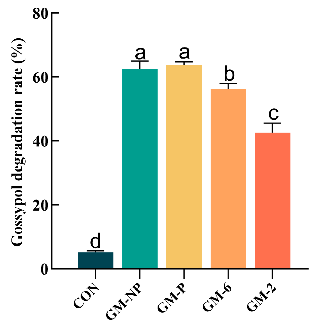
The gossypol-degrading capacity of the four LAB strains was evaluated by measuring the FG degradation rate after 24 h of incubation in GMRS medium (Figure 2). The highest degradation rate (63.75%) was exhibited by P. acidilactici GM-P, whereas the lowest (42.59%) was observed for W. confusa GM-2.

3.5. Tolerance of Four Strains of Lactic Acid Bacteria to Artificial Gastrointestinal Environments

The tolerance of the four LAB strains to simulated gastrointestinal conditions was evaluated (Table 4), as it is considered a prerequisite for probiotic functionality. After 24 h of exposure to simulated gastric juice, strain GM-P exhibited the highest survival rate (74.28%), which was significantly greater than that of strain GM-2 (68.31%), the least tolerant under these conditions. Similarly, in simulated intestinal fluid, GM-P again demonstrated superior tolerance, with a survival rate of 90.64%, compared to the lowest rate of 85.98% observed in GM-2. Furthermore, all tested strains exhibited good tolerance to 0.3% bile salts after 4 h of treatment, with survival rates exceeding 60%. Among them, GM-P displayed the highest survival rate (84.76%). These properties suggest that, upon oral administration, GM-P possesses the greatest potential to survive passage through the harsh upper gastrointestinal tract and arrive in the colon in sufficient viable numbers to initiate colonisation—a fundamental prerequisite for exerting its probiotic effects.

3.6. Thermal Tolerance of the Four Lactic Acid Bacterial Strains

To assess the heat resistance of the four LAB strains, the strains were subjected to temperatures ranging from 60 °C to 90 °C for 5 to 15 min. After 15 min at 60 °C, the average survival rate was approximately 50%. Following treatment at 70 °C for the same duration, the survival rate remained above 30%. Even after exposure to 80 °C and 90 °C, viable cells were still detected, albeit in small numbers. Among the tested strains, E. faecalis GM-6 exhibited the highest thermal tolerance (Table 5).

3.7. Auto-Aggregation and Surface Hydrophobicity of Four Lactic Acid Bacterial Strains

The auto-aggregation capabilities of the four LAB strains at different time points are summarized in Table 6. After 8 h of incubation, all strains except GM-6 exhibited auto-aggregation rates exceeding 40%. By 24 h, the auto-aggregation rates of GM-NP and GM-P both surpassed 80%, which was significantly higher than those of the other two strains (p < 0.05). Among them, GM-NP showed the highest auto-aggregation rate (82.96%), while GM-6 showed the lowest (72.89%). The hydrophobicity of the bacterial cell surfaces, as determined by their affinity for xylene, was used to evaluate potential adherence to gastrointestinal epithelial cells. GM-NP demonstrated significantly greater hydrophobicity (54.45%) compared to GM-6 (47.92%) and GM-2 (41.60%) (p < 0.05).

3.8. Antibacterial Activity of Four Lactic Acid Bacterial Strains

The antimicrobial capacity of the four LAB strains against potent intestinal pathogens further underscores their potential role as probiotics in preventing infections caused by pathogenic microorganisms in the gastrointestinal tract. The cell-free supernatants of the four strains exhibited varying inhibition zones against the four different intestinal pathogens tested (Table 7). Both the GM-NP and GM-P strains demonstrated potent inhibitory effects, with inhibition zones exceeding 14 mm, and showed the strongest activity against Salmonella spp., producing zones greater than 17 mm. The supernatant of the GM-6 strain exhibited moderate antimicrobial activity against all four pathogens, with inhibition zones larger than 10 mm. In contrast, the GM-2 strain exerted minimal inhibitory effects against the tested pathogens and showed no detectable activity against Salmonella.

3.9. Hemolytic Activity and Antioxidant Capacity of Four Lactic Acid Bacterial Strains

The safety and antioxidant potential of the four LAB strains were assessed through hemolytic activity and DPPH radical scavenging assays (Table 8). All four strains exhibited γ-hemolysis (non-hemolytic) on 5% sheep blood agar, in contrast to the β-hemolysis displayed by the S. aureus ATCC 6538 positive control. Furthermore, each strain demonstrated considerable antioxidant activity, with DPPH radical scavenging rates exceeding 25%. Notably, P. acidilactici GM-P strain showed the highest scavenging capacity at 45.29%, which was significantly greater than that of GM-6 and GM-2 (p < 0.05).

3.10. Drug Sensitivity of Four Strains of Lactic Acid Bacteria

To assess the safety of the four LAB strains for potential application, we evaluated their antibiotic susceptibility against a panel of 15 antibiotics (Table 9). P. acidilactici GM-NP and GM-P were resistant to eight antibiotics, including penicillin, levofloxacin, and gentamicin. GM-NP was sensitive to six antibiotics, such as cefalexin, clindamycin, and rifampicin, and exhibited intermediate susceptibility to chloramphenicol. Similarly, GM-P was sensitive to seven agents, including cefalexin, clindamycin, and chloramphenicol.
In contrast, GM-6 showed sensitivity to three antibiotics (e.g., penicillin, enrofloxacin, and rifampicin), intermediate susceptibility to ten others (including cefalexin, chloramphenicol, and ampicillin), and resistance to clindamycin and ciprofloxacin. GM-2 was sensitive to 14 antibiotics, including cefalexin, clindamycin, and chloramphenicol, but resistant to ofloxacin. The four LAB strains demonstrated susceptibility to a broad spectrum of antibiotics, which is critical for ensuring that fermented CSM does not contribute to the dissemination of antibiotic resistance.

3.11. Solid-State Fermentation of Cottonseed Meal and Gossypol Detoxification, as Well as Nutritional Quality Indicators

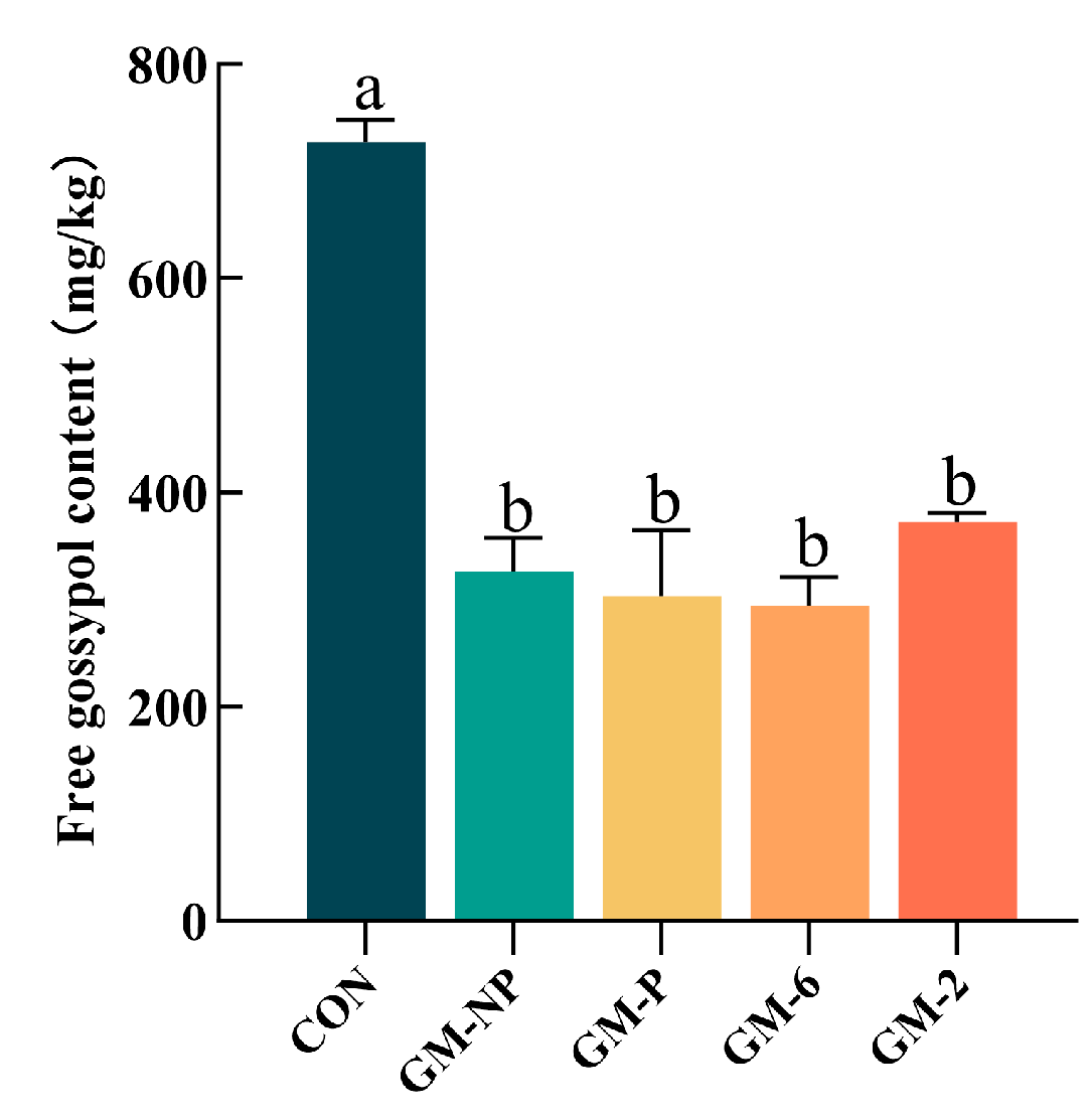
To assess the practical degradation efficacy of the four LAB strains against FG in CSM and their influence on its nutritional quality, a 72 h solid-state fermentation of CSM was performed (Figure 3 and Table 10). As shown in Figure 3, all bacterial treatments led to a significant reduction (p < 0.05) in FG content compared to the control (CON, 727.19 mg/kg). The FG values were as follows: GM-2 (355.98 mg/kg), GM-NP (326.45 mg/kg), GM-P (303.05 mg/kg), and GM-6 (294.58 mg/kg). The highest reduction in FG was achieved by GM-6 (59.49%), followed by GM-P (58.32%), GM-NP (55.11%), and GM-2 (51.05%), with all groups differing significantly from CON (p < 0.05).

4. Discussion

Eleven bacterial strains capable of utilizing free gossypol (FG) as a sole carbon source were successfully isolated from the intestinal tract of Helicoverpa armigera larvae. This insect host was selected due to its documented evolutionary resistance to gossypol. We hypothesized that its gut microbiota would represent a promising reservoir of bacteria capable of gossypol degradation or tolerance. Among these, four lactic acid bacteria—P. acidilactici GM-NP, P. acidilactici GM-P, E. faecalis GM-6, and W. confusa GM-2—were selected for further characterisation based on their well-documented probiotic properties reported in prior studies [22,29], which ensures that the candidate strains combine gossypol degradation capability with probiotic potential, providing a solid foundation for their application in animal feed. Strains not selected, such as Klebsiella oxytoca and Mammaliicoccus sciuri, were excluded based on reports of potential pathogenicity [30,31]. Subsequent solid-state fermentation experiments confirmed that all four selected LAB strains significantly reduced the FG content and enhanced the nutritional value of CSM. Moreover, comprehensive in vitro characterization revealed that these strains exhibited a promising suite of probiotic traits, including robust tolerance to gastrointestinal stresses, strong auto-aggregation, cell surface hydrophobicity, antioxidant and antimicrobial activities, and a largely favorable antibiotic susceptibility profile. Although previous studies have identified gossypol-degrading bacteria from the rumen environment [11,32], this study presents the first isolation and characterisation of such bacteria from the gut of Helicoverpa armigera, thereby providing not only a novel source of detoxifying microbes but also a targeted strategy for developing safer probiotic candidates for animal feed supplementation.

4.1. Isolation and Gossypol Degradation Capacity of Lactic Acid Bacteria Strains

To investigate the potential role of gut microbes in gossypol detoxification, bacterial strains were isolated from the intestinal contents of Helicoverpa armigera larvae reared on a CSM-based diet. Using this approach, several target strains were successfully isolated. In contrast to previous studies that primarily sourced gossypol-degrading microbes from ruminal environments or conventional substrates like mouldy cottonseed meal [11,12,13], our study specifically targeted the adapted gut microbiota of Helicoverpa armigera. This insect’s documented evolutionary resistance to gossypol provided a rationale for hypothesizing that its gut microbiome would be an enriched source of microbes with gossypol-detoxifying potential [33]. In this work, our screening protocol was designed to specifically enrich for gossypol-metabolising organisms. Four strains—GM-NP, GM-P, GM-6, and GM-2—were selected based on morphological and molecular identification, and their growth profiles were characterised, consistent with previous reports on LAB. To evaluate their gossypol degradation capacity, each strain was inoculated into a liquid culture medium containing FG as the sole carbon source. All four strains demonstrated the ability to degrade gossypol. P. acidilactici GM-P exhibited the highest degradation rate at 63.75%, whereas W. confusa GM-2 showed a comparatively lower efficiency of 42.59%. Notably, the degradation efficiencies achieved here are comparable to those reported for microbes from other sources, such as Ligilactobacillus salivarius LLK-XR1 (69.5% FG degradation) isolated from the rumen [11]. This demonstrates that our targeted approach from a novel insect gut environment can yield LAB with degradation capabilities on par with those from traditional sources. In light of the study by Yousefi et al., which demonstrated significant removal of polycyclic aromatic hydrocarbons (PAHs)—compounds characterised by planar aromatic ring systems—by LAB [34], our observation of high gossypol degradation by P. acidilacticus GM-P may suggest a broader capacity of this strain to metabolise or transform compounds with analogous structural features. Although gossypol differs in specific structure from typical PAHs, it shares the key property of aromatic planarity. Further analysis of the high-efficiency degrader GM-P is warranted. Considering the findings of Wang et al. that esterases, glutathione S-transferases (GSTs), and MarR family transcriptional regulators are crucial for gossypol detoxification in Lactobacillus rhamnosus [12], the high degradation activity of GM-P may indicate the presence of functionally similar homologous genes or gene clusters. Conversely, the relatively low degradation efficiency observed in W. confusa may reflect the absence or low expression of such efficient detoxification regulatory networks (e.g., MarR-type regulators) or key enzyme systems (e.g., esterases and GSTs). Notably, the degradation rates reported here reflect a reduction in gossypol concentration but do not unequivocally demonstrate its utilisation as a carbon source. Therefore, subsequent studies should aim to identify intermediate metabolites generated during degradation to elucidate the specific pathways involved and confirm bacterial assimilation of gossypol [13].

4.2. Gastrointestinal Tolerance and Heat Resistance

Beyond their gossypol degradation capabilities, this study systematically evaluated the gastrointestinal tolerance of four bacterial strains—GM-NP, GM-P, GM-6, and GM-2—under simulated digestive conditions to assess their potential as direct-fed probiotics or as starters for fermenting CSM. All strains exhibited robust tolerance, with survival rates exceeding 50%. Notably, P. acidilactici GM-P demonstrated exceptional performance: it achieved a survival rate of 74.28% in simulated gastric fluid (pH 2.5, 1.5 h), 90.34% in simulated intestinal fluid (containing 1% trypsin, 3 h), and maintained 84.76% viability after 4 h of exposure to 0.3% bile salts (Table 4). Previous studies have indicated that P. acidilactici generally exhibits strong bile salt tolerance [35], likely attributable to mechanisms such as the expression of bile salt hydrolase (BSH) or the ability to maintain cell membrane integrity. Furthermore, members of the Pediococcus genus have been reported to sustain high viability under intestinal conditions, demonstrating a capacity to withstand the sequential stresses of the upper gastrointestinal tract and potentially colonize the host intestine [36]. Beyond these innate molecular mechanisms, the observed tolerance of the target strains to simulated gastrointestinal fluids can also be attributed to their origin in the larval gut of Helicoverpa armigera—a highly complex microecosystem that imposes strong selective pressure for environmental adaptability [37]. The collective tolerance profiles of these LAB strains underscore their promise as probiotic candidates. However, further investigation into their adaptive mechanisms is warranted to optimise their efficacy and application. Additionally, given that probiotic processing often involves high-temperature spray-drying, strains with enhanced thermal stability are advantageous for industrial production [38]. The strains examined in this study also exhibited favourable heat resistance, supporting their suitability for large-scale use.

4.3. Auto-Aggregation and Cell Surface Hydrophobicity

To further assess the colonisation potential of these strains, we investigated their auto-aggregation capacity and cell surface characteristics. The ability of probiotic strains to adhere to the gastrointestinal tract is a critical selection criterion, as it directly influences their survival within the gut and determines their capacity to exert specific functional and health benefits. Among the direct screening techniques for evaluating probiotic adhesion, cell surface hydrophobicity, and auto-aggregation assays are widely employed. To assess the surface hydrophobicity of the strains, this study utilised the bacterial adhesion to hydrocarbons (BATH) test with xylene, a nonpolar organic solvent. The results indicated that all four tested LAB exhibited moderate hydrophobicity. P. acidilactici GM-NP showed the highest affinity to xylene, with an adsorption rate of 54.45%, indicating moderately strong hydrophobicity. After 24 h of incubation, all four strains demonstrated auto-aggregation rates exceeding 70%, reflecting a strong adhesive capacity.
These findings are consistent with previous reports [39,40]. A positive correlation between cell surface hydrophobicity and auto-aggregation ability has been established, largely mediated by hydrophobic interactions. However, this relationship is not absolute and can be significantly influenced by strain-specific surface macromolecules, such as S-layer proteins and lipoteichoic acids, which may play a decisive role in mediating high auto-aggregation [41]. Although hydrophobicity appears to be a significant contributor to auto-aggregation, our data suggest that inherent, surface molecule-mediated mechanisms are likely more critical in determining the high aggregation observed in these strains.

4.4. Antioxidant and Antimicrobial Activities

In addition to physical adherence properties, we examined the functional capabilities of these strains, beginning with their antioxidant potential. Studies have reported that LAB possess considerable antioxidant activity, attributed to their intrinsic antioxidant systems that help maintain low levels of free radicals and mitigate oxidative damage [42]. The 2,2-diphenyl-1-picrylhydrazyl (DPPH) free radical scavenging assay is widely used to evaluate antioxidant capacity, with higher scavenging rates indicating stronger antioxidant activity. In this study, all four strains exhibited DPPH radical scavenging rates consistent with previous reports. P. acidilactici GM-P showed the highest scavenging activity at 45.29%, a value exceeding those reported by Ki-Tae Kim et al. for other LAB [43]. These results suggest that GM-P may possess strong antioxidant properties and could potentially enhance host antioxidant defence mechanisms.
Another notable health benefit of probiotics is their ability to prevent and reduce gastrointestinal infectious diseases [44,45]. Commonly used probiotics, particularly LAB, can produce organic acids, ethanol, hydrogen peroxide, various enzymes, and bacteriocins, which collectively inhibit the proliferation and metabolism of pathogens, thereby helping restore gut microbial balance [44]. This functional group encompasses species such as P. acidilactici, E. faecalis, and W. confusa. Consistent with previous findings that demonstrated the inhibitory effects of Pediococcus acidilactici YH-15 against pathogens [46], the two P. acidilactici strains in this study (GM-NP and GM-P) exhibited the most pronounced antibacterial activity. As typical homofermentative or facultatively heterofermentative LAB, they are known for efficient lactic acid production [47]. The lactic acid produced likely contributes significantly to pathogen inhibition by acidifying the environment and exerting direct bactericidal effects. After 24 h of culture, the pH of both strains declined to approximately 4.3, which may contribute to their antimicrobial activity. However, whether bacteriocins or other bioactive compounds with antibacterial properties are also involved remains to be further investigated.

4.5. Safety Assessment: Antibiotic Susceptibility and Hemolytic Activity

Prior to considering practical applications, we conducted a comprehensive safety assessment of these potential probiotic strains, beginning with their antibiotic susceptibility profiles. Antibiotic resistance represents a critical criterion in the safety assessment of probiotic strains. It has been demonstrated that certain LAB may harbour antibiotic resistance genes [45,48]. The application of such probiotics—carrying transferable resistance genes—in hosts (e.g., animals or humans) poses a potential risk of horizontal gene transfer to commensal gut microbiota or pathogenic bacteria, potentially facilitating the dissemination of antibiotic resistance. Therefore, rigorous screening for antibiotic resistance is essential to ensure the safe use of probiotics [49].
In this study, the resistance of P. acidilactici strains to aminoglycosides (e.g., gentamicin, streptomycin) and ciprofloxacin is consistent with intrinsic resistance patterns commonly reported for many LAB [49]. This intrinsic resistance is often attributed to innate characteristics, such as low cell membrane permeability or the absence of target sites, rather than acquired genetic elements. In contrast, E. faecalis GM-6 exhibited intermediate susceptibility to several of these antibiotics. This phenotype appears not to be governed by a single dominant mechanism, but rather results from the synergistic activity of multiple low-efficacy or low-expression resistance strategies, such as enzymatic modification, target mutations, and efflux pumps. Conversely, W. confusa GM-2 demonstrated high susceptibility to gentamicin, possibly owing to strain-specific traits. Certain Weissella strains display variable responses to specific antibiotics due to intrinsic sensitivity or adaptive environmental responses [50]. Moreover, although prior studies have reported that most LAB are susceptible to antibiotics such as cefalotin, erythromycin, and penicillin, the observed tolerance to penicillin in P. acidilactici GM-NP and GM-P in this study deviates from this trend. This may be attributed to mutations conferring reduced affinity of penicillin-binding proteins (PBPs)—a canonical resistance mechanism in Gram-positive bacteria against β-lactam antibiotics—rather than acquired resistance genes (e.g., TEM-type β-lactamases), which are more frequently observed in enterococci [51]. Complementing our antibiotic susceptibility testing, we also evaluated the hemolytic activity of these strains. Finally, all four strains evaluated in this study exhibited γ-hemolysis (non-hemolytic activity), consistent with the non-hemolytic characteristic of probiotics reported by Tarrah et al. [52]. This indicates the absence of functional hemolysin genes, further supporting their safety profile for potential applications. Nevertheless, the risk of resistance gene transfer necessitates continued vigilance.

4.6. Solid-State Fermentation and Nutritional Improvement of CSM

Following the establishment of their probiotic potential in vitro, the efficacy of these strains in CSM detoxification was evaluated under solid-state fermentation (SSF) conditions. SSF has garnered significant attention for its effectiveness in valorizing low-cost agro-industrial by-products into high-value feed and food ingredients [53,54]. In the context of detoxifying CSM, several microorganisms—including Meyerozyma guilliermondii, Bacillus subtilis, and Lactobacillus mucosae—have been widely employed, with the choice of strain considerably influencing the final product quality [33]. This study focused on the application of LAB, specifically four strains isolated from the gut of the larvae of Helicoverpa armigera: P. acidilactici GM-NP and GM-P, E. faecalis GM-6, and W. confusa GM-2, using mono-culture SSF on CSM. The results demonstrated an effective improvement in the nutritional profile of CSM by all four LAB strains, characterized by a significant reduction in FG content, an increase in crude protein (CP) and acid-soluble protein (ASP) levels, and a decrease in neutral detergent fibre (NDF), acid detergent fiber (ADF), and ether extract (EE) (Table 10). The reduction in FG content aligns with previous reports [11,32]. The elevation in CP and ASP can be attributed to the relative enrichment of protein through consumption of non-protein components (e.g., carbohydrates and lipids), de novo microbial protein synthesis, and the action of organic acids—predominantly lactic acid—which lowers the pH. This acidic environment not only inhibits contaminating microorganisms and improves palatability but also promotes the solubilization of acid-soluble proteins and facilitates macromolecular proteolysis, thereby enhancing protein digestibility. These observations are consistent with findings from B. subtilis mono-culture fermentation [10]. The reduction in NDF and ADF is likely associated with the acid hydrolysis of fibrous compounds induced by organic acids. While LAB are not typically considered strong lipase producers, some strains, including certain Pediococcus and Enterococcus species, have been reported to exhibit lipase activity [55,56]. For instance, P. acidilactici has been reported to exhibit lipase activity up to 493 U/mL under specific conditions [55]. Given the reported lipase activities in related strains and the consistent reduction in crude fat observed across all fermentations, the decline in fat content may be partially attributed to the enzymatic hydrolysis of triglycerides. Additionally, the acidic environment generated during fermentation may further promote lipid breakdown through acidolysis. In summary, the tested LAB strains demonstrate significant potential for simultaneous FG detoxification and nutritional enhancement of CSM under SSF conditions. It is important to note that the fermentation process parameters used in this study were not systematically optimized. Further studies to optimise key fermentation conditions are therefore warranted to maximise the efficacy of LAB-based SSF for CSM detoxification [57].
The four LAB strains demonstrated promising probiotic attributes and gossypol degradation capacity, while also exhibiting γ-hemolysis (non-hemolytic activity) on 5% (v/v) defibrinated sheep blood agar, indicating a safety profile for applications. However, the molecular mechanism behind the degradation remains uncharacterized. To fully elucidate the pathway underlying gossypol detoxification, Future studies will integrate metabolomic and transcriptomic analyses. Metabolite profiling via LC-MS will delineate the degradation pathway by identifying key intermediates, while transcriptomic analysis of the most efficient degrader under gossypol stress will uncover the key genes and enzymes involved.

5. Conclusions

In summary, this study successfully isolated four LAB strains from the gut of Helicoverpa armigera, with Pediococcus acidilactici GM-P exhibiting particularly outstanding potential for application in CSM detoxification and as a novel probiotic feed additive. SSF of CSM demonstrated that all four strains significantly reduced FG content and increased crude protein levels. Furthermore, the strains exhibited strong potential for probiotic applications, showing robust tolerance to simulated gastrointestinal conditions, high auto-aggregation capacity, and a favourable safety profile—including sensitivity to a broad spectrum of antibiotics and antagonistic activity against enteric pathogens. Collectively, this work validates the effectiveness of sourcing functional bacteria from specialised environments such as insect guts, expands the resources available for probiotic development, and provides both theoretical and technical support for the application of CSM in alleviating feed resource shortages.

Author Contributions

Conceptualization, S.L. (Sijin Li); writing—original draft preparation, S.L. (Sijin Li) and C.C.; software, S.D.; resources, P.Z. and S.D.; writing—review and editing, C.C. and W.Z.; investigation, Q.L. and M.M.; formal analysis, W.P. and S.L. (Shu Li); funding acquisition, C.C. and W.Z.; project administration, C.C.; supervision, C.C. All authors have read and agreed to the published version of the manuscript.

Funding

This research was supported by the National Natural Science Foundation of China (32560832, 32060770).

Institutional Review Board Statement

Not applicable.

Informed Consent Statement

Not applicable.

Data Availability Statement

All relevant data are presented in this paper. The raw data and related analysis results of this study can be obtained from the corresponding author upon reasonable request. The 16S rRNA gene sequences of P. acidilactici GM-NP, P. acidilactici GM-P, E. faecalis GM-6, and W. confusa GM-2 have been deposited in the National Centre for Biotechnology Information (NCBI) database under accession numbers PX232694, PX242179, PX242182, and PX242193, respectively. The respective URLs for accessing these sequences are as follows: P. acidilactici GM-NP: https://www.ncbi.nlm.nih.gov/nuccore/PX232694 (accessed on 8 September 2025); P. acidilactici GM-P: https://www.ncbi.nlm.nih.gov/nuccore/PX242179 (accessed on 8 September 2025); E. faecalis GM-6: https://www.ncbi.nlm.nih.gov/nuccore/PX242182 (accessed on 8 September 2025); W. confusa GM-2: https://www.ncbi.nlm.nih.gov/nuccore/PX242193 (accessed on 8 September 2025).

Conflicts of Interest

This study involves no known financial conflicts of interest or personal relationships that could influence the report’s content.

References

  1. Rakita, S.; Kokić, B.; Manoni, M.; Mazzoleni, S.; Lin, P.; Luciano, A.; Ottoboni, M.; Cheli, F.; Pinotti, L. Cold-Pressed Oilseed Cakes as Alternative and Sustainable Feed Ingredients: A Review. Foods 2023, 12, 432. [Google Scholar] [CrossRef] [PubMed]
  2. Ninkuu, V.; Liu, Z.; Zhou, Y.; Sun, X. The Nutritional and Industrial Significance of Cottonseeds and Genetic Techniques in Gossypol Detoxification. Plants People Planet 2024, 6, 271–286. Available online: https://nph.onlinelibrary.wiley.com/doi/10.1002/ppp3.10433 (accessed on 20 August 2025). [CrossRef]
  3. Tao, A.; Wang, J.; Luo, B.; Liu, B.; Wang, Z.; Chen, X.; Zou, T.; Chen, J.; You, J. Research Progress on Cottonseed Meal as a Protein Source in Pig Nutrition: An Updated Review. Anim. Nutr. 2024, 18, 220–233. [Google Scholar] [CrossRef]
  4. Yu, J.; Yang, H.; Wang, J.; Chen, S.; Huang, Z.; Wang, J.; Wang, Z. Effects of Gossypol Acetate on Growth, Serum Biochemical Parameters, and Intestinal Health of Goslings. Poult. Sci. 2024, 103, 104025. [Google Scholar] [CrossRef]
  5. Lim, W.; Ham, J.; Park, S.; Bae, H.; You, S.; Song, G. Gossypol Induces Disruption of Spermatogenesis and Steroidogenesis in Male Mice. J. Agric. Food Chem. 2019, 67, 2075–2085. [Google Scholar] [CrossRef] [PubMed]
  6. Yu, J.; Yang, H.; Wang, J.; Huang, Z.; Chen, S.; Zhao, H.; Wang, J.; Wang, Z. Comprehensive Analysis of Histophysiology, Transcriptomics and Metabolomics in Goslings Exposed to Gossypol Acetate: Unraveling Hepatotoxic Mechanisms. Front. Vet. Sci. 2025, 12, 1527284. [Google Scholar] [CrossRef]
  7. Huang, X.; Hu, Y.; Li, Z.; Jiao, B.; Ma, X.; Guo, Q.; Wang, Q. Dephenolization Methods, Quality Characteristics, Applications, and Advancements of Dephenolized Cottonseed Protein: Review. Foods 2025, 14, 628. [Google Scholar] [CrossRef]
  8. Aguieiras, E.C.G.; de Barros, D.S.N.; Fernandez-Lafuente, R.; Freire, D.M.G. Production of Lipases in Cottonseed Meal and Application of the Fermented Solid as Biocatalyst in Esterification and Transesterification Reactions. Renew. Energy 2019, 130, 574–581. [Google Scholar] [CrossRef]
  9. Stecher, B.; Hardt, W.-D. Mechanisms Controlling Pathogen Colonization of the Gut. Curr. Opin. Microbiol. 2011, 14, 82–91. [Google Scholar] [CrossRef]
  10. Li, J.; Gao, T.; Hao, Z.; Guo, X.; Zhu, B. Anaerobic Solid-State Fermentation with Bacillus subtilis for Digesting Free Gossypol and Improving Nutritional Quality in Cottonseed Meal. Front. Nutr. 2022, 9, 1017637. [Google Scholar] [CrossRef]
  11. Lv, L.; Xiong, F.; Liu, Y.; Pei, S.; He, S.; Li, S.; Yang, H. The Rumen-Derived Lact. Mucosae LLK-XR1 Exhibited Greater Free Gossypol Degradation Capacity during Solid-State Fermentation of Cottonseed Meal and Probiotic Potential. BMC Microbiol. 2024, 24, 15. [Google Scholar] [CrossRef]
  12. Wang, W.; Zhang, F.; Chen, H.; Li, S.; Cao, Z.; Wang, W.; Yang, H. Integrative Omics and Gene Knockout Analyses Suggest a Possible Gossypol Detoxification Mechanism and Potential Key Regulatory Genes of a Ruminal Lactobacillus rhamnosus Strain. J. Agric. Food Chem. 2025, 73, 1619–1629. [Google Scholar] [CrossRef]
  13. Wang, S.; Liang, Q.; Zhan, Y.; Mukhtar, H.; Fu, X.; Zhang, F.; Wang, Y.; Mou, H. A Novel Gossypol-Degradation Approach by Meyerozyma guilliermondii WST-M1 and Its Application in the Development of Cottonseed Meal as Feed Resource. Ind. Crops Prod. 2024, 220, 119299. [Google Scholar] [CrossRef]
  14. Ceja-Navarro, J.A.; Vega, F.E.; Karaoz, U.; Hao, Z.; Jenkins, S.; Lim, H.C.; Kosina, P.; Infante, F.; Northen, T.R. Gut Microbiota Mediate Caffeine Detoxification in the Primary Insect Pest of Coffee. Nat. Commun. 2015, 6, 7618, Erratum in Nat. Commun. 2023, 14, 6306. [Google Scholar] [CrossRef] [PubMed]
  15. Kaltenpoth, M.; Engl, T. Defensive Microbial Symbionts in Hymenoptera. Funct. Ecol. 2014, 28, 315–327. [Google Scholar] [CrossRef]
  16. Engel, P.; Moran, N.A. The Gut Microbiota of Insects—Diversity in Structure and Function. FEMS Microbiol. Rev. 2013, 37, 699–735. [Google Scholar] [CrossRef]
  17. Krempl, C.; Sporer, T.; Reichelt, M.; Ahn, S.-J.; Heidel-Fischer, H.; Vogel, H.; Heckel, D.G.; Joußen, N. Potential Detoxification of Gossypol by UDP-Glycosyltransferases in the Two Heliothine Moth Species Helicoverpa armigera and Heliothis virescens. Insect Biochem. Mol. Biol. 2016, 71, 49–57. [Google Scholar] [CrossRef]
  18. Frago, E.; Dicke, M.; Godfray, H.C.J. Insect Symbionts as Hidden Players in Insect-Plant Interactions. Trends Ecol. Evol. 2012, 27, 705–711. [Google Scholar] [CrossRef]
  19. Zhao, P.; Yao, X.; Cai, C.; Li, R.; Du, J.; Sun, Y.; Wang, M.; Zou, Z.; Wang, Q.; Kliebenstein, D.J.; et al. Viruses Mobilize Plant Immunity to Deter Nonvector Insect Herbivores. Sci. Adv. 2019, 5, eaav9801. [Google Scholar] [CrossRef]
  20. Dar, M.A.; Shaikh, A.F.; Pawar, K.D.; Xie, R.; Sun, J.; Kandasamy, S.; Pandit, R.S. Evaluation of Cellulose Degrading Bacteria Isolated from the Gut-System of Cotton Bollworm, Helicoverpa armigera and Their Potential Values in Biomass Conversion. PeerJ 2021, 9, e11254. [Google Scholar] [CrossRef]
  21. Gracy, R.G.; Malathi, V.M.; Jalali, S.K.; Jose, V.L.; Thulasi, A. Variation in Larval Gut Bacteria between Insecticide-Resistant and -Susceptible Populations of Helicoverpa armigera (Hübner) (Lepidoptera: Noctuidae). Phytoparasitica 2016, 44, 477–490. Available online: https://link.springer.com/article/10.1007/s12600-016-0547-9 (accessed on 21 August 2025). [CrossRef]
  22. Woo, I.-K.; Hyun, J.-H.; Jang, H.J.; Lee, N.-K.; Paik, H.-D. Probiotic Pediococcus acidilactici Strains Exert Anti-Inflammatory Effects by Regulating Intracellular Signaling Pathways in LPS-Induced RAW 264.7 Cells. Probiotics Antimicrob. Proteins 2024. [Google Scholar] [CrossRef]
  23. Jabbari, V.; Khiabani, M.S.; Mokarram, R.R.; Hassanzadeh, A.M.; Ahmadi, E.; Gharenaghadeh, S.; Karimi, N.; Kafil, H.S. Lactobacillus plantarum as a Probiotic Potential from Kouzeh Cheese (Traditional Iranian Cheese) and Its Antimicrobial Activity. Probiotics Antimicrob. Proteins 2017, 9, 189–193. [Google Scholar] [CrossRef]
  24. Ning, Z.; Xue, B.; Wang, H. Evaluation of the Adhesive Potential of Bacteria Isolated from Meat-Related Sources. Appl. Sci. 2021, 11, 10652. Available online: https://www.mdpi.com/2076-3417/11/22/10652 (accessed on 21 August 2025). [CrossRef]
  25. Alp, D.; Kuleaşan, H. Adhesion Mechanisms of Lactic Acid Bacteria: Conventional and Novel Approaches for Testing. World J. Microbiol. Biotechnol. 2019, 35, 156. Available online: https://link.springer.com/article/10.1007/s11274-019-2730-x (accessed on 21 August 2025). [CrossRef]
  26. Dinçer, E.; Kıvanç, M. In Vitro Evaluation of Probiotic Potential of Enterococcus faecium Strains Isolated from Turkish Pastırma. Arch. Microbiol. 2021, 203, 2831–2841. [Google Scholar] [CrossRef]
  27. Li, X.; Li, W.; Zhao, L.; Li, Y.; He, W.; Ding, K.; Cao, P. Characterization and Assessment of Native Lactic Acid Bacteria from Broiler Intestines for Potential Probiotic Properties. Microorganisms 2024, 12, 749. Available online: https://www.mdpi.com/2076-2607/12/4/749 (accessed on 21 August 2025). [CrossRef] [PubMed]
  28. Comunian, R.; Daga, E.; Dupré, I.; Paba, A.; Devirgiliis, C.; Piccioni, V.; Perozzi, G.; Zonenschain, D.; Rebecchi, A.; Morelli, L.; et al. Susceptibility to Tetracycline and Erythromycin of Lactobacillus paracasei Strains Isolated from Traditional Italian Fermented Foods. Int. J. Food Microbiol. 2010, 138, 151–156. [Google Scholar] [CrossRef] [PubMed]
  29. Elnar, A.G.; Kim, G.-B. Probiotic Potential and Safety Assessment of Bacteriocinogenic Enterococcus faecalis CAUM157. Front. Microbiol. 2025, 16, 1563444. [Google Scholar] [CrossRef] [PubMed]
  30. Sara, P.; Ledala, N.; Rezaul, K.; Lin, Q.; Zhou, Y.; Provatas, A.A.; Bennett, E.; Lindberg, T.; Caimano, M.; Matson, A.P. Full Article: Cytotoxin-Producing Klebsiella oxytoca in the Preterm Gut and Its Association with Necrotizing Enterocolitis. Emerg. Microbes Infect. 2020, 9, 1321–1329. Available online: https://www.tandfonline.com/doi/full/10.1080/22221751.2020.1773743 (accessed on 3 September 2025).
  31. Gismene, C.; González, J.E.H.; de Freitas Calmon, M.; Nascimento, A.F.Z.; Santisteban, A.R.N.; Calil, F.A.; da Silva, A.D.T.; Rahal, P.; Góes, R.M.; Arni, R.K.; et al. Necrotic Activity of ExhC from Mammaliicoccus sciuri Is Mediated by Specific Amino Acid Residues. Int. J. Biol. Macromol. 2024, 254, 127741. [Google Scholar] [CrossRef] [PubMed]
  32. Wang, W.-K.; Li, W.-J.; Wu, Q.-C.; Wang, Y.-L.; Li, S.-L.; Yang, H.-J. Isolation and Identification of a Rumen Lactobacillus Bacteria and Its Degradation Potential of Gossypol in Cottonseed Meal during Solid-State Fermentation. Microorganisms 2021, 9, 2200. [Google Scholar] [CrossRef]
  33. Wang, W.-K.; Yang, H.-J.; Wang, Y.-L.; Yang, K.-L.; Jiang, L.-S.; Li, S.-L. Gossypol Detoxification in the Rumen and Helicoverpa armigera Larvae: A Review. Anim. Nutr. (Zhongguo Xu Mu Shou Yi Xue Hui) 2021, 7, 967–972. [Google Scholar] [CrossRef]
  34. Yousefi, M.; Shariatifar, N.; Tajabadi Ebrahimi, M.; Mortazavian, A.M.; Mohammadi, A.; Khorshidian, N.; Arab, M.; Hosseini, H. In Vitro Removal of Polycyclic Aromatic Hydrocarbons by Lactic Acid Bacteria. J. Appl. Microbiol. 2019, 126, 954–964. [Google Scholar] [CrossRef]
  35. Abbasiliasi, S.; Tan, J.S.; Ibrahim, T.A.T.; Ramanan, R.N.; Vakhshiteh, F.; Mustafa, S.; Ling, T.C.; Rahim, R.A.; Ariff, A.B. Isolation of Pediococcus acidilactici Kp10 with Ability to Secrete Bacteriocin-like Inhibitory Substance from Milk Products for Applications in Food Industry. BMC Microbiol. 2012, 12, 260. [Google Scholar] [CrossRef] [PubMed]
  36. Dunne, C.; O’Mahony, L.; Murphy, L.; Thornton, G.; Morrissey, D.; O’Halloran, S.; Feeney, M.; Flynn, S.; Fitzgerald, G.; Daly, C.; et al. In Vitro Selection Criteria for Probiotic Bacteria of Human Origin: Correlation with in Vivo Findings. Am. J. Clin. Nutr. 2001, 73, 386S–392S. [Google Scholar] [CrossRef]
  37. Li, S.; Tang, R.; Yi, H.; Cao, Z.; Sun, S.; Liu, T.-X.; Zhang, S.; Jing, X. Neutral Processes Provide an Insight into the Structure and Function of Gut Microbiota in the Cotton Bollworm. Front. Microbiol. 2022, 13, 849637. [Google Scholar] [CrossRef]
  38. Santivarangkna, C.; Kulozik, U.; Foerst, P. Inactivation Mechanisms of Lactic Acid Starter Cultures Preserved by Drying Processes. J. Appl. Microbiol. 2008, 105, 1–13. [Google Scholar] [CrossRef] [PubMed]
  39. Baccouri, O.; Boukerb, A.M.; Farhat, L.B.; Zébré, A.; Zimmermann, K.; Domann, E.; Cambronel, M.; Barreau, M.; Maillot, O.; Rincé, I.; et al. Probiotic Potential and Safety Evaluation of Enterococcus faecalis OB14 and OB15, Isolated from Traditional Tunisian Testouri Cheese and Rigouta, Using Physiological and Genomic Analysis. Front. Microbiol. 2019, 10, 881. [Google Scholar] [CrossRef]
  40. Krausova, G.; Hyrslova, I.; Hynstova, I. In Vitro Evaluation of Adhesion Capacity, Hydrophobicity, and Auto-Aggregation of Newly Isolated Potential Probiotic Strains. Fermentation 2019, 5, 100. Available online: https://www.mdpi.com/2311-5637/5/4/100 (accessed on 21 August 2025). [CrossRef]
  41. Racioppo, A.; Accettulli, A.; d’Amelio, A.; Corbo, M.R.; Sinigaglia, M.; Speranza, B.; Bevilacqua, A. Effects of Different Environmental Stresses on Cell Surface Hydrophobicity of Lactobacilli, Bifidobacteria and Propionibacteria. BMC Microbiol. 2025, 25, 409. [Google Scholar] [CrossRef] [PubMed]
  42. Feng, T.; Wang, J. Oxidative Stress Tolerance and Antioxidant Capacity of Lactic Acid Bacteria as Probiotic: A Systematic Review. Gut Microbes 2020, 12, 1801944. [Google Scholar] [CrossRef] [PubMed]
  43. Kim, K.-T.; Kim, J.-W.; Kim, S.-I.; Kim, S.; Nguyen, T.H.; Kang, C.-H. Antioxidant and Anti-Inflammatory Effect and Probiotic Properties of Lactic Acid Bacteria Isolated from Canine and Feline Feces. Microorganisms 2021, 9, 1971. [Google Scholar] [CrossRef] [PubMed]
  44. Williams, N.T. Probiotics. Am. J. Health-Syst. Pharm. 2010, 67, 449–458. [Google Scholar] [CrossRef]
  45. Kim, S.-K.; Guevarra, R.B.; Kim, Y.-T.; Kwon, J.; Kim, H.; Cho, J.H.; Kim, H.B.; Lee, J.-H. Role of Probiotics in Human Gut Microbiome-Associated Diseases. J. Microbiol. Biotechnol. 2019, 29, 1335–1340. [Google Scholar] [CrossRef]
  46. Li, Y.; Zhang, Y.; Zhao, J.; Zhang, X.; Liu, S.; Qi, H.; Qiao, F.; Yao, H. Isolation and Evaluation of Pediococcus acidilactici YH-15 from Cat Milk: Potential Probiotic Effects and Antimicrobial Properties. Heliyon 2024, 10, e39539. [Google Scholar] [CrossRef]
  47. Wenger, A.; Bär, C.; Portmann, R.; Schmidt, R.S.; Eugster, E.; Weisskopf, L.; Irmler, S. The Aminotransferase Aat Initiates 3-Phenyllactic Acid Biosynthesis in Pediococcus acidilactici. Front. Microbiol. 2023, 14, 1150425. [Google Scholar] [CrossRef]
  48. Duche, R.T.; Singh, A.; Wandhare, A.G.; Sangwan, V.; Sihag, M.K.; Nwagu, T.N.T.; Panwar, H.; Ezeogu, L.I. Antibiotic Resistance in Potential Probiotic Lactic Acid Bacteria of Fermented Foods and Human Origin from Nigeria. BMC Microbiol. 2023, 23, 142. [Google Scholar] [CrossRef]
  49. Mathur, S.; Singh, R. Antibiotic Resistance in Food Lactic Acid Bacteria—A Review. Int. J. Food Microbiol. 2005, 105, 281–295. [Google Scholar] [CrossRef]
  50. Yilmaz, N.; Ozogul, Y.; Coşkun Dağgeçen, E.; Akyol, İ.; Bhojraj Rathod, N.; Reddy Surasani, V.K.; Ozogul, F. Isolation, Characterization and Antibiotic Resistance of Lactic Acid Bacteria from Dairy and Seafood Sources. Food Biosci. 2025, 64, 105895. [Google Scholar] [CrossRef]
  51. Sethuvel, D.P.M.; Bakthavatchalam, Y.D.; Karthik, M.; Irulappan, M.; Shrivastava, R.; Periasamy, H.; Veeraraghavan, B. β-Lactam Resistance in ESKAPE Pathogens Mediated through Modifications in Penicillin-Binding Proteins: An Overview. Infect. Dis. Ther. 2023, 12, 829–841. [Google Scholar] [CrossRef]
  52. Tarrah, A.; da Silva Duarte, V.; de Castilhos, J.; Pakroo, S.; Lemos Junior, W.J.F.; Luchese, R.H.; Fioravante Guerra, A.; Rossi, R.C.; Righetto Ziegler, D.; Corich, V.; et al. Probiotic Potential and Biofilm Inhibitory Activity of Lactobacillus casei Group Strains Isolated from Infant Feces. J. Funct. Foods 2019, 54, 489–497. [Google Scholar] [CrossRef]
  53. Zhao, H.-M.; Guo, X.-N.; Zhu, K.-X. Impact of Solid State Fermentation on Nutritional, Physical and Flavor Properties of Wheat Bran. Food Chem. 2017, 217, 28–36. [Google Scholar] [CrossRef] [PubMed]
  54. Hu, R.; Wu, D.; Liang, X.; Wang, Z.; Zou, H.; Wu, F.; Li, H.; Jiang, Y.; Peng, Q.; Xiao, J.; et al. Solid State Fermentation Improves the Utilization Value of Cotton Stalk. Ind. Crops Prod. 2025, 230, 121113. [Google Scholar] [CrossRef]
  55. Ramakrishnan, V.; Goveas, L.C.; Narayan, B.; Halami, P.M. Comparison of Lipase Production by Enterococcus Faecium MTCC 5695 and Pediococcus acidilactici MTCC 11361 Using Fish Waste as Substrate: Optimization of Culture Conditions by Response Surface Methodology. ISRN Biotechnol. 2013, 2013, 980562. [Google Scholar] [CrossRef]
  56. Fusco, V.; Quero, G.M.; Cho, G.-S.; Kabisch, J.; Meske, D.; Neve, H.; Bockelmann, W.; Franz, C.M.A.P. The Genus Weissella: Taxonomy, Ecology and Biotechnological Potential. Front. Microbiol. 2015, 6, 155. [Google Scholar] [CrossRef]
  57. Handa, C.L.; de Lima, F.S.; Guelfi, M.F.; da Silva Fernandes, M.; Georgetti, S.R.; Ida, E.I. Parameters of the Fermentation of Soybean Flour by Monascus purpureus or Aspergillus oryzae on the Production of Bioactive Compounds and Antioxidant Activity. Food Chem. 2019, 271, 274–283. [Google Scholar] [CrossRef]
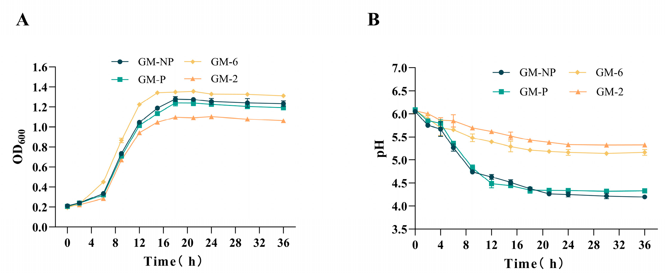
Figure 1. Growth curves (A) and acid production curves (B) of four strains of lactic acid bacteria. The values were means with three technical replicates. Abbreviations: GM-NP, Pediococcus acidilactici GM-NP; GM-P, Pediococcus acidilactici GM-P; GM-6, Enterococcus faecalis GM-6; GM-2, Weissella confusa GM-2.
Fermentation 11 00598 g001
Figure 2. Degradation of free gossypol by four strains of lactic acid bacteria in GMRS medium. All treatment groups (GM-NP, GM-P, GM-6, GM-2) and the control group (CON) were cultured in GMRS medium supplemented with 0.1 g/L free gossypol. Data are presented as the mean of three technical replicates, with error bars representing standard deviation. Different lowercase letters above bars indicate statistically significant differences (p < 0.05). Abbreviations: CON, control (saline solution); GM-NP, Pediococcus acidilactici GM-NP; GM-P, Pediococcus acidilactici GM-P; GM-6, Enterococcus faecalis GM-6; GM-2, Weissella confusa GM-2.
Fermentation 11 00598 g002
Figure 3. Effect of four strains of bacteria on the concentration of free gossypol during solid-state fermentation of CSM. The control group (CON) contained autoclaved substrate without bacterial inoculation. All treatment groups (GM-NP, GM-P, GM-6, GM-2) were inoculated with their respective bacterial culture suspensions and autoclaved substrate. Data are presented as the mean of three technical replicates, with error bars representing standard deviation. Different lowercase letters above bars indicate statistically significant differences (p < 0.05). Abbreviations: CON, control; GM-NP, Pediococcus acidilactici GM-NP; GM-P, Pediococcus acidilactici GM-P; GM-6, Enterococcus faecalis GM-6; GM-2, Weissella confusa GM-2.
Fermentation 11 00598 g003
Table 1. Composition of media used for strain cultivation.
MediaComposition
LB1%yeast extract, 2%peptone, 1%NaCl, 2% agar
MRS1%peptone, 0.8%Beef extract, 0.4%Yeast, 2%glucose, 0.2%K2HPO4, 0.2%(NH4)2HC6H5O7, 0.5%CH3COONa, 0.02%MgSO4, 0.004%MgSO4, 0.1%Tween 80
MRSA1%peptone, 0.8%Beef extract, 0.4%Yeast, 2%glucose, 0.2%K2HPO4, 0.2%(NH4)2HC6H5O7, 0.5%CH3COONa, 0.02%MgSO4, 0.004%MgSO4, 0.1%Tween 80, 2%agar
GMRS0.1%gossypol, 1%peptone, 0.8%Beef extract, 0.4%Yeast, 0.2%K2HPO4, 0.2%(NH4)2HC6H5O7, 0.5%CH3COONa, 0.02%MgSO4, 0.004%MgSO4, 0.1%Tween 80
GMRSA0.2%gossypol, 1%peptone, 0.8%Beef extract, 0.4%Yeast, 0.2%K2HPO4, 0.2%(NH4)2HC6H5O7, 0.5%CH3COONa, 0.02%MgSO4, 0.004%MgSO4, 0.1%Tween 80, 2% agar
Table 2. Sequence analysis of 16S rRNA from cotton bollworm gut bacteria.
StrainsClosest MatchGenBank Accession No.Similarity/%
GM-NPPediococcus acidilacticiNR042057.199.184%
GM-PPediococcus acidilacticiNR042057.199.515%
GM-2Weissella confusaNR040816.199.861%
GM-3Mammaliicoccus sciuriNR041327.199.859%
GM-4Enterococcus innesiiNR181755.199.718%
GM-5Enterococcus innesiiNR181755.199.858%
GM-6Enterococcus faecalisNR115765.1100.000%
GM-7Klebsiella oxytocaNR118853.199.861%
GM-8Klebsiella oxytocaNR118853.199.645%
GM-9Klebsiella oxytocaNR118853.199.363%
GM-10Klebsiella oxytocaNR118853.199.153%
Table 3. Physiological and biochemical characteristics of four strains of lactic acid bacteria.
ItemP. acidilactici
GM-NP
P. acidilactici
GM-P
E. faecalis
GM-6
W. confusa
GM-2
Aesculin+++
1%Sodium Hippurat++++
Cellobiose++
Maltose++
Mannose++
Salicin++
Sorbitol++
Sucrose++
Raffinose++
Inulin+
Lactose++
Bollworms “+” indicates utilised; “−“ indicates not utilised.
Table 4. Tolerance of four strains of lactic acid bacteria to artificial gastric conditions.
ItemsCell Numbers (Log CFU/mL)
P. acidilactici
GM-NP
P. acidilactici
GM-P
E. faecalis
GM-6
W. confusa
GM-2
Artificial gastric juice tolerance     
Initial cell number9.71 ± 0.039.36 ± 0.028.28 ± 0.058.52 ± 0.05
pH2.5, 0.3% pepsin, 1.5 h6.98 ± 0.106.95 ± 0.095.91 ± 0.205.82 ± 0.17
Survival rate (%)71.83 a ± 1.1074.28 a ± 1.0671.35 a ± 2.4068.31 b ± 2.07
Artificial intestinal fluid tolerance    
Initial cell number9.43 ± 0.029.40 ± 0.078.62 ± 0.028.37 ± 0.01
pH6.8, 1% trypsin, three h8.48 ± 0.168.52 ± 0.077.68 ± 0.077.48 ± 0.07
Survival rate (%)89.93 a ± 1.5090.64 a ± 0.8689.10 a ± 0.8685.98 b ± 0.53
Artificial bile salt tolerance    
Initial cell number8.66 ± 0.038.69 ± 0.028.56 ± 0.038.53 ± 0.02
0.3% Ox Gall, four h6.78 ± 0.137.36 ± 0.205.87 ± 0.056.26 ± 0.15
Survival rate (%)78.11 b ± 1.3284.76 a ± 2.1668.54 d ± 0.7873.46 c ± 1.79
Note: Each value shows the mean ± S.D. of triplicate values of independent experiments. Different letters indicate significant differences between groups (p < 0.05).
Table 5. Survival rate of four strains of lactic acid bacteria after high-temperature treatment.
ItemsSurvival Rate (%)
P. acidilactici
GM-NP
P. acidilactici
GM-P
E. faecalis
GM-6
W. confusa
GM-2
Heat treatment time/min    
60 °C, 5 min67.23 b ± 0.5466.36 bc ± 0.6472.20 a ± 0.4760.94 c ± 0.40
60 °C, 10 min60.29 b ± 0.957.44 c ± 0.9763.06 a ± 0.8752.51 d ± 0.42
60 °C, 15 min48.56 c ± 0.8646.72 c ± 0.8153.83 a ± 1.2643.96 d ± 0.35
70 °C, 5 min57.74 b ± 0.4652.53 c ± 0.7460.86 a ± 0.5851.64 c ± 0.48
70 °C, 10 min52.14 c ± 0.6949.01 d ± 0.9956.91 a ± 1.4544.69 e ± 0.32
70 °C, 15 min33.56 b ± 1.1432.79 b ± 0.7239.04 a ± 0.4131.31 b ± 0.38
80 °C, 5 min34.81 b ± 0.3831.87 c ± 1.0037.97 a ± 0.9125.75 d ± 0.64
80 °C, 10 min6.68 bc ± 0.405.17 c ± 0.229.04 a ± 0.877.10 bc ± 0.82
80 °C, 15 min0.00 ± 0.000.00 ± 0.000.00 ± 0.000.00 ± 0.00
90 °C, 5 min17.72 b ± 0.6915.87 bc ± 0.3321.49 a ± 1.1415.32 c ± 0.53
90 °C, 10 min0.00 ± 0.000.00 ± 0.000.00 ± 0.000.00 ± 0.00
90 °C, 15 min0.00 ± 0.000.00 ± 0.000.00 ± 0.000.00 ± 0.00
Each value shows the mean ± S.D. of triplicate values of independent experiments. Different letters indicate significant differences between groups (p < 0.05).
Table 6. Adhesion assessment of four strains of lactic acid bacteria.
ItemsStrains
P. acidilactici
GM-NP
P. acidilactici
GM-P
E. faecalis
GM-6
W. confusa
GM-2
Assessment of auto-aggregation ability (%)     
8 h48.60 a ± 1.0146.77 a ± 0.5537.21 c ± 1.1143.39 b ± 1.27
24 h82.96 a ± 0.9581.10 a ± 1.8072.89 b ± 1.7674.34 b ± 0.73
Cell surface properties (%) 54.45 b ± 1.6251.63 bc ± 1.9547.92 c ± 1.9141.60 d ± 2.29
Each value shows the mean ± S.D. of triplicate values of independent experiments. Different letters indicate significant differences between groups (p < 0.05).
Table 7. Antimicrobial activity of four strains of lactic acid bacteria.
PathogensAntimicrobial Activities
P. acidilactici
GM-NP
P. acidilactici
GM-P
E. faecalis
GM-6
W. confusa
GM-2
E. coli ATCC25922★★★★★★★★★
S. aureus ATCC6538★★★★★★★★
Salmonella ATCC14028★★★★★★★★★★-
P. aeruginosa ATCC9027★★★★★★★★
Note: The antibacterial activity of the cell-free culture supernatant of the four lactic acid bacteria strains was classified into the following grades: no inhibition, ★ weak inhibition (7–9 mm), ★★ intermediate inhibition (10–13 mm), ★★★ strong inhibition (14–17 mm), and ★★★★ very strong inhibition (>17 mm).
Table 8. Hemolytic and antioxidant properties of four strains of lactic acid bacteria.
ItemsStrains
P. acidilactici
GM-NP
P. acidilactici
GM-P
E. faecalis
GM-6
W. confusa
GM-2
Hemolytic activityγγγγ
Free radical scavenging rate (%) (DPPH)43.81 a ± 1.1145.29 a ± 1.2628.73 c ± 1.3237.62 b ± 0.50
Each value shows the mean ± S.D. of triplicate values of independent experiments. Different letters indicate significant differences between groups (p < 0.05). γ represents γ-hemolysis, indicating no hemolysis.
Table 9. Drug sensitivity of four strains of lactic acid bacteria.
Antibiotic SusceptibilityStrains
P. acidilactici
GM-NP
P. acidilactici
GM-P
E. faecalis
GM-6
W. confusa
GM-2
CefalotinSSIS
ClindamycinSSRS
ChloramphenicolISIS
PenicillinRRSS
RifampicinSSSS
ErythromycinSSIS
AmpicillinSSIS
TetracyclineSSIS
LevofloxacinRRIS
GentamicinRRIS
StreptomycinRRIR
Polymyxin BRRRR
EnrofloxacinRRSI
CiprofloxacinRRIR
OfloxacinRRII
“R” indicates resistance to antibiotics; “I” indicates intermediate susceptibility to antibiotics; “S” indicates susceptibility. Inhibition halo interpreted according to CLSI 2012 guidelines [25].
Table 10. Nutritional quality indicators and pH of fermented cottonseed meal.
ItemsCONP. acidilactici
GM-NP
P. acidilactici
GM-P
E. faecalis
GM-6
W. confusa
GM-2
CP (%)49.77 b ± 0.1653.43 a ± 1.0553.39 a ± 0.7754.35 a ± 0.5652.83 a ± 0.65
ASP (%)3.49 b ± 0.216.17 a ± 0.136.10 a ± 0.066.08 a ± 0.076.48 a ± 0.72
NDF (%)36.76 a ± 1.5228.94 cd ± 0.7130.01 cd ± 1.2929.40 cd ± 1.1331.97 bc ± 0.59
ADF (%)27.53 a ± 0.7319.87 d ± 1.4418.83 cd ± 0.2722.31 bc ± 0.7523.28 b ± 0.78
Crude fat (%) 1.30 a ± 0.060.71 b ± 0.040.51 b ± 0.130.44 b ± 0.030.59 b ± 0.10
Ash (%)6.72 ± 0.587.26 ± 0.496.41 ± 0.116.99 ± 0.327.17 ± 0.14
Ca (%)0.32 ± 0.020.37 ± 0.030.36 ± 0.030.34 ± 0.040.30 ± 0.08
P (%)0.70 ± 0.050.68 ± 0.040.79 ± 0.070.70 ± 0.010.72 ± 0.09
pH6.39 a ± 0.035.99 b ± 0.225.58 C ± 0.025.92 b ± 0.046.11 b ± 0.05
CP: Crude protein. ASP: Acid-soluble protein. NDF: Neutral detergent fiber. ADF: Acid detergent fiber. CON: did not receive any treatment; other groups received corresponding culture medium + autoclaved substrate. Each value shows the mean ± S.D. of triplicate values of independent experiments. Different letters indicate significant differences between groups (p < 0.05).
Disclaimer/Publisher’s Note: The statements, opinions and data contained in all publications are solely those of the individual author(s) and contributor(s) and not of MDPI and/or the editor(s). MDPI and/or the editor(s) disclaim responsibility for any injury to people or property resulting from any ideas, methods, instructions or products referred to in the content.