Article

Urea-Induced Enhancement of Hypocrellin A Synthesis in Shiraia bambusicola GDMCC 60438: Strategies and Mechanisms

by
Yanbo Tang
1,2,
Yongdi Wen
1,
Xiang Zhang
1,2,
Qian Gao
3,
Fuqiang Yu
4,
Zhenqiang Wu
1 and
Xiaofei Tian
1,2,*
1
School of Biology and Biological Engineering, South China University of Technology, 382 Outer Loop, University Park, Guangzhou 510006, China
2
Zhuhai Institute of Modern Industrial Innovation, South China University of Technology, 8 Fushan Road, Fushan Industrial Park, Zhuhai 519100, China
3
College of Chinese Materia Medica, Yunnan University of Chinese Medicine, 1076 Yuhua Road, Chenggong, Kunming 650500, China
4
Yunnan Key Laboratory for Fungal Diversity and Green Development, Kunming Institute of Botany, Chinese Academy of Sciences, 132 Lanhei Road, Kunming 650204, China
*
Author to whom correspondence should be addressed.
Fermentation 2024, 10(8), 381; https://doi.org/10.3390/fermentation10080381
Submission received: 22 May 2024 / Revised: 20 July 2024 / Accepted: 21 July 2024 / Published: 25 July 2024
(This article belongs to the Special Issue New Research on Fungal Secondary Metabolites, 2nd Edition)

Abstract

:
Hypocrellin A (HA) is a valuable pigment with promising applications in biotechnology and pharmaceuticals. The submerged cultivation of Shiraia bambusicola offers a strategic opportunity to enhance HA production. This study investigates the regulatory mechanisms for HA biosynthesis through urea supplementation and presents a strategy to increase HA yield. In the absence of urea, S. bambusicola (GDMCC 60438) does not synthesize HA. However, the addition of 40 g/L urea 12 h into the fermentation process results in a final HA production of 46.7 ± 8.2 mg/L. Morphological analysis reveals an optimized environment for HA synthesis, characterized by a densely intertwined and reticular hyphal structure with minute pores. RNA sequencing shows significant upregulation of genes involved in DNA repair, recombination, and metabolism. Conversely, genes related to cellular homeostasis, cell-wall chitin, and amino polysaccharide metabolism are downregulated. Urea supplementation facilitates the upregulation of amino acid metabolism and the cysteine desulfurase gene, enhancing acetyl-CoA accumulation within the mycelium and providing the necessary precursor materials for HA synthesis. Our work underscores the pivotal role of urea in regulating HA biosynthesis and proposes a practical approach to enhance HA production. The findings contribute novel insights to the fields of biotechnology for pharmaceuticals.

1. Introduction

Hypocrellins (HYPs), including hypocrellin A (HA), hypocrellin B (HB), hypocrellin C (HC), and hypocrellin D (HD), are bioactive perylenequinones (PQs) present in the stroma of Shiraia bambusicola Henn. and Hypocrella bambusae (Berk. & Broome) Sacc [1,2,3,4]. As the most abundant natural PQ in the stroma, HA is of significant interest for its potent photosensitivity and its application in photodynamic therapy (PDT) for treating neoplastic diseases, which has given it importance in clinical cancer research [5,6,7,8]. The limited natural availability of S. bambusicola stroma further underscores the need to explore scalable and sustainable production methods for HA and related compounds. The chemical synthesis of HA involves complex steps that start with 3,5-dihydroxybenzoic acid. The pathway proceeds as follows: (1) conversion of 3,5-dihydroxybenzoic acid to 3-(3,5-dimethylxybenzyl)-5-hexenoic acid, (2) formation of tetrahydroxynaphthalone, (3) conversion to propyl ketone and final conversion to HA. (4) The final step is complex and results in the formation of numerous by-products [9,10]. It usually results in a racemic mixture, which is less effective at inhibiting acetylcholinesterase (AChE) compared to its naturally derived counterpart from plant extracts [11,12]. Due to the economic and effectiveness concerns associated with the chemical-synthesis route, submerged fermentation of S. bambusicola is considered a promising alternative technique for HYP production.
Recent advancements in HYP fermentation show that techniques such as gene editing, strain mutagenesis, biological induction, and optimization of culture medium demonstrate potential for increasing HA yields [13,14,15,16]. Notably, exposure to red light has significantly enhanced HA release in both mycelium and fermentation medium, with an eight-day exposure resulting in a 3.82-fold increase in HA production, achieving levels up to 175.53 mg/L [17]. Similarly, ultrasound stimulation at 40 kHz has amplified HA content in mycelium by 177.2%, with peak production noted at 247.67 mg/L on the eighth day [18]. The composition of the fermentation medium, particularly the carbon and nitrogen sources, plays a crucial role in HA production and synthesis. Studies have highlighted the importance of optimizing these sources to boost HA production [19]. As an organic nitrogen source, urea has been effective in promoting the synthesis of essential biomolecules in other fungal species [20]. For example, the addition of urea to the fermentation medium of Cordyceps cicadae led to a 138% increase in ergosterol content, which was attributed to the role of urea in enhancing acetyl CoA synthesis through the downregulation of the tricarboxylic acid (TCA) and glyoxylic acid cycles and the upregulation of genes encoding essential enzyme involved in ergosterol biosynthesis [21]. The broader applications of urea, as evidenced by its use in processes like sweet-potato ethanol production, underscore its economic and industrial benefits. These examples highlight the potential of urea for optimizing production processes for various metabolites, offering a cost-effective strategy for enhancing yields in industrial fermentation. Such insights provide valuable guidance for industries aiming to improve production efficiency. In HA production by S. bambusicola, urea has proven effective as a nitrogen source, outperforming other options in promoting HA yield to 5.35 mg/g. However, a trade-off exists between HA concentration and mycelial growth, underscoring the complexity of nutrient composition and the regulation of HA synthesis [22].
Our initial exploration of nitrogen sources for the fermentation of S. bambusicola (GDMCC 60438) identified limitations in traditional options such as beef paste, primarily due to cost considerations for industrial applications. Among the available nitrogen sources, urea is a cost-effective option commonly employed in large-scale fermentation processes. For instance, Wechgama utilized urea instead of the more expensive yeast extract as a nitrogen source. The optimization of pH, urea concentration, and initial sugar concentration was key to achieving large-scale production of butanol, demonstrating the potential for enhancing fermentation yields using economical nutrients and straightforward fermentation conditions [23]. Yeast extract, while supporting mycelial growth, proved insufficient for inducing HA synthesis, highlighting the need to investigate alternative nitrogen sources that both support growth and enhance HA production. Currently, low productivity poses a challenge to the production of HA through fermentation. The complete biosynthetic pathway of HA has yet to be fully understood. Improving HA biosynthesis through genetic manipulation remains a significant challenge [24,25]. Compared to genetic manipulation and biological induction, using alternative substrate to enhance HA synthesis offers advantages of lower costs and simpler operations. This study focuses on the role of urea as an additional nitrogen source when combined with yeast extract, examining its regulatory and promotive effects on HA biosynthesis in S. bambusicola (GDMCC 60438). The research aims to elucidate the influence of urea on mycelial growth and HA yield, with particular attention to the optimal timing and concentration of urea supplementation. Through detailed analyses of mycelial morphology, fluctuations in acetyl coenzyme A (acetyl-CoA) content, and differential gene expression, this study seeks to illuminate the intricate regulatory mechanisms of HA synthesis facilitated by urea. This investigation represents a critical step in identifying key genes and regulatory pathways for optimizing HA biosynthesis, providing valuable insights for bioengineering and applied microbiology.

2. Materials and Methods

2.1. Production Hypocrellin A Using Submerged Fermentation of S. bambusicola GDMCC60438

The S. bambusicola (GDMCC 60438) strain was cultured on potato dextrose agar (PDA) (Huankai Microbial Sci & Tech Co., Ltd., Guangzhou, China) at 28 °C for 10 days. The mycelium on the plate was rinsed twice with 8 mL of water containing 0.1% (v/v) Tween 80 (Damao Chemical Reagent Factory, Tianjin, China) and subsequently transferred into a sterilized 15 mL tube. A 2 mL mycelium suspension was then transferred to a 250-mL conical flask (Shubo Group Co., Ltd., Chengdu, China) containing 50 mL of potato dextrose broth (PDB) (Huankai Microbial Sci & Tech Co., Ltd., Guangzhou, China). After a stationary culture period of 4–6 h at 28 °C, the seed culture was cultivated for 48 h at 150 rpm in a rotary incubator (Zhicheng Analytical Instrument Manufacturing Co., Ltd., Shanghai, China). For fermentation, a 10% (v/v) seed suspension was inoculated into a 250 mL baffled-bottom flask (Shubo Group Co., Ltd., Chengdu, China) containing 50 mL of fermentation medium. The fermentation medium was composed of 4 g/L potato extract (Yuanye Bio-Technology Co., Ltd., Shanghai, China), 12 g/L yeast extract (Huankai Microbial Sci & Tech Co., Ltd., Guangzhou, China), and 10 g/L glycerol (Macklin Biochemical Co., Ltd., Shanghai, China). The submerged fermentation was conducted at 32 °C and 150 rpm for 72 h under natural pH. In the control group, yeast extract was used as the sole nitrogen source, excluding urea. Urea was added to the fermentation medium at 12 h to achieve final concentrations of 0, 10, 20, 30, 40, and 60 g/L. The final concentrations of 40 g/L urea were added at intervals of 0, 6, 12, 24, 36, and 48 h of fermentation.

2.2. Determination of HA Yield and Mycelium Biomass

The mycelium produced by fermentation was harvested using a GM-0.5B vacuum-filtration device (Jingteng Instrument Equipment Co., Ltd., Tianjin, China). After collection, the mycelium was washed three times with distilled water and lyophilized in a SCIENTZ-10N freeze dryer (Scientz Biotechnology Co., Ltd., Ningbo, China) for 48 h until a constant weight was achieved. The dry biomass was measured using a Shimadzu ATX124 analytical balance (Shimadzu Corporation, Shimane-ken, Japan). Approximately 0.05 g of the freeze-dried mycelium, combined with 0.05 g of quartz sand (Acmec Biochemical Co., Ltd., Shanghai, China), was ground to a fine powder and transferred to a 10 mL centrifuge tube. Methylene chloride (5 mL; Zhiyuan Reagent Co., Ltd., Tianjin, China) was added, and the mixture was subjected to ultrasonic extraction at 40 kHz and 600 W for 30 min using a 100S ultrasonic cleaner (Chaojie Instrument Equipment Co., Ltd., Shenzhen, China). The extract was then filtered through a 0.22 µm filter membrane (Jinteng Experimental Equipment Co., Ltd., Tianjin, China) to prepare it for analysis by high-performance liquid chromatography (HPLC). The concentration of HA in the mycelium was determined using an e2695 HPLC system (Waters Corporation, Milford, MA, USA) equipped with an ODS analytical column (5 µm × 4.6 mm × 250 mm, Shimadzu Corporation, Shimane-ken, Japan) and a 2998 photodiode array detector. The mobile phase consisted of an aqueous solution of methanol and acetic acid (9:1, v/v, Ph = 2) with a flow rate of 1 mL/min. A 10 µL sample was injected, and HA was detected at a wavelength of 467 nm. The HA yield was calculated based on the concentration of HA detected in the mycelium, adjusted for the initial dry weight of the mycelium used in the extraction process. The HA content in the mycelia was calculated using Equation (1), as follows:
HA   content   in   mycelia ( mg / g ) = HA   concentration ( mg / ml )   ×   solvent   volume ( ml ) ground   mycelia ( g )

2.3. Morphology Observation and Diameter Determination of Mycelium Pellets

To assess the morphological characteristics and diameter of mycelium pellets, 5 mL samples of the fermentation broth were collected at 12, 24, 48, and 72 h post-inoculation. These samples were centrifuged at 8000 rpm for 5 min using a 5804 R centrifuge (Eppendorf Corporation, Hamburg, Germany). The supernatant was discarded, and the resultant pellets were washed three times with distilled water to ensure purity before morphological analysis proceeded. The morphological features of the mycelium pellets under various fermentation conditions were examined using a stereoscopic microscope (CX41, Olympus Corporation, Tokyo, Japan) at 50× magnification, allowing for detailed observation of the structural characteristics of the fungal pellets and facilitating a comparative analysis across different time intervals. The washed mycelium pellets were evenly dispersed on a Petri dish for measurement. The diameters of 20 randomly selected mycelium pellets were measured to ensure statistical relevance using Image-Pro Plus 6.0 software (Media Cybernetics Inc., Rockville, MD, USA), which provided accurate and reproducible size determinations.

2.4. Observation of Mycelium Morphology on the Surface of Mycelium Pellets

Approximately 2–5 mL of fermentation broth was centrifuged at 8000 rpm for 5 min using a 5804 R centrifuge to sediment the mycelial pellets. The supernatant was decanted, and the pellets were then immersed in 5 mL of 2.5% glutaraldehyde (Phygene Bio-Technology Co., Ltd., Fuzhou, China), then incubated at 4 °C for 4 h to ensure adequate fixation. After fixation, the mycelial pellets were centrifuged again at 8000 rpm for 5 min, the glutaraldehyde was discarded, and the pellets were washed several times with distilled water to eliminate any residual fixative. The washed pellets were then resuspended in distilled water and frozen at −80 °C overnight. Subsequently, the frozen mycelial pellets were lyophilized using a freeze dryer to obtain a dry powder. For examination of surface morphology, the dried samples were coated with gold using a 150TES sputter coater (Electron Microscopy Sciences Corporation, Hatfield, UK) to enhance electron conductivity. They were then imaged under a Merlin compact field emission scanning electron microscope (SEM) (Carl Zeiss Medical Technology Inc., Oberkochen, Germany) at a scanning voltage of 5.00 kV.

2.5. Transcriptome Sequencing and Bioinformatics Analysis

After fermentation, mycelia of S. bambusicola GDMCC60438 were harvested from cultures both with and without urea supplementation. RNA extraction was performed utilizing three biological replicates for each condition to ensure reproducibility and statistical robustness (Annoroad Gene Technology Co., Ltd., Beijing, China). A complementary DNA (cDNA) library was constructed for each replicate, following which sequencing was conducted on the MGI T7 platform (BGI Inc., Shenzhen, China), generating 150-bp paired-end reads. Quality control of the raw sequencing data was conducted using the FastQC and Skewer tools with parameters set to -q 20 -Q 30 -l 50 to filter out low-quality reads. Transcript levels were quantified as transcripts per million (TPM) using HISAT2 (v2.2.1) and StringTie (v2.2.0). Gene annotation was subsequently performed against multiple databases including Gene Ontology (GO), Kyoto Encyclopedia of Genes and Genomes (KEGG), Carbohydrate-Active enzymes (CAZy), BiGG, and Clusters of Orthologous Groups (COG) to identify functional and metabolic pathways. Further functional annotation of coding sequences was accomplished using the Pfam database. Differentially expressed genes (DEGs) between the control and urea-supplemented samples were identified using DESeq2 (v3.8). The expression patterns of DEGs were visualized using cluster-heatmap analysis in the pheatmap package. Additionally, a co-expression network of the DEGs was constructed using the Weighted Gene Co-expression Network Analysis (WGCNA) package in R, providing insights into gene interactions and regulatory mechanisms influenced by urea supplementation.

2.6. Identification of Hyaluronic Acid Synthesis and Urea-Pathway-Related Genes by qRT-PCR

The DEGs associated with HA synthesis and the urea action pathway were selected for further analysis. The DEGs were subjected to quantitative real-time PCR (qRT-PCR) to quantify their relative expression levels. Approximately 100 mg of harvested mycelia were pulverized in liquid nitrogen to ensure cell disruption. Total RNA was then isolated using an RNA extraction kit, following the manufacturer’s instructions. Complementary DNA (cDNA) was synthesized from the extracted RNA using a reverse transcription kit with gDNA Eraser. Gene-specific primers for qRT-PCR were designed using Primer Premier 5 software (Table S1). Amplification was conducted on a LightCycler 96 Instrument (F. Hoffmann-La Roche Ltd., Basel, Switzerland) under specific conditions. The housekeeping gene MH01c07g0051161 served as an internal control to normalize the expression data. Relative gene-expression levels were calculated using the 2−ΔΔCT method. Each gene was analyzed in triplicate across both biological and technical replicates to ensure data reliability and reproducibility.

2.7. Statistical Analysis

Data were collected from three independent experimental replicates. Statistical significance was evaluated using one-way analysis of variance (ANOVA) conducted in IBM SPSS Statistics 26 (IBM SPSS Inc., Armonk, NY, USA). A homogeneity test was first conducted to verify the consistency of the variance. Then, Tamhane’s T2, Dunnett’s T3, Games-Howell, and LSD tests were performed to identify statistically significant differences among experimental groups at a threshold of p < 0.05.

3. Results and Discussion

3.1. Impact of Exogenous Urea on Shiraia bambusicola Growth and HA Production

Urea is a crucial organic nitrogen source that significantly influences fungal metabolism and the biosynthesis of essential secondary metabolites. Numerous studies have shown that urea can enhance the yield of these metabolites, highlighting its importance in optimizing fermentation processes [22].Our research focused on urea’s role in the growth and secondary metabolite production of S. bambusicola GDMCC 60438. Notably, urea has been documented to affect the synthesis of compounds like ergosterol in species such as C. cicadae. For example, ergosterol levels increased from 76.01 mg/L to 180.7 mg/L when urea was used as the sole nitrogen source, demonstrating urea’s capability for metabolic induction [21]. Building on this, our study explored urea’s impact on HA synthesis, which also initiates with acetyl-CoA but follows a pathway distinct from that of ergosterol synthesis. Unlike C. cicadae, S. bambusicola exhibited neither HA production nor significant growth when it was dependent solely on urea for nitrogen, suggesting a unique metabolic response among fungi. However, when urea was combined with yeast extract as another source of nitrogen, HA synthesis occurred. A systematic increase in urea concentrations from 10 g/L to 40 g/L correlated with rising HA yield. However, further increasing the concentration to 60 g/L significantly reduced both HA production and fungal biomass. (Figure 1A). Additionally, the timing of urea addition proved critical, with the highest HA yield of 46.7 ± 8.2 mg/L (p = 0.05) achieved when urea was added at 12 h to reach a final concentration of 40 g/L in the broth (Figure 1B). This finding indicates that there is a critical window for urea’s efficacy in HA biosynthesis.
These findings underscore the dual role of urea: it is involved in facilitating HA synthesis and has a concentration-dependent impact on fungal growth. Excessive urea concentrations detrimentally affect mycelial growth, underscoring the need to optimize urea levels for effective HA production. The study determined the optimal concentration and timing of urea addition for HA synthesis, offering insights into the metabolic regulation of S. bambusicola. These results provide a foundational basis for future biotechnological applications in HA production.

3.2. Mycelium Pellet Size and Morphological Changes Influenced by Urea Supplementation

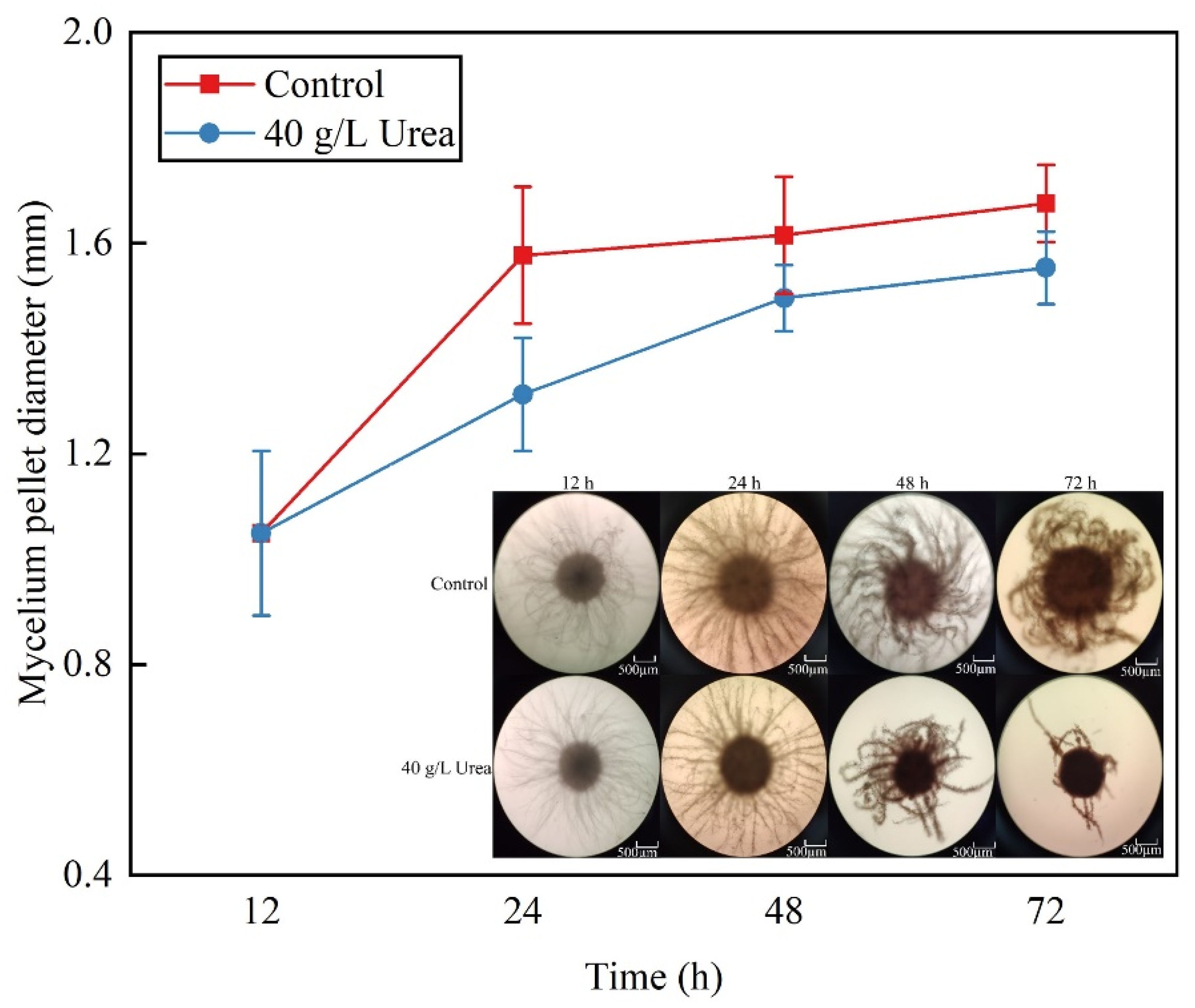
The morphology and size of fungal mycelial pellets significantly influence the efficiency of microbial secondary-metabolite synthesis during submerged fermentation [26]. To explore these dynamics in S. bambusicola GDMCC 60438, particularly regarding HA synthesis, we analyzed mycelial pellet size and morphology under varying urea treatment conditions. Mycelial pellets were collected at different fermentation stages to measure their diameter and observe morphological changes. Both with and without urea, a consistent growth pattern was observed, characterized by an initial rapid size increase followed by gradual expansion. Specifically, in the control group, rapid growth occurred between 12 to 24 h, then stabilized, whereas in the urea-supplemented group, significant growth continued from 12 to 48 h, stabilizing from 48 to 72 h. Notably, urea supplementation resulted in reduced average pellet diameter, aligning with the results of other studies in which physical or chemical stimuli affected mycelial pellet size and correlated with the efficiency of secondary-metabolite production (Figure 2).Morphological analysis revealed that urea impacts not only pellet size but also the physical structure of the mycelia. In the initial 48 h, both urea-treated and control groups exhibited robust mycelial growth. However, by 72 h post-urea treatment, a noticeable reduction in mycelium density was observed, leading to sparser mycelial distribution around the pellets—a phenomenon consistent with the findings of other studies assessing environmental or chemical impacts on mycelial morphology. Our observations align with research in which external factors, such as light−dark cycles or chemical treatments, altered mycelial pellet size and affected HA synthesis [27]. For instance, in Shiraia sp. S9 treated with sodium nitroprusside, a reduction in pellet size correlated with increased HA production. Similarly, co-fermentation studies have shown morphological changes under varied culturing conditions, affecting both biomass and metabolite yield [28,29]. These results suggest that when urea concentration is 40 g/L, urea supplementation in S. bambusicola GDMCC 60438 not only reduces the size but also alters the morphological characteristics of the mycelial pellets, potentially influencing HA yield. The observed decrease in biomass and mycelial density at higher urea concentrations may be due to the increase in pH caused by the production of CO2 and ammonia from urea metabolism, which inhibits fungal growth.

3.3. Scanning Electron Microscopy Analysis of Mycelial Morphology

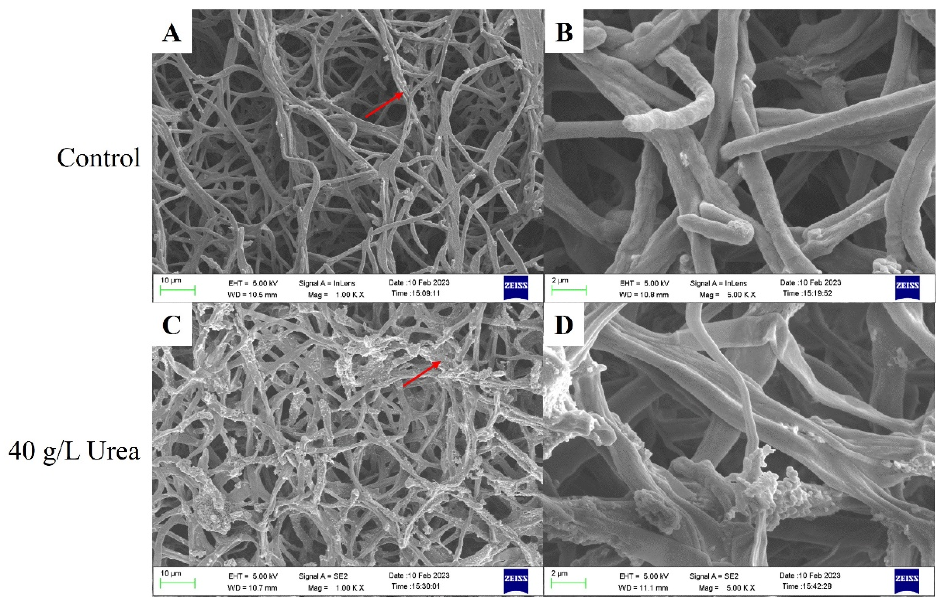
The surface morphology of S. bambusicola GDMCC 60438 mycelial pellets under varying urea-supplementation conditions was analyzed using scanning electron microscopy (SEM). In the control group, the mycelium displayed a complex, interwoven network with regions of parallel aggregation and notable surface porosity (Figure 3A). Conversely, in the urea-supplemented group, the mycelia formed a denser and more uniform mesh, characterized by reduced porosity and predominantly intertwined strands pointing in various directions (Figure 3C). Urea addition resulted in a noticeable alteration in the mycelium texture, presenting a crumpled appearance with evident surface folds and closer adhesion of mycelial strands (Figure 3B,D). These structural transformations are consistent with changes observed in other studies, such as in S. bambusicola BZ-16X1, in which external factors like protein activators significantly modified mycelial surfaces, indicating a correlation between surface morphology and metabolic activity [29,30].
Our findings corroborate previous research that suggests fermentation temperature, urea concentration, and other inducers can lead to tighter, smaller mycelial pellets in S. bambusicola, potentially enhancing the efficiency of HA synthesis. The observed morphological changes, especially the increased entanglement and decreased porosity in urea-treated samples, suggest a direct link between nutrient-induced morphological adaptation and enhanced metabolic output. The SEM analysis demonstrated that urea supplementation induces a denser mycelial structure in S. bambusicola GDMCC 60438 that is associated with improved HA production. This morphological transformation likely facilitates more efficient nutrient absorption and metabolic processing, emphasizing the importance of optimizing physical and chemical conditions to enhance secondary-metabolite yields in fungal fermentation systems [27,28,29].

3.4. Transcriptomic Analysis and Differential Expression Profiling

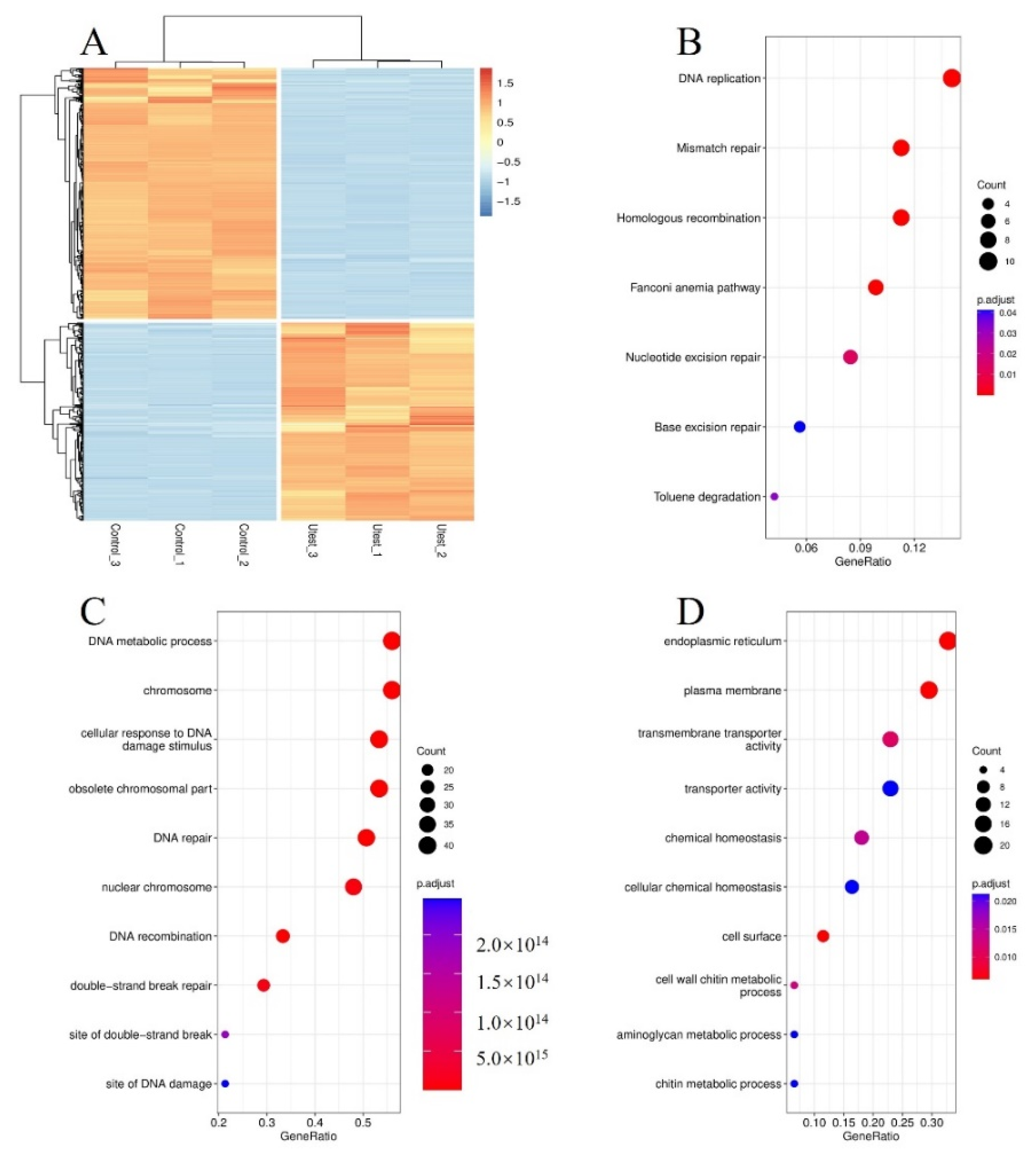
Understanding the mechanistic impact of urea on HA production could provide critical insights into the metabolic pathways and gene networks of S. bambusicola. In this study, the transcriptome of S. bambusicola GDMCC 60438 was analyzed, with urea-supplemented samples being compared to controls to elucidate the pathways influenced by urea in secondary-metabolite synthesis. A total of 12,350 genes were identified, and 816 DEGs were isolated for further investigation (Tables S3 and S4). Through clustering analysis via heat maps based on gene-expression levels, substantial transcriptional differences were highlighted, with 360 genes being up-regulated and 456 genes down-regulated following urea treatment (Figure 4A). These alterations are suggested to significantly impact the genetic landscape, particularly in relation to HA biosynthesis. Functional enrichment analyses of these DEGs were performed using KEGG and GO databases. It was revealed that up-regulated genes were predominantly involved in DNA repair and metabolic processes, such as DNA replication, mismatch repair, homologous recombination, nucleotide excision repair, and base excision repair. The upregulation of DNA repair genes suggests that the growth inhibition caused by urea may rely on DNA damage, which subsequently leads to a decrease in the mycelial biomass. (Figure 4B,C), while down-regulated genes were associated with transmembrane transport and cell-wall metabolism (Figure 4D). Notably, the down-regulation of genes related to chitin and amino sugar metabolism has been suggested to impact the structural integrity of the fungal cell wall [31,32,33,34]. Urea-induced morphological changes in fungal hyphae were corroborated by our SEM observations (Figure 3). Furthermore, genes involved in cytochemical homeostasis were found to be down-regulated, indicating a potential disruption in cellular stability. Perturbations in metabolic cycles such as the TCA and citrate-pyruvate pathways were observed to possibly affect metabolic flux, leading to an accumulation of intermediates such as acetyl-CoA (Figure 5). Through RNA-seq analysis under varying urea conditions, significant differential expression of genes associated with DNA repair, chitin metabolism, and amino acid metabolism was revealed, highlighting urea’s extensive impact on cellular functions. The integration of transcriptomic data with morphological observations demonstrates that a complex cascade of genetic and metabolic responses is triggered by urea supplementation in S. bambusicola. These findings underscore the multifaceted impact of urea on fungal secondary-metabolite production, enhancing our understanding of the underlying processes that drive metabolic responses to urea supplementation.

3.5. Acetyl-CoA Accumulation and Its Role in HA Biosynthesis

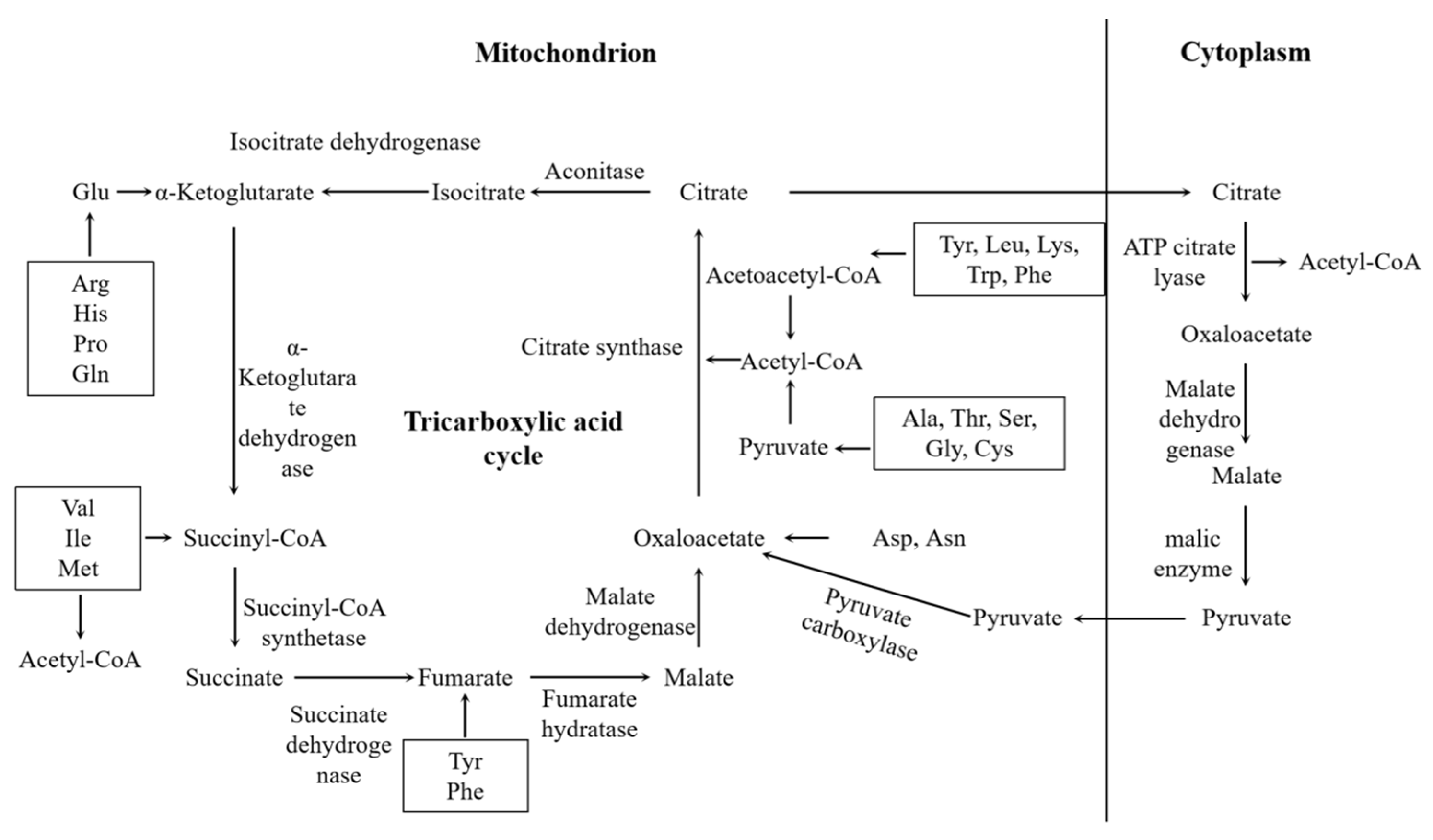
To elucidate the mechanism by which HA biosynthesis is enhanced by urea in S. bambusicola GDMCC 60438, Pfam annotations were integrated with the expression profiles of DEGs. Upon entry into the cell, urea can be metabolized through three distinct pathways: the urease pathway (Equation (2)) [35], the urea amidolyase pathway (Equations (3) and (4)) [36,37,38], and the amino acid pathway (Equations (5) and (6)) [39], each leading to the production of NH3 and CO2 or integration into amino acids. It was revealed that urea metabolism in S. bambusicola predominantly proceeds through the urease pathway, which is characterized by the conversion of urea into ammonia and carbon dioxide (Equation (2)). This process was evidenced by the upregulation of the gene encoding the urease catalytic domain, suggesting that increased urease activity facilitates this metabolic route (Table 1). The ammonia produced is then converted to ammonium, which enters the ammonium-assimilation pathway, leading to the synthesis of glutamate. Glutamate is subsequently reacted with oxaloacetate in a reaction catalyzed by aminotransferase, resulting in the production of aspartic acid and α-ketoglutarate. This reaction was evidenced by the upregulation of the gene encoding the aminotransferases. Furthermore, aspartic acid may be metabolized further to yield lysine, threonine, isoleucine, and methionine. These amino acids serve as precursors for the synthesis of other compounds, including acetyl-CoA, through various metabolic transformations. The TCA cycle, which is crucial for metabolizing amino acids, sugars, and lipids, is initiated when acetyl-CoA combines with oxaloacetic acid to form citric acid (Figure 5) [40]. The downregulation of genes related to the TCA cycle, observed upon urea addition, suggests a potential accumulation of acetyl-CoA (Table 1). The biosynthetic pathway of HYP in Shiraia sp. has not been fully delineated. However, it is understood that the pathway initiates with the synthesis of polyketides, forming the HYP core structure. This process is critically dependent on the accumulation of sufficient acetyl-CoA, which serves as an essential substrate. Compared to the control group, the group subjected to the addition of urea promoted acetyl-CoA accumulation within the mycelium during a specified 12-20 h fermentation period, contributing the stimulation for the biosynthesis of HYP (Figure 6). Although genes involved in the citrate−pyruvate cycle, one of the transport mechanisms for acetyl-CoA, were observed to be downregulated, alternative transport systems such as the carnitine shuttle might facilitate this process in fungi [41,42,43,44]. This indicates that urea supplementation might influence not only the synthesis but also the transport of acetyl-CoA, impacting HA production.
It is indicated by our findings that urea supplementation in S. bambusicola GDMCC 60438 leads to a complex interplay of metabolic pathways that results in increased acetyl-CoA availability, which is crucial for enhanced HA biosynthesis. The altered expression of genes related to amino acid metabolism and the TCA cycle, alongside the mechanisms for acetyl-CoA transport, underscores the multifaceted impact of urea on fungal secondary metabolism. Future investigations into the biosynthesis of HYP through metabolomic methods would further exploration of urea’s role in the HA biosynthesis pathway.
CO ( NH 2 ) 2 + 2 H 2 O     2 NH 3 + CO 2
CO ( NH 2 ) 2 + ATP + HCO 3     Allophanate + ADP + Pi
Allophanate     2 NH 3 + 2 CO 2
CO ( NH 2 ) 2 + Pi     Carbamoyl   phosphate + NH 3
Carbamoyl   phosphate + Ornithine     Citrulline

3.6. Screening and Validation of HA Biosynthesis and Transport Genes

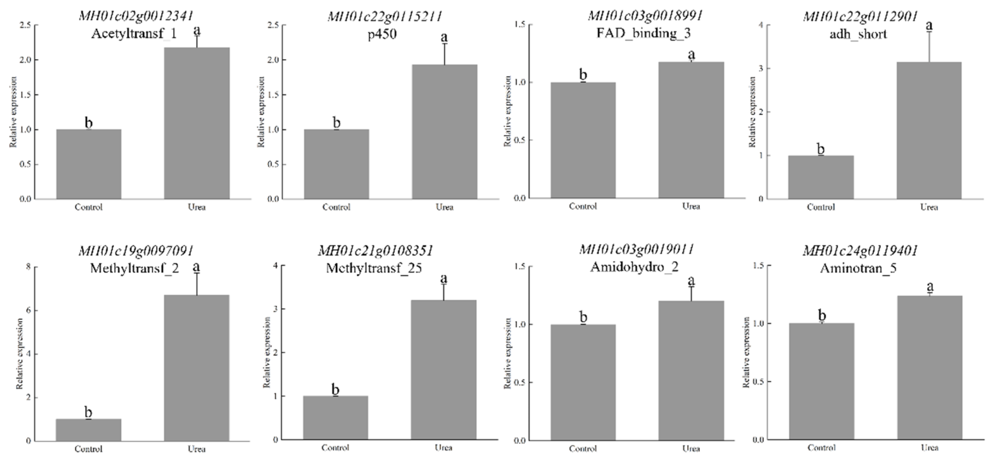
The investigation was extended to elucidate how urea influences the genetic framework underlying HA biosynthesis and transport in S. bambusicola GDMCC 60438. Beyond its role in enhancing acetyl-CoA synthesis, urea was found to upregulate genes involved in ergosterol synthesis, including those encoding enzymes such as acetyl-CoA C-acetyltransferase, hydroxymethylglutaryl-CoA synthase, and squalene monooxygenase, resulting in increased ergosterol levels [21]. This regulatory effect suggests that urea might similarly stimulate HA synthesis by modulating key enzyme-encoding genes in the pathway. Critical enzymes in HA biosynthesis, such as polyketide synthase (PKS), monooxygenase, O-methyltransferase, FAD/FMN-dependent oxidoreductase, and multicopper oxidase, were pinpointed. Notably, PKS encompasses various functional domains integral to this biosynthetic process [45,46,47,48,49]. A spectrum of genes encoding these essential enzymes, including seven PKS or PKS-domain-containing genes, were identified among the upregulated DEGs, highlighting urea’s significant impact on HA biosynthetic machinery. Furthermore, genes encoding regulatory proteins, such as calmodulin and zinc finger transcription factors, known for their roles in HA biosynthesis control, were identified [50]. The presence of two ABC transporter genes and nine major facilitator superfamily (MFS) genes among the upregulated DEGs suggests urea’s potential effect on HA-transport mechanisms, indicating an integrated influence on both synthesis and mobilization of HA [51]. To corroborate the transcriptomic insights, qRT-PCR analyses were conducted on select genes associated with HA synthesis and urea regulation. The outcomes from qRT-PCR revealed significant upregulation in the urea-treated samples, aligning with the RNA-seq data and affirming the regulatory role of urea in enhancing HA-production pathways (Figure 7). The enzyme Acetyltransf_1 (MH01c02g0012341), associated with PKS, was upregulated, with its relative expression level being twice its level in the control group. Also, urea upregulated the P450 gene (MH01c22g0115211), with a relative expression level reaching twice of its level in the control group. This finding is consistent with our previous research conclusions indicating that upregulation of the P450 gene promotes HA synthesis [51]. The gene FAD_binding_3 belongs to the monooxygenase domain and is associated with HA biosynthesis [41]. Additionally, Methyltransf_2 (MH01c19g0097091) and Methyltransf_25 (MH01c21g0108351), belonging to the O-methyltransferase domain, are key enzymes in HA biosynthesis [42]. Urea upregulated Methyltransf_2 and Methyltransf_25, with their relative expression levels being four times and three times higher than their levels in the control group, respectively. This comprehensive gene-expression analysis underscores urea’s multifaceted role in augmenting HA biosynthesis and transport. By upregulating genes encoding critical enzymes and regulatory proteins, urea not only supports the metabolic processes leading to HA production, but also potentially facilitates its transport, establishing a foundation for optimizing HA yield in microbial fermentation processes. The results provide detailed insights into the specific enzymatic factors involved in HA biosynthesis, highlighting roles for enzymes such as FAD-dependent oxidoreductase, O-methyltransferase, and cytochrome P450. The upregulation of genes encoding these enzymes in response to urea supplementation underscores urea’s regulatory role in the HA synthesis pathway. These findings deepen our understanding of how urea modulates metabolic pathways to enhance HA production in fungi.
The comprehensive analysis presented across various studies emphasizes urea’s integral role in fungal metabolism and the biosynthesis of significant metabolites like HA. By acting as a potent modulator of metabolic pathways, urea offers valuable mechanisms for optimizing industrial fermentation processes and improving metabolite production efficiency. These findings hold promise for industries seeking to enhance their production processes and explore new avenues for metabolite synthesis.

4. Conclusions

The inclusion of yeast-extract powder supplemented with urea was found to facilitate HA synthesis in S. bambusicola GDMCC 60438. Urea suppressed key enzymes in the TCA cycle while enhancing the conversion of amino acids to acetyl-CoA. This process increased the availability of precursor molecules necessary for HA synthesis. Furthermore, gene expression crucial for HA biosynthesis was upregulated by urea supplementation. These results suggest potential strategies for enhancing HA yield in fungal fermentation processes, which are of significant interest to the biotechnology and pharmaceutical industries due to the extensive applications of HA.

Supplementary Materials

The following supporting information can be downloaded at: https://www.mdpi.com/article/10.3390/fermentation10080381/s1, Table S1. List of forward and reverse primers utilized in qRT–PCR analyses. Table S2. Genes related to HA biosynthesis. Table S3. Pfam annotations of unigenes in S. bambusicola GDMCC 60438. Table S4. Expression profiles of differentially expressed genes (DEGs).

Author Contributions

Conceptualization, Y.W. and Y.T.; methodology, Y.W.; investigation, Y.W. and Y.T.; resources, X.T.; data curation, X.Z. and Q.G.; writing—original draft preparation, Y.W. and Y.T.; writing—review and editing, X.T. and F.Y.; supervision, X.T. and Z.W. All authors have read and agreed to the published version of the manuscript.

Funding

This research was supported by the Fund of Zhuhai Science and Technology Program in Social Development Area, Guangdong (2220004000339), and Yunnan Key Laboratory for Fungal Diversity and Green Development, China (2023).

Institutional Review Board Statement

Not applicable.

Informed Consent Statement

Not applicable.

Data Availability Statement

Data are contained within the article and Supplementary Materials.

Acknowledgments

The authors would acknowledge the technical support from Lining Wang, Institute of Biological and Medical Engineering, Guangdong Academy of Sciences, China.

Conflicts of Interest

The authors declare no conflicts of interest.

References

  1. Ali, S.M.; Olivo, M. Efficacy of hypocrellin pharmacokinetics in phototherapy. Int. J. Oncol. 2002, 21, 1229–1237. [Google Scholar] [CrossRef] [PubMed]
  2. Morakotkarn, D.; Kawasaki, H.; Seki, T. Molecular diversity of bamboo-associated fungi isolated from Japan. FEMS Microbiol. Lett. 2007, 266, 10–19. [Google Scholar] [CrossRef]
  3. Cheng, T.; Jia, X.; Ma, X.; Lin, H.; Zhao, Y. Phylogenetic study on Shiraia bambusicola by rDNA sequence analyses. J. Basic Microbiol. 2004, 44, 339–350. [Google Scholar] [CrossRef] [PubMed]
  4. Yang, H.; Xiao, C.; Ma, W.; He, G. The production of hypocrellin colorants by submerged cultivation of the medicinal fungus Shiraia bambusicola. Dyes Pigment. 2009, 82, 142–146. [Google Scholar] [CrossRef]
  5. Estey, E.P.; Brown, K.; Diwu, Z.; Liu, J.; Lown, J.W.; Miller, G.G.; Moore, R.B.; Tulip, J.; Mcphee, M.S. Hypocrellins as photosensitizers for photodynamic therapy: A screening evaluation and pharmacokinetic study. Cancer Chemother. Pharmacol. 1996, 37, 343–350. [Google Scholar] [CrossRef]
  6. Zhang, J.; Cao, E.H.; Li, J.F.; Zhang, T.C.; Ma, W.J. Photodynamic effects of hypocrellin A on three human malignant cell lines by inducing apoptotic cell death. J. Photochem. Photobiol. B-Biol. 1998, 43, 106–111. [Google Scholar] [CrossRef] [PubMed]
  7. Zhang, L.Z.; Tang, G.; Gao, B.; Zhang, G. Spectroscopic studies on the excited-state properties of the light-induced antiviral drug hypocrellin A loaded in the mesoporous solid. Chem. Phys. Lett. 2004, 396, 102–109. [Google Scholar] [CrossRef]
  8. Hudson, J.B.; Zhou, J.; Chen, J.; Harris, L.; Yip, L.; Towers, G.H. Hypocrellin, from Hypocrella bambuase, is phototoxic to human immunodeficiency virus. Photochem. Photobiol. 1994, 60, 253–255. [Google Scholar] [CrossRef]
  9. Morgan, B.J.; Mulrooney, C.A.; O’Brien, E.M.; Kozlowski, M.C. Perylenequinone natural products: Total syntheses of the diastereomers (+)-phleichrome and (+)-calphostin D by assembly of centrochiral and axial chiral fragments. J. Org. Chem. 2010, 75, 30–43. [Google Scholar] [CrossRef]
  10. O’Brien, E.M.; Morgan, B.J.; Mulrooney, C.A.; Carroll, P.J.; Kozlowski, M.C. Perylenequinone natural products: Total synthesis of hypocrellin A. J. Org. Chem. 2010, 75, 57–68. [Google Scholar] [CrossRef]
  11. Lei, X.Y.; Zhang, M.Y.; Ma, Y.J.; Wang, J.W. Transcriptomic responses involved in enhanced production of hypocrellin A by addition of Triton X-100 in submerged cultures of Shiraia bambusicola. J. Ind. Microbiol. Biotechnol. 2017, 44, 1415–1429. [Google Scholar] [CrossRef] [PubMed]
  12. Yan, R.; Zhang, Z.; Wang, Y.; Yang, H.; Zeng, Q.; Zhu, D. Efficient strategy for maintaining and enhancing the huperzine A production of Shiraia sp. Slf14 through inducer elicitation. J. Ind. Microbiol. Biotechnol. 2014, 41, 1175–1179. [Google Scholar] [CrossRef] [PubMed]
  13. Li, D.; Zhao, N.; Guo, B.; Lin, X.; Chen, S.; Yan, S. Gentic overexpression increases production of hypocrellin A in Shiraia bambusicola S4201. J. Microbiol. 2019, 57, 154–162. [Google Scholar] [CrossRef] [PubMed]
  14. Li, X.P.; Ma, Y.J.; Wang, J.W. Adding bamboo charcoal powder to Shiraia bambusicola preculture improves hypocrellin A production. Sustain. Chem. Pharm. 2019, 14, 100191. [Google Scholar] [CrossRef]
  15. Liu, X.Y.; Fan, L.; Gao, J.; Shen, X.Y.; Hou, C.L. Global identification of alternative splicing in Shiraia bambusicola and analysis of its regulation in hypocrellin biosynthesis. Appl. Microbiol. Biotechnol. 2020, 104, 211–223. [Google Scholar] [CrossRef] [PubMed]
  16. Gao, R.; Deng, H.; Guan, Z.; Liao, X.; Cai, Y. Enhanced hypocrellin production via coexpression of alpha-amylase and hemoglobin genes in Shiraia bambusicola. AMB Express 2018, 8, 71. [Google Scholar] [CrossRef] [PubMed]
  17. Ma, Y.J.; Sun, C.X.; Wang, J.W. Enhanced Production of Hypocrellin A in Submerged Cultures of Shiraia bambusicola by Red Light. Photochem. Photobiol. 2019, 95, 812–822. [Google Scholar] [CrossRef] [PubMed]
  18. Sun, C.X.; Ma, Y.J.; Wang, J.W. Enhanced production of hypocrellin A by ultrasound stimulation in submerged cultures of Shiraia bambusicola. Ultrason. Sonochem. 2017, 38, 214–224. [Google Scholar] [CrossRef] [PubMed]
  19. Cai, Y.; Liang, X.; Liao, X.; Ding, Y.; Sun, J.; Li, X. High-Yield Hypocrellin A Production in Solid-State Fermentation by Shiraia sp. SUPER-H168. Appl. Biochem. Biotechnol. 2010, 160, 2275–2286. [Google Scholar] [CrossRef]
  20. Dos Santos, A.; Santos, E.M.; de Oliveira, J.S.; Ribeiro, O.L.; Perazzo, A.F.; Pinho, R.; Macedo, A.; Pereira, G.A. Effects of urea addition on the fermentation of sorghum (Sorghum bicolor) silage. Afr. J. Range Forage Sci. 2018, 35, 55–62. [Google Scholar] [CrossRef]
  21. Su, Q.; Zhang, Z.; Liu, X.; Wang, F. The transcriptome analysis on urea response mechanism in the process of ergosterol synthesis by Cordyceps cicadae. Sci. Rep. 2021, 11, 10927. [Google Scholar] [CrossRef]
  22. Xiang, X. Optimization of Shiraia bambusicola P. Henn. under liquid fermentatio. J. Biotechnol. 2010, 20, 73–75. [Google Scholar]
  23. Wechgama, K.; Laopaiboon, L.; Laopaiboon, P. Enhancement of batch butanol production from sugarcane molasses using nitrogen supplementation integrated with gas stripping for product recovery. Ind. Crop Prod. 2017, 95, 216–226. [Google Scholar] [CrossRef]
  24. Deng, H.; Gao, R.; Liao, X.; Cai, Y. Characterisation of a monooxygenase in Shiraia bambusicola. Microbiology 2018, 164, 1180–1188. [Google Scholar] [CrossRef] [PubMed]
  25. Deng, H.; Gao, R.; Liao, X.; Cai, Y. Characterization of a major facilitator superfamily transporter in Shiraia bambusicola. Res. Microbiol. 2017, 168, 664–672. [Google Scholar] [CrossRef] [PubMed]
  26. Sitanggang, A.B.; Wu, H.; Wang, S.S.; Ho, Y. Effect of pellet size and stimulating factor on the glucosamine production using Aspergillus sp. BCRC 31742. Bioresour. Technol. 2010, 101, 3595–3601. [Google Scholar] [CrossRef]
  27. Sun, C.X.; Ma, Y.J.; Wang, J.W. Improved hypocrellin A production in Shiraia bambusicola by light-dark shift. J. Photochem. Photobiol. B-Biol. 2018, 182, 100–107. [Google Scholar] [CrossRef] [PubMed]
  28. Ma, Y.J.; Li, X.P.; Wang, Y.; Wang, J.W. Nitric oxide donor sodium nitroprusside-induced transcriptional changes and hypocrellin biosynthesis of Shiraia sp. S9. Microb. Cell Fact. 2021, 20, 92. [Google Scholar] [CrossRef]
  29. Ma, Y.J.; Zheng, L.P.; Wang, J.W. Inducing perylenequinone production from a bambusicolous fungus Shiraia sp. S9 through co-culture with a fruiting body-associated bacterium Pseudomonas fulva SB1. Microb. Cell Fact. 2019, 18, 121. [Google Scholar] [CrossRef]
  30. Du, W.; Sun, C.; Wang, B.; Wang, Y.; Dong, B.; Liu, J.; Xia, J.; Xie, W.; Wang, J.; Sung, J.; et al. Response mechanism of hypocrellin colorants biosynthesis by Shiraia bambusicola to elicitor PB90. AMB Express 2019, 9, 146. [Google Scholar] [CrossRef]
  31. Klis, F.M.; Boorsma, A.; De Groot, P.W.J. Cell wall construction in Saccharomyces cerevisiae. Yeast 2006, 23, 185–202. [Google Scholar] [CrossRef]
  32. Pohling, J.; Hawboldt, K.; Dave, D. Comprehensive review on pre-treatment of native, crystalline chitin using non-toxic and mechanical processes in preparation for biomaterial applications. Green Chem. Int. J. Green Chem. Resour. 2022, 24, 6790–6809. [Google Scholar] [CrossRef]
  33. Suzuki, K.; Tokoro, A.; Okawa, Y.; Suzuki, S.; Suzuki, M. Effect of N-acetylchito-oligosaccharides on activation of phagocytes. Microbiol. Immunol. 1986, 30, 777–787. [Google Scholar] [CrossRef] [PubMed]
  34. Wessels, J.G.H.U. Developmental regulation of fungal cell wall formation. Annu. Rev. Phytopathol. 1994, 32, 413–437. [Google Scholar] [CrossRef]
  35. Navarathna, D.H.M.L.; Harris, S.D.; Roberts, D.D.; Nickerson, K.W. Evolutionary aspects of urea utilization by fungi. FEMS Yeast Res. 2010, 10, 209–213. [Google Scholar] [CrossRef]
  36. Strope, P.K.; Nickerson, K.W.; Harris, S.D.; Moriyama, E.N. Molecular evolution of urea amidolyase and urea carboxylase in fungi. BMC Evol. Biol. 2011, 11, 80. [Google Scholar] [CrossRef]
  37. Fan, C.; Glibert, P.M.; Alexander, J.; Lomas, M.W. Characterization of urease activity in three marine phytoplankton species, Aureococcus anophagefferens, Prorocentrum minimum, and Thalassiosira weissflogii. Mar. Biol. 2003, 142, 949–958. [Google Scholar] [CrossRef]
  38. Solomon, C.M.; Glibert, P.M. Urease activity in five phytoplankton species. Aquat. Microb. Ecol. 2008, 52, 149–157. [Google Scholar] [CrossRef]
  39. Smith, D.G.; Russell, W.C.; Thirkell, D. Urea-hydrolysis-dependent citrulline synthesis by Ureaplasma urealyticum. FEMS Microbiol. Lett. 1992, 77, 129–131. [Google Scholar] [CrossRef] [PubMed]
  40. Fernie, A.R.; Carrari, F.; Sweetlove, L.J. Respiratory metabolism: Glycolysis, the TCA cycle and mitochondrial electron transport. Curr. Opin. Plant Biol. 2004, 7, 254–261. [Google Scholar] [CrossRef]
  41. Zhang, S.; Juan, H. Functional Characterization of Citrate Carrier in the Biosynthesis of Juvenile Hormone in Cockroach. Bioprocess 2017, 7, 1–7. [Google Scholar] [CrossRef]
  42. Brito, D.S.; Agrimi, G.; Charton, L.; Brilhaus, D.; Bitetto, M.G.; Lana-Costa, J.; Messina, E.; Nascimento, C.P.; Feitosa-Araujo, E.; Pires, M.V.; et al. Biochemical and functional characterizationof a mitochondrialcitrate carrier in Arabidopsis thaliana. Biochem. J. 2020, 477, 1759–1777. [Google Scholar] [CrossRef] [PubMed]
  43. Strijbis, K.; Distel, B. Intracellular Acetyl Unit Transport in Fungal Carbon Metabolism. Eukaryot. Cell 2010, 9, 1809–1815. [Google Scholar] [CrossRef] [PubMed]
  44. Vorapreeda, T.; Thammarongtham, C.; Cheevadhanarak, S.; Laoteng, K. Alternative routes of acetyl-CoA synthesis identified by comparative genomic analysis: Involvement in the lipid production of oleaginous yeast and fungi. Microbiology 2012, 158, 217–228. [Google Scholar] [CrossRef]
  45. Zhao, X.; Wang, Z.; Shu, S.; Wang, W.; Xu, H.; Ahn, Y.; Wang, M.; Hu, X. Ethanol and Methanol Can Improve Huperzine A Production from Endophytic Colletotrichum gloeosporioides ES026. PLoS ONE 2013, 8, e61777. [Google Scholar] [CrossRef] [PubMed]
  46. Deng, H.; Gao, R.; Liao, X.; Cai, Y. Reference genes selection and relative expression analysis from Shiraia sp. SUPER-H168 productive of hypocrellin. Gene 2016, 580, 67–72. [Google Scholar] [CrossRef] [PubMed]
  47. Deng, H.; Gao, R.; Liao, X.; Cai, Y. Genome editing in Shiraia bambusicola using CRISPR-Cas9 system. J. Biotechnol. 2017, 259, 228–234. [Google Scholar] [CrossRef] [PubMed]
  48. Jia, Y.; Du, F.; Nong, F.; Li, J.; Huang, P.; Ma, W.; Gu, Y.; Sun, X. Function of the Polyketide Synthase Domains of Schizochytrium sp. on Fatty Acid Synthesis in Yarrowia lipolytica. J. Agric. Food Chem. 2023, 71, 2446–2454. [Google Scholar] [CrossRef] [PubMed]
  49. Wen, Y.; Liao, B.; Yan, X.; Wu, Z.; Tian, X. Temperature-responsive regulation of the fermentation of hypocrellin A by Shiraia bambusicola (GDMCC 60438). Microb. Cell Fact. 2022, 21, 135. [Google Scholar] [CrossRef]
  50. Zhao, N.; Li, D.; Guo, B.; Tao, X.; Lin, X.; Yan, S.; Chen, S. Genome Sequencing and Analysis of the Hypocrellin-Producing Fungus Shiraia bambusicola S4201. Front. Microbiol. 2020, 11, 15. [Google Scholar] [CrossRef]
  51. Li, X.P.; Wang, Y.; Ma, Y.J.; Wang, J.W.; Zheng, L.P. Nitric Oxide and Hydrogen Peroxide Signaling in Extractive Shiraia Fermentation by Triton X-100 for Hypocrellin A Production. Int. J. Mol. Sci. 2020, 21, 882. [Google Scholar] [CrossRef] [PubMed]
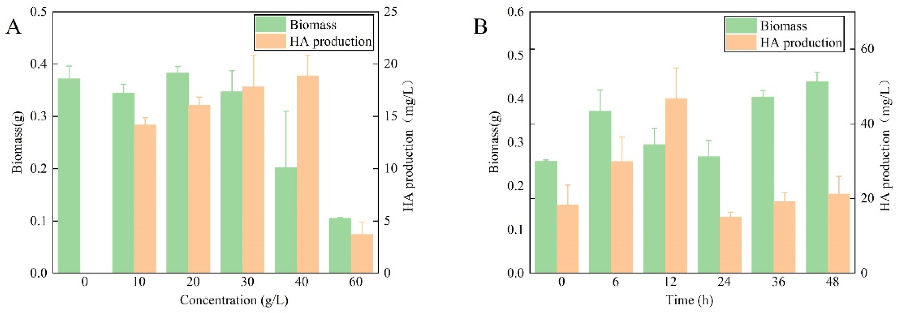
Figure 1. Effects of urea supplementation on growth and HA yield in S. bambusicola (GDMCC 60438). (A) Urea concentration; (B) Timing of urea addition to a concentration of 40 g/L.
Fermentation 10 00381 g001
Figure 2. Variations in diameter and morphology of mycelial pellets in S. bambusicola (GDMCC 60438) throughout fermentation.
Fermentation 10 00381 g002
Figure 3. Surface morphology of mycelial pellets in S. bambusicola with and without urea supplementation. Arrows indicate the region where fungal hyphae are entwined. Morphology without urea supplementation, scale bar = (A) 10 μm, or (B) 2 μm; Morphology with urea supplementation, scale bar = (C) 10 μm, or (D) 2 μm.
Fermentation 10 00381 g003
Figure 4. Cluster analysis and functional enrichment of differentially expressed genes (DEGs). (A) Cluster analysis; (B) KEGG pathway enrichment of up-regulated genes; (C) GO enrichment of up-regulated genes; (D) GO enrichment of down-regulated genes.
Fermentation 10 00381 g004
Figure 5. Illustration of interactions between the TCA cycle and the citrate−pyruvate cycle.
Fermentation 10 00381 g005
Figure 6. Variation in acetyl-CoA concentration in the mycelium of S. bambusicola (GDMCC 60438) following urea addition at 12 h to a concentration of 40 g/L in the fermentation broth. * and ** indicate a significant difference in the acetyl-CoA content within the mycelia between the control group and the urea group at the 20-h fermentation mark.
Fermentation 10 00381 g006
Figure 7. The qRT-PCR analysis for identifying genes involved in HA synthesis and urea regulation. Significant differences are indicated by distinct letters above the bars (p < 0.05).
Fermentation 10 00381 g007
Table 1. Pfam annotation of urea-regulated genes in DEGs.
Pfam IDPfam AnnotationGene ID
PF04909.13Amidohydrolase domain of ureaseMH01c03g0019011
PF02666.14Phosphatidylserine decarboxylaseMH01c07g0053961
PF00155.20Aminotransferase class I and IIMH01c20g0105931
PF00266.18Domain of amino transferase or cysteine desulphuraseMH01c24g0119401
PF01179.19Copper amine oxidaseMH01c24g0120431
PF04952.13Succinylglutamate desuccinylase/AspartoacylaseMH01c02g0010871, MH01c02g0010881
PF02786.16Carbamoyl-phosphate synthase LMH01c03g0020161,
chainMH01c09g0066391
PF02629.18Succinyl CoA synthetaseMH01c07g0049071
PF00549.18ATP-citrate lyaseMH01c07g0049071
PF00285.20Citrate synthaseMH01c07g0049071
PF03949.14Malic enzymeMH01c12g0075031
PF00682.18Pyruvate carboxylaseMH01c13g0083471
PF17763Glutaminase/asparaginaseMH01c18g0092341
Disclaimer/Publisher’s Note: The statements, opinions and data contained in all publications are solely those of the individual author(s) and contributor(s) and not of MDPI and/or the editor(s). MDPI and/or the editor(s) disclaim responsibility for any injury to people or property resulting from any ideas, methods, instructions or products referred to in the content.