Article

Eriobotrya japonica Fermentation with Plant-Derived Lactiplantibacillus plantarum MSC-5T Ameliorates Antioxidant Activity in HEK293 Cells

1
Department of Probiotic Science for Preventive Medicine, Graduate School of Biomedical and Health Sciences, Hiroshima University, Hiroshima 734-8551, Japan
2
Mitsui DM Sugar Co., Ltd., 2-28-7, Kamiochiai, Shinjuku-ku, Tokyo 161-0034, Japan
3
Department of Pharmacognosy, Graduate School of Biomedical and Health Sciences, Hiroshima University, Hiroshima 734-8551, Japan
*
Author to whom correspondence should be addressed.
Fermentation 2024, 10(4), 197; https://doi.org/10.3390/fermentation10040197
Submission received: 22 March 2024 / Revised: 31 March 2024 / Accepted: 31 March 2024 / Published: 2 April 2024
(This article belongs to the Special Issue Nutrition and Health of Fermented Foods, 3rd Edition)

Abstract

:
Oxidative stress from an excess of radical compounds generally impacts apoptosis and inflammation. The use of probiotics, therefore, has emerged as a favorable tool to suppress the formation of reactive oxygen species. In the present study, we investigated the antioxidant activity of plant-derived Lactiplantibacillus (L.) plantarum MSC-5T fermented in Eriobotrya japonica (EJ) aqueous extract. In the in vitro study, the extract fermented with the MSC-5T strain markedly decreased the cell death of H2O2-induced HEK293 cells. In addition, the fermented extract showed a protective effect against fungal toxin ochratoxin A (OTA) and citrinin (CTN). Regarding the evaluation of glutathione homeostasis, it can be clearly seen that pretreatment of HEK293 cells with fermented EJ extract greatly increased glutathione (GSH) levels, while unfermented extract did not affect the cellular GSH content. Furthermore, we identified a bioactive compound as pyrocatechol, which displayed significant antioxidation activity. The extract fermented for 48 h with the MSC-5T strain in EJ extract produces 167.4 μg/mL pyrocatechol.

1. Introduction

Probiotics are non-pathogenic microorganisms that, when administered in adequate amounts, provide health benefits for the host. It has been suggested that probiotics help optimize the health of the human digestive tract, inhibit the growth of harmful bacteria, and promote healthy digestion and nutrient absorption [1,2]. Lactic acid bacteria (LAB) are a genus of Gram-positive anaerobic inhabitants that are found in a wide variety of habitats, including vegetation and the gastrointestinal tracts of animals [3]. Among them, numerous mesophilic strains have been isolated from plants and showed higher adaptation characteristics for withstanding harsh conditions. Moreover, as demonstrated in previous studies, plant-derived LABs often have genome-encoding enzymes that convert pentoses, cellobiose, sucrose, and malic and citric acids into other compounds [4]. Our research group has isolated more than 1000 strains of plant-derived LAB strains from different plant sources, and several of these strains have demonstrated health benefits, such as immune modulation, the improvement of liver function, and the reduction of obesity [5,6,7].
Reactive oxygen species (ROS) is a general term for highly reactive oxygen species, such as superoxide (O2), hydrogen peroxide (H2O2), and hydroxyl radical (•OH). The body’s immune system deliberately produces ROS that have specialized functions, such as phagocytosis, antigen presentation, activation, cytolysis, and differentiation. However, the excessive production of ROS causes oxidative damage to various biomolecules, such as proteins and membrane lipids, and, consequently, becomes a factor leading to diseases, such as cancer, cardiovascular disease, and lifestyle-related diseases [8,9]. Given the link between ROS and inflammasome activation, it is unsurprising that the suppression of excess ROS in the body has emerged as an attractive strategy for preventing the medical conditions mentioned above. It is well known that herbal medicines, traditionally made from plant parts existing in nature, are essential for suppressing ROS generation, treating disease, enhancing general health, and balancing the immune system during chronic infection, as well as preventing dysbiosis-associated disorders. Medicinal plants’ bioactive compounds lie in alkaloids, phenolics, and glycosides, as well as flavonoids. On the other hand, the use of LABs to enhance the bioactivity of these medicinal plant remedies has been gaining favorable attention recently. In our previous experimental studies, the L. plantarum SN13T strain isolated from the banana leaf significantly increased the bioactivity of Mentha extract, and it also showed a strong ability to inhibit the release of interleukin (IL)-8 from HuH-7 cells when cultivated in Artemisia princeps Pampanini extract [10]. Furthermore, apple pomace fermented with L. plantarum KKP 1527 showed 30% higher content of the total phenolic compounds and a higher value of antioxidant activity than that of non-fermented apple pomace extracts [11]. In addition, the fermentation of Scrophularia buergeriana extract with L. brevis and L. helveticus strains inhibited the production of nitric oxide (NO) and reduced the expression of RNA for INOS, IL-1β, IL-6, TNF-α, and COX-2 in lipopolysaccharide-treated RAW264.7 cells [12].
Eriobotrya japonica Lindl. (E. japonica) is a widely studied medicinal plant used to treat cough resulting from pulmonary inflammation, stomach disorder-induced nausea, restlessness, and thirst [13]. As described by Zhu et al., the leaves of E. japonica contain 164 compounds, which are classified into triterpenes, flavonoids, sesquiterpene glycosides, megastigmane derivatives, phenylpropanoids and organic acids, and 169 volatile oils [14]. There have been numerous studies also showing the antitumor, antiviral, antioxidant, and anti-inflammatory activities of the E. japonica extract. Described by Khouya et al., the aqueous extract of leaf material was abundant with naringenin, procyanidin, epicatechin, and rutin, and the extract prevented hyperglycemia, hyperlipidemia, and oxidative stress in high-fat diet-fed mice [15]. Moreover, the procyanidin oligomer mixture of water-soluble fractions has been reported to have cytotoxic activity against human oral tumor cell lines [16]. Nevertheless, several compounds of E. japonica are considered to have antioxidant properties, and the protective mechanism of these compounds against free radicals is probably synergetic, thereby enhancing antioxidant and anti-inflammatory properties.
Despite the certain antioxidant capacities, dietary flavonoids almost exist as their glycosylated form, which, in turn, results in lower bioavailability. Thus, bacterial fermentation, especially with the LAB strains, can have profound effects on radical scavenging function if the flavonoid aglycones are generated by the action of LAB glucosidase enzymes. For this purpose, we preliminarily screened fourteen plant-derived LAB strains fermented with different combinations of plant extracts (41 medicinal plant extract) for their antioxidant activity. As a result, we further conducted research on E. japonica (EJ) extract fermented with L. plantarum MSC-5T strain isolated from sugar cane and focused on the determination of the substances responsible for the antioxidant activity.

2. Materials and Methods

2.1. Isolation and Identification of the LAB

MRS medium (Becton, Dickinson and Company, Franklin Lakes, NJ, USA) was used to isolate and maintain the bacterial culture. Sugarcane samples were obtained from Kikaijima Island, Kagoshima Prefecture; and slices of stem, leaf, and roof parts were suspended in an MRS medium. After cultivation at 28 or 37 °C, the culture of each isolate was spread on the MRS agar medium and incubated anaerobically at the given temperature for 2–3 days. Gram-staining, catalase activity, and CO2 gas production tests were performed on the purified colony. A Gram-staining test was performed using the Bermy M stain kit (Muto Pure Chemical Co., Ltd., Tokyo, Japan), and the cell morphology was observed under a microscope. Catalase activity was determined by a bubble test, using 3% hydrogen peroxide. The fermentation potential of various sugars was confirmed using the API 50 CHL kit (bioMerieux, Craponne, France) according to the manufacturer’s instructions.
The isolated strain was identified by using the entire 16S ribosomal DNA (rDNA) sequence analysis. For PCR, the genomic DNA was isolated from the LAB strain using a GenElute™ Bacterial Genomic DNA Kit (Sigma Aldrich, St. Louis, MO, USA). The 16S rDNA gene fragment was amplified by PCR, using 27f (5′-AGAGTTTGATCCTGGCTAG-3′) and 1525r (5′-AAAGGAGGTGATCCAGCC-3′) primers. The PCR products were purified by using NucleoSpin Gel and PCR Clean-Up Kit (Macherey-Nagel GmbH and Co. KG, Duren, Germany), and the sequence of the fragment was determined using the method described previously [17,18]. Sequence similarity searches were performed using the Basic Local Alignment Search Tool (BLAST) online at the National Centre for Biotechnology Information (NCBI) homepage (http://blast.ncbi.nlm.nih.gov/Blast.cgi; accessed on 17 April 2022).

2.2. Fermentation Condition of the MSC-5T Strain in EJ Extract

L. plantarum MSC-5T strain was grown at 28 °C, overnight, in MRS broth (Merck, Germany). After cultivation, the bacterial cells were collected by centrifugation and resuspended with sterile 0.85% (w/v) NaCl solution.
Ten grams of E. japonica (10 g) dried leaf (Kojima Kampo Co., Ltd., Osaka, Japan) was suspended in 100 mL of distilled water and boiled for 30 min. After cooling to room temperature, the EJ extract was obtained by centrifugation at 5000× g for 10 min and then filtrated with a 0.45 µm membrane filter (Advantec Toyo Co., Ltd., Tokyo, Japan). This filtrate was stored at 4 °C and was used further as 10% of EJ extract for all the experiments.
The overnight seed culture (9.94 log CFU/mL) of the MSC-5T strain in MRS broth was collected by centrifugation and resuspended with the same amount of sterile 0.85% (w/v) NaCl solution. This bacterial suspension was inoculated at a final 1% v/v in the EJ extract. After 12, 24, 48, and 72 h of incubation at 28 °C, cells were harvested by centrifugation at 5000× g for 10 min, and each resulting supernatant was sterilized through a 0.45 μm filter membrane.

2.3. Cell Culture and Treatment

The human embryonic kidney HEK293 cell line was seeded at 1.0 × 10⁵ cells/well in 24-well plates in DMEM medium supplemented with 10% FBS and maintained at 37 °C in a CO2 incubator. The cells were preincubated for 3 h with various samples and then treated for up to 24 h with 0.3 mM of freshly prepared H2O2, 5 μM of ochratoxin A (OTA), or 75 μM citrinin (CTN). Cytotoxicity was analyzed using a CCK-8 kit (Dojindo Laboratories, Kumamoto, Japan) in accordance with the manufacturer’s instructions. Briefly, CCK-8 solution (10 μL) was added to each well, and the cells were incubated for 2 h. Then, the absorbance at 450 nm was measured. The percentage of cell viability was determined as a value relative to that of untreated cells. The experiments were carried out in triplicate.

2.4. Measurement of Intracellular ROS Levels

The HEK293 cells were treated with H2O2, OTA, or CTN in the presence or absence of the extracts for 24 h. The cells then were incubated with 10 mM DCFH-DA for 30 min at 37 °C and washed twice with phosphate-buffered saline (PBS). Subsequently, the DCF fluorescence was measured at excitation and emission wavelengths of 485 and 530 nm (Varioskan, Thermo Scientific, Vantaa, Finland), respectively.

2.5. Preparation of the Cell Extract

The HEK293 cells treated with H2O2, OTA, or CTN in the presence or absence of the extracts or catechol for 24 h were harvested by centrifugation at 200× g for 4 min. Cell pellets were washed with PBS and lysed by sonication (BioRuptor, SonicBio, Kanagawa, Japan) on ice for 5 min, with pulsing at 10 s on and 5 s off cycles, and centrifuged at 12,000× g for 10 min to obtain the cell-free extract. Protein contents were determined using Bio-Rad Protein Assay (Bio-Rad, Hercules, CA, USA), based on the method of Bradford.

2.6. Quantification of Intracellular GSSH Levels

For the measurement of reduced glutathione (GSH) or oxidized glutathione (GSSG), a GSH/GSSG Quantitative Kit was used following the manufacturer’s instructions (Dojindo Molecular Technologies, Inc., Kumamoto, Japan).

2.7. Quantification of Caspase-3 Activity

To detect caspase-3 activity, the HEK293 cell extract (1 mg/mL in PBS) was incubated with fluorogenic peptide substrates, Ac-Asp-Glu-Val-Asp-amido-4-methyl-coumarin (Ac-DEVD-AFC, AAT Bioquest), for 3 h at 28 °C. Then, the fluorescence emission of the 7-amino-4-trifluoromethyl-coumarin (AFC), released on proteolytic cleavage of the fluorogenic substrate DEVD-AFC by active caspase-3, was measured using a Varioskan plate reader with a 380 nm excitation wavelength and a 500 nm emission wavelength.

2.8. Extraction and Identification of an Antioxidant Compound Produced in the Fermented EJ Extract

The fermented EJ broth was extracted three times with the same volume of ethyl acetate. After salting with anhydrous magnesium sulfate, the ethyl acetate fraction was dried at 50 °C under vacuum on a rotary evaporator. The residue was solved in ethyl acetate and applied to the silica gel column chromatography (chloroform/methanol from 10:1 to 0:10), and Fraction 4 was further purified by Sephadex LH-20 column chromatography with methanol. The active Fraction 3 was further purified by HPLC (JASCO system; JASCO Corporation, Tokyo, Japan) with an ODS-A (5 µm, 30 nm, Φ = 3 mm, L = 150 mm) column (YMC, Kyoto, Japan). The column was equilibrated with water containing 0.1% trifluoracetic acid, and gradient elution was performed with 0% to 40%, and 40 to 60% acetonitrile over 20 min and 10 min sequentially at a flow rate of 1 mL/min. 13C-NMR spectra of the isolated compound were taken on a JEOL JNM-LA500 spectrometer at 125 MHz at the Natural Science Center for Basic Research and Development (N-BARD), Hiroshima University. Spectroscopic data of pyrocatechol: 13C-NMR (Methanol-d4, 150 MHz): 116.41, 120.92, 146.34.

2.9. Statistical Analysis

All data were presented as the means and SD of triplicates. The significance of differences was determined via ANOVA, followed by a post hoc Tukey test, and differences with p < 0.05 were considered statistically significant.

3. Results and Discussion

3.1. The Characterization of MSC-5T Strain

We isolated and identified MSC-5T strain from sugarcane, in the present study. It was Gram-positive and was found to be negative for the catalase test. The partial 16S rRNA gene was amplified, and, subsequently, an NCBI BLAST analysis was performed. The isolate shared 99.93% 16S rRNA gene sequence similarity by BLAST and belonged to L. plantarum. Further, the profile of carbohydrate utilization, evaluated using the API 50 CHL test, demonstrated that monosaccharides metabolized by MSC-5T include D-galactose, D-fructose, D-glucose, D-mannose, D-lactose, and N-acetyl glucosamine. The utilization of disaccharide and polysaccharide by MSC-5T might indicate that it produces hydrolytic enzymes responsible for the cleavage of various sugars (α-glucosidase for trehalose, sucrose, melezitose, raffinose and maltose; and β-glucosidase for cellobiose, turanose, and d-melibiose) and also utilizes sugar alcohols (amygdalin, salicin, sorbitol, and mannitol). Carbon sources such as cellobiose and amygdalin are plant-derived carbohydrates and naturally occur in ripened fruit.

3.2. Effect of Fermented EJ Extracts on the Viability of HEK293 Cells under Oxidative Stress

In order to determine optimum EJ fermentation conditions, MSC-5T strain was fermented for 12, 24, 48, and 72 h. The bacterial growth increased with the increasing fermentation time at 28 °C and reached its stationary phase with the maximum cell population (8.69 log CFU/mL) at 48h fermentation. The data presented in Figure 1 show that the MSC-5T-fermented extract significantly increased the cell survivability of HEK293 cells. As described in Figure 1a, the survival rate of HEK293 cells pretreated with 1% of fermented EJ extract (fEJ) increased significantly from 54.6% (0 h of fermentation) to 85.6% (48 h of fermentation), while dropping slightly to 82.4% with 72 h of fermentation, against H2O2-induced oxidative damage. Thus, 48 h was the best fermentation time for the MSC-5T strain. As can be observed in Figure 1b, exposure to 0.3 mM H2O2 produced a significant decrease in cell viability of 55.9% in HEK293 cells, while exposure to fungal toxin OTA (5 μM) and CTN (75 μM) resulted in 76.3% and 80.4% viabilities, respectively. Similar effects have been obtained in the presence of unfermented EJ.
On the other hand, HEK293 cells pretreated with fEJ extract showed a significantly high protective effect against H2O2-induced cell death as compared with unfermented extract. Cells pretreated with fEJ exhibited increased cell survivability of 87.6%, 83.4%, and 88.1% when exposed to H2O2, OTA, and CTN, respectively. With regard to the antioxidative effect of EJ, the application of different extraction compounds might effectively guard against oxidative damage. Kwon et al. demonstrated that the pretreatment of 10 μg/mL ethanolic extract of EJ prevented the loss of cell viability in H2O2-exposed C2C12 cells by ~21% [19]. A similar effect has been noted in other cell lines as well. An ethanolic extract of 80 μg/mL concentration increased cell viability by 36% in HEPG2 cells exposed to 200 mM ethanol [20]. Another example where extract yield and cell viability effects have been noted is for hot water extract of EJ leaves (100 μg/mL) with a hepatoprotective effect at close to 90% in ethanol-treated HEPG2 cells [21]. Nevertheless, from a cell toxicity standpoint in the HEK293 cell line in the present study, what perhaps is more important is the observed cell protection response with unfermented EJ extract after H2O2 exposure (Figure 1b). Furthermore, nephrotoxic mycotoxins OTA and CTN are often found as contaminants in grains and agricultural products. It has been suggested that 2 μM of OTA could induce metabolic activity drop at the PK15 cell line; however, 6 μM of OTA exposure resulted in cell membrane damage of 85%. Correspondingly, 30 μM CTN significantly affected metabolic activity (82%), as well as membrane integrity (85%) [22]. We observed that pretreatment with fermented EJ extract significantly protected cells against these mycotoxins. Although very few studies have addressed the protective effects of plant compounds against OTA or CTN toxicity, Yu et al. revealed that glycyrrhizin, a major component of licorice root, dose-dependently increased the cell viability of primary chicken hepatocytes treated with OTA [23].

3.3. Antioxidant and Antiproliferative Activity of Fermented EJ Extract

To investigate the inhibitory effects of unfermented, as well as fermented, EJ extract on ROS generation in HEK293 cells, the cell-permeable probe DCF-DA was utilized as an indicator of ROS production. As shown in Table 1, the pretreatment of unfermented or fermented EJ extract markedly attenuated ROS production. As compared to control exposed cells, non-fermented EJ extract suppressed ROS by 19%, 7.7%, and 19.7%, respectively. As with fEJ pretreatment, ROS production induced by all three oxidative agents was significantly lower than that of unfermented EJ extract (29.5%, 17.5%, and 31.9%, respectively), which suggests that the fermentation of EJ with the MSC-5T strain may prevent the formation of ROS.
Several studies have demonstrated that the elevated production of intracellular ROS under oxidative stress can be suppressed by extraction components present in different parts of EJ. For example, Eraso et al. showed that EJ fruit extract markedly decreased ROS in leukocytes and erythrocytes [24]. Similarly, the pretreatment of the human neuroblastoma cell line PC12 with 5% ethanolic extract of EJ leaves dose-dependently suppressed the levels of intracellular ROS induced with β-amyloid [25]. We evaluated, further, the effects of unfermented or fermented EJ on the apoptotic pathway in HEK293 cells. The exposures of H2O2, OTA, or CTN enhanced the activation of caspase-3; this activation was not suppressed by unfermented EJ and, therefore, is not effective for oxidation-induced cell apoptosis. On the other hand, the addition of fermented EJ extract greatly reduced caspase-3 activity as compared to that of an unfermented control (Table 1). Interestingly, an early study by Kikuchi et al. showed caspase-dependent apoptotic cell death of the HL-60 cell line by 3-O-(E)-p-coumaroyl tormentic acid, a compound isolated from methanolic extract of EJ leaves [26]. These findings are in agreement with another study by Uto et al., where the corosolic acid from the methanolic extract of EJ leaves involved HL-60 cell apoptosis via the activation of initiator caspases. However, corosolic acid did not inhibit cell proliferation in the normal skin fibroblast cell lines NGSF46 and NB1RGB [27].

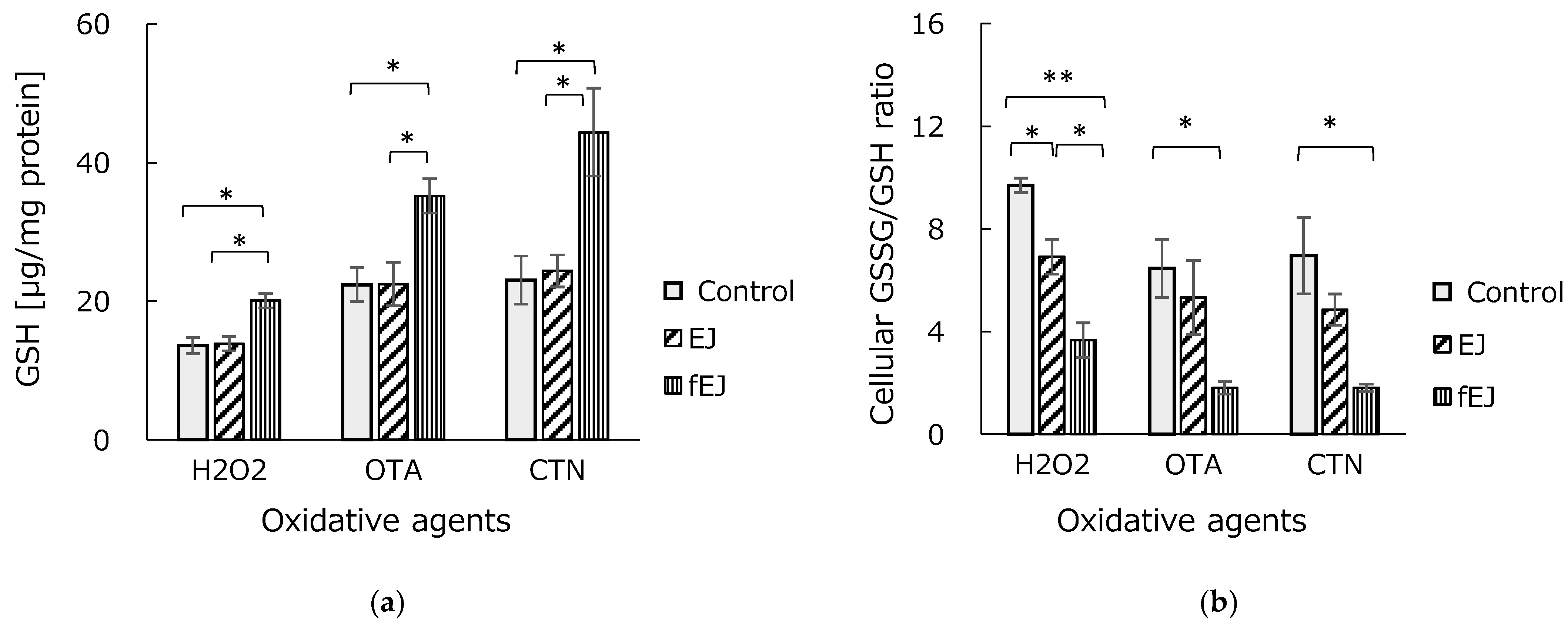
3.4. Effect of Fermented EJ Extract on GSH Homeostasis

Hepatic GSH, a ubiquitous intracellular peptide, is an important non-enzymatic antioxidant that protects cells against oxidative injury by scavenging ROS and maintaining enzymatic antioxidants. When cells are damaged by oxidation, GSH participates in the antioxidant reaction to be oxidized to GSSG, resulting in an increase in the ratio of GSSG/GSH. As regards the evaluation of glutathione homeostasis (Figure 2a), it can be clearly seen that the pretreatment of HEK293 cells with fermented EJ extract greatly increased GSH levels, while unfermented extract did not affect the cellular GSH content. Accordingly, concentrations of GSH enzyme levels with the pretreatment of fermented EJ were 20.1 µg/mg in H2O2, 35.2 µg/mg in OTA, and 44.4 µg/mg in CTN—values that are statistically significant (p < 0.01).
Subsequently, a decreased GSSG/GSH ratio resulted as compared to untreated cells exposed to H2O2 (p < 0.01). Similarly, with OTA or CTN exposure, fEJ treatment exhibited a significant reduction in the GSSG/GSH ratio, indicating that the antioxidative effects of MSC-5T fermented EJ extract on oxidative damage of HEK293 cells are achieved by restoring GSH homeostasis (Figure 2b). This is consistent with what has been reported by Mun et al., where pretreatment with 50 or 100 μg/mL hot water extract of EJ resulted in increased GSH enzyme activities of HEPG2/2E1 cells exposed with either 200 mM ethanol or 1 mM free fatty acid mixture [21].

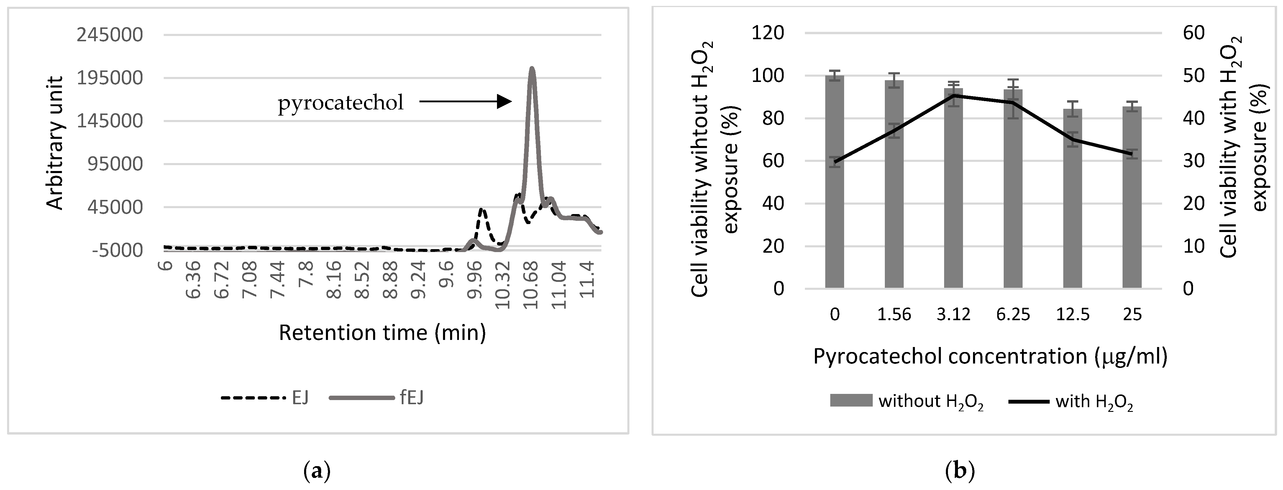
3.5. Analysis of Antioxidant Components

With regard to the specificity of the antioxidant effect of fermented EJ extract, the active compound was isolated. After 48 h of incubation, cell-free extract was collected and extracted with organic solvents. Subsequently, the ethyl acetate fraction was loaded to a silica gel column. The final purification of the target compound was conducted with HPLC. It is well known that EJ leaf extract contains ellagic acid, chlorogenic acid, and caffeic acid, as well as ursolic acid [28]. Furthermore, the leaf extract of EJ may have higher antioxidant and anticancer potential than the fruit extract. Of note, chlorogenic acid and caffeic acids occurred most abundantly in the leaf [29]. As shown in Figure 3a, we observed a new peak at 10.68 min retention time in fermented extract; however, no product peak could be detected in unfermented EJ extract. Through the chemical structure, 13C-NMR, it was identified that the antioxidant compound generated in EJ extract fermented with MSC-5T is pyrocatechol. The highest concentration of pyrocatechol produced was 167.4 μg/mL at 48 h of fermentation. In our previous study, another plant-derived LAB strain, L. plantarum SN13T, displayed the generation of highly bioactive compound dihydrocaffeic acid (DHCA) from rosmarinic acid and caffeic acid through the Mentha arvensis aqueous extract fermentation [30]. However, we could not detect significant change in the content of caffeic acid, as well as chlorogenic acid, in the present study. We should note here that the generated pyrocatechol protects HEK293 cells from oxidizing agents, but not the sole antioxidant compound. Nevertheless, our results of the high antioxidative effect of MSC-5T-fermented extract compared with unfermented EJ extract are likely to be caused by the involvement of other phenolic metabolites produced.
Furthermore, pyrocatechol’s effect on the percentage viability of HEK293 cells was dose dependent, and the viability decreased markedly as the dose exceeded 12.5 μg/mL. On the other hand, the highest cell survivability was detected at 3.12 μg/mL of pyrocatechol (45.3%), as compared to that of non-treated control cells (29.7%) (Figure 3b). Kosobutskii studied the effect of pyrocatechol and its derivatives on gamma radiation-induced oxidation and demonstrated that pyrocatechol and dihalocatechols exhibited antioxidant properties [31]. With regard to the anti-inflammatory responses of pyrocatechol, treatment with 5% (v/v) coffee extract containing approximately 2.5 μM pyrocatechol inhibited the LPS-induced activation of NF-κB in RAW264.7 macrophages [32]. Similarly, pyrocatechol treatment significantly prevented cisplatin-induced oxidative stress and ROS-induced JNK/P38 activation in HK-2, as well as HEK293T, cells [33].

4. Conclusions

In the present study, we demonstrated that the medicinal plant Eriobotrya japonica Lindl. fermented with L. plantarum MSC-5T strain isolated from sugarcane increases the survival of HEK293 cells from oxidizing agents. One of the main metabolic compounds is a pyrocatechol, which, at optimal concentrations (up to 12 μg/mL), greatly enhances cell survival in H2O2 exposure. We conclude that medicinal herbal extract fermented with plant-derived LAB strains could be a practical solution for enhancing the therapeutic potential of medicinal plants.

Author Contributions

Conceptualization, N.D. and Y.I.; methodology, Y.I.; software, Y.I.; formal analysis, N.D., Y.I., S.S. (Shrijana Shakya) and S.S. (Sachiko Sugimoto); writing—original draft preparation, N.D.; writing—review and editing, N.D., M.N. and M.S.; supervision, N.D. All authors have read and agreed to the published version of the manuscript.

Funding

This research received no external funding.

Institutional Review Board Statement

Not applicable.

Informed Consent Statement

Not applicable.

Data Availability Statement

Data are contained within the article.

Acknowledgments

We thank Hitoshi Fujitaka and Tomoko Amimoto, the Natural Science Center for Basic Research and Development (N-BARD), Hiroshima University, for the measurement of NMR. This work was also supported by the Program of the Network-Type Joint Usage/Research Center for Radiation Disaster Medical Science.

Conflicts of Interest

Author Yusuke Inoue was employed by Mitsui DM Sugar Co., Ltd. The remaining authors declare that the research was conducted in the absence of any commercial or financial relationships that could be construed as a potential conflict of interest.

References

  1. Williams, N.T. Probiotics. Am. J. Health Syst. Pharm. 2010, 67, 449–458. [Google Scholar] [CrossRef]
  2. Gupta, V.; Garg, R. Probiotics. Indian J. Med. Microbiol. 2009, 27, 202–209. [Google Scholar] [CrossRef]
  3. Liu, W.; Pang, H.; Zhang, H.; Cai, Y. Biodiversity of lactic acid bacteria. In Lactic Acid Bacteria: Fundamentals and Practice; Springer: Dordrecht, The Netherlands, 2014; pp. 103–203. [Google Scholar]
  4. Nomura, M.; Kobayashi, M.; Narita, T.; Kimoto-Nira, H.; Okamoto, T. Phenotypic and molecular characterization of Lactococcus lactis from milk and plants. J. Appl. Microbiol. 2006, 101, 396–405. [Google Scholar] [CrossRef]
  5. Noda, M.; Danshiitsoodol, N.; Kanno, K.; Uchida, T.; Sugiyama, M. The Exopolysaccharide produced by Lactobacillus paracasei IJH-SONE68 prevents and ameliorates inflammatory responses in DSS-induced ulcerative colitis. Microorganisms 2021, 9, 2243. [Google Scholar] [CrossRef] [PubMed]
  6. Danshiitsoodol, N.; Noda, M.; Kanno, K.; Uchida, T.; Sugiyama, M. Plant-derived Lactobacillus paracasei IJH-SONE68 improves the gut microbiota associated with hepatic disorders: A randomized, double-blind, and placebo-controlled clinical trial. Nutrients 2022, 14, 4492. [Google Scholar] [CrossRef] [PubMed]
  7. Higashikawa, F.; Danshiitsoodol, N.; Kanno, K.; Ishida, R.; Tazuma, T.; Sugiyama, M. Lactobacillus plantarum SN13T cells improve hepatic dysfunction and fecal microbiota: A randomized pilot study. Arch. Clin. Biomed. Res. 2020, 4, 605–625. [Google Scholar] [CrossRef]
  8. Brieger, K.; Schiavone, S.; Miller, F.J., Jr.; Krause, K.H. Reactive oxygen species: From health to disease. Swiss Med. Wkly. 2012, 142, 13659. [Google Scholar] [CrossRef] [PubMed]
  9. Davalli, P.; Mitic, T.; Caporali, A.; Lauriola, A.; D’Arca, D. ROS, Cell senescence, and novel molecular mechanisms in aging and age-related diseases. Oxid. Med. Cell. Longev. 2016, 2016, 3565127. [Google Scholar] [CrossRef]
  10. Okamoto, T.; Sugimoto, S.; Noda, M.; Yokooji, T.; Danshiitsoodol, N.; Higashikawa, F.; Sugiyama, M. Interleukin-8 release inhibitors generated by fermentation of artemisia princeps pampanini herb extract with Lactobacillus plantarum SN13T. Front. Microbiol. 2020, 11, 1159. [Google Scholar] [CrossRef]
  11. Kalinowska, M.; Gołebiewska, E.; Zawadzka, M.; Choińska, R.; Koronkiewicz, K.; Piasecka-Jóźwiak, K.; Bujak, M. Sustainable extraction of bioactive compound from apple pomace through lactic acid bacteria (LAB) fermentation. Sci. Rep. 2023, 13, 19310. [Google Scholar] [CrossRef]
  12. Pham, T.N.A.; Kim, H.L.; Lee, D.R.; Choi, B.K.; Yang, S.H. Anti-inflammatory effects of Scrophularia buergeriana extract mixture fermented with Lactic Acid Bacteria. Biotechnol. Bioproc. Eng. 2022, E27, 370–378. [Google Scholar] [CrossRef]
  13. Liu, Y.; Zhang, W.; Xu, C.; Li, X. Biological activities of extracts from loquat (Eriobotrya japonica Lindl.): A Review. Int. J. Mol. Sci. 2016, 17, 1983. [Google Scholar] [CrossRef]
  14. Zhu, X.; Wang, L.; Zhao, T.; Jiang, Q. Traditional uses, phytochemistry, pharmacology, and toxicity of Eriobotrya japonica leaves A summary. J. Ethnopharmacol. 2022, 298, 115566. [Google Scholar] [CrossRef]
  15. Khouya, T.; Ramchoun, M.; Elbouny, H.; Hmidani, A.; Bouhlali, E.D.T.; Alem, C. Loquat (Eriobotrya japonica (Thunb) Lindl.): Evaluation of nutritional value, polyphenol composition, antidiabetic effect, and toxicity of leaf aqueous extract. J. Ethnopharmacol. 2022, 296, 115473. [Google Scholar]
  16. Ito, H.; Kobayashi, E.; Takamatsu, Y.; Li, S.H.; Hatano, T.; Sakagami, H.; Kusama, K.; Satoh, K.; Sugita, D.; Shimura, S.; et al. Polyphenols from Eriobotrya japonica and their cytotoxicity against human oral tumor cell lines. Chem. Pharm. Bull. 2000, 48, 687–693. [Google Scholar] [CrossRef]
  17. Hiraishi, A. Direct automated sequencing of 16S rDNA amplified by a polymerase chain reaction from bacterial cultures without DNA purification. Lett. Appl. Microbiol. 1992, 15, 210–213. [Google Scholar] [CrossRef]
  18. Weisburg, W.G.; Barns, S.M.; Pelletier, D.A.; Lane, D.J. 16S ribosomal DNA amplification for phylogenetic study. J. Bacteriol. 1991, 173, 697–703. [Google Scholar] [CrossRef]
  19. Kwon, Y.H.; Jang, J.Y.; Lee, J.H.; Choi, Y.W.; Choi, Y.H.; Kim, N.D. Loquat leaf extract inhibits oxidative stress-induced DNA damage and apoptosis via AMPK and Nrf2/HO-1 signaling pathways in C2C12 cells. Appl. Sci. 2023, 13, 572. [Google Scholar] [CrossRef]
  20. Bae, D.; You, Y.; Yoon, H.G.; Kim, K.; Lee, Y.H.; Kim, Y.; Baek, H.; Kim, S.; Lee, J.; Jun, W. Protective effects of loquat (Eriobotrya japonica) leaves against ethanol-induced toxicity in HepG2 cells transfected with CYP2E1. Food Sci. Biotechnol. 2010, 19, 1093–1096. [Google Scholar] [CrossRef]
  21. Mun, J.; Park, J.; Yoon, H.J.; You, Y.; Choi, K.C.; Lee, Y.H.; Kim, K.; Lee, J.; Kim, O.K.; Jun, W. Effects of Eriobotrya japonica water extract on alcoholic and nonalcoholic fatty liver impairment. J. Med. Food 2019, 22, 1262–1270. [Google Scholar] [CrossRef]
  22. Klarić, M.Š.; Želježić, D.; Rumora, L.; Peraica, M.; Pepeljnjak, S.; Domijan, A.M. A potential role of calcium in apoptosis and aberrant chromatin forms in porcine kidney PK15 cells induced by individual and combined ochratoxin A and citrinin. Arch. Toxicol. 2012, 86, 97–107. [Google Scholar] [CrossRef]
  23. Yu, Z.; Wu, F.; Tian, J.; Guo, X.; An, R. Protective effects of compound ammonium glycyrrhizin, L-arginine, silymarin and glucurolactone against liver damage induced by ochratoxin A in primary chicken hepatocytes. Mol. Med. Rep. 2018, 18, 2551–2560. [Google Scholar] [CrossRef] [PubMed]
  24. Eraso, A.J.; Albesa, I. Eriobotrya japonica counteracts reactive oxygen species and nitric oxide stimulated by chloramphenicol. Am. J. Chin. Med. 2007, 35, 875–885. [Google Scholar] [CrossRef]
  25. Kim, M.J.; Lee, J.; Seong, A.R.; Lee, Y.H.; Kim, Y.J.; Baek, H.Y.; Kim, Y.J.; Jun, W.J.; Yoon, H.J. Neuroprotective effects of Eriobotrya japonica against β-amyloid-induced oxidative stress and memory impairment. Food Chem. Toxicol. 2011, 49, 780–784. [Google Scholar] [CrossRef]
  26. Kikuchi, T.; Akazawa, H.; Tabata, K.; Manosroi, A.; Manosroi, J.; Suzuki, T.; Akihisa, T. 3-O-(E)-p-coumaroyl tormentic acid from Eriobotrya japonica leaves induces caspase-dependent apoptotic cell death in human leukemia cell line. Chem. Pharm. Bull. 2011, 59, 378–381. [Google Scholar] [CrossRef] [PubMed]
  27. Uto, T.; Sakamoto, A.; Tung, N.H.; Fujiki, T.; Kishihara, K.; Oiso, S.; Kariyazono, H.; Morinaga, O.; Shoyama, Y. Anti-proliferative activities and apoptosis induction by triterpenes derived from Eriobotrya japonica in human leukemia cell lines. Int. J. Mol. Sci. 2013, 14, 4106–4120. [Google Scholar] [CrossRef]
  28. Zhang, J.; Li, Y.; Chen, S.S.; Zhang, L.; Wang, J.; Yang, Y.; Zhang, S.; Pan, Y.; Wang, Y.; Yang, L. Systems pharmacology dissection of the anti-inflammatory mechanism for the medicinal herb Folium eriobotryae. Int. J. Mol. Sci. 2015, 16, 2913–2941. [Google Scholar] [CrossRef]
  29. Pawłowska, A.M.; Żurek, N.; Kapusta, I.; De Leo, M.; Braca, A. Antioxidant and antiproliferative activities of phenolic extracts of Eriobotrya japonica (Thunb.) Lindl. fruits and leaves. Plants 2023, 12, 3221. [Google Scholar] [CrossRef] [PubMed]
  30. Shakya, S.; Danshiitsoodol, N.; Noda, M.; Sugiyama, M. Role of phenolic acid metabolism in enhancing bioactivity of Mentha extract fermented with plant-derived Lactobacillus plantarum SN13T. Probiotics Antimicrob. Proteins 2023, 6. [Google Scholar] [CrossRef]
  31. Kosobutskii, V.S. Pyrocatechol and its derivatives as antioxidants and prooxidants. Russ. J. Gen. Chem. 2014, 84, 839–842. [Google Scholar] [CrossRef]
  32. Funakoshi-Tago, M.; Nonaka, Y.; Tago, K.; Takeda, M.; Ishihara, Y.; Sakai, A.; Matsutaka, M.; Kobata, K.; Tamura, H. Pyrocatechol, a component of coffee, suppresses LPS-induced inflammatory responses by inhibiting NF-κB and activating Nrf2. Sci. Rep. 2020, 10, 2584. [Google Scholar] [CrossRef] [PubMed]
  33. Xie, X.; Wu, F.; Tian, J.; Liu, Z.; He, H.; Bao, D.; Li, G.; Li, H.; Chen, J.; Lai, Y.; et al. Pyrocatechol alleviates cisplatin-induced acute kidney injury by inhibiting ROS production. Oxid. Med. Cell. Longev. 2022, 9, 2158644. [Google Scholar] [CrossRef] [PubMed]
Figure 1. EJ extract fermented with the MSC-5T strain inhibits various oxidizing agent-induced apoptotic cell death of HEK293 cells. HEK293 cells were pre-incubated with EJ or fEJ for 3 h prior to H2O2, OTA, or CTN exposure for 24 h. (a) Correlation between cell survivability of H2O2-treated HEK293 cells and the fermentation time of the EJ extract by the MSC-5T strain. (b) HEK293 cells were pretreated with EJ or 48 h-fermented EJ extract, and cell viability was assessed. Mean ± S.D. (n = 3); Tukey HSD test (* p < 0.05, ** p < 0.01) vs. treatment with control cells.
Fermentation 10 00197 g001
Figure 2. Glutathione homeostasis in HEK293 cells exposed to different oxidizing agents. HEK293 cells were pretreated with EJ or fEJ for 3 h, followed by exposure to oxidizing agents for 24 h, and a glutathione homeostasis assay was conducted. (a) Cellular GSH level. (b) Cellular GSSG/GSH ratio. Mean ± S.D. (n = 3), Turkey HSD test (* p < 0.05, ** p < 0.01) vs. treatment with control cells.
Fermentation 10 00197 g002
Figure 3. HPLC chromatogram and pyrocatechol profiling at H2O2-induced HEK293 cells. (a) HPLC chromatogram of EJ extract without or with MSC-5T fermentation. (b) Relative cell viability of HEK293 cells treated without (bar) or with H2O2 (line) in the presence of pyrocatechol at 1.56, 3.12, 6.25, 12.5, and 25 µg/mL. Error bars represent +/− standard deviation.
Fermentation 10 00197 g003
Table 1. Suppression of unfermented or fermented EJ extract on the cellular ROS and caspase-3 activity in various oxidative agent-treated HEK293 cells.
Unfermented EJ ExtractFermented EJ Extract
Intracellular ROS (%)H2O281 ± 4.170.5 ± 3.7
OTA93.3 ± 4.182.5 ± 3.7
CTN80.3 ± 2.568.1 ± 5.2
Intracellular Caspase-3 (%)H2O2103.1 ± 6.242.6 ± 3.0
OTA95 ± 5.168.9 ± 5.1
CTN92.8 ± 2.383.6 ± 1.9
Disclaimer/Publisher’s Note: The statements, opinions and data contained in all publications are solely those of the individual author(s) and contributor(s) and not of MDPI and/or the editor(s). MDPI and/or the editor(s) disclaim responsibility for any injury to people or property resulting from any ideas, methods, instructions or products referred to in the content.

