Article

Probiotic Evaluation of Lactiplantibacillus pentosus 68-1, a Rutin Conversion Strain Isolated from Jiangshui, by Genomic Analysis and Tests In Vitro

1
Shaanxi Institute of Microbiology, Xi’an 710043, China
2
National Engineering Laboratory for Resource Developing of Endangered Chinese Crude Drugs in Northwest China, College of Life Sciences, Shaanxi Normal University, Xi’an 710119, China
*
Author to whom correspondence should be addressed.
Fermentation 2024, 10(2), 87; https://doi.org/10.3390/fermentation10020087
Submission received: 20 December 2023 / Revised: 20 January 2024 / Accepted: 25 January 2024 / Published: 31 January 2024
(This article belongs to the Special Issue Application of Lactobacillus in Fermented Food and Beverages)

Abstract

:
To assess the probiotic potential of strain 68-1 with rutin conversion capabilities, isolated from Chinese traditional Jiangshui, a complete genomic analysis and in vitro tests were conducted. The Oxford Nanopore Technologies (ONT, Oxford, UK)–Illumina (San Diego, CA, USA) hybrid sequencing platform was used for whole genome sequencing and the results showed that strain 68-1 had a chromosome sequence of 3,482,151 bp, with 46.53% GC content and five plasmids with a sequence length ranging from 2009 bp to 48,711 bp. Strain 68-1 was identified as Lactiplantibacillus pentosus based on the whole genome sequence. A total of 133 genes encoding for carbohydrate-active enzymes (CAZymes) were identified and genes that may be involved in rutin conversion were found in the L. pentosus 68-1 genome. L. pentosus 68-1 showed excellent tolerance to gastrointestinal juice and adhesion properties, and corresponding genes were identified. In addition, L. pentosus 68-1 exhibited strong antibacterial and antifungal activity, where organic acids may play a crucial role in its antagonistic ability. Moreover, the gene cluster for plantaricin_EF production was detected. No high virulence factor was found in the L. pentosus 68-1 genome and no hemolytic effect was observed. In addition, L. pentosus 68-1 showed resistance to ampicillin, gentamycin, and kanamycin, and the genomic analysis indicated that horizontal ARG transfer should not be possible. The results show that L. pentosus 68-1 could be developed as a novel probiotic candidate to improve rutin bioavailability in the food and feed industry.

1. Introduction

Jiangshui, also known as a serofluid dish, is a traditional fermented vegetable in Northwest China [1]. It is considered that Jiangshui can promote digestion, lower blood pressure, and reduce cholesterol levels [2]. The most abundant bacteria in Jiangshui products are lactic acid bacteria (LAB) [1,2,3], which may contribute to the health benefits of Jiangshui. LAB are widely used as probiotics due to their “generally recognized as safe” (GRAS) status and their great beneficial effects [4,5,6]. However, few studies have focused on the probiotic properties of LAB strains isolated from Jiangshui.
Probiotics are defined as “live microorganisms which when administered in adequate amounts confer a health benefit on the host” according to an FAO/WHO report [7]. Therefore, probiotics should be resistant to stress conditions and stay viable under the gastrointestinal environment [8,9]. In addition, antagonistic abilities, cholesterol assimilation, and other functional properties are also important for probiotics in exerting beneficial effects. On the other hand, strains must be safe and should be free of virulence factors and toxins and have no risk of antibiotic resistance gene transfer for application as a probiotic [7,9,10]. It is worth noting that the evaluation of the probiotic potential of LAB strains should be conducted before their application in the food and feed industries since the probiotic properties of LAB are strain-specific [6,11]. The available probiotic evaluation methods mainly include in vitro tests, in vivo tests, and human clinical studies [6,7,8,9,10,11]. With recent developments in genome sequencing technologies, the assessment of the genome-scale safety and probiotic characterization of LAB has become practical and feasible. The combination of genomic analysis and characteristic experiments could promote the evaluation of probiotics, providing more information about their probiotic potential [12,13,14].
Quercitin-3-O-rutinoside (rutin) is a flavonol glycoside composed of quercitin and rutinose and represents the most consumed dietary flavonol [15]. Rutin has been reported to have beneficial effects on human health, including antioxidant, anti-inflammatory, and anti-cancer activity [16,17,18]. On the other hand, the bioavailability of the dietary flavonoid depends on intestinal absorption, which is determined largely by the nature of glycosylation. Generally, the aglycone forms of flavonoids are more efficiently absorbed than flavonoid glycosides because of their ability to cross through the cell membrane [15]. Therefore, researchers have endeavored to improve the rutin bioavailability by deglycosylation transformation [15,19,20] and several LAB strains are reported to have rutin biotransformation ability [21,22]. However, as far as we know, no LAB strains with rutin biotransformation ability have been isolated from Chinese traditional fermented vegetables.
In this study, strain 68-1 was isolated from traditional Jiangshui, which is usually obtained from celery, in Shaanxi, China. Celery is a commonly consumed vegetable containing high content of flavonoid glycosides and dietary fiber [23]. Therefore, we supposed that the LAB responsible for Jiangshui fermentation might possess rich glycoside hydrolase, which could be used for the biotransformation of flavonoids. The objective of this study was to assess the probiotic potential of strain 68-1 by whole genomic analysis and in vitro tests.

2. Materials and Methods

2.1. Strains and Culture Conditions

The strain 68-1 was previously isolated from Jiangshui in Shaanxi, China and deposited in the China General Microbiological Culture Collection Center (CGMCC24424). The probiotic strain Lacticaseibacillus rhamnosus GG (LGG, CGMCC1.3724 (=ATCC53103)), which was purchased from the China General Microbiological Culture Collection Center (CGMCC, Beijing, China), was used as a reference strain. The above two strains were cultured in de Man, Rogosa, and Sharpe (MRS) broth at 37 °C for 18 h.
Listeria monocytogenes CICC21635, Enterococcus faecalis CICC10396, Enterococcus faecium CGMCC1.101, Escherichia coli CMCC44102, Salmonella paratyphi B CMCC50094, Shigella flexneri CMCC51574, and Staphylococcus aureus CGMCC1.0089 were used as indicator bacteria, which were cultured at 37 °C and 180 r/min in Luria–Bertani (LB) broth for 12 h.
The used fungal pathogens were Rhizoctonia solani CICC40529, Candida albicans GGMCC2.2086, Aspergillus flavus CICC40375, Fusarium oxysporum CICC2532. The fungal strains were cultured at 25 °C on PDA for 5–7 days and then spore suspensions were prepared by adding 15 mL of sterile ultrapure water and counted by a hematocytometer.
All indicators were previously obtained from the CGMCC and China Center of Industrial Culture Collection (CICC), Beijing, China.

2.2. Complete Genome Sequencing and Genome Assembly

After 18 h incubation at 37 °C in MRS broth, the bacterial genomic DNA was extracted by using the cetyltrimethyl ammonium bromide (CTAB) method with minor modification, and then the DNA concentration, quality, and integrity were determined by using a Qubit Flurometer (Invitrogen, Carlsbad, CA, USA) and a NanoDrop Spectrophotometer (Thermo Scientific, Waltham, MA, USA). Sequencing libraries were generated using the TruSeq DNA Sample Preparation Kit (Illumina, San Diego, CA, USA) and the SQK-LSK109 connection kit (Oxford Nanopore Technologies, Oxford, UK). Genome sequencing was then performed using the Illumina NovaSeq platform and the Oxford Nanopore PromethION platform. The genome was assembled using the software Unicycler v0.4.8 [24]. Both the genome sequencing and assembly were carried out by a commercial service (Personalbio Technology Co., Ltd., Shanghai, China). The complete genome sequence of strain 68-1 was deposited in NCBI (CP104714-CP104719) (https://www.ncbi.nlm.nih.gov/assembly/GCA_025398935.1, accessed on 25 September 2022).

2.3. Genome Annotation

The genome of strain 68-1 was annotated with GenemarkS [25] after evaluations of the assembly quality. The tRNAscan-SE (version 1.3.1) software [26] and Barrnap v0.9 software were used to predict the tRNA and rRNA in the strain 68-1 genome, respectively. The online software CRISPRCasFinder (https://crisprcas.i2bc.paris-saclay.fr/, accessed on 19 December 2023) was used to search clustered palindromic interspaced palindromic repeat (CRISPR) regions and cas genes [27]. Genomic islands of strain 68-1 were identified with the IslandViewer 4 software [28]. Prophages and insertion sequences (IS) were searched with the PHASTER webserver [29,30] and ISfinder [31], respectively. In addition, the Non-Redundant Protein Sequence Database (NR), Swiss-Prot, Kyoto Encyclopedia of Genes and Genomes (KEGG), and EggNOG were used for general function annotation. Bacteriocin gene clusters were searched using BAGEL4 [32].

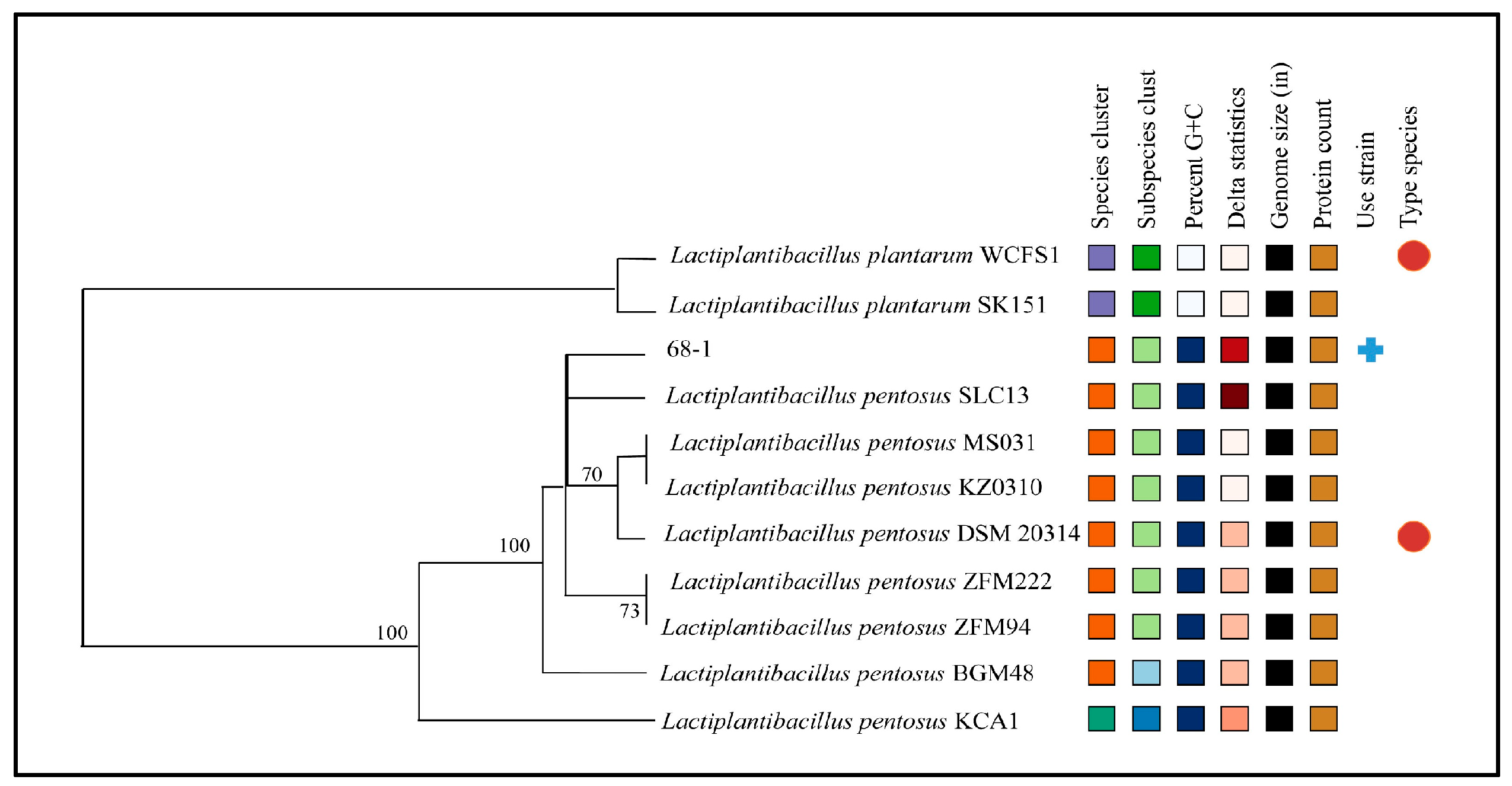
2.4. Phylogenetic Analysis

The taxonomy of strain 68-1 was investigated based on the complete genome. The complete genomes of Lactiplantibacillus pentosus DSM20314, BGM48, ZFM222, ZFM94, SLC13, MS031, KZ0310 (Table S2), and Lactiplantibacillus plantarum WCFS1 and SK151, downloaded from NCBI, were used as references. The ANIb, ANIm, and TETRA frequencies were calculated using the JSpeciesWS software (http://jspecies.ribohost.com/, accessed on 19 December 2023) [33]. The genome-to-genome distance (GGDC) was calculated using a web service (https://ggdc.dsmz.de/, accessed on 19 December 2023). In addition, the Type Strain Genome Server (TYGS, https://tygs.dsmz.de, accessed on 19 December 2023) was used to build a phylogenomic tree based on the complete genome [34].

2.5. Identification of Safety-Related Genes

Virulence factor genes were detected using the Virulence Factor Database (VFDB) [35]. The search criteria of the cut-off values of >60% identity, >70% coverage, and E-value < 0.00001 were used to identify the possible virulence genes. The bacterial pathogenicity was predicted using the web server PathogenFinder [36]. The Comprehensive Antibiotic Resistance Database (CARD) was used to search for antibiotic resistance genes (ARGs) using the Resistance Gene Identifier (RGI) tool under perfect and rigorous hits only and high quality/coverage criteria [37]. In addition, the KEGG database was used to detect toxin and ARG genes [38].

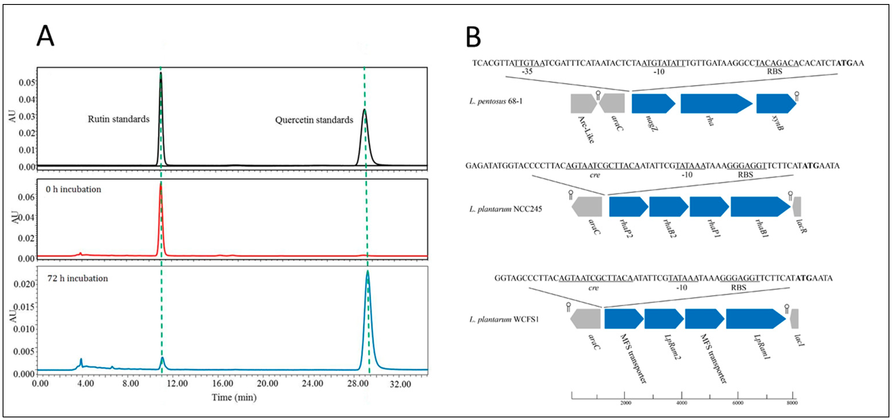
2.6. Rutin Biotransformation by Strain 68-1

The overnight cultures of strain 68-1 were inoculated into MRS medium (1%, v/v) supplemented with 100 mg/L rutin. After 72 h incubation at 37 °C, the cultures were freeze-dried using a freeze dryer (Heto PowerDry LL3000, Thermo, Waltham, MA, USA). The dried samples were dissolved in 10 mL of DMSO and then filtered using a 0.2 µm membrane filter (Millipore, MA, USA). The filtrates were analyzed by HPLC (Waters, alliance separation module 2695, 2998 detector; Waters, Milford, MA, USA) using a C18 column (YMC-Pack ODS-AQ, 250 mm × 4.6 mm, 5 μm, YMC, Kyoto, Japan). The mobile phase was composed of 50% (v/v) of methanol and 50% (v/v) of phosphoric acid solution (0.4%). The pump flow rate was set at 1.0 mL/min and the column temperature was 30 °C. All samples were detected by absorption at 256 nm with an injection volume of 10 μL.

2.7. In Vitro Assessment of Probiotic Properties of Strain 68-1

2.7.1. Tolerance to Simulated Gastric Juice and Simulated Intestinal Juice

In vitro tolerance to simulated gastric juice (SGJ, 3 g/L pepsin (p7000, Sigma, Shanghai, China), pH 2.0) and simulated intestinal juice (SIJ, 1 g/L trypsin (T105532, Aladdin, Shanghai, China), 0.3% bile salt, pH 8.0) was evaluated according to a previous study, with slight modifications [6]. The cells after overnight culture were harvested and re-suspended in SGJ (pH 3.0) at a concentration of 109 CFU/mL at 37 °C for 0 h, 1 h, 2 h, and 3 h, respectively. Then, the viable cells were counted. To assess intestinal fluid tolerance, viable bacterial cells were determined after incubation at 37 °C in SIJ (0.3% bile salt) for 0 h, 1 h, 2 h, and 4 h. The survival rate (%) was calculated according to a previous report [6].

2.7.2. Cholesterol Assimilation

Cholesterol assimilation was evaluated according to a previous study [6]. After incubation in MRS broth supplemented with 0.1 g/L cholesterol at 37 °C for 24 h, bacterial cells were removed. The cholesterol concentration of the cell-free broth was determined by the o-phthalaldehyde method described previously [9] and the cholesterol removal percentage was expressed as follows:
Cholesterol   removal   percentage   ( % ) = C 0 C t C 0 × 100%
where C0 is the concentration of cholesterol in the initial medium and Ct is the concentration of cholesterol at the end of inoculation.

2.7.3. Evaluation of Adhesion and Aggregation Properties In Vitro

The hydrophobicity, auto-aggregation, and adhesion ability to Caco-2 cells were evaluated according to a previous study [6]. For hydrophobicity tests, the collected strain 68-1 and LGG cells were adjusted to achieve an OD600 value of approximately 0.4. The bacterial cells were removed and the OD600 was measured after 30 min incubation with chloroform (3:1, v/v). For the auto-aggregation assay, the cells were re-suspended in PBS buffer (pH 7.2) to achieve a concentration of 108 CFU/mL. After incubation at 37 °C for 0–28 h, the OD600 was measured for the upper bacterial suspension. For the adhesion assay, Caco-2 cells were seeded into 24-well cell culture plates and incubated at 37 °C, 5% CO2 for 15 days in a humidified atmosphere to obtain cell differentiation. Then, the bacterial cell suspension (1 × 10⁸ CFU/mL, re-suspended in DMEM after an overnight culture at 37 °C) was added to the 24-well cell culture plate and co-incubated with Caco-2 monolayers for 1 h at 37 °C with 5% CO2. After removing the non-attached bacterial cells, adherent bacterial cells were detached and counted. The LGG strain was used as a control.

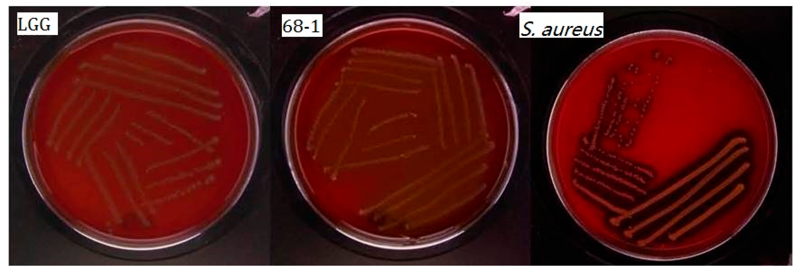
2.7.4. Antimicrobial Activity against Pathogens

Antibacterial activity was evaluated based on a previous study [6]. An overlay assay was used to evaluate the bacterial antifungal activity [39]. The cells were inoculated in 2-cm lines on MRS agar plates after growth for 16 h in MRS broth at 37 °C and then incubated at 37 °C for 48 h. Subsequently, plates were overlaid with cooled soft PDA (0.7% agar) containing a mold spore suspension (104 spores/mL). After incubation at 25 °C for 3 days, the presence of a clear zone of inhibition around the bacterial smears was observed visually.

2.8. In Vitro Safety Assessment of Strain 68-1

2.8.1. Hemolytic Activity

The hemolytic activity was tested according to a previous report [9]. Fresh bacterial cultures were inoculated on Columbia agar plates containing 7% (v/v) sheep blood (PB001 land bridge, Beijing, China). After incubation at 37 °C for 48 h, the blood agar plates were examined visually. The presence of a greenish zone around the colony was considered as α-hemolysis, while a clear zone and no halo were considered for β-hemolysis and γ-hemolysis, respectively. LGG was used as a negative control and S. aureus CGMCC1.0089 was used as a positive control.

2.8.2. Antibiotic Resistance

According to the EFSA guidelines [10], antibiotics including ampicillin, kanamycin, chloramphenicol, clindamycin, erythromycin, gentamicin, and tetracycline were used to determine the minimum inhibitory concentration (MIC) against strain 68-1 in this study. Various concentrations of each antibiotic, namely 1024, 512, 256, 128, 64, 32, 16, 8, 4, 2, and 1 µg/mL, were examined for growth (OD600 nm) after 24 h incubation at 37 °C in a microplate reader.

2.9. Statistical Analysis

All experiments were performed in triplicate. The experimental data are presented as means ± SEM. Statistical analysis of data was carried out using SPSS (Ver. 19.0 SPSS, Chicago, IL, USA). A one-way analysis of variance (ANOVA) test was performed for multiple comparisons by GraphPad Prism 9.0. In this study, all values of p < 0.05 were considered statistically significant.

3. Results

3.1. General Genome Characteristics

The ONP–Illumina hybrid sequencing platform was used to sequence the complete genome of strain 68-1 in this study. The results showed that the complete genome sequence of the strain was composed of a circular 3,482,151 bp chromosome and five plasmids with a sequence length ranging from 2009 bp to 48,711 bp (Table 1 and Figure S1). The GC content of the chromosome was 46.53% and that of plasmids ranged from 36.54% to 41.93%. The GC content and size of the strain 68-1 chromosome was similar to that of other L. pentosus strains (Table S1). There were 3114 ORFs, 16 rRNA (6 5S rRNAs, 5 16S rRNAs, and 5 23S rRNAs), and 64 tRNAs in the chromosome. A total of 106 ORFs were found in the five plasmids.
As shown in Table S2, 2000 genes (64.2%) could be assigned to 21 functional categories in COG families, while 483 genes were not included in COG and 631 genes were assigned to the function unknown. The most abundant gene category (8.77%) was predicted for carbohydrate transport and metabolism, followed by 8.38% encoding for transcription and 6.71% coded for proteins related to amino acid transport and metabolism.

3.2. Phylogenetic Analysis

Based on the 16S rRNA sequence analysis, strain 68-1 was identified as either L. plantarum or L. pentosus. Therefore, the strain was identified by phylogenetic analysis based on the complete genome. The ANIb, ANIm, TETRA, and GGDC algorithms were used to compare the strain 68-1 genome with the reference genomes. As shown in Table S3, high similarity values, which were above the limit of each algorithm except TETRA, were observed between strain 68-1 and almost all the selected genomes of L. pentosus. The similarity values were also within the range of the limit (>0.989) of the TETRA algorithm. Relatively low similarity was observed between strain KCA1 and other selected strains. L. pentosus KZ0310 and MS031 were identified as the closest neighbors of strain 68-1, with similarity of 97.52% for ANIb, 98.02% for ANIm, 0.99903 for TETRA, and 81.5% for GGDC. In addition, the phylogenetic tree was built using TYGS and the result confirmed the high similarity of strain 68-1 compared with the selected L. pentosus strains (Figure 1).

3.3. Identification of Carbohydrate-Active Enzymes

In the genome of L. pentosus 68-1, 133 genes have been identified in five classes of carbohydrate-active enzymes (CAZymes), including glycosyltransferase (GT), glycoside hydrolase (GH), enzymes for auxiliary activity (AA), carbohydrate-binding modules (CBM), and carbohydrate esterase (CE). No polysaccharide lyase (PL) family was found in this genome. The number of CAZymes ranged from 91 to 115 in the six L. pentosus strains collected in the CAZy database (Table S4). Compared with them, the genome of L. pentosus 68-1 contained the highest number of CAZymes (133 genes). In addition, it is worth noting that the strain possessed 26 CE family genes and 7 AA family genes, which were considerably higher than those of the other selected L. pentosus strains. The CAZymes analysis also showed that the most abundant class, GH, included 54 genes of 22 GH families in the strain. GH13 enzymes (10 genes) were the most abundant family, followed by the GH1 family (nine genes).
In addition, L. pentosus 68-1 could transform rutin (Figure 2), which may be attributed to the β-glucosidase and α-L-rhamnosidase activity [40,41]. Interestingly, a GH3 gene (chr_2965), which may code for β-glucosidase, was adjacent to the only gene of α-L-rhamnosidase (GH78, chr_2966) in the genome. The genomic organization of the region of the gene coding for putative α-L-rhamnosidase (GH78) in strain 68-1, including the gene araC, the gene coding for AraC-like ligand-binding domain containing a protein, and three GH genes, is shown in Figure 2b. The araC gene seems to be a transcriptional regulator of the probable rha/nagZ/xynB operon.

3.4. Resistance to the Gastrointestinal Environment

As shown in Figure 3A, the survival rate of L. pentosus 68-1 remained at 97.9%, while that of LGG was 85.9% after being exposed to SGJ for 3 h. Meanwhile, the survival rate of the strain was 96.5%, which was much higher than that of LGG (80.6%), after 1 h incubation in SIJ, and it then decreased to 67% after 4 h exposure (Figure 3B). In addition, the strain showed a cholesterol assimilation rate of 20.51% ± 1.65%, which was higher than that of LGG (15.38% ± 0.58%) (Figure 3C).
To understand the molecular basis of the excellent stress resistance of L. pentosus 68-1, genome analysis was carried out (Table S5). Genes encoding the complete proton pump F0F1-ATPase system (atpF1ABDEG, atpF0ABC) and 10 genes that may encode Na+/H+ antiporters were detected in the genome of L. pentosus 68-1. In addition, five L-lactate dehydrogenase genes, one D-lactate dehydrogenase gene, and one arginine decarboxylase gene were also identified in the genome. The genome of L. pentosus 68-1 contained three bile salt hydrolase genes (chr_55, chr_2245, and chr_2869). Four types of multidrug efflux transporters, including the MATE, SMR, MFS, and ABC families, were identified in the genome. In addition, the general stress response factors, including the proteins involved in macromolecule repair (DnaK-DnaJ-GrpE-HrcA and GroES-GroEL chaperone complexes and proteases ClpBCPXL and HslOU), cold response factors CspAC and Hsp20, and alkaline shock response factors (AmaP, Asp23, Gls24) were identified in the genome of L. pentosus 68-1.

3.5. Adhesion and Aggregation Properties

The self-aggregation rate of L. pentosus 68-1 increased with time and then reached a maximum value (82.67%) after 24 h incubation (Figure 3E). The self-aggregation rates of L. pentosus 68-1 and LGG were 80.79% and 29.58%, respectively, at 20 h of incubation. The cell surface hydrophobicity of L. pentosus 68-1 was 34.57%, which was lower than that of LGG (53.28%) (Figure 3D). Moreover, the strain showed higher adhesion (9.79%) to Caco-2 cells than LGG (4.19%) (Figure 3F).
The genome analysis showed that there were three genes coding for MucBP domain proteins, one gene coding for a collagen-binding protein, two genes coding for a fibronectin-binding protein, and two genes coding for the zinc/manganese transport system substrate-binding protein in the genome of L. pentosus 68-1 (Table S6). In addition, poly-β-1,6-N-acetyl-D-glucosamine synthase was identified. Genes coding for moonlighting proteins, including glutamine synthetase (GS), glucose-6-phosphate isomerase (GPI), glyceraldehyde-3-phosphate dehydrogenase (GAPDH), enolase, elongation factor Tu, triosephosphate isomerase (TPI), phosphoglycerate mutase (PGM), phosphoglycerate kinase (PGK), and molecular chaperone DnaK-DnaJ-GroES-GroEL, were also detected in the genome of L. pentosus 68-1 (Table S6).

3.6. Antimicrobial Activity

The antagonistic activity of L. pentosus 68-1 against pathogenic bacteria and fungi is shown in Table 2. Among seven pathogenic bacteria, the strain the showed strongest activity against E. coli CMCC44102, S. paratyphi B CMCC50094, S. flexneri CMCC51574, and S. aureus CGMCC1.0089, while it exhibited the least antagonistic activity against E. faecium CGMCC1.101. Moreover, the strain showed antagonistic activity against all tested pathogenic fungi, including R. solani CICC40529, C. albicans GGMCC2.2086, A. flavus CICC10375, and F. oxysporum CICC2532, while LGG only showed antagonistic activity against F. oxysporum CICC2532.
Two area of interest (AOI) regions containing genes encoding for bacteriocins, including pediocin (159.2) and plantaricin_EF (171.2), were predicted in the genome of L. pentosus 68-1 with BAGEL4 (Table S7). However, a further manual BLAST showed that the putative pediocin gene was actually pediocin PA-1 immunity protein in UniProtKB and no pediocin gene was found in the predicted AOI. The plnE and plnF were identified at 56 aa and 52 aa, showing 100% and 92.3% amino acid sequence identity with those of L. plantaram WCFS1 (UniRef90_A0A7H4UG25 and UniRef90_A0A097A2A0, respectively) (Figure 4). We compared the bacteriocin gene clusters of L. pentosus 68-1 with those of the other eight L. pentosus strains. As shown in Figure S2, two AOI regions were predicted in the five selected strains of L. pentosus (including the strain 68-1), while only one was predicted in the other four selected strains of L. pentosus. The pediocin gene cluster was widespread in L. pentosus strains, while the gene cluster of plantaricin_EF was only predicted in the genome of L. pentosus 68-1. The plantaricin_EF gene cluster of the strain, with approximately 20 kb, showed 82.1% identity and 48% coverage with that of L. plantarum WCFS1, indicating the low similarity between the two gene clusters (Figure 4).

3.7. Safety Assessment and Identification of Related Genes

According to the EFSA guidelines [10] and the literature [12,42], the safety of strain 68-1 was evaluated in terms of the genotype and phenotype, including the assessment of its toxigenicity, pathogenicity, and antimicrobial susceptibility.
A total of eight virulence genes, including Clp protease ATP-binding subunit ClpP and ClpL, and six capsules were found under the criteria of identity > 60%, coverage > 70%, and E-value < 0.00001 (Table S8) using VFDB. Subsequently, two bacterial toxin genes (hlyIII (chr_2805) and tlyC (chr_2329)) were identified in the L. pentosus 68-1 genome using the KEGG database. However, hemolysis was not induced by strain 68-1 in the in vitro test (Figure 5). The risk score for L. pentosus 68-1 was 0.166, which indicated that the strain was a non-human pathogen, as calculated using PathogenFinder.
Antibiotic sensitivity tests indicated that L. pentosus 68-1 was resistant to kanamycin, gentamicin, and ampicillin, while it was susceptible to tetracycline, erythromycin, clindamycin, and chloramphenicol (Table 3). However, only one antibiotic resistance gene (ARG) (chr_909, vanY) was identified using the CARD databases based on strict hits. Further research with the KEEG database showed that L. pentosus 68-1 possessed genes relating to vancomycin, macrolide, β-lactam, chloramphenicol, and cationic antimicrobial peptide (CAMP) resistance, as well as genes encoding for multidrug efflux pumps and transporters (Table S9).
According to previous studies, the presence of mobile genetic elements including plasmids, prophages, and genomic islands could represent vehicles for the horizontal gene transfer of ARGs to other microorganisms [12,42]. A total of 5 plasmids, 1 intact prophage (57 genes) (Table S10), and 24 genomic islands (208 genes) (Table S11) were predicted in the genome of L. pentosus 68-1. None of the ARGs were found in any of the plasmids or the prophage, while one gene (nagZ, chr_2965) was identified in the genomic islands of L. pentosus 68-1. The gene nagZ, coding for β-N-acetylhexosaminidase, is involved in β-Lactam resistance by KEGG annotation. Subsequently, chr_2965 was manually BLASTed using CARD and the result showed that the bit scores were under 30 and E-values were above 0.5 for all hits, which indicated that chr_2965 posed no safety risk to the strain. Moreover, the identified insertion sequences (ISs) in the genome belonged to the species Lactiplantibacillus plantarum (ISP1, ISLpl3, ISP2, ISLpl2), Paucilactobacillus hokkaidonensis (ISLho3), and Lactobacillus helveticus (SLhe65) (Table S12). No reported ARG-related ISs were found in L. pentosus 68-1 [43]. Five CRISPR regions were predicted and three CRISPR regions of evidence level 4 were adjacent to cas gene clusters (Table S13).

4. Discussion

Rutin has been gaining attention due to its valuable pharmacological functions, such as antioxidant, anti-cancerous, antibacterial, and antifungal properties. To exert its beneficial effects on human health, the deglycosylation transformation of rutin by the gut microbiota is necessary because there are no endogenous glycosidases in the small intestine of humans to hydrolyze the glycosidic bonds of rutin. However, there was substantial variability in the abundance of rutin deglycosylation microbiota among different individuals [15]. Therefore, it was supposed that probiotics with rutin conversion capabilities could be applied for improvements in rutin bioavailability in humans [22]. According to a previous study, rutin is converted to isoquercitrin by α-L-rhamnosidase and then β-D-glucosidase hydrolyzes isoquercitrin to quercetin by the gut microbiota [15,21,44]. It was reported that LAB have both α-L-rhamnosidase and β-D-glucosidase activity and could be used as biocatalysts for the biotransformation of flavonoids to aglycones. Park et al. [21] isolated 34 LAB strains from kimchi and found that all LAB strains exhibited β-D-glucosidase activity and 12 LAB strains showed α-L-rhamnosidase activity. Among the 12 LAB strains, only L. pentosus NGI01 could produce quercetin from rutin after whole-cell biotransformation. Tests in vitro showed that L. pentosus 68-1, previously isolated from Chinese traditional Jiangshui, could also biotransform rutin to quercitin, which is quite attractive in improving rutin bioavailability and producing quercitin in the food and chemical industries. In addition, genes that may be involved in rutin conversion were found in the L. pentosus 68-1 genome. Similar genomic organization patterns for the rha region consisting of GH genes and araC, which may contribute to the hydrolysis of rutin, have been reported for L. plantarum [22,41]. Interestingly, three different GH genes (rha, nagZ, and xynB) were predicted on a probable operon in strain 68-1, which may contribute to the hydrolysis of various complex carbohydrates present in plant-based fermentation, while only rha genes were found in L. plantarum [22,41]. Nonetheless, the specific regulation mechanisms involved in rutin conversion need to be further investigated.
To assess the probiotic potential of strain 68-1, whole genomic analysis and in vitro tests were conducted in this study. It is not surprising that the LAB isolated from Jiangshui have excellent acidic tolerance, while the resistance to bile is strain-specific due to the acidic environment of the product. The genome analysis confirmed that acidic stress-related genes, bile tolerance-related genes, and general stress response genes were present in L. pentosus 68-1. Similar stress-related genes were also identified in L. plantarum [45,46], Pediococcus pentosaceus [12], and Bacillus velezensis [47]. It is worth noting that L. pentosus 68-1 showed much stronger adhesion to Caco-2 cells and a higher self-aggregation ability but lower surface hydrophobicity compared to LGG. Several L. pentosus strains have been reported to have an ability to adhere to epithelial cells and adherence-related genes were identified in their genomes [48]. Similar genes, including those coding for mucus-binding proteins, collagen-binding proteins, fibronectin-binding protein, moonlighting proteins, and exopolysaccharides, were also identified in L. pentosus 68-1. In addition, L. pentosus 68-1 showed better antagonistic activity against almost all pathogens than LGG, probably due to the phenomenon whereby the pH value of the cell-free supernatant of the strain (3.71) was lower than that of LGG (3.83). However, their antagonistic activity disappeared when the pH of the fermentation broth supernatant was adjusted to 6.0. Effective antibacterial substances of the strain existed in the supernatant and the antagonistic activity was pH-dependent, which was also found in other strains of Lactiplantibacillus [49], where organic acids, including lactic acid, citric acid, isobutyric acid, and acetic acid, may play an important role in their antagonistic ability. In addition, genomic analysis showed that the strain encoded for plantaricin_EF, which is a class IIb bacteriocin consisting of two peptides [12,45]. Interestingly, although all the selected L. pentosus strains contained AIO regions, some of them were reported not to be functionally active. It was reported that L. pentosus L33 does not code for functional bacteriocins, due to the lack of motifs crucial for their inhibitory action [50]. Ye et al. [51] failed to identify genes related to antibacterial peptide production in L. pentosus ZFM94. In this study, the predicted AOI region of plantaricin_EF showed low similarity with those of L. plantaram WCFS1 and no bacteriocin production was found. Nevertheless, the knowledge of the bacteriocin gene clusters in L. pentosus is very limited and their biological function needs to be further studied.
Tests in vitro, including a hemolytic assay and antibiotic resistance test, together with risk-related gene detection were used to evaluate the safety of L. pentosus 68-1. Several virulence factors, including Clp protease and capsular polysaccharide biosynthesis proteins, were detected using VFDB. Nevertheless, these genes are also present in the other Lactiplantibacillus strains and could be regarded as beneficial to the bacterium since they increase the bacterial fitness without other pathogenesis mechanisms [42,49]. In addition, the identified hemolysin III gene is widespread in Lactiplantibacillus spp. and the phenomenon whereby the hemolysin genes do not induce hemolysis was also observed by other groups [12,42,52]. Antibiotic sensitivity tests indicated that L. pentosus 68-1 exhibited resistance to ampicillin, gentamycin, and kanamycin, and dozens of ARGs were found by searching with the KEEG database. However, the main concern regarding ARGs in LAB is the possibility of their transfer to other bacteria, mainly pathogens [42]. Using the CARD databases, in which ARGs of non-pathogenic bacteria are usually not included, only VanY was identified. Considering the long history of the safe consumption of fermented vegetables containing Lactiplantibacillus, the authors speculated that the ARGs in L. pentosus 68-1 have no risk of transfer to pathogens. The analysis of mobile genetic elements also suggested that horizontal ARG transfer in the strain should not be possible. In addition, it is worth noting that many Lactiplantibacillus strains have antibiotic resistance levels that exceed those recommended by the EFSA [42,53] and researchers have suggested that these cutoff values should be reexamined in light of the genetic basis for resistance [53,54]. Actually, resistance to antibiotics, if not transferable, can be considered as a beneficial phenomenon, especially among potent bacterial pathogens during combination treatment with antibiotics [54].
In addition, it is worth noting that strain 68-1 possessed abundant CAZymes, especially the AA and CE families, which may be useful for biotechnological applications. However, L. pentosus has received much less research attention compared with its closely related species L. plantarum, which has been used widely due to its diverse probiotic properties. Further investigation should be carried out to obtain more knowledge of L. pentosus and to explore their potential application in the food, feed, nutraceutical, and pharmaceutical fields.

5. Conclusions

In this study, the probiotic potential of L. pentosus 68-1 with rutin conversion capabilities, isolated from Chinese traditional Jiangshui, was evaluated by whole genomic analysis and in vitro tests. The results showed that L. pentosus 68-1 exhibited great tolerance to simulated gastro and intestinal juices, excellent adhesion properties, and strong antibacterial and antifungal activity, compared with LGG. In addition, L. pentosus 68-1 was resistant to ampicillin, gentamycin, and kanamycin, and the genomic analysis indicated that horizontal ARG transfer should not be possible. No hemolytic activity was observed. Altogether, L. pentosus 68-1 could be used as a potential probiotic to improve rutin bioavailability in the food and feed industry.

Supplementary Materials

The following supporting information can be downloaded at: https://www.mdpi.com/article/10.3390/fermentation10020087/s1, Figure S1: Genome map of Lactiplantibacillus pentosus 68-1. Circles (from inside to outside): circle 1 shows scale; circle 2 (purple and green) represents the GC skew; circle 3 (black) shows the GC plot; circles 4 and 7 are color-coded according to the COG classification of the genes present on the forward and reverse strands, respectively; circles 5 and 6 show CDS, tRNA, and Rrna; Figure S2: Bacteriocin clusters predicted in Lactiplantibacillus pentosus strains using BAGEL 4; Table S1: Genome information of the selected strains of Lactiplantibacillus pentosus; Table S2: EggNOG category distribution of functional annotation results of Lactiplantibacillus pentosus 68-1 chromosome; Table S3: Relatedness of the sequenced genomes of Lactiplantibacillus pentosus 68-1 to those of reference strains; Table S4: CAZy function classification of the selected Lactiplantibacillus pentosus strains; Table S5: Acid and bile stress-related genes in the genome of Lactiplantibacillus pentosus 68-1; Table S6: Adhesion-related genes in the genome of Lactiplantibacillus pentosus 68-1; Table S7: Bacteriocin clusters predicted in Lactiplantibacillus pentosus 68-1 using BAGEL4; Table S8: The virulence factors in the genome of Lactiplantibacillus pentosus 68-1; Table S9: Antibiotic resistance genes in the genome of Lactiplantibacillus pentosus 68-1; Table S10: Prophages predicted in Lactiplantibacillus pentosus 68-1 using PHASTER; Table S11: Genomic islands predicted in Lactiplantibacillus pentosus 68-1 using IslandViewer 4; Table S12: Insertion elements (IS) predicted in Lactiplantibacillus pentosus 68-1 using ISfinder; Table S13: CPISPR/cas predicted in Lactiplantibacillus pentosus 68-1 using CRISPRCasFinder.

Author Contributions

Conceptualization, W.X.; methodology, C.L., S.Z. and S.M.; formal analysis, W.X. and C.L.; investigation, C.L., H.D. and Q.Z.; data curation, Y.L. and S.Z.; writing—original draft preparation, W.X.; writing—review and editing, Y.L. and C.A. All authors have read and agreed to the published version of the manuscript.

Funding

This research was jointly supported by the Science and Technology Program of Shaanxi Academy of Sciences (2023K-12), Shaanxi Science and Technology Project (2023-YBNY-198), and Shaanxi Advanced Program of Scientific and Technological Activities for Returned Overseas Scholars (2021001).

Data Availability Statement

The datasets presented in this study can be found in online repositories. The names of the repositories and accession number(s) can be found in the article/Supplementary Materials.

Conflicts of Interest

The authors declare no conflicts of interest.

References

  1. Zhang, J.; Wu, S.; Zhao, L.; Ma, Q.; Li, X.; Ni, M.; Zhou, T.; Zhu, H. Culture-dependent and -independent analysis of bacterial community structure in Jiangshui, a traditional Chinese fermented vegetable food. LWT-Food Sci. Technol. 2018, 96, 244–250. [Google Scholar]
  2. Wei, J.; Ren, W.; Wang, L.; Liu, M.; Tian, X.; Ding, G.; Ma, Z. Microbial dynamics, metabolomic profiles, and the correlation between them during fermentation of serofluid dish. J. Sci. Food Agric. 2020, 100, 5627–5636. [Google Scholar] [CrossRef]
  3. Li, Q.; Kang, J.; Ma, Z.; Li, X.; Liu, L.; Hu, X. Microbial succession and metabolite changes during traditional serofluid dish fermentation. LWT-Food Sci. Technol. 2017, 84, 771–779. [Google Scholar] [CrossRef]
  4. Son, S.-H.; Jeon, H.-L.; Jeon, E.B.; Lee, N.-K.; Park, Y.-S.; Kang, D.-K.; Paik, H.-D. Potential probiotic Lactobacillus plantarum Ln4 from kimchi: Evaluation of β-galactosidase and antioxidant activities. LWT-Food Sci. Technol. 2017, 85, 181–186. [Google Scholar] [CrossRef]
  5. Rao, Y.; Tao, Y.; Li, Y.; She, X.; Yang, J.; Qian, Y.; Du, H.; Liu, L.; Xiao, H. Characterization of a probiotic starter culture with anti-Candida activity for Chinese pickle fermentation. Food Funct. 2019, 10, 6936–6944. [Google Scholar] [CrossRef]
  6. Liu, C.; Xue, W.J.; Ding, H.; An, C.; Ma, S.J.; Liu, Y. Probiotic potential of Lactobacillus strains isolated from fermented vegetables in Shaanxi, China. Front. Microbiol. 2021, 12, 774903. [Google Scholar] [CrossRef]
  7. FAO/WHO. Guidelines for the Evaluation of Probiotics in Food; FAO/WHO: London, ON, Canada, 2002. [Google Scholar]
  8. Ramos, C.L.; Thorsen, L.; Schwan, R.F.; Jespersen, L. Strain-specific probiotics properties of Lactobacillus fermentum, Lactobacillus plantarum and Lactobacillus brevis isolates from Brazilian food products. Food Microbiol. 2013, 36, 22–29. [Google Scholar] [CrossRef] [PubMed]
  9. Nami, Y.; Vaseghi Bakhshayesh, R.; Mohammadzadeh Jalaly, H.; Lotfi, H.; Eslami, S.; Hejazi, M.A. Probiotic properties of Enterococcus isolated from artisanal dairy products. Front. Microbiol. 2019, 10, 300. [Google Scholar] [CrossRef] [PubMed]
  10. EFSA. Guidance on the characterisation of microorganisms used as feed additives or as production organisms. EFSA J. 2018, 16, e05206. [Google Scholar]
  11. Argyri, A.A.; Zoumpopoulou, G.; Karatzas, K.A.; Tsakalidou, E.; Nychas, G.J.; Panagou, E.Z.; Tassou, C.C. Selection of potential probiotic lactic acid bacteria from fermented olives by in vitro tests. Food Microbiol. 2013, 33, 282–291. [Google Scholar] [CrossRef]
  12. Oliveira, F.S.; da Silva Rodrigues, R.; de Carvalho, A.F.; Nero, L.A. Genomic analyses of Pediococcus pentosaceus ST65ACC, a bacteriocinogenic strain isolated from artisanal raw-milk cheese. Probiotics Antimicrob. 2023, 15, 630–645. [Google Scholar] [CrossRef]
  13. Sun, Y.; Zhang, S.; Li, H.; Zhu, J.; Liu, Z.; Hu, X.; Yi, J. Assessments of Probiotic potentials of Lactiplantibacillus plantarum strains isolated from Chinese traditional fermented food: Phenotypic and genomic analysis. Front. Microbiol. 2022, 13, 895132. [Google Scholar] [CrossRef]
  14. Wu, Y.-P.; Liu, D.-M.; Zhao, S.; Huang, Y.-Y.; Yu, J.-J.; Zhou, Q.-Y. Assessing the safety and probiotic characteristics of Bacillus coagulans 13002 based on complete genome and phenotype analysis. LWT-Food Sci. Technol. 2022, 155, 112847. [Google Scholar] [CrossRef]
  15. Riva, A.; Kolimar, D.; Spittler, A.; Wisgrill, L.; Herbold, C.W.; Abranko, L.; Berry, D. Conversion of rutin, a prevalent dietary flavonol, by the human gut microbiota. Front. Microbiol. 2020, 11, 585428. [Google Scholar] [CrossRef]
  16. Juca, M.M.; Cysne Filho, F.M.S.; de Almeida, J.C.; Mesquita, D.D.S.; Barriga, J.R.M.; Dias, K.C.F.; Barbosa, T.M.; Vasconcelos, L.C.; Leal, L.; Ribeiro, J.E.; et al. Flavonoids: Biological activities and therapeutic potential. Nat. Prod. Res. 2020, 34, 692–705. [Google Scholar] [CrossRef]
  17. Kopustinskiene, D.M.; Jakstas, V.; Savickas, A.; Bernatoniene, J. Flavonoids as anticancer agents. Nutrients 2020, 12, 457. [Google Scholar] [CrossRef]
  18. Pei, R.; Liu, X.; Bolling, B. Flavonoids and gut health. Curr. Opin. Biotech. 2020, 61, 153–159. [Google Scholar] [CrossRef] [PubMed]
  19. Goris, T.; Cuadrat, R.R.C.; Braune, A. Flavonoid-modifying capabilities of the human gut microbiome—An in silico study. Nutrients 2021, 13, 2688. [Google Scholar] [CrossRef] [PubMed]
  20. Amaretti, A.; Raimondi, S.; Leonardi, A.; Quartieri, A.; Rossi, M. Hydrolysis of the rutinose-conjugates flavonoids rutin and hesperidin by the gut microbiota and bifidobacteria. Nutrients 2015, 7, 2788–2800. [Google Scholar] [CrossRef] [PubMed]
  21. Park, C.M.; Kim, G.M.; Cha, G.S. Biotransformation of flavonoids by newly isolated and characterized Lactobacillus pentosus NGI01 strain from kimchi. Microorganisms 2021, 9, 1075. [Google Scholar] [CrossRef] [PubMed]
  22. Avila, M.; Jaquet, M.; Moine, D.; Requena, T.; Pelaez, C.; Arigoni, F.; Jankovic, I. Physiological and biochemical characterization of the two alpha-L-rhamnosidases of Lactobacillus plantarum NCC245. Microbiology 2009, 155, 2739–2749. [Google Scholar] [CrossRef] [PubMed]
  23. Lin, L.-Z.; Lu, S.; Harnly, J.M. Detection and quantification of glycosylated flavonoid malonates in celery, Chinese celery, and celery seed by LC-DAD-ESI/MS. J. Agric. Food Chem. 2007, 55, 1321–1326. [Google Scholar] [CrossRef]
  24. Wick, R.R.; Judd, L.M.; Gorrie, C.L.; Holt, K.E. Unicycler: Resolving bacterial genome assemblies from short and long sequencing reads. PLoS Comput. Biol. 2017, 13, e1005595. [Google Scholar] [CrossRef]
  25. Besemer, J.; Lomsadze, A.; Borodovsky, M. GeneMarkS: A self-trainingmethod for prediction of gene starts in microbial genomes. Implications for finding sequencemotifs in regulatory regions. Nucleic Acids Res. 2001, 29, 2607–2618. [Google Scholar] [CrossRef]
  26. Lowe, T.M.; Eddy, S.R. tRNAscan-SE: A program for improved detection of transfer RNA genes in genomic sequence. Nucleic Acids Res. 1997, 25, 955–964. [Google Scholar] [CrossRef] [PubMed]
  27. Grissa, I.; Vergnaud, G.; Pourcel, C. CRISPRFinder: A web tool to identify clustered regularly interspaced short palindromic repeats. Nucleic Acids Res. 2007, 35, W52–W57. [Google Scholar] [CrossRef] [PubMed]
  28. Bertelli, C.; Laird, M.R.; Williams, K.P.; Simon Fraser University Research Computing Group; Lau, B.Y.; Hoad, G.; Winsor, G.L.; Brinkman, F.S.L. IslandViewer 4: Expanded prediction of genomic islands for larger-scale datasets. Nucleic Acids Res. 2017, 45, W30–W35. [Google Scholar] [CrossRef]
  29. Zhou, Y.; Liang, Y.; Lynch, K.H.; Dennis, J.J.; Wishart, D.S. PHAST: A fast phage search tool. Nucleic Acids Res. 2011, 39, W347–W352. [Google Scholar] [CrossRef]
  30. Arndt, D.; Grant, J.R.; Marcu, A.; Sajed, T.; Pon, A.; Liang, Y.; Wishart, D.S. PHASTER: A better, faster version of the PHAST phage search tool. Nucleic Acids Res. 2016, 44, W16–W21. [Google Scholar] [CrossRef]
  31. Siguier, P.; Perochon, J.; Lestrade, L.; Mahillon, J.; Chandler, M. ISfinder: The reference centre for bacterial insertion sequences. Nucleic Acids Res. 2006, 34, D32–D36. [Google Scholar] [CrossRef]
  32. van Heel, A.J.; de Jong, A.; Song, C.; Viel, J.H.; Kok, J.; Kuipers, O.P. BAGEL4: A user-friendly web server to thoroughly mine RiPPs and bacteriocins. Nucleic Acids Res. 2018, 46, W278–W281. [Google Scholar] [CrossRef]
  33. Richter, M.; Rossello-Mora, R.; Oliver Glockner, F.; Peplies, J. JSpeciesWS: A web server for prokaryotic species circumscription based on pairwise genome comparison. Bioinformatics 2016, 32, 929–931. [Google Scholar] [CrossRef] [PubMed]
  34. Meier-Kolthoff, J.P.; Carbasse, J.S.; Peinado-Olarte, R.L.; Goker, M. TYGS and LPSN: A database tandem for fast and reliable genome-based classification and nomenclature of prokaryotes. Nucleic Acids Res. 2022, 50, D801–D807. [Google Scholar] [CrossRef]
  35. Chen, L.; Zheng, D.; Liu, B.; Yang, J.; Jin, Q. VFDB 2016: Hierarchical and refined dataset for big data analysis—10 years on. Nucleic Acids Res. 2016, 44, D694–D697. [Google Scholar] [CrossRef] [PubMed]
  36. Cosentino, S.; Larsen, M.V.; Aarestrup, F.M.; Lund, O. PathogenFinder-distinguishing friend from foe using bacterial whole genome sequence data. PLoS ONE 2013, 8, e77302. [Google Scholar] [CrossRef]
  37. Alcock, B.P.; Raphenya, A.R.; Lau, T.T.Y.; Tsang, K.K.; Bouchard, M.; Edalatmand, A.; Huynh, W.; Nguyen, A.V.; Cheng, A.A.; Liu, S.; et al. CARD 2020: Antibiotic resistome surveillance with the comprehensive antibiotic resistance database. Nucleic Acids Res. 2020, 48, D517–D525. [Google Scholar] [CrossRef] [PubMed]
  38. Moriya, Y.; Itoh, M.; Okuda, S.; Yoshizawa, A.C.; Kanehisa, M. KAAS: An automatic genome annotation and pathway reconstruction server. Nucleic Acids Res. 2007, 35, W182–W185. [Google Scholar] [CrossRef]
  39. Quattrini, M.; Korcari, D.; Ricci, G.; Fortina, M.G. A polyphasic approach to characterize Weissella cibaria and Weissella confusa strains. J. Appl. Microbiol. 2020, 128, 500–512. [Google Scholar] [CrossRef]
  40. Michlmayr, H.; Kneifel, W. Beta-Glucosidase activities of lactic acid bacteria: Mechanisms, impact on fermented food and human health. FEMS Microbiol. Lett. 2014, 352, 1–10. [Google Scholar] [CrossRef]
  41. Beekwilder, J.; Marcozzi, D.; Vecchi, S.; de Vos, R.; Janssen, P.; Francke, C.; van Hylckama Vlieg, J.; Hall, R.D. Characterization of rhamnosidases from Lactobacillus plantarum and Lactobacillus acidophilus. Appl. Environ. Microbiol. 2009, 75, 3447–3454. [Google Scholar] [CrossRef]
  42. Chokesajjawatee, N.; Santiyanont, P.; Chantarasakha, K.; Kocharin, K.; Thammarongtham, C.; Lertampaiporn, S.; Vorapreeda, T.; Srisuk, T.; Wongsurawat, T.; Jenjaroenpun, P.; et al. Safety assessment of a Nham starter culture Lactobacillus plantarum BCC9546 via whole-genome analysis. Sci. Rep. 2020, 10, 10241. [Google Scholar] [CrossRef]
  43. Partridge, S.R.; Kwong, S.M.; Firth, N.; Jensen, S.O. Mobile genetic elements associated with antimicrobial resistance. Clin. Microbiol. Rev. 2018, 31, e00088-17. [Google Scholar] [CrossRef] [PubMed]
  44. Shin, N.R.; Moon, J.S.; Shin, S.Y.; Li, L.; Lee, Y.B.; Kim, T.J.; Han, N.S. Isolation and characterization of human intestinal Enterococcus avium EFEL009 converting rutin to quercetin. Lett. Appl. Microbiol. 2016, 62, 68–74. [Google Scholar] [CrossRef]
  45. Liu, D.; Huang, Y.; Liang, M. Analysis of the probiotic characteristics and adaptability of Lactiplantibacillus plantarum DMDL 9010 to gastrointestinal environment by complete genome sequencing and corresponding phenotypes. LWT-Food Sci. Technol. 2022, 158, 113129. [Google Scholar] [CrossRef]
  46. Jia, F.F.; Zhang, L.J.; Pang, X.H.; Gu, X.X.; Abdelazez, A.; Liang, Y.; Sun, S.R.; Meng, X.C. Complete genome sequence of bacteriocin-producing Lactobacillus plantarum KLDS1.0391, a probiotic strain with gastrointestinal tract resistance and adhesion to the intestinal epithelial cells. Genomics 2017, 109, 432–437. [Google Scholar] [CrossRef]
  47. Soni, R.; Keharia, H.; Dunlap, C.; Pandit, N.; Doshi, J. Functional annotation unravels probiotic properties of a poultry isolate, Bacillus velezensis CGS1.1. LWT-Food Sci. Technol. 2022, 153, 112471. [Google Scholar] [CrossRef]
  48. Abriouel, H.; Perez Montoro, B.; Casimiro-Soriguer, C.S.; Perez Pulido, A.J.; Knapp, C.W.; Caballero Gomez, N.; Castillo-Gutierrez, S.; Estudillo-Martinez, M.D.; Galvez, A.; Benomar, N. Insight into potential probiotic markers predicted in Lactobacillus pentosus MP-10 genome sequence. Front. Microbiol. 2017, 8, 891. [Google Scholar] [CrossRef]
  49. Wei, C.; Luo, K.; Wang, M.; Li, Y.; Pan, M.; Xie, Y.; Qin, G.; Liu, Y.; Li, L.; Liu, Q.; et al. Evaluation of potential probiotic properties of a strain of Lactobacillus plantarum for shrimp farming: From beneficial functions to safety assessment. Front. Microbiol. 2022, 13, 854131. [Google Scholar] [CrossRef]
  50. Stergiou, O.S.; Tegopoulos, K.; Kiousi, D.E.; Tsifintaris, M.; Papageorgiou, A.C.; Tassou, C.C.; Chorianopoulos, N.; Kolovos, P.; Galanis, A. Whole-genome sequencing, phylogenetic and genomic analysis of Lactiplantibacillus pentosus L33, a potential probiotic strain isolated from fermented sausages. Front. Microbiol. 2021, 12, 746659. [Google Scholar] [CrossRef]
  51. Ye, K.; Li, P.; Gu, Q. Complete genome sequence analysis of a strain Lactobacillus pentosus ZFM94 and its probiotic characteristics. Genomics 2020, 112, 3142–3149. [Google Scholar] [CrossRef]
  52. Syrokou, M.K.; Paramithiotis, S.; Drosinos, E.H.; Bosnea, L.; Mataragas, M. A comparative genomic and safety assessment of six Lactiplantibacillus plantarum subsp. argentoratensis strains isolated from spontaneously fermented Greek wheat sourdoughs for potential biotechnological application. Int. J. Mol. Sci. 2022, 23, 2487. [Google Scholar] [CrossRef] [PubMed]
  53. Campedelli, I.; Mathur, H.; Salvetti, E.; Clarke, S.; Rea, M.C.; Torriani, S.; Ross, R.P.; Hill, C.; O’Toole, P.W. Genus-wide assessment of antibiotic resistance in Lactobacillus spp. Appl. Environ. Microbiol. 2019, 85, e01738-18. [Google Scholar] [CrossRef] [PubMed]
  54. Das, D.J.; Shankar, A.; Johnson, J.B.; Thomas, S. Critical insights into antibiotic resistance transferability in probiotic Lactobacillus. Nutrition 2020, 69, 110567. [Google Scholar] [CrossRef] [PubMed]
Figure 1. Phylogenomic tree of Lactiplantibacillus pentosus 68-1 based on the complete genome.
Fermentation 10 00087 g001
Figure 2. Rutin biotransformation by Lactiplantibacillus pentosus 68-1. (A) HPLC chromatogram; (B) genomic organization.
Fermentation 10 00087 g002
Figure 3. In vitro assessment of probiotic characteristics of Lactiplantibacillus pentosus 68-1. (A) Resistance to simulated gastric juice, (B) resistance to simulated intestinal juice, (C) cholesterol removal rate, (D) hydrophobicity, (E) self-aggregation ability, and (F) adhesion ability to Caco-2 cells. LGG was used as positive control (* p < 0.05; ** p < 0.01; *** p < 0.001).
Fermentation 10 00087 g003
Figure 4. Comparison of (A) gene clusters, and (B) protein BLAST of antimicrobial peptides predicted in L. pentosus 68-1 and L. plantarum WCFS1.
Fermentation 10 00087 g004
Figure 5. Hemolytic activity of Lactiplantibacillus pentosus 68-1.
Fermentation 10 00087 g005
Table 1. Assembly statistics of L. pentosus 68-1 genome.
FeatureChromosomePlasmid 1Plasmid 2Plasmid 3Plasmid 4Plasmid 5All
Size (bp)3,482,151886248,71137,700431920093,583,752
GC content (%)46.5336.5439.7141.9339.8537.9846.32
Number of ORF3114124938613220
tRNA genes640000064
rRNA genes160000016
Size (bp)3,482,151886248,71137,700431920093,583,752
Table 2. Antimicrobial activity of the cell-free supernatants of Lactiplantibacillus pentosus 68-1.
CategoryIndicator StrainLGG068-1
Indicator bacteria 1Listeria monocytogenes
CICC21635
++++
Enterococcus faecalis
CICC10396
+++
Enterococcus faecium
CGMCC1.101
+++
Escherichia coli
CMCC44102
+++++
Salmonella paratyphi B
CMCC50094
-+++
Shigella flexneri
CMCC51574
+++++
Staphylococcus aureus
CGMCC1.0089
++++++
Indicator fungi 2Rhizoctonia solani
CICC40529
-+
Candida albicans
GGMCC2.2086
-+
Aspergillus flavus
CICC10375
-+
Fusarium oxysporum
CICC2532
++
1 Results of independent experiments (n = 3) of inhibition zone (diameter in mm): -, no inhibition; +, 0–3; ++, 3–6; +++, >6. Diameter of well (8 mm) was deducted. 2 Results of independent experiments (n = 3): -, no inhibition; +, strong inhibition.
Table 3. Minimum inhibitory concentration (MIC) for Lactiplantibacillus pentosus 68-1 against tested antibiotics.
AntibioticsMIC (μg/mL)
EFSA Breakpoints 1Test Values 2
Ampicillin216 (R)
Gentamicin16128 (R)
Kanamycin641024(R)
Erythromycin1<1 (S)
Clindamycin4<2 (S)
Tetracycline328 (S)
Chloramphenicol84 (S)
1 EFSA breakpoints for L. plantarum/L. pentosus. 2 S means that L. pentosus 68-1 was sensitive to the antibiotic, and R indicates resistance to the antibiotic.
Disclaimer/Publisher’s Note: The statements, opinions and data contained in all publications are solely those of the individual author(s) and contributor(s) and not of MDPI and/or the editor(s). MDPI and/or the editor(s) disclaim responsibility for any injury to people or property resulting from any ideas, methods, instructions or products referred to in the content.

