Hydrogels as Local Structural-Protective Platforms in Rheumatoid Arthritis: An Evidence-Graded Review Across the Synovium–Cartilage–Bone Axis
 ,
,
Abstract
1. Introduction
2. Literature-Search Strategy and Evidence Classification
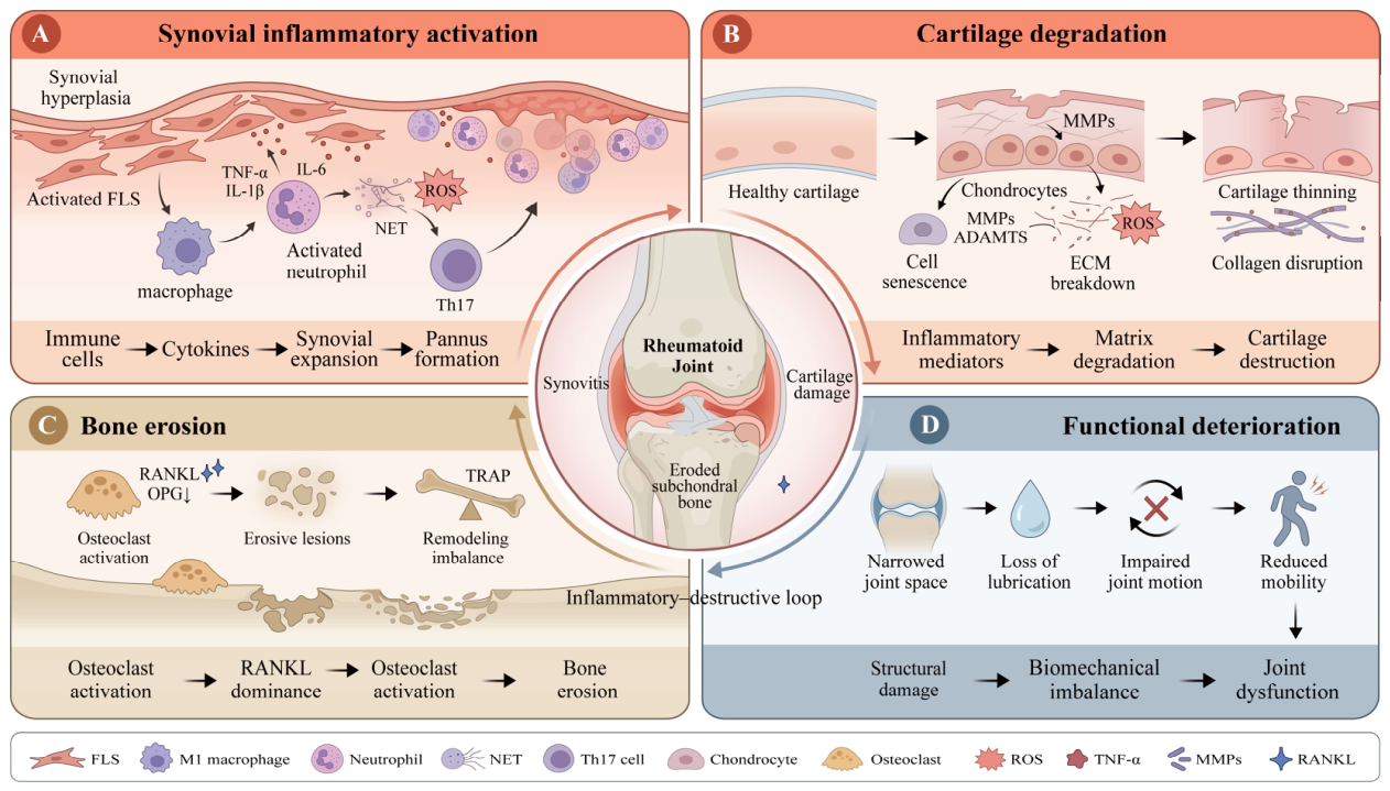
3. Pathological Coupling of the Synovium–Cartilage–Bone Axis in RA
3.1. The Engine of Synovial Inflammation: The Macrophage–FLS Network
3.2. Cartilage Matrix Degradation and Impaired Lubrication
3.3. Bone Erosion Driven by RANKL/OPG Imbalance
3.4. Multitissue Positive Feedback and Its Implications for Hydrogel Design
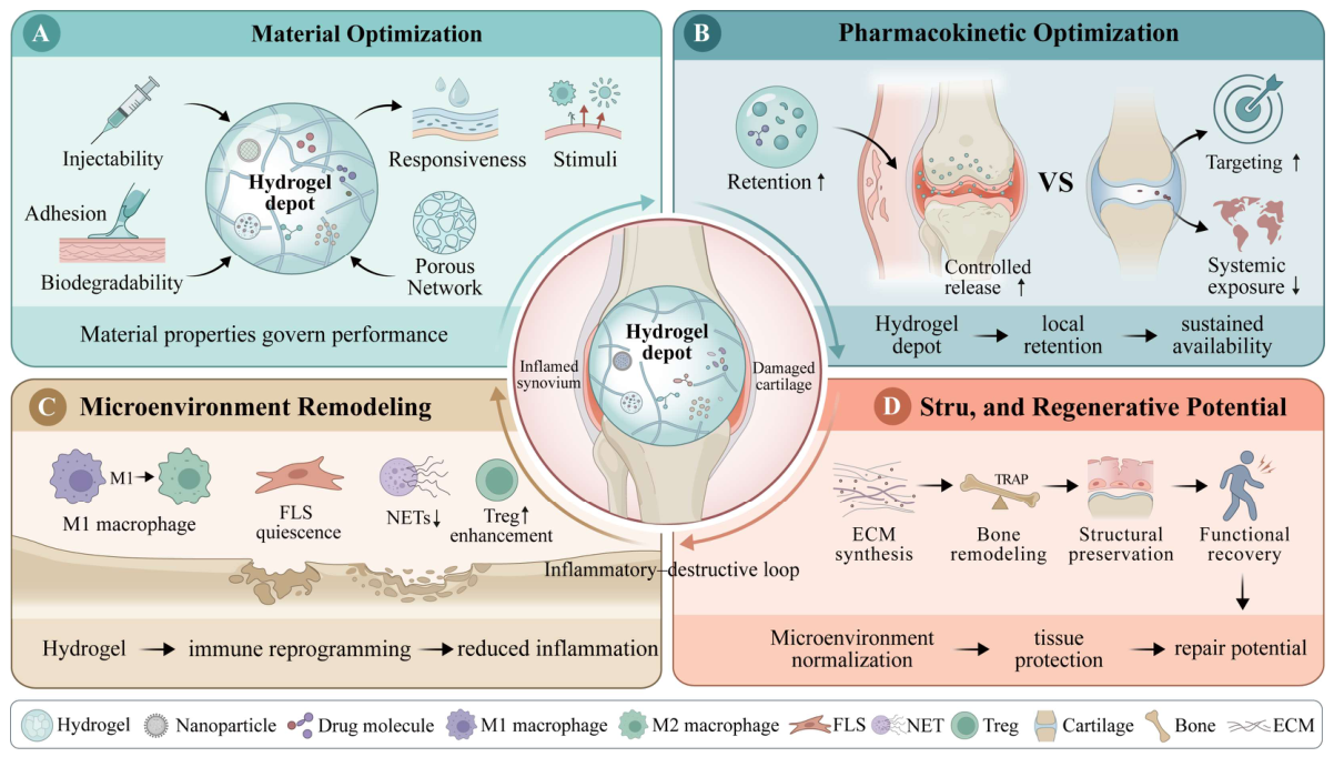
4. Hydrogel Design Requirements for Local Structural Protection and Disease-Modifying Potential
4.1. Injectability, In Situ Gelation, and Intra-Articular Retention
4.2. Adhesion, Self-Healing, Fatigue Stability, and Lubrication
4.3. Responsiveness to Pathological Microenvironmental Signals, Including ROS, pH, and MMPs
4.4. Coupled Design of Mechanical Properties, Degradation, and Release Behavior
5. Local Modulation of the Synovial Inflammatory Microenvironment
5.1. Modulation of Macrophage States and Functions
5.2. Inhibition and Selective Targeting of Pathological FLS
5.3. Coordinated Regulation of Macrophage–FLS Crosstalk
5.4. Adaptive Immune Tolerance: Potential and Evidentiary Boundaries
6. Cartilage Protection, Repair, and Boundaries of Regeneration
6.1. Antioxidant and MMP-Inhibitory Strategies and Chondrocyte Protection
6.2. Restoration of Joint Lubrication and Reduction of Mechanical Wear
6.3. Delivery of Stem Cells, Extracellular Vesicles, and Chondrogenic Factors
6.4. From Cartilage Protection to Functional Regeneration: A Conservative Evidence Hierarchy
7. Control of Bone Erosion and Osteoimmune Modulation
7.1. Inhibition of Osteoclast Formation and Bone Resorption
7.2. Regulation of the RANKL/OPG Axis and Inflammation–Osteoclast Coupling
7.3. Osteogenic Support and Repair of Bone Erosions
7.4. Remodeling the Osteoimmune Microenvironment of Bone-Erosion Sites
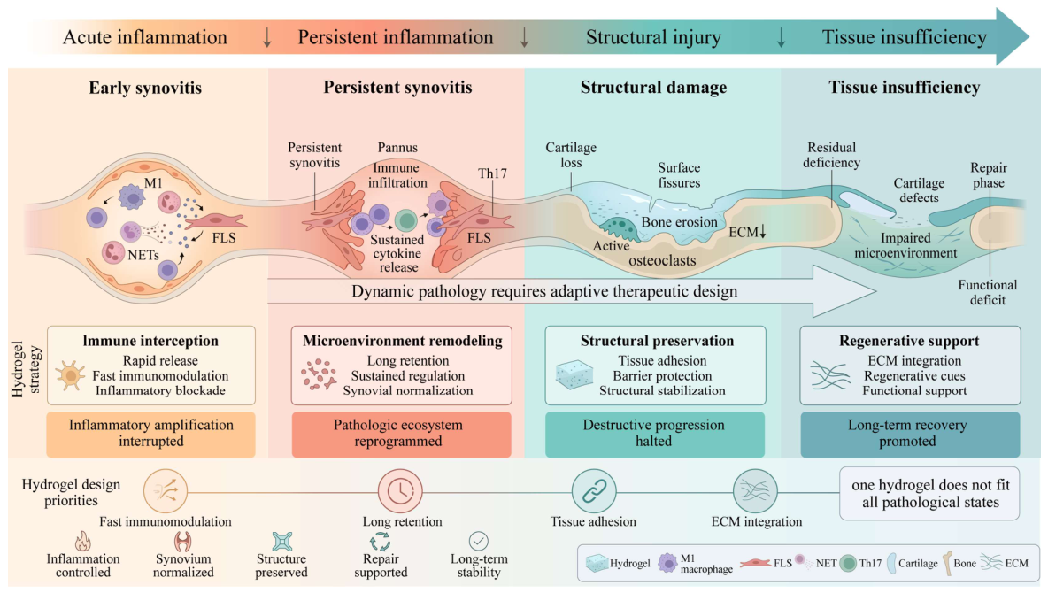
8. Multitissue Coverage and Adaptation to Pathological States
8.1. From Single-Compartment Intervention to Multitissue Coverage
8.2. Prioritizing Treatment According to Pathological State
8.3. Spatial and Sequential Release: Design Rationale and Validation Requirements
9. Evidence Evaluation and Clinical Translation
9.1. From Endpoint Listing to a Study-Level Four-Dimensional Evidence Matrix
9.2. Immune Response, Structural Protection, Tissue Repair, and Functional Structural Modification

| Hydrogel System | RA Model and Model Characteristics | Administration Route and Observation Design | Principal In Vivo Evaluation Endpoints | Highest Level of Evidence | Evidentiary Boundaries and Major Limitations | Reference |
|---|---|---|---|---|---|---|
| Dexamethasone-loaded HA–Tyr hydrogel | Collagen-induced arthritis (CIA) model; primarily recapitulates immune-mediated synovitis and secondary joint damage | Intra-articular injection; short-term therapeutic effects following sustained local release were evaluated | IL-6, PGE2, and multiple cytokines; H&E staining of joint tissues | Level I: Immune response | Evidence was based mainly on reductions in inflammatory mediators and improvement in overall histopathology; the cartilage matrix, bone erosion, and joint function were not separately quantified, precluding a conclusion of structural modification | [4] |
| MTX-loaded click-crosslinked Cx-HA drug depot | RA rat model; used to evaluate intra-articular drug retention and local treatment | Single intra-articular injection; free MTX, MTX-HA, and MTX-Cx-HA were compared | Intra-articular drug distribution, arthritis index, cartilage thickness, chondrocytes and glycosaminoglycan deposition, inflammatory mediators, and histological indicators related to new bone formation | Level III: Signals of tissue repair | Chondrocytes, glycosaminoglycan deposition, and new bone formation were reported, but the mechanical properties of the newly formed tissue, interfacial integration, and standardized joint-function assessments were lacking; histological repair cannot be directly equated with mature functional regeneration | [212] |
| IFX-loaded F127–HA–PGA thermosensitive hydrogel | Ovalbumin/complete Freund’s adjuvant (OVA/CFA)-induced rabbit knee arthritis model; suitable for evaluating intra-articular cartilage and pain-related behavior in a relatively large joint | A single 0.5 mL dose was injected into the affected knee after model establishment; follow-up lasted 6 weeks | Joint temperature and diameter, inflammatory mediators in synovial fluid, synovial histology, gross cartilage examination, H&E, toluidine blue, and Safranin O staining, COL I/COL II, weight-bearing index, and paw-withdrawal threshold | Level II: Structural protection with pain and weight-bearing benefits | Clear evidence of cartilage protection and behavioral benefit was obtained, but bone erosion was not evaluated; a 6-week observation period is insufficient to demonstrate durable structural modification, and pain relief does not indicate tissue regeneration | [93] |
| IND/MTX/MMP-9 siRNA in situ hydrogel | Mouse arthritis model; simultaneously targets inflammation and MMP-9-mediated matrix degradation | Intra-articular injection of the composite nanogel; compared with formulations containing individual or partial cargos | Paw and ankle swelling, TNF-α, IL-6, and MMP-9, ankle morphological parameters, and histology | Level II: Structural protection | The ankle morphology approached normal and suggested reduced cartilage injury, but the formation, integration, and mechanical properties of newly formed hyaline cartilage were not demonstrated; direct functional endpoints were lacking | [92] |
| DNase I-functionalized dynamic hydrogel | CIA mouse model; focuses on inflammatory amplification driven by the persistent presence of NETs | Intra-articular injection of the DNase-functionalized hydrogel; combined with MTX in some experiments | NET- and citrullinated histone-related indicators, inflammatory mediators, arthritis scores, paw swelling, and joint histology | Level I: Immune response | Direct evidence showed that sustained NET degradation alleviated inflammation, but cartilage, bone, and functional endpoints were incomplete; general histological improvement is insufficient to elevate the evidence to structural modification | [73] |
| TAP2-loaded click-crosslinked HA hydrogel | RA animal model; uses Toll-like receptor 4 (TLR4)-associated innate immune signaling to model persistent synovitis and structural damage | Intra-articular injection; free peptide, non-crosslinked carrier, and click-crosslinked hydrogel were compared | In vivo peptide stability and retention, arthritis index, inflammatory mediators, cartilage thickness, glycosaminoglycans, and bone-related histology | Level II: Structural protection | Supports cartilage-matrix preservation and shows signals associated with tissue repair, but evidence of interfacial integration of newly formed tissue, mechanical testing, and direct functional evaluation was lacking, preventing classification as mature Level III tissue repair | [95] |
| HP@CEL HA–nanodrug supramolecular hydrogel | RA rodent model; focuses on macrophage–FLS crosstalk | Local intra-articular administration; free drug, nanocarrier, and composite hydrogel were compared | Joint swelling and arthritis scores, macrophage phenotypes, FLS activation, inflammatory mediators, cartilage ECM, and histological indicators related to bone structure | Level II: Multitissue structural protection | Synovial, cartilage, and bone-related indicators were assessed, but cartilage and bone outcomes relied mainly on histology; long-term micro-CT, tissue mechanics, and functional endpoints were lacking | [18] |
| SPT@TPL dual dynamically crosslinked hydrogel | RA rodent model; oxidative stress and the inflammatory microenvironment serve as the principal therapeutic targets | Intra-articular injection; the synergistic effects of material-mediated ROS regulation and sustained triptolide release were evaluated | Paw swelling, arthritis scores, inflammatory mediators, ROS, macrophage states, and articular-cartilage histology | Level II: Structural protection | Improvements in cartilage staining and surface morphology support a protective effect, but the formation of new hyaline cartilage, integration with subchondral bone, and restoration of joint function were not demonstrated | [80] |
| Anti-inflammatory–osteogenic HA/collagen interpenetrating-network hydrogel | RA bone-erosion microenvironment model incorporating a focal bone-erosion/defect repair setting | The injectable hydrogel was adapted to the bone-erosion region; staged anti-inflammatory, anti-osteoclastic, and osteogenic effects were evaluated | Macrophage states, TRAP and osteoclast-related indicators, osteogenic markers, micro-CT-derived bone-volume fraction and trabecular parameters, new bone formation, and tissue integration | Level III: Bone tissue repair | Provides relatively direct evidence of new bone formation and bone-defect filling; however, an experimentally created focal defect differs from naturally progressive marginal erosion in RA, and recovery of overall joint mechanics or motor function was not demonstrated | [6] |
| DNRS dual-gas-regulating self-healing hydrogel | RA animal model characterized by excess NO, insufficient H2S, macrophage dysregulation, and osteoclast activation | Intra-articular injection; the hydrogel scavenged NO and released H2S and MTX under pathological conditions | Joint swelling and scores, inflammatory mediators, macrophage states, TRAP, bone histology, and micro-CT bone parameters | Level II: Structural protection with signals of bone repair | Demonstrated suppression of inflammation and osteoclast activity together with improved bone microstructure, but stable filling with newly formed bone and long-term integration with host tissue were not adequately established | [97] |
| DAGQD@Cu@KGN–SO3−/DA-HA adhesive lubricating hydrogel | Two-stage model: CIA rats at an early stage and OIA rabbits with superimposed standardized full-thickness osteochondral defects at a later stage | CIA rats received intra-articular administration on the day of the second immunization and were observed for 4 weeks; OIA rabbits were observed for 8 weeks after treatment of the defect site | Rats: arthritis scores, micro-CT, cartilage histology, SOX9, type II collagen (COL II), aggrecan (ACAN), and inflammatory mediators; rabbits: gross defect examination, micro-CT, HSS/Osteoarthritis Research Society International (OARSI) scores, bone volume/tissue volume (BV/TV), trabecular number (Tb.N), and indentation modulus of newly formed tissue | Level III: Cartilage/osteochondral tissue repair | This is one of the systems with the most comprehensive structural assessments and includes local mechanical testing of newly formed tissue; however, the later-stage model combined an inflammatory background with an artificial defect, and Level IV functional structural modification was not demonstrated by long-term gait, weight-bearing, or range-of-motion assessments | [13] |
| CuS-T/ChSMA MMP-9-binding hydrogel | Adjuvant-induced arthritis (AIA) mouse model; emphasizes synovial pannus and MMP-9-mediated cartilage injury | Intra-articular injection of a photocrosslinked hydrogel; non-targeted CuS and MMP-9-binding CuS-T were compared | Arthritic symptoms, synovial inflammation, RA-FLS invasion, macrophage states, MMP-9, MAPK signaling, COL II, aggrecan, and cartilage histology | Level II: Synovium–cartilage dual-compartment structural protection | Demonstrated an association between local MMP-9 binding and improvement in the cartilage matrix, but mature cartilage-defect filling, interfacial integration, cartilage mechanics, and bone-erosion assessment were lacking | [72] |
| Polymer-modified DNA hydrogel co-delivering functional mitochondria and Prussian blue nanozymes | RA animal model; primarily targets intracellular and extracellular oxidative stress and mitochondrial dysfunction | Intra-articular injection of a composite DNA hydrogel containing functional mitochondria and nanozymes | ROS and mitochondrial function, inflammatory mediators, macrophage and synovial states, and cartilage and osteochondral histology | Level II: Multitissue structural protection | Cartilage- and bone-related repair signals were observed, but the in vivo survival, tissue localization, and long-term effects of the functional organelles remain unclear; standardized functional assessments and long-term recurrence monitoring were lacking | [16] |
| ChSMA@SPD spermidine hydrogel | CIA mouse model supplemented by an RA patient-derived synovial organoid–chondrocyte coculture model | Intra-articular administration; eight animals per group were used in the CIA in vivo experiments | Arthritis scores and incidence, paw-pad thickness, H&E and Safranin O staining, COL2A, MMP3, macrophage states, micro-CT, BV/TV, trabecular thickness (Tb.Th), and trabecular separation (Tb.Sp) | Level II: Cartilage and bone structural protection | Provides evidence of both cartilage preservation and bone structural parameters, but the in vivo bone findings mainly indicate reduced erosion or structural preservation; integrated new bone formation within erosion sites was not demonstrated, and direct functional endpoints were not included | [52] |
| Gel-MTX/Mg supramolecular hydrogel | RA rat model; focuses on inflammation and osteochondral destruction | Single intra-articular administration; free MTX, individual components, and the complete Gel-MTX/Mg formulation were compared | Paw swelling and arthritis scores, inflammatory mediators, cartilage histology, osteoclast-related indicators, and bone microstructure | Level II: Multitissue structural protection | A single administration improved inflammatory and osteochondral indicators, but the evidence primarily supports structural preservation rather than new tissue formation within defect sites; standardized gait, weight-bearing, and range-of-motion assessments were absent | [7] |
9.3. Model Extrapolation, Intra-Articular Fate, and Long-Term Safety
9.4. Manufacturing, Sterilization, Quality Control, and Clinical Positioning
10. Conclusions and Perspectives
Author Contributions
Funding
Institutional Review Board Statement
Informed Consent Statement
Data Availability Statement
Acknowledgments
Conflicts of Interest
Abbreviations
| ACAN | Aggrecan |
| ADAMTS | A disintegrin and metalloproteinase with thrombospondin motifs |
| ADAMTS-5 | A disintegrin and metalloproteinase with thrombospondin motifs 5 |
| ADSCs | Adipose-derived stem cells |
| AIA | Adjuvant-induced arthritis |
| ALP | Alkaline phosphatase |
| ASK1 | Apoptosis signal-regulating kinase 1 |
| BMP-2 | Bone morphogenetic protein 2 |
| BMP-7 | Bone morphogenetic protein 7 |
| BMP9 | Bone morphogenetic protein 9 |
| BP | Black phosphorus |
| Bregs | Regulatory B cells |
| BV/TV | Bone volume/tissue volume |
| CCL18 | C-C motif chemokine ligand 18 |
| CD80 | Cluster of differentiation 80 |
| CD86 | Cluster of differentiation 86 |
| CD147 | Basigin/cluster of differentiation 147 |
| CD206 | Cluster of differentiation 206 |
| CFU/g | Colony-forming units per gram |
| CIA | Collagen-induced arthritis |
| COL I | Type I collagen |
| COL II | Type II collagen |
| COL2A | Type II collagen alpha chain marker |
| CQAs | Critical quality attributes |
| CSF1 | Colony-stimulating factor 1 |
| CTSK | Cathepsin K |
| DCs | Dendritic cells |
| DMARD/DMARDs | Disease-modifying antirheumatic drug(s) |
| DNA | Deoxyribonucleic acid |
| DNase I | Deoxyribonuclease I |
| ECM | Extracellular matrix |
| EVs | Extracellular vesicles |
| FAPI | Fibroblast activation protein inhibitor |
| FK506 | Tacrolimus |
| FLS | Fibroblast-like synoviocytes |
| GelMA | Gelatin methacryloyl |
| HA | Hyaluronic acid |
| HAPLN1 | Hyaluronan and proteoglycan link protein 1 |
| H&E | Hematoxylin and eosin |
| HRP | Horseradish peroxidase |
| H2O2 | Hydrogen peroxide |
| H2S | Hydrogen sulfide |
| IDO | Indoleamine 2,3-dioxygenase |
| IFX | Infliximab |
| IGF-1 | Insulin-like growth factor 1 |
| IL-1β | Interleukin-1β |
| IL-4 | Interleukin-4 |
| IL-6 | Interleukin-6 |
| IL-17 | Interleukin-17 |
| IL-32 | Interleukin-32 |
| IL-6/JAK2/STAT3 | Interleukin-6/Janus kinase 2/signal transducer and activator of transcription 3 |
| IND | Indomethacin |
| JAK2 | Janus kinase 2 |
| JNK | c-Jun N-terminal kinase |
| KGN | Kartogenin |
| MAPK | Mitogen-activated protein kinase |
| METTL3 | Methyltransferase-like 3 |
| m6A | N6-methyladenosine |
| miRNA/miRNAs | MicroRNA(s) |
| miR-124-3p | MicroRNA-124-3p |
| miR-140 | MicroRNA-140 |
| miR-195a | MicroRNA-195a |
| miR-221/222 | MicroRNA-221/222 |
| circRNA_28313 | Circular RNA 28313 |
| MMP/MMPs | Matrix metalloproteinase(s) |
| MMP-1 | Matrix metalloproteinase-1 |
| MMP-2 | Matrix metalloproteinase-2 |
| MMP-3/MMP3 | Matrix metalloproteinase-3 |
| MMP-9 | Matrix metalloproteinase-9 |
| MMP-13 | Matrix metalloproteinase-13 |
| MRI | Magnetic resonance imaging |
| MSCs | Mesenchymal stem cells |
| MTX | Methotrexate |
| mRNA | Messenger RNA |
| NET/NETs | Neutrophil extracellular trap(s) |
| NETosis | Neutrophil extracellular trap formation |
| NF-κB | Nuclear factor-κB |
| NFATc1 | Nuclear factor of activated T cells 1 |
| NLRP3 | NOD-like receptor family pyrin domain-containing 3 |
| NO | Nitric oxide |
| NSAIDs | Nonsteroidal anti-inflammatory drugs |
| OA | Osteoarthritis |
| OARSI | Osteoarthritis Research Society International |
| OCN | Osteocalcin |
| OIA | Osteoarthritis-induced arthritis/osteochondral-injury-associated arthritis model, as used in the manuscript context |
| OPG | Osteoprotegerin |
| OVA/CFA | Ovalbumin/complete Freund’s adjuvant |
| PAD4 | Peptidyl arginine deiminase 4 |
| PD-L1 | Programmed death-ligand 1 |
| PGE2 | Prostaglandin E2 |
| PK | Pharmacokinetic(s) |
| PLXNC1 | Plexin C1 |
| PRG4 | Proteoglycan 4 |
| PRP | Platelet-rich plasma |
| PVA | Poly(vinyl alcohol) |
| RA | Rheumatoid arthritis |
| RA-FLS | Rheumatoid arthritis-derived fibroblast-like synoviocytes |
| RANK | Receptor activator of nuclear factor-κB |
| RANKL | Receptor activator of nuclear factor-κB ligand |
| RANKL/OPG | RANKL/osteoprotegerin ratio or balance |
| RANKL/TNF | RANKL/tumor necrosis factor-related strategy |
| RGD | Arginine–glycine–aspartic acid |
| RNA | Ribonucleic acid |
| ROS | Reactive oxygen species |
| RUNX2 | Runt-related transcription factor 2 |
| SAL | Sterility assurance level |
| SIN | Sinomenine hydrochloride |
| SIS | Small intestinal submucosa |
| siRNA | Small interfering RNA |
| SOX9 | SRY-box transcription factor 9 |
| SPARCL1 | Secreted protein acidic and rich in cysteine-like 1 |
| SPD | Spermidine |
| STAT3 | Signal transducer and activator of transcription 3 |
| TAP | Toll-like receptor antagonist peptide |
| Tb.N | Trabecular number |
| Tb.Sp | Trabecular separation |
| Tb.Th | Trabecular thickness |
| TGF-β | Transforming growth factor-β |
| TGF-β3 | Transforming growth factor-β3 |
| Tfh | T follicular helper cell |
| Th17 | T helper 17 cell |
| TLR4 | Toll-like receptor 4 |
| TLRs | Toll-like receptors |
| TNF | Tumor necrosis factor |
| TNF-α | Tumor necrosis factor-α |
| TNFR1 | Tumor necrosis factor receptor 1 |
| TNFR2 | Tumor necrosis factor receptor 2 |
| TPL | Triptolide |
| TRAF | Tumor necrosis factor receptor-associated factor |
| TRAP | Tartrate-resistant acid phosphatase |
| Tregs | Regulatory T cells |
| Tr1 cells | Type 1 regulatory T cells |
| VEGF | Vascular endothelial growth factor |
| W9 | W9 peptide |
| WYRGRL | Cartilage-binding peptide sequence WYRGRL |
| c-Fos | Fos proto-oncogene |
| c-Src | Cellular Src tyrosine kinase |
| micro-CT | Micro-computed tomography |
| Material/system abbreviation | Full term/explanation |
| BiNS/PEI | BiNS/polyethyleneimine nanosheets |
| CeNZs | Cerium-based nanozymes/ceria nanozymes |
| ChSMA | Methacrylated chondroitin sulfate |
| CS/BP | Chitosan/black phosphorus |
| CuS-T | MMP-9-binding CuS-T component |
| Cx-HA | Click-crosslinked hyaluronic acid |
| Cx-SIS | Click-crosslinked small intestinal submucosa |
| DA-HA | Dopamine-modified hyaluronic acid |
| DAGQD | Dopamine-hybrid graphene quantum dots |
| DAGQD@Cu@KGN–SO3−/DA-HA | DAGQD-, Cu-, KGN-, sulfonate-, and DA-HA-based multifunctional hydrogel |
| DNRS | Dual-gas-regulating self-healing hydrogel |
| DS | Diclofenac sodium |
| DSP | Dexamethasone sodium phosphate |
| F127 | Pluronic F127/poloxamer 407 |
| F68 | Pluronic F68/poloxamer 188 |
| Gel-MTX/Mg | Methotrexate- and magnesium-containing supramolecular hydrogel |
| GG | Gellan gum |
| HA–Tyr | Tyramine-modified hyaluronic acid |
| HC@PTM | Composite hydrogel containing MTX-loaded polymeric micelles |
| HP@CEL | Hydrophobically modified HA/celastrol-loaded nanoparticle hydrogel |
| HPAP | HPAP hydrogel system |
| IND–MTX | Indomethacin–methotrexate |
| IND–MTX–siMMP-9 | Indomethacin–methotrexate–MMP-9 siRNA system |
| KGN@P407 | Kartogenin-loaded Pluronic P407 system |
| M-NO | Nitric oxide-responsive or NO-scavenging gel system |
| MOS | Manno-oligosaccharide-modified system |
| NanoIGUR | Iguratimod-loaded nanomicelles |
| PCL | Polycaprolactone |
| PECT | PECT nanoparticles |
| PEG | Polyethylene glycol |
| PEGDMA | Poly(ethylene glycol) dimethacrylate |
| PEI | Polyethyleneimine |
| PEI-SS | Disulfide-containing polyethyleneimine component |
| PGA | Poly(γ-glutamic acid) |
| PLA | Polylactic acid/polylactide |
| PRP–CS/BP | Platelet-rich plasma–chitosan/black phosphorus hydrogel |
| SPT@TPL | Triptolide-loaded dynamically crosslinked hydrogel system |
| TAP/Cx-HA | Toll-like receptor antagonist peptide-loaded click-crosslinked HA hydrogel |
| TAP2 + Cx-HA | TAP2-loaded click-crosslinked HA system |
| Tyr–GG | Tyramine-modified gellan gum |
References
- Kadiri, M.; Charbonneau, M.; Lalanne, C.; Harper, K.; Balg, F.; Marotta, A.; Dubois, C.M. 14-3-3η Promotes Invadosome Formation via the FOXO3-Snail Axis in Rheumatoid Arthritis Fibroblast-like Synoviocytes. Int. J. Mol. Sci. 2021, 23, 123. [Google Scholar] [CrossRef] [PubMed]
- Baig, M.; Wong, L.K.; Zia, A.W.; Wu, H. Development of biomedical hydrogels for rheumatoid arthritis treatment. Asian J. Pharm. Sci. 2024, 19, 100887. [Google Scholar] [CrossRef] [PubMed]
- Singh, H.; Dan, A.; Kumawat, M.K.; Pawar, V.; Chauhan, D.S.; Kaushik, A.; Bhatia, D.; Srivastava, R.; Dhanka, M. Pathophysiology to advanced intra-articular drug delivery strategies: Unravelling rheumatoid arthritis. Biomaterials 2023, 303, 122390. [Google Scholar] [CrossRef] [PubMed]
- Kim, K.S.; Park, S.J.; Yang, J.A.; Jeon, J.H.; Bhang, S.H.; Kim, B.S.; Hahn, S.K. Injectable hyaluronic acid-tyramine hydrogels for the treatment of rheumatoid arthritis. Acta Biomater. 2011, 7, 666–674. [Google Scholar] [CrossRef] [PubMed]
- Song, Y.; Yang, P.; Guo, W.; Lu, P.; Huang, C.; Cai, Z.; Jiang, X.; Yang, G.; Du, Y.; Zhao, F. Supramolecular Hydrogel Dexamethasone-Diclofenac for the Treatment of Rheumatoid Arthritis. Nanomaterials 2024, 14, 645. [Google Scholar] [CrossRef] [PubMed]
- Liu, X.; Zhang, Q.; Cao, Y.; Hussain, Z.; Xu, M.; Liu, Y.; Ullah, I.; Lu, Z.; Osaka, A.; Lin, J.; et al. An Injectable Hydrogel Composing Anti-Inflammatory and Osteogenic Therapy toward Bone Erosions Microenvironment Remodeling in Rheumatoid Arthritis. Adv. Healthc. Mater. 2024, 13, e2304668. [Google Scholar] [CrossRef] [PubMed]
- Li, H.; Lyu, Y.; Wang, R.; Yu, H.; Lin, M.; Li, Z.; Zhong, Y.; Sheng, P.; Zhang, K.; Liao, W.; et al. Supramolecular drug-laden hydrogel based on structural tautomerization enhances drug delivery for rheumatoid arthritis treatment. Bioact. Mater. 2025, 53, 495–506. [Google Scholar] [CrossRef] [PubMed]
- Cui, J.; Cui, Y.; Zhao, Y. Research Progress of Hydrogels in the Treatment of Rheumatoid Arthritis and Osteoarthritis. Biomed. Eng. Res. 2022, 41, 405–410. [Google Scholar] [CrossRef]
- Shunan, G.; Qi, L. Research Progress of Hydrogel Drug Delivery Systems for the Treatment of Rheumatoid Arthritis. Chin. J. Jt. Surg. (Electron. Ed.) 2020, 14, 597–601. [Google Scholar]
- Zhu, H.; Wu, X.; Liu, R.; Zhao, Y.; Sun, L. ECM-Inspired Hydrogels with ADSCs Encapsulation for Rheumatoid Arthritis Treatment. Adv. Sci. 2025, 10, e2206253, Correction in Adv. Sci. 2019, 12, e2417661. https://doi.org/10.1002/advs.202417661. [Google Scholar] [CrossRef]
- Haloi, P.; Chawla, S.; Konkimalla, V.B. Thermosensitive smart hydrogel of PEITC ameliorates the therapeutic efficacy in rheumatoid arthritis. Eur. J. Pharm. Sci. 2023, 181, 106367. [Google Scholar] [CrossRef] [PubMed]
- Zhao, Y.; Gao, C.; Liu, H.; Liu, H.; Feng, Y.; Li, Z.; Liu, H.; Wang, J.; Yang, B.; Lin, Q. Infliximab-based self-healing hydrogel composite scaffold enhances stem cell survival, engraftment, and function in rheumatoid arthritis treatment. Acta Biomater. 2021, 121, 653–664. [Google Scholar] [CrossRef] [PubMed]
- He, H.; Zhang, Q.; Zhang, Y.; Qu, S.; Li, B.; Lin, J.; Lu, X.; Xie, C. Injectable bioadhesive and lubricating hydrogel with polyphenol mediated single atom nanozyme for rheumatoid arthritis therapy. Nat. Commun. 2025, 16, 2768. [Google Scholar] [CrossRef] [PubMed]
- Bahatibieke, A.; Zhao, J.; Fan, D.; Zhou, Z.; Li, J.; Wang, X.; Zhao, H.; Wang, T.; Fang, Z.; Xie, Y.; et al. Sea-Island Micelle Structured Hydrogel Scaffold: A Dual-Action Approach to Combat Cartilage Damage under RA Conditions. ACS Appl. Mater. Interfaces 2025, 17, 2911–2923, Correction in ACS Appl. Mater. Interfaces 2025, 17, 19200. https://doi.org/10.1021/acsami.5c03006. [Google Scholar] [CrossRef]
- Back, W.; Jeong, M.; Le, H.T.; Park, J.H. Immunosuppressive Cytokine-Tethered Hydrogel for Treating Rheumatoid Arthritis. Adv. Healthc. Mater. 2025, 14, e2501613. [Google Scholar] [CrossRef] [PubMed]
- Wang, F.; Han, Y.; Zhou, Q.; Sheng, S.; Hu, Y.; Zhang, H.; Chen, X.; He, C.; Tan, H.; Bai, L.; et al. Polymer-modified DNA hydrogels for living mitochondria and nanozyme delivery in the treatment of rheumatoid arthritis. Bioact. Mater. 2025, 47, 448–459. [Google Scholar] [CrossRef] [PubMed]
- Menarim, B.C.; Gillis, K.H.; Oliver, A.; Ngo, Y.; Werre, S.R.; Barrett, S.H.; Rodgerson, D.H.; Dahlgren, L.A. Macrophage Activation in the Synovium of Healthy and Osteoarthritic Equine Joints. Front. Vet. Sci. 2020, 7, 568756. [Google Scholar] [CrossRef] [PubMed]
- Wang, Y.; Wang, J.; Ma, M.; Gao, R.; Wu, Y.; Zhang, C.; Huang, P.; Wang, W.; Feng, Z.; Gao, J. Hyaluronic-Acid-Nanomedicine Hydrogel for Enhanced Treatment of Rheumatoid Arthritis by Mediating Macrophage-Synovial Fibroblast Cross-Talk. Biomater. Res. 2024, 28, 0046. [Google Scholar] [CrossRef] [PubMed]
- Saisai, H.; Cheng, Z.; Jun, L.; Xianyan, L.; Shiying, W. Mechanism of Macrophage Extracellular Traps Inducing the Activation of Fibroblast-Like Synoviocytes in the Pathogenesis of Rheumatoid Arthritis. J. Clin. Med. Pract. 2022, 26, 97–102. [Google Scholar] [CrossRef]
- Miao, T.; Qiu, Y.; Chen, J.; Li, P.; Li, H.; Zhou, W.; Shen, W. METTL3 knockdown suppresses RA-FLS activation through m(6)A-YTHDC2-mediated regulation of AMIGO2. Biochim. Biophys. Acta Mol. Basis Dis. 2024, 1870, 167112. [Google Scholar] [CrossRef] [PubMed]
- Chen, Y.H.; Zhang, X.; Chou, C.H.; Hsueh, M.F.; Attarian, D.; Li, Y.J.; Kraus, V.B. Association of Dipeptidylpeptidase 4 (CD26) With Chondrocyte Senescence and Radiographic Progression in Knee Osteoarthritis. Arthritis Rheumatol. 2023, 75, 1120–1131. [Google Scholar] [CrossRef] [PubMed]
- Lv, Y.; Gao, W. Interleukin-17 Induces the Release of Inflammatory and Bone Erosive Factors from Fibroblast-Like Synoviocytes in Rheumatoid Arthritis via Signal Transducer and Activator of Transcription 3. Chin. J. Rheumatol. 2017, 21, 338–341. [Google Scholar]
- Wang, C.; Chen, L.; Zhu, P.; Fan, C.; Wang, Y.; Jia, J. CD147 stimulates the angiogenesis in rheumatoid synovium via the activation of vascular endothelial growth factor. Chin. J. Cell. Mol. Immunol. 2007, 23, 426–428. [Google Scholar]
- Blackler, G.; Jiang, H.; Appleton, C.T. Synovial joint organ homeostasis: Mechanisms and biological considerations. Connect. Tissue Res. 2025, 66, 331–338. [Google Scholar] [CrossRef] [PubMed]
- Dong, W.J.; Zhu, P.; Fan, C.M.; Wang, Y.H.; Xiao, L.B. Expression of CD147 and Matrix Metalloproteinases in Synovium of Patients with Rheumatoid Arthritis. Chin. J. Rheumatol. 2004, 135–138+193. [Google Scholar]
- Chen, L.; Jing, J.; Yang, L.; Ma, J.; Mo, Y. CC Chemokine Ligand 18 Enriched in Synovial Fluid Participates in Joint Destruction by Promoting the Migration of Fibroblast-Like Synoviocytes in Rheumatoid Arthritis. Chin. J. Front. Med. Sci. (Electron. Version) 2023, 15, 55–64. [Google Scholar]
- Xie, P.; Dan, F.; Yu, G.; Ruan, W.; Yu, H. Laquinimod Mitigated IL-1β-Induced Impairment of the Cartilage Extracellular Matrix in Human ATDC5 Chondrocytes. Chem. Res. Toxicol. 2020, 33, 933–939. [Google Scholar] [CrossRef] [PubMed]
- Gao, J.B.; Lin, L.; Men, X.Q.; Zhao, J.B.; Zhang, M.H.; Jin, L.P.; Gao, S.J.; Zhao, S.Q.; Dong, J.T. Fibulin-5 protects the extracellular matrix of chondrocytes by inhibiting the Wnt/β-catenin signaling pathway and relieves osteoarthritis. Eur. Rev. Med. Pharmacol. Sci. 2020, 24, 5249–5258. [Google Scholar] [CrossRef] [PubMed]
- Arra, M.; Abu-Amer, Y. Cross-talk of inflammation and chondrocyte intracellular metabolism in osteoarthritis. Osteoarthr. Cartil. 2023, 31, 1012–1021. [Google Scholar] [CrossRef] [PubMed]
- Meng, N.; Mao, L.; Jiang, Q.; Yuan, J.; Liu, L.; Wang, L. PLXNC1 interference alleviates the inflammatory injury, apoptosis and extracellular matrix degradation of IL-1β-exposed chondrocytes via suppressing GRP78 expression. J. Orthop. Surg. Res. 2023, 18, 784. [Google Scholar] [CrossRef] [PubMed]
- Miao, Y.; Wu, S.; Gong, Z.; Chen, Y.; Xue, F.; Liu, K.; Zou, J.; Feng, Y.; Li, G. SPARCL1 promotes chondrocytes extracellular matrix degradation and inflammation in osteoarthritis via TNF/NF-κB pathway. J. Orthop. Transl. 2024, 46, 116–128. [Google Scholar] [CrossRef] [PubMed]
- Yao, Q.; Chen, X.; Sheng, H.; Zhang, Y.; Chen, R.; Fan, P.; Kou, L. Lubricating nano/micro particles for osteoarthritis therapy. Mater. Horiz. 2025, 12, 9476–9508. [Google Scholar] [CrossRef] [PubMed]
- Das, N.; Schmidt, T.A.; Krawetz, R.J.; Dufour, A. Proteoglycan 4: From Mere Lubricant to Regulator of Tissue Homeostasis and Inflammation: Does proteoglycan 4 have the ability to buffer the inflammatory response? Bioessays 2019, 41, e1800166. [Google Scholar] [CrossRef] [PubMed]
- Wang, X.; Fu, Y.; Lin, W. Liposome-based nanocolloids for restoring lubrication and targeted therapy in osteoarthritis. Colloids Surf. B Biointerfaces 2026, 265, 115723. [Google Scholar] [CrossRef] [PubMed]
- Chen, W.; Ye, Q.; Zhang, M.; Xie, R.; Xu, C. Lubrication for Osteoarthritis: From Single-Function to Multifunctional Lubricants. Int. J. Mol. Sci. 2025, 26, 1856. [Google Scholar] [CrossRef] [PubMed]
- Ge, W.; Qi, L.; Wang, Y.; Wang, J.; Fang, X.; Lei, S.; Lin, D.; Zhang, L.; Zhang, S. Engineered M2 macrophage-derived extracellular vesicles reprogram mitochondrial metabolism to alleviate temporomandibular joint cartilage degeneration. Mater. Today Bio 2026, 36, 102674. [Google Scholar] [CrossRef] [PubMed]
- Danks, L.; Komatsu, N.; Guerrini, M.M.; Sawa, S.; Armaka, M.; Kollias, G.; Nakashima, T.; Takayanagi, H. RANKL expressed on synovial fibroblasts is primarily responsible for bone erosions during joint inflammation. Ann. Rheum. Dis. 2016, 75, 1187–1195. [Google Scholar] [CrossRef] [PubMed]
- Yu, L.; Zhou, X. RANKL and Osteoclasts. Int. J. Endocrinol. Metab. 2003, 23, 123–126. [Google Scholar] [CrossRef]
- Xi, Z.; Hu, J. RANKL-RANK Signaling, Osteoclastogenesis and Bone Diseases. Chin. J. Osteoporos. 2008, 14, 285–293. [Google Scholar]
- Liang, G. Osteoblasts, Osteoclasts and OPG/RANKL/RANK Axis in Osteoporosis. J. Tradit. Chin. Orthop. Traumatol. 2010, 22, 41–44+47. [Google Scholar]
- Zhang, C.; Lin, Y. Regulatory Effects of Traditional Chinese Medicine on Osteoclasts Based on OPG/RANKL/RANK Signaling Pathway. J. Rehabil. 2018, 28, 59–64+70. [Google Scholar]
- Zhong, L.; Yang, B.; Huang, X.; Sun, Y. The Role of OPG/RANKL/RANK System in the Mutual Regulation Between Osteoblasts and Osteoclasts. Chin. J. Osteoporos. 2011, 17, 1010–1013. [Google Scholar]
- Yang, X.; Hong, F.; Xie, W.; Zhang, J.; Jin, Z.; Qin, Z. Effects of Combined Exposure to Fluoride and Arsenic on OPG/RANKL-Mediated Osteoclast Differentiation. J. Environ. Health 2019, 36, 305–310. [Google Scholar] [CrossRef]
- Yao, C.; Kong, F.; Chen, Y.; Mei, Y. Study on the Relationship Between Simvastatin and the Inhibition of Osteoclasts and Regulation of RANKL/OPG System. Chin. J. Conserv. Dent. 2016, 26, 525–530+535. [Google Scholar] [CrossRef]
- Wang, S.; Zhang, Z.; Chen, J. RANKL Induces the Differentiation and Maturation of Osteoclast Precursor Cells. Chin. J. Gerontol. 2011, 31, 963–965. [Google Scholar] [CrossRef]
- Liu, Y.; Wang, T.; Chen, H.; Tao, P.; Ma, B.; Zhang, Y.; Guan, H.; Xu, W.; Li, H.; Chen, A.; et al. Effects of RANKL Concentration on the Formation and Activity of Osteoclasts. Orthopaedics 2014, 5, 5–8. [Google Scholar] [CrossRef]
- Hong, Y.; Jiang, H.; Wang, J.; Yu, P.; You, W. Experimental Study on Formononetin Inhibiting RANKL-Induced Osteoclast Differentiation. China J. Orthop. Traumatol. 2020, 33, 64–70. [Google Scholar]
- Zhang, J.; Zhuang, Y.; Shang, B.; Sun, H.; Wang, Y. Quercitrin Inhibits RANKL-Induced Osteoclast Formation and Bone Resorption. Orthop. J. China 2015, 23, 2180–2185. [Google Scholar]
- Kim, H.N.; Xiong, J.; MacLeod, R.S.; Iyer, S.; Fujiwara, Y.; Cawley, K.M.; Han, L.; He, Y.; Thostenson, J.D.; Ferreira, E.; et al. Osteocyte RANKL is required for cortical bone loss with age and is induced by senescence. JCI Insight 2020, 5, 138815. [Google Scholar] [CrossRef] [PubMed]
- Lu, G.; Liu, O.; Xu, L. Preliminary Study on Semagacestat Regulating RANKL-Induced Osteoclast Formation via Notch Signaling Pathway. Oral. Biomed. 2021, 12, 160–165. [Google Scholar]
- Zhang, M. Denosumab and Osteoclast RANKL/RANK Signaling Pathway. In Proceedings of the 2021 Annual Conference of Chinese Society for Gerontology and Geriatrics—Actively Responding to Population Aging: Promoting Coordinated Development of Elderly Care Undertakings and Industries, Beijing, China, 19–20 December 2021; pp. 310–315. [Google Scholar]
- Wang, X.; He, J.; Hong, Y.; Jie, L.; Wu, B.; Shen, J.; Zhou, G.; Wang, Q. Spermidine-encapsulated chondroitin sulfate methacryloyl hydrogel delivery system for remodeling bone homeostasis in rheumatoid arthritis. Mater. Today Bio 2025, 35, 102531. [Google Scholar] [CrossRef] [PubMed]
- Yan, Z.; Chen, Y.; Xiao, J.; Lu, S. Effects of Sinomenine Hydrogel on Autophagy and Inflammation via IL-6/JAK2/STAT3 Signaling Pathway in Collagen-Induced Rheumatoid Arthritis Mice. J. Intern. Med. 2024, 41, 633–637. [Google Scholar] [CrossRef]
- Song, Y.; Hao, B. Preparation and In Vitro Transdermal Permeation of Sinomenine Hydrochloride Ethosomal Hydrogel Ointment. Cent. South. Pharm. 2017, 15, 1216–1220. [Google Scholar]
- Zhou, C.; Ma, X.; Wang, B.; Shen, J.; Ji, W. Study on CeNZs/KGN@P407 Thermosensitive Hydrogel for the Treatment of Collagen-Induced Arthritis in Rats. J. Med. Res. 2026, 55, 124–129. [Google Scholar]
- Shen, W.; Xu, A.; Li, S.; Deng, J.; Wang, J.; Yang, F.; Chen, Y.; Zhang, H. Creatine Phosphate-Modified Chitosan Hydrogel Regulates Macrophage Polarization and Inflammatory Factor Expression in Rat Bone Marrow-Derived Macrophages. Chin. J. Tissue Eng. Res. 2022, 26, 5040–5046. [Google Scholar]
- Thambi, T.; Li, Y.; Lee, D.S. Injectable hydrogels for sustained release of therapeutic agents. J. Control Release 2017, 267, 57–66. [Google Scholar] [CrossRef] [PubMed]
- Dimatteo, R.; Darling, N.J.; Segura, T. In situ forming injectable hydrogels for drug delivery and wound repair. Adv. Drug Deliv. Rev. 2018, 127, 167–184. [Google Scholar] [CrossRef] [PubMed]
- Mathew, A.P.; Uthaman, S.; Cho, K.H.; Cho, C.S.; Park, I.K. Injectable hydrogels for delivering biotherapeutic molecules. Int. J. Biol. Macromol. 2018, 110, 17–29. [Google Scholar] [CrossRef] [PubMed]
- Macaya, D.; Spector, M. Injectable hydrogel materials for spinal cord regeneration: A review. Biomed. Mater. 2012, 7, 012001. [Google Scholar] [CrossRef] [PubMed]
- Li, X.; Zhao, D.; Shea, K.J.; Li, X.; Lu, X. In situ formed thermogelable hydrogel photonic crystals assembled by thermosensitive IPNs. Mater. Horiz. 2021, 8, 932–938. [Google Scholar] [CrossRef] [PubMed]
- Pouso, M.R.; Melo, B.L.; Gonçalves, J.J.; Mendonça, A.G.; Correia, I.J.; de Melo-Diogo, D. Development of dual-crosslinked Pluronic F127/Chitosan injectable hydrogels incorporating graphene nanosystems for breast cancer photothermal therapy and antibacterial applications. Eur. J. Pharm. Biopharm. 2024, 203, 114476. [Google Scholar] [CrossRef] [PubMed]
- Morgen, M.; Tung, D.; Boras, B.; Miller, W.; Malfait, A.M.; Tortorella, M. Nanoparticles for improved local retention after intra-articular injection into the knee joint. Pharm. Res. 2013, 30, 257–268. [Google Scholar] [CrossRef] [PubMed]
- Bae, J.W.; Choi, J.H.; Lee, Y.; Park, K.D. Horseradish peroxidase-catalysed in situ-forming hydrogels for tissue-engineering applications. J. Tissue Eng. Regen. Med. 2015, 9, 1225–1232. [Google Scholar] [CrossRef] [PubMed]
- Luo, Y.; Zhao, Y.; Chen, L.; Guan, Y.; Zhang, Y. In Situ-Forming, Adhesive, and Antioxidant Chitosan Hydrogels for Accelerated Wound Healing. Biomacromolecules 2025, 26, 1219–1233. [Google Scholar] [CrossRef] [PubMed]
- Singh, A.; Agarwal, R.; Diaz-Ruiz, C.A.; Willett, N.J.; Wang, P.; Lee, L.A.; Wang, Q.; Guldberg, R.E.; García, A.J. Nanoengineered particles for enhanced intra-articular retention and delivery of proteins. Adv. Healthc. Mater. 2014, 3, 1562–1567, 1525. [Google Scholar] [CrossRef] [PubMed]
- Sandker, M.J.; Duque, L.F.; Redout, E.M.; Chan, A.; Que, I.; Löwik, C.; Klijnstra, E.C.; Kops, N.; Steendam, R.; van Weeren, R.; et al. Degradation, intra-articular retention and biocompatibility of monospheres composed of [PDLLA-PEG-PDLLA]-b-PLLA multi-block copolymers. Acta Biomater. 2017, 48, 401–414. [Google Scholar] [CrossRef] [PubMed]
- Guo, Z.; Han, Y.; Peng, J.; Peng, C.; Zhao, M.; Hu, H.; Chen, D. Polyglutamic acid and Pluronic F127-based hydrogel for loading sinomenine hydrochloride liposomes in the treatment of rheumatoid arthritis. Colloids Surf. B Biointerfaces 2025, 256, 115060. [Google Scholar] [CrossRef] [PubMed]
- Ma, Z.; Tao, C.; Sun, L.; Qi, S.; Le, Y.; Wang, J.; Li, C.; Liu, X.; Zhang, J.; Zhao, J. In Situ Forming Injectable Hydrogel For Encapsulation Of Nanoiguratimod And Sustained Release Of Therapeutics. Int. J. Nanomed. 2019, 14, 8725–8738. [Google Scholar] [CrossRef] [PubMed]
- Zhou, Y.; He, X.; Zhang, W.; Zhang, W.; Zhao, H.; Zhou, X.; Gu, Q.; Shen, H.; Yang, H.; Liu, X.; et al. Cell-recruited microspheres for OA treatment by dual-modulating inflammatory and chondrocyte metabolism. Mater. Today Bio 2024, 27, 101127. [Google Scholar] [CrossRef] [PubMed]
- Luo, Z.; Zhang, S.; Pan, J.; Shi, R.; Liu, H.; Lyu, Y.; Han, X.; Li, Y.; Yang, Y.; Xu, Z.; et al. Time-responsive osteogenic niche of stem cells: A sequentially triggered, dual-peptide loaded, alginate hybrid system for promoting cell activity and osteo-differentiation. Biomaterials 2018, 163, 25–42. [Google Scholar] [CrossRef] [PubMed]
- Xue, Z.; Li, N.; Du, K.; Shu, J.; Huang, Z.; Gao, Z.; Xie, X.; Li, Q.; Lu, Y. Inhibiting synovial inflammation and promoting cartilage repair in rheumatoid arthritis using a matrix metalloproteinase-binding hydrogel. Mater. Today Bio 2025, 32, 101792. [Google Scholar] [CrossRef] [PubMed]
- Wang, N.; Ma, J.; Song, W.; Zhao, C. An injectable hydrogel to disrupt neutrophil extracellular traps for treating rheumatoid arthritis. Drug Deliv. 2023, 30, 2173332. [Google Scholar] [CrossRef] [PubMed]
- Liu, X.; Chen, X.; Fei, Y.; Zhang, J.; Yue, O.; Wang, X.; Jiang, H. Locally Injectable, ROS-Scavenging, and ROS-/pH-Responsive Polymeric-Micelles-Embedded Hydrogels for Precise Minimally Invasive and Long-Lasting Rheumatoid Therapy. Adv. Healthc. Mater. 2025, 14, e2403579. [Google Scholar] [CrossRef] [PubMed]
- Oliveira, I.M.; Gonçalves, C.; Shin, M.E.; Lee, S.; Reis, R.L.; Khang, G.; Oliveira, J.M. Enzymatically crosslinked tyramine-gellan gum hydrogels as drug delivery system for rheumatoid arthritis treatment. Drug Deliv. Transl. Res. 2021, 11, 1288–1300. [Google Scholar] [CrossRef] [PubMed]
- Pan, W.; Dai, C.; Li, Y.; Yin, Y.; Gong, L.; Machuki, J.O.; Yang, Y.; Qiu, S.; Guo, K.; Gao, F. PRP-chitosan thermoresponsive hydrogel combined with black phosphorus nanosheets as injectable biomaterial for biotherapy and phototherapy treatment of rheumatoid arthritis. Biomaterials 2020, 239, 119851. [Google Scholar] [CrossRef] [PubMed]
- Zhang, A.; Ma, Y.; Liu, Y.; Dong, S.; Torres, M.N.; Kim, B.Y.S.; Liu, C.; Sun, L.; Yuan, Y.; Jiang, W. Mannose Oligosaccharide-Conjugated In Situ Pore-Forming Injectable Hydrogels for Rheumatoid Arthritis Treatment by Reprogramming Macrophage Extracellular Vesicles. Small Methods 2026, 10, e2500605. [Google Scholar] [CrossRef] [PubMed]
- Huang, X.; Ye, Y.; Zhang, J.; Zhang, X.; Ma, H.; Zhang, Y.; Fu, X.; Tang, J.; Jiang, N.; Han, Y.; et al. Reactive Oxygen Species Scavenging Functional Hydrogel Delivers Procyanidins for the Treatment of Traumatic Brain Injury in Mice. ACS Appl. Mater. Interfaces 2022, 14, 33756–33767. [Google Scholar] [CrossRef] [PubMed]
- An, Z.; Zhang, L.; Liu, Y.; Zhao, H.; Zhang, Y.; Cao, Y.; Zhang, Y.; Pei, R. Injectable thioketal-containing hydrogel dressing accelerates skin wound healing with the incorporation of reactive oxygen species scavenging and growth factor release. Biomater. Sci. 2021, 10, 100–113. [Google Scholar] [CrossRef] [PubMed]
- Wang, T.; Huang, C.; Fang, Z.; Bahatibieke, A.; Fan, D.; Wang, X.; Zhao, H.; Xie, Y.; Qiao, K.; Xiao, C.; et al. A dual dynamically cross-linked hydrogel promotes rheumatoid arthritis repair through ROS initiative regulation and microenvironment modulation-independent triptolide release. Mater. Today Bio 2024, 26, 101042. [Google Scholar] [CrossRef] [PubMed]
- Li, H.; Wei, S.; Ling, Q.; Wang, R.; Liu, T.; Yu, H.; Zhao, P.; Zhang, K.; Bian, L.; Liao, W. Nanozyme-Reinforced Hydrogel Spray as a Reactive Oxygen Species-Driven Oxygenator to Accelerate Diabetic Wound Healing. Adv. Mater. 2025, 37, e2504829. [Google Scholar] [CrossRef] [PubMed]
- Kim, Y.E.; Choi, S.W.; Kim, M.K.; Nguyen, T.L.; Kim, J. Therapeutic Hydrogel Patch to Treat Atopic Dermatitis by Regulating Oxidative Stress. Nano Lett. 2022, 22, 2038–2047. [Google Scholar] [CrossRef] [PubMed]
- Wu, J.; Ma, J.; Li, Z.; Li, J.; Hu, J.; Ma, F.; Wu, Y.; Han, F.; Li, B.; Xiao, C. Smart Microneedles Regulate Reactive Oxygen Species and Deliver Matrix Metalloproteinases for Pathological Scar Treatment. ACS Nano 2026, 20, 15153–15170. [Google Scholar] [CrossRef] [PubMed]
- Zhu, R.; Liao, H.Y.; Huang, Y.C.; Shen, H.L. Application of Injectable Hydrogels as Delivery Systems in Osteoarthritis and Rheumatoid Arthritis. Br. J. Hosp. Med. 2024, 85, 1–41. [Google Scholar] [CrossRef] [PubMed]
- Wang, K.; Yin, C.; Ye, X.; Chen, Q.; Wu, J.; Chen, Y.; Li, Y.; Wang, J.; Duan, C.; Lu, A.; et al. A Metabolic Driven Bio-Responsive Hydrogel Loading Psoralen for Therapy of Rheumatoid Arthritis. Small 2023, 19, e2207319. [Google Scholar] [CrossRef] [PubMed]
- Zewail, M.; Nafee, N.; Boraie, N. Intra-Articular Dual Drug Delivery for Synergistic Rheumatoid Arthritis Treatment. J. Pharm. Sci. 2021, 110, 2808–2822. [Google Scholar] [CrossRef] [PubMed]
- Tsai, W.B.; Chen, C.J. Development of hyaluronic acid hydrogel containing prednisolone-encapsulated nonphospholipid liposomes for the treatment of rheumatoid arthritis. J. Biomed. Mater. Res. B Appl. Biomater. 2024, 112, e35453. [Google Scholar] [CrossRef] [PubMed]
- Kim, K.; Park, J.H.; Park, S.H.; Lee, H.Y.; Kim, J.H.; Kim, M.S. An Injectable, Click-Cross-Linked Small Intestinal Submucosa Drug Depot for the Treatment of Rheumatoid Arthritis. Adv. Healthc. Mater. 2016, 5, 3105–3117. [Google Scholar] [CrossRef] [PubMed]
- Wu, H.; Wang, K.; Wang, H.; Chen, F.; Huang, W.; Chen, Y.; Chen, J.; Tao, J.; Wen, X.; Xiong, S. Novel self-assembled tacrolimus nanoparticles cross-linking thermosensitive hydrogels for local rheumatoid arthritis therapy. Colloids Surf. B Biointerfaces 2017, 149, 97–104. [Google Scholar] [CrossRef] [PubMed]
- Park, J.H.; Park, S.H.; Lee, H.Y.; Lee, J.W.; Lee, B.K.; Lee, B.Y.; Kim, J.H.; Kim, M.S. An injectable, electrostatically interacting drug depot for the treatment of rheumatoid arthritis. Biomaterials 2018, 154, 86–98. [Google Scholar] [CrossRef] [PubMed]
- Yin, N.; Guo, X.; Sun, R.; Liu, H.; Tang, L.; Gou, J.; Yin, T.; He, H.; Zhang, Y.; Tang, X. Intra-articular injection of indomethacin-methotrexate in situ hydrogel for the synergistic treatment of rheumatoid arthritis. J. Mater. Chem. B 2020, 8, 993–1007. [Google Scholar] [CrossRef] [PubMed]
- Yin, N.; Tan, X.; Liu, H.; He, F.; Ding, N.; Gou, J.; Yin, T.; He, H.; Zhang, Y.; Tang, X. A novel indomethacin/methotrexate/MMP-9 siRNA in situ hydrogel with dual effects of anti-inflammatory activity and reversal of cartilage disruption for the synergistic treatment of rheumatoid arthritis. Nanoscale 2020, 12, 8546–8562. [Google Scholar] [CrossRef] [PubMed]
- Chen, W.; Li, Z.; Wang, Z.; Gao, H.; Ding, J.; He, Z. Intraarticular Injection of Infliximab-Loaded Thermosensitive Hydrogel Alleviates Pain and Protects Cartilage in Rheumatoid Arthritis. J. Pain. Res. 2020, 13, 3315–3329. [Google Scholar] [CrossRef] [PubMed]
- Kim, T.; Suh, J.; Kim, W.J. Polymeric Aggregate-Embodied Hybrid Nitric-Oxide-Scavenging and Sequential Drug-Releasing Hydrogel for Combinatorial Treatment of Rheumatoid Arthritis. Adv. Mater. 2021, 33, e2008793. [Google Scholar] [CrossRef] [PubMed]
- Lee, S.; Seo, J.; Kim, Y.H.; Ju, H.J.; Kim, S.; Ji, Y.B.; Lee, H.B.; Kim, H.S.; Choi, S.; Kim, M.S. Enhanced intra-articular therapy for rheumatoid arthritis using click-crosslinked hyaluronic acid hydrogels loaded with toll-like receptor antagonizing peptides. Acta Biomater. 2023, 172, 188–205. [Google Scholar] [CrossRef] [PubMed]
- Liao, H.; Qi, W.; Xue, Z.; Wu, K.; Jiang, L.; Wu, C.; Huang, Z.; Li, Q.; Lu, Y. A multifunctional supramolecular hydrogel that rapidly binds TNF-α for efficient reduction of synovial inflammation and cartilage destruction in rheumatoid arthritis. Chem. Eng. J. 2023, 477, 147125. [Google Scholar] [CrossRef]
- Geng, W.; Zhao, J.; Tao, B.; Yang, Y.; Duan, Q.; Gao, P.; He, T.; Liu, S.; Feng, Q.; Zhao, P.; et al. Regulation of rheumatoid arthritis microenvironment via a self-healing injectable hydrogel for improved inflammation elimination and bone repair. Bioact. Mater. 2024, 36, 287–300. [Google Scholar] [CrossRef] [PubMed]
- Xu, M.; Fu, T.; Zhang, C.; An, Z.; Yan, J.; Lu, Z.; Wu, H.; Liu, J.; Qiu, L.; Shi, L.; et al. Prolonged, staged, and self-regulated methotrexate release coupled with ROS scavenging in an injectable hydrogel for rheumatoid arthritis therapy. J. Control Release 2024, 375, 60–73. [Google Scholar] [CrossRef] [PubMed]
- Yang, R.; Yan, L.; Xu, T.; Zhang, K.; Lu, X.; Xie, C.; Fu, W. Injectable bioadhesive hydrogel as a local nanomedicine depot for targeted regulation of inflammation and ferroptosis in rheumatoid arthritis. Biomaterials 2024, 311, 122706. [Google Scholar] [CrossRef] [PubMed]
- Wu, Y.; Ge, Y.; Wang, Z.; Zhu, Y.; Tian, T.; Wei, J.; Jin, Y.; Zhao, Y.; Jia, Q.; Wu, J.; et al. Synovium microenvironment-responsive injectable hydrogel inducing modulation of macrophages and elimination of synovial fibroblasts for enhanced treatment of rheumatoid arthritis. J. Nanobiotechnol. 2024, 22, 188. [Google Scholar] [CrossRef] [PubMed]
- Wang, M.; Liu, K.; She, P.; Gao, T.; Shi, Z.; Wang, Y.; Ding, X.; Yang, Y.; Yin, D.; Shen, W.; et al. In Situ Ca2+-Reinforced Full-Active Hydrogels for the Treatment of Rheumatoid Arthritis by Modulating the NF-κB and NLRP3 Pathways. ACS Appl. Mater. Interfaces 2026, 18, 6563–6577. [Google Scholar] [CrossRef] [PubMed]
- Xue, L.; Qing, L. The Role of Macrophage Polarization in Rheumatoid Arthritis. Chin. Bull. Life Sci. 2018, 30, 261–266. [Google Scholar] [CrossRef]
- Huang, Y.; Sun, G. Research Progress of Macrophage Polarization in Rheumatoid Arthritis. World Latest Med. Inf. 2020, 20, 46–48. [Google Scholar]
- Van den Bossche, J.; Baardman, J.; Otto, N.A.; van der Velden, S.; Neele, A.E.; van den Berg, S.M.; Luque-Martin, R.; Chen, H.J.; Boshuizen, M.C.; Ahmed, M.; et al. Mitochondrial Dysfunction Prevents Repolarization of Inflammatory Macrophages. Cell Rep. 2016, 17, 684–696. [Google Scholar] [CrossRef] [PubMed]
- Wang, Q.; Su, Y.Y.; Li, Y.Q.; Zhang, Y.F.; Yang, S.; Wang, J.L.; Li, H.Y. Atorvastatin alleviates renal ischemia-reperfusion injury in rats by promoting M1-M2 transition. Mol. Med. Rep. 2017, 15, 798–804. [Google Scholar] [CrossRef] [PubMed]
- Hu, Q.; Xie, N.; Liao, K.; Huang, J.; Yang, Q.; Zhou, Y.; Liu, Y.; Deng, K. An injectable thermosensitive Pluronic F127/hyaluronic acid hydrogel loaded with human umbilical cord mesenchymal stem cells and asiaticoside microspheres for uterine scar repair. Int. J. Biol. Macromol. 2022, 219, 96–108. [Google Scholar] [CrossRef] [PubMed]
- Su, S.; Wang, H.; Cao, X.; Xu, Y.; Qi, H.; Sun, J.; Wang, C.; Zhen, M. Macrophage-Targeted Fullerene Potentiates Redox Homeostasis Regulation and Reprograms Macrophage Polarization to Ameliorate Hepatic Steatosis. ACS Appl. Bio Mater. 2026, 9, 3066–3075. [Google Scholar] [CrossRef] [PubMed]
- Zhao, W.; Xu, D.; Hong, W.; Zhang, L.; Wu, Q.; Gao, M.; Wang, J. Grossamide attenuates inflammation by balancing macrophage polarization through metabolic reprogramming of macrophages in mice. Int. Immunopharmacol. 2022, 112, 109190. [Google Scholar] [CrossRef] [PubMed]
- Galván-Peña, S.; O’Neill, L.A. Metabolic reprograming in macrophage polarization. Front. Immunol. 2014, 5, 420. [Google Scholar] [CrossRef] [PubMed]
- Nakamizo, S.; Kabashima, K. Metabolic reprogramming and macrophage polarization in granuloma formation. Int. Immunol. 2024, 36, 329–338. [Google Scholar] [CrossRef] [PubMed]
- Guo, T.; Li, J.; Lin, J.; Xie, M.; Du, Y.; Fu, J.; Wang, Q.; Chen, X.; Luo, S. Mitochondrial transfer from adipose-derived stem cells reprograms macrophage lipid metabolism to improve fat graft survival. J. Transl. Med. 2026. [Google Scholar] [CrossRef] [PubMed]
- Liu, S.; Wang, H.; Li, J.; Gao, J.; Yu, L.; Wei, X.; Cui, M.; Zhao, Y.; Liang, Y.; Wang, H. Loss of Bcl-3 regulates macrophage polarization by promoting macrophage glycolysis. Immunol. Cell Biol. 2024, 102, 605–617. [Google Scholar] [CrossRef] [PubMed]
- Yu, Y.; Cai, W.; Zhou, J.; Wei, F. Metabolic Reprogramming Regulates Macrophage Polarization and Its Roles in Rheumatoid Arthritis. Acta Pharm. Sin. 2020, 55, 2027–2034. [Google Scholar] [CrossRef]
- Zhao, J.; Lu, J.; Lu, Y.; Guo, J.; Zhang, X. Research Progress on Lactate and Lactylation Modification Regulating Macrophages in the Pathogenesis of Rheumatoid Arthritis. J. Youjiang Med. Univ. Natl. 2025, 47, 870–876. [Google Scholar]
- Roumelioti, F.; Tzaferis, C.; Konstantopoulos, D.; Papadopoulou, D.; Prados, A.; Sakkou, M.; Liakos, A.; Chouvardas, P.; Meletakos, T.; Pandis, Y.; et al. Mir221/222 drive synovial hyperplasia and arthritis by targeting cell cycle inhibitors and chromatin remodeling components. eLife 2024, 13, e84698. [Google Scholar] [CrossRef] [PubMed]
- Mnich, S.J.; Blanner, P.M.; Hu, L.G.; Shaffer, A.F.; Happa, F.A.; O’Neil, S.; Ukairo, O.; Weiss, D.; Welsh, E.; Storer, C.; et al. Critical role for apoptosis signal-regulating kinase 1 in the development of inflammatory K/BxN serum-induced arthritis. Int. Immunopharmacol. 2010, 10, 1170–1176. [Google Scholar] [CrossRef] [PubMed]
- Kloesch, B.; Becker, T.; Dietersdorfer, E.; Kiener, H.; Steiner, G. Anti-inflammatory and apoptotic effects of the polyphenol curcumin on human fibroblast-like synoviocytes. Int. Immunopharmacol. 2013, 15, 400–405. [Google Scholar] [CrossRef] [PubMed]
- Han, Z.; Feng, Y.; Zhang, Y.; Ren, X.; Hu, M.; Li, M.; Lv, Z.; Zhang, Y.; Fan, Y.; Yang, R.; et al. Chemotherapeutic Agent-Loaded Nanoparticles Synergizing with X-ray Irradiation to Regulate Fibroblast-like Synoviocytes for Rheumatoid Arthritis Treatment. ACS Nano 2026, 20, 13830–13840. [Google Scholar] [CrossRef] [PubMed]
- Chen, L.; Zhao, M.; Kang, W.; Yu, L.; Zhang, C.; Wu, S.; Song, X.; Zhao, K.; Liu, P.; Liu, Q.; et al. Endogenous Melanin and Hydrogen-Based Specific Activated Theranostics Nanoagents: A Novel Multi-Treatment Paradigm for Rheumatoid Arthritis. Adv. Sci. 2024, 11, e2401046. [Google Scholar] [CrossRef] [PubMed]
- Li, F.; Tang, Y.; Song, B.; Yu, M.; Li, Q.; Zhang, C.; Hou, J.; Yang, R. Nomenclature clarification: Synovial fibroblasts and synovial mesenchymal stem cells. Stem Cell Res. Ther. 2019, 10, 260. [Google Scholar] [CrossRef] [PubMed]
- Tong, S.; Liu, J.; Zhang, C. Platelet-rich plasma inhibits inflammatory factors and represses rheumatoid fibroblast-like synoviocytes in rheumatoid arthritis. Clin. Exp. Med. 2017, 17, 441–449. [Google Scholar] [CrossRef] [PubMed]
- Browning, S.R.; Weiser, A.M.; Woolf, N.; Golish, S.R.; SanGiovanni, T.P.; Scuderi, G.J.; Carballo, C.; Hanna, L.S. Platelet-rich plasma increases matrix metalloproteinases in cultures of human synovial fibroblasts. J. Bone Jt. Surg. Am. 2012, 94, e1721–e1727. [Google Scholar] [CrossRef] [PubMed]
- Thangadurai, M.; Sethuraman, S.; Subramanian, A. A 2D inflammatory co-culture model for investigating synovial fibroblast and macrophage interactions in rheumatoid arthritis. Sci. Rep. 2025, 15, 30105. [Google Scholar] [CrossRef] [PubMed]
- Liu, B.; Xian, Y.; Chen, X.; Shi, Y.; Dong, J.; Yang, L.; An, X.; Shen, T.; Wu, W.; Ma, Y.; et al. Inflammatory Fibroblast-Like Synoviocyte-Derived Exosomes Aggravate Osteoarthritis via Enhancing Macrophage Glycolysis. Adv. Sci. 2024, 11, e2307338. [Google Scholar] [CrossRef] [PubMed]
- Yu, S.; Li, J.; Xu, Z.; Cheng, Y.; Zong, M.; Fan, L. Exosomes Derived from Rheumatoid Arthritis Fibroblast-Like Synoviocytes Promote M1 Macrophage Polarization. J. Tongji Univ. (Med. Sci.) 2023, 44, 785–791. [Google Scholar] [CrossRef]
- Zheng, Y.; Wei, K.; Jiang, P.; Zhao, J.; Shan, Y.; Shi, Y.; Zhao, F.; Chang, C.; Li, Y.; Zhou, M.; et al. Macrophage polarization in rheumatoid arthritis: Signaling pathways, metabolic reprogramming, and crosstalk with synovial fibroblasts. Front. Immunol. 2024, 15, 1394108. [Google Scholar] [CrossRef] [PubMed]
- Nakamachi, Y.; Uto, K.; Hayashi, S.; Okano, T.; Morinobu, A.; Kuroda, R.; Kawano, S.; Saegusa, J. Exosomes derived from synovial fibroblasts from patients with rheumatoid arthritis promote macrophage migration that can be suppressed by miR-124-3p. Heliyon 2023, 9, e14986. [Google Scholar] [CrossRef] [PubMed]
- Bartok, B.; Firestein, G.S. Fibroblast-like synoviocytes: Key effector cells in rheumatoid arthritis. Immunol. Rev. 2010, 233, 233–255. [Google Scholar] [CrossRef] [PubMed]
- Zhang, W. Indoleamine 2,3-Dioxygenase, Dendritic Cells and Immune Tolerance. Int. J. Immunol. 2006, 29, 364–367. [Google Scholar] [CrossRef]
- Chen, Y.; Wang, B.; Xu, G. Dendritic Cells and Immune Tolerance. Int. J. Immunol. 2006, 29, 314–318. [Google Scholar] [CrossRef]
- Mitosek-Szewczyk, K.; Tabarkiewicz, J.; Wilczynska, B.; Lobejko, K.; Berbecki, J.; Nastaj, M.; Dworzanska, E.; Kolodziejczyk, B.; Stelmasiak, Z.; Rolinski, J. Impact of cladribine therapy on changes in circulating dendritic cell subsets, T cells and B cells in patients with multiple sclerosis. J. Neurol. Sci. 2013, 332, 35–40. [Google Scholar] [CrossRef] [PubMed]
- Sage, P.T.; Schildberg, F.A.; Sobel, R.A.; Kuchroo, V.K.; Freeman, G.J.; Sharpe, A.H. Dendritic Cell PD-L1 Limits Autoimmunity and Follicular T Cell Differentiation and Function. J. Immunol. 2018, 200, 2592–2602. [Google Scholar] [CrossRef] [PubMed]
- Moore, C.; Sauma, D.; Reyes, P.A.; Morales, J.; Rosemblatt, M.; Bono, M.R.; Fierro, J.A. Dendritic cells and B cells cooperate in the generation of CD4(+)CD25(+)FOXP3(+) allogeneic T cells. Transplant. Proc. 2010, 42, 371–375. [Google Scholar] [CrossRef] [PubMed]
- Hu, S.H.; Zhang, L.H.; Gao, J.; Guo, J.H.; Xun, X.D.; Xiang, X.; Cheng, Q.; Li, Z.; Zhu, J.Y. NKG2D Enhances Double-Negative T Cell Regulation of B Cells. Front. Immunol. 2021, 12, 650788. [Google Scholar] [CrossRef] [PubMed]
- Volchenkov, R.; Karlsen, M.; Jonsson, R.; Appel, S. Type 1 regulatory T cells and regulatory B cells induced by tolerogenic dendritic cells. Scand. J. Immunol. 2013, 77, 246–254. [Google Scholar] [CrossRef] [PubMed]
- Huo, F.; Shi, X.; Zou, X.; Wang, S.; Zhang, Y.; Yang, W.; Li, Y. Aire deficient dendritic cells promote the T follicular helper cells differentiation. Immunobiology 2019, 224, 539–550. [Google Scholar] [CrossRef] [PubMed]
- Boldison, J.; Da Rosa, L.C.; Davies, J.; Wen, L.; Wong, F.S. Dendritic cells license regulatory B cells to produce IL-10 and mediate suppression of antigen-specific CD8 T cells. Cell Mol. Immunol. 2020, 17, 843–855. [Google Scholar] [CrossRef] [PubMed]
- Fan, K.; Jin, L.; Zhao, C.; Zhou, S.; Tan, S.; Lai, J.; Yao, C.; Long, B.; Gao, Y.; Yu, S. Der p1 Dendritic Cells Promote Regulatory B Cell Induced Immunotolerance Through IL-10/STAT3 in Allergic Rhinitis. Biomedicines 2026, 14, 206. [Google Scholar] [CrossRef] [PubMed]
- Liu, H.; Liu, Y.; Tian, Z.; Li, J.; Li, M.; Zhao, Z. Coordinating Macrophage Targeting and Antioxidation by Injectable Nanocomposite Hydrogel for Enhanced Rheumatoid Arthritis Treatment. ACS Appl. Mater. Interfaces 2024, 16, 37656–37668. [Google Scholar] [CrossRef] [PubMed]
- Djordjevic, K.; Pindovic, B.; Mihajlovic, K.; Ilic, I.; Terzic, J.; Milojevic-Samanovic, A.; Stanic, Z.; Postolovic, K.; Ekkert, N.; Reshetnikov, V.; et al. Exploring the beneficial effects of curcumin-based hydrogel beads in rats with rheumatoid arthritis induced by Freund’s complete adjuvant. Mol. Cell Biochem. 2026, 481, 213–229. [Google Scholar] [CrossRef] [PubMed]
- Zhao, Y.; Song, S.; Wang, D.; Liu, H.; Zhang, J.; Li, Z.; Wang, J.; Ren, X.; Zhao, Y. Nanozyme-reinforced hydrogel as a H(2)O(2)-driven oxygenerator for enhancing prosthetic interface osseointegration in rheumatoid arthritis therapy. Nat. Commun. 2022, 13, 6758. [Google Scholar] [CrossRef] [PubMed]
- Zhao, Y.P.; Han, J.F.; Zhang, F.Y.; Liao, T.T.; Na, R.; Yuan, X.F.; He, G.B.; Ye, W. Flexible nano-liposomes-based transdermal hydrogel for targeted delivery of dexamethasone for rheumatoid arthritis therapy. Drug Deliv. 2022, 29, 2269–2282. [Google Scholar] [CrossRef] [PubMed]
- Guan, P.; Cui, R.; Wang, Q.; Sun, Y. 3D Hydrogel Loaded with Bone Marrow Stem Cell-Derived Exosomes Promotes Damaged Cartilage Repair via Immunomodulation. J. South. Med. Univ. 2022, 42, 528–537. [Google Scholar]
- Qin, X.; Li, T.; Chen, C.; Yang, L. Anti-Inflammatory Peptide-Loaded Zirconium Metal-Organic Framework Encapsulated in Hydrogel Enhances Chondrocyte Extracellular Matrix Production. J. Army Med. Univ. 2022, 44, 673–683. [Google Scholar] [CrossRef]
- Birtwistle, L.; Chen, X.M.; Pollock, C. Mesenchymal Stem Cell-Derived Extracellular Vesicles to the Rescue of Renal Injury. Int. J. Mol. Sci. 2021, 22, 6596. [Google Scholar] [CrossRef] [PubMed]
- Rezaie, J.; Nejati, V.; Mahmoodi, M.; Ahmadi, M. Mesenchymal stem cells derived extracellular vesicles: A promising nanomedicine for drug delivery system. Biochem. Pharmacol. 2022, 203, 115167. [Google Scholar] [CrossRef] [PubMed]
- Park, H.S.; Cetin, E.; Siblini, H.; Seok, J.; Alkelani, H.; Alkhrait, S.; Liakath Ali, F.; Mousaei Ghasroldasht, M.; Beckman, A.; Al-Hendy, A. Therapeutic Potential of Mesenchymal Stem Cell-Derived Extracellular Vesicles to Treat PCOS. Int. J. Mol. Sci. 2023, 24, 11151. [Google Scholar] [CrossRef] [PubMed]
- Abid, A.I.; Conzatti, G.; Toti, F.; Anton, N.; Vandamme, T. Mesenchymal stem cell-derived exosomes as cell free nanotherapeutics and nanocarriers. Nanomedicine 2024, 61, 102769. [Google Scholar] [CrossRef] [PubMed]
- Wang, D.; Cao, H.; Hua, W.; Gao, L.; Yuan, Y.; Zhou, X.; Zeng, Z. Mesenchymal Stem Cell-Derived Extracellular Vesicles for Bone Defect Repair. Membranes 2022, 12, 716. [Google Scholar] [CrossRef] [PubMed]
- Balaraman, A.K.; Arockia Babu, M.; Afzal, M.; Sanghvi, G.; M, M.R.; Gupta, S.; Rana, M.; Ali, H.; Goyal, K.; Subramaniyan, V.; et al. Exosome-based miRNA delivery: Transforming cancer treatment with mesenchymal stem cells. Regen. Ther. 2025, 28, 558–572. [Google Scholar] [CrossRef] [PubMed]
- Tsai, Y.C.; Cheng, T.S.; Liao, H.J.; Chuang, M.H.; Chen, H.T.; Chen, C.H.; Zhang, K.L.; Chang, C.H.; Lin, P.C.; Huang, C.F. Mesenchymal Stem Cell Secreted-Extracellular Vesicles are Involved in Chondrocyte Production and Reduce Adipogenesis during Stem Cell Differentiation. Tissue Eng. Regen. Med. 2022, 19, 1295–1310. [Google Scholar] [CrossRef] [PubMed]
- Wang, X.; Thomsen, P. Mesenchymal stem cell-derived small extracellular vesicles and bone regeneration. Basic. Clin. Pharmacol. Toxicol. 2021, 128, 18–36. [Google Scholar] [CrossRef] [PubMed]
- Liao, H.J.; Yang, Y.P.; Liu, Y.H.; Tseng, H.C.; Huo, T.I.; Chiou, S.H.; Chang, C.H. Harnessing the potential of mesenchymal stem cells-derived exosomes in degenerative diseases. Regen. Ther. 2024, 26, 599–610. [Google Scholar] [CrossRef] [PubMed]
- Torrecillas-Baena, B.; Pulido-Escribano, V.; Dorado, G.; Gálvez-Moreno, M.; Camacho-Cardenosa, M.; Casado-Díaz, A. Clinical Potential of Mesenchymal Stem Cell-Derived Exosomes in Bone Regeneration. J. Clin. Med. 2023, 12, 4385. [Google Scholar] [CrossRef] [PubMed]
- Wei, W.; Ao, Q.; Wang, X.; Cao, Y.; Liu, Y.; Zheng, S.G.; Tian, X. Mesenchymal Stem Cell-Derived Exosomes: A Promising Biological Tool in Nanomedicine. Front. Pharmacol. 2020, 11, 590470. [Google Scholar] [CrossRef] [PubMed]
- Matsuzaka, Y.; Yashiro, R. Therapeutic Strategy of Mesenchymal-Stem-Cell-Derived Extracellular Vesicles as Regenerative Medicine. Int. J. Mol. Sci. 2022, 23, 6480. [Google Scholar] [CrossRef] [PubMed]
- Asensio, G.; Benito-Garzón, L.; Ramírez-Jiménez, R.A.; Guadilla, Y.; Gonzalez-Rubio, J.; Abradelo, C.; Parra, J.; Martín-López, M.R.; Aguilar, M.R.; Vázquez-Lasa, B.; et al. Biomimetic Gradient Scaffolds Containing Hyaluronic Acid and Sr/Zn Folates for Osteochondral Tissue Engineering. Polymers 2021, 14, 12. [Google Scholar] [CrossRef] [PubMed]
- Li, Y.; Zhu, H.; Liu, R.; Zhao, Y.; Sun, L. Hierarchical Microcarriers Loaded with Peptide Dendrimer-Grafted Methotrexate for Rheumatoid Arthritis Treatment. Small Sci. 2024, 4, 2300097. [Google Scholar] [CrossRef] [PubMed]
- Wang, Y.; Zhou, X.; Zheng, J.; Zhou, Y. Human β-Defensin 3-Loaded Hydrogel for the Treatment of Periodontitis in Rats. Chin. J. Tissue Eng. Res. 2024, 28, 2690–2695. [Google Scholar]
- Liu, X.; Li, W.; Zhao, Z.; Zhang, X.; Ma, Z.; Li, C. Experimental Study on the Therapeutic Effect of Drynaria Rhizome Flavonoids on Periodontitis in Rats. J. Conserv. Dent. Periodontol. 2026, 31, 2–9. [Google Scholar] [CrossRef]
- Zhao, Z.; Wu, C.; Huangfu, Y.; Zhang, Y.; Zhang, J.; Huang, P.; Dong, A.; Wang, Y.; Deng, J.; Wang, W.; et al. Bioinspired Glycopeptide Hydrogel Reestablishing Bone Homeostasis through Mediating Osteoclasts and Osteogenesis in Periodontitis Treatment. ACS Nano 2024, 18, 29507–29521. [Google Scholar] [CrossRef] [PubMed]
- Dai, Z.; Wang, D. Preparation Methods and Biological Characteristics of Strontium-Substituted Hydroxyapatite. Chin. J. Tissue Eng. Res. 2018, 22, 938–944. [Google Scholar]
- Xuan, S.; Zhu, Y.; Zhang, L.; Tang, Z.; Xu, F.; Chen, T.; Wang, Z. Controlled Release and Cellular Biological Study of Recombinant Human Parathyroid Hormone (1-34) Mediated by Hyaluronic Acid Thermosensitive Hydrogel. Acta Pharm. Sin. 2022, 57, 809–817. [Google Scholar] [CrossRef]
- Deng, W.; Ding, Z.; Wang, Y.; Zou, B.; Zheng, J.; Tan, Y.; Yang, Q.; Ke, M.; Chen, Y.; Wang, S.; et al. Dendrobine attenuates osteoclast differentiation through modulating ROS/NFATc1/MMP9 pathway and prevents inflammatory bone destruction. Phytomedicine 2022, 96, 153838. [Google Scholar] [CrossRef] [PubMed]
- Lin, Z.; Zhou, Z.; Ye, J.; Wei, J.; Chen, S.; Zhou, W.; Bi, Y.; Zhou, Z.; Xie, G.; Yuan, G.; et al. Trifolirhizin protects ovariectomy-induced bone loss in mice by inhibiting osteoclast differentiation and bone resorption. Heliyon 2024, 10, e34250. [Google Scholar] [CrossRef] [PubMed]
- Li, S.; Ren, X.; Pu, Y.; Li, A.; Liu, X.; Huang, Y.; Guo, L. Injectable Hydrogel Loaded with Magnolol and Nanohydroxyapatite for Treating Periodontitis in Diabetes. ACS Appl. Mater. Interfaces 2026, 18, 6478–6497. [Google Scholar] [CrossRef] [PubMed]
- Chen, X.; Ouyang, Z.; Shen, Y.; Liu, B.; Zhang, Q.; Wan, L.; Yin, Z.; Zhu, W.; Li, S.; Peng, D. CircRNA_28313/miR-195a/CSF1 axis modulates osteoclast differentiation to affect OVX-induced bone absorption in mice. RNA Biol. 2019, 16, 1249–1262. [Google Scholar] [CrossRef] [PubMed]
- Kumar, A.; Ali, A.; Kanika; Vyawahare, A.; Ahmad, A.; Mishra, R.K.; Ansari, M.M.; Nadeem, A.; Siddiqui, N.; Raza, S.S.; et al. Highly Biocompatible Smart Injectable Hydrogel for the Management of Rheumatoid Arthritis. ACS Biomater. Sci. Eng. 2023, 9, 5312–5321. [Google Scholar] [CrossRef] [PubMed]
- Jiao, Y.; Wang, H.; Zhao, M.; Liu, H.; Shen, P.; Wu, G.; Forouzanfar, T.; Tay, F.R.; Miao, L.; Liu, H.; et al. Multifunctional nanocomposite hydrogel with dual-factor controlled release for stem cell recruitment, immunomodulation and bone remodeling. J. Control Release 2025, 384, 113893. [Google Scholar] [CrossRef] [PubMed]
- Kou, Y.; Li, C.; Yang, P.; Li, D.; Lu, X.; Liu, H.; Li, M. The W9 peptide inhibits osteoclastogenesis and osteoclast activity by downregulating osteoclast autophagy and promoting osteoclast apoptosis. J. Mol. Histol. 2022, 53, 27–38. [Google Scholar] [CrossRef] [PubMed]
- Komatsu, N.; Takayanagi, H. Mechanisms of joint destruction in rheumatoid arthritis—Immune cell-fibroblast-bone interactions. Nat. Rev. Rheumatol. 2022, 18, 415–429. [Google Scholar] [CrossRef] [PubMed]
- Braun, T.; Zwerina, J. Positive regulators of osteoclastogenesis and bone resorption in rheumatoid arthritis. Arthritis Res. Ther. 2011, 13, 235. [Google Scholar] [CrossRef] [PubMed]
- Geusens, P. The role of RANK ligand/osteoprotegerin in rheumatoid arthritis. Ther. Adv. Musculoskelet. Dis. 2012, 4, 225–233. [Google Scholar] [CrossRef] [PubMed]
- Chen, L.; Liang, L. Osteoimmunology of Osteoporosis in Rheumatoid Arthritis: Emerging Mechanisms and Therapeutic Implications. Int. J. Gen. Med. 2025, 18, 7813–7822. [Google Scholar] [CrossRef] [PubMed]
- O’Neil, L.J.; Oliveira, C.B.; Wang, X.; Navarrete, M.; Barrera-Vargas, A.; Merayo-Chalico, J.; Aljahdali, R.; Aguirre-Aguilar, E.; Carlucci, P.; Kaplan, M.J.; et al. Neutrophil extracellular trap-associated carbamylation and histones trigger osteoclast formation in rheumatoid arthritis. Ann. Rheum. Dis. 2023, 82, 630–638. [Google Scholar] [CrossRef] [PubMed]
- Schneider, A.H.; Taira, T.M.; Públio, G.A.; da Silva Prado, D.; Donate Yabuta, P.B.; Dos Santos, J.C.; Machado, C.C.; de Souza, F.F.L.; Rodrigues Venturini, L.G.; de Oliveira, R.D.R.; et al. Neutrophil extracellular traps mediate bone erosion in rheumatoid arthritis by enhancing RANKL-induced osteoclastogenesis. Br. J. Pharmacol. 2024, 181, 429–446. [Google Scholar] [CrossRef] [PubMed]
- Vasilyev, A.V.; Kuznetsova, V.S.; Bukharova, T.B.; Grigoriev, T.E.; Zagoskin, Y.D.; Nedorubova, I.A.; Babichenko, I.I.; Chvalun, S.N.; Goldstein, D.V.; Kulakov, A.A. Influence of the Degree of Deacetylation of Chitosan and BMP-2 Concentration on Biocompatibility and Osteogenic Properties of BMP-2/PLA Granule-Loaded Chitosan/β-Glycerophosphate Hydrogels. Molecules 2021, 26, 261. [Google Scholar] [CrossRef] [PubMed]
- Tavakol, S.; Rasoulian, B.; Ramezani, F.; Hoveizi, E.; Tavakol, B.; Rezayat, S.M. Core and biological motif of self-assembling peptide nanofiber induce a stronger electrostatic interaction than BMP2 with BMP2 receptor 1A. Mater. Sci. Eng. C Mater. Biol. Appl. 2019, 101, 148–158. [Google Scholar] [CrossRef] [PubMed]
- Zhao, W.; Li, X.; Liu, X.; Zhang, N.; Wen, X. Effects of substrate stiffness on adipogenic and osteogenic differentiation of human mesenchymal stem cells. Mater. Sci. Eng. C Mater. Biol. Appl. 2014, 40, 316–323. [Google Scholar] [CrossRef] [PubMed]
- Elvitigala, K.C.M.L.; Sakai, S. MSC-derived osteogenic cell sheets on stiffness-tuned hyaluronic acid-gelatin hydrogels. J. Mater. Chem. B 2026, 14, 2134–2144. [Google Scholar] [CrossRef] [PubMed]
- Sun, W.; Wu, H.; Yan, Y.; Zhang, X.; Yao, X.; Li, R.; Zuo, J.; Li, W.; Ouyang, H. Living joint prosthesis with in-situ tissue engineering for real-time and long-term osteoarticular reconstruction. Bioact. Mater. 2025, 48, 431–442. [Google Scholar] [CrossRef] [PubMed]
- Zhang, H.; Dang, Z.; Wang, X.; Wang, C.; Zhang, H.; Zhang, Y. BMP9 enhances osteogenic differentiation in rheumatoid arthritis: A potential therapeutic approach. J. Transl. Med. 2025, 23, 241. [Google Scholar] [CrossRef] [PubMed]
- Xie, Z.; Liu, G.; Tang, P.; Sun, X.; Chen, S.; Qin, A.; Zhu, P.; Zhang, J.; Fan, S. Bone-targeted methotrexate-alendronate conjugate inhibits osteoclastogenesis in vitro and prevents bone loss and inflammation of collagen-induced arthritis in vivo. Drug Deliv. 2018, 25, 187–197. [Google Scholar] [CrossRef] [PubMed]
- Song, M.H.; Yan, Y.; Chen, B.; Gong, L.; Chen, L.; Feng, J.; Han, M.; Liu, C.; Xiao, C.; Jin, M.; et al. Advances in Intra-Articular Injection Hydrogel Drug Delivery Systems in the Treatment of Rheumatoid Arthritis. Pharmaceutics 2025, 17, 1118. [Google Scholar] [CrossRef] [PubMed]
- Ostrowska, M.; Maśliński, W.; Prochorec-Sobieszek, M.; Nieciecki, M.; Sudoł-Szopińska, I. Cartilage and bone damage in rheumatoid arthritis. Reumatologia 2018, 56, 111–120. [Google Scholar] [CrossRef] [PubMed]
- Niu, R.; Hang, X.; Feng, Y.; Zhang, Y.; Qian, X.; Song, S.; Wang, C.; Tao, J.; Peng, X.; Chen, F. ASIC1a promotes synovial invasion of rheumatoid arthritis via Ca2+/Rac1 pathway. Int. Immunopharmacol. 2020, 79, 106089. [Google Scholar] [CrossRef] [PubMed]
- Karmakar, S.; Kay, J.; Gravallese, E.M. Bone damage in rheumatoid arthritis: Mechanistic insights and approaches to prevention. Rheum. Dis. Clin. N. Am. 2010, 36, 385–404. [Google Scholar] [CrossRef] [PubMed]
- Guo, Q.; Wang, Y.; Xu, D.; Nossent, J.; Pavlos, N.J.; Xu, J. Rheumatoid arthritis: Pathological mechanisms and modern pharmacologic therapies. Bone Res. 2018, 6, 15. [Google Scholar] [CrossRef] [PubMed]
- Mota, L.M.; Laurindo, I.M.; Santos Neto, L.L. Early rheumatoid arthritis: Concepts. Rev. Assoc. Med. Bras. 2010, 56, 227–229. [Google Scholar] [CrossRef] [PubMed]
- Demoruelle, M.K.; Deane, K.D. Treatment strategies in early rheumatoid arthritis and prevention of rheumatoid arthritis. Curr. Rheumatol. Rep. 2012, 14, 472–480. [Google Scholar] [CrossRef] [PubMed]
- Haraoui, B.; Pope, J. Treatment of early rheumatoid arthritis: Concepts in management. Semin. Arthritis Rheum. 2011, 40, 371–388. [Google Scholar] [CrossRef] [PubMed]
- Tan, Y.K.; Conaghan, P.G. Imaging in rheumatoid arthritis. Best. Pract. Res. Clin. Rheumatol. 2011, 25, 569–584. [Google Scholar] [CrossRef] [PubMed]
- Dou, Y.; Li, C.; Li, L.; Guo, J.; Zhang, J. Bioresponsive drug delivery systems for the treatment of inflammatory diseases. J. Control Release 2020, 327, 641–666. [Google Scholar] [CrossRef] [PubMed]
- Ghosh, S.; Mukherjee, B.; Chaudhuri, S.; Roy, T.; Mukherjee, A.; Sengupta, S. Methotrexate Aspasomes Against Rheumatoid Arthritis: Optimized Hydrogel Loaded Liposomal Formulation with In Vivo Evaluation in Wistar Rats. AAPS PharmSciTech 2018, 19, 1320–1336. [Google Scholar] [CrossRef] [PubMed]
- Yeo, J.; Lee, Y.M.; Lee, J.; Park, D.; Kim, K.; Kim, J.; Park, J.; Kim, W.J. Nitric Oxide-Scavenging Nanogel for Treating Rheumatoid Arthritis. Nano Lett. 2019, 19, 6716–6724. [Google Scholar] [CrossRef] [PubMed]
- Wang, Q.S.; Xu, B.X.; Fan, K.J.; Li, Y.W.; Wu, J.; Wang, T.Y. Dexamethasone-Loaded Thermosensitive Hydrogel Suppresses Inflammation and Pain in Collagen-Induced Arthritis Rats. Drug Des. Devel Ther. 2020, 14, 4101–4113. [Google Scholar] [CrossRef] [PubMed]
- Zewail, M.; Nafee, N.; Helmy, M.W.; Boraie, N. Synergistic and receptor-mediated targeting of arthritic joints via intra-articular injectable smart hydrogels containing leflunomide-loaded lipid nanocarriers. Drug Deliv. Transl. Res. 2021, 11, 2496–2519. [Google Scholar] [CrossRef] [PubMed]
- Maeda, Y.; Farina, N.H.; Matzelle, M.M.; Fanning, P.J.; Lian, J.B.; Gravallese, E.M. Synovium-Derived MicroRNAs Regulate Bone Pathways in Rheumatoid Arthritis. J. Bone Miner. Res. 2017, 32, 461–472. [Google Scholar] [CrossRef] [PubMed]
- Stefania, S.; Rotondo, C.; Mele, A.; Trotta, A.; Cantatore, F.P.; Corrado, A. Role of denosumab in bone erosions in rheumatoid arthritis. Postgrad. Med. J. 2023, 99, 976–984. [Google Scholar] [CrossRef] [PubMed]
- Karabhari, A.; Rathee, S.; Soni, S.; Patil, U.K. Rheumatoid Arthritis: A Comprehensive Review of Etiology, Pathophysiology, and Innovative Therapeutic Advancements. Curr. Pharm. Des. 2026, 32, e13816128404108. [Google Scholar] [CrossRef] [PubMed]
- Zhu, W.; Zhao, Z.; Yuwen, W.; Qu, L.; Duan, Z.; Zhu, C.; Fan, D. Engineered Chondrocalcin From Pichia pastoris: A Dual-Functional Biomaterial for Cartilage Regeneration and Rheumatoid Arthritis Modulation. Biotechnol. Bioeng. 2026. Epub ahead of printing. [Google Scholar] [CrossRef] [PubMed]
- Majood, M.; Agrawal, O.; Garg, P.; Selvam, A.; Yadav, S.K.; Singh, S.; Kalyansundaram, D.; Verma, Y.K.; Nayak, R.; Mohanty, S.; et al. Carbon quantum dot-nanocomposite hydrogel as Denovo Nexus in rapid chondrogenesis. Biomater. Adv. 2024, 157, 213730. [Google Scholar] [CrossRef] [PubMed]
- Rui, K.; Tang, X.; Shen, Z.; Jiang, C.; Zhu, Q.; Liu, S.; Che, N.; Tian, J.; Ling, J.; Yang, Y. Exosome inspired photo-triggered gelation hydrogel composite on modulating immune pathogenesis for treating rheumatoid arthritis. J. Nanobiotechnol. 2023, 21, 111. [Google Scholar] [CrossRef]
- Zhang, Y.; Yang, W.; Xiong, W.; Chen, Y.; Zhuang, P.; Wang, H.; Toivola, D.M.; Du, Y.; Zhang, H.; Cui, W. Disrupting Complement-Inflammation Positive Feedback Circuit via Oligonucleotide Hydrogel Microspheres for Reversing Joint Inflammation. Adv. Mater. 2026, 38, e18378. [Google Scholar] [CrossRef] [PubMed]
- Meng, X.H.; Wang, Z.; Zhang, X.N.; Xu, J.; Hu, Y.C. Rheumatoid Arthritis of Knee Joints: MRI-Pathological Correlation. Orthop. Surg. 2018, 10, 247–254. [Google Scholar] [CrossRef] [PubMed]
- Zhang, H.; Chen, Q.; Yu, H.; Zhu, M.; Zhang, X.; Fu, X.; Wu, S.; Wang, G. Role of IL-32 in RA pathology and potential as a drug target. Front. Immunol. 2026, 17, 1696081. [Google Scholar] [CrossRef] [PubMed]
- Martínez-Calatrava, M.J.; Prieto-Potín, I.; Roman-Blas, J.A.; Tardio, L.; Largo, R.; Herrero-Beaumont, G. RANKL synthesized by articular chondrocytes contributes to juxta-articular bone loss in chronic arthritis. Arthritis Res. Ther. 2012, 14, R149. [Google Scholar] [CrossRef] [PubMed]
- Shimomura, S.; Inoue, H.; Arai, Y.; Nakagawa, S.; Fujii, Y.; Kishida, T.; Ichimaru, S.; Tsuchida, S.; Shirai, T.; Ikoma, K.; et al. Treadmill Running Ameliorates Destruction of Articular Cartilage and Subchondral Bone, Not Only Synovitis, in a Rheumatoid Arthritis Rat Model. Int. J. Mol. Sci. 2018, 19, 1653. [Google Scholar] [CrossRef] [PubMed]
- Singh, R.; Jadhav, K.; Kamboj, R.; Malhotra, H.; Ray, E.; Jhilta, A.; Dhir, V.; Verma, R.K. Self-actuating inflammation responsive hydrogel microsphere formulation for controlled drug release in rheumatoid arthritis (RA): Animal trials and study in human fibroblast like synoviocytes (hFLS) of RA patients. Biomater. Adv. 2024, 160, 213853. [Google Scholar] [CrossRef] [PubMed]
- Ma, S.; Gu, S.; Zhang, J.; Qi, W.; Lin, Z.; Zhai, W.; Zhan, J.; Li, Q.; Cai, Y.; Lu, Y. Robust drug bioavailability and safety for rheumatoid arthritis therapy using D-amino acids-based supramolecular hydrogels. Mater. Today Bio 2022, 15, 100296. [Google Scholar] [CrossRef] [PubMed]
- Haiyang, M.; Jing, L.; Gan, C.; Xiaoqi, Y.; Chunlei, J.; Yuling, Z.; Yanli, Z. Lycium Barbarum Polysaccharide-Loaded Hydrogel Microneedles Alleviate Arthritis via Regulating Tfh Cells in CIA Rats. J. Ningxia Med. Univ. 2025, 47, 980–986. [Google Scholar] [CrossRef]
- Seo, J.; Park, S.H.; Kim, M.J.; Ju, H.J.; Yin, X.Y.; Min, B.H.; Kim, M.S. Injectable Click-Crosslinked Hyaluronic Acid Depot To Prolong Therapeutic Activity in Articular Joints Affected by Rheumatoid Arthritis. ACS Appl. Mater. Interfaces 2019, 11, 24984–24998, Correction in ACS Appl. Mater. Interfaces 2023, 15, 35751. https://doi.org/10.1021/acsami.3c09828. [Google Scholar] [CrossRef]
- Qian, E.; MacLeod, R.S.; Liu, C.J. TNFR2 signaling in musculoskeletal diseases: Implications for rheumatoid arthritis and osteoarthritis. J. Leukoc. Biol. 2025, 118, qiaf178. [Google Scholar] [CrossRef] [PubMed]
- Li, Y.; Duan, Q.; Huang, J.; Zhao, P.; Cai, K. Advances in injectable drug delivery systems for the treatment of rheumatoid arthritis. Biomater. Transl. 2025, 6, 40–54. [Google Scholar] [CrossRef] [PubMed]
- Morici, L.; Jordan, O.; Allémann, E.; Rodríguez-Nogales, C. Recent advances in nanocrystals for arthritis drug delivery. Expert. Opin. Drug Deliv. 2025, 22, 1031–1042. [Google Scholar] [CrossRef] [PubMed]
- Melrose, J. The Importance of the Knee Joint Meniscal Fibrocartilages as Stabilizing Weight Bearing Structures Providing Global Protection to Human Knee-Joint Tissues. Cells 2019, 8, 324. [Google Scholar] [CrossRef] [PubMed]
- Chen, Y.M.; Wong, C.C.; Weng, P.W.; Chiang, C.W.; Lin, P.Y.; Lee, P.W.; Jheng, P.R.; Hao, P.C.; Chen, Y.T.; Cho, E.C.; et al. Bioinspired and self-restorable alginate-tyramine hydrogels with plasma reinforcement for arthritis treatment. Int. J. Biol. Macromol. 2023, 250, 126105. [Google Scholar] [CrossRef] [PubMed]
- Garg, N.K.; Singh, B.; Tyagi, R.K.; Sharma, G.; Katare, O.P. Effective transdermal delivery of methotrexate through nanostructured lipid carriers in an experimentally induced arthritis model. Colloids Surf. B Biointerfaces 2016, 147, 17–24. [Google Scholar] [CrossRef]
- Agostini, S.B.N.; Malta, I.H.S.; Rodrigues, R.F.; Freitas, J.T.J.; Lino, M.E.S.; Dos Santos, R.S.; Elisei, L.S.; Moraes, T.R.; Giusto, L.; de Oliveira, M.K.; et al. Preclinical evaluation of methotrexate-loaded polyelectrolyte complexes and thermosensitive hydrogels as treatment for rheumatoid arthritis. Eur. J. Pharm. Sci. 2021, 163, 105856. [Google Scholar] [CrossRef] [PubMed]
- Haloi, P.; Lokesh, B.S.; Chawla, S.; Konkimalla, V.B. Formulation of a dual drug-loaded nanoparticulate co-delivery hydrogel system and its validation in rheumatoid arthritis animal model. Drug Deliv. 2023, 30, 2184307. [Google Scholar] [CrossRef] [PubMed]
- Miyanroodan, S.M.; Sohail, M. Injectable thermosensitive hydrogels for sustained intra-articular release of TNF-α inhibitors in rheumatoid arthritis. Int. J. Pharm. 2026, 694, 126726. [Google Scholar] [CrossRef] [PubMed]
- Shah, D.K.; Ghosh, S.; More, N.; Choppadandi, M.; Sinha, M.; Srivalliputtur, S.B.; Velayutham, R.; Kapusetti, G. ECM-mimetic, NSAIDs loaded thermo-responsive, immunomodulatory hydrogel for rheumatoid arthritis treatment. BMC Biotechnol. 2024, 24, 26. [Google Scholar] [CrossRef] [PubMed]
- Jiang, H.; Lu, Q.; Huang, X.; Zhang, H.; Zeng, J.; Wang, M.; Xu, J.; Yuan, Z.; Wei, Q.; Xiao, E.; et al. Sinomenine-glycyrrhizic acid self-assembly enhanced the anti-inflammatory effect of sinomenine in the treatment of rheumatoid arthritis. J. Control Release 2025, 382, 113718. [Google Scholar] [CrossRef] [PubMed]


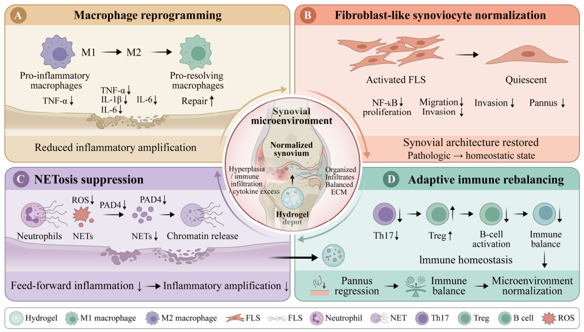
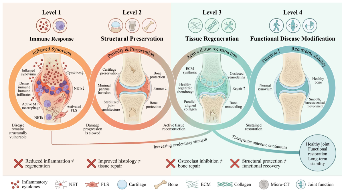

| Evidence or Outcome Level | Operational Definition Used in This Review | Examples of Acceptable Endpoints | Interpretation Allowed |
|---|---|---|---|
| Direct RA evidence | Evidence generated using human RA-derived material, clinical RA samples, or established RA-relevant experimental inflammatory arthritis models | CIA, AIA, K/BxN or RA-FLS; synovitis, pannus, cartilage matrix, erosion, gait. | Supports RA-relevant efficacy within the tested model, route, and endpoint |
| Transferable evidence | OA, bone-defect, wound-healing, inflammation, or tissue-engineering model used to support a material-design principle. | Lubrication, stiffness-tuned osteogenesis, generic ROS/MMP-responsive release. | Supports design rationale only; requires RA validation. |
| Conceptual evidence | Mechanistic or materials-science data without disease-specific validation. | Crosslinking chemistry, in vitro release trigger, isolated cell mechanism. | Hypothesis-generating; not proof of RA efficacy. |
| Level I: local immunomodulation | Reduction in inflammatory activity without proven structural benefit. | Cytokines, macrophage/FLS markers, paw swelling, arthritis score. | Anti-inflammatory or immunomodulatory activity. |
| Level II: structural protection | Less cartilage or bone damage than controls but no confirmed new tissue formation. | Safranin O, COL2A/aggrecan, TRAP, micro-CT erosion metrics. | Structural protection or anti-erosive effect. |
| Level III: tissue repair | Defect filling or new matrix/bone formation with host-tissue integration assessed. | New cartilage/bone, matrix organization, integration, remodeling. | Repair-associated evidence; not automatically functional disease modification. |
| Level IV: functionally validated structural disease modification | Durable multitissue structural benefit plus functional recovery. | Long-term imaging/histology plus gait, weight bearing, range of motion, pain behavior. | Highest preclinical evidence for disease-modifying potential. |
| Hydrogel System | Hydrogel Matrix and Gelation Method | Cargo or Functional Component | Primary Delivery/Response Mechanism | Study Model and Administration Route | Main Translational Limitation/Evidentiary Boundary | Reference |
|---|---|---|---|---|---|---|
| HA–Tyr hydrogel | Tyramine-modified hyaluronic acid; horseradish peroxidase/hydrogen peroxide (HRP/H2O2)-catalyzed oxidative coupling of phenolic groups to form a covalent network | Dexamethasone | The gel network restricts drug diffusion and prolongs local exposure | CIA animal model; intra-articular injection | evaluation focused mainly on inflammation and short-term histological changes, without establishing complete intra-articular pharmacokinetics | [4] |
| Cx-SIS drug depot | Tetrazine-modified and trans-cyclooctene-modified small intestinal submucosa (SIS) matrix rapidly crosslinked through a bioorthogonal click reaction | Methotrexate (MTX) | Gelation occurs within seconds after injection; the extracellular matrix (ECM) network improves intra-articular retention and enables sustained MTX release | RA animal model; intra-articular injection | histological findings indicated preservation of cartilage and glycosaminoglycans, but these findings should not be directly interpreted as functional evidence | [88] |
| FK506 nanoparticle–thermosensitive hydrogel | Self-assembled tacrolimus nanoparticles incorporated into a thermosensitive polymer hydrogel | Tacrolimus | The nanocarrier and thermosensitive gel form a secondary delivery system that reduces rapid diffusion | RA animal model; local intra-articular administration | the long-term intra-articular fates of the gel, nanoparticles, and free drug were not systematically compared | [89] |
| Electrostatic-interaction drug depot | A carboxyl-containing mPEG-b-[PCL-ran-PLA] block copolymer forms an injectable polyelectrolyte depot | Minocycline or sulfasalazine | Electrostatic attraction between oppositely charged drug and polymer components retards release | RA rat model; intra-articular injection | this system more closely resembles an injectable polymer depot than a conventional highly hydrated crosslinked scaffold | [90] |
| NanoIGUR/HA hydrogel | Acrylated HA and dithiol-terminated poly(ethylene glycol) (PEG) form a covalent network through Michael addition | Iguratimod-loaded poly(vinyl alcohol) (PVA) nanomicelles | A secondary “nanomicelle–hydrogel” diffusion barrier | CIA rats; subcutaneous injection | because the material was not administered intra-articularly, the study cannot demonstrate intra-articular retention | [69] |
| PRP–CS/BP hydrogel | Chitosan-based thermosensitive hydrogel | Platelet-rich plasma (PRP) and black phosphorus (BP) nanosheets | Temperature-induced gelation; BP mediates near-infrared photothermal effects, whereas PRP provides bioactive signals | RA animal model; local injection combined with near-infrared irradiation | efficacy depends on external irradiation, and the long-term safety of BP degradation and repeated irradiation requires further evaluation | [76] |
| IND–MTX in situ hydrogel | PEI-SS/drug nanoparticles dispersed in a 27% F127/10% F68 thermosensitive network | Indomethacin and MTX | Thermosensitive in situ gelation acts synergistically with sustained nanoparticle release to enable local dual-drug delivery | Arthritis animal model; intra-articular injection | complete in vivo concentration–time data were lacking | [91] |
| IND–MTX–siMMP-9 hydrogel | Thermosensitive poloxamer network containing PEI-SS composite nanoparticles | Indomethacin, MTX, and MMP-9 small interfering RNA (siRNA) | Co-delivery of small-molecule drugs and nucleic acids to simultaneously target inflammation and matrix degradation | RA animal model; intra-articular injection | the independent contribution, tissue distribution, and release synchronization of each cargo were not adequately established | [92] |
| IFX thermosensitive hydrogel | Thermosensitive composite network comprising F127, HA, and poly(γ-glutamic acid) | Infliximab (IFX) | Body-temperature-induced gelation retards protein diffusion | RA rabbit model; intra-articular injection | the findings should be described as sustained local delivery of a protein therapeutic and structural protection rather than cartilage regeneration | [93] |
| Tyr–GG hydrogel | Tyramine-modified gellan gum; HRP/H2O2-mediated enzymatic crosslinking | Betamethasone | Crosslinking density regulates swelling, degradation, and drug diffusion | Primarily material characterization and in vitro drug-release evaluation | evidence of structural efficacy in RA animals remains insufficient, and the system is more appropriately regarded as an RA-oriented delivery material | [75] |
| M-NO gel | Click-crosslinked network containing nitric oxide (NO)-cleavable crosslinkers and embedded self-assembled drug-loaded polymeric aggregates | Dexamethasone and NO-scavenging functional groups | Direct scavenging of excess NO; NO triggers network changes and drug release according to the degree of inflammation | RA animal model; local injection | “on-demand release” is supported by NO-response experiments, but long-term self-regulation during recurrent disease has not been validated | [94] |
| IFX self-healing composite scaffold | An IFX-loaded self-healing hydrogel combined with a three-dimensional (3D)-printed porous metal scaffold | IFX and adipose-derived mesenchymal stem cells | The self-healing hydrogel improves local retention of cells and biologics, whereas the metal scaffold provides structural support | RA rabbit bone-defect model; local implantation | it was used mainly to evaluate cell survival, implantation, and osteochondral repair | [12] |
| DNase-functionalized hydrogel | Deoxyribonuclease I (DNase I) conjugated to oxidized HA, followed by formation of a dynamic network with carboxymethyl chitosan through a Schiff base reaction | DNase I; combined with MTX in some experiments | Prolongs the local activity of DNase I and continuously degrades neutrophil extracellular traps (NETs) | CIA animal model; intra-articular injection | the evidence primarily supports NET clearance and anti-inflammatory activity and cannot independently demonstrate structural regeneration | [73] |
| TAP/Cx-HA hydrogel | Click-crosslinked HA hydrogel | Toll-like receptor antagonist peptide (TAP) | The covalently crosslinked network improves intra-articular peptide stability and prolongs exposure | RA animal model; intra-articular injection | structural improvement was assessed mainly using histological and matrix-related indicators, with limited evidence of functional repair | [95] |
| Metabolism-driven responsive hydrogel | Composite hydrogel responsive to the disease-associated metabolic environment | Psoralen and oxygen-supplying/microenvironment-modulating components | Exploits hypoxia, enzymatic activity, or metabolic state to achieve local responsiveness and drug release | RA animal model; local injection | actual pathological thresholds, interpatient variability, and reproducibility of triggering require further validation | [85] |
| TNF-α-binding supramolecular hydrogel | Supramolecular network based on reversible noncovalent interactions and incorporating TNF-α-binding sites | The material itself provides TNF-α-capturing functionality | Rapidly binds and locally neutralizes TNF-α rather than relying solely on drug diffusion | RA animal model; intra-articular injection | TNF-α-binding capacity, inflammatory improvement, and long-term structural modification should be evaluated as distinct outcomes | [96] |
| DSP–DS supramolecular hydrogel | Dexamethasone sodium phosphate and diclofenac sodium self-assemble into a gel through noncovalent interactions | Both drugs serve simultaneously as therapeutic components and gel-forming units | Drug self-assembly without an additional polymer carrier; thixotropy supports injection | AIA rats; local administration | network stability depends on drug–drug interactions, and in vivo dissociation and local pharmacokinetics remain to be defined | [5] |
| Anti-inflammatory–osteogenic interpenetrating network | HA–collagen interpenetrating network containing bisphosphonate-functionalized components and zinc-doped calcium phosphate | Anti-inflammatory components and bone-affinitive/mineralization-supporting components | Combines local anti-inflammatory activity, bone-surface localization, and mineralization support | RA bone-erosion model; local injection or defect filling | stable integration of newly formed bone with pre-existing erosion defects requires long-term confirmation | [6] |
| HP@CEL hydrogel | Supramolecular composite network formed by hydrophobically modified HA and celastrol-loaded PECT nanoparticles | Celastrol | Rapid formation of a nanodrug–hydrogel depot after injection, followed by sustained release | RA animal model; intra-articular injection | stricter component-dissection controls are needed to distinguish the relative contributions of the cargo and the material itself | [18] |
| Dual dynamically crosslinked TPL hydrogel | Adaptive network formed by two types of reversible dynamic bonds | Triptolide (TPL) and ROS-modulating components | The dynamic network provides injectability and self-healing; the material regulates ROS while enabling sustained drug release | RA animal model; intra-articular injection | “microenvironment-independent release” does not imply the absence of dose fluctuations and still requires confirmation by in vivo pharmacokinetic (PK) studies | [80] |
| Self-healing gas-regulating hydrogel | Dynamic, self-healing injectable network | Functional modules for NO scavenging and hydrogen sulfide (H2S) delivery | Simultaneously regulates oxidative/nitrosative stress and the gaseous-signaling microenvironment | RA animal model; intra-articular injection | gas-release dose, local concentration, and the long-term safety window remain to be quantified | [97] |
| Staged MTX-release hydrogel | ROS-scavenging injectable hydrogel incorporating a hierarchical drug-binding structure | MTX | Staged release comprising initial, sustained, and pathological-feedback-associated phases, together with simultaneous ROS scavenging | RA animal model; intra-articular injection | “self-regulation” should nevertheless be validated using in vivo local pharmacokinetics rather than cumulative in vitro release curves | [98] |
| Anti-inflammatory–antiferroptotic adhesive hydrogel | Tissue-adhesive injectable network serving as a local nanotherapeutic depot | Inflammation-modulating and antiferroptotic nanotherapeutic components | Wet-tissue adhesion improves retention while simultaneously targeting inflammation and ferroptosis-related pathways in synovial cells | RA animal model; intra-articular injection | improvement in ferroptosis-related indicators cannot substitute for structural endpoints in cartilage and bone | [99] |
| pH-responsive peptide hydrogel | Injectable, pH-sensitive, self-assembling peptide hydrogel | MTX and BiNS/PEI nanosheets | Responds to the acidic synovial microenvironment; MTX regulates macrophages, whereas photothermal/photodynamic effects eliminate excessive proliferation | RA animal model; intra-articular injection combined with irradiation | reliance on external irradiation raises concerns regarding damage to normal synovium and the risk of nonselective cell elimination | [100] |
| DAGQD@Cu@KGN–SO3−/DA-HA hydrogel | Double network comprising dopamine-modified HA and sulfonated HA | Cu single-atom nanozyme and dopamine-hybrid graphene quantum dots grafted with kartogenin (KGN) | Dopamine provides adhesion, sulfonate groups enhance hydration lubrication, the nanozyme scavenges ROS, and KGN is released continuously | CIA rats and rabbits with RA-associated osteochondral defects; intra-articular injection/defect application | “full-cycle treatment” remains limited to preclinical models | [13] |
| IL-4-immobilized HA hydrogel | HA-based hydrogel in which interleukin-4 (IL-4) is covalently immobilized within the network | Immobilized IL-4 | Local immobilized presentation replaces rapid diffusional release and withstands repeated mechanical loading in the joint | RA animal model; intra-articular application | the local effects of an immobilized cytokine should be distinguished from the pharmacokinetic concept applicable to a releasable drug | [15] |
| Polymer-modified DNA hydrogel | Polymer-modified deoxyribonucleic acid (DNA) forms a gel through sequence pairing and network assembly | Functional mitochondria and nanozymes | The DNA network co-immobilizes biological organelles and catalytic components, thereby reducing local clearance | RA animal model; intra-articular injection | preservation of mitochondrial activity, batch-to-batch consistency, immunogenicity, and storage conditions are major translational barriers | [16] |
| ChSMA@SPD hydrogel | Methacrylated chondroitin sulfate (ChSMA) network | Spermidine (SPD) | The ECM-like network locally encapsulates SPD and modulates inflammation and chondrocyte catabolism | RA-related coculture and animal models; local application | the quality of newly formed tissue, interfacial integration, and functional recovery have not been adequately demonstrated | [52,55] |
| SIN liposome/PGA–F127 hydrogel | Composite hydrogel comprising poly(γ-glutamic acid) (PGA) and Pluronic F127 | Sinomenine hydrochloride (SIN)-loaded liposomes | The liposomes and thermosensitive/composite gel form a secondary release barrier | RA animal model; local administration | the duration of effective intra-articular concentrations should not be inferred solely from in vitro release data | [68] |
| MMP-binding hydrogel | Functionalized network containing MMP-binding sites | Material-based binding sites serve as the primary functional module | Locally captures or restricts pathological MMPs, thereby reducing matrix-degradation pressure | RA animal model; intra-articular injection | MMP binding does not imply inhibition of all matrix-degrading enzyme activity | [72] |
| HC@PTM composite hydrogel | Dynamic Schiff base network formed by aldehyde-functionalized HA and chitosan and containing MTX-loaded polymeric micelles | MTX and ROS-scavenging/ROS-responsive micelles | The hydrogel provides injectability, self-healing, and shape adaptability; the micelles respond to ROS and pH | RA animal model; intra-articular injection | the in vivo timescales of hydrogel degradation, micelle dissociation, and MTX exposure require separate validation | [74] |
| Gel-MTX/Mg supramolecular hydrogel | Supramolecular network driven by structural rearrangement and noncovalent interactions | MTX and Mg2+ | Therapeutically active components participate in network formation and enable sustained local delivery | RA animal model; intra-articular injection | the local Mg2+ concentration, release stability, and independent contribution of Mg2+ require further clarification | [7] |
| MOS in situ pore-forming hydrogel | Injectable, in situ pore-forming network comprising manno-oligosaccharide-modified chondroitin sulfate and HA | Manno-oligosaccharide targeting units intrinsic to the material | Pore formation promotes cell–material interactions, whereas manno-oligosaccharides act on cluster of differentiation 206 (CD206)-associated macrophages | RA animal model; intra-articular injection | its mechanism should not be reduced to a conventional binary M1/M2 polarization model | [77] |
| Ca2+-reinforced all-active hydrogel | Therapeutically active components participate in network formation, which is reinforced through in situ Ca2+ coordination | Network-forming units with intrinsic pharmacological activity | Reduces the proportion of inert carrier materials; Ca2+ enhances network stability and regulates release | RA animal model; local injection | “all-active” indicates that the components participate in treatment but does not imply a dose–response relationship or long-term safety | [101] |
| Hydrogel System and Principal Intervention Target | Evidence from the Synovium and Local Immune Environment | Cartilage-Related Evidence | Bone-Related Evidence | Evidence of Pain Relief or Joint Function | Cautious Interpretation Based on Measured Endpoints | Reference |
|---|---|---|---|---|---|---|
| Dexamethasone-loaded HA–Tyr hydrogel | Reduced interleukin-6, prostaglandin E2 (PGE2), and multiple inflammatory cytokines in a CIA model | Cartilage thickness, type II collagen, aggrecan, and newly formed cartilage were not independently quantified | TRAP, micro-CT, and quantitative bone-erosion outcomes were not reported | No direct pain or motor-function endpoints were reported | cartilage or bone structural modification cannot be concluded solely from overall H&E findings | [4] |
| Infliximab-loaded F127–HA–PGA thermosensitive hydrogel | Reduced TNF-α, IL-1β, IL-6, and IL-17 levels in synovial fluid and cartilage; alleviated joint swelling and increased surface temperature | Histological findings showed reduced cartilage destruction, supporting cartilage structural protection | Bone erosion and bone-remodeling endpoints were not included | Weight-bearing index and pain-related behavior indicated pain relief | the short follow-up period and absence of bone endpoints are insufficient to demonstrate whole-joint structural modification | [93] |
| Indomethacin/MTX/MMP-9 siRNA in situ hydrogel | Reduced local inflammation and suppressed MMP-9-associated catabolism through siRNA | Histological and cartilage matrix-related findings indicated reduced or partially reversed cartilage destruction | Three-dimensional bone structure and bone-erosion volume were not systematically reported | Gait, weight bearing, and range of motion were not reported | “reversal of cartilage destruction” does not demonstrate the formation of mature hyaline cartilage | [92] |
| DNase I-functionalized injectable hydrogel | Preserved DNase I activity and promoted NET degradation; reduced the NET burden, local inflammation, and arthritis severity | Overall joint histology improved, but evaluation of cartilage-specific matrix and repair endpoints was limited | Micro-CT-based bone erosion and new bone formation were not adequately reported | No direct functional endpoints were reported | cartilage regeneration or bone repair cannot be inferred from reduced inflammation | [73] |
| TAP2-loaded click-crosslinked HA hydrogel | Prolonged the intra-articular retention of the TLR4-antagonistic peptide and suppressed TLR4-associated inflammatory responses and synovial pathology | Cartilage thickness, glycosaminoglycan preservation, and histological improvement were reported | Bone-related histological changes were reported, but adequate long-term evidence of three-dimensional bone reconstruction was lacking | No direct functional endpoints were reported | functional evaluation and long-term integration remain lacking | [95] |
| HP@CEL HA–nanodrug supramolecular hydrogel | Regulated macrophage–FLS crosstalk through sustained celastrol release and suppressed proinflammatory macrophage states, FLS activation, and inflammation | Cartilage histology and ECM-related indicators improved | Improvements in joint bone structure were reported, but independent quantitative evidence of bone repair was limited | Gait, weight bearing, and joint range of motion were not reported | sustained and functional regeneration of cartilage or bone has not been demonstrated | [18] |
| SPT@TPL dual dynamically crosslinked hydrogel | Scavenged or regulated excessive ROS, reduced inflammation, and promoted the transition of macrophages toward repair-associated states | Improvements in cartilage-surface morphology and matrix staining indicated cartilage protection or early repair | Bone structure was not a principal validation endpoint | No direct functional endpoints were reported | histological improvement alone should not be described as functional cartilage regeneration | [80] |
| Anti-inflammatory–osteogenic interpenetrating-network hydrogel | Regulated local inflammation and the macrophage-associated osteoimmune microenvironment | Cartilage was not the primary repair target, and cartilage-specific outcomes were relatively limited | Osteoclast activity, osteogenic differentiation, and micro-CT bone structure were evaluated | The direct mechanical function of newly formed bone and recovery of overall joint movement were not reported | an experimentally created erosion defect is not fully equivalent to naturally progressive bone erosion in RA | [6] |
| DNRS dual-gas-regulating self-healing hydrogel | Scavenged excessive NO, released H2S, regulated macrophages and the inflammatory microenvironment, and reduced synovial inflammation | Cartilage was not the principal repair endpoint, and structural evidence was limited | TRAP staining and micro-CT indicated inhibition of osteoclast activity and improvement in bone structure | Direct assessments of joint mechanics, gait, and range of motion were not reported | the quality of newly formed bone, integration with host tissue, and stability after inflammatory recurrence require further validation | [97] |
| Adhesive and lubricating DAGQD@Cu@KGN double-network hydrogel | The Cu single-atom nanozyme scavenged ROS and reduced local inflammatory responses | Reduced friction and cartilage wear in an early-stage model | Bone was not a principal assessment target, and a complete evidence chain for bone-erosion repair was not established | Material tribological properties were evaluated, but adequate evidence from animal gait, weight-bearing, and joint-range-of-motion assessments was lacking | reduced friction in vitro cannot substitute for restoration of whole-joint function | [13] |
| MMP-9-binding CuS-T/ChSMA hydrogel | Locally bound MMP-9, inhibited RA-FLS invasion and proinflammatory macrophage states, and alleviated synovial inflammation | Increased expression of type II collagen- and aggrecan-related markers | Bone erosion and new bone formation were not adequately evaluated | No direct functional endpoints were reported | mature cartilage regeneration cannot be concluded solely from collagen expression and improved staining | [72] |
| Polymer-modified DNA hydrogel co-delivering functional mitochondria and Prussian blue nanozymes | Reduced oxidative stress and inflammation through extracellular ROS scavenging and intracellular mitochondrial renewal | Improvements in cartilage histology, chondrocyte status, and matrix repair-associated indicators were reported | Improvements in bone and osteochondral structures were reported, but long-term bone reconstruction and mechanical quality remain unclear | Standardized recovery of gait or weight bearing was not reported | preservation of mitochondrial potency, immune safety, and batch-to-batch consistency remain critical limitations | [16] |
| Spermidine-loaded ChSMA hydrogel | Reduced local inflammation and modulated inflammation-associated cells and the osteoimmune microenvironment | Reduced chondrocyte apoptosis and MMP expression and improved type II collagen, aggrecan, and cartilage histology | Suppressed osteoclast-related signaling, improved bone-erosion or bone-microstructural indicators, and regulated the osteoblast–osteoclast balance | No direct functional endpoints were reported | the mechanical properties of newly formed tissue, long-term integration, and joint function remain unproven | [52] |
| Gel-MTX/Mg supramolecular drug-loaded hydrogel | Reduced inflammatory mediators and clinical arthritis scores and modulated the local immune state | Preservation of the cartilage surface and matrix was reported | Suppressed osteoclast differentiation and improved indicators of bone erosion and bone microstructure | Standardized functional endpoints were not reported | evidence remains insufficient to conclude that erosion defects underwent stable bone regeneration | [7] |
| IL-4-covalently immobilized immunosuppressive hydrogel | Immobilized IL-4 exerted sustained effects on local macrophages and reduced proinflammatory states and synovial inflammation | Reduced cartilage degradation or joint-tissue damage was reported, primarily indicating structural protection | Bone erosion and new bone formation were not adequately validated | No direct functional endpoints were reported | changes in macrophage phenotype should not be equated with complete restoration of synovial homeostasis | [15] |
| Infliximab-loaded self-healing hydrogel–porous metal composite scaffold delivering adipose-derived stem cells (ADSCs) | Reduced local inflammatory pressure and provided a more favorable microenvironment for transplanted cells | Improvements in tissue repair and cell engraftment in the osteochondral region were reported | The scaffold supported the bone defect, and osteogenesis, implantation, and bone-tissue repair were reported | Recovery of overall RA joint mobility and load-bearing function was not adequately demonstrated | its conclusions should not be extrapolated to diffuse structural damage in RA | [12] |
Disclaimer/Publisher’s Note: The statements, opinions and data contained in all publications are solely those of the individual author(s) and contributor(s) and not of MDPI and/or the editor(s). MDPI and/or the editor(s) disclaim responsibility for any injury to people or property resulting from any ideas, methods, instructions or products referred to in the content. |
© 2026 by the authors. Licensee MDPI, Basel, Switzerland. This article is an open access article distributed under the terms and conditions of the Creative Commons Attribution (CC BY) license.
Share and Cite
Liao, R.; Mu, K.; Ran, F.; Yang, L.; Feng, Y.; Xu, T.; Zhong, X.; Wei, F.; Pang, Y.; Liu, G.; et al. Hydrogels as Local Structural-Protective Platforms in Rheumatoid Arthritis: An Evidence-Graded Review Across the Synovium–Cartilage–Bone Axis. Gels 2026, 12, 601. https://doi.org/10.3390/gels12070601
Liao R, Mu K, Ran F, Yang L, Feng Y, Xu T, Zhong X, Wei F, Pang Y, Liu G, et al. Hydrogels as Local Structural-Protective Platforms in Rheumatoid Arthritis: An Evidence-Graded Review Across the Synovium–Cartilage–Bone Axis. Gels. 2026; 12(7):601. https://doi.org/10.3390/gels12070601
Chicago/Turabian StyleLiao, Ruiqi, Kailang Mu, Fei Ran, Lixia Yang, Yunqian Feng, Tianrui Xu, Xuemei Zhong, Fudao Wei, Yuxin Pang, Gang Liu, and et al. 2026. "Hydrogels as Local Structural-Protective Platforms in Rheumatoid Arthritis: An Evidence-Graded Review Across the Synovium–Cartilage–Bone Axis" Gels 12, no. 7: 601. https://doi.org/10.3390/gels12070601
APA StyleLiao, R., Mu, K., Ran, F., Yang, L., Feng, Y., Xu, T., Zhong, X., Wei, F., Pang, Y., Liu, G., & Liu, Y. (2026). Hydrogels as Local Structural-Protective Platforms in Rheumatoid Arthritis: An Evidence-Graded Review Across the Synovium–Cartilage–Bone Axis. Gels, 12(7), 601. https://doi.org/10.3390/gels12070601