Article

Vat Photopolymerization-Fabricated Theranostic Hydrogels for Smart Wound Management

by
Karl Albright Tiston
1,2,*,
Laureen Ida Ballesteros
1,
Jo Marie Venus Agad
1,
Patrick Meracandayo
1,
Karlos Mayo Silva
1,
Toni Beth Lopez
1,
Nadnudda Rodthongkum
2,3,
Voravee P. Hoven
2 and
Rigoberto Advincula
4
1
Department of Science and Technology, Metals Industry Research and Development Center, Taguig City 1631, Philippines
2
Department of Chemistry, Faculty of Science, Chulalongkorn University, Bangkok 10330, Thailand
3
Center of Excellence in Responsive Wearable Materials, Chulalongkorn University, Bangkok 10330, Thailand
4
Department of Chemical and Biomolecular Engineering, University of Tennessee, Knoxville, TN 37996, USA
*
Author to whom correspondence should be addressed.
Gels 2026, 12(5), 393; https://doi.org/10.3390/gels12050393
Submission received: 25 March 2026 / Revised: 28 April 2026 / Accepted: 29 April 2026 / Published: 2 May 2026
(This article belongs to the Special Issue 3D Printing of Gel-Based Materials (2nd Edition))

Abstract

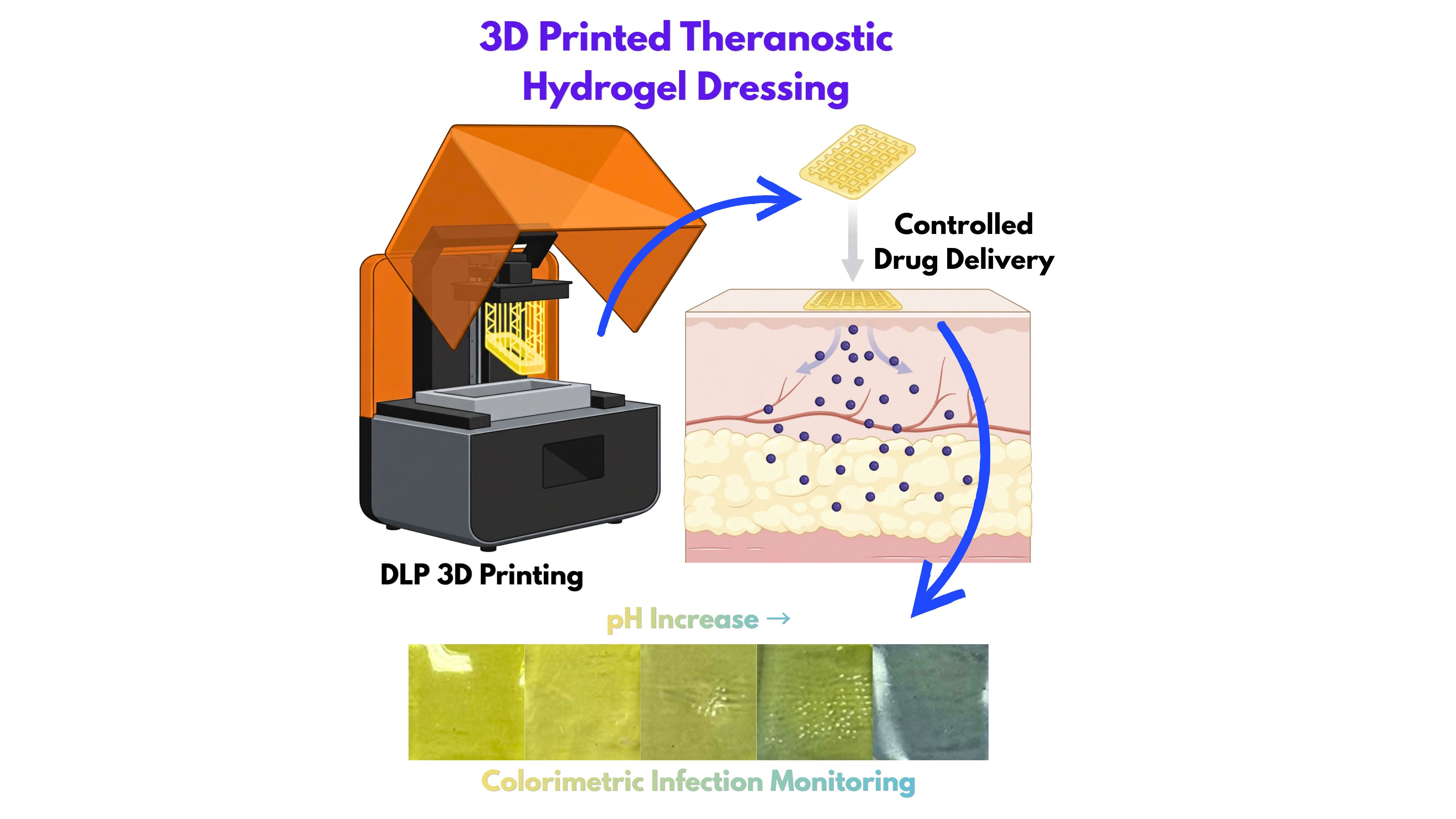
Despite the demand for personalized wound care, integrating diagnostics and therapeutics into a unified platform remains a significant challenge. To address this, we developed a 3D-printed theranostic hydrogel using vat photopolymerization, enabling precise, multifunctional wound management. The hydrogel matrix, composed of poly(acrylamide-co-hydroxyethyl acrylate) and carboxymethyl cellulose, was chemically crosslinked with poly(ethylene glycol) diacrylate. Bromocresol purple was integrated into the photosensitive resin to enhance printing fidelity and serve as a diagnostic indicator, providing a distinct colorimetric shift upon skin infection. For controlled drug delivery, graphene oxide (GO) and levofloxacin were incorporated into the system. The 3D-printed hydrogel demonstrated superior swelling capacity (>600%), ideal for absorbing wound exudate. A semi-quantitative linear colorimetric response was observed across varying pH levels, allowing for clear differentiation between healthy healing skin (pH 4.0–6.0) and infected conditions (pH 7.0 and above). Furthermore, the hydrogel exhibited infection-stimulated therapy, with a cumulative levofloxacin release of 92.63% at pH 8, significantly higher than in acidic conditions. Moreover, the incorporation of GO further optimized the delivery profile by tuning absorption and release rates. Synergizing real-time monitoring and on-demand therapeutic action, this 3D-printed system offers a scalable, robust solution for future-ready, personalized wound management.

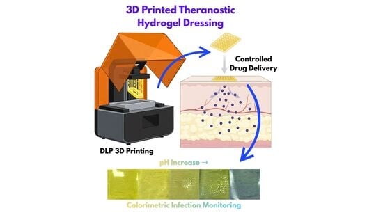
Graphical Abstract

1. Introduction

Chronic wounds represent an escalating global health and socioeconomic challenge, predominantly affecting the elderly and individuals with diabetes, vascular diseases, or traumatic injuries [1,2]. Unlike acute wounds, chronic wounds, such as diabetic foot, venous, and pressure ulcers, fail to progress through the normal stages of tissue repair and are frequently complicated by persistent infection, prolonged inflammation, and impaired angiogenesis [3]. This kind of wound environment may render conventional passive dressings, thus necessitating more effective, active therapeutic interventions [4].
Current chronic wound management is primarily guided by the utilization of moisture-balancing dressings, targeted debridement, and advanced adjuncts such as negative-pressure wound therapy and biologic matrices to facilitate tissue repair [5]. However, these conventional interventions remain largely passive, relying on episodic clinical evaluations, standard cultures, and distinct diagnostic tests that fail to capture the dynamic physiological fluctuations of the healing process [6,7]. To address the limitations of separated therapeutic and diagnostic protocols, recent innovations are shifting toward “smart” wound dressings integrated with novel biosensing technologies [8,9,10]. By continuously tracking critical local biomarkers including pH, temperature, oxygenation, and bacterial burden via colorimetric or electrochemical sensors, these devices provide real-time, actionable data [9,10]. Ultimately, bridging the gap between passive treatment and active monitoring through fully integrated platforms capable of simultaneous diagnosis and therapy represents a critical frontier in optimizing chronic wound care.
While healthy skin and progressing, acute wounds maintain a protective, mildly acidic environment, chronic wounds are characterized by a sustained alkaline state with pH values often ranging from 7.4 to nearly 9 [7]. This prolonged alkalinity is driven by persistent inflammation and infection, which in turn delays healing by promoting bacterial proliferation and destructive protease activity [7,10]. Consequently, tracking this distinct shift toward higher pH serves as a highly valuable, non-invasive biomarker for continuously monitoring wound status and providing early detection of infection risk [6].
Traditionally, wound dressing has played an enormous role in wound management as these provide protective barriers for the wound to promote adequate healing. Wound dressing material ranges from fabrics such as cotton or viscose, polymers like polyurethane and silicone, and hydrogels [11]. Hydrogels have emerged as highly effective materials for advanced wound dressings due to their biocompatible, hydrated polymer networks that excel at localized, sustained drug delivery [3,12]. Capable of encapsulating a diverse array of therapeutics, ranging from small-molecule antibiotics to sensitive biologics like extracellular vesicles, hydrogels improve treatment efficacy directly at the wound site while minimizing systemic side effects [13,14]. Furthermore, the development of “smart” hydrogels allows for highly tailorable, stimuli-responsive systems that react to specific wound microenvironmental cues, such as the elevated pH or high reactive oxygen species (ROS) levels characteristic of chronic infections, to trigger on-demand drug release [15,16]. While their structural adaptability and responsive therapeutic capabilities make hydrogels exceptionally well-suited for treating complex wounds, current designs primarily function as passive therapeutic reservoirs, leaving a crucial gap for integrating real-time diagnostic feedback.
By combining therapeutic and diagnostic capabilities, the concept of “theranostics” arises as an integrated mechanism for smarter, simplified biomedical devices [17]. In this aspect, theranostic hydrogels represent a transformative approach to wound management by seamlessly integrating real-time diagnostic monitoring with localized, on-demand therapeutic delivery within a single “sense-and-respond” platform [17,18,19]. Unlike conventional materials, these smart dressings actively track critical physiological biomarkers such as pH, temperature, and specific biochemical cues, through embedded colorimetric, optical, or electrochemical sensors [6,19]. In response to these real-time inputs, such as an alkaline shift indicative of infection, the hydrogel matrix can trigger the targeted release of appropriate interventions, including antibiotics, anti-inflammatory agents, or regenerative biomolecules [13,15,19,20]. By bridging the gap between continuous physiological monitoring and adaptive drug delivery, theranostic hydrogels enable earlier detection of complications, reduce the need for frequent dressing changes, and provide a highly customized, dynamic healing environment for chronic wounds [17,18,19].
While hydrogels are highly effective platforms for localized wound management, fine-tuning controlled drug release remains a considerable problem. To mitigate this limitation, various functional additives, such as metal nanoparticles, mesoporous silica, and carbon nanotubes, are frequently incorporated into the hydrogel matrix to modulate delivery profiles. Among these, graphene oxide (GO) has emerged as an exceptional nanocarrier. The two-dimensional planar structure and immense surface area of GO allow exceptionally high drug-loading capacity, which mitigates proper dosage delivery [21]. GO also contains a highly functionalized amphiphilic surface which allows suitable drug and polymer network interactions. The unoxidized hydrophobic basal plane stably binds aromatic therapeutic agents, while abundant hydrophilic oxygen-containing functional groups ensure excellent compatibility with the surrounding hydrogel network [22,23]. Furthermore, GO controls drug delivery through specific non-covalent interactions, primarily π–π stacking, and can be disrupted through localized environmental triggers such as pH changes [22,24,25]. These specific interactions make GO an excellent candidate for fine-tuning drug delivery in hydrogel-based platforms.
To fully realize the potential of dynamic theranostic platforms, additive manufacturing (AM) offers a highly versatile approach for fabricating precise, patient-specific hydrogel geometries directly from digital models [26,27]. By building constructs layer-by-layer, AM allows for the meticulous adjustment of scaffold dimensions, pore distribution, and mechanical stiffness to match user needs [28,29]. Currently, direct ink writing (DIW) is the most widely used 3D printing technique for hydrogels; however, this extrusion-based method relies heavily on the shear-thinning behavior of continuous inks and often requires support materials or post-processing to maintain shape fidelity [30]. Consequently, DIW is inherently constrained by the scale of its extruded filaments, making the precise control necessary for fine micro-architectures challenging [31,32]. To overcome these structural limitations, this study utilizes vat photopolymerization (VPP), specifically digital light processing (DLP), to engineer the proposed theranostic hydrogel dressings. By employing patterned light to selectively cure photosensitive resins, DLP achieves exceptionally high print resolution and smooth surface finishes that are difficult to replicate with DIW [33,34]. More importantly, this light-based technique excels at generating complex, intricate internal geometries, such as compartmentalized constructs, microlattices, and macropore structures [35,36]. From a drug delivery perspective, these advanced architectures are crucial. These significantly increase the functional surface area, modulate internal diffusion pathways, and allow for locally varied crosslink densities [37,38,39]. Ultimately, leveraging DLP to integrate such structural complexity empowers the hydrogel dressing with highly programmable release kinetics, seamlessly linking sophisticated physical design with advanced, localized wound therapy.
Herein, we report a high-fidelity, DLP 3D-printed hydrogel platform for the simultaneous sensing and treatment of wound infections. We have formulated a semi-interpenetrating polymer network made from a carboxymethyl cellulose-based matrix crosslinked with a multicomponent poly(acrylamide-co-hydroxyethyl acrylate-co-polyethylene glycol diacrylate), poly(AAm-co-HEA-co-PEGDA). Notably, bromocresol purple was utilized as a dual-functional additive, serving both as a photoabsorber to ensure superior printing resolution and as a real-time colorimetric biosensor for pH-based infection diagnostics. Beyond sensing, the platform provides autonomous therapeutic intervention as the loaded antibiotic, levofloxacin, exhibited accelerated release at pH above 7.0 via diffusion. Moreover, graphene oxide (GO) was also added to the formulation to further modulate drug release, ensuring a more controlled and sustained delivery of the therapeutic agent. Lastly, we demonstrated that the advanced spatial resolution of 3D printing enables the precise fabrication of customized macroporous architectures, which later provided further control in drug release. By deliberately engineering these internal diffusion pathways and optimizing the overall surface area-to-volume ratio, the system achieves highly tunable and inherently more efficient drug release. This multifunctional system represents a versatile, scalable advancement in the design of next-generation “smart” wound management materials.

2. Results and Discussion

2.1. Formulation of Photopolymerizable Resin and DLP 3D Printing of Hydrogels

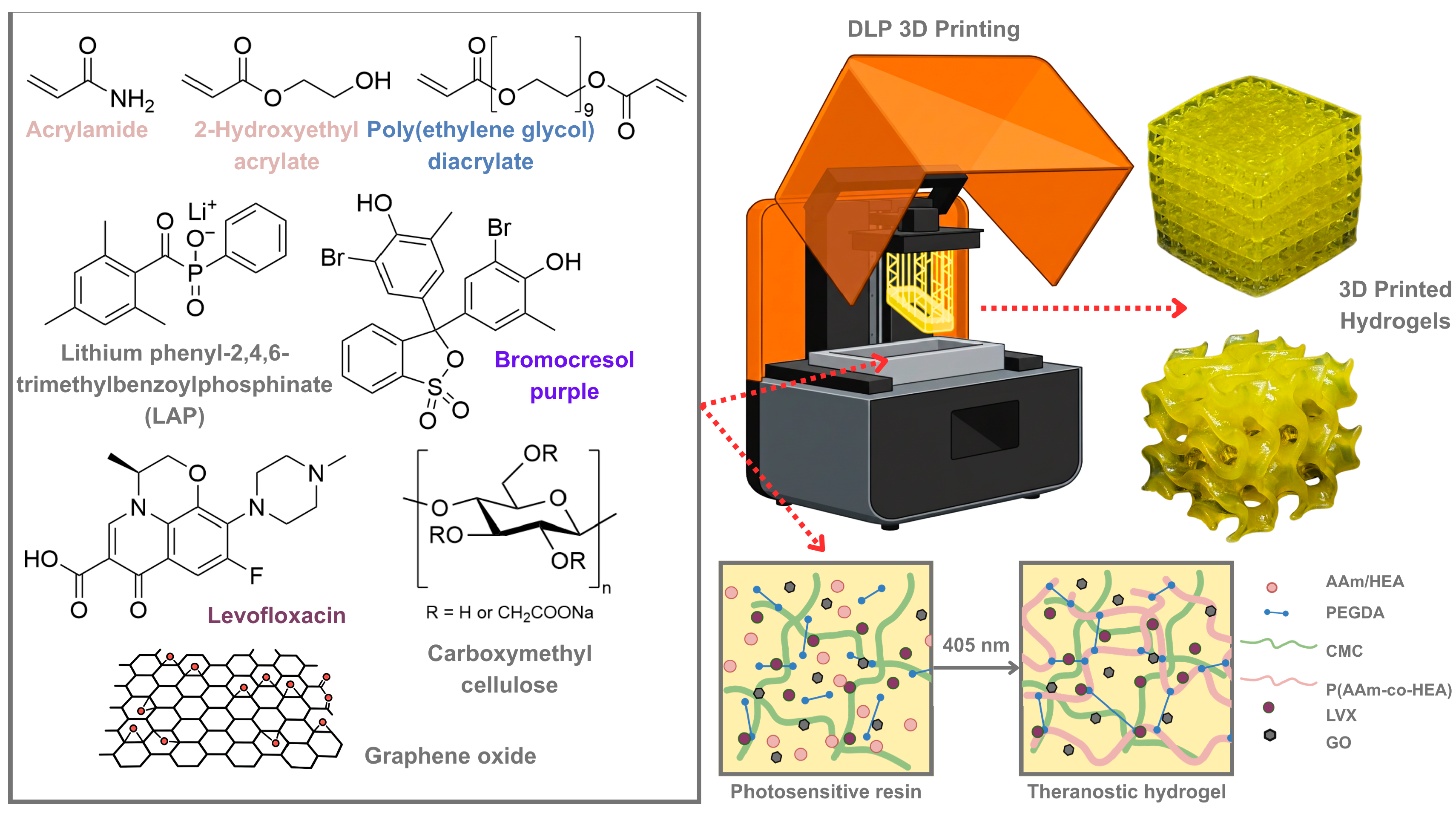
The preparation of the photopolymerizable hydrogel resin was done by dissolving acrylamide (AAm), 2-hydroxyethyl acrylate (HEA), polyethylene glycol-9 (PEGDA), carboxymethyl cellulose (CMC), Lithium phenyl-2,4,6-trimethylbenzoylphosphinate (LAP), graphene oxide (GO), levofloxacin (LVX), and bromocresol purple (BCP) in distilled water (Figure 1). The resulting solution was used as a photosensitive resin in a DLP 3D printer. CMC is a cellulose-derived water-soluble biopolymer added to hydrogel formulation for mechanical reinforcement and increased hydrophilicity, suitable for wound dressing applications [40,41]. The hydrogel was formed after exposing the liquid resin precursor to 405 nm light where the LAP photoinitiator decomposes and produces radicals, initiating the photopolymerization. The main polymer network is made from poly(AAm-co-HEA) crosslinked with PEGDA. Overall, a semi-interpenetrating polymer network (semi-IPN) hydrogel was formed upon printing. The pH-responsive dye, BCP, acts as the photoabsorber in the resin formulation, which inhibits further photopolymerization in previously printed layers, thus allowing higher-resolution prints [42,43]. BCP acts similarly to the more commonly used tartrazine dye, as both afford a yellow hue upon dissolution in the photopolymerizable resin [44]. In this case, BCP has a dual function of acting as a photoabsorber, improving the printing fidelity, and later as a pH-responsive dye for potential infection monitoring. Ultimately, the successful combination of these constituents results in a robust material system that balances mechanical reinforcement with precise printability. This approach ensures that the final hydrogel construct possesses the necessary structural and functional properties required for complex biomedical devices, such as 3D-printed smart wound dressings.
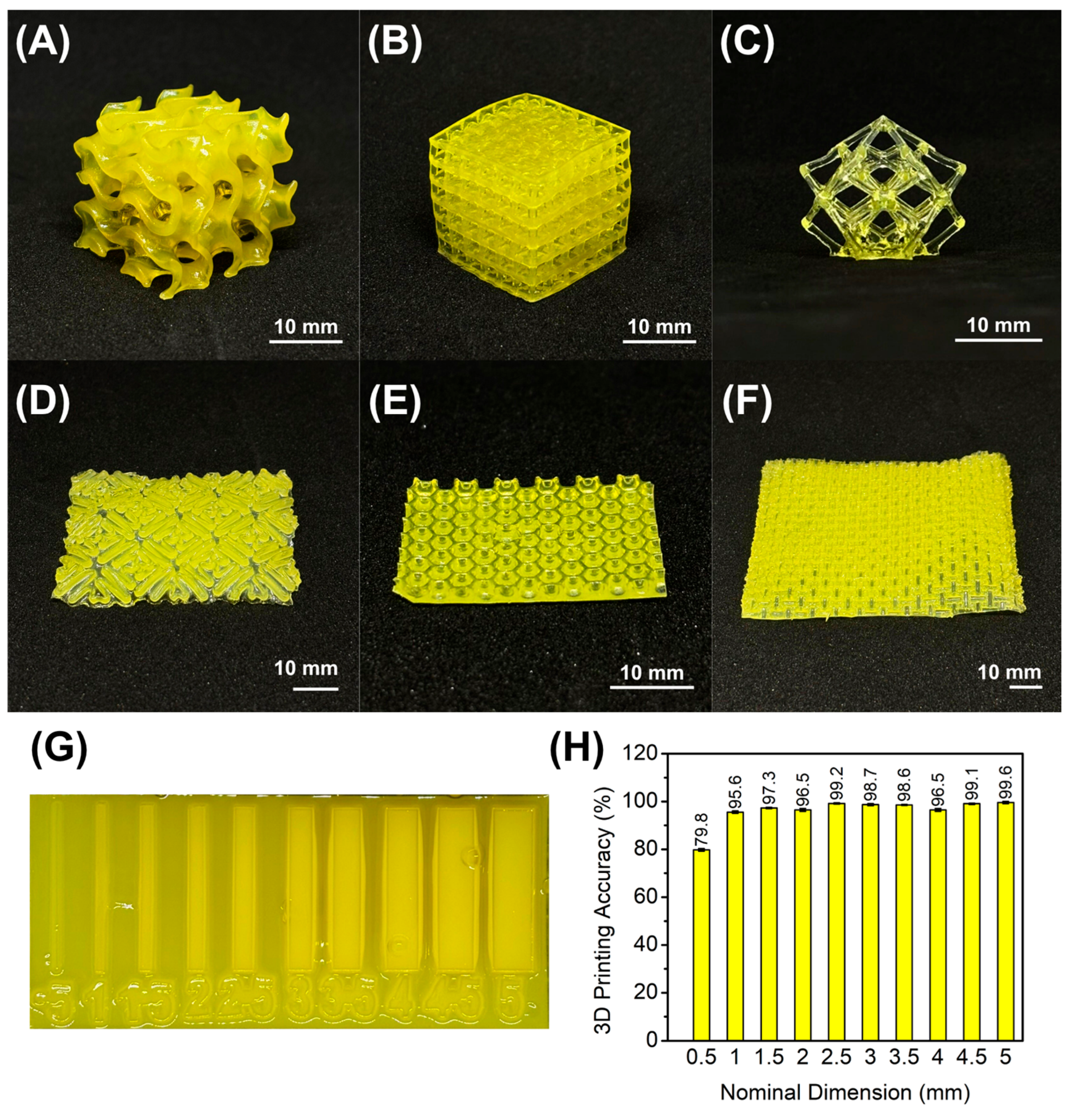
To demonstrate the 3D printability of the formulated hydrogel resins, various structures were fabricated using the Creality Halot One, a commercial DLP 3D printer with 2K resolution. Architectures with complex motifs were printed to showcase the resin capabilities, including a gyroid infill, a square grid, a lattice calibration specimen, and various patterned wound dressing prototypes (Figure 2A–F). Vat photopolymerization enables the creation of intricate patterns, such as lattices and hollowed structures, which are often difficult to achieve with extrusion-based techniques [42,45,46]. Consequently, applying DLP 3D printing to theranostic hydrogels facilitates the fabrication of smart wound dressings with personalized designs. After printing, the hydrogels were quickly rinsed with distilled water to remove excess resin and then exposed to 405 nm light to complete photopolymerization. Printing fidelity was assessed by comparing the dimensions of a 3D-printed thickness calibration sample to the nominal dimensions (Figure 2G). While the printed hydrogels generally exhibited high accuracy (Figure 2H), walls with the finest dimensions (0.5 mm) were larger than the nominal value. This discrepancy is likely attributable to gel swelling during the printing process. Additionally, the incorporation of BCP effectively prevented overexposure by absorbing excess UV light and controlling the depth of cure (Figure S1, Supporting Information). Overall, the 3D-printed constructs demonstrated suitable dimensional accuracy, confirming the high printing fidelity of the hydrogel resin.

2.2. Characterization of the 3D-Printed Hydrogels

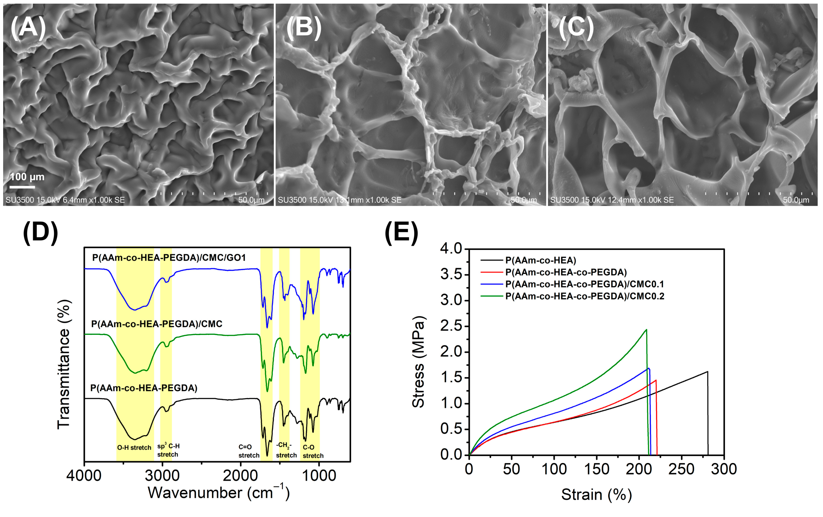
Scanning electron microscopy revealed porous structures for the formulated hydrogel (Figure 3A–C). The base copolymer, poly(AAm-co-HEA-co-PEGDA), exhibits a relatively dense and tightly packed polymeric network with irregularly shaped, smaller pores. Upon the incorporation of CMC to form poly(AAm-co-HEA-co-PEGDA)/CMC, the microstructure transitions into a more defined, interconnected, and distinctly open porous architecture, suggesting that the CMC chains aid in organizing and stabilizing the polymer matrix [47]. The subsequent incorporation of graphene oxide (GO) transforms the morphology into a regular, macroporous, honeycomb-like architecture. These significantly enlarged and well-defined pores suggest that the dispersed GO nanosheets function as a reinforcing agent within the pore walls, preventing structural collapse and facilitating highly interconnected porosity [48]. Good gel porosity is highly advantageous for exudate absorption, improved drug diffusion and gas exchange in wound management applications.
Fourier transform infrared (FT-IR) spectroscopy was utilized to characterize the chemical structures and verify the successful integration of components within the hydrogel networks (Figure 3D). Across all formulations, the spectra exhibit characteristic absorption bands that confirm the expected functional groups. The broad band centered around 3400 cm−1 corresponds to the overlapping O-H and N-H stretching vibrations, reflecting the presence of hydroxyl and amine groups from the primary monomers, alongside contributions from CMC and GO. The absorption at 2960 cm−1 is attributed to sp3 C-H stretching, while the prominent peak at around 1700 cm−1 confirms the C=O stretching of the constituent amide and ester groups. Additionally, the peaks at 1460 cm−1 and 1160 cm−1 represent -CH2- bending and C-O stretching vibrations, respectively. Moreover, only a slight widening of peaks around 1460 cm−1 and 1160 cm−1 was observed in the spectrum of the hydrogel containing 1 ppm GO. This might be due to the GO interaction with the polymer matrix [49]. Aside from this observation, significant changes in the peaks after the addition of 1 ppm GO were not evident as this amount typically falls below the instrument’s limit of detection [50,51]. Furthermore, any weak signals from the trace graphene oxide would be entirely masked by the overlapping infrared absorption bands of the host polymer matrix [51,52]. The overall similarity of the spectral profiles upon the successive addition of CMC and graphene oxide suggests that the fundamental polymeric backbone is preserved, indicating that the composite materials are primarily stabilized through robust physical interactions, such as intermolecular hydrogen bonding, rather than alterations to the covalent network
The tensile mechanical properties of the hydrogels were evaluated to understand the structural impact of the crosslinker and carboxymethyl cellulose (CMC) on the polymer matrix (Figure 3E). The base copolymer, poly(AAm-co-HEA), exhibited the highest elasticity with an elongation at break of 281%, coupled with a tensile strength of 1.623 MPa. The introduction of PEGDA to form poly(AAm-co-HEA-co-PEGDA) resulted in a tighter crosslinked network, which predictably restricted chain mobility, reducing the maximum strain to 221%, with tensile strength of 1.456 MPa. Most notably, the incorporation of CMC into the hydrogel system served as an effective reinforcing agent. As the CMC concentration increased from 0.1 to 0.2 wt%, the ultimate tensile stress significantly enhanced, peaking at nearly 2.440 MPa for poly(AAm-co-HEA-co-PEGDA)/CMC0.2. While this reinforcement came with a marginal trade-off in stretchability, the dramatic increase in stiffness and tensile strength suggests that the rigid CMC chains successfully established strong intermolecular interactions with the primary polymer network, effectively transferring stress and improving the overall mechanical robustness of the composite hydrogel [53]. Ultimately, this synergistic balance of enhanced tensile strength and sustained elasticity makes the CMC-reinforced hydrogels highly promising for wound dressing applications, ensuring conformation to dynamic body movements while resisting mechanical failure [30].

2.3. Swelling Behavior of the 3D-Printed Hydrogels

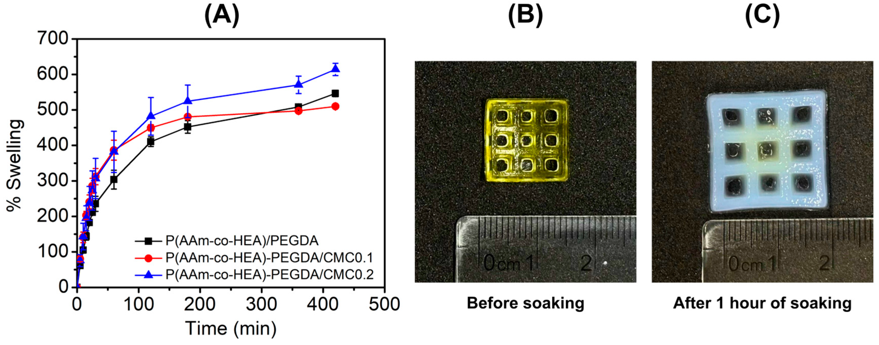
The water absorbance of the formulated hydrogels was assessed by determining the swelling behavior of representative formulations. Dried 3D-printed specimens were soaked in pH 7.4 PBS and changes in weight were recorded. Figure 4 illustrates the time-dependent swelling percentage over a period of roughly seven hours, revealing that all three compositions follow a characteristic kinetic profile: a rapid initial uptake of water followed by a gradual plateau as the materials approach equilibrium. This rapid water uptake can be attributed to the previously discussed porous structure of the hydrogel. Moreover, the hydrogel containing 0.2 wt% CMC demonstrates the superior swelling capacity throughout the entire duration, achieving the highest maximum swelling of approximately 610%. This suggests that the higher concentration of CMC introduces sufficient hydrophilic groups and ionic charges to maximize the osmotic pressure within the network, driving substantial water absorption [53,54,55]. The addition of 0.1 wt% CMC also significantly increased initial water uptake as compared to the control formulation. Interestingly, the water uptake eventually plateaued at a slightly lower or comparable level (513%) to the control (536%) by the end of the experiment. This indicates that while small amounts of CMC accelerate the kinetics of hydration, they do not necessarily expand the ultimate equilibrium volume as effectively as the 0.2 wt% loading. Aside from CMC, the high water uptake can also be attributed to the addition of GO to the formulation. This is driven by the interaction between water and the hydrophilic sites on GO, specifically the –OH groups, which foster hydrogen bonding and enhance the diffusion and perfusion of water into the matrix [23,56,57]. Such high swelling capabilities are particularly advantageous for wound dressing applications, where the ability to absorb excess exudate while maintaining a moist healing environment is essential [57,58]. Furthermore, the significant swelling of these hydrogels is essential for the sustained release of encapsulated therapeutic agents [57]. Their exceptional capacity for biofluid absorption positions these synthetic bioactive hydrogels as highly promising candidates for advanced wound healing applications.

2.4. Colorimetric Response of 3D-Printed Hydrogels

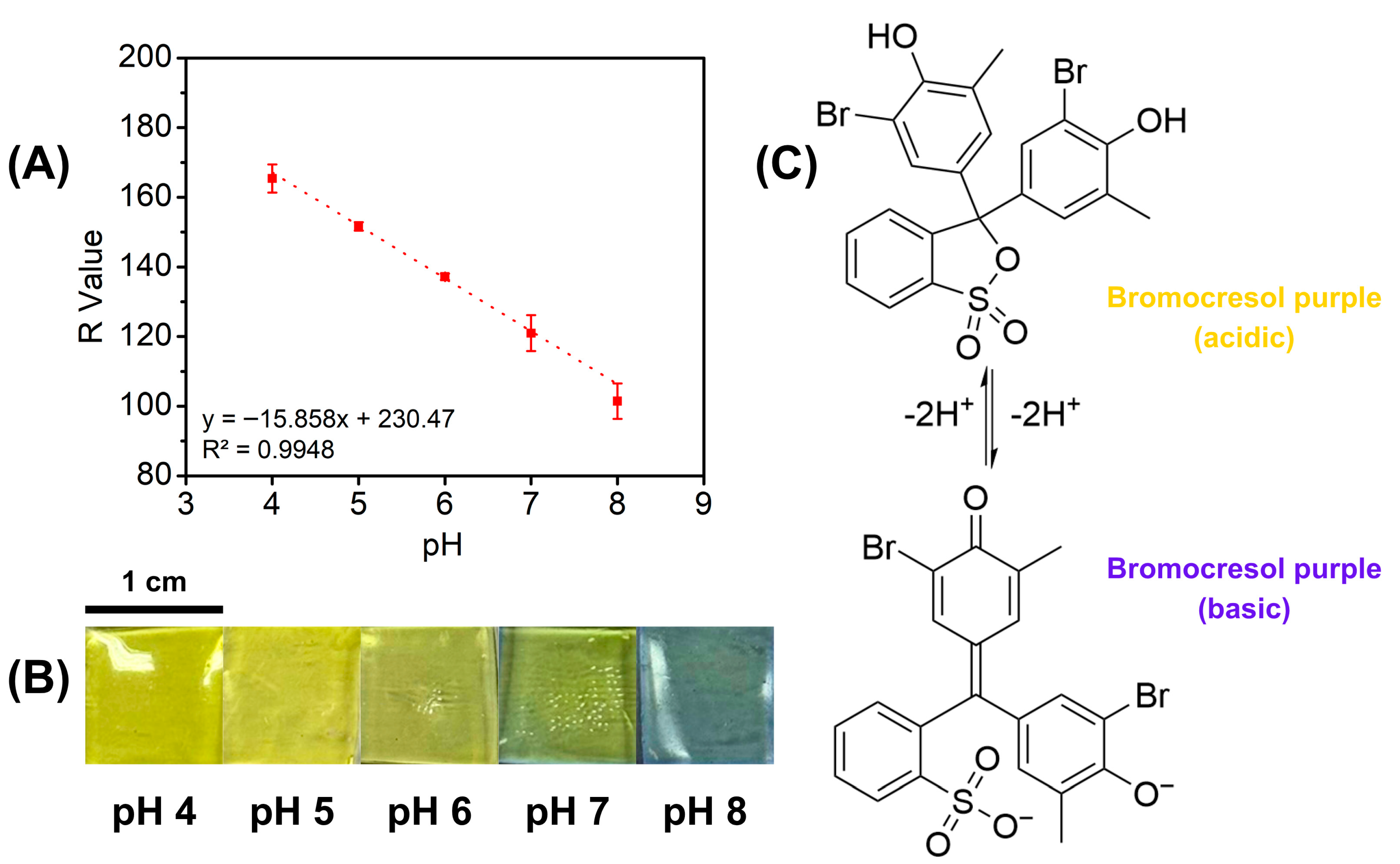
To assess the colorimetric response of the 3D-printed hydrogels, 3D-printed hydrogel patches were immersed in solutions of varying pH (pH 4–8) for one minute. This procedure was designed to simulate the pH variations associated with wound infection. The 3D-printed hydrogels exhibited an initial yellow hue, attributed to the presence of bromocresol purple (BCP) in the resin. A significant color shift was observed at pH 6, where the hydrogel transitioned to a slight green. At pH 7, this green coloration became more pronounced, and at pH 8, the hydrogel ultimately shifted to blue (Figure 5B). These observations align with the expected behavior of BCP, which remains yellow below pH 5.2 and transitions to purple above pH 6.8 [59]. Consequently, this study demonstrates the dual functionality of BCP, acting as a photoabsorber during DLP 3D printing and serving as a pH-responsive indicator for wound infection monitoring. In a clinical setting, the gel functions by reacting to the bacterial metabolites, which typically shift the wound environment from acidic to alkaline as an infection develops. As the wound fluid permeates the porous hydrogel matrix, the embedded BCP molecules undergo a structural rearrangement (Figure 5C) that alters their light absorption, providing a real-time visual warning through a clear color change from yellow to purple.
While visual inspection allows for a rapid qualitative assessment of the significant color shifts associated with pH increases, a quantitative analysis was performed to rigorously validate these variations and construct a calibration curve. To generate a semi-quantitative model for colorimetric response, digital images of the hydrogels were processed using ImageJ software according to previously described methods [60]. Data points representing the red, green, and blue (RGB) channels, along with grayscale intensities, were extracted and correlated with specific pH values. The resulting plots demonstrated a highly linear relationship between color intensity and pH level. Specifically, the red (R) channel yielded the highest correlation coefficient (R2 = 0.9948, Figure 5A), closely followed by the green channel (R2 = 0.9919, Figure S2, Supporting Information) and grayscale values (R2 = 0.9675, Figure S3, Supporting Information). This strong linearity confirms the reliability of the formulation as a sensor for monitoring infection progression. Furthermore, this correlation establishes a framework for smart wound-monitoring systems that integrate qualitative visual inspection with smartphone-based digital colorimetry. This method permits the non-invasive, real-time measurement of wound status, effectively connecting patient self-care with professional clinical supervision to ensure prompt and suitable medical treatment.

2.5. In Vitro Drug Release Study

In this study, we investigated the influence of two primary factors on drug release kinetics: GO content and 3D-printed structural geometry. Levofloxacin (LVX) was used as the model drug, with release profiles monitored via UV-Vis spectrophotometry. To simulate the dynamic wound microenvironment, drug release was evaluated under conditions mimicking healthy skin (pH 5.0), physiological fluid (pH 7.4), and infected tissue (pH 8.0). The cumulative release data demonstrated clear pH-responsive behavior, where drug release increased significantly with rising pH. The lowest release was observed at pH 5.0, followed by moderate release at pH 7.4, with the maximum release occurring at pH 8.0. This trend is characteristic of carboxylated hydrogels, such as the CMC and CMC/GO matrix used in this system [61,62,63]. As the environment becomes more alkaline, the carboxylic acid groups (-COOH) within the polymer network deprotonate into negatively charged carboxylate ions (-COO). The resulting electrostatic repulsion between polymer chains causes the hydrogel network to swell and expand, thereby increasing the pore size and facilitating faster drug diffusion [63]. This behavior aligns well with the physiological requirements of wound management. The minimal release at pH 5.0 helps prevent premature drug loss on healthy skin. However, as the wound environment shifts towards the alkaline pH typical of bacterial infection (pH 7.4–8.9), the hydrogel’s swelling response triggers a significant increase in levofloxacin release. This infection-responsive mechanism ensures that the highest therapeutic dose is delivered precisely when bacterial proliferation is most aggressive, optimizing antibacterial efficacy while minimizing unnecessary exposure in non-infected states.
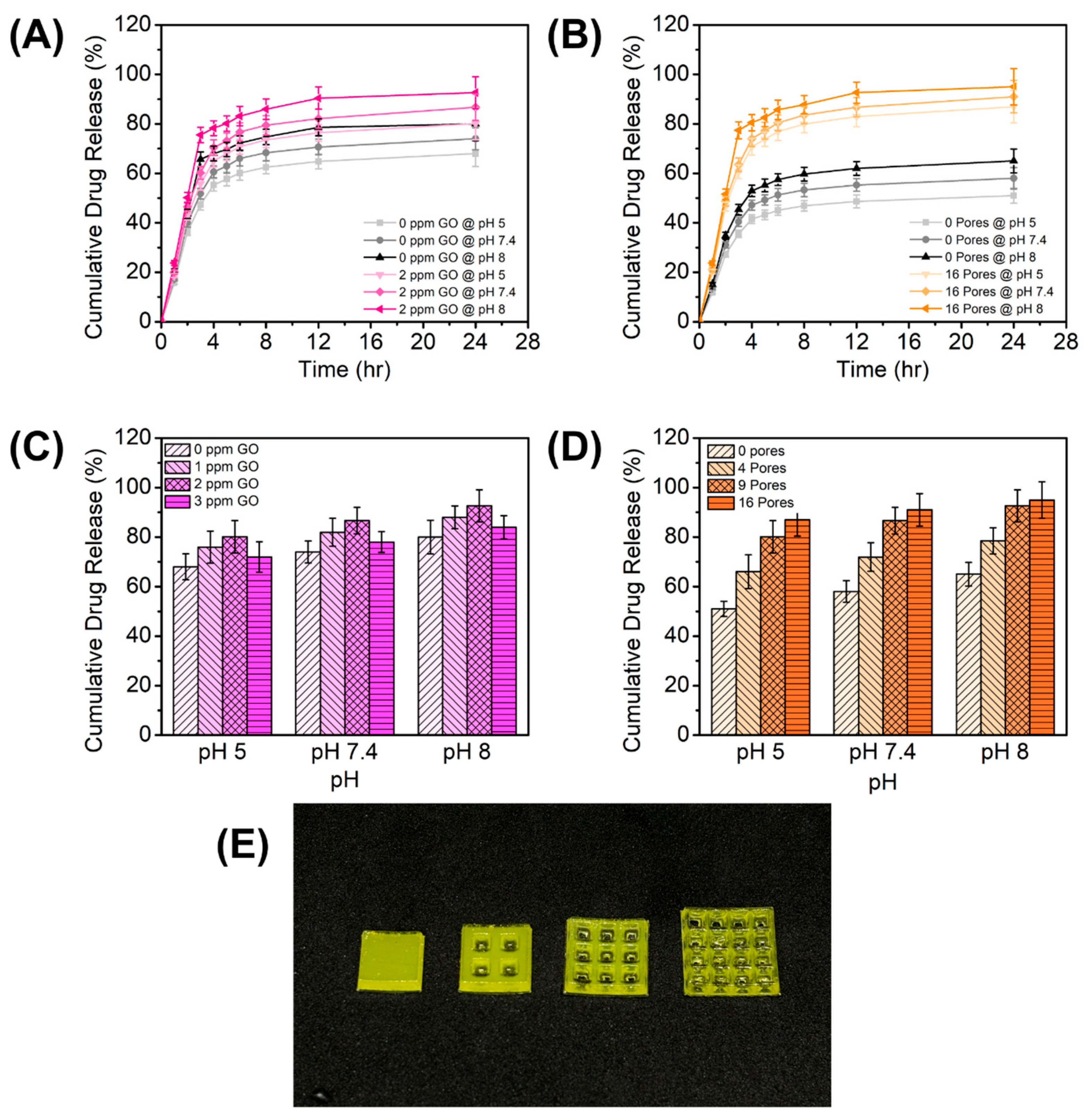
Comparing the drug release of 3D-printed hydrogels with and without GO, higher drug release was consistently seen with hydrogels with GO than those without GO (Figure 6A). To test the effect of GO, a standard nine-pore gel pad was 3D-printed and subjected to a drug release experiment. Among the concentrations tested, the 2 ppm GO formulation exhibited the highest cumulative drug release (pH 8) at 92.63%, followed by 1 ppm GO at 88.01%. Interestingly, increasing the concentration further to 3 ppm GO resulted in the lowest drug release among the GO-loaded samples, at 83.98% (Figure 6C). This non-linear trend suggests an optimal dispersion threshold at 2 ppm. At this concentration, the hydrophilic functional groups on the GO sheets likely enhance water uptake and matrix swelling, facilitating drug diffusion [64,65]. However, at 3 ppm, the excess GO nanosheets likely undergo π-π stacking and agglomeration [66]. These aggregates reduce the effective surface area and create tortuous physical barriers within the hydrogel network, thereby hindering the release of the drug [66,67]. Nonetheless, the observed behavior suggests that the GO nanosheets act as structural spacers, potentially interrupting the polymer chain packing to create a more permeable network for drug diffusion [68]. Additionally, the superior surface-to-volume ratio of GO facilitates high-capacity drug anchoring and release, governed by the dynamic interplay between the drug molecules and the graphene surface [68,69]. The observed enhancement can also be attributed to the high affinity between LVX and the GO surface, mediated by two primary mechanisms: π-π stacking between the GO lattice and the fluoroquinolone rings of LVX, and hydrogen bonding between the GO’s oxygen-containing groups and the drug’s amine moieties [20]. Furthermore, the release profile is governed by the pH-dependent ionization of these functional groups. Variations in pH alter the electrostatic interplay between the drug and the filler, thereby tuning the adsorption strength and controlling the release rate [66,68,70]. From the results, the hydrogel containing GO consistently demonstrated a significantly higher cumulative drug release compared to the GO-free formulation. Mechanistically, we hypothesize that GO acts as a high-capacity reservoir within the hydrogel matrix [21]. During loading, LVX is strongly adsorbed onto the GO surface via π-π stacking between the aromatic rings of GO and the fluoroquinolone structure of LVX, alongside hydrogen bonding between the oxygen-containing functional groups (hydroxyl and carboxyl) of GO and the amine groups of LVX [65]. The enhanced, pH-dependent release profile is driven by the alteration of these non-covalent interactions. As the environment becomes more alkaline, mimicking infected wound exudate, the ionization states of both LVX and the functional groups on GO shift [71]. This induces electrostatic repulsion and weakens the drug–nanomaterial interactions, facilitating rapid diffusion of the drug from the matrix, with the maximum LVX release observed at pH 8. Ultimately, the strategic incorporation of GO creates a synergistic release mechanism, coupling the structural expansion of the polymer network with dynamic, pH-responsive chemical interactions to precisely govern the sustained delivery of the drug.
One advantage of additive manufacturing is that it allows the incorporation of specialized design and architecture that can further enhance the functionality of the printed object [30]. The drug release capability of the 3D-printed hydrogels was evaluated to understand the influence of structural design, specifically the inclusion of pore channels, on the release profile. As illustrated in Figure 6D, the introduction of porous channels into the hydrogel matrix had a profound impact on the total drug released. A clear positive correlation was observed between pore number and cumulative release percentage. The solid, bulk hydrogels (0 pores) consistently demonstrated the lowest drug release across all pH conditions, maxing out at approximately 65% release at pH 8. In contrast, the scaffolds designed with 16 pores achieved significantly higher release rates, reaching nearly 95% under the same conditions. It is critical to note that the total mass of the hydrogel was standardized across all samples, regardless of the pore count. Therefore, the observed increase in drug release is not a function of varying drug payloads or polymer content but is attributable strictly to the geometric modification. We hypothesize that the inclusion of macropore structures in the 3D-printed gel has increased the surface area available for drug diffusion, facilitating a more efficient mass transfer of the drug from the gel to the surrounding environment. Looking at the time-dependent release profiles (Figure 6B), the 16-pore scaffolds exhibited a rapid initial burst release within the first 4 h, followed by a sustained plateau. This rapid onset is advantageous for therapeutic applications requiring an immediate high dose to combat infection or inflammation. Conversely, the 0-pore bulk samples showed a much slower, more gradual release profile, struggling to release much of their payload within the 24 h window. These results underscore the distinct advantage of utilizing 3D printing technologies for pharmaceutical applications. Unlike traditional molding techniques, 3D printing allows for the precise, digital manipulation of scaffold geometry to increase drug delivery efficiency [72,73]. By simply modifying the CAD model to include 4, 9, or 16 pores, the drug release profile can be tuned to meet specific patient needs. A patient requiring rapid drug availability could be prescribed a highly porous 16-pore dressing, while a condition requiring slow, sustained release might benefit from a solid or low-porosity design. This ability to decouple geometric complexity from fabrication difficulty represents a significant leap forward in the design of personalized, tunable drug delivery systems.

2.6. Drug Release Kinetics

With the cumulative drug release data, the in vitro drug release profiles were mathematically modeled using the Higuchi, Korsmeyer–Peppas, and Peppas–Sahlin equations to elucidate the underlying transport mechanisms governing the release of the drug from the 3D-printed hydrogel scaffolds. The kinetic parameters obtained for hydrogels with varying GO concentrations (GO-0 to GO-3) reveal a release mechanism that is predominantly anomalous (non-Fickian) transport (Table 1). Across all formulations at pH 5 and 7.4, the release exponent n from the Korsmeyer–Peppas model consistently hovered around 0.69. This value, falling between 0.5 and 1.0, indicates that the drug release is driven by a coupling of two phenomena: Fickian diffusion of the drug through the hydrogel matrix and the relaxation (swelling) of the polymer chains [74]. Interestingly, the release kinetics demonstrated pH-sensitivity. At pH 8, n increased to values between 0.81 and 0.86. This shift towards Case II transport suggests that at higher pH, the contribution of polymer relaxation and potentially erosion becomes more significant compared to simple diffusion [74,75]. This is likely due to the increased ionization of the carboxyl groups in the CMC and polymer network at basic pH, leading to greater electrostatic repulsion, higher swelling ratios, and faster chain mobility [76]. In addition, dispersion of GO nanosheets creates a tortuous path for the drug molecules. Instead of moving in a straight line through the water-filled pores of the hydrogel, the drug molecules must navigate around the impermeable graphene sheets [77,78]. The Peppas–Sahlin model further corroborated this, showing mostly positive values for both the Fickian (k1) and relaxation (k2) constants, confirming that both mechanisms are operative and competitive.
The introduction of 3D-printed pores had a profound impact on both the rate and mechanism of drug release. As expected, the release rate increased substantially with pore density. For instance, at pH 8, the Higuchi rate constant kH nearly doubled from 24.52 for the non-porous control to 41.25 for the 16-pore scaffold (Table 2). This acceleration is directly improved by the increased surface area-to-volume ratio provided by the printed macro-channels, which facilitates faster solvent penetration and shorter diffusion path lengths for the drug [79,80]. More notably, the mechanism of release shifted drastically in the highly porous scaffolds. While the non-porous and low-porosity (4 pores) samples maintained anomalous transport characteristics (n ≈ 0.58–0.74), the high-porosity samples (9 and 16 pores) exhibited Super Case II transport kinetics (n > 0.89, reaching 1.05 at pH 8). An n value exceeding 0.89 indicates that the release is dominated by polymer relaxation and erosion rather than diffusion. The presence of macropores likely allows for rapid bulk water uptake, leading to immediate swelling stresses and accelerated degradation or macro-erosion of the hydrogel lattice [81,82]. This structural breakdown facilitates a rapid “dumping” of the drug, overpowering the diffusion-controlled regime.
Among the three kinetic models applied, the Peppas–Sahlin model consistently provided the best fit across all formulations and pH conditions, with R2 values exceeding 0.99. This superior fit is attributed to the model’s ability to decouple and quantify the individual contributions of Fickian diffusion (k1) and Case II relaxation (k2). The Korsmeyer–Peppas model also showed high applicability, whereas the Higuchi model, which assumes purely diffusion-controlled release from a non-swelling matrix, showed the poorest fit. This statistical comparison confirms that the swelling and relaxation of the 3D-printed hydrogel matrix are critical factors that cannot be ignored in the design of these drug delivery systems. Overall, these results indicate that both GO incorporation and scaffold porosity serve as critical handles for modulating the drug release mechanism, offering dual strategies to control the transition between diffusive and relaxational kinetics for optimized LVX delivery.

3. Conclusions

We report on the successful development of a novel vat photopolymerization 3D-printed hydrogel designed for simultaneous wound monitoring and therapeutic delivery. By integrating bromocresol purple directly into the resin, we achieved high-fidelity digital light processing (DLP) of customizable prototypes that function as high-sensitivity colorimetric sensors (R2 = 0.9948). The resulting scaffolds exhibit robust mechanical integrity (1.456 MPa) and high extensibility (221%), coupled with an optimized macroporous architecture that ensures effective exudate management. Furthermore, the synergistic incorporation of graphene oxide enables a sophisticated pH-responsive mechanism, achieving a 92.63% cumulative release of levofloxacin at infection-simulating alkaline levels (pH 8.0). Further development of this material can include other compatible therapeutics such as ciprofloxacin, gentamicin, moxifloxacin, and others with aromatic or ionizable groups. Moreover, testing with other buffer systems to simulate complex wound microenvironments, leading to in vivo testing, can be done. Overall, this dual-functional platform demonstrates immense potential for the scalable manufacturing of smart dressings, providing a versatile foundation for personalized, theranostic medical interventions.

4. Materials and Methods

4.1. Materials

Acrylamide (AAm), 2-hydroxyethyl acrylate (HEA), poly(ethylene glycol) diacrylate (n = approx. 9) (PEGDA), lithium phenyl-2,4,6-trimethylbenzoylphosphinate (LAP), and levofloxacin (LVX) were obtained from Tokyo Chemical Industry (Tokyo, Japan). Sodium carboxymethyl cellulose (~90 KDa) was purchased from SinoCMC (Qingdao, China). Bromocresol purple (BCP) was purchased from Techno PharmaChem (New Delhi, India). Graphene oxide (v20) was acquired from Standard Graphene (Ulsan, Republic of Korea).

4.2. Preparation of Photosensitive Resin and 3D Printing of Poly(AAm-co-HEA-co-PEGDA)/CMC/GO Hydrogels

A simple one-pot mixing was done to prepare the photosensitive hydrogel resin. The monomers, CMC, LAP, GO, LVX, and BCP were mixed with distilled water at room temperature until a clear solution was achieved. The hydrogel resins were stored in amber-colored glass vials and kept away from light. Before printing, the resins were sonicated to remove trapped gas and air bubbles. Detailed formulations are listed in Table 3. Various objects were then 3D-printed using a DLP 3D printer (Creality Halot One, Shenzhen, China). Printed gels were washed with distilled water to remove excess unpolymerized resin, then exposed to 6 W 405 nm UV light for 5 min to allow full curing.

4.3. Characterization Tests

Tensile tests of 3D-printed hydrogels were done using a universal testing machine (Instron 3343, Instron, Norwood, MA, USA). 3D-printed standard dogbone specimens were tested with a crosshead speed of 50 mm min−1 and a load cell of 500 N. All ultimate tensile strength and strain tests were performed in triplicate. Dimensional accuracy of the 3D printing process was assessed by printing a standard wall thickness structure, then measured by a coordinate measuring machine (Hexagon Optiv M, Hexagon Manufacturing Intelligence, Stockholm, Sweden). To check gel porosity, samples were observed under a scanning electron microscope (Hitachi SU3500, Hitachi, Tokyo, Japan) using 15 kV accelerating voltage at 12.5 mm working distance. Fourier transform infrared (FT-IR) spectra of representative hydrogels were obtained using an FT-IR spectrometer (Perkin Elmer Frontier, Waltham, MA, USA) with an attenuated total reflectance accessory.

4.4. Swelling Test

To determine the swelling capacity of the optimized formulation, a freeze-dried 3D-printed hydrogel was immersed in a phosphate-buffered saline (PBS) solution at pH 7.4. After a set incubation period, the weight of the hydrated gel was measured and compared against its initial weight. Excess water was blotted and removed before weighing. The swelling percentage was calculated using the following formula:
% S w e l l i n g = W s W d W d × 100
where Ws is the weight of swollen gel and Wd is the initial weight.

4.5. Colorimetric Response

To check the colorimetric response, a 10 × 10 × 2 mm3 piece of hydrogel was soaked in PBS with varying pH (4–8) at room temperature (25 °C). After 10 min, the color change was captured using a phone camera (iPhone 15, Apple Inc., Cupertino, CA, USA). RGB and grayscale values were measured using ImageJ software (Version 1.54s), and the corresponding values were plotted versus pH change to assess semi-quantitative color change correlation.

4.6. Drug Release Profile

To evaluate the in vitro release profile of LVX under conditions mimicking both healthy and infected wounds, hydrogel patch samples were placed into individual well plates. Each well was filled with 5.0 mL of PBS adjusted to a specific pH (5.0, 7.4, or 8.0). At predetermined time intervals, 1.0 mL aliquots were withdrawn for analysis and immediately replaced with an equal volume of fresh buffer to maintain sink conditions. The concentration of the released drug was then quantified using a Shimadzu UV mini-1240 UV–Visible Spectrophotometer. Initial drug release kinetics were determined by modeling the calculated % cumulative drug released (Q) using the following equations [77]:
Higuchi Model:
Q = k H t 1 2
Korsmeyer–Peppas:
Q = k K P t n
where n is the release exponent.
Peppas–Sahlin:
Q = k 1 t m + k 2 t 2 m
where m = 0.43 for a sphere, m = 0.45 for a cylinder, and m = 0.5 for a thin film.

Supplementary Materials

The following supporting information can be downloaded at https://www.mdpi.com/article/10.3390/gels12050393/s1: Figure S1. 3D-printed hydrogels without BCP (left), and with BCP (right); Figure S2. Hydrogel green color value vs. pH; Figure S3. Hydrogel grayscale value vs. pH; Figure S4. 3D printing accuracy as a function of measured dimensions vs. nominal dimensions.

Author Contributions

Conceptualization, K.A.T.; methodology, K.A.T. and L.I.B.; software, K.A.T.; validation, K.A.T. and L.I.B.; investigation, K.A.T., L.I.B., J.M.V.A., P.M. and K.M.S.; data curation, K.A.T.; writing—original draft, K.A.T. and L.I.B.; writing—review and editing, T.B.L., V.P.H., N.R. and R.A.; visualization, K.A.T.; supervision, T.B.L., V.P.H., N.R. and R.A.; project administration, K.A.T.; funding acquisition, K.A.T. All authors have read and agreed to the published version of the manuscript.

Funding

The funding, including APC, for this project was supported by the Department of Science and Technology (DOST) through DOST Grants-in-Aid Project No. 11233, under DOST-PCIEERD as granting agency. The project is led by Fred P. Liza of the DOST—Metals Industry Research and Development Center.

Institutional Review Board Statement

Not applicable.

Informed Consent Statement

Not applicable.

Data Availability Statement

The original contributions presented in this study are included in the article/supplementary material. Further inquiries can be directed to the corresponding author.

Acknowledgments

K.A.T., L.I.B., J.M.V.A. and P.A. would like to thank Fred P. Liza for providing financial assistance with this study as part of Project TRIAMPH (DOST GIA Project No. 11233). Additionally, K.A.T. would like to express his gratitude to the DOST S&T Fellows Program. This research was also supported partially by the International Research Partnership Fund—Faculty of Science, and the Graduate School, Chulalongkorn University.

Conflicts of Interest

The authors declare no conflicts of interest.

Abbreviations

The following abbreviations are used in this manuscript:
DLPDigital light processing
AAmAcrylamide
HEA2-Hydroxyethyl acrylate
PEGDAPolyethylene glycol diacrylate
LAPLithium phenyl-2,4,6-trimethylbenzoylphosphinate
BCPBromocresol purple
LVXLevofloxacin
CMCCarboxymethyl cellulose
GOGraphene oxide
PBSPhosphate-buffered saline

References

  1. Zhao, R.; Liang, H.; Clarke, E.; Jackson, C.; Xue, M. Inflammation in Chronic Wounds. Int. J. Mol. Sci. 2016, 17, 2085. [Google Scholar] [CrossRef] [PubMed]
  2. Monika, P.; Chandraprabha, M.N.; Rangarajan, A.; Waiker, P.V.; Chidambara Murthy, K.N. Challenges in Healing Wound: Role of Complementary and Alternative Medicine. Front. Nutr. 2022, 8, 791899. [Google Scholar] [CrossRef] [PubMed]
  3. Serpico, L.; Iacono, S.D.; Cammarano, A.; Stefano, L.D. Recent Advances in Stimuli-Responsive Hydrogel-Based Wound Dressing. Gels 2023, 9, 451. [Google Scholar] [CrossRef]
  4. Youssef, J.R.; Boraie, N.A.; Ibrahim, H.F.; Ismail, F.A.; El-Moslemany, R.M. Glibenclamide Nanocrystal-Loaded Bioactive Polymeric Scaffolds for Skin Regeneration: In Vitro Characterization and Preclinical Evaluation. Pharmaceutics 2021, 13, 1469. [Google Scholar] [CrossRef]
  5. Frykberg, R.G.; Banks, J. Challenges in the Treatment of Chronic Wounds. Adv. Wound Care 2015, 4, 560–582. [Google Scholar] [CrossRef]
  6. Brown, M.S.; Ashley, B.; Koh, A. Wearable Technology for Chronic Wound Monitoring: Current Dressings, Advancements, and Future Prospects. Front. Bioeng. Biotechnol. 2018, 6, 47. [Google Scholar] [CrossRef]
  7. Vo, D.-K.; Trinh, K.T.L. Advances in Wearable Biosensors for Wound Healing and Infection Monitoring. Biosensors 2025, 15, 139. [Google Scholar] [CrossRef]
  8. Farahani, M.; Shafiee, A. Wound Healing: From Passive to Smart Dressings. Adv. Healthc. Mater. 2021, 10, 2100477. [Google Scholar] [CrossRef]
  9. Derakhshandeh, H.; Kashaf, S.S.; Aghabaglou, F.; Ghanavati, I.O.; Tamayol, A. Smart Bandages: The Future of Wound Care. Trends Biotechnol. 2018, 36, 1259–1274. [Google Scholar] [CrossRef] [PubMed]
  10. Mirani, B.; Hadisi, Z.; Pagan, E.; Dabiri, S.M.H.; van Rijt, A.; Almutairi, L.; Noshadi, I.; Armstrong, D.G.; Akbari, M. Smart Dual-Sensor Wound Dressing for Monitoring Cutaneous Wounds. Adv. Healthc. Mater. 2023, 12, 2203233. [Google Scholar] [CrossRef]
  11. Shi, C.; Wang, C.; Liu, H.; Li, Q.; Li, R.; Zhang, Y.; Liu, Y.; Shao, Y.; Wang, J. Selection of Appropriate Wound Dressing for Various Wounds. Front. Bioeng. Biotechnol. 2020, 8, 47. [Google Scholar] [CrossRef]
  12. Delgado-Pujol, E.J.; Martínez, G.; Casado-Jurado, D.; Vázquez, J.; León-Barberena, J.; Rodríguez-Lucena, D.; Torres, Y.; Alcudia, A.; Begines, B. Hydrogels and Nanogels: Pioneering the Future of Advanced Drug Delivery Systems. Pharmaceutics 2025, 17, 215. [Google Scholar] [CrossRef] [PubMed]
  13. Du, L.; Lin, C.; Hu, H.; Zhao, Y.; Liao, J.; Al-Smadi, F.; Mi, B.; Hu, Y.; Liu, G. Recent Advances and Challenges in Hydrogel-Based Delivery of Immunomodulatory Strategies for Diabetic Wound Healing. Theranostics 2026, 16, 516–544. [Google Scholar] [CrossRef] [PubMed]
  14. Yang, Y.; Chen, H.; Li, Y.; Liang, J.; Huang, F.; Wang, L.; Miao, H.; Nanda, H.S.; Wu, J.; Peng, X.; et al. Hydrogel Loaded with Extracellular Vesicles: An Emerging Strategy for Wound Healing. Pharmaceuticals 2024, 17, 923. [Google Scholar] [CrossRef]
  15. Jia, X.; Dou, Z.; Zhang, Y.; Li, F.; Xing, B.; Hu, Z.; Li, X.; Liu, Z.; Yang, W.; Liu, Z. Smart Responsive and Controlled-Release Hydrogels for Chronic Wound Treatment. Pharmaceutics 2023, 15, 2735. [Google Scholar] [CrossRef]
  16. Zhang, Y.; Wu, B.M. Current Advances in Stimuli-Responsive Hydrogels as Smart Drug Delivery Carriers. Gels 2023, 9, 838. [Google Scholar] [CrossRef]
  17. Yasir, M.; Mishra, R.; Tripathi, A.S.; Maurya, R.K.; Shahi, A.; Zaki, M.E.A.; Al Hussain, S.A.; Masand, V.H. Theranostics: A Multifaceted Approach Utilizing Nano-Biomaterials. Discov. Nano 2024, 19, 35. [Google Scholar] [CrossRef]
  18. Ran, P.; Zheng, H.; Cao, W.; Jia, X.; Zhang, G.; Liu, Y.; Li, X. On-Demand Changeable Theranostic Hydrogels and Visual Imaging-Guided Antibacterial Photodynamic Therapy to Promote Wound Healing. ACS Appl. Mater. Interfaces 2022, 14, 49375–49388. [Google Scholar] [CrossRef]
  19. Karthikeyan, L.; Kang, H.W. Recent Progress in Multifunctional Theranostic Hydrogels: The Cornerstone of next-Generation Wound Care Technologies. Biomater. Sci. 2025, 13, 4358–4389. [Google Scholar] [CrossRef]
  20. Tao, J.; Yang, J.; Ma, C.; Li, J.; Du, K.; Wei, Z.; Chen, C.; Wang, Z.; Zhao, C.; Deng, X. Cellulose Nanocrystals/Graphene Oxide Composite for the Adsorption and Removal of Levofloxacin Hydrochloride Antibiotic from Aqueous Solution. R. Soc. Open Sci. 2020, 7, 200857. [Google Scholar] [CrossRef] [PubMed]
  21. Oliveira, A.M.L.; Machado, M.; Silva, G.A.; Bitoque, D.B.; Ferreira, J.T.; Pinto, L.A.; Ferreira, Q. Graphene Oxide Thin Films with Drug Delivery Function. Nanomaterials 2022, 12, 1149. [Google Scholar] [CrossRef] [PubMed]
  22. Yu, W.; Sisi, L.; Haiyan, Y.; Jie, L. Progress in the Functional Modification of Graphene/Graphene Oxide: A Review. RSC Adv. 2020, 10, 15328–15345. [Google Scholar] [CrossRef]
  23. Li, S.; Wang, J.; Zhang, H.; Zhang, X. Advances in Graphene Oxide-Based Polymeric Wound Dressings for Wound Healing. Front. Mater. 2025, 12, 1635502. [Google Scholar] [CrossRef]
  24. Jin, C.; Zheng, H.; Chen, J. Advances in the Application of Graphene and Its Derivatives in Drug Delivery Systems. Pharmaceuticals 2025, 18, 1245. [Google Scholar] [CrossRef] [PubMed]
  25. Phookum, T.; Siripongpreda, T.; Tiston, K.A.; Rerknimitr, P.; Henry, C.S.; Narupai, B.; Rodthongkum, N. Dual-Functional 3D-Printed Hydrogels for pH-Responsive Wound Monitoring and on-Demand Therapy. J. Mater. Chem. B 2026, 14, 1088–1098. [Google Scholar] [CrossRef]
  26. Wu, H.; Chen, J.; Zhao, P.; Liu, M.; Xie, F.; Ma, X. Development and Prospective Applications of 3D Membranes as a Sensor for Monitoring and Inducing Tissue Regeneration. Membranes 2023, 13, 802. [Google Scholar] [CrossRef]
  27. Serrano, D.R.; Kara, A.; Yuste, I.; Luciano, F.C.; Ongoren, B.; Anaya, B.J.; Molina, G.; Diez, L.; Ramirez, B.I.; Ramirez, I.O.; et al. 3D Printing Technologies in Personalized Medicine, Nanomedicines, and Biopharmaceuticals. Pharmaceutics 2023, 15, 313. [Google Scholar] [CrossRef]
  28. Zhang, J.; Wehrle, E.; Rubert, M.; Müller, R. 3D Bioprinting of Human Tissues: Biofabrication, Bioinks, and Bioreactors. Int. J. Mol. Sci. 2021, 22, 3971. [Google Scholar] [CrossRef]
  29. Kandasamy, M.; Vijayananth, K.; Parasuraman, A.; Ayrilmis, N. 3D Bioprinting of Biomaterials: A Review of Advances in Techniques, Materials, and Applications. Polym. Adv. Technol. 2025, 36, e70324. [Google Scholar] [CrossRef]
  30. Tiston, K.A.; Tipachan, C.; Yimnoi, T.; Cheacharoen, R.; Hoven, V.P.; Narupai, B. 3D Printing of Ultrastretchable and Tough Double—Network Hydrogel for Strain Sensor. Adv. Mater. Technol. 2025, 10, 2400751. [Google Scholar] [CrossRef]
  31. Tagliaferri, S.; Panagiotopoulos, A.; Mattevi, C. Direct Ink Writing of Energy Materials. Mater. Adv. 2021, 2, 540–563. [Google Scholar] [CrossRef]
  32. Zheng, Q.; Xie, B.; Xu, Z.; Wu, H. A Systematic Printability Study of Direct Ink Writing towards High-Resolution Rapid Manufacturing. Int. J. Extrem. Manuf. 2023, 5, 035002. [Google Scholar] [CrossRef]
  33. Agrawal, A.; Hussain, C.M. 3D-Printed Hydrogel for Diverse Applications: A Review. Gels 2023, 9, 960. [Google Scholar] [CrossRef]
  34. Wu, H.; Diao, J.; Li, X.; Yue, D.; He, G.; Jiang, X.; Li, P. Hydrogel-Based 3D Printing Technology: From Interfacial Engineering to Precision Medicine. Adv. Colloid Interface Sci. 2025, 341, 103481. [Google Scholar] [CrossRef]
  35. Grigoryan, B.; Paulsen, S.J.; Corbett, D.C.; Sazer, D.W.; Fortin, C.L.; Zaita, A.J.; Greenfield, P.T.; Calafat, N.J.; Gounley, J.P.; Ta, A.H.; et al. Multivascular Networks and Functional Intravascular Topologies within Biocompatible Hydrogels. Science 2019, 364, 458–464. [Google Scholar] [CrossRef]
  36. Huang, B.; Zhou, Y.; Wei, L.; Hu, R.; Zhang, X.; Coates, P.; Sefat, F.; Zhang, W.; Lu, C. Visible Light 3D Printing of High-Resolution Superelastic Microlattices of Poly(Ethylene Glycol) Diacrylate/Graphene Oxide Nanocomposites via Continuous Liquid Interface Production. Ind. Eng. Chem. Res. 2022, 61, 13052–13062. [Google Scholar] [CrossRef]
  37. Tsegay, F.; Hisham, M.; Elsherif, M.; Schiffer, A.; Butt, H. 3D Printing of pH Indicator Auxetic Hydrogel Skin Wound Dressing. Molecules 2023, 28, 1339. [Google Scholar] [CrossRef]
  38. Muralidharan, A.; Uzcategui, A.C.; McLeod, R.R.; Bryant, S.J. Stereolithographic 3D Printing for Deterministic Control over Integration in Dual-Material Composites. Adv. Mater. Technol. 2019, 4, 1900592. [Google Scholar] [CrossRef]
  39. Vaupel, S.; Mau, R.; Kara, S.; Seitz, H.; Kragl, U.; Meyer, J. 3D Printed and Stimulus Responsive Drug Delivery Systems Based on Synthetic Polyelectrolyte Hydrogels Manufactured via Digital Light Processing. J. Mater. Chem. B 2023, 11, 6547–6559. [Google Scholar] [CrossRef] [PubMed]
  40. Chand, R.; Janarthanan, G.; Elkhoury, K.; Vijayavenkataraman, S. Digital Light Processing 3D Bioprinting of Biomimetic Corneal Stroma Equivalent Using Gelatin Methacryloyl and Oxidized Carboxymethylcellulose Interpenetrating Network Hydrogel. Biofabrication 2025, 17, 025011. [Google Scholar] [CrossRef]
  41. Alberts, A.; Moldoveanu, E.-T.; Niculescu, A.-G.; Grumezescu, A.M. Hydrogels for Wound Dressings: Applications in Burn Treatment and Chronic Wound Care. J. Compos. Sci. 2025, 9, 133. [Google Scholar] [CrossRef]
  42. Yang, Y.; Zhou, Y.; Lin, X.; Yang, Q.; Yang, G. Printability of External and Internal Structures Based on Digital Light Processing 3D Printing Technique. Pharmaceutics 2020, 12, 207. [Google Scholar] [CrossRef]
  43. He, N.; Wang, X.; Shi, L.; Li, J.; Mo, L.; Chen, F.; Huang, Y.; Liu, H.; Zhu, X.; Zhu, W.; et al. Photoinhibiting via Simultaneous Photoabsorption and Free-Radical Reaction for High-Fidelity Light-Based Bioprinting. Nat. Commun. 2023, 14, 3063. [Google Scholar] [CrossRef]
  44. Xiang, Z.; Li, N.; Rong, Y.; Zhu, L.; Huang, X. 3D-Printed High-Toughness Double Network Hydrogels via Digital Light Processing. Colloids Surf. A Physicochem. Eng. Asp. 2022, 639, 128329. [Google Scholar] [CrossRef]
  45. Ding, H.; Dong, M.; Zheng, Q.; Wu, Z.L. Digital Light Processing 3D Printing of Hydrogels: A Minireview. Mol. Syst. Des. Eng. 2022, 7, 1017–1029. [Google Scholar] [CrossRef]
  46. Mo, X.; Ouyang, L.; Xiong, Z.; Zhang, T. Advances in Digital Light Processing of Hydrogels. Biomed. Mater. 2022, 17, 042002. [Google Scholar] [CrossRef]
  47. Kumar, B.; Priyadarshi, R.; Sauraj; Deeba, F.; Kulshreshtha, A.; Gaikwad, K.K.; Kim, J.; Kumar, A.; Negi, Y.S. Nanoporous Sodium Carboxymethyl Cellulose-g-Poly (Sodium Acrylate)/FeCl3 Hydrogel Beads: Synthesis and Characterization. Gels 2020, 6, 49. [Google Scholar] [CrossRef] [PubMed]
  48. Khan, M.U.A.; Stojanović, G.M.; Hassan, R.; Anand, T.J.S.; Al-Ejji, M.; Hasan, A. Role of Graphene Oxide in Bacterial Cellulose−Gelatin Hydrogels for Wound Dressing Applications. ACS Omega 2023, 8, 15909–15919. [Google Scholar] [CrossRef] [PubMed]
  49. Sieradzka, M.; Fabia, J.; Biniaś, D.; Fryczkowski, R.; Janicki, J. The Role of Reduced Graphene Oxide in the Suspension Polymerization of Styrene and Its Effect on the Morphology and Thermal Properties of the Polystyrene/rGO Nanocomposites. Polymers 2020, 12, 1468. [Google Scholar] [CrossRef]
  50. Cuthbertson, A.A.; Lincoln, C.; Miscall, J.; Stanley, L.M.; Maurya, A.K.; Asundi, A.S.; Tassone, C.J.; Rorrer, N.A.; Beckham, G.T. Characterization of Polymer Properties and Identification of Additives in Commercially Available Research Plastics. Green. Chem. 2024, 26, 7067–7090. [Google Scholar] [CrossRef]
  51. Layek, R.K.; Ramakrishnan, K.R.; Sarlin, E.; Orell, O.; Kanerva, M.; Vuorinen, J.; Honkanen, M. Layered Structure Graphene Oxide/Methylcellulose Composites with Enhanced Mechanical and Gas Barrier Properties. J. Mater. Chem. A 2018, 6, 13203–13214. [Google Scholar] [CrossRef]
  52. Mahmoodi, H.; Fattahi, M.; Motevassel, M. Graphene Oxide–Chitosan Hydrogel for Adsorptive Removal of Diclofenac from Aqueous Solution: Preparation, Characterization, Kinetic and Thermodynamic Modelling. RSC Adv. 2021, 11, 36289–36304. [Google Scholar] [CrossRef] [PubMed]
  53. Uyanga, K.A.; Daoud, W.A. Carboxymethyl Cellulose-Chitosan Composite Hydrogel: Modelling and Experimental Study of the Effect of Composition on Microstructure and Swelling Response. Int. J. Biol. Macromol. 2021, 181, 1010–1022. [Google Scholar] [CrossRef]
  54. Rahman, M.S.; Hasan, M.S.; Nitai, A.S.; Nam, S.; Karmakar, A.K.; Ahsan, M.S.; Shiddiky, M.J.A.; Ahmed, M.B. Recent Developments of Carboxymethyl Cellulose. Polymers 2021, 13, 1345. [Google Scholar] [CrossRef]
  55. Hendy, J.M.; Mansour, M.S.; Abdel-Megeed, A.; Al-Oufy, A.K.; Salem, M.Z.M. Swelling Properties of Superabsorbent Hydrogels Based on Carboxymethyl Cellulose With Silica, Kaolin, and Bentonite Particles for Agricultural Applications. ChemistrySelect 2025, 10, e04489. [Google Scholar] [CrossRef]
  56. Asmat, R.; Farooq, A.; Islam, A.; Ara, C.; Shafiq, N.; Imtiaz, F.; Khan, R.U.; Mohammed, O.A.; Iqbal, M. Graphene Oxide-Reinforced Chitosan/Polyethylene Glycol/Poly(Acrylic Acid) Ternary Blend Hydrogels: A Promising Material for Biomedical Applications. Environ. Technol. Innov. 2025, 40, 104430. [Google Scholar] [CrossRef]
  57. Cheng, L.; Zhang, S.; Zhang, Q.; Gao, W.; Wang, B.; Mu, S. Fabrication of pH-Stimuli Hydrogel as Bioactive Materials for Wound Healing Applications. Heliyon 2024, 10, e32864. [Google Scholar] [CrossRef]
  58. Silva, R.; Medeiros, M.; Paula, C.T.B.; Saraiva, S.; Rebelo, R.C.; Pereira, P.; Coelho, J.F.J.; Serra, A.C.; Fonseca, A.C. Light-Mediated 3D-Printed Wound Dressings Based on Natural Polymers with Improved Adhesion and Antioxidant Properties. Polymers 2025, 17, 1114. [Google Scholar] [CrossRef]
  59. Ito, S.; Yamamoto, D. Mechanism for the Color Change in Bromocresol Purple Bound to Human Serum Albumin. Clin. Chim. Acta 2010, 411, 294–295. [Google Scholar] [CrossRef]
  60. Doughan, S.; Shahmuradyan, A. At-Home Real-Life Sample Preparation and Colorimetric-Based Analysis: A Practical Experience Outside the Laboratory. J. Chem. Educ. 2021, 98, 1031–1036. [Google Scholar] [CrossRef]
  61. Rumon, M.M.H. Advances in Cellulose-Based Hydrogels: Tunable Swelling Dynamics and Their Versatile Real-Time Applications. RSC Adv. 2025, 15, 11688–11729. [Google Scholar] [CrossRef]
  62. Kumar, R.; Mehdi, H.; Bhati, S.S.; Arunkumar, M.; Mishra, S.; Lohumi, M.K. A Comprehensive Review of Advancements in Additive Manufacturing for 3D Printed Medical Components Using Diverse Materials. Discov. Mater. 2025, 5, 152. [Google Scholar] [CrossRef]
  63. Tan, R.Y.H.; Lee, C.S.; Pichika, M.R.; Cheng, S.F.; Lam, K.Y. PH Responsive Polyurethane for the Advancement of Biomedical and Drug Delivery. Polymers 2022, 14, 1672. [Google Scholar] [CrossRef]
  64. Thang, N.H.; Chien, T.B.; Cuong, D.X. Polymer-Based Hydrogels Applied in Drug Delivery: An Overview. Gels 2023, 9, 523. [Google Scholar] [CrossRef] [PubMed]
  65. Gao, J.; Bao, F.; Feng, L.; Shen, K.; Zhu, Q.; Wang, D.; Chen, T.; Ma, R.; Yan, C. Functionalized Graphene Oxide Modified Polysebacic Anhydride as Drug Carrier for Levofloxacin Controlled Release. RSC Adv. 2011, 1, 1737–1744. [Google Scholar] [CrossRef]
  66. Frigerio, G.; Motta, S.; Siani, P.; Donadoni, E.; Di Valentin, C. Unveiling the Drug Delivery Mechanism of Graphene Oxide Dots at the Atomic Scale. J. Control. Release 2025, 379, 344–362. [Google Scholar] [CrossRef]
  67. Ferreira, H.P.; Moura, D.; Pereira, A.T.; Henriques, P.C.; Barrias, C.C.; Magalhães, F.D.; Gonçalves, I.C. Using Graphene-Based Materials for Stiff and Strong Poly(Ethylene Glycol) Hydrogels. Int. J. Mol. Sci. 2022, 23, 2312. [Google Scholar] [CrossRef] [PubMed]
  68. Shim, G.; Kim, M.-G.; Park, J.Y.; Oh, Y.-K. Graphene-Based Nanosheets for Delivery of Chemotherapeutics and Biological Drugs. Adv. Drug Deliv. Rev. 2016, 105, 205–227. [Google Scholar] [CrossRef]
  69. Pan, Y.; Sahoo, N.G.; Li, L. The Application of Graphene Oxide in Drug Delivery. Expert Opin. Drug Deliv. 2012, 9, 1365–1376. [Google Scholar] [CrossRef]
  70. Miao, P.; Gao, J.; Han, X.; Zhao, Y.; Chen, T. Adsorption of Levofloxacin onto Graphene Oxide/Chitosan Composite Aerogel Microspheres. Gels 2024, 10, 81. [Google Scholar] [CrossRef] [PubMed]
  71. Tao, C.; Wang, J.; Qin, S.; Lv, Y.; Long, Y.; Zhu, H.; Jiang, Z. Fabrication of pH-Sensitive Graphene Oxide–Drug Supramolecular Hydrogels as Controlled Release Systems. J. Mater. Chem. 2012, 22, 24856–24861. [Google Scholar] [CrossRef]
  72. Oh, Y.C.; Ong, J.J.; Alfassam, H.; Díaz-Torres, E.; Goyanes, A.; Williams, G.R.; Basit, A.W. Fabrication of 3D Printed Mutable Drug Delivery Devices: A Comparative Study of Volumetric and Digital Light Processing Printing. Drug Deliv. Transl. Res. 2025, 15, 1595–1608. [Google Scholar] [CrossRef]
  73. Zhu, H.; Kuang, H.; Huang, X.; Li, X.; Zhao, R.; Shang, G.; Wang, Z.; Liao, Y.; He, J.; Li, D. 3D Printing of Drug Delivery Systems Enhanced with Micro/Nano-Technology. Adv. Drug Deliv. Rev. 2025, 216, 115479. [Google Scholar] [CrossRef]
  74. Ritger, P.L.; Peppas, N.A. A Simple Equation for Description of Solute Release II. Fickian and Anomalous Release from Swellable Devices. J. Control. Release 1987, 5, 37–42. [Google Scholar] [CrossRef]
  75. Sultana, N.; Hasan, M.; Alim, M.S.-U.; Tahia, F.; Komatsu, N.; Salem, K.S.; Rahman, A.F.M.M. A Study on Drug Delivery and Release Kinetics of Polyethylene Glycol-Functionalized Few-Layer Graphene (FLG) Incorporated into a Gelatin–Chitosan Bio-Composite Film. RSC Adv. 2025, 15, 46048–46062. [Google Scholar] [CrossRef] [PubMed]
  76. Manzoor, H.; Arshad, N.; Qureshi, M.A.U.R.; Qadar, S. Novel pH-Responsive Pectin-Based Hybrid Smart Hydrogels for in Vitro Drug Release and in Vivo Wound Healing Applications. RSC Adv. 2026, 16, 5515–5534. [Google Scholar] [CrossRef] [PubMed]
  77. Escobar, C.; Figueroa, T.; González, L.; Ruíz, I.; Aguayo, C.R.; Toledo, J.R.; Garrido, Á.R.R.; Larenas-Muñoz, F.; Fernández, K. Poly(Vinyl Alcohol) (PVA)/Graphene Oxide (GO)/Vitamin A Palmitate (VAP) Hydrogels for Wound Care: Integrating Mechanical Robustness, Photoprotection, and Enhanced Bioactivity. ACS Appl. Polym. Mater. 2026, 8, 901–918. [Google Scholar] [CrossRef]
  78. Puvirajesinghe, T.M.; Zhi, Z.L.; Craster, R.V.; Guenneau, S. Tailoring Drug Release Rates in Hydrogel-Based Therapeutic Delivery Applications Using Graphene Oxide. J. R. Soc. Interface 2018, 15, 20170949. [Google Scholar] [CrossRef] [PubMed]
  79. Sadia, M.; Arafat, B.; Ahmed, W.; Forbes, R.T.; Alhnan, M.A. Channelled Tablets: An Innovative Approach to Accelerating Drug Release from 3D Printed Tablets. J. Control. Release 2018, 269, 355–363. [Google Scholar] [CrossRef]
  80. Li, J.; Wu, C.; Chu, P.K.; Gelinsky, M. 3D Printing of Hydrogels: Rational Design Strategies and Emerging Biomedical Applications. Mater. Sci. Eng. R Rep. 2020, 140, 100543. [Google Scholar] [CrossRef]
  81. Boere, K.W.M.; Visser, J.; Seyednejad, H.; Rahimian, S.; Gawlitta, D.; van Steenbergen, M.J.; Dhert, W.J.A.; Hennink, W.E.; Vermonden, T.; Malda, J. Covalent Attachment of a Three-Dimensionally Printed Thermoplast to a Gelatin Hydrogel for Mechanically Enhanced Cartilage Constructs. Acta Biomater. 2014, 10, 2602–2611. [Google Scholar] [CrossRef] [PubMed]
  82. Vo, T.S.; Vo, T.T.B.C.; Tran, T.T.; Pham, N.D. Enhancement of Water Absorption Capacity and Compressibility of Hydrogel Sponges Prepared from Gelatin/Chitosan Matrix with Different Polyols. Prog. Nat. Sci. Mater. Int. 2022, 32, 54–62. [Google Scholar] [CrossRef]
Figure 1. Schematic diagram showing the DLP 3D printing of a theranostic hydrogel for wound infection, including the hydrogel precursors and polymer network formation process.
Gels 12 00393 g001
Figure 2. 3D-printed poly(AAm-co-HEA-co-PEGDA)/CMC/GO hydrogels: (A) Gyroid cube, (B) lattice cube, (C) torture specimen, (D) auxetic mesostructured dressing, (E) honeycomb dressing, (F) auxetic grid dressing; (G) 3D-printed wall calibration specimen; (H) 3D printing accuracy.
Gels 12 00393 g002
Figure 3. SEM images of (A) poly(AAm-co-HEA-co-PEGDA), (B) poly(AAm-co-HEA-co-PEGDA)/CMC, (C) poly(AAm-co-HEA-co-PEGDA)/CMC/GO; (D) FT-IR spectra of selected 3D-printed hydrogel samples; (E) tensile stress–strain curves of selected 3D-printed hydrogel samples.
Gels 12 00393 g003
Figure 4. Swelling behavior of 3D-printed hydrogels. (A) Percentage of swelling vs. time. Change in 3D-printed hydrogel size (B) before soaking (C) after soaking.
Gels 12 00393 g004
Figure 5. (A) Hydrogel red color value vs. pH, (B) color change of the hydrogel, (C) change in bromocresol purple structure due to pH shift.
Gels 12 00393 g005
Figure 6. Drug release profile: (A) Effect of 0 vs 2 ppm GO, (B) effect of 0 vs 16 pores structure. Total cumulative drug release: (C) Effect of GO, (D) effect of number of pores. (E) Weight-normalized 3D-printed gel pads with different numbers of pores (0, 4, 9, 16 pores).
Gels 12 00393 g006
Table 1. Kinetic parameters for LVX release from 3D-printed hydrogels with varying GO content fitted to Higuchi, Korsmeyer–Peppas, and Peppas–Sahlin models.
SamplepHHiguchiKorsmeyer–PeppasPeppas–Sahlin
kHR2nkKPR2k1
(Fickian Term)
k2
(Relaxation Term)
R2
GO-0522.180.9490.6916.920.9936.847.970.998
7.424.360.9470.6918.520.9947.158.940.999
826.680.9320.6322.840.98511.237.90.999
GO-1524.770.9470.6918.860.9947.379.040.999
7.426.960.9440.6920.480.9957.939.870.999
833.260.9780.8124.890.9835.7519.340.997
GO-2526.070.9470.6919.920.9957.829.470.999
7.428.240.9470.6921.570.9958.4910.250.999
834.690.9780.8125.990.9835.9120.140.997
GO-3523.510.9480.6917.910.9946.948.60.999
7.425.660.9440.6919.520.9947.429.470.999
832.70.9850.8623.230.99311.0221.280.999
Table 2. Kinetic parameters for LVX release from 3D-printed hydrogels with varying macropores fitted to Higuchi, Korsmeyer–Peppas, and Peppas–Sahlin models.
SamplepHHiguchiKorsmeyer–PeppasPeppas–Sahlin
kHR2nkKPR2k1
(Fickian Term)
k2
(Relaxation Term)
R2
0 Pores519.640.8870.5815.320.9813.1212.870.985
7.422.180.9010.6216.450.9844.8511.230.989
824.520.9230.6818.210.9917.4510.950.996
4 Pores523.410.9120.6418.10.9885.2312.650.992
7.426.850.9350.6920.350.9936.8913.540.998
830.120.9560.7422.450.9955.1217.890.999
9 Pores527.560.9280.6721.050.9917.1514.230.995
7.432.480.9610.7823.650.9942.1522.450.998
838.650.9840.8826.120.9928.5433.120.996
16 Pores529.850.9350.6823.150.9928.4515.10.997
7.434.150.9650.8224.850.9932.4528.650.998
841.250.9911.0524.10.99618.2542.650.999
Table 3. 3D-printed theranostic hydrogel formulation.
EntryWater (% w/w)AAm (% w/w)HEA (% w/w)CMC (% w/v)GO (ppm)
A40303000
B40303001
C4030300.011
D4030300.021
E4030300.011
F4030300.012
G4030300.013
All entries were formulated with 0.01% w/w PEGDA, 0.25% w/w LAP, 0.004% w/w BCP, and 1000 ppm LVX.
Disclaimer/Publisher’s Note: The statements, opinions and data contained in all publications are solely those of the individual author(s) and contributor(s) and not of MDPI and/or the editor(s). MDPI and/or the editor(s) disclaim responsibility for any injury to people or property resulting from any ideas, methods, instructions or products referred to in the content.