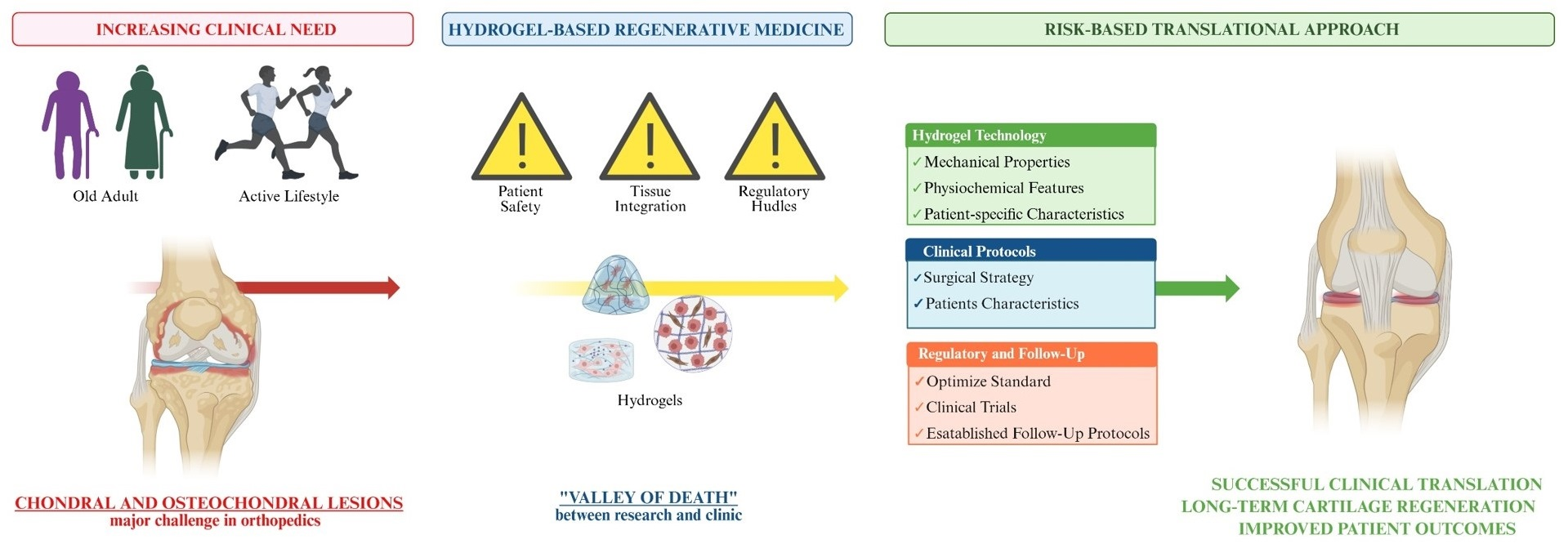
Challenges and Strategies in Hydrogel-Based Cartilage Regeneration


Abstract
1. Introduction

| Approach | Therapy Type | Pros | Cons | Refs. |
|---|---|---|---|---|
| Palliative | Physiotherapy/exercise: muscular strengthening and joint mobilization | Non-invasive, low cost, improves stability, exerts additional effects on inflammation and muscle hypertrophy | Does not repair tissue damage; temporary relief; efficacy moderated by the patient’s age and comorbidities, the intervention time, and the follow-up duration | [12] |
| Drug administration: systemic use of non-steroidal anti-inflammatory drugs (NSAIDs) or analgesics | Easy to administer, rapid pain control | Side effects (gastrointestinal, renal). Does not stop degeneration; long-term use could accelerate the progression to total knee replacement by markedly exacerbating symptoms | [13] | |
| Intra-articular injections of analgesics, and viscosupplementation with hyaluronic acid, glucosamine, and chondroitin sulfate | Minimally invasive | Risk of infection; temporary effect | [14] | |
| Use of orthotics: braces or specialized footwear | Corrects alignment, reduces load on the lesion | Patient compliance; skin irritation; bulky | [15] | |
| Arthroscopic debridement/chondral shaving/chondroplasty: removal of all the debris deriving from the damaged joint: inflammatory cells, unstable chondral flaps, osteophytes, superfluous synovia, degenerated meniscus, and torn ligament fragments | Minimally invasive; rapid improvement of pain symptoms | Short-term relief; does not regenerate cartilage; may lead to further thinning of cartilage | [16] | |
| Joint lavage: rinsing of the joint with physiological fluids to wash out degradation products | Flushes out inflammatory cytokines and debris, minimally invasive | Short-term effect | [17] | |
| Reparative | Bone marrow stimulation: abrasions, drilling, and microfracture: enclose a phase consisting of the subchondral bone penetration, thus inducing bleeding, and the migration of bone marrow MSCs to the site of injury, along with blood clot formation | Technically easy, low-cost, one-stage procedure; excellent short-term clinical outcomes have been demonstrated | The resulting repair tissue is composed of fibrocartilage. The clinical durability of the repair tissue has shown a functional decline with further follow-up | [18] |
| Restorative/reconstructive | Knee arthroplasty: total or partial joint replacement prosthetic implants | Decrease pain and improve mobility in people suffering from end-stage lesions | Invasive procedure, risk of stiffness, instability, aseptic loosening, infection, prosthesis failure, and malalignment | [19] |
| Regenerative | Osteochondral grafting: autologous osteochondral transplantation (AOT) or mosaicplasty, harvest of plugs from non-weight-bearing areas | Use of autologous hyaline cartilage; immediate availability; no risk of disease transmission | Limited graft supply; donor site morbidity; graft failure due to marginal chondrocyte death | [20] |
| Osteochondral grafting: Allogeneic (allograft transplantation): transplantation of donor grafts | No donor site morbidity; treats large defects | Graft availability, possible disease transmission; short cell viability; biomechanical integrity of the donor graft | ||
| Cell therapy implantation of a suspension of previously GMP *-expanded chondrocytes: Autologous Chondrocyte Implantation (ACI) (1st generation ACI) | Uses autologous cells; long-term results | Two surgeries; periosteal hypertrophy risk; high cost of expansion; regulatory hurdles; safety of media supplements; de-differentiation of chondrocytes in monolayer cultures (switch to a fibroblastic phenotype) | [7,21] | |
| Cell therapy: implantation of a suspension of previously GMP *-expanded chondrocytes under a sutured, inert collagen membrane, (C-ACI), (2nd generation ACI) | Avoid periosteal flap harvesting as for ACI 1st generation | High cost; requires two surgical procedures | [21] | |
| Cell therapy: implantation of a suspension of previously GMP * expanded mesenchymal stem cells MSCs)/progenitors | High number of cells; multipotent potential; immunomodulatory effects; immune privilege; scalability | Risk of hypertrophy/ossification; high cost of expansion; regulatory hurdles; safety of media supplements | [22,23] | |
| Cell therapy: implantation of a suspension of previously GMP * expanded Adipose tissue(-Derived) Stem Cells (ASCs or ADSCs) | Abundant tissue source; high proliferative potential; easier to harvest than BM; immunomodulatory properties; low immunogenicity: | High cost of expansion; regulatory hurdles; safety of media supplements | [24] | |
| Cell therapy: implantation of a suspension of previously GMP * expanded Synovial Tissue-Derived Stem Cells (SDSCs) | High chondrogenic potential; tissue-specific lineage (shares niche with cartilage) | [25] | ||
| Cell therapy: implantation of a suspension of previously GMP * expanded allogeneic Umbilical Cord Mesenchymal Stem Cells (UCMSCs)s | High proliferative capacity | Ethical/regulatory hurdles; long-term safety issues; high cost of expansion | [26] | |
| Cell therapy: implantation of a suspension of previously GMP * expanded induced Pluripotent Stem Cells (iPSCs) | Theoretically unlimited supply and scalability; reduced immunogenicity; genetic customization; high-quality control | Still in the pre-clinical stage (stringent safety controls required); tumorigenic risk; genetic and epigenetic instability; high cost and complexity of expansion; regulatory hurdles | [10,27] | |
| Cell-free therapy: scaffold alone used to guide tissue growth. Can be combined with bone marrow stimulation (e.g., microfracture) | One-stage; no cell expansion costs; provides structural support for endogenous cell recruitment; when in combination with bone marrow stimulation, the scaffold acts to recruit native MScs from the patient’s own bone marrow | Requires surrounding cells for migration; risk of inhomogeneous cell infiltration | [28] | |
| Cell-free therapy: acellular cartilage matrix (ACM), allogenic, or xenogenic | Mimics natural ECM; promotes host integration | Risk of immunogenicity | [29] | |
| Cell-free therapy: extracellular vesicles/exosomes from MSCs or chondrocytes | Low immunogenicity; no risk of cell transformation | Still in experimental stages, dosage is not standardized | [9] | |
| Tissue engineering: implantation of a construct composed of previously GMP * expanded chondrocytes seeded onto or into a 3D scaffold (3rd generation ACI) | The 3D scaffold improves cell retention and supports phenotype stability | Cost of two procedures; demanding GMP compliance | [21] | |
| Tissue engineering: implantation of a construct composed of GMP * expanded MSCs seeded onto or into a 3D scaffold | The 3D scaffold improves cell retention; possibility to use three-layered scaffolds and growth factors stimulating MSCs differentiation towards the chondrogenic or the osteogenic lineage (osteochondral regeneration) | [30] | ||
| Scaffold-free therapy: Implantation of High-Density Autologous Chondrocyte (HD-ACI) | Promotes robust cell-to-cell signaling; minimizes dedifferentiation of chondrocytes | Mimics natural tissue density; no synthetic material risks | [31] | |
| Scaffold-free therapy: implants of spherical aggregates of GMP * expanded chondrocytes with a self-synthesized extracellular matrix (ECM), for instance Chondrocyte-based spheroids (third-generation ACI) | Uniform cell distribution; avoids scaffold-induced inflammation | Higher cost due to specialized culture techniques | [21] | |
| Synthetic growth factors: Transforming Growth Factor-beta (TGF-β), Kartogenin, Bone Morphogenetic Protein-2 (BMP-2), Insulin-like Growth Factor-1 (IGF-1) | Potentiates natural healing; minimally invasive (if injected) | Short half-life; side effects | [32] | |
| Biologic growth factors: Platelet-rich plasma (PRP) + hyaluronic acid, multiple injections | Autologous cocktails; minimally invasive application | High inter-patient variability; mechanisms not fully elucidated | [33] | |
| Concentrates: bone marrow Aspirate Concentrate (BMAC) | One-stage procedure; cocktail of cells/growth factors | Variability between patients; lower cell concentration than cultured cells; lack of standardization | [34] | |
| Concentrates: stromal vascular fraction (SVF) from adipose tissue | [35] | |||
| Concentrates and biologic growth factors: combination of BMAC and PRP | Synergistic effect of BMAC and PRP | Increased procedure time; lack of standardized mixing ratios | [8] | |
| Concentrated associated with scaffolds and biologic growth factors: Combination of PRP and BMAC on a scaffold (for instance, collagen) | Scaffold provides mechanical stability and localized concentration of bioactive factors | Variability between patients; lower cell concentration than cultured cells; lack of standardization | [35] | |
| Concentrates associated with scaffolds: SVF on a biomaterial | [21] | |||
| Minimally manipulated cells: cartilage chondrons mixed with allogeneic MSCs (4th-generation ACI, one-stage ACI) | One-stage procedure | Limited cell numbers | [36] | |
| Minimally manipulated cells: morselized cartilage implantation mixed with fibrin sealant or PRP (4th-generation ACI, one-stage ACI) | Technically easy; low cost; utilizes hyaline cartilage cues | Variable tissue quality; limited to smaller defects | [21,37] | |
| Minimally manipulated cells: juvenile allogeneic cartilage fragments and micromosaic plastic (4th-generation ACI, one-stage ACI, off-the-shelf) | One-stage; immediate hyaline matrix cues; no donor morbidity | Risk of immunogenicity | [21] | |
| Gene Therapy: 1. viral or non-viral local delivery of genes encoding growth factors, transcription factors, or anti-inflammatory proteins and non-coding RNAs through viral and non-viral vectors: 2. In vivo or direct delivery of the target gene to the lesion site by surgical incision or local injection 3. Ex vivo, GMP * transfection or infection of the cells, followed by delivery of the cells to the target tissue (ex vivo) 4. Biomaterials as gene delivery carriers | Long-term expression of therapeutic factors | Safety concerns (viral vectors); potential for uncontrolled over-expression | [11] | |
| Gene editing, CRISPR/Cas9 technology: precise ex vivo or in vivo modification of inflammatory or hypertrophic genes within cells used for repair | Precise modification of specific genes | Off-target effects; regulatory issues | [38] |
1.1. Rationale and Aim
1.2. Search Strategy
2. Hydrogels
2.1. Source
2.2. Composition
2.3. Crosslinking
2.4. Configuration
2.5. Ionic Charge
2.6. Properties
2.7. Responsiveness
3. Hydrogels for Cartilage Regeneration
3.1. Characteristics
3.2. Challenges and Strategies
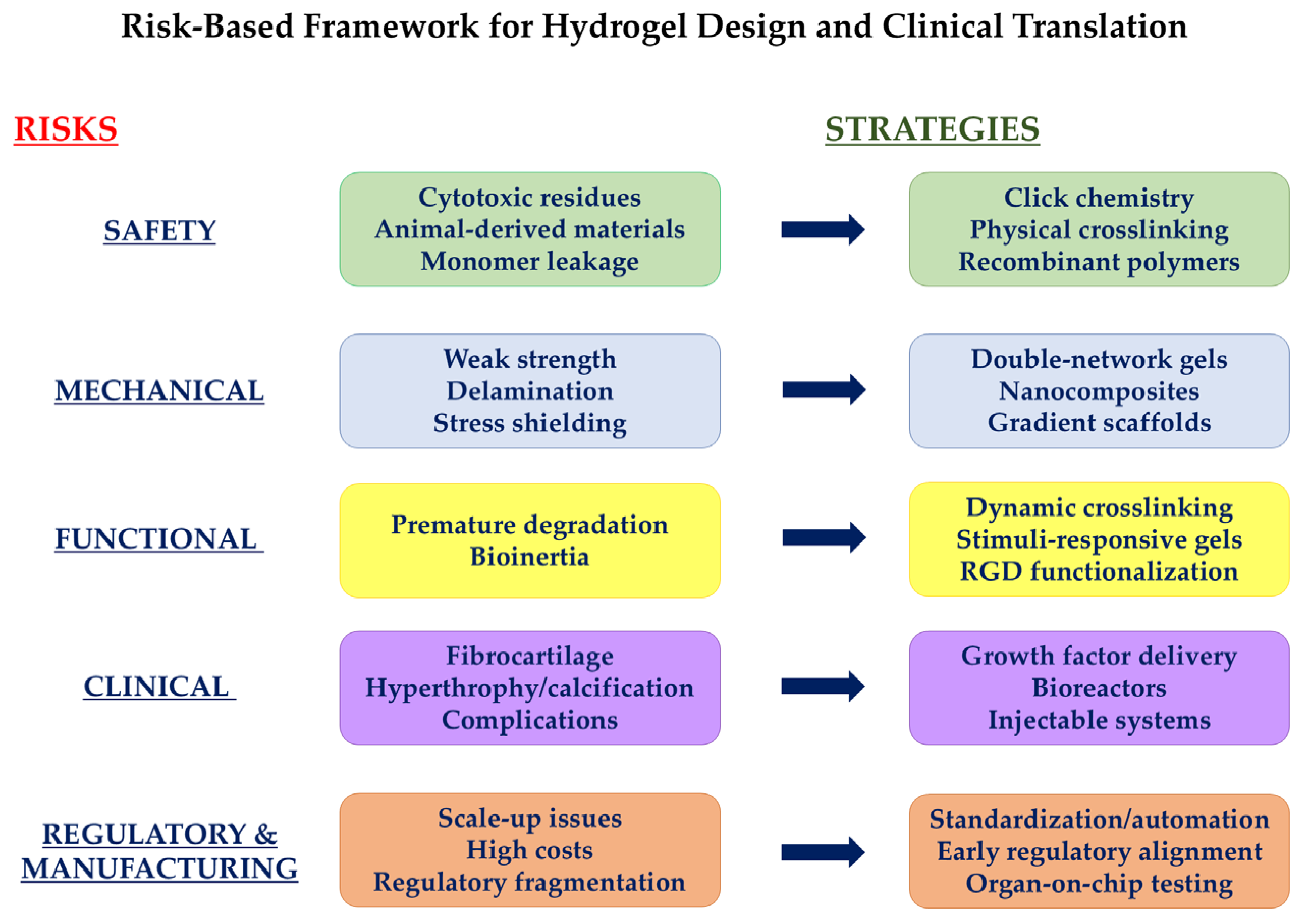
| Risk | Impact | Mitigation Strategy | References |
|---|---|---|---|
| Weak Mechanical Properties | Natural hydrogels lack the strength to withstand high joint loads, leading to an altered load distribution on subchondral bone and accelerating OA development or progression | Hydrogel reinforcement to enhance compressive and shear modulus through nanofillers (e.g., carbon nanotubes, nanocellulose) organic material (i.e., hydroxyapatite) hybridization with synthetic polymers | [68,80] |
| Delamination and detachment | Failure of integration between the hydrogel and the native cartilage, causing implant failure and freely movable fragments (bodies) in the joint that cause pain | Mollusk catechol modification (inspired by the exceptional underwater adhesion of mussels) | [81] |
| Covalent bonds to create injectable, self-healing hydrogels | [82] | ||
| Nanocomposite fillers to restore fluid pressure and seal the surface | [85] | ||
| Design of gradient or bi/three-layer scaffolds to support osteochondral tissue regeneration (with the transitional layer serving as an interface with intermediate stiffness to prevent delamination of the upper and lower layers) | [86] | ||
| Stress shielding effect | If the hydrogel is too rigid compared to the surrounding cartilage, it can alter joint load distribution, accelerating osteoarthritis degeneration | Double-network (DN) hydrogels, which combine a sacrificial brittle network to dissipate energy and an elastic one to maintain shape, thus better mimicking cartilage viscoelasticity | [83,84] |
| Risk | Impact | Mitigation Strategy | References |
|---|---|---|---|
| Premature material degradation | Material degrades before new cartilage formation. If reabsorption is faster than matrix production, the structural support collapses, leading to intervention failure. | Optimization of cross-linking: dynamic bonds, such as disulfide bonds, with self-healing properties that allow the hydrogel to autonomously reform after damage | [92,95] |
| Development of stimulus-reactive materials | |||
| Bioinertia | Synthetic hydrogels, although mechanically strong, may not offer the biological signals necessary for cell adhesion, migration, and proliferation. | Functionalization with RGD peptides or growth factors (TGF-β) | [92] |
| Blending with natural polymers (collagen, hyaluronic acid) to provide intrinsic biocompatibility |
| Risk | Impact | Mitigation Strategy | References |
|---|---|---|---|
| Formation of fibrocartilage | The neo-formation of fibrocartilage, which has lower mechanical properties and tends to degrade rapidly, at the expense of naïve hyaline cartilage | Controlled release of growth factors: development of controlled release hydrogels. For example, the growth factor TGF-β drives the differentiation of stem cells towards the hyaline chondrocyte phenotype. | [80,92] |
| Modifying polymeric hydrogel chains with adhesive peptide sequences, most notably RGD. | [81,96] | ||
| Porosity modulation: human bone marrow-derived MSC-based spheroids associated with 3D porous PEGDA hydrogels supported the viability and chondrogenic differentiation, as demonstrated by the positive staining of the ECM for glycosaminoglycans and upregulation of chondrogenesis marker genes, like collagen type II. | [97] | ||
| Pre-implantation Bioreactors: Before clinical use, apply cell-seeded hydrogel to dynamic compression cycles in specific bioreactors to “train” the cells to produce hyaline ECM. | [95] | ||
| Hypertrophic chondrocytes differentiation and ECM calcification | Hyaluronic acid-based hydrogels with a high crosslinking density, preventing the migration of nutrients, can promote the production of a calcified ECM, leading chondrogenic-stimulated MSCs to differentiate into a hypertrophic phenotype (expressing type X collagen) | Low-density or low-molecular-weight hydrogels: methacrylated hyaluronic acid (MeHA) can protect and promote the formation of articular cartilage. They achieve this by improving integration into the cartilage ECM, reducing degradation, and providing a favorable microenvironment for proper tissue regeneration. | [98,99] |
| Invasive treatment | Patient discomfort during treatment | Development of minimally invasive options: hydrogel microneedles. | [66] |
| Post-operative complications | Risks of infection or joint stiffness | Injectable hydrogels allow for the treatment of irregularly shaped defects. | [66] |
| Follow-up monitoring: for example, the use of magnetic resonance imaging with the Magnetic Resonance Observation of Cartilage Repair Tissue (MOCART) score to detect pilot release or filling abnormalities. | [100] | ||
| Design control: Use of computational models to predict the behavior of the hydrogel under load before implantation. | [63] | ||
| Long-term uncertainty | Most clinical trials on hydrogel-based applications for cartilage regeneration are still in the initial stages, which means that long-term safety data (over 10 years) is still being consolidated | Solid clinical data on the duration of results beyond 5–10 years are still lacking. | [101] |
| Risk | Mitigation Strategies | Reference |
|---|---|---|
| Long R&D complications: the R&D phase is complex, multi-faceted, multi-step, costly, long, and time-consuming | Milestone-based approach: pilot studies and targeted pre-clinical testing to validate data before investing in large-scale clinical trials | [112] |
| Complex and expensive production process: (for instance, nanocomposites, organ-on-a-chip) | Scalability and standardization: optimizing manufacturing processes and investing in automation | [111] |
| Biocompatibility failures: ISO 10993 ensures device safety for human use by evaluating biocompatibility | Advanced in vitro testing: organ-on-a-chip models to test biocompatibility, avoiding failures during mandatory in vivo testing | [113] |
| Ethical and animal testing barriers: without animal testing, researchers may fail to see joint and systemic changes | Adoption of validated alternative methods, such as 3D bioprinted human tissue models | [115] |
| Regulatory compliance issues: lack of communication with regulatory bodies like the FDA (USA) or EU-designated authorities | Early regulatory consultation: contact regulatory bodies or engage regulatory affairs experts just at the initial design phase | [109] |
| Regulatory panorama fragmentation: regulations vary, which impedes manufacturers from selling products across multiple countries | Strategic harmonization: international standards (like ISO 13485) and guidance to facilitate mutual recognition across global markets | [110] |
| Unclear classification of exosomes: Exosomes may be classified as drugs, biological products, or ATMPs depending on the region | Meticulous characterization: Define chemistry, manufacturing, and control (CMC) requirements early to align with biological medicinal product standards | [116] |
| Unclear classification of 3D-bioprinting products: Ambiguity between classification as medical devices, biologics, or Advanced Therapy Medicinal Products (ATMPs) | Case-by-case classification: Early engagement with regulatory bodies to determine the primary mode of action and appropriate pathway | [117] |
| Lack of organ-on-a-chip standardization: Design variability leads to poor data reproducibility and skepticism | Unified Roadmap: Adopt standardized protocols for chip manufacturing and cell differentiation to ensure data robustness. | [118] |
4. Future Perspectives
Author Contributions
Funding
Institutional Review Board Statement
Informed Consent Statement
Data Availability Statement
Acknowledgments
Conflicts of Interest
References
- Florescu, S.; Zaharia, C.; Drăghici, G.A.; Nica, D.V.; Damian, C.G. The Impact of Aging on Meniscal Tears and Chondral Lesions in Men: Insights from First-Time Arthroscopic Knee Evaluation. Life 2025, 15, 1305. [Google Scholar] [CrossRef] [PubMed]
- Merkely, G.; Ackermann, J.; Lattermann, C. Articular Cartilage Defects: Incidence, Diagnosis, and Natural History. Oper. Tech. Sports Med. 2018, 26, 156–161. [Google Scholar] [CrossRef]
- Tsujii, A.; Ohori, T.; Hanai, H.; Nakamura, N. Next Generation Approaches for Cartilage Repair and Joint Preservation. J. Cartil. Jt. Preserv. 2024, 4, 100177. [Google Scholar] [CrossRef]
- Huey, D.J.; Hu, J.C.; Athanasiou, K.A. Unlike Bone, Cartilage Regeneration Remains Elusive. Science 2012, 338, 917–921. [Google Scholar] [CrossRef]
- Simon, T.M.; Jackson, D.W. Articular Cartilage: Injury Pathways and Treatment Options. Sports Med. Arthrosc. Rev. 2006, 14, 146–154. [Google Scholar] [CrossRef]
- Nordberg, R.C.; Bielajew, B.J.; Takahashi, T.; Dai, S.; Hu, J.C.; Athanasiou, K.A. Recent Advancements in Cartilage Tissue Engineering Innovation and Translation. Nat. Rev. Rheumatol. 2024, 20, 323–346. [Google Scholar] [CrossRef]
- Brittberg, M.; Lindahl, A.; Nilsson, A.; Ohlsson, C.; Isaksson, O.; Peterson, L. Treatment of Deep Cartilage Defects in the Knee with Autologous Chondrocyte Transplantation. N. Engl. J. Med. 2026, 331, 889–895. [Google Scholar] [CrossRef]
- Balusani, P.; Shrivastava, S.; Pundkar, A.; Kale, P. Navigating the Therapeutic Landscape: A Comprehensive Review of Platelet-Rich Plasma and Bone Marrow Aspirate Concentrate in Knee Osteoarthritis. Cureus 2024, 16, e54747. [Google Scholar] [CrossRef]
- Su, J.; Qi, Y.; Niu, L.; Wang, Y.; Wei, B.; Ma, B.; Kong, K.; Zhai, Z.; Xu, Y. The Role of Synovial Mesenchymal Stem Cell-Derived Exosomes in Cartilage Repair: A Systematic Review. Front. Pharmacol. 2025, 16, 1617874. [Google Scholar] [CrossRef]
- Lach, M.S.; Rosochowicz, M.A.; Richter, M.; Jagiełło, I.; Suchorska, W.M.; Trzeciak, T. The Induced Pluripotent Stem Cells in Articular Cartilage Regeneration and Disease Modelling: Are We Ready for Their Clinical Use? Cells 2022, 11, 529. [Google Scholar] [CrossRef]
- Li, X.; Shen, L.; Deng, Z.; Huang, Z. New Treatment for Osteoarthritis: Gene Therapy. Precis. Clin. Med. 2023, 6, pbad014. [Google Scholar] [CrossRef]
- Lin, C.L.; Chen, H.C.; Huang, M.H.; Huang, S.W.; Liao, C. De Comparative Efficacy of Various Exercise Therapies and Combined Treatments on Inflammatory Biomarkers and Morphological Measures of Skeletal Muscle among Older Adults with Knee Osteoarthritis: A Network Meta-Analysis. Biomedicines 2024, 12, 1524. [Google Scholar] [CrossRef]
- Salis, Z.; Sainsbury, A. Association of Long-Term Use of Non-Steroidal Anti-Inflammatory Drugs with Knee Osteoarthritis: A Prospective Multi-Cohort Study over 4-to-5 Years. Sci. Rep. 2024, 14, 6593. [Google Scholar] [CrossRef] [PubMed]
- Mass, H.; Collins, J.E.; Yang, C.; Hunter, D.J.; Jones, M.H.; Tsai, L.; Messier, S.P.; Neogi, T.; Katz, J.N.; Losina, E. Intra-Articular Injections for Knee Osteoarthritis Management: Analysis of Cost-Effectiveness. Osteoarthr. Cartil. Open 2025, 7, 100641. [Google Scholar] [CrossRef] [PubMed]
- Bashir, A.Z.; Dinkel, D.M.; Pipinos, I.I.; Johanning, J.M.; Myers, S.A. Patient Compliance with Wearing Lower Limb Assistive Devices: A Scoping Review. J. Manip. Physiol. Ther. 2022, 45, 114–126. [Google Scholar] [CrossRef] [PubMed]
- Mufti, Y.; Sachs, J.P.; Bi, A.S.; Franzia, C.; Ebersole, J.; Giordano, T.; Cole, B.J. Articular Cartilage Debridement Results in Short-Term Significant Improvements in Patient-Reported Outcomes for Large Areas of Cartilage Loss of the Femur in the Setting of Mild to Moderate Knee Osteoarthritis. J. Cartil. Jt. Preserv. 2024, 4, 100221. [Google Scholar] [CrossRef]
- Dinç, M.; Cevdet Soydemir, Ö. Exploring the Efficacy of Joint Lavage in Knee Osteoarthritis: A Focus on Cytokines, Degrading Enzymes, and Oxidative Stress. Cartilage 2025, 16, 453–466. [Google Scholar] [CrossRef]
- Douleh, D.; Frank, R.M. Marrow Stimulation: Microfracture, Drilling, and Abrasion. Oper. Tech. Sports Med. 2018, 26, 170–174. [Google Scholar] [CrossRef]
- Li, Y.; Ajia, A.; Wu, Z.; Fan, J.; Wu, M.; Mao, P.; Yang, F.; He, Q.; Wang, P. Previous Knee Surgery Increases Risks of Revision, Infection, Pain and Stiffness after Knee Replacement Arthroplasty: A Systematic Review and Meta-Analysis. BMC Musculoskelet. Disord. 2025, 26, 869. [Google Scholar] [CrossRef]
- Medina, G.; Görtz, S. Osteochondral Techniques: Where Are We Now? J. Cartil. Jt. Preserv. 2023, 3, 100105. [Google Scholar] [CrossRef]
- Brittberg, M. Cartilage Repair with Autologous Chondrocytes (ACI Generations 1–4). Clin. Sports Med. 2025, 44, 527–540. [Google Scholar] [CrossRef]
- Carneiro, D.d.C.; Araújo, L.T.d.; Santos, G.C.; Damasceno, P.K.F.; Vieira, J.L.; Santos, R.R.d.; Barbosa, J.D.V.; Soares, M.B.P. Clinical Trials with Mesenchymal Stem Cell Therapies for Osteoarthritis: Challenges in the Regeneration of Articular Cartilage. Int. J. Mol. Sci. 2023, 24, 9939. [Google Scholar] [CrossRef]
- Copp, G.; Robb, K.P.; Viswanathan, S. Culture-Expanded Mesenchymal Stromal Cell Therapy: Does It Work in Knee Osteoarthritis? A Pathway to Clinical Success. Cell. Mol. Immunol. 2023, 20, 626–650. [Google Scholar] [CrossRef] [PubMed]
- Perdisa, F.; Gostyńska, N.; Roffi, A.; Filardo, G.; Marcacci, M.; Kon, E. Adipose-Derived Mesenchymal Stem Cells for the Treatment of Articular Cartilage: A Systematic Review on Preclinical and Clinical Evidence. Stem Cells Int. 2015, 2015, 597652. [Google Scholar] [CrossRef] [PubMed]
- Sekiya, I.; Muneta, T.; Horie, M.; Koga, H. Arthroscopic Transplantation of Synovial Stem Cells Improves Clinical Outcomes in Knees with Cartilage Defects. Clin. Orthop. Relat. Res. 2015, 473, 2316–2326. [Google Scholar] [CrossRef]
- Jung, S.H.; Nam, B.J.; Choi, C.H.; Kim, S.; Jung, M.; Chung, K.; Park, J.; Jung, Y.; Kim, S.H. Allogeneic Umbilical Cord Blood-Derived Mesenchymal Stem Cell Implantation versus Microdrilling Combined with High Tibial Osteotomy for Cartilage Regeneration. Sci. Rep. 2024, 14, 3333. [Google Scholar] [CrossRef] [PubMed]
- Chen, M.; Jiang, Z.; Zou, X.; You, X.; Cai, Z.; Huang, J. Advancements in Tissue Engineering for Articular Cartilage Regeneration. Heliyon 2024, 10, e25400. [Google Scholar] [CrossRef]
- De Marziani, L.; Boffa, A.; Andriolo, L.; Di Martino, A.; Romandini, I.; Solaro, L.; Zaffagnini, S.; Filardo, G. Cell-Free Biomimetic Osteochondral Scaffold for the Treatment of Knee Articular Surface Lesions: Clinical Outcomes Differ Based on Patient and Lesion Characteristics. Knee Surg. Sports Traumatol. Arthrosc. 2025, 33, 544–554. [Google Scholar] [CrossRef]
- Demmer, W.; Schinacher, J.; Wiggenhauser, P.S.; Giunta, R.E. Use of Acellular Matrices as Scaffolds in Cartilage Regeneration: A Systematic Review. Adv. Wound Care 2024, 13, 625–638. [Google Scholar] [CrossRef]
- Wróbel, M.; Rytel, H.; Jaszczyszyn, I.; Maj, M.; Malejczyk, J.; Janiuk, I.R. Models of Cartilage Repair with Autologous Mesenchymal Stem Cells Seeded on Scaffolds: A Systematic Narrative Review. Front. Bioeng. Biotechnol. 2026, 14, 1762579. [Google Scholar] [CrossRef]
- Guillén-García, P.; Guillén-Vicente, I.; Rodríguez-Iñigo, E.; Guillén-Vicente, M.; Fernández-Jaén, T.F.; Navarro, R.; Aboli, L.; Torres, R.; Abelow, S.; López-Alcorocho, J.M. Cartilage Defect Treatment Using High-Density Autologous Chondrocyte Implantation (HD-ACI). Bioengineering 2023, 10, 1083. [Google Scholar] [CrossRef]
- Morici, L.; Nikolic, I. Update on Clinical Trials of Intra-Articular Disease-Modifying Osteoarthritis Drugs Inducing Cartilage Regeneration. Eur. J. Pharm. Sci. 2026, 217, 107403. [Google Scholar] [CrossRef] [PubMed]
- Wang, C.; Yao, B. Efficacy and Safety of Platelet-Rich Plasma Injections for the Treatment of Knee Osteoarthritis: A Systematic Review and Meta-Analysis of Randomized Controlled Trials. Eur. J. Med. Res. 2025, 30, 992. [Google Scholar] [CrossRef] [PubMed]
- Pabinger, C.; Lothaller, H.; Kobinia, G.S. Intra-Articular Injection of Bone Marrow Aspirate Concentrate (Mesenchymal Stem Cells) in KL Grade III and IV Knee Osteoarthritis: 4 Year Results of 37 Knees. Sci. Rep. 2024, 14, 2665. [Google Scholar] [CrossRef] [PubMed]
- Lu, J.; Wen, T.; Huang, H.; Xie, C.; Kang, J.; Huang, J.; Hu, J.; Wu, T.; Wen, Y. Intraarticular Injection of the Stromal Vascular Fraction for the Treatment of Knee Osteoarthritis a Prospective Randomized Controlled Clinical Trial. Sci. Rep. 2025, 15, 28109. [Google Scholar] [CrossRef]
- Saris, T.F.F.; de Windt, T.S.; Kester, E.C.; Vonk, L.A.; Custers, R.J.H.; Saris, D.B.F. Five-Year Outcome of 1-Stage Cell-Based Cartilage Repair Using Recycled Autologous Chondrons and Allogenic Mesenchymal Stromal Cells: A First-in-Human Clinical Trial. Am. J. Sports Med. 2021, 49, 941–947. [Google Scholar] [CrossRef]
- Salzmann, G.M.; Calek, A.K.; Preiss, S. Second-Generation Autologous Minced Cartilage Repair Technique. Arthrosc. Tech. 2017, 6, e127–e131. [Google Scholar] [CrossRef]
- Li, C.; Du, Y.; Zhang, T.; Wang, H.; Hou, Z.; Zhang, Y.; Cui, W.; Chen, W. “Genetic Scissors” CRISPR/Cas9 Genome Editing Cutting-Edge Biocarrier Technology for Bone and Cartilage Repair. Bioact. Mater. 2023, 22, 254–273. [Google Scholar] [CrossRef]
- Carton, F.; Rizzi, M.; Canciani, E.; Sieve, G.; Di Francesco, D.; Casarella, S.; Di Nunno, L.; Boccafoschi, F. Use of Hydrogels in Regenerative Medicine: Focus on Mechanical Properties. Int. J. Mol. Sci. 2024, 25, 11426. [Google Scholar] [CrossRef]
- Naranđa, J.; Bračič, M.; Maver, U.; Trojner, T. Recent Advancements in Smart Hydrogel-Based Materials in Cartilage Tissue Engineering. Materials 2025, 18, 2576. [Google Scholar] [CrossRef]
- Submission of Comments on European Commission—Guideline on Risk Proportionate Approaches in Clinical Trials (Reference)—Recommendations of the Expert Group on Clinical Trials for the Implementation of Regulation (EU) No 536/2014 on Clinical Trials on Medicinal Products for Human Use. Available online: https://health.ec.europa.eu/document/download/1141def8-ea3f-4c8e-bf1c-2550045c89c7_en (accessed on 13 April 2026).
- Ho, T.C.; Chang, C.C.; Chan, H.P.; Chung, T.W.; Shu, C.W.; Chuang, K.P.; Duh, T.H.; Yang, M.H.; Tyan, Y.C. Hydrogels: Properties and Applications in Biomedicine. Molecules 2022, 27, 2902. [Google Scholar] [CrossRef] [PubMed]
- Kang, Y.; Guan, Y.; Li, S. Innovative Hydrogel Solutions for Articular Cartilage Regeneration: A Comprehensive Review. Int. J. Surg. 2024, 110, 7984–8001. [Google Scholar] [CrossRef] [PubMed]
- Chauhan, D. A review of hydrogels: Their classifications, and applications. In Advances in Chemical Science: Exploring New Frontiers V4B43; Iterative International Publishers IIP: Novi, MI, USA, 2024; pp. 183–202. ISBN 978-93-6252-178-1. [Google Scholar] [CrossRef]
- Zhao, L.; Zhou, Y.; Zhang, J.; Liang, H.; Chen, X.; Tan, H. Natural Polymer-Based Hydrogels: From Polymer to Biomedical Applications. Pharmaceutics 2023, 15, 2514. [Google Scholar] [CrossRef] [PubMed]
- Madduma-Bandarage, U.S.K.; Madihally, S.V. Synthetic Hydrogels: Synthesis, Novel Trends, and Applications. J. Appl. Polym. Sci. 2021, 138, 50376. [Google Scholar] [CrossRef]
- Rana, M.M.; De la Hoz Siegler, H. Evolution of Hybrid Hydrogels: Next-Generation Biomaterials for Drug Delivery and Tissue Engineering. Gels 2024, 10, 216. [Google Scholar] [CrossRef]
- Erkoc, C.; Yildirim, E.; Yurtsever, M.; Okay, O. Roadmap to Design Mechanically Robust Copolymer Hydrogels Naturally Cross-Linked by Hydrogen Bonds. Macromolecules 2022, 55, 10576–10589. [Google Scholar] [CrossRef]
- León-Campos, M.I.; Mendoza, J.J.; Aguayo-Morales, H.; Cobos-Puc, L.E.; Cabrera-Munguía, D.A.; Claudio-Rizo, J.A. The Biological Applications of IPN Hydrogels. ADMET DMPK 2024, 12, 581–621. [Google Scholar] [CrossRef]
- Segneanu, A.E.; Bejenaru, L.E.; Bejenaru, C.; Blendea, A.; Mogoşanu, G.D.; Biţă, A.; Boia, E.R. Advancements in Hydrogels: A Comprehensive Review of Natural and Synthetic Innovations for Biomedical Applications. Polymers 2025, 17, 2026. [Google Scholar] [CrossRef]
- Han, Y.; Cao, Y.; Lei, H. Dynamic Covalent Hydrogels: Strong yet Dynamic. Gels 2022, 8, 577. [Google Scholar] [CrossRef]
- Rumon, M.M.H.; Rahman, M.S.; Akib, A.A.; Sohag, M.S.; Rakib, M.R.A.; Khan, M.A.R.; Yesmin, F.; Shakil, M.S.; Rahman Khan, M.M. Progress in Hydrogel Toughening: Addressing Structural and Crosslinking Challenges for Biomedical Applications. Discov. Mater. 2025, 5, 5. [Google Scholar] [CrossRef]
- Gajurel, B.; Tamang, K.B.; Das, D.; Adhikari, R. Advances in Synthetic Strategies and Applications of Polymeric Hydrogels. Polym. Eng. Sci. 2025, 65, 2803–2840. [Google Scholar] [CrossRef]
- Revete, A.; Aparicio, A.; Cisterna, B.A.; Revete, J.; Luis, L.; Ibarra, E.; Segura González, E.A.; Molino, J.; Reginensi, D. Advancements in the Use of Hydrogels for Regenerative Medicine: Properties and Biomedical Applications. Int. J. Biomater. 2022, 2022, 3606765. [Google Scholar] [CrossRef] [PubMed]
- Schuiringa, G.H.; Mihajlovic, M.; van Donkelaar, C.C.; Vermonden, T.; Ito, K. Creating a Functional Biomimetic Cartilage Implant Using Hydrogels Based on Methacrylated Chondroitin Sulfate and Hyaluronic Acid. Gels 2022, 8, 457. [Google Scholar] [CrossRef] [PubMed]
- Zhu, S.; Li, Y.; He, Z.; Ji, L.; Zhang, W.; Tong, Y.; Luo, J.; Yu, D.; Zhang, Q.; Bi, Q. Advanced Injectable Hydrogels for Cartilage Tissue Engineering. Front. Bioeng. Biotechnol. 2022, 10, 954501. [Google Scholar] [CrossRef]
- Asadikorayem, M.; Surman, F.; Weber, P.; Weber, D.; Zenobi-Wong, M. Zwitterionic Granular Hydrogel for Cartilage Tissue Engineering. Adv. Healthc. Mater. 2024, 13, e2301831. [Google Scholar] [CrossRef]
- Ferrante, M.; Alvarez, V.; Narain, R.; Gonzalez, J. Properties of Hydrogels. In Natural and Synthetic Hydrogels: Rational Design, Synthesis and Biomedical Applications; Elsevier: Amsterdam, The Netherlands, 2025; pp. 3–15. [Google Scholar] [CrossRef]
- Akolpoglu, M.B.; Bozuyuk, U.; Erkoc, P.; Kizilel, S. Biosensing-Drug Delivery Systems for In Vivo Applications. In Advanced Biosensors for Health Care Applications; Elsevier: Amsterdam, The Netherlands, 2019; pp. 249–262. [Google Scholar] [CrossRef]
- Hennink, W.E.; van Nostrum, C.F. Novel Crosslinking Methods to Design Hydrogels. Adv. Drug Deliv. Rev. 2012, 64, 223–236. [Google Scholar] [CrossRef]
- Wan, L.Q.; Jiang, J.; Arnold, D.E.; Guo, X.E.; Lu, H.H.; Mow, V.C. Calcium Concentration Effects on the Mechanical and Biochemical Properties of Chondrocyte-Alginate Constructs. Cell. Mol. Bioeng. 2008, 1, 93–102. [Google Scholar] [CrossRef]
- Maitra, J.; Kumar Shukla, V. Cross-Linking in Hydrogels-A Review. Am. J. Polym. Sci. 2014, 4, 25–31. [Google Scholar] [CrossRef]
- Hashemi-Afzal, F.; Fallahi, H.; Bagheri, F.; Collins, M.N.; Eslaminejad, M.B.; Seitz, H. Advancements in Hydrogel Design for Articular Cartilage Regeneration: A Comprehensive Review. Bioact. Mater. 2025, 43, 1–31. [Google Scholar] [CrossRef]
- Jahanbekam, S.; Asare-Addo, K.; Alipour, S.; Nokhodchi, A. Smart Hydrogels and the Promise of Multi-Responsive in-Situ Systems. J. Drug Deliv. Sci. Technol. 2025, 107, 106758. [Google Scholar] [CrossRef]
- Yuan, Y.; Zhang, Q.; Lin, S.; Li, J. Water: The Soul of Hydrogels. Prog. Mater. Sci. 2025, 148, 101378. [Google Scholar] [CrossRef]
- Chen, D.; Shen, J.; Zhao, W.; Wang, T.; Han, L.; Hamilton, J.L.; Im, H.J. Injectable Hydrogels: An Emerging Therapeutic Strategy for Cartilage Regeneration. Adv. Colloid. Interface Sci. 2023, 321, 103030. [Google Scholar] [CrossRef]
- Li, A.; Huang, J.; Chen, J.; Wu, L.; Zeng, H.; Deng, Z.; Liu, P.; Lin, J. Evolving Functional Hydrogel Strategies for Cartilage Engineering: From Fundamentals to Functional Regeneration. Burn. Trauma 2025, 13, tkaf041. [Google Scholar] [CrossRef] [PubMed]
- Sun, L.; Xu, Y.; Han, Y.; Cui, J.; Jing, Z.; Li, D.; Liu, J.; Xiao, C.; Li, D.; Cai, B. Collagen-Based Hydrogels for Cartilage Regeneration. Orthop. Surg. 2023, 15, 3026–3045. [Google Scholar] [CrossRef] [PubMed]
- Wang, Y.; Koole, L.H.; Gao, C.; Yang, D.; Yang, L.; Zhang, C.; Li, H. The Potential Utility of Hybrid Photo-Crosslinked Hydrogels with Non-Immunogenic Component for Cartilage Repair. NPJ Regen. Med. 2021, 6, 54. [Google Scholar] [CrossRef]
- Zhou, L.; Guo, P.; D’Este, M.; Tong, W.; Xu, J.; Yao, H.; Stoddart, M.J.; van Osch, G.J.V.M.; Ho, K.K.W.; Li, Z.; et al. Functionalized Hydrogels for Articular Cartilage Tissue Engineering. Engineering 2022, 13, 71–90. [Google Scholar] [CrossRef]
- Liu, Z.; Xin, W.; Ji, J.; Xu, J.; Zheng, L.; Qu, X.; Yue, B. 3D-Printed Hydrogels in Orthopedics: Developments, Limitations, and Perspectives. Front. Bioeng. Biotechnol. 2022, 10, 845342. [Google Scholar] [CrossRef]
- Trinadha Rao, M.; Phanindra, C.H.V.S.; Yamini, M.; Prasad, C.H. Hydrogels the Three Dimensional Networks: A Review. Int. J. Curr. Pharm. Res. 2021, 13, 12–17. [Google Scholar] [CrossRef]
- Li, X.; Xiong, Y. Application of “Click” Chemistry in Biomedical Hydrogels. ACS Omega 2022, 7, 36918–36928. [Google Scholar] [CrossRef]
- Wei, F.; Liu, S.; Chen, M.; Tian, G.; Zha, K.; Yang, Z.; Jiang, S.; Li, M.; Sui, X.; Chen, Z.; et al. Host Response to Biomaterials for Cartilage Tissue Engineering: Key to Remodeling. Front. Bioeng. Biotechnol. 2021, 9, 664592. [Google Scholar] [CrossRef]
- Zhang, M.; Ye, Q.; Zhu, Z.; Shi, S.; Xu, C.; Xie, R.; Li, Y. Hyaluronic Acid-Based Dynamic Hydrogels for Cartilage Repair and Regeneration. Gels 2024, 10, 703. [Google Scholar] [CrossRef] [PubMed]
- Park, H.; Lee, K.Y. Cartilage Regeneration Using Biodegradable Oxidized Alginate/Hyaluronate Hydrogels. J. Biomed. Mater. Res. A 2014, 102, 4519–4525. [Google Scholar] [CrossRef] [PubMed]
- Catalano, E. Biophysical and Biomechanical Properties of Cartilage. arXiv 2023, arXiv:2305.01529. [Google Scholar] [CrossRef]
- Aamodt, J.M.; Grainger, D.W. Extracellular Matrix-Based Biomaterial Scaffolds and the Host Response. Biomaterials 2016, 86, 68–82. [Google Scholar] [CrossRef]
- Li, J.; Mooney, D.J. Designing Hydrogels for Controlled Drug Delivery. Nat. Rev. Mater. 2016, 1, 16071. [Google Scholar] [CrossRef]
- Bratovcic, A. Nanocomposite Hydrogels Reinforced by Carbon Nanotubes. Int. J. Eng. Res. Appl. 2020, 10, 30–41. [Google Scholar] [CrossRef]
- Duan, W.L.; Zhang, L.N.; Bohara, R.; Martin-Saldaña, S.; Yang, F.; Zhao, Y.Y.; Xie, Y.; Bu, Y.Z.; Pandit, A. Adhesive Hydrogels in Osteoarthritis: From Design to Application. Mil. Med. Res. 2023, 10, 4. [Google Scholar] [CrossRef]
- Li, M.; Li, F.; Xu, J.; Zhu, L.; Xiang, J.; Zhu, C.; Dai, Z.; Tang, S.; Ouyang, F.; Yu, J.; et al. Bioadhesive Chitosan Hydrogel with Dynamic Covalent Bonds and Sustained Kartogenin Release for Endogenous Cartilage Regeneration. Front. Bioeng. Biotechnol. 2025, 13, 1606726. [Google Scholar] [CrossRef]
- Li, W.; Wu, D.; Hu, D.; Zhu, S.; Pan, C.; Jiao, Y.; Li, L.; Luo, B.; Zhou, C.; Lu, L. Stress-Relaxing Double-Network Hydrogel for Chondrogenic Differentiation of Stem Cells. Mater. Sci. Eng. C 2020, 107, 110333. [Google Scholar] [CrossRef]
- Singh, J.; Kadir, J.C.; Orlando, J.D.; Sydlik, S.A. Biomimetic Double Network Hydrogels of Chondroitin Sulfate and Synthetic Polypeptides for Cartilage Tissue Engineering. Biomater. Sci. 2025, 13, 4211–4231. [Google Scholar] [CrossRef]
- Ao, Y.; Zhang, E.; Liu, Y.; Yang, L.; Li, J.; Wang, F. Advanced Hydrogels with Nanoparticle Inclusion for Cartilage Tissue Engineering. Front. Bioeng. Biotechnol. 2022, 10, 951513. [Google Scholar] [CrossRef]
- Zhang, N.; Wang, Y.; Zhang, J.; Guo, J.; He, J. Controlled Domain Gels with a Biomimetic Gradient Environment for Osteochondral Tissue Regeneration. Acta Biomater. 2021, 135, 304–317. [Google Scholar] [CrossRef] [PubMed]
- Vega, S.L.; Kwon, M.Y.; Burdick, J.A. Recent Advances in Hydrogels for Cartilage Tissue Engineering. Eur. Cell. Mater. 2017, 33, 59–75. [Google Scholar] [CrossRef] [PubMed]
- Gong, J.P.; Katsuyama, Y.; Kurokawa, T.; Osada, Y. Double-Network Hydrogels with Extremely High Mechanical Strength. Adv. Mater. 2003, 15, 1155–1158. [Google Scholar] [CrossRef]
- Rahimkhoei, V.; Akinay, Y.; Akbari, A.; Aljeboree, A.M.; Alsultany, F.H.; Salavati-Niasari, M. Harnessing Biopolymeric Double Network Hydrogels for Cartilage Repair: A Review of Current Strategies. J. Mater. Sci. Mater. Med. 2026, 37, 55. [Google Scholar] [CrossRef]
- Cai, Z.; Tang, Y.; Wei, Y.; Wang, P.; Zhang, H. Double—Network Hydrogel Based on Exopolysaccharides as a Biomimetic Extracellular Matrix to Augment Articular Cartilage Regeneration. Acta Biomater. 2022, 152, 124–143. [Google Scholar] [CrossRef]
- Milner, P.E.; Parkes, M.; Puetzer, J.L.; Chapman, R.; Stevens, M.M.; Cann, P.; Jeffers, J.R.T. A Low Friction, Biphasic and Boundary Lubricating Hydrogel for Cartilage Replacement. Acta Biomater. 2018, 65, 102–111. [Google Scholar] [CrossRef]
- Gan, X.; Wang, X.; Huang, Y.; Li, G.; Kang, H. Applications of Hydrogels in Osteoarthritis Treatment. Biomedicines 2024, 12, 923. [Google Scholar] [CrossRef]
- Sun, Q.; Zhang, Y.; Hu, B.; Feng, Q.; Xia, Y.; Yu, L.; Zhang, C.; Liu, W.; Liu, Z.; Yao, H.; et al. Development of a Dual-Responsive Injectable GelMA/F127DA Hydrogel for Enhanced Cartilage Regeneration in Osteoarthritis: Harnessing MMP-Triggered and Mechanical Stress-Induced Release of Therapeutic Agents. Int. J. Biol. Macromol. 2025, 304, 140823. [Google Scholar] [CrossRef]
- Qiong Liu, S.; Tian, Q.; Wang, L.; Hedrick, J.L.; Po Hui, J.H.; Yan Yang, Y.; Ee, P.L.R. Injectable Biodegradable Polyethylene Glycol/RGD Peptide Hybrid Hydrogels for in Vitro Chondrogenesis of Human Mesenchymal Stern Cellsa. Macromol. Rapid Commun. 2010, 31, 1148–1154. [Google Scholar] [CrossRef]
- Fu, L.; Li, P.; Li, H.; Gao, C.; Yang, Z.; Zhao, T.; Chen, W.; Liao, Z.; Peng, Y.; Cao, F.; et al. The Application of Bioreactors for Cartilage Tissue Engineering: Advances, Limitations, and Future Perspectives. Stem Cells Int. 2021, 2021, 6621806. [Google Scholar] [CrossRef]
- Manferdini, C.; Trucco, D.; Saleh, Y.; Gabusi, E.; Dolzani, P.; Lenzi, E.; Vannozzi, L.; Ricotti, L.; Lisignoli, G. RGD-Functionalized Hydrogel Supports the Chondrogenic Commitment of Adipose Mesenchymal Stromal Cells. Gels 2022, 8, 382. [Google Scholar] [CrossRef]
- Gonella, S.; Domingues, M.F.; Miguel, F.; Moura, C.S.; Rodrigues, C.A.V.; Ferreira, F.C.; Silva, J.C. Fabrication and Characterization of Porous PEGDA Hydrogels for Articular Cartilage Regeneration. Gels 2024, 10, 422. [Google Scholar] [CrossRef] [PubMed]
- Bian, L.; Hou, C.; Tous, E.; Rai, R.; Mauck, R.L.; Burdick, J.A. The Influence of Hyaluronic Acid Hydrogel Crosslinking Density and Macromolecular Diffusivity on Human MSC Chondrogenesis and Hypertrophy. Biomaterials 2013, 34, 413–421. [Google Scholar] [CrossRef] [PubMed]
- Brackin, R.B.; McColgan, G.E.; Pucha, S.A.; Kowalski, M.A.; Drissi, H.; Doan, T.N.; Patel, J.M. Improved Cartilage Protection with Low Molecular Weight Hyaluronic Acid Hydrogel. Bioengineering 2023, 10, 1013. [Google Scholar] [CrossRef] [PubMed]
- Trattnig, S.; Ohel, K.; Mlynarik, V.; Juras, V.; Zbyn, S.; Korner, A. Morphological and Compositional Monitoring of a New Cell-Free Cartilage Repair Hydrogel Technology—GelrinC by MR Using Semi-Quantitative MOCART Scoring and Quantitative T2 Index and New Zonal T2 Index Calculation. Osteoarthr. Cartil. 2015, 23, 2224–2232. [Google Scholar] [CrossRef]
- Zhang, Z.; Schon, L. The Current Status of Clinical Trials on Biologics for Cartilage Repair and Osteoarthritis Treatment: An Analysis of ClinicalTrials.Gov Data. Cartilage 2022, 13, 19476035221093065. [Google Scholar] [CrossRef]
- Iwamoto, M.; Ohta, Y.; Larmour, C.; Enomoto-Iwamoto, M. Toward Regeneration of Articular Cartilage. Birth Defects Res. C Embryo Today 2013, 99, 192–202. [Google Scholar] [CrossRef]
- Kocher, M.S.; Steadman, J.R.; Briggs, K.K.; Sterett, W.I.; Hawkins, R.J. Reliability, Validity, and Responsiveness of the Lysholm Knee Scale for Various Chondral Disorders of the Knee. J. Bone Jt. Surg. 2004, 86, 1139–1145. [Google Scholar] [CrossRef]
- Irrgang, J.J.; Anderson, A.F.; Boland, A.L.; Harner, C.D.; Kurosaka, M.; Neyret, P.; Richmond, J.C.; Donald Shelborne, K. Development and Validation of the International Knee Documentation Committee Subjective Knee Form. Am. J. Sports Med. 2001, 29, 600–613. [Google Scholar] [CrossRef]
- Bekkers, J.E.J.; de Windt, T.S.; Raijmakers, N.J.H.; Dhert, W.J.A.; Saris, D.B.F. Validation of the Knee Injury and Osteoarthritis Outcome Score (KOOS) for the Treatment of Focal Cartilage Lesions. Osteoarthr. Cartil. 2009, 17, 1434–1439. [Google Scholar] [CrossRef]
- Schreiner, M.M.; Raudner, M.; Marlovits, S.; Bohndorf, K.; Weber, M.; Zalaudek, M.; Röhrich, S.; Szomolanyi, P.; Filardo, G.; Windhager, R.; et al. The MOCART (Magnetic Resonance Observation of Cartilage Repair Tissue) 2.0 Knee Score and Atlas. Cartilage 2021, 13, 571S–587S. [Google Scholar] [CrossRef] [PubMed]
- Löffler, M.T.; Akkaya, Z.; Bhattacharjee, R.; Link, T.M. Biomarkers of Cartilage Composition. Semin. Musculoskelet. Radiol. 2024, 28, 26–38. [Google Scholar] [CrossRef] [PubMed]
- Elahi, S.A.; Castro-Viñuelas, R.; Tanska, P.; Maes, L.; Famaey, N.; Korhonen, R.K.; Jonkers, I. An in Silico Mechanoregulatory Model of Depth-Dependent Adaptations to Mechanical Loading in Intact and Damaged Cartilage: A Proof of Concept Study. Biomech. Model. Mechanobiol. 2026, 25, 2. [Google Scholar] [CrossRef] [PubMed]
- Wizemann, T.M. Public Health Effectiveness of the FDA 510(k) Clearance Process: Balancing Patient Safety and Innovation: Workshop Report; National Academies Press: Washington, DC, USA, 2010. [Google Scholar]
- Rimpi; Verma, S.J.; Pinky; Baldi, A. Evidence-Based Recommendations for Comprehensive Regulatory Guidelines in Medical Devices: The Imperative for Global Harmonization. Naunyn-Schmiedeberg’s Arch. Pharmacol. 2025, 398, 7697–7711. [Google Scholar] [CrossRef]
- Ali, F.; Khan, I.; Chen, J.; Akhtar, K.; Bakhsh, E.M.; Khan, S.B. Emerging Fabrication Strategies of Hydrogels and Its Applications. Gels 2022, 8, 205. [Google Scholar] [CrossRef]
- EU. Risk Proportionate Approaches in Clinical Trials Recommendations of the Expert Group on Clinical Trials for the Implementation of Regulation (EU) No 536/2014 on Clinical Trials on Medicinal Products for Human Use; EU: Brussels, Belgium, 2017; Available online: https://health.ec.europa.eu/system/files/2017-08/2017_04_25_risk_proportionate_approaches_in_ct_0.pdf (accessed on 13 April 2026).
- Karami, P.; Stampoultzis, T.; Guo, Y.; Pioletti, D.P. A Guide to Preclinical Evaluation of Hydrogel-Based Devices for Treatment of Cartilage Lesions. Acta Biomater. 2023, 158, 12–31. [Google Scholar] [CrossRef]
- Choi, Y.J.; Park, H.; Ha, D.H.; Yun, H.S.; Yi, H.G.; Lee, H. 3D Bioprinting of in Vitro Models Using Hydrogel-Based Bioinks. Polymers 2021, 13, 366. [Google Scholar] [CrossRef]
- Gao, D.; Li, R.; Pan, J.; Li, C.; Zhang, W.; Qin, L.; Lai, Y. 3D Bioprinting Bone/Cartilage Organoids: Construction, Applications, and Challenges. J. Orthop. Transl. 2025, 55, 75–93. [Google Scholar] [CrossRef]
- Wang, C.K.; Tsai, T.H.; Lee, C.H. Regulation of Exosomes as Biologic Medicines: Regulatory Challenges Faced in Exosome Development and Manufacturing Processes. Clin. Transl. Sci. 2024, 17, e13904. [Google Scholar] [CrossRef]
- Mladenovska, T.; Choong, P.F.; Wallace, G.G.; O’connell, C.D. The Regulatory Challenge of 3D Bioprinting. Regen. Med. 2023, 18, 659–674. [Google Scholar] [CrossRef]
- Sampaio, A.R.; Maia, R.F.; Ciardulli, M.C.; Santos, H.A.; Sarmento, B. Organ-on-Chip Platforms for Nanoparticle Toxicity and Efficacy Assessment: Advancing beyond Traditional in Vitro and in Vivo Models. Mater. Today Bio 2025, 33, 102053. [Google Scholar] [CrossRef] [PubMed]

| Classification | Type | Characteristics | Refs. |
|---|---|---|---|
| Source | Natural | Derived from biological sources (e.g., collagen, chitosan, alginate) | [45] |
| Synthetic | Synthesized using chemical polymerization (e.g., PEG, PAA) | [46] | |
| Semi-synthetic | Natural polymers modified with synthetic groups to enhance properties | [47] | |
| Composition | Homopolymer | Consists of only one type of monomer in the network | [42] |
| Copolymer | Formed by two or more different types of monomers | [48] | |
| Semi-IPN | One crosslinked network with a second “linear” polymer trapped inside | [49] | |
| IPN | Two or more independent, cross-linked networks interlaced at a molecular level | ||
| Crosslinking | Physical junction | Reversible bonds (hydrogen bonds, ionic interactions, or chain entanglements) | [50] |
| Chemical binding | Permanent, stable covalent bonds between polymer chains | [51,52] | |
| Configuration | Amorphous | Disordered, random molecular arrangement (transparent and flexible) | [53] |
| Crystalline | Highly ordered, tight molecular packing (stronger and opaquer) | ||
| Semi-Crystalline | A mixture of both ordered crystalline and disordered amorphous regions | ||
| Ionic Charge | Nonionic | Neutral network with no electrical charge | [54] |
| Anionic | Carries a negative charge (often responds to higher pH) | [55] | |
| Cationic | Carries a positive charge (often responds to lower pH) | [56] | |
| Ampholytic | Contains both positive and negative charges (zwitterionic) | [57] | |
| Property | Mechanical strength | The ability of the gel to withstand physical stress or load | [58] |
| Biocompatibility | Compatibility with living tissues without causing harm | ||
| Biodegradability | Capacity to break down naturally in a biological environment | ||
| Swelling ability | The ability to absorb and hold large amounts of water or fluids | ||
| Stimuli sensitivity | Ability to change volume or shape in response to environmental cues | ||
| Chemical Response | Stimuli Factors | Responds to pH, ionic strength, solvent composition, molecular species, and redox reactions | [59] |
| Physical Response | Stimuli Factors | Responds to temperature, electric field, magnetic field, light, pressure, sound, and humidity |
| Advantage | Disadvantage | |
|---|---|---|
| Biomimicry: high water content and gel structure resembling cartilage ECM support cell survival, proliferation, and differentiation | Low mechanical strength: lack of compressive and shear strength required to withstand the high-load joint environment | [43] |
| Biocompatibility: natural-based hydrogels (hyaluronic acid, collagen, or chitosan) have a minimal risk of adverse or toxic reactions | Batch variability: natural polymers can suffer from inconsistent properties between batches, affecting reproducibility in clinical settings | [68] |
| Non-immunogenicity: the ability to trigger an appropriate host response without causing an inappropriate immune response | Degradation-induced inflammation: some synthetic components or cross-linking agents may trigger a delayed foreign-body response during degradation | [69] |
| Biodegradability: controlled breakdown of the material into non-toxic byproducts | Mismatch in rates: Difficulty in matching the hydrogel degradation rate with the speed of new tissue formation, potentially leading to premature structural failure | [43] |
| Design Flexibility: hydrogels can be engineered to be bioadhesive, biodegradable, or to actively release growth factors and drugs to promote healing | Complex release kinetics: controlling the burst effect or maintaining a sustained release of bioactive factors over long periods is technically difficult | [70] |
| Customizability: patient-tailored, stimuli-responsive, or “smart” behavior hydrogels can be engineered from the molecular level (composition and cross-linking) to macroscopic architecture (shape and responsiveness) | Technical complexity: high manufacturing costs and complex regulatory pathways for “smart” or highly engineered materials | [71] |
| Minimal invasiveness: hydrogels can be injected directly into the joint, allowing for precise, arthroscopic, minimally invasive surgery rather than open joint surgery | Leakage and migration risk: low-viscosity precursor solutions may leak from the defect site before gelation (cross-linking) is complete | [66] |
| Effective for focal defects: hydrogels can fill irregularly shaped cartilage voids, providing a better fit and structural support for a specific injury | Poor lateral integration: difficulty in achieving a seamless biological and mechanical bond between the hydrogel and the surrounding native cartilage | [66] |
| Risk | Impact | Mitigation Strategy | References |
|---|---|---|---|
| Cytotoxic chemical residues from the crosslinking process | Toxic or inflammatory responses leading to fibrocartilage formation | Physical crosslinking (UV/temperature) or Bio-orthogonal click chemistry to eliminate toxic catalysts | [73] |
| Animal origin residues | Rejection, allergic reactions, cartilage hypertrophy, and fibrosis | De-antigenization protocols for natural polymers or the use of ultra-pure synthetic precursors | [50,74,75] |
| Leaking of monomers (e.g., acrylic acid, acrylamide) | Cytotoxic and genotoxic effects | Controlled degradation | [76] |
Disclaimer/Publisher’s Note: The statements, opinions and data contained in all publications are solely those of the individual author(s) and contributor(s) and not of MDPI and/or the editor(s). MDPI and/or the editor(s) disclaim responsibility for any injury to people or property resulting from any ideas, methods, instructions or products referred to in the content. |
© 2026 by the authors. Licensee MDPI, Basel, Switzerland. This article is an open access article distributed under the terms and conditions of the Creative Commons Attribution (CC BY) license.
Share and Cite
Cavallo, C.; Amore, E.; Carpentieri, S.; Roseti, L. Challenges and Strategies in Hydrogel-Based Cartilage Regeneration. Gels 2026, 12, 350. https://doi.org/10.3390/gels12050350
Cavallo C, Amore E, Carpentieri S, Roseti L. Challenges and Strategies in Hydrogel-Based Cartilage Regeneration. Gels. 2026; 12(5):350. https://doi.org/10.3390/gels12050350
Chicago/Turabian StyleCavallo, Carola, Emanuela Amore, Sara Carpentieri, and Livia Roseti. 2026. "Challenges and Strategies in Hydrogel-Based Cartilage Regeneration" Gels 12, no. 5: 350. https://doi.org/10.3390/gels12050350
APA StyleCavallo, C., Amore, E., Carpentieri, S., & Roseti, L. (2026). Challenges and Strategies in Hydrogel-Based Cartilage Regeneration. Gels, 12(5), 350. https://doi.org/10.3390/gels12050350