Article

The Effect of Antibiotics on the Infant Gut Fungal Microbiota

1
Human Microbiome Research Program, Faculty of Medicine, University of Helsinki, 00014 Helsinki, Finland
2
Folkhälsan Research Center, 00250 Helsinki, Finland
3
Children’s Hospital, Helsinki University, 00029 Helsinki, Finland
4
Department of Paediatrics and Adolescent Medicine, Turku University Hospital, University of Turku, 20014 Turku, Finland
5
Laboratory of Microbiology, Wageningen University, 6708 WE Wageningen, The Netherlands
6
Department of Pediatrics, Tampere University, 33520 Tampere, Finland
*
Author to whom correspondence should be addressed.
J. Fungi 2022, 8(4), 328; https://doi.org/10.3390/jof8040328
Submission received: 14 February 2022 / Revised: 15 March 2022 / Accepted: 18 March 2022 / Published: 22 March 2022
(This article belongs to the Special Issue New Insights into Current Understanding of Host–Fungal Interactions)

Abstract

:
Antibiotics are commonly used drugs in infants, causing disruptions in the developing gut microbiota with possible detrimental long-term effects such as chronic inflammatory diseases. The focus has been on bacteria, but research shows that fungi might have an important role as well. There are only a few studies on the infant gut fungal microbiota, the mycobiota, in relation to antibiotic treatment. Here, the aim was to investigate the impact of antibiotics on the infant gut mycobiota, and the interkingdom associations between bacteria and fungi. We had 37 antibiotic-naïve patients suffering from respiratory syncytial virus, of which 21 received one to four courses of antibiotics due to complications, and 16 remained antibiotic-naïve throughout the study. Fecal samples were collected before, during and after antibiotic treatment with a follow-up period of up to 9.5 months. The gut mycobiota was studied by Illumina MiSeq sequencing of the ITS1 region. We found that antibiotic use affected the gut mycobiota, most prominently seen as a higher relative abundance of Candida (p < 0.001), and a higher fungal diversity (p = 0.005–0.04) and richness (p = 0.03) in the antibiotic-treated infants compared to the antibiotic-naïve ones at multiple timepoints. This indicates that the gut mycobiota could contribute to the long-term consequences of antibiotic treatments.

Graphical Abstract

1. Introduction

The gut microbiota is known to include bacteria, fungi, archaea, viruses and sometimes protozoa and is a major factor contributing to human health [1]. Most microbiota-related studies have focused on the bacteria, although the fungal microbiota, the mycobiota, might have a significant role as well. Imbalance in the intestinal fungal composition has been associated with diseases such as inflammatory bowel disease (IBD) [2,3,4,5,6], celiac disease [7] and colorectal cancer [8]. The antifungal drug fluconazole worsens inflammation in mice with induced intestinal- and lung inflammation, indicating that the fungal microbiota also has a protective role [9].
Compared to the bacterial gut microbiota, the gut mycobiota has fewer taxa and seems to be less stable [10,11]. Variation both between and within individuals makes it difficult to determine whether certain fungi are actual colonizers or only transient fungi derived from food or environment as previously reviewed [12]. The gut mycobiota consists of the phyla Ascomycota and Basidiomycota, with Ascomycota being more abundant in both adults [11,13,14,15] and in children [16].
The first major exposure to microorganisms is through birth, and the infant gut has been observed to be colonized by fungi [17] as early as 10 days after birth [16]. The gut microbiota continues to develop during the first years of life, affected by multiple factors such as delivery mode and diet [14,18], and particularly the use of antibiotics [19]. At two years of age, the infant gut mycobiota shows similarities to that of adults [16].
Many newborns receive antibiotics due to an infection during early years of life. In Finland, 26.5% of children < 5 years of age received at least one course of antibiotics in 2019 (Kela statistics, www.kela.fi/tilastot, accessed on 2 February 2022; and statistics in Finland, www.stat.fi/til, accessed on 2 February 2022). The use of antibiotics during the critical time from conception to 2 years of age has been reported to cause both short-term problems including dramatic shifts of the gut fungal and bacterial microbiota and even sepsis, and further long-term consequences such as asthma, atopy, pediatric onset of Crohn’s disease and metabolic diseases [20,21]. The use of antibiotics is also associated with higher body mass index (BMI) [19,22,23,24], and in a study conducted on Finnish pre-school children the use of antibiotics was associated with increased risk for overweight and asthma later in life [25,26].
Antibiotics, evidently only affecting prokaryotes, may induce fungal growth, particularly of Candida spp. in the mouse gut [27,28,29], and cause major changes in the adult gut bacterial microbiota composition, with an even more persistent shift in the gut mycobiota [30]. Additionally, antibiotics have been observed to cause increased risk of candidiasis and bloodstream infection [31,32]. Bacteria and fungi co-exist in the gut, and competition for nutrient sources and the production of antifungal compounds by gut bacteria and antibacterial compounds by fungi are characteristic of the interkingdom interactions [30,33,34]. Candida is a part of the healthy gut mycobiota [13], but simultaneously it seems to be a driver of aberrant composition of gut microbiota and is associated with multiple diseases [2,3,4,5,6,7,8]. In a recent study it was observed that the decrease of short-chain fatty acids and anaerobic bacteria correlates with an increase of fungal growth [35].
There are many detrimental long-term effects in using antibiotics, particularly in children, that stem from an aberrant gut microbiota composition. Studies on the effect of antibiotics on the gut mycobiota are very limited. The impact of antibiotics on the gut bacterial microbiota has been published for these data [19] with the observation that the administration of only a single course of amoxicillin or macrolide already caused a consistent shift in the bacterial classes Actinobacteria, Gammaproteobacteria and Clostridia.
Here, our aim was to investigate the impact of antibiotics on the gut mycobiota of infants by investigating the gut mycobiota composition in antibiotic-treated compared to antibiotic-naïve infants. Further, we aimed to study the interkingdom correlations and to combine both fungal and bacterial gut microbiota.

2. Materials and Methods

2.1. Study Design

The cohort consisted of 37 children at a median age of 2 months who were hospitalized at the Children’s Hospital, University of Helsinki, or Turku University Hospital between December 2013 and May 2014 due to a respiratory syncytial virus (RSV) infection. Only antibiotic-naïve children were included. Fecal samples were collected before, during and after antibiotic treatment with a follow-up of up to 9.5 months. All fecal samples were immediately frozen at −20 °C until transported frozen to −70 °C where they were stored until analysis. At each sampling timepoint, questionnaires were filled out containing information on diet, probiotics and use of antibiotics. Additionally, information of birth mode, other possible diseases, growth and weight gain were available from the patient files. Preterm infants (birth < 37 gestational week or birth weight < 2500 g) or those with congenital malformations or syndromes were excluded. None of the infants had been traveling abroad. In analyses, the timepoints were calculated as time since the start of antibiotics, or time since the first sample for antibiotic-naïve patients. In the cases where multiple courses of antibiotics were administered to a patient, the days from the most recent antibiotic course were used in the timepoints. Therefore, the time of follow-up might be longer than the timepoint used in analyses.

2.2. DNA Extraction and MiSeq Library Preparation

The fecal samples were thawed, and the bacterial and fungal DNA was extracted using the bead-beating method as previously described [19]. The 16S rRNA gene amplicon data were obtained from the previous study by Korpela et al. [19]. The gut fungal microbiota composition was studied by Illumina MiSeq sequencing of the internal transcribed sequence 1 (ITS1) targeting a conserved region of fungi. The PCR primer pair ITS1F (FWD, CTTGGTCATTTAGAGGAAGTAA) and ITS2 (REV, GCTGCGTTCTTCATCGATGC) [36] were used to amplify the fungal DNA. The sequencing library was prepared as previously described [37]. Illumina MiSeq paired-end sequencing was performed in the Functional Genomics Unit, University of Helsinki, Helsinki, Finland. The ITS rDNA amplicon sequences of this study are available at the ENA database [PRJEB50378].

2.3. Analysis of Sequencing Data

The ITS MiSeq sequencing read data were processed according to the DADA2 ITS pipeline [38]. The reads were annotated as previously described [37] by annotating the amplicon sequence variants (ASVs) with BLAST [39]. The BLAST hits were limited to >75% query coverage and percentage identity and anything below this was discarded. The annotations were gut- or environment-specific. Before pre-processing, the median number of reads per sample was 66,791 (17,942–154,451). After pre-processing, the median read count was 7556 (134–32,846) and finally, after annotation the median read count was 1786 (121–22,932). Successful annotation to fungal taxa was obtained from 123 samples (out of 151 samples) from 37 patients and a total of 113 samples (75%) from 37 patients were annotated successfully with more than 100 reads.

2.4. Statistical Analysis

The R package mare [40] was used for analysis and visualizing the ITS relative abundance data, including the use of the packages vegan [41], MASS [42] and nlme [43]. p-values for taxon-specific differences were corrected for false discovery rate (FDR; Benjamini–Hochberg [44]). Generalized linear models with negative binomial distribution (glm.nb) from the MASS package [42] and Generalized Least Squares (gls) from the nlme package [43] were used to analyze differences in the microbiota composition between the control group and the antibiotic-treated patients. Samples with less than 100 reads were discarded from the analysis and sex, breastfeeding and hospital location were used as confounding variables within the models. Background variables were analyzed by multivariate permutational analysis of variance at both baseline and all timepoints, and the factors included in the statistical analyses of individual taxa were chosen based on their impact on the total fungal microbiota. The diversity was calculated as the inverse Simpson diversity index and richness as the number of ASVs. The Spearman correlations and p-values between fecal fungal and bacterial taxa at different timepoints were calculated with “rcorr” from the Hmisc package [45].

2.5. Ethics Statement

The study was approved by the Ethics committee of Helsinki and Uusimaa Hospital district. All guardians signed an informed consent to include their child in the study.

3. Results

3.1. Patient Characteristics

The aim was to study the effect of antibiotics on the gut mycobiota. We analyzed samples from 37 antibiotic-naïve infants who were recruited prospectively and included in our previous report on the bacterial gut microbiota [19]. All children were recruited while treated because of RSV infection. Out of these, 21 patients received antibiotics prescribed by a clinician due to complications such as otitis media. Sixteen infants did not receive any antibiotics and were considered controls in this study. The antibiotics used were amoxicillin and macrolides and the infants received one to four courses of antibiotics. Amoxicillin was given to 21 infants as their first antibiotic, and 8 of these received a second course of penicillin-group antibiotics and 4 patients received macrolides. The number of samples at each timepoint stratified by antibiotics used are presented in Table 1. The timepoints are calculated from the most recent course of antibiotics, or from the first sample for the controls.
Fungal amplicons generated from a total of 123 fecal samples obtained from the 37 recruited patients were sequenced. Out of these, 68 samples were derived from the 21 antibiotic-treated patients and 56 samples from the 16 control patients. Baseline samples of the antibiotic-treated patients were obtained from 12 patients. Background data of the study participants are presented in Table 2. During the follow-up (up to 9.5 months), there were no infants who developed any diseases.

3.2. Overview of the Gut Mycobiota Composition

Across all samples the fungal genus Saccharomyces was most prevalent (97%) with a relative abundance of 71%, followed by Malassezia (prevalence 44%) with a relative abundance of 8%, Candida (prevalence 30%) with a relative abundance of 9% and Cladosporium (prevalence 11%) with a relative abundance of 1%. The prevalence and relative abundance of all genera with a prevalence >5% is presented in Supplementary Table S1. The gut mycobiota composition at genus level was analyzed stratified by timepoints and patient groups (Figure 1). Location of hospital, sex and breastfeeding were used as background factors and the analyses were adjusted for these factors. All three background factors impacted the gut mycobiota significantly (p < 0.05) both at baseline and at all timepoints, except for the location of hospital, which was significant only when including all timepoints (Supplementary Figure S1).

3.3. Difference between the Control Group and the Group Treated with Antibiotics

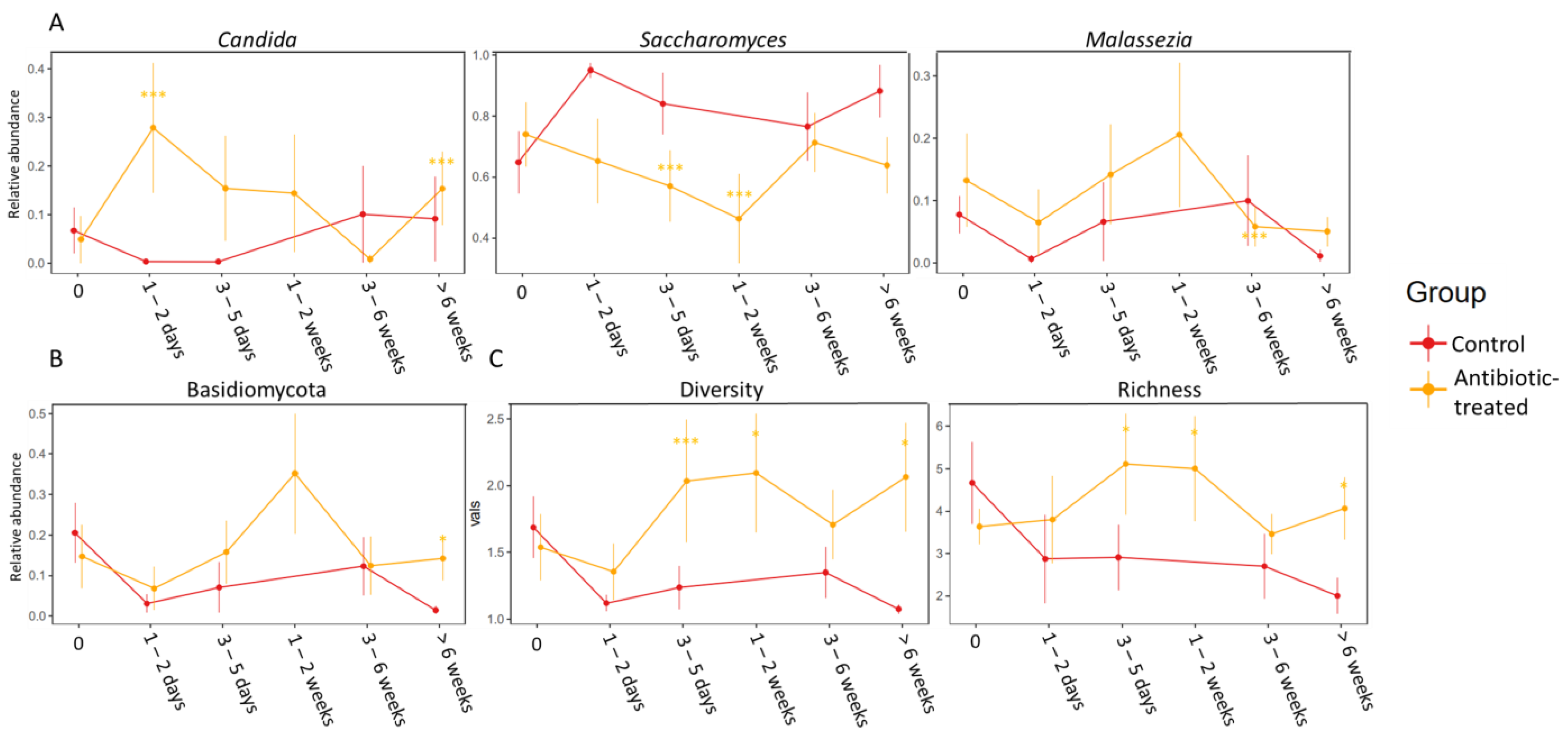
Before the start of antibiotic treatment there was no significant difference in gut mycobiota composition between the groups. At 1–2 days after the start of treatment (during treatment), the amoxicillin-treated group had significantly higher relative abundance of the fungal genus Candida (p FDR < 0.001, fold change = 132) (Figure 2A). When comparing antibiotic-treated patients (both amoxicillin and macrolides combined) 1–2 weeks after start of treatment to controls at 3–5 days after first sample, Saccharomyces was more abundant in the control group (p FDR < 0.001, fold change = 3.8) (Figure 2A). Further, Saccharomyces was more abundant in the control group at 3–5 days after the start of treatment compared to the antibiotic-treated patients (p FDR < 0.001, fold change = 1.44). At the end of the study (>6 weeks after start of treatment) the phylum Basidiomycota was more abundant in the antibiotic-treated group (both amoxicillin and macrolide combined) (p FDR = 0.009, fold change = 10.9) (Figure 2B). Detailed results of the statistical differences between the antibiotic-treated and antibiotic-naïve infants are presented in Supplementary Tables S2–S6.
When comparing the groups of antibiotics used, the amoxicillin-treated infants had significantly higher relative abundance of Candida at >6 weeks after start of treatment compared to controls (p FDR = 0.028, fold change = 13.14). This difference was not significant for macrolide-treated infants, although the trend was similar with non-FDR-corrected p-value < 0.05, and fold change 22. Both the amoxicillin- (p FDR < 0.001, fold change = 1.68), but notably the macrolide-treated (p FDR < 0.001, fold change = 48.45) infants had significantly higher relative abundance of Malassezia at 3–6 weeks after the start of antibiotic treatment. Further, Basidiomycota was more abundant in the macrolide-treated group compared to controls at 3–6 weeks after the start of treatment (p FDR = 0.033, fold change = 20.01).
Finally, the antibiotic-treated group had significantly higher fungal diversity and richness at 3–5 days (diversity, p = 0.04, richness, p = 0.03), 1–2 weeks ((when comparing to control at 3–5 days) diversity, p = 0.005, richness, p = 0.03)) and >6 weeks (diversity, p = 0.03, richness = 0.03) after the start of antibiotic treatment as seen in Figure 2C. At >6 weeks the difference in diversity was even more significant between the control and macrolide-treated group (p = 0.0027).

3.4. Spearman Correlations between Fungi and Bacteria

Interkingdom Spearman correlations were calculated at each timepoint for both the antibiotic-treated group and for those who remained antibiotic-naïve throughout the study. There were significant correlations (p < 0.05) at all timepoints, and these are presented in Supplementary Figures S2–S4. No robust interkingdom correlations were detected.

4. Discussion

Here we investigated the effect of antibiotics on the infant gut mycobiota. This was carried out by MiSeq sequencing of the PCR-amplified ITS1 region from the DNA of fecal samples collected from both controls and antibiotic-treated infants at six different timepoints with a follow-up ranging over 9 months. All patients were naïve to any antibiotics at the first timepoint and controls were antibiotic-naïve throughout the follow-up. We observed that the use of antibiotics affected the gut mycobiota, characterized by an increased relative abundance of Candida and higher diversity and richness in antibiotic-treated infants.
Antibiotics may have a detrimental effect on both the bacterial and fungal gut microbiota, with consequences such as antibiotic-associated diarrhea and recurrent Clostridioides difficile infection. In children, however, C. difficile infections after antibiotic use is rare [46]. The frequent use of antibiotics in infants and the many potential long-term effects, as presented in the introduction, however, highlight the importance to study the effects of antibiotics on the infant gut microbiota.
The healthy gut bacterial microbiota of infants is characterized by facultative anaerobic bacteria such as streptococci, enterococci and enterobacteria dominating the composition [47]. There are few studies on the infant gut mycobiota, and although the composition varies between studies, Saccharomyces, Malassezia, Candida and Debaromyces were reported to make up the core gut mycobiota of infants across these studies [10,16,17,48,49,50,51]. This is in line with our results, apart from Debaromyces, which interestingly was not one of the most abundant genera. In addition to the difficulty in distinguishing colonizers from transient fungi [12] of the gut, the methodologies used differ and this should also be considered when interpreting and comparing results [14]. In a Norwegian study following 298 healthy mother–offspring pairs, 56–76% of the infants had detectable fungi in their stool [16]. In our study we observed fungi in 75% of the samples. Since we have longitudinal sampling, we can observe that although we found high variability in the gut fungal composition both within patients and between patients, which is in line with a recent study [10], the most abundant fungi are present throughout the timepoints. This increases the probability that these are real colonizers of the infant gut. It is also worth noting that the infants in this study were not healthy, since both antibiotic-treated patients and controls had an active RSV infection at baseline and can therefore not fully be compared to the healthy infants in other studies. Viral infection may impact the gut fungal microbiota in an unknown manner since a shift in the bacterial gut microbiota has been established for infants suffering from RSV infection [52].
In our previous study we showed that after introducing antibiotics the bacterial diversity and richness dropped significantly at 1–4 days, and then it rose again after cessation of antibiotic treatment. Additionally, we observed a rapid change in the bacterial composition during the antibiotic course with bifidobacteria already replaced by enterobacteria 4 days after the start of antibiotics. After the treatment ended, the bacterial microbiota started to recover by an increase of bifidobacteria, but there was still an increase of Firmicutes 6 months after the start of antibiotics [19]. Here we show that the fungal diversity and richness is higher in the antibiotic-treated group at both 3–5 days, 1–2 weeks and still at the last timepoint, >6 weeks after start of antibiotics compared to the controls, with an even more significant difference between macrolide and control patients. These results agree with a recent adult study, where it was observed that a single course of antibiotics caused an increase in fungal diversity [30]. In other words, as the bacterial diversity decreased, the fungal diversity increased. Previously, it has been observed that the aberrant composition of microbiota caused by antibiotics lasts longer in the gut mycobiota compared to the bacterial microbiota in adults [30]. We observed persisting changes in the fungal composition as well as in the bacterial composition, as shown previously [19].
The gut mycobiota was comparable between the control group and antibiotic-treated group before start of antibiotic treatment, while the antibiotic-treated group already had a higher relative abundance of the bacteria belonging to Bacteroidaceae, Bifidobacterium, Enterococcus, Bacillus and Haemophilus at baseline in our previous analysis with the same cohort [19]. This could be attributed to the overall lower abundance of the fungal versus the bacterial communities. After the start of antibiotic treatment, the relative abundance of Candida was observed to be significantly more abundant in the antibiotic-treated infants compared to controls, while Saccharomyces was more abundant in the controls. The same trend was seen throughout. We also found that the fungal phylum Basidiomycota was relatively more abundant in the antibiotic-treated group at the end of the study. Further, we found that Basidiomycota was more abundant in the macrolide-treated infants compared to controls at >6 weeks after start of treatment, while Malassezia was more abundant in both amoxicillin- and macrolide-treated infants. Basidiomycota has been observed to be more abundant in IBD previously [16], suggesting a persisting fungal dysbiosis more than 6 weeks after antibiotics in these patients. In the bacterial gut microbiota, we found macrolides to be more detrimental than amoxicillin, and together with the fungal gut microbiota, these and earlier results indicate that macrolides might have a more persistent effect on the gut microbiota compared to amoxicillin [25].
While being a part of the healthy gut microbiota [13], Candida has been observed to be elevated in inflammatory diseases [2,3,4,5,6,7,8], and further increased in antibiotic-treated mice [28,29]. Candida is a yeast characterized by polymorphism, including both different growth forms and morphologies, of which particularly hyphae formation is observed to be associated with virulence [53]. In a recent study it was found that bacterial metabolites control the growth and virulence of Candida albicans by limiting hyphae formation [30], which also has been observed previously [54]. Since antibiotics do not directly affect fungi, the observed changes in the gut mycobiota are likely mediated by the changing bacterial composition. Our results strongly suggest that commensal gut bacteria regulate the fungi, keeping them in check. When the bacteria are disrupted by antibiotics, fungi, especially Candida, have the opportunity to grow. Unfortunately, we do not have data on the absolute abundances of the fungi, but other studies have shown that overgrowth of Candida is a common side-effect of antibiotic treatments [30]. Further, Candida is elevated after antibiotic treatment and prevents re-establishment of Lactobacillus spp. in microbiome-disturbed mice [55]. It has been studied that gut fungi, particularly C. albicans, induce Th17 cells [56,57]. Th17 regulation is important in the development of chronic inflammatory diseases, such as IBD [58]. However, it is debated whether Candida is a driver of inflammation, or whether it simply benefits from inflammation [12].
All infants included in this study had an RSV infection at baseline and therefore the observed differences between the study groups are unlikely to be due to the infection, but due to the antibiotics. Additionally, the infants had not received any antibiotics prior to being included in this study, i.e., all infants were antibiotic-naïve at recruitment. The relatively low number of patients, particularly the controls, is a limitation of this study, as in most pediatric studies. Finally, the fungal data were relative abundances and highly zero-inflated, meaning there was a high number of samples devoid of the given fungal taxa, which was considered by taking prevalence into account in all statistical analyses. Zero-inflation is observed in most, if not all, fungal microbiota studies, and therefore statistical tests designed for normally distributed data may produce unreliable results.

5. Conclusions

Introduction of only a single course of antibiotics during the development of the gut mycobiota in infants caused a shift in the fungal gut composition, characterized by a higher relative abundance of Candida, and higher diversity and richness. This could indicate that aberrant gut mycobiota composition after antibiotic treatment could, together with the bacterial microbiota, be a cause of the long-term effects that antibiotics have on human health.

Supplementary Materials

The following supporting information can be downloaded at: https://www.mdpi.com/article/10.3390/jof8040328/s1, Supplementary Table S1. The prevalence and relative abundance of the fungal genera with a prevalence > 5%. Supplementary Tables S2–S6. The statistically significant differences in the gut mycobiota between the antibiotic-treated and -naïve infants at different timepoints. Supplementary Figure S1. PCoA plots of all background factors including all samples and samples at baseline that were significantly impacting the gut fungal microbiota composition, and therefore included as background factors in the analyses. Supplementary Figure S2. Interkingdom correlations at genus level before start of antibiotics stratified by the group that remained antibiotic-naïve and the group that received antibiotics during the study. AB = antibiotics. Supplementary Figure S3. Interkingdom correlations at genus level at four different timepoints after start of antibiotics or after first sample stratified by the group that remained antibiotic-naïve and the group that received antibiotics during the study. AB = antibiotics. Supplementary Figure S4. Interkingdom correlations at genus level at 1–2 weeks after starting antibiotic treatment. There were no samples collected from the antibiotic-naïve patients at this timepoint. AB = antibiotics.

Author Contributions

Conceptualization and methodology, R.V.-H., K.K. and K.-L.K.; software and formal analysis, R.V.-H., S.S. and K.K.; patient recruitment, sample collection and investigation of demographics, A.N. and V.P.; resources, A.S., W.M.d.V. and K.-L.K.; data curation, R.V.-H., K.K. and A.N.; writing—original draft preparation, R.V.-H. and K.-L.K.; writing—review and editing, R.V.-H. and K.-L.K.; visualization, R.V.-H. and S.S.; supervision, K.-L.K.; project administration, K.-L.K.; funding acquisition, K.-L.K., A.S. and W.M.d.V. All authors have read and agreed to the published version of the manuscript.

Funding

This research was funded by the Pediatric Research Foundation (Finland), Helsinki University Hospital Research Fund grant number TYH2021216, and Academy of Finland, grant number 308255.

Institutional Review Board Statement

The study was conducted in accordance with the Declaration of Helsinki, and approved by the Institutional Review Board (or Ethics Committee) of Helsinki and Uusimaa Hospital District (protocol code 245/13/03/03/2011 on 19 September 2013).

Informed Consent Statement

Informed consent was obtained from the guardians of all subjects.

Data Availability Statement

The ITS rDNA amplicon sequences of this study are available at the ENA database (PRJEB50378).

Acknowledgments

We would like to acknowledge Tinja Kanerva for assistance in DNA extraction and library preparation and Tytti Jaakkola for assisting in recruiting patients. Open access funding provided by University of Helsinki.

Conflicts of Interest

The authors declare no conflict of interest.

References

  1. Fan, Y.; Pedersen, O. Gut microbiota in human metabolic health and disease. Nat. Rev. Microbiol. 2021, 19, 55–71. [Google Scholar] [CrossRef] [PubMed]
  2. Chehoud, C.; Albenberg, L.G.; Judge, C.; Hoffmann, C.; Grunberg, S.; Bittinger, K.; Baldassano, R.N.; Lewis, J.D.; Bushman, F.D.; Wu, G.D. Fungal signature in the gut microbiota of pediatric patients with inflammatory bowel disease. Inflamm. Bowel. Dis. 2015, 21, 1948–1956. [Google Scholar] [CrossRef] [PubMed]
  3. Standaert-Vitse, A.; Sendid, B.; Joossens, M.; Francois, N.; Vandewalle-El Khoury, P.; Branche, J.; Van Kruiningen, H.; Jouault, T.; Rutgeerts, P.; Gower-Rousseau, C.; et al. Candida albicans colonization and ASCA in familial Crohn’s disease. Am. J. Gastroenterol. 2009, 104, 1745–1753. [Google Scholar] [CrossRef]
  4. Kumamoto, C.A. Inflammation and gastrointestinal Candida colonization. Curr. Opin. Microbiol. 2011, 14, 386–391. [Google Scholar] [CrossRef] [PubMed] [Green Version]
  5. Sokol, H.; Leducq, V.; Aschard, H.; Pham, H.P.; Jegou, S.; Landman, C.; Cohen, D.; Liguori, G.; Bourrier, A.; Nion-Larmurier, I.; et al. Fungal microbiota dysbiosis in IBD. Gut 2017, 66, 1039–1048. [Google Scholar] [CrossRef] [PubMed] [Green Version]
  6. Iliev, I.D.; Funari, V.A.; Taylor, K.D.; Nguyen, Q.; Reyes, C.N.; Strom, S.P.; Brown, J.; Becker, C.A.; Fleshner, P.R.; Dubinsky, M.; et al. Interactions between commensal fungi and the C-type lectin receptor Dectin-1 influence colitis. Science 2012, 336, 1314–1317. [Google Scholar] [CrossRef] [Green Version]
  7. El Mouzan, M.; Al-Hussaini, A.; Fanelli, B.; Assiri, A.; AlSaleem, B.; Al Mofarreh, M.; Al Sarkhy, A.; Alasmi, M. Fungal Dysbiosis in Children with Celiac Disease. Dig. Dis. Sci. 2021, 67, 216–223. [Google Scholar] [CrossRef] [PubMed]
  8. Wang, T.; Fan, C.; Yao, A.; Xu, X.; Zheng, G.; You, Y.; Jiang, C.; Zhao, X.; Hou, Y.; Hung, M.C.; et al. The adaptor protein CARD9 protects against colon cancer by restricting mycobiota-mediated expansion of myeloid-derived suppressor cells. Immunity 2018, 49, 504–514.e4. [Google Scholar] [CrossRef] [Green Version]
  9. Wheeler, M.L.; Limon, J.J.; Bar, A.S.; Leal, C.A.; Gargus, M.; Tang, J.; Brown, J.; Funari, V.A.; Wang, H.L.; Crother, T.R.; et al. Immunological consequences of intestinal fungal dysbiosis. Cell Host. Microbe 2016, 19, 865–873. [Google Scholar] [CrossRef] [PubMed] [Green Version]
  10. Boutin, R.C.; Sbihi, H.; McLaughlin, R.J.; Hahn, A.S.; Konwar, K.M.; Loo, R.S.; Dai, D.; Petersen, C.; Brinkman, F.S.L.; Winsor, G.L.; et al. Composition and Associations of the Infant Gut Fungal Microbiota with Environmental Factors and Childhood Allergic Outcomes. Mbio 2021, 12, e03396-20. [Google Scholar] [CrossRef] [PubMed]
  11. Nash, A.K.; Auchtung, T.A.; Wong, M.C.; Smith, D.P.; Gesell, J.R.; Ross, M.C.; Stewart, C.J.; Metcalf, G.A.; Muzny, D.M.; Gibbs, R.A.; et al. The gut mycobiome of the Human Microbiome Project healthy cohort. Microbiome 2017, 5, 153. [Google Scholar] [CrossRef] [PubMed]
  12. Santus, W.; Devlin, J.R.; Behnsen, J. Crossing Kingdoms: How the Mycobiota and Fungal-Bacterial Interactions Impact Host Health and Disease. Infect. Immun. 2021, 89, e00648-20. [Google Scholar] [CrossRef] [PubMed]
  13. Richard, M.L.; Sokol, H. The gut mycobiota: Insights into analysis, environmental interactions and role in gastrointestinal diseases. Nat. Rev. Gastroenterol. Hepatol. 2019, 16, 331–345. [Google Scholar] [CrossRef]
  14. Wu, X.; Xia, Y.; He, F.; Zhu, C.; Ren, W. Intestinal mycobiota in health and diseases: From a disrupted equilibrium to clinical opportunities. Microbiome 2021, 9, 60. [Google Scholar] [CrossRef] [PubMed]
  15. Huseyin, C.E.; O’Toole, P.W.; Cotter, P.D.; Scanlan, P.D. Forgotten fungi—The gut mycobiome in human health and disease. FEMS Microbiol. Rev. 2017, 41, 479–511. [Google Scholar] [CrossRef] [PubMed] [Green Version]
  16. Schei, K.; Avershina, E.; Øien, T.; Rudi, K.; Follestad, T.; Salamati, S.; Ødegård, R.A. Early gut mycobiota and mother-offspring transfer. Microbiome 2017, 5, 107. [Google Scholar] [CrossRef] [Green Version]
  17. Henderickx, J.G.E.; de Weerd, H.; Groot Jebbink, L.J.; van Zoeren-Grobben, D.; Hemels, M.A.C.; van Lingen, R.A.; Knol, J.; Belzer, C. The first fungi: Mode of delivery determines early life fungal colonization in the intestine of preterm infants. Microbiome Res. Rep. 2022, 1, 7. [Google Scholar] [CrossRef]
  18. Stewart, C.J.; Ajami, N.J.; O’Brien, J.L.; Hutchinson, D.S.; Smith, D.P.; Wong, M.C.; Ross, M.C.; Lloyd, R.E.; Doddapaneni, H.; Metcalf, G.A.; et al. Temporal development of the gut microbiome in early childhood from the TEDDY study. Nature 2018, 562, 583–588. [Google Scholar] [CrossRef] [PubMed]
  19. Korpela, K.; Salonen, A.; Saxen, H.; Nikkonen, A.; Peltola, V.; Jaakkola, T.; de Vos, W.; Kolho, K.L. Antibiotics in early life associate with specific gut microbiota signatures in a prospective longitudinal infant cohort. Pediatr. Res. 2020, 88, 438–443. [Google Scholar] [CrossRef] [PubMed]
  20. Bejaoui, S.; Poulsen, M. The impact of early life antibiotic use on atopic and metabolic disorders: Meta-analyses of recent insights. Evol. Med. Public Health 2020, 24, 279–289. [Google Scholar] [CrossRef] [PubMed]
  21. Aires, J. First 1000 Days of Life: Consequences of Antibiotics on Gut Microbiota. Front. Microbiol. 2021, 12, 681427. [Google Scholar] [CrossRef]
  22. Korpela, K.; Zijlmans, M.A.C.; Kuitunen, M.; Kukkonen, K.; Savilahti, E.; Salonen, A.; de Weerth, C.; de Vos, W.M. Childhood BMI in relation to microbiota in infancy and lifetime antibiotic use. Microbiome 2017, 5, 26. [Google Scholar] [CrossRef] [PubMed] [Green Version]
  23. Schei, K.; Simpson, M.R.; Avershina, E.; Rudi, K.; Øien, T.; Júlíusson, P.B.; Underhill, D.; Salamati, S.; Ødegård, R.A. Early Gut Fungal and Bacterial Microbiota and Childhood Growth. Front. Pediatr. 2020, 8, 572538. [Google Scholar] [CrossRef] [PubMed]
  24. Saari, A.; Virta, L.J.; Sankilampi, U.; Dunkel, L.; Saxen, H. Antibiotic exposure in infancy and risk of being overweight in the first 24 months of life. Pediatrics 2015, 135, 617–626. [Google Scholar] [CrossRef] [PubMed] [Green Version]
  25. Korpela, K.; Salonen, A.; Virta, L.J.; Kekkonen, R.A.; Forslund, K.; Bork, P.; de Vos, W.M. Intestinal microbiome is related to lifetime antibiotic use in Finnish pre-school children. Nat. Commun. 2016, 7, 10410. [Google Scholar] [CrossRef] [PubMed]
  26. Korpela, K.; de Vos, W.M. Antibiotic use in childhood alters the gut microbiota and predisposes to overweight. Microb. Cell 2016, 3, 296–298. [Google Scholar] [CrossRef] [PubMed]
  27. Dollive, S.; Chen, Y.-Y.; Grunberg, S.; Bittinger, K.; Hoffmann, C.; Vandivier, L.; Cuff, C.; Lewis, J.D.; Wu, G.D.; Bushman, F.D. Fungi of the murine gut: Episodic variation and proliferation duringantibiotic treatment. PLoS ONE 2013, 8, e71806. [Google Scholar] [CrossRef]
  28. Noverr, M.C.; Noggle, R.M.; Toews, G.B.; Huffnagle, G.B. Role of antibiotics and fungal microbiota in driving pulmonary allergic responses. Infect. Immun. 2004, 72, 4996–5003. [Google Scholar] [CrossRef] [PubMed] [Green Version]
  29. Noverr, M.C.; Falkowski, N.R.; McDonald, R.A.; McKenzie, A.N.; Huffnagle, G.B. Development of allergic airway disease in mice following antibiotic therapy and fungal microbiota increase: Role of host genetics, antigen, and interleukin-13. Infect. Immun. 2005, 73, 30–38. [Google Scholar] [CrossRef] [PubMed] [Green Version]
  30. Seelbinder, B.; Chen, J.; Brunke, S.; Vazquez-Uribe, R.; Santhaman, R.; Meyer, A.C.; de Oliveira Lino, F.S.; Chan, K.-F.; Loos, D.; Imamovic, L.; et al. Antibiotics create a shift from mutualism to competition in human gut communities with a longer-lasting impact on fungi than bacteria. Microbiome 2020, 8, 133. [Google Scholar] [CrossRef] [PubMed]
  31. Dadar, M.; Tiwari, R.; Karthik, K.; Chakraborty, S.; Shahali, Y.; Dhama, K. Candida albicans-Biology, molecular characterization, pathogenicity, and advances in diagnosis and control—An update. Microb. Pathog. 2018, 117, 128–138. [Google Scholar] [CrossRef] [PubMed]
  32. Hani, U.; Shivakumar, H.G.; Vaghela, R.; Osmani, R.A.; Shrivastava, A. Candidiasis: A fungal infection—Current challenges and progress in prevention and treatment. Infect. Disord. Drug Targets 2015, 15, 42–52. [Google Scholar] [CrossRef] [PubMed]
  33. Cabral, D.J.; Penumutchu, S.; Norris, C.; Morones-Ramirez, J.R.; Belenky, P. Microbial competition between Escherichia coli and Candida albicans reveals a soluble fungicidal factor. Microb. Cell 2018, 5, 249–255. [Google Scholar] [CrossRef]
  34. Peleg, A.Y.; Hogan, D.A.; Mylonakis, E. Medically important bacterial–fungal interactions. Nat. Rev. Microbiol. 2010, 8, 340–349. [Google Scholar] [CrossRef] [PubMed]
  35. Haak, B.W.; Argelaguet, R.; Kinsella, C.M.; Kullberg, R.F.; Lankelma, J.M.; Deijs, M.; Klein, M.; Jebbink, M.F.; Hugenholtz, F.; Kostidis, S.; et al. Integrative Transkingdom Analysis of the Gut Microbiome in Antibiotic Perturbation and Critical Illness. mSystems 2021, 6, e01148-20. [Google Scholar] [CrossRef] [PubMed]
  36. White, T.J.; Bruns, T.; Lee, S.; Taylor, J.W. Amplification and direct sequencing of fungal ribosomal RNA genes for phylogenetics. In PCR Protocols: A Guide to Methods and Applications; Elsevier Academic Press: Cambridge, MA, USA, 1990; Volume 18, pp. 315–322. [Google Scholar]
  37. Ventin-Holmberg, R.; Eberl, A.; Saqib, S.; Korpela, K.; Virtanen, S.; Sipponen, T.; Salonen, A.; Saavalainen, P.; Nissilä, E. Bacterial and Fungal Profiles as Markers of Infliximab Drug Response in Inflammatory Bowel Disease. J. Crohns. Colitis. 2020, 15, 1019–1031. [Google Scholar] [CrossRef]
  38. Callahan, B.J.; McMurdie, P.J.; Rosen, M.J.; Han, A.W.; Johnson, A.J.; Holmes, S.P. DADA2: High-resolution sample inference from Illumina amplicon data. Nat. Methods 2016, 13, 581–583. [Google Scholar] [CrossRef] [Green Version]
  39. Altschul, S.F.; Gish, W.; Miller, W.; Myers, E.W.; Lipman, D.J. BLAST PROGRAMS. Basic local alignment search tool. J. Mol. Biol. 1990, 215, 403–410. [Google Scholar] [CrossRef] [PubMed]
  40. Korpela, K. Mare: Microbiota Analysis in R Easily; R Package Version 1.0.; GitHub: San Francisco, CA, USA, 2016; Available online: https://github.com/katrikorpela/mare (accessed on 18 January 2021).
  41. Oksanen, J.; Blanchet, F.G.; Kindt, R.; O’Hara, B.; Stevens, M.H.H.; Oksanen, M.J. Vegan: Community Ecology Package; Version 2.5-7; R Core Team: Vienna, Austria, 2020; Available online: https://cran.r-project.org/web/packages/vegan/index.html (accessed on 2 February 2022).
  42. Venables, W.; Ripley, B. Modern Applied Statistics with S; Springer: Berlin/Heidelberg, Germany, 2002. [Google Scholar] [CrossRef] [Green Version]
  43. Pinheiro, J.; Bates, D.; DebRoy, S.; Sarkar, D.R. Nlme: Linear and Nonlinear Mixed Effects Models; R Package Version 3.1-14.; R Core Team: Vienna, Austria, 2022; Available online: https://cran.r-project.org/web/packages/nlme/index.html (accessed on 2 February 2022).
  44. Benjamini, Y.; Hochberg, Y. Controlling the false discovery rate: A practical and powerful approach to multiple testing. J. R. Stat. Soc. 1995, 57, 289–300. [Google Scholar]
  45. Harrell, F.E. Hmisc: Harrell Miscellaneous; R Package Version 4.6-0; R Core Team: Vienna, Austria, 2021; Available online: https://CRAN.R-project.org/package=Hmisc (accessed on 2 February 2022).
  46. Borali, E.; De Giacomo, C. Clostridium difficile infection in children: A review. J. Pediatr. Gastroenterol. Nutr. 2016, 63, e130–e140. [Google Scholar] [CrossRef] [PubMed]
  47. Korpela, K.; de Vos, W.M. Early life colonization of the human gut: Microbes matter everywhere. Curr. Opin. Microbiol. 2018, 44, 70–78. [Google Scholar] [CrossRef] [PubMed] [Green Version]
  48. Strati, F.; Cavalieri, D.; Albanese, D.; De Felice, C.; Donati, C.; Hayek, J.; Jousson, O.; Leoncini, S.; Pindo, M.; Renzi, D.; et al. Altered gut microbiota in Rett syndrome. Microbiome 2016, 4, 41. [Google Scholar] [CrossRef] [Green Version]
  49. James, S.A.; Phillips, S.; Telatin, A.; Baker, D.; Ansorge, R.; Clarke, P.; Hall, L.J.; Carding, S.R. Preterm Infants Harbour a Rapidly Changing Mycobiota That Includes Candida Pathobionts. J. Fungi 2020, 6, 273. [Google Scholar] [CrossRef] [PubMed]
  50. Amenyogbe, N.; Adu-Gyasi, D.; Enuameh, Y.; Asante, K.P.; Konadu, D.G.; Kaali, S.; Dosoo, D.; Panigrahi, P.; Kollmann, T.R.; Mohn, W.W.; et al. Bacterial and fungal gut community dynamics over the first five years of life in predominantly rural communities in Ghana. Front. Microbiol. 2021, 12, 664407. [Google Scholar] [CrossRef] [PubMed]
  51. Mok, K.; Suratanon, N.; Roytrakul, S.; Charoenlappanit, S.; Patumcharoenpol, P.; Chatchatee, P.; Vongsangnak, W.; Nakphaichit, M. ITS2 Sequencing and Targeted Meta-Proteomics of Infant Gut Mycobiome Reveal the Functional Role of Rhodotorula sp. during Atopic Dermatitis Manifestation. J. Fungi 2021, 7, 748. [Google Scholar] [CrossRef] [PubMed]
  52. Harding, J.N.; Siefker, D.; Vu, L.; You, D.; DeVincenzo, J.; Pierre, J.F.; Cormier, S.A. Altered gut microbiota in infants is associated with respiratory syncytial virus disease severity. BMC Microbiol. 2020, 20, 140. [Google Scholar] [CrossRef] [PubMed]
  53. d’Enfert, C.; Kaune, A.K.; Alaban, L.R.; Chakraborty, S.; Cole, N.; Delavy, M.; Kosmala, D.; Marsaux, B.; Fróis-Martins, R.; Morelli, M.; et al. The impact of the Fungus-Host-Microbiota interplay upon Candida albicans infections: Current knowledge and new perspectives. FEMS Microbiol. Rev. 2020, 45, fuaa060. [Google Scholar] [CrossRef] [PubMed]
  54. Allonsius, C.N.; Vandenheuvel, D.; Oerlemans, E.F.M.; Petrova, M.I.; Donders, G.G.G.; Cos, P.; Delputte, P.; Lebeer, S. Inhibition of Candida albicans morphogenesis by chitinase from Lactobacillus rhamnosus GG. Sci. Rep. 2019, 9, 2900. [Google Scholar] [CrossRef] [PubMed]
  55. Mason, K.L.; Erb Downward, J.R.; Falkowski, N.R.; Young, V.B.; Kao, J.Y.; Huffnagle, G.B. Interplay between the gastric bacterial microbiota and Candida albicans during post antibiotic recolonization and gastritis. Infect. Immun. 2012, 80, 150–158. [Google Scholar] [CrossRef] [PubMed] [Green Version]
  56. Acosta-Rodriguez, E.V.; Rivino, L.; Geginat, J.; Jarrossay, D.; Gattorno, M.; Lanzavecchia, A.; Sallusto, F.; Napolitani, G. Surface phenotype and antigenic specificity of human interleukin 17-producing T helper memory cells. Nat. Immunol. 2007, 8, 639–646. [Google Scholar] [CrossRef] [PubMed]
  57. Bacher, P.; Hohnstein, T.; Beerbaum, E.; Röcker, M.; Blango, M.G.; Kaufmann, S.; Röhmel, J.; Eschenhagen, P.; Grehn, C.; Seidel, K.; et al. Human Anti-fungal Th17 Immunity and Pathology Rely on Cross-Reactivity against Candida albicans. Cell 2019, 176, 1340–1355.e15. [Google Scholar] [CrossRef] [PubMed] [Green Version]
  58. Schirmer, M.; Garner, A.; Vlamakis, H.; Xavier, R.J. Microbial genes and pathways in inflammatory bowel disease. Nat. Rev. Microbiol. 2019, 17, 497–511. [Google Scholar] [CrossRef] [PubMed]
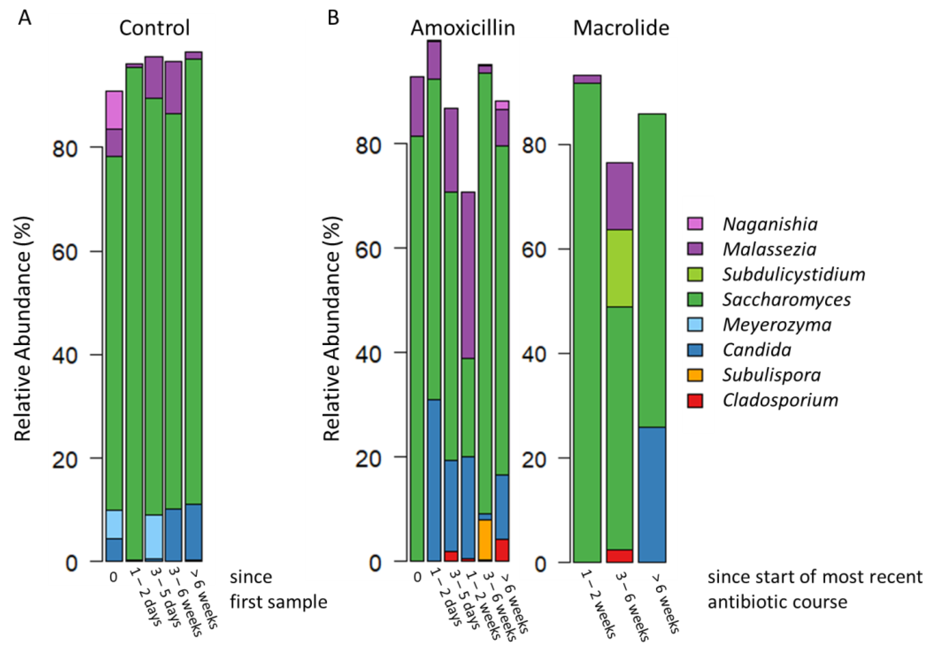
Figure 1. The gut fungal microbiota (mycobiota) composition presented as mean relative abundances of the most abundant fungal genera (presented on the right side of the figure) in (A) infants that were antibiotic-naïve throughout the follow-up and (B) infants that were antibiotic-naïve at baseline (marked as 0) but received amoxicillin or macrolide antibiotic treatment during follow-up. For the infants who received multiple antibiotics, the timepoints are calculated as time since the most recent antibiotic. All macrolide-treated patients received at least one course of amoxicillin prior to the macrolide course. Detailed results of the statistical differences between the antibiotic-treated and antibiotic-naïve groups are presented in Supplementary Tables S2–S6.
Jof 08 00328 g001
Figure 2. Differences between the control group and antibiotic-treated group over 6 timepoints starting before antibiotic treatment (marked as 0) stratified by (A) differences in the relative abundances at genus level, (B) at phylum level in the gut fungal microbiota (mycobiota), and further (C) differences in diversity and richness between control (red) and antibiotic-treated group (yellow). Significant p-values marked as * <0.05 and *** <0.001. All antibiotics are grouped together in the antibiotic group and for the infants who received multiple antibiotics, the timepoints are calculated as time since the most recent antibiotic. Detailed results of the statistical differences between the groups are presented in Supplementary Tables S2–S6.
Jof 08 00328 g002
Table 1. Number of infants stratified by control, amoxicillin and macrolide divided into time since most recent course of antibiotics or since first sample. All infants were antibiotic-naïve at timepoint 0.
Time Since Most Recent Course of Antibiotics
or Since First Sample
0 Days1–2 Days3–5 Days1–2 Weeks3–6 Weeks>6 Weeks
Control (no. of infants—16; no. of samples—50)14890109
Amoxicillin (no. of infants—21; no. of samples—51)10985910
Macrolide (no. of infants—4; no. of samples—12)0003 *5 *4
* one cephalosporin.
Table 2. Patient characteristics stratified by control and antibiotic use.
PatientsAll (N = 37)Antibiotics during Admission (N = 21)No Antibiotics during Admission (N = 16)
Antibiotic-naïve at recruitment, N(%)37 (100)21 (100)16 (100)
Female, N(%)15 (40.5)7 (33)8 (50)
Age at hospitalization, median (range, months)2.3 (0.8–9.3)2.5 (0.8–9.3)2.1 (0.8–6.8)
Vaginal delivery, N(%)31 (83.8)17 (81.0)14 (87.5)
Exclusive or partial breast-feeding, N(%)32 (86.5)17 (81.0)15 (93.8)
Birth weight, median (range, kg)3.7 (2.5–4.9)3.5 (2.5–4.9)3.8 (2.7–4.5)
Length of hospital stay, median (range, days)4 (1–9)4 (2–9)4 (1–7)
Annotated samples with >100 reads, N1136350
Fecal samples obtained and successfully annotated, median (range, N/patient)3 (1–6)3 (1–5)3.5 (1–6)
Length of follow-up, median (range, months)3 (0–9.5)3 (0–9.5)2 (0–6)
Publisher’s Note: MDPI stays neutral with regard to jurisdictional claims in published maps and institutional affiliations.