Article

Anti-Adipogenic Lanostane-Type Triterpenoids from the Edible and Medicinal Mushroom Ganoderma applanatum

1
State Key Laboratory of Phytochemistry and Plant Resources in West China, Kunming Institute of Botany, Chinese Academy of Sciences, Kunming 650201, China
2
University of the Chinese Academy of Sciences, Beijing 100049, China
3
Key Laboratory of Medicinal Chemistry for Natural Resource, Ministry of Education, Yunnan Provincial Center for Research & Development of Natural Products, School of Chemical Science and Technology, Yunnan University, Kunming 650091, China
*
Authors to whom correspondence should be addressed.
These authors contributed equally to this work.
J. Fungi 2022, 8(4), 331; https://doi.org/10.3390/jof8040331
Submission received: 22 February 2022 / Revised: 17 March 2022 / Accepted: 18 March 2022 / Published: 22 March 2022

Abstract

:
Our previous research has shown that lanostane triterpenoids from Ganoderma applanatum exhibit significant anti-adipogenesis effects. In order to obtain more structurally diverse lanostane triterpenoids to establish a structure–activity relationship, we continued the study of lanostane triterpenoids from the fruiting bodies of G. applanatum, and forty highly oxygenated lanostane-type triterpenoinds (140), including sixteen new compounds (116), were isolated. Their structures were elucidated using NMR spectra, X-ray crystallographic analysis, and Mosher’s method. In addition, some of their parts were evaluated to determine their anti-adipogenesis activities in the 3T3-L1 cell model. The results showed that compounds 16, 22, 28, and 32 exhibited stronger anti-adipogenesis effects than the positive control (LiCl, 20 mM) at the concentration of 20 μM. Compounds 15 and 20 could significantly reduce the lipid accumulation during the differentiation process of 3T3-L1 cells, comparable to the untreated group. Their IC50 values were 6.42 and 5.39 μM, respectively. The combined results of our previous and present studies allow us to establish a structure-activity relationship of lanostane triterpenoids, indicating that the A-seco-23→26 lactone skeleton could play a key role in anti-adipogenesis activity.

1. Introduction

Macro-fungi provide crucial food and medicinal resources [1]. Tricholoma matsutake, [2,3] Lentinula edodes, [4,5], and Collybia albuminosa [6] are delicious mushrooms which contain plentiful amino acids, fatty acids, vitamins, crude fiber, and protein. In addition, Fomitopsis pinicola (SW.) [7] Karst, Inonotus obliquus [8,9], Phellinus igniarius [10,11], Ganoderma lucidum [12,13], and Ganoderma sinense [14,15] have been used as edible and medicinal mushrooms for preventing and treating various diseases. The Ganoderma genus plays an important role in the history of Chinese medicine [16]. Shennong’s Herbal Classics recorded its traditional effects to include improving eyesight, strengthening muscles and bones, reinforcing kidney function, soothing the nerves, and prolonging the lifespan. G. lucidum and G. sinense have been registered in the Chinese Pharmacopoeia (2015 version). Meanwhile, G. lucidum was also included in the catalog of the latest edition of “Homology of medicine and Food” in 2020. Modern pharmacological research has further demonstrated that Ganoderma has a variety of pharmacological activities [17,18,19,20,21]. Thus, Ganoderma has great prospects in preventing and treating diseases.
Ganoderma applanatum, belonging to the genus Ganoderma, has traditionally been used to treat various chronic diseases, such as chronic hepatitis, immunological disorders, neurasthenia, arthritis, and nephritis [22]. Meanwhile, G. applanatum has been made into capsules and injections to cure chronic liver fibrosis and inflammation in a clinical setting [23,24,25,26]. G. applanatum is rich in chemical constituents, including polysaccharides, triterpenoids, meroterpenoids, alkaloids, and steroids. The majority of studies relating to it focus on the application and development of polysaccharides [23,27,28,29,30,31]. However, our previous research proved that highly oxygenated triterpenoids showed significant anti-adipogenesis activities [27,28]. In order to search for more active compounds to clarify the structure–activity relationship to lay the foundations for the discovery of lead compounds, we continued to investigate triterpenoids isolated from G. applanatum and 40 lanostane-type triterpenoids; of these (140), 16 were new compounds (116, Figure 1). Furthermore, their anti-adipogenesis effects were evaluated in the 3T3-L1 cell model, and their structure–activity relationship was established.

2. Materials and Methods

2.1. General Experimental Procedures

NMR spectra were recorded on a Bruker AV-600 MHz (Bruker, Zurich, Switzerland) using TMS as an internal standard for chemical shifts with reference to the TMS resonance. ESIMS and HRTOF-ESIMS were measured on an API QSTAR Pulsar spectrometer. UV spectra were recorded on a Shimadzu UV-2401PC spectrometer. IR was recorded on the Bruker Tensor-27 instrument using KBr pellets. Optical rotations were recorded on a Horiba SEPA-300 polarimeter. CD spectra were measured on a Chirascan instrument. An Agilent 1100 series instrument equipped with an Agilent ZORBAX SB-C18 column (5 μm, 9.6 mm × 250 mm) was used for high-performance liquid chromatography (HPLC) separation.
TLC was performed on precoated TLC plates (200–250 µm thickness, F254 Si gel 60, Qingdao Marine Chemical, Inc., Qingdao, China), with compounds visualized by spraying the dried plates with 10% aqueous H2SO4 followed by heating until they were dry. Silica gel ((200–300) mesh, Qingdao Marine Chemical, Inc.), Lichroprep RP-18 (40–63 μm, Fuji), and Sephadex LH-20 (20–150 μm, Pharmacia) were used for column chromatography. Methanol, chloroform, ethyl acetate, acetone, petroleum ether, n-hexane, and 2-propanol were purchased from Tianjing Chemical Reagents Co. (Tianjing, China). All other materials were of the highest grade available.

2.2. Fungal Materials

Ganoderma applanatum (39 kg) was purchased in December 2019 from a traditional Chinese medicine market in Kunming, Yunnan, China, which was identified by Prof. Yang Zhuliang, Kunming Institute of Botany, Chinese Academy of Science (voucher No. 19122201).

2.3. Extraction and Isolation

G. applanatum (39 kg) was chipped and extracted with 95% EtOH under reflux three times (three hours each time). The combined EtOH extracts were evaporated under reduced pressure. The residue was suspended in H2O and extracted with EtOAc. The volume of the combined EtOAc extracts was reduced to one-third under a reduced pressure. The residue was fractionated by macroporous resin (D-101; MeOH−H2O, 50:50, 70:30, and 90:10, v/v): fractions I−III. Fraction III (245 g) was further fractioned by a silica gel column with petroleum ether (PE)/ethyl acetate (EA) as the mobile phase, which gave six subfractions (Fr. III-1→Fr. III-6).
Fr. III-2 (156 g) was treated by a silica gel column and CHCl3/MeOH (80:1→20:1, v/v) was used as an eluent. Ten fractions (Fr. III-2-1→Fr. III-2-10) were obtained, of which Fr. III-2-4 (20 g) was separated using Sephadex LH-20 (MeOH) to obtain three subfractions (Fr. III-2-4a→Fr. III-2-4c). Compound 29 (235 mg) was purified through recrystallization from Fr. III-2-4b. The remaining solution was isolated using semi-preparative HPLC (CH3CN/H2O = 52%, v/v) to gain compound 6 (8 mg, tR = 28.6 min). Fr. III-2-5 (10 g) was treated by a silica gel column, being eluted with PE/EA (20:1, v/v) to obtain five parts (Fr. III-2-5a→Fr. III-2-5e). Subsequently, 5b and 5d were purified using P-TLC (CHCl3/MeOH = 40:1, v/v) to obtain compounds 18 (11 mg), 16 (26 mg), and 21 (9.2 mg). Fr. III-2-6 (12 g) was separated by Rp-C18 with the elution of MeOH/H2O (50%→55%) to obtain five fractions. Compounds 36 (6.2 mg) and 13 (12 mg) were obtained from Fr. III-2-6c and Fr. III-2-6d through P-TLC (CHCl3/MeOH = 40:1, v/v), respectively.
Fr. III-2-7 (25 g) was fractioned by an Rp-C18 column, being eluted with MeOH/H2O (50%→65% containing 0.3% CF3COOH, v/v); nine subfractions (7a→7i) were obtained. Furthermore, 7d, 7g, and 7h were purified by semi-preparative HPLC (CH3CN/H2O = 45%→60%, v/v) to obtain compounds 14 (5.3 mg, tR = 19.1 min), 4 (8.3 mg, tR = 19.1 min), 8 (3.4 mg, tR = 14.8 min), 9 (3.2 mg, tR = 17.4 min), 12 (4.2 mg, tR = 21.3 min), and 17 (5.1 mg, tR = 22.2 min). Similarly, Fr. III-2-8 (31 g) was also treated using an Rp-C18 column with MeOH/H2O (50%→55%) to obtain nine subfractions (8a→8i), from which compounds 37 (3.2 mg, tR = 26.6 min), 38 (3.6 mg, tR = 27.6 min), 31 (6.1 mg, tR = 22.1 min), 22 (3.1 mg, tR = 21.5 min), 25 (14 mg, tR = 25.9 min), 19 (12.5 mg, tR = 27.5 min), and 20 (7.2 mg, tR = 20.7 min) were purified by semi-preparative HPLC (CH3CN/H2O = 43%→60% containing 0.3% CF3COOH, v/v). The Rp-C18 column and semi-preparative HPLC were used to treat Fr. III-2-9 (20 g), and compounds 35 (2.5 mg, tR = 20.8 min), 28 (2.9 mg, tR = 20.3 min), 13 (2.1 mg, tR = 18.3 min), 7 (2.2 mg, tR = 19.5 min), and 23 (2.9 mg, tR = 18.5 min) were isolated from 9d-1 and 9d-2. 9e (15 mg) was purified by P-TLC (CHCl3/MeOH = 30:1, v/v) to obtain compounds 39 (4.2 mg) and 40 (2.8 mg).
The combination of Fr. III-4 and Fr. III-5 weighing 52 g was fractioned using Rp-C18 column elution with MeOH/H2O (35%→100%, v/v) to obtain six subfractions (Fr. III-4-1→Fr. III-4-6). Among these, Fr. III-4-2→Fr. III-4-5 were treated using Sephadex LH-20 (MeOH). Subsequently, the triterpenoid parts were purified by P-TLC (CHCl3/MeOH = 20:1 containing 0.3% CF3COOH, v/v) and semi-preparative HPLC (CH3CN/H2O = 38%→53% containing 0.3% CF3COOH, v/v) to obtain compounds 27 (4.2 mg), 24 (2.1 mg), 30 (6.2 mg), 5 (3.1 mg), 33 (3 mg), 26 (4.8 mg), 17 (5 mg, tR = 23.5 min), 3 (7.2 mg, tR = 22.5 min), 34 (6.1 mg, tR = 12.1 min), 32 (3.3 mg, tR = 17.8 min), 1 (3.0 mg, tR = 17.5 min), 10 (2.9 mg), and 2 (3.7 mg).
Ganoapplic acid A (1): white powder (MeOH); [α]28D −1.2 (c 0.25, MeOH); UV (MeOH); λmax (log ε): 230 (3.36), and 196 (3.33); IR (KBr) vmax 3428, 2953, 2943, 1653, 1636, 1423, 1364, 1212, and 1131 cm−1; 1H NMR and 13C NMR data: see Table 1 and Table 2; HRMS (ESI-TOF) m/z: 561.2696 [M + H]+ (calcd for C30H40O10, 561.2694).
Ganoapplic acid B (2): white powder (MeOH); [α]28D +48.0 (c 0.13, MeOH); UV (MeOH); λmax (log ε): 251 (3.75), and 196 (4.08); IR (KBr) vmax 3430, 2953, 2928, 1653, 1636, 1473, 1344, 1211, and 1147 cm−1; 1H NMR and 13C NMR data: see Table 1 and Table 2; HRMS (ESI-TOF) m/z: 527.2653 [M − H] (calcd for C30H40O8, 527.2650).
Ganoapplic acid C (3): white powder (MeOH); [α]28D +42.0 (c 0.09, MeOH); UV (MeOH); λmax (log ε): 251 (3.59), and 196 (3.93); IR (KBr) vmax 3423, 2955, 2930, 1673, 1635, 1428, 1380, 1219, and 1132 cm−1; 1H NMR and 13C NMR data: see Table 1 and Table 2; HRMS (ESI-TOF) m/z: 527.2654 [M − H] (calcd for C30H40O8, 527.2650).
Methyl ganoapplate C (4): white powder (MeOH); [α]28D +39.5 (c 0.07, MeOH); UV (MeOH); λmax (log ε): 251 (3.52), and 196 (3.85); IR (KBr) vmax 3458, 2957, 2928, 1689, 1606, 1473, 1375, 1210, and 1132 cm−1; 1H NMR and 13C NMR data: see Table 1 and Table 2; HRMS (ESI-TOF) m/z: 565.2773 [M + Na]+ (calcd for C31H42O8Na, 565.2772).
Ganoapplic acid D (5): white powder (MeOH); [α]28D −20.1 (c 0.21, MeOH); UV (MeOH); λmax (log ε): 242 (3.71), and 195 (3.79); IR (KBr) vmax 3503, 2967, 2935, 1663, 1635, 1452, 1390, 1200, and 1125 cm−1; 1H NMR and 13C NMR data: see Table 1 and Table 2; HRMS (ESI-TOF) m/z: 511.2707 [M − H] (calcd for C30H40O7, 511.2701).
Methyl gibbosate M (6): white powder (MeOH); [α]28D −49.64 (c 0.11, MeOH); UV (MeOH); λmax (log ε): 307 (3.21), 243 (3.80), and 196 (3.77); IR (KBr) vmax 3438, 2966, 2912, 1688, 1634, 1453, 1364, 1212, and 1132 cm−1; 1H NMR and 13C NMR data: see Table 1 and Table 2; HRMS (ESI-TOF) m/z: 533.2873 [M + Na]+ (calcd for C31H42O6Na, 533.2874).
Methyl ganoapplate E (7): white powder (MeOH); [α]28D −69.3 (c 0.13, MeOH); UV (MeOH); λmax (log ε): 316 (3.13), 243 (3.74), and 196 (3.72); IR (KBr) vmax 3413, 2953, 2916, 1657, 1620, 1454, 1374, 1209, and 1142 cm−1; 1H NMR and 13C NMR data: see Table 1 and Table 2; HRMS (ESI-TOF) m/z: 535.3033 [M + Na]+ (calcd for C31H44O6Na, 535.3030).
Ganoapplic acid E (8): white powder (MeOH); [α]28D −143.5 (c 0.19, MeOH); UV (MeOH); λmax (log ε): 317 (3.57), 244 (4.15), and 196 (4.05); IR (KBr) vmax 3445, 2980, 2906, 1712, 1690, 1458, 1380, 1214, and 1028 cm−1; 1H NMR and 13C NMR data: see Table 1 and Table 2; HRMS (ESI-TOF) m/z: 497.2911 [M − H] (calcd for C30H41O6, 497.2909).
Methyl gibbosate L (9): white powder (MeOH); [α]28D +20.27 (c 0.15, MeOH); UV (MeOH); λmax (log ε): 291 (3.70), 244 (3.94), and 196 (3.85); IR (KBr) vmax 3444, 2976, 2911, 1673, 1658, 1423, 1374, 1219, and 1140 cm−1; 1H NMR and 13C NMR data: see Table 3 and Table 4; HRMS (ESI-TOF) m/z: 533.2876 [M + Na]+ (calcd for C31H42O6Na, 533.2874).
Ganoapplic acid F (10): white powder (MeOH); [α]28D +20.86 (c 0.14, MeOH); UV (MeOH); λmax (log ε): 299 (3.77), and 196 (3.98); IR (KBr) vmax 3437, 2953, 2924, 1678, 1656, 1433, 1374, 1215, and 1132 cm−1; 1H NMR and 13C NMR data: see Table 3 and Table 4; HRMS (ESI-TOF) m/z: 511.2708 [M − H] (calcd for C30H40O7, 511.2701).
Methyl ganoapplate F (11): white powder (MeOH); [α]28D +5.80 (c 0.10, MeOH); UV (MeOH); λmax (log ε): 301 (3.46), and 196 (3.87); IR (KBr) vmax 3439, 2953, 2929, 1723, 1638, 1445, 1334, 1219, and 1138 cm−1; 1H NMR and 13C NMR data: see Table 3 and Table 4; HRMS (ESI-TOF) m/z: 549.2827 [M + Na]+ (calcd for C31H42O7Na, 549.2823).
Methyl gannosate I (12): white powder (MeOH); [α]28D +95.0(c 0.14, MeOH); UV (MeOH); λmax (log ε): 249 (3.99), and 195 (3.73); IR (KBr) vmax 3452, 2985, 2921, 1673, 1618, 1425, 1376, 1221, and 1132 cm−1; 1H NMR and 13C NMR data: see Table 3 and Table 4; HRMS (ESI-TOF) m/z: 565.2769 [M + Na]+ (calcd for C31H42O8Na, 565.2772).
Ganoapplic acid G (13): white powder (MeOH); [α]28D −32.86 (c 0.14, MeOH); UV (MeOH); λmax (log ε): 242 (3.99), and 196 (3.86); IR (KBr) vmax 3440, 2978, 2965, 1683, 1628, 1403, 1364, 1200, and 1151 cm−1; 1H NMR and 13C NMR data: see Table 3 and Table 4; HRMS (ESI-TOF) m/z: 535.0000 [M + Na]+ (calcd for C30H40O7Na, 535.0000).
Methyl ganoapplate G (14): white powder (MeOH); [α]28D +0.80 (c 0.07, MeOH); UV (MeOH); λmax (log ε): 243 (3.79), and 196 (3.77); IR (KBr) vmax 3445, 2963, 2931, 1683, 1638, 1453, 1384, 1209, and 1142 cm−1; 1H NMR and 13C NMR data: see Table 3 and Table 4; HRMS (ESI-TOF) m/z: 549.2823 [M + Na]+ (calcd for C31H42O7Na, 549.2823).
Methyl applate C (15): white powder (MeOH); [α]28D +43.44 (c 0.18, MeOH); UV (MeOH); λmax (log ε): 248 (3.99), and 196 (3.95); IR (KBr) vmax 3443, 2956, 2915, 1665, 1624, 1433, 1376, 1205, and 1132 cm−1; 1H NMR and 13C NMR data: see Table 3 and Table 4; HRMS (ESI-TOF) m/z: 547.2672 [M + Na]+ (calcd for C31H40O7Na, 547.2666).
Methyl gibbosate A (16): white powder (MeOH); [α]28D +18.94 (c 0.15, MeOH); UV (MeOH); λmax (log ε): 237 (3.78), and 196 (3.82); IR (KBr) vmax 3430, 2953, 2929, 1703, 1628, 1433, 1354, 1229, and 1147 cm−1; 1H NMR and 13C NMR data: see Table 3 and Table 4; HRMS (ESI-TOF) m/z: 563.2612 [M + Na]+ (calcd for C31H40O8Na, 563.2615)
X-ray Crystallographic Data for Ganoapplic acid A (1): C30H40O10·CH4O·H2O, M = 610.68, a = 13.9761(7) Å, b = 6.9089(3) Å, c = 15.8732(7) Å, α = 90°, β = 90.948(2)°, γ = 90°, V = 1532.50(12) Å3, T = 100.(2) K, space group P1211, Z = 2, μ(Cu Kα) = 0.844 mm−1, 26,208 reflections measured, 5973 independent reflections (Rint = 0.0593). The final R1 values were 0.0504 (I > 2σ(I)). The final wR(F2) values were 0.1381 (I > 2σ(I)). The final R1 values were 0.0543 (all data). The final wR(F2) values were 0.1436 (all data). The goodness of fit for F2 was 1.042. Flack parameter = 0.07(9).

2.4. Mosher’s Method

The specific esterification of compound 4 was performed based on the previous method [32]. The 1H NMR spectroscopic data of the (R)-MTPA ester derivative (4r) of 4 (600 MHz, pyridine-d5; data were obtained from the reaction NMR tube directly and assigned on the basis of correlations of the 1H-1H COSY spectrum): δ 4.253 (1H, m, H-15), δ 2.158 (1H, m, H-16a), δ 2.756 (1H, m, H-16b), δ 3.566 (1H, m, H-17), δ 5.619 (1H, s, H-21a), δ 5.632 (1H, s, H-21b), δ 5.007 (1H, m, H-22), δ 5.067 (1H, m, H-23), δ 2.759 (1H, m, H-24a), δ 1.905 (1H, m, H-24b), δ 3.080 (1H, m, H-25), δ 1.198 (3H, s, Me-26). Meanwhile, the 1H NMR spectroscopic data of the (S)-MTPA ester derivative (4s) of 4 were: δ 4.255 (1H, m, H-15), δ 2.156 (1H, m, H-16a), δ 2.756 (1H, m, H-16b), δ 3.568 (1H, m, H-17), δ 5.639 (1H, s, H-21a), δ 5.633 (1H, s, H-21b), δ 5.003 (1H, m, H-22), δ 5.062 (1H, m, H-23), δ 2.756 (1H, m, H-24a), δ 1.903 (1H, m, H-24b), δ 3.078 (1H, m, H-25), δ 1.196 (3H, s, Me-26) (see Figures S29–S31 in Section S10).

2.5. Inhibition of Lipogenesis Assay

2.5.1. Cell Culture and Adipocyte Differentiation

3T3-L1 cells were purchased from the American Type Culture Collection (ATCC, Manassas, VA, U.S.A.). The culture and differentiation of 3T3-L1 cells were performed based on the description reported previously [28]. Firstly, Dulbecco’s modified Eagle’s medium (DMEM) containing 10% bovine calf serum (CS) was used to cultivated 3T3-L1 cells. The whole system was incubated at 37 ℃ in a humidified atmosphere of 5% CO2 and 95% air. Secondly, different medium systems were used for the different differentiated phases. Confluent cells were grown in DMEM medium containing 1 μg/mL insulin, 1 μM dexamethasone (DEX), 0.5 mM 3-isobutyl-1-methylxanthine (IBMX), and 1 μM rosiglitazone (Rosi). Then, on the third day, post-differentiation medium—namely, DMEM with 10% fetal bovine serum (FBS) and 1 μg/mL insulin—was used to continually cultivate the cells. From the fourth day, DMEM + 10% FBS was used as a maintenance medium for cell differentiation. In this process, it commonly takes two days for the mature adipocytes to form. During the whole differentiation process, the tested compounds or 0.1% DMSO were added to the differentiated 3T3-L1 cells, for which the 0.1% DMSO group was used as the vehicle.

2.5.2. Cell Viability Assay

The viability of cell treated with compounds 15 and 20 was determined by the MTS method. The detailed experimental procedures were similar to those described in our previous study [29].

2.5.3. Lipid Content Analysis

The intracellular lipid contents of 3T3-L1 adipocytes were determined by Oil Red O staining [33]. Briefly, differentiated 3T3-L1 cells were washed twice with PBS and fixed with 10% formaldehyde for 1 h. After another washing with PBS, the fixed cells were stained with 0.5% Oil Red O in 3:2 of Oil Red O/H2O for 15 min at room temperature and then washed with 60% isopropanol and distilled water. The lipid content was imaged with an inverted light microscope Nikon TS100 (Tokyo, Japan). Finally, 100% isopropanol was used to elute Oil Red O dye and it was quantified at 492 nm absorbance.

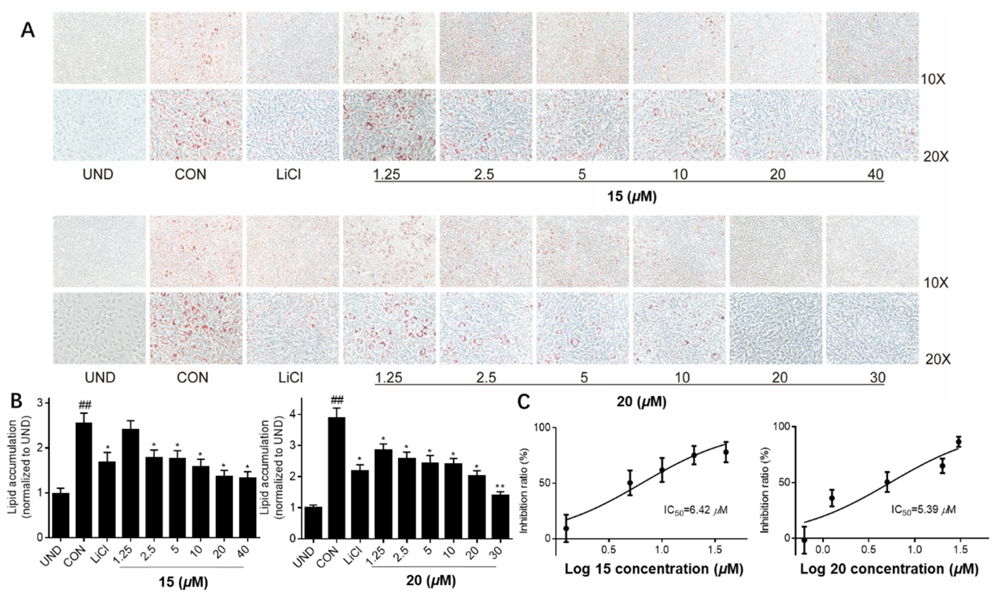
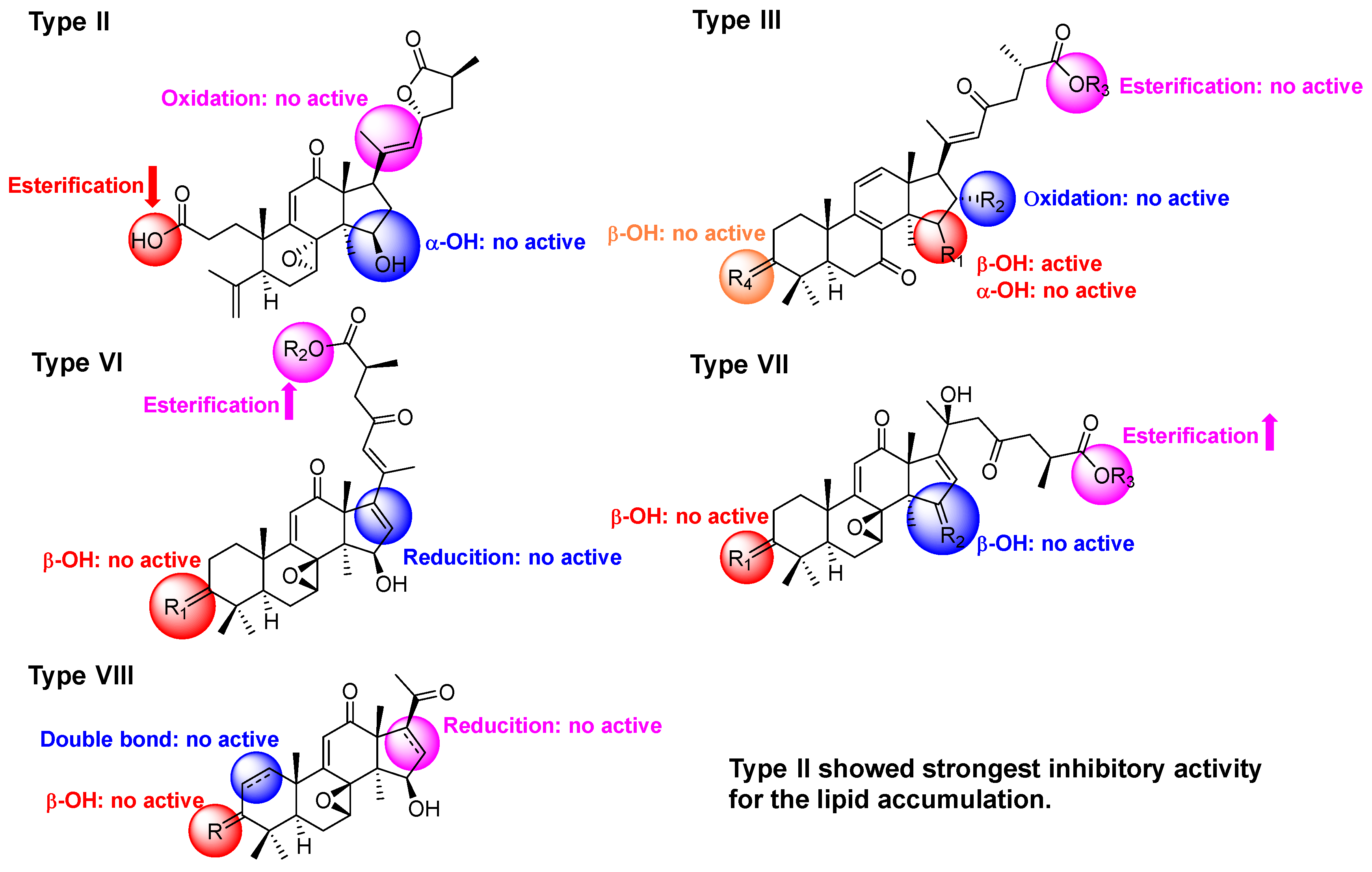
3. Results and Discussion

The molecular formula of ganoapplic acid A (1) was established to be C30H40O10 by HRESIMS ion at m/z 561.2696 [M + H]+ (calcd. 561.2694), suggesting 11 degrees of unsaturation. Its 13C NMR spectra displayed 30 carbon resonances, of which seven methyls, three ketone carbonyls (δC 215.8, δC 205.9, and δC 209.9), an α,β-unsaturated carbonyl (δC 165.0, δC 127.3, and δC 204.0), one carboxyl (δC 179.7), two oxygenated methines (δC 80.0 and δC 61.2), and three quaternary carbons containing oxygen (δC 85.5, δC 71.4, and δC 73.4) were assigned based on the HSQC and 13C-DEPT NMR spectra (see Figures S2 and S4 in Section S1). These data indicated that the structure of compound 1 was similar to that of gibbosic acid G with a 7,12,23-trioxo-8,20-dihydroxy-lanosta-9,11-en-26-oic acid skeleton [34], except for the presence of one oxygenated methine and one oxygenated quaternary carbon and the absence of a double bond at C-16 and C-17 in 1. Furthermore, the HMBC spectrum (Figure 2A) of 1 revealed the correlations of H3-21 with C-20 (δC 73.4), C-22, and the oxygenated quaternary carbon (δC 71.4); of H3-30 with C-15 (δC 80.0); and of H-15 (δH 4.83, s) with the oxygenated methine (δC 61.2) and quaternary carbons (δC 71.4). Meanwhile, the proton of the methine containing oxygen (δH 3.36, s) showed the HMBC correlations with C-13, C-14, C-15, and C-20, as well as the 1H-1H COSY correlation with H-15 (Figure 2A), which certified that C-16 and C-17 was substituted by hydroxyls. According to the molecular formula of 1, an epoxy was present in 1 at C-16 and C-17.
In the ROESY spectrum (Figure 2B) of 1, H-15 showed an apparent cross peak with H3-30, while H-16 correlated with H3-18, suggesting that H-15 and H-16 were α- and β-oriented, respectively. The X-ray crystallographic analysis (Figure 3A) of 1 (Cu κα) further confirmed that the absolute configurations of C-15, C-16, C-20, and C-25 were R, S, S, and S (see Table S1 in Section S1). Finally, the structure of 1 was determined to be (20S,25S)-7,12,23-trioxo-8β,20-dihydroxy-16α,17α-epoxy-lanosta-9,11-en-26-oic acid.
Ganoapplic acid B (2) was isolated as a colorless solid. Its molecular formula was determined to be C30H40O8 by the HRESIMS. The 1D NMR and HSQC data of 2 supported the presence of five methyls, an α,β-unsaturated ketone, two carboxyl or ester carbonyls, one oxygenated methylene (δH 4.37, s; δC 61.8), three oxygenated methines (δH 3.95, d, J = 6.5 Hz, δC 77.4; δH 4.93, s, δC 64.7; δH 5.59, dd, J = 8.6 and 6.6 Hz, δC 76.8), one quaternary carbon containing oxygen (δC 67.4), a terminal double bond (δC 146.5; δC 115.9), and a double bond (δC 144.1; δH 5.78, d, J = 8.6 Hz, δC 130.8), which indicated that compound 2 was an A-seco-lanostane triterpenoid and had the similar structure to that of gibbosicolid F. [35] However, five methyls and an oxygenated methylene were observed in 2, rather than the six methyls found in gibbosicolid F. The detailed analysis of the 2D NMR spectra (see Figures S10–S13 in Section S2) exhibited the long-range HMBC correlations (Figure 2A) of the oxygenated methylene with C-17, C-20, and C-22; of H3-18 with C-12, C-13, C-14, and C-17; and of H-22 with C-17, C-20, C-23, and C-24, together with the 1H-1H COSY correlations of H-22/H-23/H2-24/H-25/H3-26. These pieces of evidence confirmed that the hydroxyl was connected to C-21.
The ROESY cross peaks (Figure 2B) of H-7/H3-18 and H-15/H3-30 demonstrated that both H-15 and the epoxy between C-7 and C-8 were α. H2-21 showed a ROESY correlation with H-23, suggesting that the double bond at C-20 and C-22 was Z. In addition, H3-26 displayed the ROESY correlation with H-23, hinting that H3-26 and H-23 were on the same face. Moreover, the dd-peak type of H-23 (J = 8.6 and 6.6 Hz) was consistent with that of gibbosicolid B (δH 5.31, dd, J = 13.2 and 8.0 Hz), which further indicated that the absolute configurations of C-23 and C-25 were R and S, respectively. [35] Therefore, the structure of 2 was assigned as (23R,25S)-15β,21-dihydroxyl-7α,8α-epoxy-12-oxo-3,4-seco-lanosta-4(28),9-(11),20E(22)-trien-23,26-olide-3-oic acid and named ganoapplic acid B (2).
The molecular formula of ganoapplic acid C (3) was established to be C30H40O8 by the HRESIMS. The 1D NMR spectra (see Figures S15 and S16 in Section S6) of 3 showed a high similarity with those of ganoapplic acid B (2), suggesting that compound 3 was an A-seco lanostane triterpenoid. However, the comparison of the 1D NMR spectroscopic data of 2 and 3 revealed that another terminal double bond (δC 117.6 and δC 148.7) and oxygenated methine (δC 76.5) were present in 3, while a double bond (C-20-C-22) and an oxygenated methylene (C-21) were observed in 2. Furthermore, the obvious 1H-1H COSY correlations of H3-26/H-25/H2-24/H-23/H-22, as well as the HMBC correlations of H3-26 with C-24, C-25, and C-27 and of H2-24 with C-22, C-23, and C-26, indicated that the hydroxyl was located at C-22 (Figure 2A). Meanwhile, H-22 (δH 4.47, d, J = 5.6 Hz) and H-17 displayed HMBC correlations of the terminal double bond, proving that the terminal double bond was at C-21 and C-20.
The 1D NMR spectra and molecular weight (see Figures S22 and S23 in Section S8, and Figure S28 in Section S9) of methyl ganoapplate C (4) showed that compound 4 was the ester derivative of 3, which was confirmed by the HMBC correlation of OMe with C-27 (δC 175.6). The analysis of the ROESY spectra of 3 and 4 exhibited cross peaks of H-7/H3-18, H-23/H3-26, and H-22/H-25, indicating that the epoxy between C-7 and C-8 was α; meanwhile, H-23 and H3-26 were cofacial (Figure 2B). Biogenetically, the absolute configuration of C-25 from G. applanatum was S. Thus, C-23 was determined to be R. The absolute configuration of C-22 was established as R based on the revised Mosher’s method (Figure 3B) [33]. Thus, the structures of 3 and 4 were elucidated as (22R,23R,25S)-15β,22-dihydroxyl-7α,8α-epoxy-12-oxo-3,4-seco-lanosta-4(28),9(11),20(21)-trien-23,26-olide-3-oic acid and methyl (22R,23R,25S)-15β,22-dihydroxyl-7α,8α-epoxy-12-oxo-3,4-seco-lanosta-4(28),9(11),20(21)-trien-23,26-olide-3-oate, respectively.
Ganoapplic acid D (5) was isolated as a white powder and its molecular formula was determined to be C30H40O7 based on the HRESIMS at m/z 511.2701 [M − H] (calcd. 511.2707). Its 13C-DEPT spectra (see Figures S32 and S33 in Section S11) showed thirty carbon resonances belonging to seven methyls, four methylenes, eight methines (including three sp2 and two oxygenated), and eleven quaternary carbons (including three ketone carbonyls, one carboxyl, and three sp2). These data indicated that compound 5 was a lanostane-type triterpenoid and had similar structure to that of gibbosic acid M (22) [35], except for the replacement of the methylene (C-16) in 22 with an oxygenated methine in 5. The HMBC spectrum of 5 revealed the correlations of H3-30 with C-15 (δC 84.9), C-13, and C-14; of H3-18 with C-12, C-13, C-14, and C-17; and of H-17 with C-16, C-15, C-20, C-21, and C-22, suggesting that C-16 was an oxygenated methine in 5, together with the 1H-1H COSY correlations of H-15/H-16/H-17 (Figure 2A). The ROESY correlations of H-15/H3-30 and of H-16/H3-18 illustrated that H-15 and H-16 were α- and β-oriented, respectively. The E-configuration of ∆20,22 was determined by the ROESY correlation of H-22/H-17/H-16. Therefore, the structure of 5 was assigned as 15β,16α-dihydroxy-3,7,23-trioxolanosta-8,11,20E(22)-trien-26-oic acid and named ganoapplic acid D (5).
Methyl gibbosate M (6) had the molecular formula of C31H42O6 based on the positive HRESIMS at m/z 533.2874 [M + Na]+ (calcd. 533.2873). The 1D NMR spectra (see Figures S39 and S40 in Section S13) of 6 were the same as those of gibbosic acid M (22) [35], except that the carboxyl at C-27 in 22 was replaced by the ester carbonyl in 6. The key HMBC correlation of OMe with C-27 confirmed the above deduction. The characteristic d-coupling type of H-15 and the ROESY correlation of H-15/H3-30 indicated that the 15-OH was β-oriented [35]. Finally, the structure of 6 was established to be methyl 15β-hydroxy-3,7,23-trioxolanosta-8,11,20E(22)-trien-26-oate and named methyl gibbosate M (6).
Methyl ganoapplate E (7) was isolated as a white powder and its molecular formula was determined to be C31H44O6 by the HRESIMS. The analysis of the 1D NMR spectra (see Figures S46 and S47 in Section S15) of 7 showed that compound 7 had a similar structure to that of 6, with the only difference being in the replacement of the ketone carbonyl at C-3 in 6 with the oxygenated methine in 7, which was confirmed by the HMBC correlations of H3-28, H3-29, and H-5 with the oxygenated methine (δC 78.3). The ROESY correlations of H-3/H-5 and of H-15/H3-30 indicated that 3-OH and 15-OH were β.
Ganoapplic acid E (8) was deduced to be the demethylated derivative of 7 on the basis of the HMBC correlation regarding the lack of the OMe at C-27 and the low-field shift of C-27. Thus, the structures of compounds 7 and 8 were elucidated as methyl (25S)-3β,15β-dihydroxy-7,23-dioxolanosta-8,11,20E(22)-trien-26-oate and (25S)-3β,15β-dihydroxy-7,23-dioxolanosta-8,11,20E(22)-trien-26-oic acid, respectively.
Methyl gibbosate O (9) was found to be similar to the known compound gibbosic acid O (24) [35] based on the 1D NMR spectroscopic data, except for distinct differences in the chemical shift of C-27 and the presence of an additional methoxyl. The HMBC spectrum of 9 showed the correlation of OMe with C-27, suggesting that 9 was a methyl ester derivative of gibbosic acid O (24). Therefore, the structure of 9 was determined to be methyl 15α-hydroxy-3,12,23-trioxolanosta-7,9(11),20E(22)-trien-26-oate.
The molecular formula of ganoapplic acid F (10) was deduced to be C30H40O7 based on the HRESIMS and NMR data. Its 1D NMR spectra (see Figures S67 and S68 in Section S21) showed a similar tetracyclic skeleton to that of gibbosic acid O (24) [35] with a 15-hydroxy-3,12-dioxolanosta-7(8),9(11)-diene skeleton, which was confirmed by the 2D NMR spectra. In addition, an oxygenated quaternary carbon signal (δC 72.6) and two sp2 carbon signals (δC 127.5 and δC 158.9) were characteristic for the quaternary carbon containing oxygen at C-20 and the double bond at C-16 and C-17. Furthermore, the HMBC correlations (Figure 2A) of H3-30 with C-15; of H-15 with C-16 and C-17; of H3-18 with C-17; of H3-21 with C-17, C-20, and C-22; and of H-22 with C-20, C-23, and C-24 confirmed the above deduction.
Methyl ganoapplate F (11) was an ester derivative at C-27 of ganoapplic acid H (10), according to the HMBC correlation of OMe with C-27. The ROESY spectra of 10 and 11 showed cross peaks of H-15/H3-30, indicating the β-orientation of 15-OH. Therefore, the structures of 10 and 11 were determined to be 15β,20-dihydroxy-3,12, 23-trioxo-5α-lanosta-7,9(11),16-trien-26-oic acid and methyl 15β,20-dihydroxy-3,12,23-trioxo-5α-lanosta-7,9(11),16-trien-26-oate, respectively.
Based on the NMR data analysis, methyl gibbosate I (12) was found to be close to that of 26 [35], with a 12,15-dihydroxy-3,7,11,23-tetraoxolanosta-8,20(22)-dien structure. The 2D NMR spectra further confirmed its structure and 12 had an additional methoxyl at C-27, which was proven by the key HMBC correlation of OMe with C-27. Moreover, the ROESY correlations of H-12/H3-18 and H-15/H3-30 demonstrated that 12-OH was α while 15-OH was β. Thus, the structure of 12 was deduced to be methyl 12α,15β-dihydroxy-3,7,11,23-tetraoxolanosta-8,20(22)-dien-26-ate.
Ganoapplic acid G (13) was isolated as a white powder and its molecular formula was determined to be C30H40O7 based on the HRMS (ESI-TOF) m/z 535.0000 [M + Na]+ (calcd. 535.0000). The 1D NMR spectra of 13 showed the presence of the ketone carbonyl at C-3, 7,8-epoxyl, α,β-unsaturated ketones (C-9/C-11/C-12 and C-20/C-22/C-23), 15-OH, and 27-oic acid, which was further confirmed by the 2D NMR spectra (Figure 2A). The aforementioned information indicated that compound 13 had the same planar structure as gibbosic acid N. [35] The comparison of the ROESY spectra of 13 and gibbosic acid N revealed that they were 15-isomers due to the existence of the ROESY correlation of H-15/H3-30 and the d-coupling of H-15 [35]. Therefore, the structure of 13 was established to be 15β-hydroxy-7β,8β-epoxy-3,12,23-trioxolanosta-9(11),20E(22)-dien-26-oic acid. In addition, methyl ganoapplate G (14) was deduced to be the methylation product of 13 on the basis of the HMBC correlation of OMe with C-27.
Methyl applanate C (15) was found to have a similar structure to methyl ganoapplate F (14), except for the presence of a double bond in 15, rather than one methylene and one methine in 14. Furthermore, in the HMBC spectrum of 15, the correlations (Figure 2A) of H3-30 with C-15, of H-15 with the sp2 methine and quaternary carbon, and of H3-18 and H3-21 with the sp2 quaternary carbon were observed, which proved that the double bond was located at C-16 and C-17. The ROESY correlation of H-16/H3-21 and H3-18/H-22 suggested that the geometry of the 16,20(22)-conjugated diene was 17,20-Z-(16Z, 20E). Additionally, the ROESY correlation of H3-30/H-15 demonstrated that 15-OH was β. Finally, the structure of 15 was determined to be methyl 15β-hydroxy-7β,8β-epoxy-3,12,23-trioxolanosta-9(11),16Z,20E(22)-trien-26-oate and named methyl applanate C (15).
Methyl gibbosate A (16) was considered to be the methylation derivative of gibbosic acid A (29) [34] because of their similar 1D and 2D spectra (see Figures S104–S109 in Section S33) and the HMBC correlation of OMe with C-27. 7β,8β-epoxy was proven by the ROESY correlation of H-17/H3-30. Thus, the structure of 16 was established to be methyl 20-hydroxy-7β,8β-epoxy-3,12,15,23-tetraoxo-lanosta-9,16-dien-26-oate.
In addition, 24 known compounds were identified by comparing their 1D NMR spectra with those reported in the literature, and they were assigned as gibbosic acid G (17) [34], applanoic acid B (18) [36], gibbosicolid E (19) [35], gibbosicolid F (20) [35], gibbosicolid G (21) [35], gibbosic acid M (22) [35], gibbosic acid L (23) [35], gibbosic acid O (24) [35], applanoic acid D (25) [36], gibbosic acid I (26) [35], ganodapplanoic acid D (27) [27], applanoic acid C (28) [36], ganoapplanic acid F (29) [37], elfvingic acid B (30) [37], applanoxidic acid G methyl ester (31) [37], gibbosic acid C (32) [34], gibbosic acid B (33) [34], elfvingic acid C (34) [38], methyl ganoapplaniate D (35) [37], applanone E (36) [36], ganoapplanoid K (37) [28], ganoapplanoid L (38) [28], ganoapplanilactone B (39) [37], and ganoapplanilactone A (40) [37].
Parts of the isolated compounds were evaluated to determine their anti-adipogenesis activities. At a concentration of 20 μM, compounds 16, 22, 28, and 32 showed comparable inhibition for lipid accumulation compared to the positive control (LiCl, 20 mM). Meanwhile, compounds 15 and 20 displayed stronger inhibitory effects than the positive control, even resembling the untreated group (Figure 4). Furthermore, compounds 15 and 20 did not show any toxicity for the 3T3-L1 cells when the concentration was less than 100 μM. At the concentrations of 1.25, 2.5, 5, 10, 20, and 30 or 40, compounds 15 and 20 showed significantly inhibitory activities in a dose-dependent manner, with IC50 values of 6.42 and 5.39 μM, respectively (Figure 5).
The structures of the isolates were divided into nine types, including type I with a 7,12-dioxo-8-hydroxy-9,11-en fraction, type II with a A-seco-7,8-epoxy-9,11-en-12-oxo-23→27 lactone fraction, type III with a 7,23-dioxo-8(9),11(12),20(22)-trien fraction, type IV with a 12-oxo-7(8),9(11)-dien fraction, type V with a 7,11-dioxo-12-hydroxy-8(9)-en fraction, type VI with a 7,8-epoxy-12,23-dioxo-9(11),16(17),20(22)-trien fraction, type VII with a 20-hydroxy-7,8-epoxy-12,23-dioxo-9(11)-en fraction, type VIII with a 12-oxo-7,8-epoxy-9(11)-en-21,22,23,24,25,26,27-norlanostane, and type IX with a 12,23-epoxy-23→27 lactone fraction. The combined results of the previous and present studies showed that bioactive compounds were mainly present in type II (20 and 21), type III (22), type VI (15 and 28), type VII (16 and 32), and type VIII (37). For type II, when the relative configuration of 15-OH was α, the activity was decreased, similar to compound 19, while any changes in the side chain decreased their activities, such as compounds 24. For type III, no matter which reactions happened in type III, compounds 58 and 23 did not show inhibitory activity. For type VI, compound 27 was the 3-OH analogue of 15 and 28, leading to a decrease in inhibition. In type VII, the carbonyl at C-3 and the carbonyl or hydroxyl at C-15 could be the crucial active functionalities. C24 lanostane triterpenoids possessing a double bond at C-16 and C-17 displayed anti-adipogenesis activity [27,28]. Compared to the other compounds, compound 20 belonging to type II showed the strongest inhibitory activity, suggesting that A-seco-15β-hydroxy-7,8-epoxy-12-oxolanosta-9,11-en-23→27 lactone could play a significant role in the anti-adipogenesis effect (Figure 6).

4. Conclusions

Overall, inspired by our previous studies, we investigate the lanostane-type triterpenoids of G. applanatum; 40 triterpenoids, including 16 new compounds, were isolated. Their anti-adipogenesis activities were evaluated and the results showed that compounds 15 and 20 can significantly inhibit lipid accumulation, with the IC50 values of 6.42 and 5.39 μM, respectively. Furthermore, we established a structure–activity relationship for the lanostane-type triterpenoids from G. applanatum, suggesting that the structure skeleton (A-seco-15β-hydroxy-7,8-epoxy-12-oxolanosta-9,11-en-23→27 lactone) could be of importance for the anti-adipogenic effect. In the next step, we can use type III as a template for further structural modification in order to find the lead compound.

Supplementary Materials

The following supporting information can be downloaded at: https://www.mdpi.com/article/10.3390/jof8040331/s1, Section S1: 1D and 2D NMR spectra of compound 1; Section S2: HRESIMS spectrum of 1; Section S3: X-ray crystallographic data of 1; Section S4: 1D and 2D NMR spectra of 2; Section S5: HRESIMS spectrum of 2; Section S6: 1D and 2D NMR spectra of 3; Section S7: HRESIMS spectrum of 3; Section S8: 1D and 2D NMR spectra of 4; Section S9: HRESIMS spectrum of 4; Section S10: Comparison of 1H NMR and 1H-1H COSY spectra between 4r and 4s; Section S11: 1D and 2D NMR spectra of 5; Section S12: HRESIMS spectrum of 5; Section S13: 1D and 2D NMR spectra of 6; Section S14: HRESIMS spectrum of 6; Section S15: 1D and 2D NMR spectra of 7; Section S16: HRESIMS spectrum of 7; Section S17: 1D and 2D NMR spectra of 8; Section S18: HRESIMS spectrum of 8; Section S19: 1D and 2D NMR spectra of 9; Section S20: HRESIMS spectrum of 9; Section S21: 1D and 2D NMR spectra of 10; Section S22: HRESIMS spectrum of 10; Section S23: 1D and 2D NMR spectra of 11; Section S24: HRESIMS spectrum of 11; Section S25: 1D and 2D NMR spectra of 12; Section S26: HRESIMS spectrum of 12; Section S27: 1D and 2D NMR spectra of 13; Section S28: HRESIMS spectrum of 13; Section S29: 1D and 2D NMR spectra of 14; Section S30: HRESIMS spectrum of 14; Section S31: 1D and 2D NMR spectra of 15; Section S32: HRESIMS spectrum of 15; Section S33: 1D and 2D NMR spectra of 16; Section S34: HRESIMS spectrum of 16.

Author Contributions

Conceptualization, X.-R.P. and M.-H.Q.; methodology, Q.W., X.-R.P., H.-G.S., W.-Y.X. and M.-H.Q.; resources, X.-R.P., L.Z., W.-Y.X. and M.-H.Q.; data curation, X.-R.P. and Q.W.; writing—original draft preparation, X.-R.P.; writing—review and editing, X.-R.P., W.-Y.X. and M.-H.Q.; project administration, X.-R.P.; funding acquisition, X.-R.P. All authors have read and agreed to the published version of the manuscript.

Funding

This research was funded by the Basic Research Project of Yunnan Province (201901T070239) and the Youth Innovation Promotion Association of CAS (2019383).

Institutional Review Board Statement

Not applicable.

Informed Consent Statement

Not applicable.

Data Availability Statement

X-ray crystallographic data of 1 (CIF) are available free of charge.

Acknowledgments

This work was supported by grants from the Basic Research Program of Yunnan Province (202001AT070070), and the Youth Innovation Promotion Association of CAS (2019383). The authors are grateful to the Analytical and Testing Center at Kunming Institute of Botany for the NMR and ECD data collection.

Conflicts of Interest

The authors declare no conflict of interest. The funders had no role in the design of the study; in the collection, analysis, or interpretation of data; in the writing of the manuscript; or in the decision to publish the results.

References

  1. Sudheer, S.; Bai, R.G.; Muthoosamy, K.; Tuvikene, R.; Gupta, V.K.; Manickam, S. Biosustainable production of nanoparticles via mycogenesis for biotechnological applications: A critical review. Environ. Res. 2022, 204, 111963–111979. [Google Scholar] [CrossRef] [PubMed]
  2. Li, M.; Lv, R.; Xu, X.; Ge, Q.; Lin, S. Tricholoma matsutake-Derived Peptides Show Gastroprotective Effects against Ethanol-Induced Acute Gastric Injury. J. Agric. Food Chem. 2021, 69, 14985–14994. [Google Scholar] [CrossRef] [PubMed]
  3. Li, M.; Ge, Q.; Du, H.; Lin, S. Tricholoma matsutake-derived peptides ameliorate inflammation and mitochondrial dysfunction in RAW264.7 macrophages by modulating the NF-κB/COX-2 pathway. Foods 2021, 10, 2680. [Google Scholar] [CrossRef] [PubMed]
  4. Yu, H.; Shen, X.; Chen, H.; Dong, H.; Zhang, L.; Yuan, T.; Zhang, D.; Shang, X.; Tan, Q.; Liu, J.; et al. Analysis of heavy metal content in Lentinula edodes and the main influencing factors. Food Control 2021, 130, 108198–108205. [Google Scholar] [CrossRef]
  5. Shi, D.; Yin, C.; Fan, X.; Yao, F.; Qiao, Y.; Xue, S.; Lu, Q.; Feng, C.; Meng, J.; Gao, H. Effects of ultrasound and gamma irradiation on quality maintenance of fresh Lentinula edodes during cold storage. Food Chem. 2022, 373, 131478. [Google Scholar] [CrossRef]
  6. Chen, Y.-h.; Chen, J. Optimization of extraction conditions for polysaccharides from Collybia albuminosa via response surface methodology. Mod. Food Sci. Technol. 2012, 28, 541. [Google Scholar]
  7. Peng, X.R.; Su, H.G.; Liu, J.H.; Huang, Y.J.; Yang, X.Z.; Li, Z.R.; Zhou, L.; Qiu, M.H. C30 and C31 triterpenoids and triterpene sugar esters with cytotoxic activities from edible mushroom Fomitopsis pinicola (Sw. Ex Fr.) Krast. J. Agric. Food. Chem. 2019, 67, 10330–10341. [Google Scholar] [CrossRef]
  8. Ying, Y.M.; Yu, H.F.; Tong, C.P.; Shan, W.G.; Zhan, Z.J. Spiroinonotsuoxotriols A and B, two highly rearranged triterpenoids from Inonotus obliquus. Org. Lett. 2020, 22, 3377–3380. [Google Scholar] [CrossRef]
  9. Yu, J.; Xiang, J.Y.; Xiang, H.; Xie, Q. Cecal butyrate (Not Propionate) was connected with metabolism-related chemicals of mice, based on the different effects of the two Inonotus obliquus extracts on obesity and their mechanisms. ACS Omega 2020, 5, 16690–16700. [Google Scholar] [CrossRef]
  10. Li, Y.-T.; Zhang, Z.; Feng, Y.; Cheng, Y.; Li, S.; Li, C.; Tian, L.-W. Cardioprotective 22-hydroxylanostane triterpenoids from the fruiting bodies of Phellinus igniarius. Phytochemistry 2021, 191, 112907–112914. [Google Scholar] [CrossRef]
  11. Cao, Y.; Liu, Y.; Wang, G.; Wang, W.; Li, Y.; Xuan, L. Styryl pyranones with apoptosis activities from the sporocarps of Phellinus igniarius. Phytochem. Lett. 2021, 44, 154–159. [Google Scholar] [CrossRef]
  12. Shao, W.; Xiao, C.; Yong, T.; Zhang, Y.; Hu, H.; Xie, T.; Liu, R.; Huang, L.; Li, X.; Xie, Y.; et al. A polysaccharide isolated from Ganoderma lucidum ameliorates hyperglycemia through modulating gut microbiota in type 2 diabetic mice. Int. J. Biol. Macromol. 2022, 197, 23–38. [Google Scholar] [CrossRef] [PubMed]
  13. Wen, L.; Sheng, Z.; Wang, J.; Jiang, Y.; Yang, B. Structure of water-soluble polysaccharides in spore of Ganoderma lucidum and their anti-inflammatory activity. Food Chem. 2022, 373, 131374. [Google Scholar] [CrossRef] [PubMed]
  14. Han, W.; Chen, H.; Zhou, L.; Zou, H.; Luo, X.; Sun, B.; Zhuang, X. Polysaccharides from Ganoderma Sinense—rice bran fermentation products and their anti-tumor activities on non-small-cell lung cancer. BMC Complementary Med. Ther. 2021, 21, 169–179. [Google Scholar] [CrossRef]
  15. Teseo, S.; Houot, B.; Yang, K.; Monnier, V.; Liu, G.; Tricoire, H. G. sinense and P. notoginseng extracts improve healthspan of aging flies and provide protection in a huntington disease model. Aging Dis. 2021, 12, 425–440. [Google Scholar] [CrossRef]
  16. Ahmad, R.; Riaz, M.; Khan, A.; Aljamea, A.; Algheryafi, M.; Sewaket, D.; Alqathama, A. Ganoderma lucidum (Reishi) an edible mushroom; a comprehensive and critical review of its nutritional, cosmeceutical, mycochemical, pharmacological, clinical, and toxicological properties. Phytother. Res. 2021, 35, 6030–6062. [Google Scholar] [CrossRef]
  17. Zeng, M.; Qi, L.; Guo, Y.; Zhu, X.; Tang, X.; Yong, T.; Xie, Y.; Wu, Q.; Zhang, M.; Chen, D. Long-term administration of triterpenoids From Ganoderma lucidum mitigates age-associated brain physiological decline via regulating sphingolipid metabolism and enhancing autophagy in Mice. Front. Aging Neurosci. 2021, 13, 628860–628881. [Google Scholar] [CrossRef] [PubMed]
  18. Xu, J.; Xiao, C.; Xu, H.; Yang, S.; Chen, Z.; Wang, H.; Zheng, B.; Mao, B.; Wu, X. Anti-inflammatory effects of Ganoderma lucidum sterols via attenuation of the p38 MAPK and NF-κB pathways in LPS-induced RAW 264.7 macrophages. Food Chem. Toxicol. 2021, 150, 112073–112082. [Google Scholar] [CrossRef]
  19. Shao, C.S.; Feng, N.; Zhou, S.; Zheng, X.X.; Wang, P.; Zhang, J.S.; Huang, Q. Ganoderic acid T improves the radiosensitivity of HeLa cells via converting apoptosis to necroptosis. Toxicol. Res. 2021, 10, 531–541. [Google Scholar] [CrossRef]
  20. Kou, R.W.; Gao, Y.Q.; Xia, B.; Wang, J.Y.; Liu, X.N.; Tang, J.J.; Yin, X.; Gao, J.M. Ganoderterpene A, a New Triterpenoid from Ganoderma lucidum, Attenuates LPS-Induced Inflammation and Apoptosis via Suppressing MAPK and TLR-4/NF-kappaB Pathways in BV-2 Cells. J. Agric. Food. Chem. 2021, 69, 12730–12740. [Google Scholar] [CrossRef]
  21. Wang, Y.-Q.; Wang, N.-X.; Luo, Y.; Yu, C.-Y.; Xiao, J.-H. Ganoderal A effectively induces osteogenic differentiation of human amniotic mesenchymal stem cells via cross-talk between Wnt/β-catenin and BMP/SMAD signaling pathways. Biomed. Pharmacother. 2020, 123, 109807–109817. [Google Scholar] [CrossRef] [PubMed]
  22. Chen, Y.; Gao, J.; Chen, Q.; Liu, W.; Qi, Y.; Aisa, H.A.; Yuan, T. Applanaic acids A-C, three new highly oxygenated lanostane triterpenoids from the fruiting bodies of Ganoderma applanatum. Nat. Prod. Res. 2020, 35, 3918–3924. [Google Scholar] [CrossRef] [PubMed]
  23. Song, X.; Cui, W.; Gao, Z.; Zhang, J.; Jia, L. Structural characterization and amelioration of sulfated polysaccharides from Ganoderma applanatum residue against CCl4-induced hepatotoxicity. Int. Immunopharmacol. 2021, 96, 107554–107563. [Google Scholar] [CrossRef]
  24. Mfopa, A.; Mediesse, F.K.; Mvongo, C.; Nkoubatchoundjwen, S.; Lum, A.A.; Sobngwi, E.; Kamgang, R.; Boudjeko, T. Antidyslipidemic potential of water-soluble polysaccharides of Ganoderma applanatum in MACAPOS-2-induced obese rats. Evid. Based Complement. Alternat. Med. 2021, 2021, 2452057. [Google Scholar] [CrossRef] [PubMed]
  25. Hossen, S.M.M.; Islam, M.J.; Hossain, M.R.; Barua, A.; Uddin, M.G.; Emon, N.U. CNS anti-depressant, anxiolytic and analgesic effects of Ganoderma applanatum (mushroom) along with ligand-receptor binding screening provide new insights: Multi-disciplinary approaches. Biochem. Biophys. Rep. 2021, 27, 101062. [Google Scholar] [CrossRef]
  26. Hossain, M.S.; Barua, A.; Tanim, M.A.H.; Hasan, M.S.; Islam, M.J.; Hossain, R.M.; Emon, N.U.; Hossen, S.M.M. Ganoderma applanatum mushroom provides new insights into the management of diabetes mellitus, hyperlipidemia, and hepatic degeneration: A comprehensive analysis. Food Sci. Nutr. 2021, 9, 4364–4374. [Google Scholar] [CrossRef] [PubMed]
  27. Su, H.G.; Wang, Q.; Zhou, L.; Peng, X.R.; Xiong, W.Y.; Qiu, M.H. Functional triterpenoids from medicinal fungi Ganoderma applanatum: A continuous search for antiadipogenic agents. Bioorg. Chem. 2021, 112, 104977–104986. [Google Scholar] [CrossRef]
  28. Su, H.G.; Wang, Q.; Zhou, L.; Peng, X.R.; Xiong, W.Y.; Qiu, M.H. Highly oxygenated lanostane triterpenoids from Ganoderma applanatum as a class of agents for inhibiting lipid accumulation in adipocytes. Bioorg. Chem. 2020, 104, 104263–104276. [Google Scholar] [CrossRef]
  29. Peng, X.R.; Wang, Q.; Wang, H.R.; Hu, K.; Xiong, W.Y.; Qiu, M.H. FPR2-based anti-inflammatory and anti-lipogenesis activities of novel meroterpenoid dimers from Ganoderma. Bioorg. Chem. 2021, 116, 105338–105348. [Google Scholar] [CrossRef]
  30. Peng, X.; Su, H.; Wang, H.; Hu, G.; Hu, K.; Zhou, L.; Qiu, M. Applanmerotic acids A and B, two meroterpenoid dimers with an unprecedented polycyclic skeleton from Ganoderma applanatum that inhibit formyl peptide receptor 2. Org. Chem. Front. 2021, 8, 3381–3389. [Google Scholar] [CrossRef]
  31. Lee, S.Y.; Kim, J.S.; Lee, S.; Kang, S.S. Polyoxygenated ergostane-type sterols from the liquid culture of Ganoderma applanatum. Nat. Prod. Res. 2011, 25, 1304–1311. [Google Scholar] [CrossRef]
  32. Peng, X.; Liu, J.; Xia, J.; Wang, C.; Li, X.; Deng, Y.; Bao, N.; Zhang, Z.; Qiu, M. Lanostane triterpenoids from Ganoderma hainanense J. D. Zhao. Phytochemistry 2015, 114, 137–145. [Google Scholar] [CrossRef]
  33. Wang, Q.; Mu, R.F.; Liu, X.; Zhou, H.M.; Xu, Y.H.; Qin, W.Y.; Yang, C.R.; Wang, L.B.; Li, H.Z.; Xiong, W.Y. Steaming changes the composition of saponins of Panax notoginseng (Burk.) F.H. Chen that function in treatment of hyperlipidemia and obesity. J. Agric. Food. Chem. 2020, 68, 4865–4875. [Google Scholar] [CrossRef] [PubMed]
  34. Pu, D.; Li, X.; Lin, J.; Zhang, R.; Luo, T.; Wang, Y.; Gao, J.; Zeb, M.A.; Zhang, X.; Li, X.; et al. Triterpenoids from Ganoderma gibbosum: A class of sensitizers of FLC-Resistant candida albicans to fluconazole. J. Nat. Prod. 2019, 82, 2067–2077. [Google Scholar] [CrossRef] [PubMed]
  35. Pu, D.-B.; Zheng, X.; Gao, J.-B.; Zhang, X.-J.; Qi, Y.; Li, X.-S.; Wang, Y.-M.; Li, X.-N.; Li, X.-L.; Wan, C.-P.; et al. Highly oxygenated lanostane-type triterpenoids and their bioactivity from the fruiting body of Ganoderma gibbosum. Fitoterapia 2017, 119, 1–7. [Google Scholar] [CrossRef] [PubMed]
  36. Peng, X.; Li, L.; Dong, J.; Lu, S.; Lu, J.; Li, X.; Zhou, L.; Qiu, M. Lanostane-type triterpenoids from the fruiting bodies of Ganoderma applanatum. Phytochemistry 2019, 157, 103–110. [Google Scholar] [CrossRef] [PubMed]
  37. Li, L.; Peng, X.-R.; Dong, J.-R.; Lu, S.-Y.; Li, X.-N.; Zhou, L.; Qiu, M.-H. Rearranged lanostane-type triterpenoids with anti-hepatic fibrosis activities from Ganoderma applanatum. RSC Adv. 2018, 8, 31287–31295. [Google Scholar] [CrossRef] [Green Version]
  38. Yoshikawa, K.; Nishimura, N.; Bando, S.; Arihara, S.; Matsumura, E.; Katayama, S. New lanostanoids, elfvingic acids A-H, from the fruit body of Elfvingia applanata. J. Nat. Prod. 2002, 65, 548–552. [Google Scholar] [CrossRef]
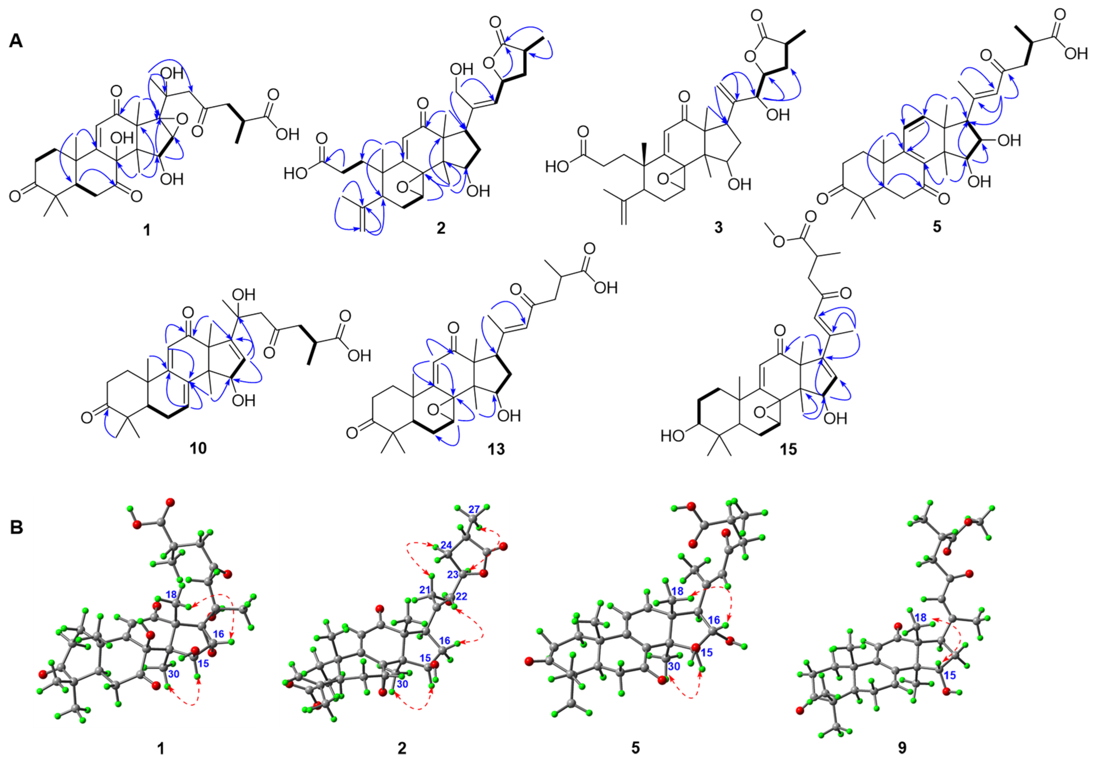
Figure 1. Structures and classifications of isolates from G. applanatum (red: new compounds).
Jof 08 00331 g001
Figure 2. (A) Selected HMBC (H→C) and 1H-1H COSY (H Jof 08 00331 i001H) correlations of compounds 13, 5, 10, 13, and 15. (B) Selected ROESY correlations of compounds 1, 2, 5, and 9.
Jof 08 00331 g002
Figure 3. (A) X-ray crystallographic structure of 1; (B) values of δSδR of the MTPA esters of 4.
Jof 08 00331 g003
Figure 4. Effects of compounds (1, 2, 4, 5, 7, 8, 1220, 22, 24, 26, 2834, and 39) at a level of 20 μM on lipid accumulation during 3T3-L1 adipocyte differentiation (A). LiCl (20 mM) was used as a positive control. Quantification of intracellular lipids in Oil Red O-stained adipocytes (B). Cell viability of compounds 15 and 20 on 3T3-L1 pre-adipocytes when treated for 24 h with an MTS assay (C). Data are representative results from three independent experiments. Data are shown as mean ± SD (n = 3) versus undifferentiated cells (UND). (##) p < 0.01 versus undifferentiated cells (UND). (*) p < 0.05 and (**) p < 0.01 versus fully differentiated cells (CON).
Jof 08 00331 g004
Figure 5. Effects of compounds 15 and 20 on lipid accumulation in 3T3-L1 adipocytes. (A) Oil Red O staining of cells administrated with serial doses of compounds 15 and 20. (B) Quantification of intracellular lipid in Oil Red O-stained adipocytes. (C) The IC50 values of compounds 15 and 20. LiCl (20 mM) was used as a positive control. Data are representative results from three independent experiments. Data are shown as mean ± SD (n = 3), versus undifferentiated cells (UND). (##) p < 0.01 versus undifferentiated cells (UND). (*) p < 0.05 and (**) p < 0.01 versus fully differentiated cells (CON).
Jof 08 00331 g005
Figure 6. The proposed structure–activity relationship of triterpenoids from G. applanatum.
Jof 08 00331 g006
Table 1. 1H NMR spectra of compounds 18 (600 MHz, methanol-d4, J in Hz, δ in ppm).
Position12345678
11.87 dt (13.7 4.5)
2.21 dd (15.4 2.8)
1.86 m
2.20 m
1.82 m
2.16 m
1.85 m
2.17 m
1.91 m
2.29 m
1.87 m
2.16 m
2.02 m
1.51 m
1.50 m
2.02 m
22.40 m
2.95 m
2.19 m
2.36 m
2.17 m
2.34 m
2.25 m
2.40 m
2.55 m
2.74 m
2.51 m
2.71 m
1.78 m1.77 m
3 3.24 t (8.4)3.23 t (8.4)
51.74 dd (14.8 2.8)2.97 t (8.3)2.94 t (8.4)2.95 dd (9.3 7.5)2.28 m2.19 m1.66 dd (14.4 3.6)1.66 dd (14.4 3.6)
62.30 dd (15.4 2.8)
3.30 m
2.03 m1.99 m2.03 m2.44 dd (16.3 3.3)2.52 m
2.66 m
2.62 m
2.48 m
2.48 m
2.59 m
7 4.93 s4.95 s4.98 s
115.98 s6.04 s6.02 s6.04 s6.28 d (9.9)6.14 d (10.2)6.31 d (10.2)6.31 d (10.2)
12 6.59 d (9.9)6.59 d (10.2)6.64 d (10.2)6.65 d (10.2)
154.83 s3.95 d (6.5)3.91 d (6.5)3.94 d (6.4)4.20 s4.44 d (7.8)4.39 d (7.2)4.39 d (7.2)
163.36 s1.80 m
4.65 m
1.73 m
2.64 m
1.76 m
2.67 m
4.57 d (7.8)2.20 m
2.42 m
2.16 m
2.51 m
2.16 m
2.51 m
17 3.33 m3.21 dd (10.7 8.7)3.23 m2.73 m2.73 m2.87 m2.83 m
181.97 s1.30 s1.37 s1.39 s0.99 s1.01 s1.00 s0.99 s
191.65 s1.01 s1.04 s1.05 s1.14 s1.27 s1.16 s1.16 s
211.34 s4.37 s5.33 s
5.39 s
5.36 s
5.43 s
1.21 s2.23 s2.19 s2.19 s
222.66 d (14.9)
3.00 d (14.9)
5.78 d (8.6)4.47 d (5.6)4.50 d (5.6)6.36 s6.22 s6.40 s6.32 s
23 5.59 dd (8.6 6.6)4.72 m4.73 ddd (8.6 5.6 3.2)
242.62 dd (18.3 5.6)
3.07 dd (18.3 7.7)
2.19 m1.97 m
2.16 m
2.00 m
2.56 m
2.61 m
2.90 m
2.54 m
2.94 m
2.64 m
2.90 m
2.53 m
2.55 m
251.74 dd (14.8 2.8)2.82 q (7.7)2.84 m2.86 m2.74 m2.97 m2.85 m2.84 m
261.16 d (6.6)1.29 d (7.3)1.22 d (6.5)1.25 d (6.5)1.18 d (7.0)1.19 d (7.2)1.17 d (7.2)1.17 d (7.2)
281.14 s4.80 s
4.98 s
4.77 s
4.95 s
4.77 s
4.95 s
1.12 s1.13 s1.00 s1.00 s
291.07 s1.80 s1.78 s1.80 s1.10 s1.14 s0.90 s0.90 s
300.74 s1.06 s1.01 s1.04 s1.10 s0.94 s0.95 s0.95 s
OMe 3.65 s 3.68 s3.65 s
Table 2. 13C NMR spectra of compounds 18 (150 MHz, methanol-d4).
Position12345678
137.6 CH237.7 CH237.7 CH237.5 CH236.5 CH235.6 CH236.3 CH236.3 CH2
235.1 CH230.4 CH230.5 CH230.5 CH235.0 CH234.0 CH228.1 CH228.1 CH2
3215.8 C177.4 C177.4 C175.6 C217.1 C214.3 C78.3 CH78.3 CH
448.7 C146.5 C146.5 C146.3 C51.9 C46.9 C39.7 C39.8 C
548.5 CH45.0 CH45.3 CH45.3 CH50.7 CH49.3 CH50.8 CH50.8 CH
635.8 CH228.2 CH228.1 CH228.1 CH237.5 CH236.8 CH237.0 CH237.0 CH2
7205.9 C64.7 CH64.8 CH65.0 CH203.0 C210.4 C204.3 C204.3 C
885.5 C67.4 C67.5 C67.5 C135.8 C134.9 C135.7 C135.7 C
9165.0 C167.2 C167.8 C167.3 C161.4 C160.6 C164.5 C164.5 C
1041.0 C45.0 C45.2 C45.3 C39.2 C37.9 C39.9 C39.7 C
11127.3 CH130.7 CH130.6 CH130.6 CH123.2 CH122.0 CH123.3 CH123.3 CH
12204.0 C206.5 C207.3 C207.0 C147.4 CH147.5 CH148.3 CH148.3 C
1362.3 C60.8 C60.4 C60.4 C52.2 C50.2 C51.4 C51.4 C
1447.0 C54.4 C54.4 C54.4 C48.0 C52.5 C53.9 C53.8 C
1580.0 CH77.4 CH77.2 CH77.2 CH84.9 CH75.0 CH76.5 CH76.6 CH
1661.2 CH41.3 CH243.7 CH243.5 CH283.6 CH36.2 CH237.2 CH237.2 CH2
1771.4 C43.3 CH41.6 CH41.6 CH58.7 CH48.5 CH49.7 CH49.7 C
1824.6 CH320.2 CH320.2 CH320.2 CH319.2 CH317.1 CH320.2 CH321.2 CH3
1919.9 CH324.2 CH323.4 CH323.4 CH321.1 CH319.5 CH318.0 CH318.0 CH3
2073.4 C144.1 C148.7 C148.7 C156.5 C157.3 C158.8 C158.4 C
2127.8 CH361.8 CH2117.6 CH2117.5 CH219.7 CH321.4 CH321.4 CH321.3 CH3
2253.9 CH2130.8 CH76.5 CH76.5 CH126.9 CH124.8 CH126.0 CH126.4 CH
23209.9 C76.8 CH80.9 CH80.9 CH200.8 C198.3 C200.6 C201.3 C
2448.7 CH238.3 CH232.2 CH232.2 CH248.7 CH247.7 CH248.8 CH249.0 CH2
2535.8 CH35.7 CH35.3 CH35.3 CH36.2 CH34.8 CH36.6 CH36.6 CH
2617.4 CH315.9 CH316.3 CH316.0 CH317.5 CH317.1 CH317.4 CH317.7 CH3
27179.7 C182.7 C183.1 C183.2 C179.7 C176.4 C178.1 C180.8 C
2822.3 CH3115.9 CH2115.9 CH2115.9 CH221.0 CH325.6 CH327.8 CH327.8 CH3
2924.9 CH323.5 CH323.4 CH323.4 CH325.9 CH320.4 CH315.7 CH315.7 CH3
3030.7 CH321.3 CH321.4 CH321.4 CH319.7 CH320.6 CH321.2 CH320.2 CH3
OMe 52.3 CH3 51.8 CH352.3 CH3
Table 3. 1H NMR spectra of compounds 916 (600 MHz, J in Hz, δ in ppm).
Position9 a10 b11 b12 b13 b14 b15 b16 a
12.27 m
1.86 m
1.83 m
2.39 m
1.83 m
2.38 m
2.80 m
2.95 m
1.82 m
2.26 m
1.79 m
2.23 m
1.81 m
2.23 m
1.80 m
1.80 m
22.42 m
2.78 m
2.37 m
2.93 m
2.37 m
2.94 m
2.53 m
2.59 m
2.31 m
2.95 m
2.27 m
2.94 m
2.28 m
2.93 m
2.36 m
2.89 m
51.65 m 1.78 dd (12.8 3.1)1.76 dd (11.4 3.602.36 dd (15.0 3.0)1.29 dd (12.4 5.9)1.67 dd (12.6 6.0)1.66 m1.56 dd (12.66.0)
61.11 m
2.28 m
2.33 m
2.49 m
2.34 m
2.53 m
2.54 m
2.68 m
2.25 m2.25 m2.30 m2.16 m
76.50 br s6.62 m6.63 d (7.2) 3.96 d (5.7)3.93 d (5.4)3.90 d (3.6)4.45 d (5.4)
115.65 s5.74 s5.74 s 6.04 s6.01 s5.98 s6.03 s
12 3.70 s
154.58 t (8.3)4.47 d (3.0)4.47 d (3.0)4.40 d (7.8)4.08 d (6.0)4.05 d (6.0)4.35 d (3.0)
161.80 m
2.48 m
5.75 d (3.0)5.74 overlapped2.07 m
2.43 m
1.94 m
2.43 m
1.91 m
2.42 m
6.20 d (3.0)5.66 s
173.26 m 3.28 t (9.6)3.24 dd (10.7 7.3)3.21 dd (10.8 7.8)
180.80 s1.54 s1.54 s−0.096 s1.46 s1.43 s1.90 s 1.76 s
191.28 s1.39 s1.40 s1.31 s1.47 s1.45 s1.47 s1.44 s
20
212.22 s1.42 s1.40 s2.24 s2.28 s2.25 s2.30 s1.49 s
226.37 s2.82 d (14.2)
3.01 d (14.2)
2.79 d (14.4)
2.79 dd (14.4)
6.31 s6.56 s6.52 s6.50 s2.92 d (15.0)
2.98 d (15.0)
242.54 m
2.94 m
2.66 dd (18.4 5.2)
3.02 m
2.79 dd (18.6 5.4)
3.02 dd (18.6 8.4)
2.54 m
2.94 m
2.62 m
2.92 m
2.62 m
2.88 m
2.58 m
2.93 m
3.04 m
2.59 dd (18.0 5.4)
252.93 m2.80 m2.81 m1.19 m2.89 m2.89 m2.89 m2.89 m
261.17 d (6.7)1.15 d (7.2)113 d (7.2)1.19 d (7.2)1.20 d (7.0)1.16 d (7.2)1.20 d (7.0)1.15 d (7.2)
281.11 s 1.20 s1.20 s1.15 s1.09 s1.09 s1.10 s1.11 s
291.15 s1.11 s1.06 s1.13 s1.13 s1.13 s1.38 s1.12 s
301.08 s1.06 s3.63 s1.24 s0.96 s0.96 s1.00 s1.28 s
OMe3.68 s 3.56 s3.69 s 3.65 s3.63 s3.65 s
a Measured in CDCl3; b measured in methanol-d4.
Table 4. 13C NMR spectra of compounds 916 (150 MHz).
Position9 a10 b11 b12 b13 b14 b15 b16 a
135.6 CH236.7 CH236.8 CH235.1 CH238.4 CH238.4 CH238.4 CH237.2 CH2
234.2 CH235.4 CH235.4 CH234.3 CH234.9 CH234.9 CH234.8 CH233.8 CH2
3214.8 C217.2 C217.2 C214.9 C216.4 C216.4 C216.5 C213.6 C
447.0 C48.4 C48.8 C47.1 C48.7 C48.5 C48.8 C47.8 C
549.5 CH51.1 CH51.2 CH49.8 CH50.7 CH50.7 CH51.2 CH49.9 CH
623.9 CH225.2 CH225.2 CH237.6 CH222.6 CH222.6 CH222.4 CH221.6 CH2
7130.9 CH135.5 CH135.5 CH203.4 C59.7 CH59.7 CH58.8 CH57.1 CH
8139.8 C137.3 C137.3 C150.2 C65.7 C65.8 C64.4 C59.0 C
9161.0 C165.5 C165.6 C151.9 C162.2 C162.3 C163.0 C165.0 C
1038.0 C39.6 C39.7 C39.3 C39.0 C39.0 C38.6 C38.2 C
11118.2 CH118.3 CH118.3 CH203.9 C127.6 CH127.6 CH127.2 CH125.0 CH
12202.5 C206.5 C206.5 C79.6 CH204.6 C204.6 C201.9 C200.6 C
1352.3 C64.0 C64.0 C52.9 C59.4 C59.4 C63.1 C61.8 C
1457.8 C54.3 C54.3 C50.0 C51.3 C51.3 C50.0 C54.4 C
1572.9 CH79.3 CH79.4 CH77.5 CH79.0 CH79.0 CH79.9 CH202.7 C
1636.5 CH2127.5 CH127.6 CH34.2 CH238.2 CH238.2 CH2134.9 CH124.4 CH
1745.7 CH158.9 C159.0 C46.9 CH49.6 CH49.6 CH148.8 C181.9 C
1817.8 CH329.0 CH329.1 CH318.6 CH320.3 CH320.3 CH327.0 CH330.9 CH3
1921.3 CH321.5 CH321.5 CH319.0 CH321.0 CH321.0 CH321.2 CH320.8 CH3
20157.6 C72.6 C72.6 C157.5 C159.0 C159.2 C156.0 C72.6 C
2120.4 CH329.0 CH329.1 CH320.3 CH321.5 CH321.5 CH317.5 CH329.1 CH3
22125.7 CH54.6 CH254.7 CH2125.1 CH127.5 CH127.5 CH127.1 CH52.7 CH2
23198.3 C209.8 C209.7 C198.6 C201.0 C201.0 C201.7 C206.3 C
2447.7 CH248.8 CH248.7 CH247.9 CH248.7 CH248.7 CH248.9 CH247.8 CH2
2534.7 CH35.8 CH35.9 CH35.0 CH36.3 CH36.3 CH36.3 CH34.5 CH
2617.0 CH317.4 CH317.3 CH316.9 CH317.4 CH317.4 CH317.5 CH317.0 CH3
27176.4 C179.7 C178.2 C176.7 C179.7 C178.2 C180.1 C176.2 C
2825.2 CH322.9 CH325.5 CH326.9 CH324.9 CH324.9 CH324.9 CH324.5 CH3
2922.3 CH325.2 CH322.9 CH320.4 CH322.3 CH322.3 CH322.5 CH322.1 CH3
3018.1 CH329.0 CH329.1 CH327.4 CH322.5 CH322.5 CH324.9 CH325.9 CH3
OMe51.8 CH3 52.2 CH352.0 CH3 52.3 CH352.0 CH351.9 CH3
a Measured in CDCl3; b measured in methanol-d4.
Publisher’s Note: MDPI stays neutral with regard to jurisdictional claims in published maps and institutional affiliations.

