Article

The Importance of Nitric Oxide as the Molecular Basis of the Hydrogen Gas Fumigation-Induced Alleviation of Cd Stress on Ganoderma lucidum

1
Laboratory Center of Life Sciences, College of Life Sciences, Nanjing Agricultural University, Nanjing 210095, China
2
Center of Hydrogen Science, Shanghai Jiao Tong University, Shanghai 200240, China
3
Microbiology Section, Botany Department, Faculty of Science, Tanta University, Tanta 31527, Egypt
4
State Key Laboratory for Crop Genetics and Germplasm Enhancement, National Center for Soybean Improvement, Nanjing Agricultural University, Nanjing 210095, China
5
Key Laboratory of Crop Physiology and Ecology, Ministry of Agriculture, Nanjing Agricultural University, Nanjing 210095, China
6
Botany and Microbiology Department, Faculty of Science, Benha University, Benha 13511, Egypt
7
Department of Food Science and Engineering, College of Food Science and Technology, Nanjing Agricultural University, Nanjing 210095, China
8
College of Animal Science and Technology, Nanjing Agricultural University, Nanjing 210095, China
9
Key Laboratory of Monitoring and Management of Crop Diseases and Pest Insects, Nanjing Agricultural University, Nanjing 210095, China
10
Department of Food Science, Faculty of Agriculture, Zagazig University, Zagazig 44511, Egypt
*
Author to whom correspondence should be addressed.
J. Fungi 2022, 8(1), 10; https://doi.org/10.3390/jof8010010
Submission received: 21 November 2021 / Revised: 19 December 2021 / Accepted: 20 December 2021 / Published: 23 December 2021
(This article belongs to the Special Issue Signal Transductions in Fungi)

Abstract

:
Whether or not hydrogen gas (H2) can reduce cadmium (Cd) toxicity in Ganoderma lucidum has remained largely unknown. Here, we report that Cd-induced growth inhibition in G. lucidum was significantly alleviated by H2 fumigation or hydrogen-rich water (HRW), evaluated by lower oxidative damage and Cd accumulation. Moreover, the amelioration effects of H2 fumigation were better than of HRW in an optimum concentration of H2 under our experimental conditions. Further results showed that H2-alleviated growth inhibition in G. lucidum was accompanied by increased nitric oxide (NO) level and nitrate reductase (NR) activity under Cd stress. On the other hand, the mitigation effects were reversed after removing endogenous NO with its scavenger cPTIO or inhibiting H2-induced NR activity with sodium tungstate. The role of NO in H2-alleviated growth inhibition under Cd stress was proved to be achieved through a restoration of redox balance, an increase in cysteine and proline contents, and a reduction in Cd accumulation. In summary, these results clearly revealed that NR-dependent NO might be involved in the H2-alleviated Cd toxicity in G. lucidum through rebuilding redox homeostasis, increasing cysteine and proline levels, and reducing Cd accumulation. These findings may open a new window for H2 application in Cd-stressed economically important fungi.

Graphical Abstract

1. Introduction

Ganodermalucidum is one of the large basidiomycetous fungi with a highly significant medicinal importance [1]. Nevertheless, the development of its commercial value is impeded by the limitations of its basic biological research. Cadmium (Cd) is one of the most harmful heavy metals due to its carcinogenic and neurotoxic effects on animals and humans [2]. Due to its high solubility in water, it can readily enter into different life forms, including microbes, plants, and animals [3]. Excess Cd accumulation inside living organisms can disrupt physiological processes such as protein metabolism, respiration, and nutrients uptake [4], as it induces the production of reactive oxygen species (ROS) [5,6]. ROS, in turn, oxidize vital biological molecules such as proteins, lipids, and nucleic acids, leading to membrane damage, inactivation of enzymes, and lipid peroxidation [5]. The mushrooms’ cultivation medium mainly consists of rice and wheat straw that might grow in soil contaminated with Cd [7]. So, G. lucidum accumulates Cd while consuming contaminated straw, leading to reduced mycelial growth and biomass. Furthermore, the contaminated mushrooms are finally eaten by humans, and their health is adversely affected.
Hydrogen gas (H2) application, commonly in the form of hydrogen-rich saline or hydrogen-rich water (HRW), has been utilized in medicine for the treatment of many diseases [8,9,10]. The first study was conducted on animals, wherein H2 efficacy was explained by its ability to reduce reactive oxygen species (ROS), mitigating the oxidative damage caused by ischemia/reperfusion injury in the rat brain [8]. Numerous studies followed this study in the medical field. Meanwhile, H2 was extensively applied in agriculture, mainly in the form of HRW, for enhancing plant tolerance to abiotic stresses such as drought [11] and heavy metals [12,13,14,15,16], as well as regulating plant growth and development [17,18,19]. In contrast to animals and plants, few studies concerning the effects of H2 on fungi were documented. The first study on fungi reported that HRW could ameliorate acetic acid stress by modulating the impact of ROS balance on morphology, growth, and secondary metabolism via enhancing glutathione peroxidase in G. lucidum [20]. Another study on the mushroom Hypsizygus marmoreus reported that, though strengthening the antioxidant system, HRW could alleviate the toxicities of NaCl, CdCl2, and H2O2 [21], improve the post-harvest quality, and reduce rot incidence [22].
The direct supply of H2 in its gaseous form, called fumigation, was rarely applied and was confined to H2-modified atmosphere studies, targeting fresh food storage [23,24,25,26] as well as soil treatment to enhance the plant growth via improving the rhizosphere micro-environment [27,28,29]. The concentration of H2 by direct gas supply is higher than when it is bubbled into water (HRW), and the hydrogen solubility and retention time in water are short, so HRW preparation technology needs improvements, particularly in agricultural applications [30]. Regarding mushrooms, there are additional problems concerning the HRW delivery method, such as the necessity for optimum pH and humidity levels, which are critical factors for fungal growth [31,32]. Therefore, HRW is an unsuitable form of H2 delivery for fungi, particularly for solid-state growth, because the water content in HRW would interfere with the humidity content and may alter the pH value in the culture environment. Moreover, the application of HRW on the solid-state culture is relatively inconvenient, unlike the H2 gas fumigation technique. Therefore, our research aims to overcome the possible HRW problems by directly utilizing H2 gas, which can spread rapidly and evenly in the cultivation area. Since H2 is neither flammable nor explosive at concentrations less than 4% [33], we examined different H2 gas fumigation concentrations within the safe range for its ability to alleviate the Cd-induced mycelial growth inhibition and regulate Cd accumulation in G. lucidum and compared its efficacy to the HRW’s.
Nitric oxide (NO) is another key signaling molecule that regulates several physiological and biochemical responses to abiotic stresses in animals, plants, and fungi. In fungi, it was reported that 6 h heat treatment raised the concentration of NO in Trichoderma harzianum mycelia [34]. Additionally, sodium nitroprusside (SNP), the NO-releasing compound, relieved the oxidative damage caused by heat stress in Pleurotus eryngii var. tuoliensis [35] and alleviated copper toxicity in Saccharomyces cerevisiae [36]. As a second messenger, NO was confirmed as a participant in many key signaling pathways in animals and plants. However, its role in microorganisms is still largely unknown [37].
The enhancement of the antioxidant system, and hence the regulation of redox homeostasis, is a well-known mechanism of the H2 alleviation effect under stresses in plants and fungi [30]. It was confirmed that the H2-induced NO acts upstream of the antioxidant system in higher plants under different stresses, such as drought [38] and Cd exposure [39], and participates in root organogenesis [18,40]; however, the possible link between H2 and NO in fungi is still unidentified. Thus, in the present study on the fungus G. lucidum, the early minute-scale time course of the endogenous H2 and NO signals was observed under Cd stress, and we found that H2 signal preceded NO signal. This gave us a clue that NO may be induced by H2 in fungi. So, exogenous H2 and/or NO were added under Cd stress to mimic the physiological effects of these molecules and examine their possible link through their effects on phenotype and physiology. Nitrate reductase (NR) is the well-known enzyme responsible for NO production in plants and fungi [41,42]. Additionally, it was reported that NO synthesis was regulated by NR during development in Aspergillus nidulans [43]. Therefore, the NO scavenger, cPTIO, and the NR inhibitor, sodium tungstate (Na2WO4), were administered after H2 in our study to investigate the NO’s involvement in the H2-induced responses.
Cysteine (Cys) was demonstrated to effectively reduce Cd toxicity in Arabidopsis [44]. Previous studies suggested the role of Cys in the alleviation of stresses via the enhancement of the glutathione (GSH) system and the stimulation of cell proliferation in humans [45,46]. The availability of Cys is a limiting factor for glutathione biosynthesis [47,48]. GSH is a major non-enzymatic antioxidant and precursor of phytochelatins, which have metal binding affinity [49]. Cys could enhance chromium tolerance by enhancing GSH generation and heavy metal chelation in Arabidopsis [50]. Recently, Cys has been reported to act downstream of NO, mitigating chromium toxicity in tomato seedlings [51]. The report on fungi demonstrated that exogenous Cys treatment participated in the regulation of morphogenesis and the mitochondrial activity of Histoplasma capsulatum [52]. Similarly, proline is another significant amino acid that was revealed to participate in the stress response of different organisms [53]. It was reported that proline accumulation was enhanced by adding exogenous NO under osmotic stress in wheat seedlings [54]. However, the corresponding role of cysteine and proline in fungi is still unclear.
Previous research suggested a link among H2, NO, some amino acids such as cysteine and proline, as well as their effects on stress alleviation. Thus, in this study, we examined the role of NO in H2-alleviated Cd stress in G. lucidum and further explored the mediation role of cysteine and proline. More importantly, it provides new ideas for improving the production of economically important fungi.

2. Materials and Methods

2.1. Chemicals

Unless stated otherwise, all the chemicals used in this study were purchased from Sigma-Aldrich (St Louis, MO, USA). Cadmium chloride (CdCl2, CAS 10108-64-2) was purchased from Shanghai Aladdin Biochemical Technology Co., Ltd. The concentrations of the following chemicals were determined in pilot experiments. Hydrogen gas (purity ≥ 99.99%) was prepared by a generator (SHC-300, Saikesaisi Hydrogen Energy Co., Ltd., Jinan, Shandong, China). The chemicals utilized in our treatments were: sodium tungstate (Na2WO4; 500 μM, CAS 10213-10-2, purity 99%), an inhibitor of nitrate reductase, the enzyme responsible for NO formation [55]; sodium nitroprusside (SNP; 500 μM, CAS 13755-38-9, purity 98%), a well-known NO-releasing compound [56,57]; 2-(4-carboxyphenyl)-4,4,5,5-tetramethylimidazoline-1-oxyl-3-oxide potassium salt (cPTIO; 500 μM, CAS 148819-94-7; purity 97%), a well-known NO scavenger [41,58,59]; and proline (10 μM, CAS 147-85-3, purity 99%), an antioxidant. Moreover, cysteine was used at 300 µM and GSH at 325 µM. The GSH synthesis inhibitor DL-buthionine-[S,R]-sulfoximine (BSO) was used at 2.5 mM. To ensure NO’s role, old SNP was used as a negative control and was prepared by keeping 500 μM SNP solution in an open tube in the light for 10 d to eliminate the entire NO [60].

2.2. Preparation of Hydrogen-Rich Water (HRW)

The procedures were carried out according to the previously mentioned method [60], with slight modifications. The H2 gas generator (SHC-300, Saikesaisi Hydrogen Energy Co., Ltd., Shandong, China) was utilized to produce hydrogen gas (99.99%, v/v). To prepare saturated HRW, the H2 gas generated was bubbled into 1000 mL of sterile water at a rate of 150 mL per minute for 30 min. The concentration of H2 at saturation point was 0.8 mM. Subsequently, 10, 25, 50, 75, or 100 mL of the saturated HRW was immediately mixed with 90, 75, 50, 25, or 0 mL ddH2O, respectively, to prepare 10%, 25%, 50%, 75%, or 100% HRW concentrations (v/v). Freshly prepared HRW was pre-sterilized through a 0.22 µM membrane before application to mycelia. The mycelia plates were treated with 15 mL for each treatment, and 15 mL pre-sterilized ddH2O was added to the control plates.

2.3. Fungal Materials, Growth Condition, and Screening of Cd Inhibitory Concentrations

The wild-type strain (Ganoderma lucidum HG) was obtained from the Edible Fungi Institute, Shanghai Academy of Agricultural Science, China. Potato dextrose agar (PDA) was used as a culture medium for the strains of G. lucidum incubated at 28 °C in the dark for 7 d (solid seed). From the solid seed, 6 mm-diameter mycelial discs were inoculated into solid complete yeast medium (CYM) plates (2% glucose, 1% maltose, 0.05% MgSO4·7H2O, 0.2% yeast extract, 0.2% tryptone, 0.46% KH2PO4, 1% agar, initial pH = 5.5), supplied with CdCl2 at different concentrations, 0, 1, 1.5, 1.75, and 2 mM. The cultures were incubated in the dark at 28 °C for 8 d; then, the mycelial growth diameters were immediately measured. Meanwhile, eight mycelial discs, each of 6 mm diameter, were cut from the same growth diameter at the edge of the solid seed colony. Each set of 8 mycelial discs was grown on 100 mL liquid CYM media containing different concentrations of CdCl2 0, 50, 100, 200, 300, and 400 μM. The cultures were incubated in shaking incubator in the dark at 28 °C for 8 d. Afterward, the mycelia were harvested and washed with distilled water and then oven-dried at 60 °C to a constant biomass, and the mycelial biomass dry weight values were instantly determined.

2.4. Experimental Design: Response of G. lucidum to Different Concentrations of H2 Fumigation

G. lucidum mycelia were grown on control and stressed CYM Petri-plates, which were let open in pre-sterilized plastic containers (10.5 L, Lock & Lock, Suzhou, China), sealed with vaseline. Fumigation of G. lucidum with H2 was carried out in the plastic containers according to the previous method [23], with slight modifications. An airbag with a sampling plug was filled with H2 from the H2 gas generator. Gas syringes (Hamilton Co., Reno, NV, USA) were used for H2 gas sampling from the airbag and then were used to inject H2 through 0.22 µM sterilization filter that ends with a sterilized needle into the fumigation containers. Mycelia were incubated in a hydrogen-containing atmosphere at 28 ± 1 °C for 24 h. Another set of non-fumigated mycelia was incubated under the same conditions as control.
Then, 6 mm-diameter mycelial discs were cut from a seven-day-old G. lucidum (solid seed), and each disc was inoculated into one of the solid CYM plates. Then, the culture plates were aseptically transferred into the fumigation containers and grown in the dark for 2 days at 28 °C. On the 3rd day, mycelia were either pre-treated with different H2 fumigation concentrations, 1%, 2%, 3%, 5%, or 10%, or with 50% HRW, each for 24 h, twice, once every 12 h, or not treated with either hydrogen forms and used as control (Con). On the 4th day, 6 mm-diameter mycelial discs from H2- or HRW-treated or non-treated mycelia were cut and grown on plain solid CYM (−Cd) or 1.75 mM CdCl2-containing solid CYM (+Cd) for 8 d at 28 °C in the dark; then, the diameters of fungal growth were measured. Simultaneously, the effect of H2 on the mycelial biomass dry weight was determined by cutting 6 mm-diameter mycelial discs on the 4th day from H2- or HRW-treated or non-treated mycelia from the same growth diameter. Then, each set of 8 mycelial discs was inoculated into 100 mL plain liquid CYM (−Cd) or 200 μM CdCl2-containing liquid CYM (+Cd) and grown for 8 d in shaking incubator at 28 °C in the dark. Afterward, the mycelia were harvested and washed with distilled water and then oven-dried at 60 °C to a constant biomass. The biomass dry weight values were immediately measured. Additionally, mycelial samples were collected and immediately used or stored at −80 °C for further analysis.
To address the possible oxygen deficiency that might occur in the fumigation containers, in our preliminary trials, the mycelial growth in the plates inside the containers was compared to that in parafilm-sealed plates that were not enclosed inside containers, under the same incubation conditions, and we found no phenotypic difference between them. Additionally, the containers were recurrently opened under sterilized conditions for 15 min across a 12 h interval. Moreover, 10 g calcium hydroxide Ca(OH)2, the CO2 absorbent, was placed in the containers to absorb the excess CO2.

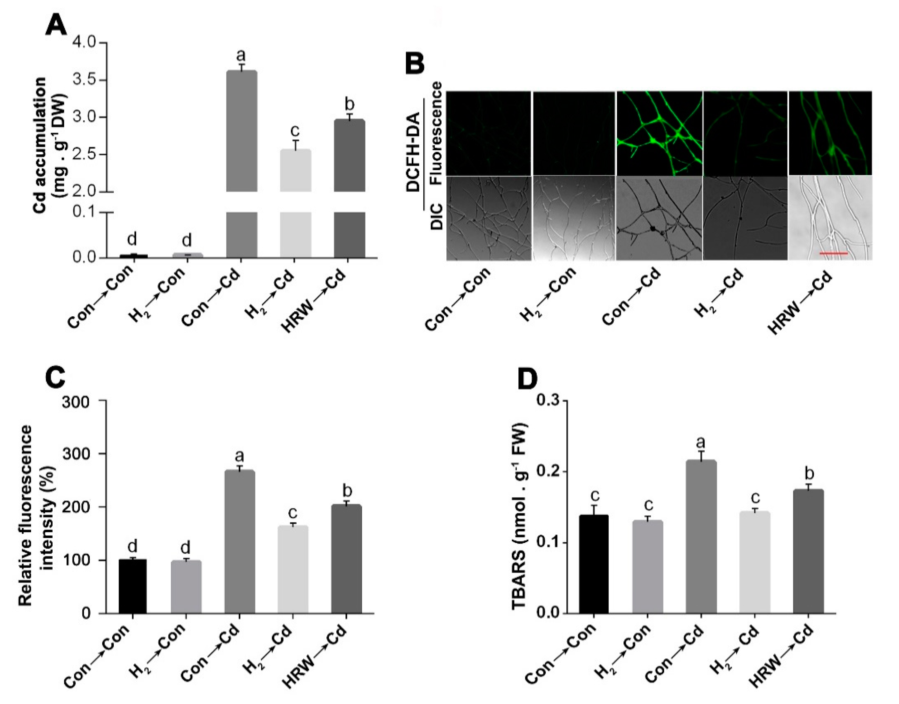
2.5. Determination of Intracellular Cadmium Content

Cd content in the fungal mycelia was assessed according to the previous method [61], with a slight modification. Briefly, mycelia were washed with distilled water to remove the non-specific bound Cd. Then, mycelia were dried in the oven at 60 °C till constant weight. Afterward, 0.2 g dry samples were ground to a fine powder and then mixed with 6 mL of HNO3 and 2 mL of H2O2. The mixture was digested via microwave digestion (Milestone Ethos T). The digested sample was transferred into a quartz crucible at 60 °C for 45 min to evaporate excess reagents. ddH2O was used to complete the sample volume to 10 mL, and inductively coupled plasma optical emission spectroscopy (ICP-OES; PerkinElmer Optima 2100DV) was utilized to determine the Cd content in the samples. Three replicas for each sample were prepared. The parameters were set according to the previous method [61]. A range of Cd concentrations was measured to generate a standard curve for Cd.

2.6. Effect of H2 Fumigation on Reactive Oxygen Species (ROS) and TBARS Contents

The detection of ROS fluorescence intensity was carried out according to the previous method [62]. The pre-sterilized glass coverslips that had been inserted in the solid CYM media and had mycelial growth on their surface were picked and stained with 2, 7 dichlorodihydrofluorescein diacetate (DCHF-DA; 10 mM phosphate-buffered saline (PBS) (pH 7.5)) for 20 min in the dark. Then, the hyphae were gently washed with 10 mM PBS (pH 7.5) to remove the excess stain. Confocal laser microscope (TCS SP2; Leica) at 488 nm emission wavelength was utilized to visualize the ROS fluorescence produced by an argon laser and a 525 to 530 nm filter. The fluorescence intensity was analyzed by ZEN lite (Zeiss software).
TBARS content, the indicator of lipid peroxidation, was measured using the previously described method [63], with a slight modification. A small amount of liquid nitrogen was added to 0.1 g of mycelia before grinding it. After that, 2 mL of 5% TCA (CAS 76-03-9; purity 99%) was added and then centrifuged at 10,000× g at 4 °C for 10 min. Then, 2 mL of the supernatant was collected and added to 2 mL 0.67% 2-thiobarbituric acid (TBA; CAS 504-17-6; purity 98%). The mixture was inserted into a boiling water bath and exposed to heat for 15 min. Afterward, the sample was centrifuged at 10,000× g for 10 min at 4 °C. The supernatant was collected, and its level of absorbance was quantified at 532 nm. The results obtained were corrected for non-specific absorption by deducting the reading of absorbance at 600 nm. The levels of TBARS were estimated using the extinction coefficient of 155 mM−1 cm−1.

2.7. Measurement of the Antioxidant Enzyme Activities

The following procedures were followed to determine the activities of different enzymes. Mycelial samples were frozen and ground in liquid N2. Briefly, 0.3 g of frozen mycelia were minced into a uniform consistency using liquid N2 and 5 mL of potassium phosphate-buffered solution (50 mM, pH 7.0). Then, the homogenate was centrifuged at 15,000× g for 20 min at 4 °C, and the resulting supernatant was instantly utilized for the following enzyme assays. Protein contents were measured according to Bradford method using bovine serum albumin (BSA) as a standard [64].
Catalase (CAT) activity was evaluated by determining the decomposition rate of hydrogen peroxide (H2O2) at 240 nm according to the previous procedures [65]. The assay of SOD activity was carried out according to the previous method [66] by tracking the inhibition of the photochemical reduction in nitro blue tetrazolium (NBT). One unit of SOD activity was represented as the amount of enzyme consumed to produce 50% of NBT reduction inhibition, observed at 560 nm. Glutathione Peroxidase Cellular Activity Assay Kit (Beyotime Institute of Biotechnology) was used to measure glutathione peroxidase (GPX) activity. During the assay, GPX in the supernatant oxidizes GSH to GSSG, and this reaction is coupled to the recycling of GSSG back to GSH via glutathione reductase (GR), consuming NADPH. The decrease in the absorbance of NADPH at 340 nm is the indicator of GPX activity, since the GPX is the factor limiting the rate of the coupled reactions. GR activity was detected using an assay kit (Beyotime Institute of Biotechnology) by monitoring the decrease in absorbance at 340 nm.

2.8. Detection of Endogenous H2

Endogenous H2 content in G. lucidum mycelia was measured according to the previous method [67], with some modifications. Headspace sampling of H2 was followed by gas chromatography (GC). Mycelia (0.3 g) were homogenized for 1 min, placed in a vial, and then 7 mL ddH2O, 5 mL octanol (purity 99%), and 0.5 mL sulfuric acid (5 M, purity 99.9%) were added. Afterward, nitrogen gas was added to the vial to replace the air. Afterward, the vial was instantly capped and shaken strongly by hand for 1 min. Finally, the vials were heated for 1 h at 70 °C to liberate H2 gas from the fungal mycelia and then left to cool at room temperature before GC analysis. The chromatographic system and its parameters were according to the previous method [67].

2.9. Detection of Nitrate Reductase (NR) Activity

Nitrate reductase assay was carried out according to the previous method [68]. The nitrite produced was determined by spectrophotometry at 540 nm. The results were represented as the formation of nmol of nitrite/min/mg protein.

2.10. Detection of Nitric Oxide by Confocal Laser Microscopy and Spectrophotometry

Nitric oxide content was determined by the fluorescent probe diaminofluorescein-FM diacetate (DAF-FM DA; CAS number 254109-22-3, purity ≥ 98%) according to the previous methods [43]. Staining of mycelia was carried out for 20 min with DAF-FM DA to visualize nitric oxide. The fluorescence was detected by a confocal laser microscope with excitation at 488 nm, and ZEN lite (Zeiss software) was used to analyze the fluorescence intensities. NO contents were also measured by spectrophotometric Griess reagent method, with slight modification according to [59].

2.11. Measurement of Proline, Cysteine, and Glutathione Contents

Proline contents were analyzed according to the previous procedures [69] with minor modifications. Briefly, the quantification of proline was carried out by its derivatization with phenylisothiocyanate, and a reversed-phase HPLC (HITACHID-2000, Tokyo, Japan) was used to separate the resulting derivatives. Cysteine and glutathione levels were detected according to the method previously described [70].

2.12. Determination of Gene Expression in G. lucidum

The expression levels of the indicated genes in G. lucidum were determined by RT-qPCR according to the previous procedures [71]. The sequences of the primers used in the gene analysis and the reference gene are listed in Supplementary Table S1.

2.13. Statistical Analysis

Data are the means ± SD of three independent experiments with three replicates at least for each. For data analysis, a t-test (p < 0.05) or multiple-comparison one-way analysis of variance (ANOVA) followed by Tukey’s test (p < 0.05) was chosen depending on the experiment type.

3. Results

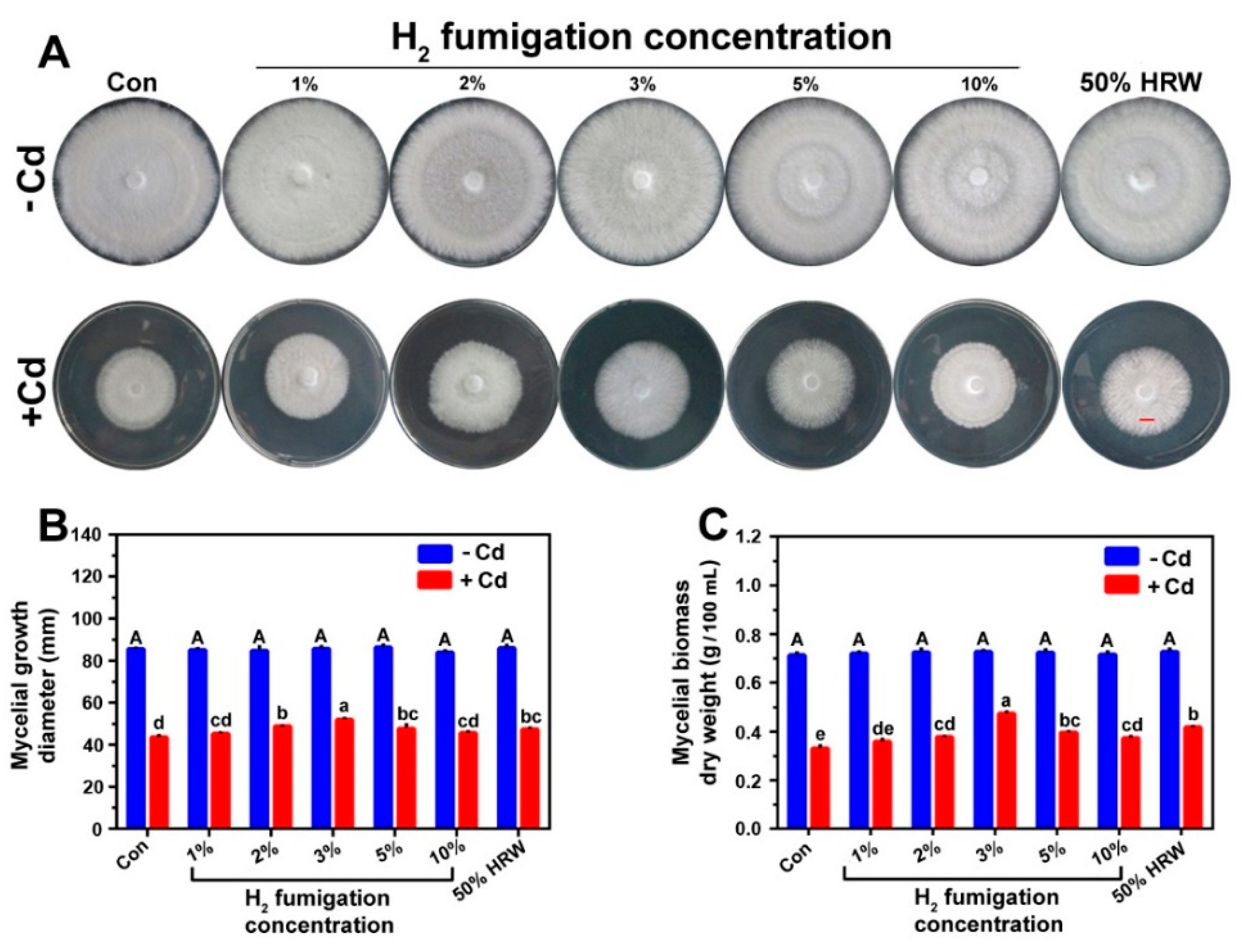
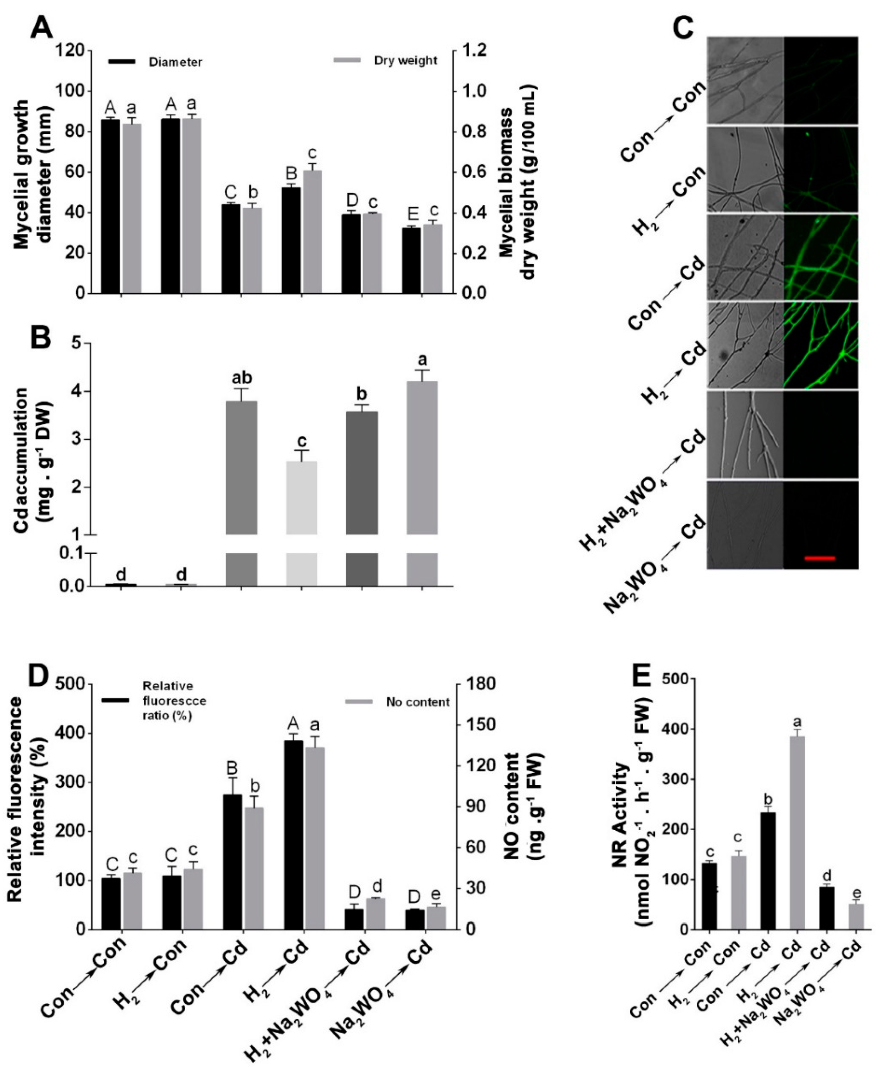
3.1. H2 Fumigation Alleviated Cd-Induced Inhibition of Mycelial Growth Diameter and Biomass Dry Weight in Ganoderma lucidum Mycelia

In our experiments, the mycelia of G. lucidum grown on solid CYM media were treated with different concentrations of CdCl2: 0, 1, 1.5, 1.75, and 2 mM (Supplementary Figure S1A). Among them, 1.75 mM CdCl2 resulted in about 50% inhibition in the mycelial growth diameter in the solid culture. However, in the liquid culture, 200 µM CdCl2 caused about 50% inhibition of the biomass dry weight (Supplementary Figure S1B). Therefore, 1.75 mM or 200 μM CdCl2 in solid or liquid cultures, respectively, were selected for all subsequent stress treatments. Additionally, our results showed that 50% HRW pre-treatment elicited the highest alleviation in diameter and dry weight of the stressed mycelia (Supplementary Figure S2). So, 50% HRW was used as a positive control in the subsequent H2 fumigation experiments.
To determine the most effective H2 fumigation concentration that significantly alleviated the Cd-caused growth inhibition, another experiment was performed in which the mycelia were grown in CYM plates and were pre-treated with the H2 fumigation concentrations 1%, 2%, 3%, 5%, or 10% or with 50% HRW (as a positive control) before exposure to Cd stress. As shown in Figure 1A–C, among the H2 concentrations, 2%, 3% (in particular), and 5% showed significant alleviation of the inhibition caused by Cd stress, whereas lower and higher concentrations brought about weaker responses. The maximum mitigation effect on mycelial growth diameter and biomass was observed at 3% H2 concentration, with a higher alleviation effect than the 50% HRW pre-treatment.

3.2. H2 Fumigation Reduced Cd Accumulation, Decreased the Production of ROS and Alleviated Lipid Peroxidation in G. lucidum under Cd Stress

As shown in Figure 2A, the mycelia accumulated Cd when treated with 1.75 mM CdCl2 for 24 h. Cd accumulation in the mycelia pre-treated with 3% H2 fumigation showed a significant decline by 29.13% compared to Cd stress alone mycelia. The reduction in Cd accumulation by H2 gas fumigation was more pronounced than that achieved by 50% HRW, suggesting the higher efficacy of H2 fumigation relative to HRW. This is in agreement with a previous study that showed that HRW significantly alleviated mycelial growth inhibition under acetic acid stress in G. lucidum but did not affect it under normal conditions [20].
The effect of H2 fumigation on the ROS production caused by Cd stress was detected by loading the mycelia with 2′, 7′ dichlorofluorescein diacetate (DCFH-DA) probe. A sharp decline was detected in the DCFH-DA fluorescence intensity in the H2-fumigated mycelia by 39.63% compared to Cd alone sample (Figure 2B,C). Moreover, the redox alleviation in the H2-fumigated mycelia was more evident than HRW-treated mycelia, which exhibited a decrease in DCFH-DA fluorescence intensity by only 24.15%. These findings suggested the advantages of H2 fumigation over HRW in maintaining redox homeostasis under Cd stress. We also noticed that H2 fumigation did not significantly alter the ROS accumulation under normal conditions.
The intracellular TBARS contents were also measured to detect the impact of Cd and H2 on lipid peroxidation. As shown in Figure 2D, despite no significant difference in the TBARS content between the H2-treated and non-treated mycelia under normal conditions, there was a marked decline in TBARS contents in the H2- and Cd-treated mycelia than those samples with only Cd stress, reaching a similar level of unstressed control.

3.3. H2 Fumigation Enhanced the Antioxidant Enzymes’ Activities and Their Corresponding Transcripts under Oxidative Stress Caused by Cd

To understand the mechanism by which H2 achieved redox homeostasis, we detected the activities of the antioxidant enzymes: catalase (CAT), superoxide dismutase (SOD), glutathione reductase (GR), and glutathione peroxidase (GPX). Our results showed that, under Cd stress, the activities of CAT, GR, and GPX were decreased by 55.40%, 46.43%, and 43.67%, respectively; however, SOD activity was increased by 24.31%. Interestingly, in the H2-pretreated Cd-stressed mycelia, all these antioxidant enzyme activities were significantly increased relative to the Cd stress alone of mycelia. Moreover, the transcript levels of the related genes were further quantified by RT-PCR. Gene expression levels of cat1/2, sod1/4, gr, and gpx showed similar tendencies to the corresponding antioxidant enzyme activities (Supplementary Figure S3).

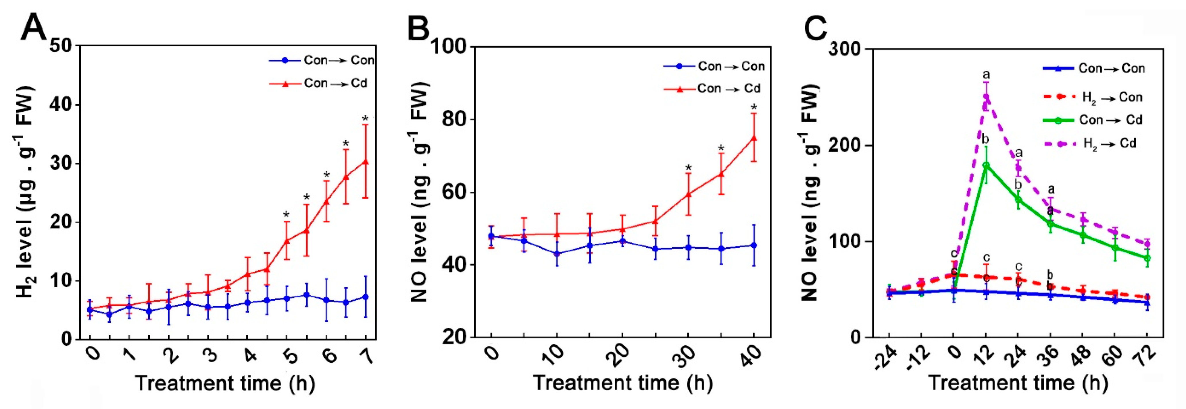
3.4. Endogenous NO Was Involved in the H2-Induced Cd Stress Tolerance

To know whether or not NO proceeded as a downstream signal of H2 in the Cd tolerance in G. lucidum, the early signals of H2 and NO were observed. The endogenous H2 content increased steadily in the Cd-treated mycelia with the time extension, and it showed a marked difference between control and Cd stress only after 5 min (Figure 3A). However, the NO content in the Con- and Cd-treated mycelia had a slight difference during the first 20 min and then increased significantly in the Cd-stressed mycelia after 30 min (Figure 3B), suggesting a possible inherent role of the endogenous H2 in enhancing NO production in G. lucidum under Cd stress. Thus, we investigated the effect of H2 fumigation on the endogenous NO content under stress and control conditions across a 4-day time course (Figure 3C), and we found that, in comparison to the control samples, the mycelia treated with Cd alone exhibited a rapid burst of NO release at 12 h and then declined until 72 h. Moreover, the Cd-induced NO production in the mycelia was markedly enhanced by H2 fumigation relative to the Cd alone treated samples. Moreover, H2-fumigation for 24 h also led to a slight NO accumulation in the control samples. These results gave us a clue about NO’s importance in the H2-mediated alleviation of Cd stress in G. lucidum.

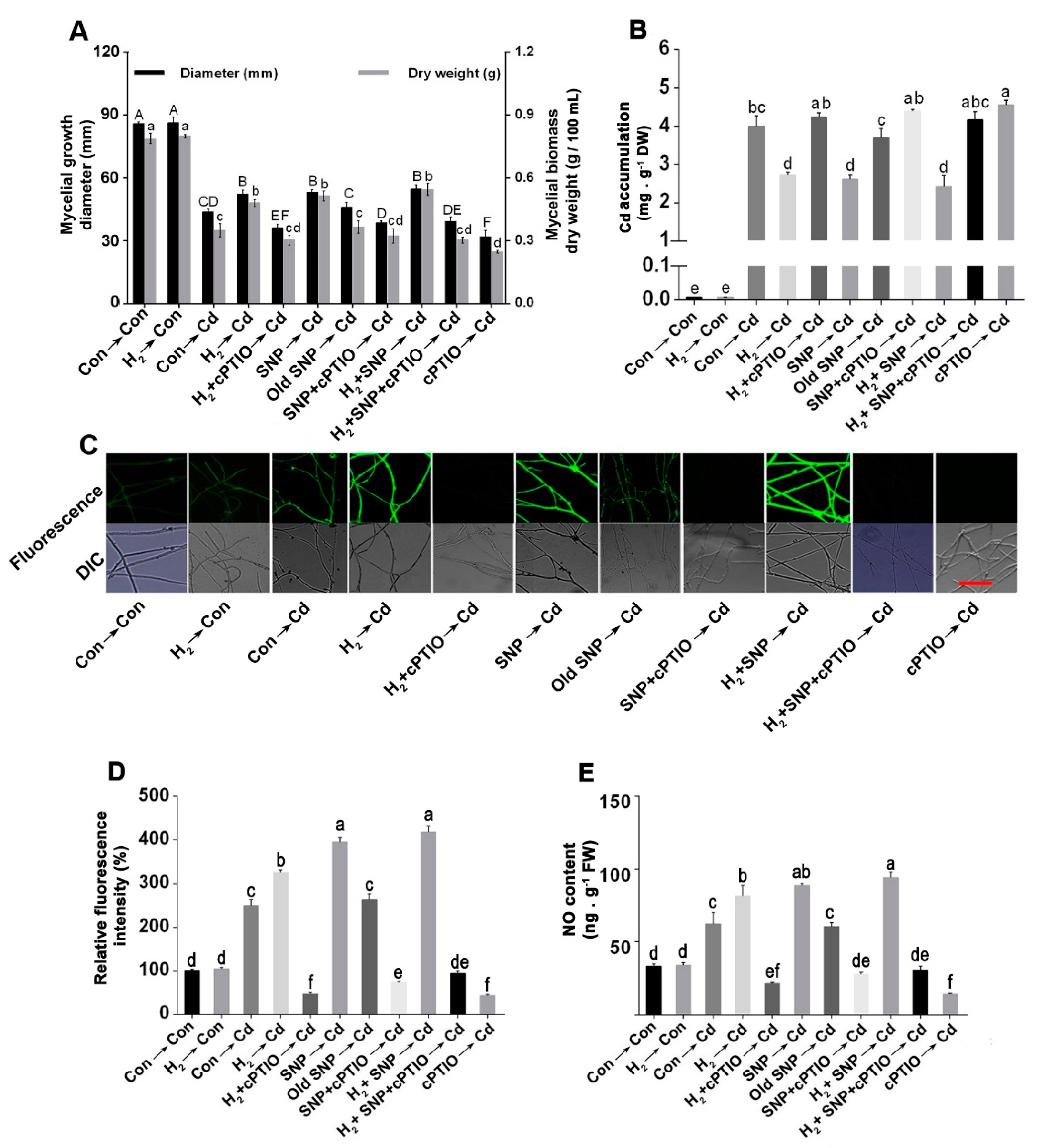
3.5. H2-Induced Alleviation of the Cd-Caused Mycelial Growth Inhibition and Excess Cd Accumulation Was Reversed by the NO Scavenger, cPTIO

To further test the possibility of NO involvement in the H2-elicited response under Cd stress, the NO scavenger cPTIO was used to eliminate the endogenous NO. Interestingly, the H2-induced responses were severely diminished by cPTIO, indicating a potential participation of NO in the H2-induced amelioration of the Cd-elicited growth inhibition. On the contrary, by adding the NO-releasing compound SNP, Cd-induced inhibition of mycelial growth diameter (Figure 4A; left) and biomass dry weight (Figure 4A; right) were ameliorated, while old SNP, the negative control of SNP, failed to induce such responses. These results demonstrated the signifi-cance of NO in the fungus tolerance against Cd stress. Moreover, further mitigation was observed when exogenous NO was administered after H2, suggesting a possible link between the two signaling molecules in the stress tolerance.
For further examination of the NO’s role in Cd stress alleviation, Cd accumulation was also analyzed (Figure 4B). A remarkable reduction in Cd accumulation was detected in the H2 and/or NO treated mycelia, and the highest alleviation was achieved under H2 and NO successive treatments. However, reversion of these responses occurred after combining treatment with cPTIO. The treatment with cPTIO alone before Cd exposure caused considerably higher Cd accumulation relative to that in the Cd alone treatment. Moreover, the administration of old SNP exhibited no significant response. These results corroborated the contribution of NO in the H2-induced reduction in Cd accumulation.
Furthermore, NO’s participation was confirmed by quantifying its contents in the mycelia by confocal laser microscopy (Figure 4C), and the respective fluorescence intensities were analyzed (Figure 4D). Relative to control mycelia, Cd-stressed mycelia exhibited higher NO contents. However, mycelia showed markedly higher NO contents when pre-treated with H2 and/or SNP under Cd stress. Furthermore, the H2 and/or NO-triggered response in the stressed mycelia was prevented by cPTIO. When old SNP was added before Cd stress, it did not alter NO level, while cPTIO pre-treatment caused a significant depletion in the NO level. Analogous results in NO content were observed using spectrophotometric Griess reagent analysis (Figure 4E).

3.6. The H2-Induced Cd Tolerance Was Linked to the NR-Dependent NO Formation

The source of NO in G. lucidum was further investigated by determining the impact of sodium tungstate (Na2WO4), an inhibitor of NR. A significant reduction in the mycelial growth diameter (Figure 5A; left) and the biomass dry weight (Figure 5A; right) was observed when Na2WO4 was applied. Interestingly, Na2WO4 addition markedly blocked the alleviation role of H2 in the Cd-elicited growth inhibition. Similarly, the H2-induced Cd accumulation decrease was reversed by Na2WO4 (Figure 5B). Na2WO4 pre-treatment alone showed higher Cd accumulation in the Cd-stressed mycelia.
The images of confocal laser microscopy exhibited that Cd- and/or H2-induced NO levels were suppressed by Na2WO4 (Figure 5C), which verified the NR source of NO under Cd and/or H2 treatments. Furthermore, results from corresponding fluorescence intensities (Figure 5D, left) and spectrophotometric analysis of NO contents (Figure 5D, right) suggested the potent inhibition of endogenous NO generation by Na2WO4 in mycelia. Furthermore, NR activity was determined (Figure 5E), and the results were consistent with NO contents. Therefore, these results demonstrate that the magnification of NO content by H2 and Cd was NR-dependent, and this further suggested that H2-triggered amelioration of Cd stress is possibly mediated by NR-related NO production. To minimize artefacts, mycelia were treated with H2, HRW, SNP, NO’s scavenger, cPTIO or NO’s inhibitor, and Na2WO4 alone or in the indicated combinations to investigate whether or not these chemicals have some effect that interferes with the response elicited by Cd stress in the combined treatment of Cd with these chemicals. This experiment showed no significant effect of either of these treatments on the mycelial growth or NO content under normal unstressed conditions (Supplementary Figure S7), consistent with our previous experiments that demonstrated no significant effect of the upstream signal (H2) under normal conditions.

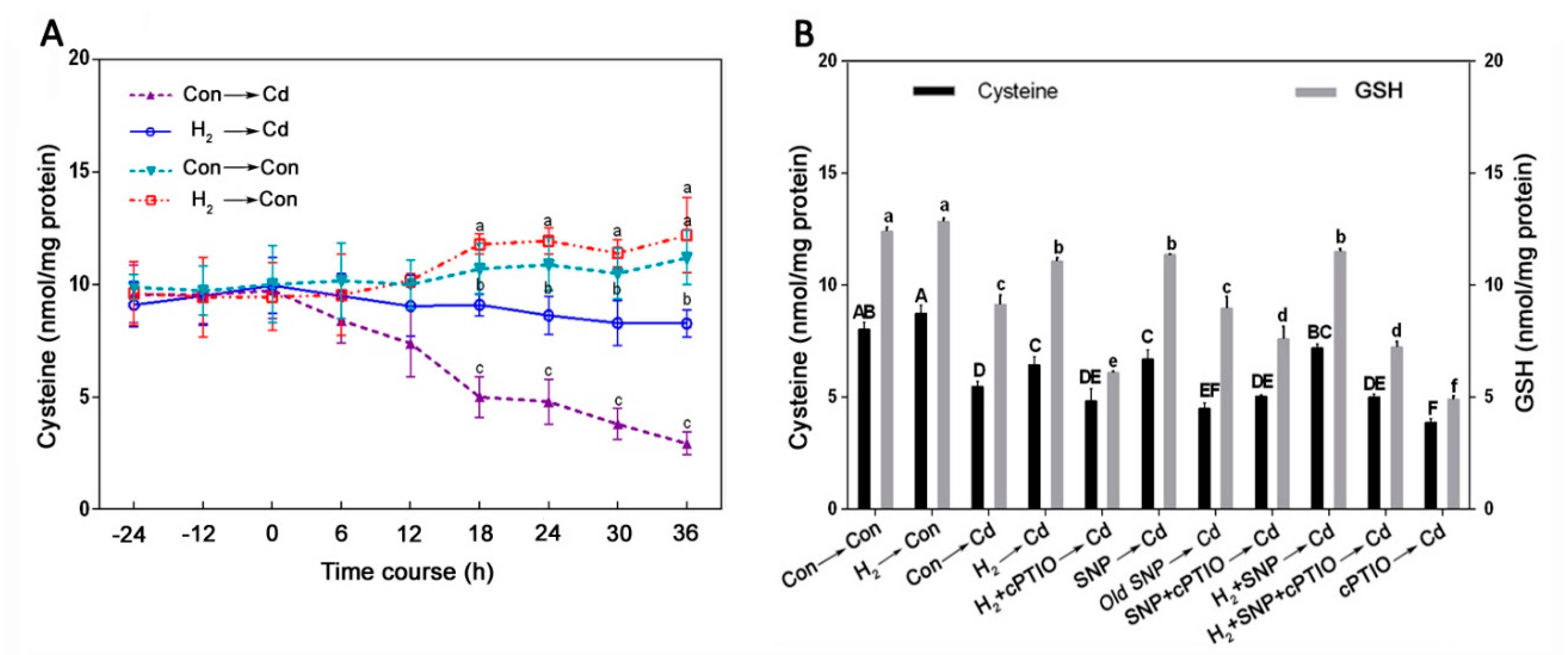
3.7. Cysteine Was Involved in the H2-Induced, NO-Mediated Cd Tolerance

To address the underlying mechanism, time-dependent cysteine (Cys) accumulation was analyzed under H2 and Cd treatments. We noticed that, under Cd alone treatment, Cys levels were swiftly reduced through 36 h of Cd stress, while H2 pretreatment rescued the Cd-elicited drop in Cys content, especially after 18 h of Cd stress (Figure 6A). The role of Cys as a downstream component of NO signal in the alleviation of plant stresses was documented [51,72]. Thus, we further examined the possible involvement of NO in the H2-induced enhancement of Cys under Cd stress. Interestingly, pre-treatment with H2 and/or SNP resulted in the recovery of the Cd-elicited Cys decline. However, these responses were prevented by the treatment with cPTIO plus H2. We also noticed that old SNP did not ameliorate Cys level, and cPTIO pre-treatment caused further depletion of Cys (Figure 6B; left). Interestingly, similar trends were observed in the GSH levels under the same treatments (Figure 6B; right).
The effect of exogenous Cys on mycelial growth under Cd stress was also investigated, and the exogenous Cys-elicited response was compared to that of exogenous GSH. Exogenous Cys mitigated the inhibition of mycelial growth caused by Cd. However, this alleviation effect was significantly reduced in the samples pre-treated with BSO. Moreover, the treatment with BSO alone resulted in further depletion in the mycelial growth (Supplementary Figure S4A). Meanwhile, the evaluation of GSH levels was also performed. GSH levels were decreased under Cd stress and could be alleviated by exogenous Cys application (Supplementary Figure S4B). In addition, Cd accumulation was also measured under exogenous Cys treatment, and we observed that exogenous Cys markedly alleviated Cd accumulation in G. lucidum. However, the effects of exogenous Cys were weakened in the mycelia pre-treated with BSO (Supplementary Figure S4B).

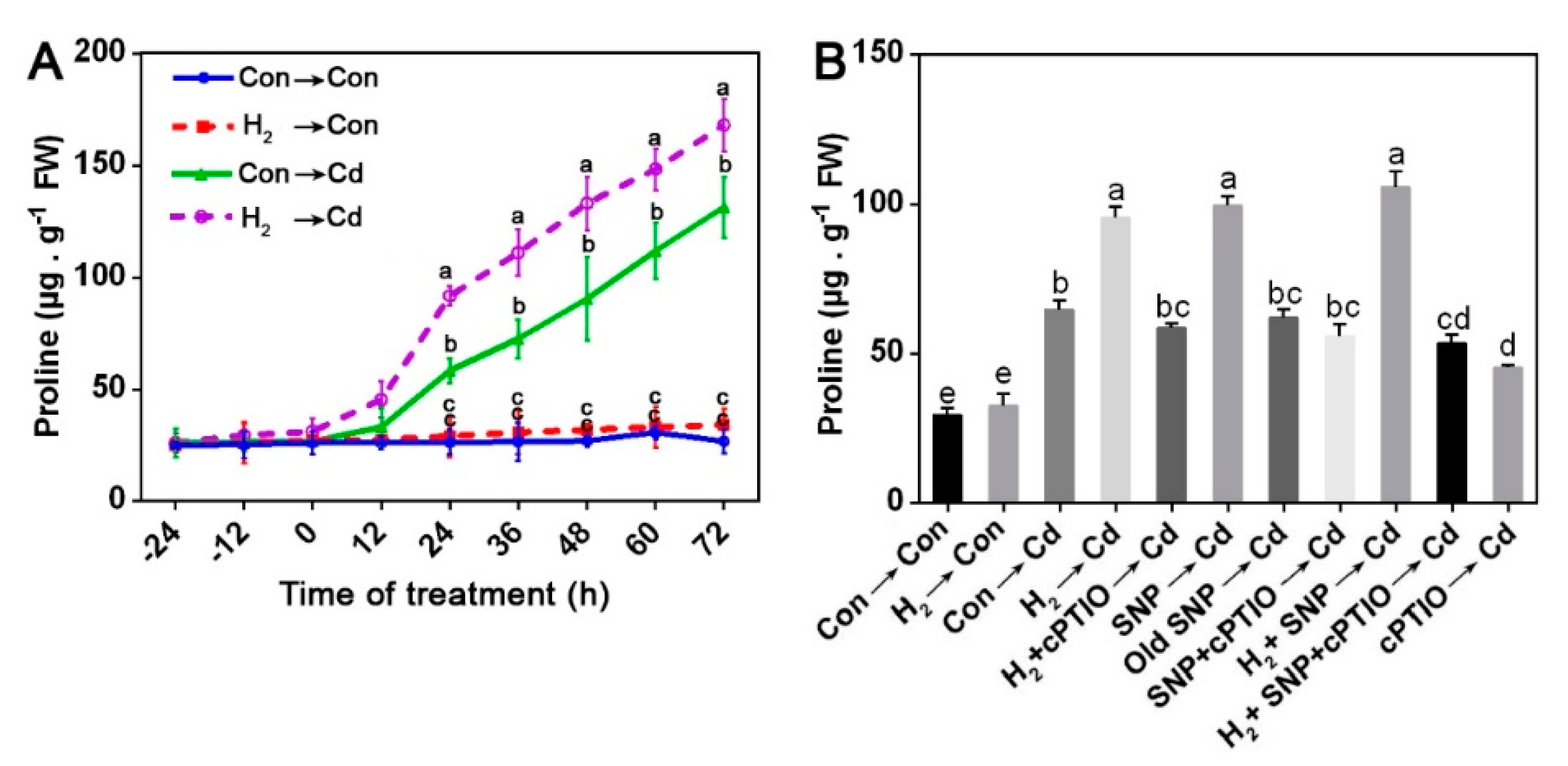
3.8. H2-Enhanced Proline Content Was Susceptible to cPTIO

Proline participates in the non-enzymatic antioxidation processes in the cells under metal stresses [73]. Thus, HPLC was used to assess proline content in G. lucidum mycelia under H2 and Cd treatments. As shown in Figure 7A, proline content was progressively increased over 72 h of Cd stress. The samples pre-treated with H2 showed significantly higher proline content than Cd stress alone samples after 24 h of Cd treatment. Then, we evaluated the proline contents after H2, SNP, and cPTIO treatment to examine the likelihood link between H2/NO and proline. As expected, proline in the mycelia was augmented by either H2 or SNP, and a maximum increase was observed under H2 and SNP successive treatments (Figure 7B). However, cPTIO severely decreased proline accumulation, and old SNP caused no marked effect. In addition, by comparison to the results of the NO peak at 12 h in the H2-treated stressed mycelia (Figure 3C), we inferred that the proline rise might be, at least partially, mediated by the H2-induced NO under Cd stress.
Further results showed that exogenous proline brought about a significant alleviation in the Cd-caused inhibition of the mycelial growth diameter by 15.23% and the biomass dry weight by 28.94%. However, proline exhibited no significant effect on the mitigation of Cd accumulation (Supplementary Figure S5).

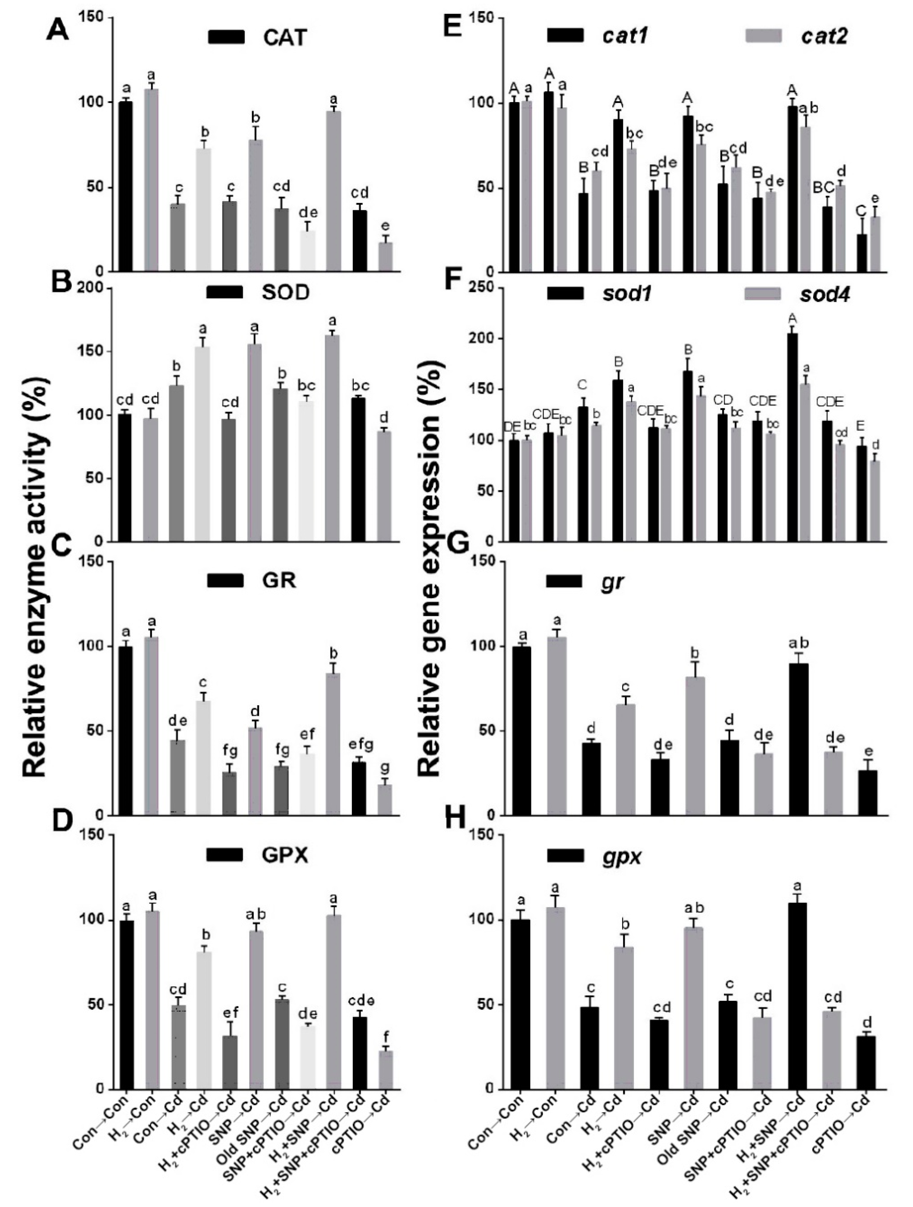
3.9. H2-Induced Reestablishment of Redox Homeostasis Was Sensitive to cPTIO

Given the knowledge that both cysteine and proline have a powerful influence on oxidative stress, we detected ROS accumulation after H2, SNP, cPTIO, and/or Cd treatments (Figure 8). Cd-stressed mycelia showed extensive ROS fluorescence intensities; however, lower ROS were observed in the H2- and/or SNP-treated stressed mycelia. When cPTIO was added alone or after H2 and/or SNP, it prevented the previous observations. Moreover, TBARS contents exhibited similar trends (Figure 8B). These results revealed that NO contributed to the H2-elicited mitigation of the Cd-caused oxidative stress.
The role of the H2-induced NO in the re-establishment of redox homeostasis under Cd stress was also confirmed by applying Na2WO4. Similar to the NO and/or cPTIO elicited responses (Figure 8A,B), the H2-elicited decline in ROS and TBARS was reversed by administering Na2WO4, which could inhibit the NR activity, and consequently reversed the H2-induced mitigation (Supplementary Figure S6A,B). Collectively, these results signify that ROS and TBARS were regulated by the H2 signaling, which was mediated by NR-related NO functioning under Cd stress.

3.10. The NO Scavenger cPTIO Reversed the H2-Induced Regulation of the Activities and Related Transcripts of the Antioxidant Enzymes
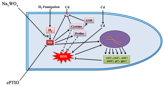
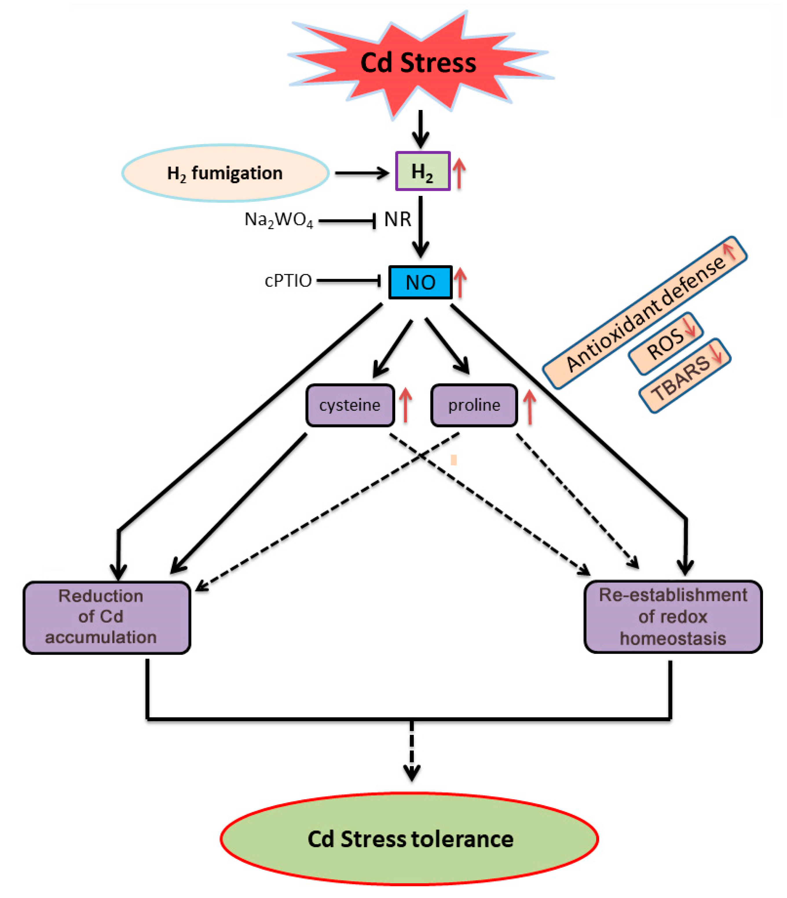
The activities of the antioxidant enzymes and their respective gene expression levels were also measured to confirm the NO role in the H2-induced effects. After mycelia were exposed to Cd stress for 24 h, the relative activities of CAT, GR, and GPX decreased markedly by 60.07%, 55.40%, and 50.35%, respectively; however, an increase was noticed in SOD by 22.12% relative to the control unstressed mycelia (Figure 9A–D). H2 or SNP treatment significantly increased all the antioxidant enzyme activities under Cd stress, and the highest rise was detected under the H2 and NO successive treatments by 136.58%, 31.76%, 88.45%, and 107.24% in CAT, SOD, GR, and GPX enzymes, respectively, relative to the Cd alone stressed samples. However, cPTIO reversed the previous responses, and old SNP had no significant effect. Furthermore, the corresponding gene expression levels showed similar tendencies (Figure 9E–H).

4. Discussion

Besides the broad application of HRW in medicine and agriculture, it was also utilized to alleviate stresses on fungi, namely mushrooms. However, the water content in HRW may alter the pH and humidity level in the fungi cultivation area, not to mention the possible inconvenience of its application at a large scale on the solid-state cultures of the macroscopic fungi. These reasons make it a less appropriate form. So, we adopted the hydrogen fumigation method in different concentrations on Ganoderma lucidum and used 50% HRW as a positive control. Interestingly, we found that 3% H2 fumigation initiated a higher alleviation effect of the Cd-elicited inhibition in mycelial growth diameter and biomass dry weight and a lesser Cd accumulation relative to the 50% HRW.
Previous studies on fungi showed the beneficial role of exogenous H2 in alleviating different stresses on Hypsizygus marmoreus [21], besides the amelioration effects of exogenous NO, individually, on different stresses such as Cd stress on G. lucidum [56] and heat shock in Pleurotus eryngii [35]. Thus, we investigated a possible plant-like link between the two gaseous signaling molecules, H2 and NO, in the fungus G. lucidum under Cd stress. Interestingly, our experiments suggested that NO might be involved in the H2-motivated alleviation of Cd stress in G. lucidum mycelia. We reached this conclusion by analyzing the NO content under 3% H2 fumigation pre-treatment. H2 strengthened NO release over 72 h of Cd treatment, with a peak at 12 h of Cd exposure (Figure 3C). When the exogenous NO-releasing compound, SNP alone, was added before Cd stress, it also elicited NO release (Figure 4C–E). However, when cPTIO was added, it not only impaired the release of endogenous NO but also reversed both the H2-induced amelioration of the mycelial growth inhibition triggered by Cd stress (Figure 4A) and the reduction in heavy metal accumulation (Figure 4B), suggesting the possible participation of NO in the H2-induced mitigation response in G. lucidum mycelia.
In more detail, we recognized that NR is the source of NO production in the H2-pretreated Cd-stressed mycelia. The participation of NR was confirmed by the addition of sodium tungstate (Na2WO4), an inhibitor of NR alone or in combinations with H2. Interestingly, Na2WO4 reversed the H2-elicited amelioration responses in G. lucidum (Figure 5). These results are in agreement with a plant study that showed that the osmotic stress tolerance mediated by the exogenous H2-elicited NO was prevented in the NR mutant [38]. Cumulatively, the enhancement of NR activity, and hence the amplified NO release in the H2-treated stressed mycelia, along with the reversion of H2-induced mitigation by the NO scavenger or the NR inhibitor, altogether confirm that the NR-dependent NO evolution was, at least partially, involved in the H2-triggered G. lucidum tolerance to Cd stress. A recent study showed similar findings where H2-enhanced NR-dependent NO alleviated the toxic effects of Cd stress in Brassica campestris [39]. More recently, H2-induced lateral root formation in tomato seedlings through increasing NR-dependent NO [40]. Based on the above findings, besides many reports of the NO’s regulatory role under stress in fungi [41,74], we concluded that the link between H2 and NO is responsible for the H2-initiated amelioration of Cd stress in G. lucidum. Nevertheless, few conflicting studies revealed an interaction between H2 and NO in which the H2-induced response was achieved by downregulating NO [60,75]. These contrasting responses may indicate the intricate interactions of these signaling pathways in the different biological systems.
ROS regulation is a key mechanism of the H2-initiated alleviation responses to stresses in many animal and plant studies [30]. In fungi, HRW helped the regeneration of H. marmoreus mycelia stressed by scratching, improved fruiting body development [76], and prolonged the post-harvest period [22]. The related mechanism was the enhancement of the antioxidant system and redox homeostasis, yet the intermediate signals underlying H2 are still largely unknown. Our results illustrated that H2 and/or NO reduced ROS and TBARS in the stressed mycelia, and these responses were reversed by the NO scavenger, cPTIO (Figure 8), and the NR inhibitor, Na2WO4 (Supplementary Figure S6). The above evidence suggests that NO might act downstream of H2 in mitigating the ROS abundance elicited by Cd stress in G. lucidum (Figure 8). Furthermore, the activities of the antioxidant enzymes CAT, SOD, GPX, and GR and the respective amounts of transcripts cat1, cat2, sod1, sod4, gr, and gpx were regulated by H2 and/or NO treatments under stress. However, this regulatory effect was blocked by cPTIO (Figure 9). Together with the phenotypic evidence, which showed reversion of the H2 ameliorating effect by cPTIO (Figure 4) or Na2WO4 (Figure 5) addition, we conclude that the H2-induced NO might act upstream of the antioxidant system in alleviating Cd stress in G. lucidum. The above results are consistent with previous plant and animal findings, indicating a common mechanism in ROS regulation that involves the interaction of H2 and NO signals.
Cysteine (Cys) is a thiol-containing amino acid and acts as an indispensable precursor of glutathione (GSH) [77]. In this study, our results showed positive effects of H2 on Cys and GSH contents under Cd stress (Figure 6). The time course of Cys levels showed that H2 elicited the recovery of the Cd-elicited decline in the Cys levels noticeably after 18 h. This suggested that Cys acted downstream of NO. This signaling pathway was reported in a previous study [72]. Results revealed that H2-induced NO mediated an elevation in both Cys and GSH contents under Cd stress. However, this response was reversed by cPTIO, confirming the role of the H2-induced NO in the restoration of Cys and GSH levels. The H2- and/or NO-induced increase in GSH contents are in harmony with our previous experiments, which showed marked increases in GR and GPX enzymes and transcripts under the same treatments. Some studies suggested the participation of exogenous Cys in the enhancement of heavy metal tolerance in plants [78,79]. Similarly, in our experiments, Cys could alleviate the Cd-caused mycelial growth inhibition, rescue GSH decline, and ameliorate Cd accumulation (Supplementary Figure S4). However, these responses were blocked by the GSH synthesis inhibitor, BSO, suggesting the role of Cys-mediated GSH enhancement in the Cd stress alleviation in G. lucidum. This role of GSH in heavy metals chelation and amelioration of the heavy metal toxicity was confirmed individually or through the formation of phytochelatin in previous studies in yeast, protists, and plants [80].
Another significant molecule, proline, participated in the mitigation of different stresses in many plants [81,82,83] and some microorganisms [73], as it protected Colletotrichum trifolii fungus against different stresses such as UV light, salt, heat, hydrogen peroxide, and paraquat. Our results revealed that H2 fumigation caused a significant increase in proline level after 24 h of Cd stress on G. lucidum (Figure 7A), which followed the NO signal peak (Figure 3C). This indicates that NO might act upstream of proline under Cd stress in G. lucidum. A previous study suggested that NO-mediated proline aided the Arabidopsis’s resistance to cold stress [58]. NO also mediated the accumulation of proline under copper stress in the alga Chlamydomonas reinhardtii [84]. Similarly, our experiments revealed that NO alone or in combination with H2 pre-treatment enhanced proline accumulation; however, the NO scavenger, cPTIO, severely reduced it relative to the Cd alone stress treatment (Figure 7B). This is consistent with a previous study that suggested that the H2-triggered NO signal elevated proline levels by enhancing its synthesis and decreasing its degradation, contributing to osmotic stress tolerance in alfalfa [85].
Proline was also administered under stress to further investigate its participation. Additionally, it significantly alleviated the Cd-triggered inhibition in the mycelial growth diameter and the mycelial biomass dry weight (Supplementary Figure S5A). Cumulatively, previous results suggest that the H2-initiated augmentation of proline, intermediated by NO might, at least partially, participate in the amelioration of Cd stress in G. lucidum. Nevertheless, proline did not lessen Cd accumulation (Supplementary Figure S5B), implying that it works through other pathways rather than Cd sequestration in G. lucidum, possibly through antioxidation mechanism, and this requires further investigation.
Overall, our report provides the underlying mechanism of the H2-induced mitigation of Cd stress in G. lucidum, mainly through the NO-mediated regulation of ROS homeostasis, the reduction in Cd accumulation, and possibly, the increase in cysteine and proline accumulation (Figure 10). Moreover, this is the first report on fungi that reveals the advantages of H2 fumigation in stress alleviation.

5. Conclusions

The link we found between H2 and NO in the alleviation of Cd stress on G. lucidum will provide new insights into the mechanisms of H2 stress alleviation in G. lucidum, and this may also be further deployed on the molecular level in other valuable pathways beyond enhancing stress mitigation in G. lucidum and other fungi. Most importantly, our report provides a convenient model for hydrogen application on fungi (hydrogen fumigation), which brought about a higher response than HRW in alleviating Cd heavy metal stress. This model could be harnessed on a large scale in the economically important macroscopic fungi farms by fumigating fungi at a suitable rate through perforated pipelines connected to a large hydrogen source, aiming to achieve higher stress alleviation and higher productivity, similar to our in vitro findings.

Supplementary Materials

The following are available online at https://www.mdpi.com/article/10.3390/jof8010010/s1, Table S1: The sequences of primers used for RT-qPCR in Ganoderma lucidum. Figure S1: Effect of different Cd concentrations on mycelial growth of G. lucidum; Figure S2: Screening for the optimum HRW concentration for the alleviation of 1.75 mM CdCl2 stress on G. lucidum. Figure S3: H2-fumigation pre-treatment activated the antioxidant enzyme activities and their corresponding transcript levels under oxidative stress caused by Cd stress. Figure S4: Cd-elicited negative effects on mycelial growth, GSH and Cd accumulation was rescued by cysteine. Figure S5: Proline alleviated the growth inhibition caused by Cd stress but did not mitigate Cd accumulation. Figure S6: H2-reestablished redox balance was sensitive to sodium tungstate, an inhibitor of NR enzyme activity. Figure S7: H2, HRW, SNP, NO’s scavenger, cPTIO, Na2WO4 or proline had no significant effect on mycelial growth or NO content under normal conditions.

Author Contributions

Conceptualization, D.A., G.Z., P.C. and W.S.; methodology, D.A. and G.Z., J.A.B.; software, D.A., G.Z. and I.A.; validation, P.C., F.A., I.A. and S.A.K.; formal analysis, D.A., J.A.B., M.K.A.E. and W.S.; investigation, D.A., S.A.K., Y.A.-G.M., M.K.A.E. and W.S.; resources, W.S.; data curation, D.A., P.C., W.A.K. and Q.A.; writing—original draft preparation, D.A., J.A.B., F.A. and W.S.; writing—review and editing, D.A., M.G.A., Y.A.-G.M., W.C. and W.S.; visualization, D.A., W.A.K., M.G.A. and Q.A.; supervision, W.S.; project administration, G.Z., P.C. and W.S.; funding acquisition, W.S. All authors have read and agreed to the published version of the manuscript.

Funding

This work was supported by the Foshan Agriculture Science and Technology Project (Foshan City Budget, No. 140, 2019) and the Funding from the Center of Hydrogen Science, Shanghai Jiao Tong University, China.

Institutional Review Board Statement

Not applicable.

Informed Consent Statement

Not applicable.

Data Availability Statement

All data generated or analyzed during this study are included in this published article and its Supplementary Materials.

Acknowledgments

We are grateful to Mingwen Zhao and Ang Ren and their lab, the Key Laboratory of Microbiological Engineering of Agricultural Environment, Microbiology Department, College of Life Sciences, Nanjing Agricultural University for their assistance through the experimental work.

Conflicts of Interest

The authors declare that they have no competing interest. The funders had no role in the design of the study; in the collection, analyses, or interpretation of data; in the writing of the manuscript, or in the decision to publish the results.

References

  1. Jia, J.; Zhang, X.; Hu, Y.-S.; Wu, Y.; Wang, Q.-Z.; Li, N.-N.; Guo, Q.-C.; Dong, X.-C. Evaluation of in vivo antioxidant activities of Ganoderma lucidum polysaccharides in STZ-diabetic rats. Food Chem. 2009, 115, 32–36. [Google Scholar] [CrossRef]
  2. Wang, D.; Liu, Y.; Tan, X.; Liu, H.; Zeng, G.; Hu, X.; Jian, H.; Gu, Y. Effect of exogenous nitric oxide on antioxidative system and S-nitrosylation in leaves of Boehmeria nivea (L.) Gaud under cadmium stress. Environ. Sci. Pollut. Res. 2015, 22, 3489–3497. [Google Scholar] [CrossRef] [PubMed]
  3. Hart, J.J.; Welch, R.M.; Norvell, W.A.; Sullivan, L.A.; Kochian, L. Characterization of Cadmium Binding, Uptake, and Translocation in Intact Seedlings of Bread and Durum Wheat Cultivars. Plant Physiol. 1998, 116, 1413–1420. [Google Scholar] [CrossRef] [PubMed] [Green Version]
  4. di Toppi, L.S.; Gabbrielli, R. Response to cadmium in higher plants. Environ. Exp. Bot. 1999, 41, 105–130. [Google Scholar] [CrossRef]
  5. Deng, X.; Xia, Y.; Hu, W.; Zhang, H.; Shen, Z. Cadmium-induced oxidative damage and protective effects of N-acetyl-l-cysteine against cadmium toxicity in Solanum nigrum L. J. Hazard. Mater. 2010, 180, 722–729. [Google Scholar] [CrossRef]
  6. DalCorso, G.; Farinati, S.; Furini, A. Regulatory networks of cadmium stress in plants. Plant Signal. Behav. 2010, 5, 663–667. [Google Scholar] [CrossRef]
  7. Zhang, R.; Li, X.; Fadel, J. Oyster mushroom cultivation with rice and wheat straw. Bioresour. Technol. 2002, 82, 277–284. [Google Scholar] [CrossRef]
  8. Ohsawa, I.; Ishikawa, M.; Takahashi, K.; Watanabe, M.; Nishimaki, K.; Yamagata, K.; Katsura, K.-I.; Katayama, Y.; Asoh, S.; Ohta, S. Hydrogen acts as a therapeutic antioxidant by selectively reducing cytotoxic oxygen radicals. Nat. Med. 2007, 13, 688–694. [Google Scholar] [CrossRef]
  9. Fukuda, K.-I.; Asoh, S.; Ishikawa, M.; Yamamoto, Y.; Ohsawa, I.; Ohta, S. Inhalation of hydrogen gas suppresses hepatic injury caused by ischemia/reperfusion through reducing oxidative stress. Biochem. Biophys. Res. Commun. 2007, 361, 670–674. [Google Scholar] [CrossRef]
  10. Nakashima-Kamimura, N.; Mori, T.; Ohsawa, I.; Asoh, S.; Ohta, S. Molecular hydrogen alleviates nephrotoxicity induced by an anti-cancer drug cisplatin without compromising anti-tumor activity in mice. Cancer Chemother. Pharmacol. 2009, 64, 753–761. [Google Scholar] [CrossRef]
  11. Jin, Q.; Zhu, K.; Cui, W.; Li, L.; Shen, W. Hydrogen-Modulated Stomatal Sensitivity to Abscisic Acid and Drought Tolerance Via the Regulation of Apoplastic pH in Medicago sativa. J. Plant Growth Regul. 2016, 35, 565–573. [Google Scholar] [CrossRef]
  12. Cui, W.; Gao, C.; Fang, P.; Lin, G.; Shen, W. Alleviation of cadmium toxicity in Medicago sativa by hydrogen-rich water. J. Hazard. Mater. 2013, 260, 715–724. [Google Scholar] [CrossRef] [PubMed]
  13. Wu, Q.; Su, N.; Cai, J.; Shen, Z.; Cui, J. Hydrogen-rich water enhances cadmium tolerance in Chinese cabbage by reducing cadmium uptake and increasing antioxidant capacities. J. Plant Physiol. 2015, 175, 174–182. [Google Scholar] [CrossRef]
  14. Wu, X.; Zhu, Z.B.; Chen, J.H.; Huang, Y.F.; Liu, Z.L.; Zou, J.W.; Chen, Y.H.; Na Su, N.; Cui, J. Transcriptome analysis revealed pivotal transporters involved in the reduction of cadmium accumulation in pak choi (Brassica chinensis L.) by exogenous hydrogen-rich water. Chemosphere 2019, 216, 684–697. [Google Scholar] [CrossRef] [PubMed]
  15. Wu, X.; Su, N.; Yue, X.; Fang, B.; Zou, J.; Chen, Y.; Shen, Z.; Cui, J. IRT1 and ZIP2 were involved in exogenous hydrogen-rich water-reduced cadmium accumulation in Brassica chinensis and Arabidopsis thaliana. J. Hazard. Mater. 2021, 407, 124599. [Google Scholar] [CrossRef] [PubMed]
  16. Xu, D.; Cao, H.; Fang, W.; Pan, J.; Chen, J.; Zhang, J.; Shen, W. Linking hydrogen-enhanced rice aluminum tolerance with the reestablishment of GA/ABA balance and miRNA-modulated gene expression: A case study on germination. Ecotoxicol. Environ. Saf. 2017, 145, 303–312. [Google Scholar] [CrossRef]
  17. Cao, Z.; Duan, X.; Yao, P.; Cui, W.; Cheng, D.; Zhang, J.; Jin, Q.; Chen, J.; Dai, T.; Shen, W. Hydrogen Gas Is Involved in Auxin-Induced Lateral Root Formation by Modulating Nitric Oxide Synthesis. Int. J. Mol. Sci. 2017, 18, 2084. [Google Scholar] [CrossRef]
  18. Zhu, Y.; Liao, W.; Wang, M.; Niu, L.; Xu, Q.; Jin, X. Nitric oxide is required for hydrogen gas-induced adventitious root formation in cucumber. J. Plant Physiol. 2016, 195, 50–58. [Google Scholar] [CrossRef]
  19. Li, C.; Gong, T.; Bian, B.; Liao, W. Roles of hydrogen gas in plants: A review. Funct. Plant Biol. 2018, 45, 783–792. [Google Scholar] [CrossRef]
  20. Ren, A.; Liu, R.; Miao, Z.-G.; Zhang, X.; Cao, P.-F.; Chen, T.-X.; Li, C.-Y.; Shi, L.; Jiang, A.-L.; Zhao, M.-W. Hydrogen-rich water regulates effects of ROS balance on morphology, growth and secondary metabolism via glutathione peroxidase inGanoderma lucidum. Environ. Microbiol. 2017, 19, 566–583. [Google Scholar] [CrossRef]
  21. Zhang, J.; Hao, H.; Chen, M.; Wang, H.; Feng, Z.; Chen, H. Hydrogen-rich water alleviates the toxicities of different stresses to mycelial growth in Hypsizygus marmoreus. AMB Express 2017, 7, 107. [Google Scholar] [CrossRef] [PubMed] [Green Version]
  22. Chen, H.; Zhang, J.; Hao, H.; Feng, Z.; Chen, M.; Wang, H.; Ye, M. Hydrogen-rich water increases postharvest quality by enhancing antioxidant capacity in Hypsizygus marmoreus. AMB Express 2017, 7, 221. [Google Scholar] [CrossRef] [PubMed] [Green Version]
  23. Hu, H.; Zhao, S.; Li, P.; Shen, W. Hydrogen gas prolongs the shelf life of kiwifruit by decreasing ethylene biosynthesis. Postharvest Biol. Technol. 2018, 135, 123–130. [Google Scholar] [CrossRef]
  24. Alwazeer, D.; Tan, K.; Örs, B. Reducing atmosphere packaging as a novel alternative technique for extending shelf life of fresh cheese. J. Food Sci. Technol. 2020, 57, 3013–3023. [Google Scholar] [CrossRef]
  25. Alwazeer, D.; Delbeau, C.; Divies, C.; Cachon, R. Use of redox potential modification by gas improves microbial quality, color retention, and ascorbic acid stability of pasteurized orange juice. Int. J. Food Microbiol. 2003, 89, 21–29. [Google Scholar] [CrossRef]
  26. Alwazeer, D.; Örs, B. Reducing atmosphere drying as a novel drying technique for preserving the sensorial and nutritional notes of foods. J. Food Sci. Technol. 2019, 56, 3790–3800. [Google Scholar] [CrossRef]
  27. Dong, Z.; Wu, L.; Kettlewell, B.; Caldwell, C.D.; Layzell, D.B. Hydrogen fertilization of soils—is this a benefit of legumes in rotation? Plant Cell Environ. 2003, 26, 1875–1879. [Google Scholar] [CrossRef]
  28. Stein, S.; Selesi, D.; Schilling, R.; Pattis, I.; Schmid, M.; Hartmann, A. Microbial activity and bacterial composition of H2-treated soils with net CO2 fixation. Soil Biol. Biochem. 2005, 37, 1938–1945. [Google Scholar] [CrossRef]
  29. Liu, H.; Wang, W.; Cao, G.; Tang, M. Effect of hydrogen on microbial population and enzyme activity in Robinia pseudoacacia rhizosphere soil. Chin. J. Appl. Environ. Biol. 2010, 16, 515–518. [Google Scholar] [CrossRef]
  30. Li, L.; Lou, W.; Kong, L.; Shen, W. Hydrogen Commonly Applicable from Medicine to Agriculture: From Molecular Mechanisms to the Field. Curr. Pharm. Des. 2021, 27, 747–759. [Google Scholar] [CrossRef]
  31. Kapoor, P.; Sharma, B.M. Studies on different growth parameters of Ganoderma lucidum. Int. J. Sci. Environ. Tech. 2014, 3, 1515–1524. [Google Scholar]
  32. Tang, W.; Kuehn, T.H.; Simcik, M.F. Effects of Temperature, Humidity and Air Flow on Fungal Growth Rate on Loaded Ventilation Filters. J. Occup. Environ. Hyg. 2015, 12, 525–537. [Google Scholar] [CrossRef] [PubMed]
  33. Chao, C.H. Clinical Applications of Magnesium Hydride. In Magnesium Alloys—Selected Issue; Tański, T., Borek, W., Król, M., Eds.; IntechOpen: London, UK, 2018; pp. 115–128. [Google Scholar] [CrossRef]
  34. Yü, Y.; Yang, Z.; Guo, K.; Li, Z.; Zhou, H.; Wei, Y.; Li, J.; Zhang, X.; Harvey, P.; Yang, H. Oxidative Damage Induced by Heat Stress Could be Relieved by Nitric Oxide in Trichoderma harzianum LTR-2. Curr. Microbiol. 2015, 70, 618–622. [Google Scholar] [CrossRef] [PubMed]
  35. Kong, W.; Huang, C.; Chen, Q.; Zou, Y.; Zhang, J. Nitric oxide alleviates heat stress-induced oxidative damage in Pleurotus eryngii var. tuoliensis. Fungal Genet. Biol. 2012, 49, 15–20. [Google Scholar] [CrossRef]
  36. Chiang, K.T.; Shinyashiki, M.; Switzer, C.H.; Valentine, J.S.; Gralla, E.B.; Thiele, D.J.; Fukuto, J.M. Effects of Nitric Oxide on the Copper-Responsive Transcription Factor Ace1 in Saccharomyces cerevisiae: Cytotoxic and Cytoprotective Actions of Nitric Oxide. Arch. Biochem. Biophys. 2000, 377, 296–303. [Google Scholar] [CrossRef] [Green Version]
  37. Liu, R.; Zhu, T.; Yang, T.; Yang, Z.; Ren, A.; Shi, L.; Zhu, J.; Yu, H.; Zhao, M. Nitric oxide regulates ganoderic acid biosynthesis by the S-nitrosylation of aconitase under heat stress in Ganoderma lucidum. Environ. Microbiol. 2021, 23, 682–695. [Google Scholar] [CrossRef]
  38. Xie, Y.; Mao, Y.; Zhang, W.; Lai, D.; Wang, Q.; Shen, W. Reactive Oxygen Species-Dependent Nitric Oxide Production Contributes to Hydrogen-Promoted Stomatal Closure in Arabidopsis. Plant Physiol. 2014, 165, 759–773. [Google Scholar] [CrossRef] [Green Version]
  39. Su, N.; Wu, Q.; Chen, H.; Huang, Y.; Zhu, Z.; Chen, Y.; Cui, J. Hydrogen gas alleviates toxic effects of cadmium in Brassica campestris seedlings through up-regulation of the antioxidant capacities: Possible involvement of nitric oxide. Environ. Pollut. 2019, 251, 45–55. [Google Scholar] [CrossRef]
  40. Wang, Y.; Lv, P.; Konga, L.; Shenac, W.; Hebc, Q. Nanomaterial-mediated sustainable hydrogen supply induces lateral root formation via nitrate reductase-dependent nitric oxide. Chem. Eng. J. 2021, 405, 126905. [Google Scholar] [CrossRef]
  41. Liu, R.; Shi, L.; Zhu, T.; Yang, T.; Ren, A.; Zhu, J.; Zhao, M.-W. Cross Talk between Nitric Oxide and Calcium-Calmodulin Regulates Ganoderic Acid Biosynthesis in Ganoderma lucidum under Heat Stress. Appl. Environ. Microbiol. 2018, 84. [Google Scholar] [CrossRef] [Green Version]
  42. Chamizo-Ampudia, A.; Sanz-Luque, E.; Llamas, A.; Galvan, A.; Fernandez, E.P.-A. Nitrate Reductase Regulates Plant Nitric Oxide Homeostasis. Trends Plant Sci. 2017, 22, 163–174. [Google Scholar] [CrossRef] [PubMed]
  43. Marcos, A.T.; Ramos, M.S.; Marcos, J.F.; Carmona, L.; Strauss, J.; Cánovas, D. Nitric oxide synthesis by nitrate reductase is regulated during development in A. spergillus. Mol. Microbiol. 2016, 99, 15–33. [Google Scholar] [CrossRef] [PubMed] [Green Version]
  44. Jia, H.; Wang, X.; Dou, Y.; Liu, D.; Si, W.; Fang, H.; Zhao, C.; Chen, S.; Xi, J.; Li, J. Hydrogen sulfide-cysteine cycle system enhances cadmium tolerance through alleviating cadmium-induced oxidative stress and ion toxicity in Arabidopsis roots. Sci. Rep. 2016, 6, 39702. [Google Scholar] [CrossRef]
  45. Yamauchi, A.; Bloom, E.T. Requirement of thiol compounds as reducing agents for IL-2-mediated induction of LAK activity and proliferation of human NK cells. J. Immunol. 1993, 151, 5535–5544. [Google Scholar] [PubMed]
  46. Yamauchi, A.; Bloom, E.T. Control of Cell Cycle Progression in Human Natural Killer Cells Through Redox Regulation of Expression and Phosphorylation of Retinoblastoma Gene Product Protein. Blood 1997, 89, 4092–4099. [Google Scholar] [CrossRef] [PubMed]
  47. Bannai, S. Transport of cystine and cysteine in mammalian cells. Biochim. Biophys. Acta Rev. Biomembr. 1984, 779, 289–306. [Google Scholar] [CrossRef]
  48. Lu, S.C. Regulation of hepatic glutathione synthesis: Current concepts and controversies. FASEB J. 1999, 13, 1169–1183. [Google Scholar] [CrossRef] [Green Version]
  49. Cobbett, C.; Goldsbrough, P. PHYTOCHELATINS AND METALLOTHIONEINS: Roles in Heavy Metal Detoxification and Homeostasis. Annu. Rev. Plant Biol. 2002, 53, 159–182. [Google Scholar] [CrossRef] [Green Version]
  50. Fang, H.; Liu, Z.; Jin, Z.; Zhang, L.; Liu, D.; Pei, Y. An emphasis of hydrogen sulfide-cysteine cycle on enhancing the tolerance to chromium stress in Arabidopsis. Environ. Pollut. 2016, 213, 870–877. [Google Scholar] [CrossRef]
  51. Alamri, S.; Ali, H.M.; Khan, M.I.R.; Singh, V.P.; Siddiqui, M.H. Exogenous nitric oxide requires endogenous hydrogen sulfide to induce the resilience through sulfur assimilation in tomato seedlings under hexavalent chromium toxicity. Plant Physiol. Biochem. 2020, 155, 20–34. [Google Scholar] [CrossRef]
  52. Maresca, B.; Lambowitz, A.M.; Kumar, V.B.; Grant, G.A.; Kobayashi, G.S.; Medoff, G. Role of cysteine in regulating morphogenesis and mitochondrial activity in the dimorphic fungus Histoplasma capsulatum. Proc. Natl. Acad. Sci. USA 1981, 78, 4596–4600. [Google Scholar] [CrossRef] [PubMed] [Green Version]
  53. Szabados, L.; Savouré, A. Proline: A multifunctional amino acid. Trends Plant Sci. 2010, 15, 89–97. [Google Scholar] [CrossRef] [PubMed]
  54. Lei, Y.; Yin, C.; Ren, J.; Li, C. Effect of osmotic stress and sodium nitroprusside pretreatment on proline metabolism of wheat seedlings. Biol. Plant. 2007, 51, 386–390. [Google Scholar] [CrossRef]
  55. Shi, F.M.; Wu, R.W.; Wang, C.; Li, J. Nitrate reductase-dependent NO production is critical for Arabidopsis roots response to ABA. Life Sci. J. 2013, 10, 297–303. [Google Scholar]
  56. Guo, S.; Yao, Y.; Zuo, L.; Shi, W.; Gao, N.; Xu, H. Enhancement of tolerance ofGanoderma lucidumto cadmium by nitric oxide. J. Basic Microbiol. 2015, 56, 36–43. [Google Scholar] [CrossRef] [PubMed]
  57. Bright, J.; Desikan, R.; Hancock, J.T.; Weir, I.S.; Neill, S.J. ABA-induced NO generation and stomatal closure in Arabidopsis are dependent on H2O2 synthesis. Plant J. 2006, 45, 113–122. [Google Scholar] [CrossRef]
  58. Zhao, M.-G.; Chen, L.; Zhang, L.-L.; Zhang, W.-H. Nitric Reductase-Dependent Nitric Oxide Production Is Involved in Cold Acclimation and Freezing Tolerance in Arabidopsis. Plant Physiol. 2009, 151, 755–767. [Google Scholar] [CrossRef] [Green Version]
  59. Qi, F.; Xiang, Z.; Kou, N.; Cui, W.; Xu, D.; Wang, R.; Zhu, D.; Shen, W. Nitric oxide is involved in methane-induced adventitious root formation in cucumber. Physiol. Plant. 2016, 159, 366–377. [Google Scholar] [CrossRef] [Green Version]
  60. Chen, M.; Cui, W.; Zhu, K.; Xie, Y.; Zhang, C.; Shen, W. Hydrogen-rich water alleviates aluminum-induced inhibition of root elongation in alfalfa via decreasing nitric oxide production. J. Hazard. Mater. 2014, 267, 40–47. [Google Scholar] [CrossRef]
  61. Su, J.; Nie, Y.; Zhao, G.; Cheng, D.; Wang, R.; Chen, J.; Zhang, S.; Shen, W. Endogenous hydrogen gas delays petal senescence and extends the vase life of lisianthus cut flowers. Postharvest Biol. Technol. 2019, 147, 148–155. [Google Scholar] [CrossRef]
  62. Mu, D.; Li, C.; Zhang, X.; Li, X.; Shi, L.; Ren, A.; Zhao, M. Functions of the nicotinamide adenine dinucleotide phosphate oxidase family in Ganoderma lucidum: An essential role in ganoderic acid biosynthesis regulation, hyphal branching, fruiting body development, and oxidative-stress resistance. Environ. Microbiol. 2013, 16, 1709–1728. [Google Scholar] [CrossRef] [PubMed]
  63. Heath, R.L.; Packer, L. Photoperoxidation in isolated chloroplasts. i. kinetics and stoichiometry of fatty acid peroxidation. Arch. Biochem. Biophys. 1968, 125, 189–198. [Google Scholar] [CrossRef]
  64. Kula, I.; Solak, M.H.; Uğurlu, M.; Işıloğlu, M.; Arslan, Y. Determination of Mercury, Cadmium, Lead, Zinc, Selenium and Iron by ICP-OES in Mushroom Samples from Around Thermal Power Plant in Muğla, Turkey. Bull. Environ. Contam. Toxicol. 2011, 87, 276–281. [Google Scholar] [CrossRef] [PubMed]
  65. Hammond, J.B.W.; Kruger, N.J. The Bradford Method for Protein Quantitation. In New Protein Techniques; Springer: Berlin/Heidelberg, Germany, 1988; Volume 3, pp. 25–32. [Google Scholar]
  66. Aebi, H. Catalase in vitro. Methods Enzymol. 1984, 105, 121–126. [Google Scholar] [CrossRef]
  67. Giannopolitis, C.N.; Ries, S.K. Superoxide Dismutases: I. Occurrence in higher plants. Plant Physiol. 1977, 59, 309–314. [Google Scholar] [CrossRef]
  68. Garrett, R.H.; Cove, D.J. Formation of NADPH-nitrate reductase activity in vitro from Aspergillus nidulans niaD and cnx mu-tants. Mol. Gen. Genet. 1976, 149, 179–186. [Google Scholar] [CrossRef]
  69. Planchet, E.; Verdu, I.; Delahaie, J.; Cukier, C.; Girard, C.; Morere-Le Paven, M.-C.; Limami, A.M. Abscisic acid-induced nitric oxide and proline accumulation in independent pathways under water-deficit stress during seedling establishment in Medicago truncatula. J. Exp. Bot. 2014, 65, 2161–2170. [Google Scholar] [CrossRef] [Green Version]
  70. Reed, D.; Babson, J.; Beatty, P.; Brodie, A.; Ellis, W.; Potter, D. High-performance liquid chromatography analysis of nanomole levels of glutathione, glutathione disulfide, and related thiols and disulfides. Anal. Biochem. 1980, 106, 55–62. [Google Scholar] [CrossRef]
  71. Mu, D.; Shi, L.; Ren, A.; Li, M.; Wu, F.; Jiang, A.; Zhao, M. The Development and Application of a Multiple Gene Co-Silencing System Using Endogenous URA3 as a Reporter Gene in Ganoderma lucidum. PLoS ONE 2012, 7, e43737. [Google Scholar] [CrossRef]
  72. Khan, M.N.; Mobin, M.; Abbas, Z.K.; Siddiqui, M.H. Nitric oxide-induced synthesis of hydrogen sulfide alleviates osmotic stress in wheat seedlings through sustaining antioxidant enzymes, osmolyte accumulation and cysteine homeostasis. Nitric Oxide 2017, 68, 91–102. [Google Scholar] [CrossRef]
  73. Siripornadulsil, S.; Traina, S.; Verma, D.P.S.; Sayre, R.T. Molecular Mechanisms of Proline-Mediated Tolerance to Toxic Heavy Metals in Transgenic Microalgae. Plant Cell 2002, 14, 2837–2847. [Google Scholar] [CrossRef] [PubMed]
  74. Chen, C.; Li, Q.; Wang, Q.; Lu, D.; Zhang, H.; Wang, J.; Fu, R. Transcriptional profiling provides new insights into the role of nitric oxide in enhancing Ganoderma oregonense resistance to heat stress. Sci. Rep. 2017, 7, 15694. [Google Scholar] [CrossRef] [Green Version]
  75. Itoh, T.; Hamada, N.; Terazawa, R.; Ito, M.; Ohno, K.; Ichihara, M.; Nozawa, Y.; Ito, M. Molecular hydrogen inhibits lipopolysaccharide/interferon γ-induced nitric oxide production through modulation of signal transduction in macrophages. Biochem. Biophys. Res. Commun. 2011, 411, 143–149. [Google Scholar] [CrossRef]
  76. Chen, H.; Hai, H.; Wang, H.; Wang, Q.; Chen, M.; Feng, Z.; Ye, M.; Zhang, J. Hydrogen-rich water mediates redox regulation of the antioxidant system, mycelial regeneration and fruiting body development in Hypsizygus marmoreus. Fungal Biol. 2018, 122, 310–321. [Google Scholar] [CrossRef] [PubMed]
  77. Noda, T.; Iwakiri, R.; Fujimoto, K.; Rhoads, C.A.; Aw, T.Y. Exogenous cysteine and cystine promote cell proliferation in CaCo-2 cells. Cell Prolif. 2002, 35, 117–129. [Google Scholar] [CrossRef] [PubMed]
  78. Yuan, H.; Zhang, Y.; Huang, S.; Yang, Y.; Gu, C. Effects of exogenous glutathione and cysteine on growth, lead accumulation, and tolerance of Iris lactea var. chinensis. Environ. Sci. Pollut. Res. 2014, 22, 2808–2816. [Google Scholar] [CrossRef] [PubMed]
  79. Terzi, H.; Yıldız, M. Proteomic analysis reveals the role of exogenous cysteine in alleviating chromium stress in maize seedlings. Ecotoxicol. Environ. Saf. 2021, 209, 111784. [Google Scholar] [CrossRef]
  80. Mendoza-Cózatl, D.; Loza-Tavera, H.; Hernández-Navarro, A.; Moreno-Sanchez, R. Sulfur assimilation and glutathione metabolism under cadmium stress in yeast, protists and plants. FEMS Microbiol. Rev. 2005, 29, 653–671. [Google Scholar] [CrossRef] [Green Version]
  81. Khedr, A.H.A.; Abbas, M.A.; Wahid, A.A.A.; Quick, W.P.; Abogadallah, G.M. Proline induces the expression of salt-stress-responsive proteins and may improve the adaptation of Pancratium maritimum L. to salt-stress. J. Exp. Bot. 2003, 54. [Google Scholar] [CrossRef] [Green Version]
  82. Kaur, G.; Asthir, B. Proline: A key player in plant abiotic stress tolerance. Biol. Plant. 2015, 59, 609–619. [Google Scholar] [CrossRef]
  83. Chen, C.; Dickman, M.B. From The Cover: Proline suppresses apoptosis in the fungal pathogen Colletotrichum trifolii. Proc. Natl. Acad. Sci. USA 2005, 102, 3459–3464. [Google Scholar] [CrossRef] [PubMed] [Green Version]
  84. Zhang, L.P.; Mehta, S.K.; Liu, Z.P.; Yang, Z.M. Copper-Induced Proline Synthesis is Associated with Nitric Oxide Generation in Chlamydomonas reinhardtii. Plant Cell Physiol. 2008, 49, 411–419. [Google Scholar] [CrossRef] [PubMed]
  85. Su, J.; Zhang, Y.; Nie, Y.; Cheng, D.; Wang, R.; Hu, H.; Chen, J.; Zhang, J.; Du, Y.; Shen, W. Hydrogen-induced osmotic tolerance is associated with nitric oxide-mediated proline accumulation and reestablishment of redox balance in alfalfa seedlings. Environ. Exp. Bot. 2018, 147, 249–260. [Google Scholar] [CrossRef]
Figure 1. H2 fumigation increased the mycelial growth diameter and the biomass dry weight of G. lucidum. Mycelial inocula were grown on solid CYM media for 2 days; then, on the 3rd day, were either pre-treated with 1%, 2%, 3%, 5%, or 10% H2 fumigation or 50% HRW for 24 h or not treated with either hydrogen forms (Con). On the 4th day, 6 mm-diameter mycelial discs from different treatments were cut and grown on solid CYM with or without CdCl2 for 8 d; then, photographs of the plates were taken (A). Scale bar = 1 cm. Diameters of mycelial growth were detected (B). Simultaneously, on the 4th day, mycelial discs of 6 mm-diameter were cut from H2- or HRW-treated or non-treated mycelia. Each set of 8 mycelial discs was inoculated into liquid CYM media with or without CdCl2 then grown in shaking incubator for 8 days. Then, the biomass dry weight values were immediately determined (C). Bars with different letters are significantly different at p < 0.05 according to one-way ANOVA with multiple comparison using Tukey’s test.
Jof 08 00010 g001
Figure 2. H2-fumigation pre-treatment reduced Cd accumulation and decreased ROS and TBARS contents in G. lucidum. Mycelia were cultured on solid CYM media for 2 days. Then, on the 3rd day, mycelia were pre-treated with 3% H2 fumigation, 50% HRW for 24 h, or not treated with either hydrogen form (control). On the 4th day, 6-mm-diameter mycelial discs from these treatments were transferred to solid CYM or 1.75 mM CdCl2-containing solid CYM, each with or without inserted glass coverslips then grown for 5 days. Afterward, samples from the plates without coverslips were immediately used for Cd content analysis (A). Coverslips with mycelia on their surface were loaded with DCFH−DA, and then the ROS fluorescence intensities were analyzed via confocal laser microscopy (B). Scale bar = 100 µm. DIC, differential interference contrast. Respective fluorescence intensities were measured (C). Mycelia were also sampled for TBARS analysis (D). Mycelia without chemicals were used as a control for each analysis (Con → Con). Bars with different letters are significantly different at p < 0.05 according to one-way ANOVA with multiple comparison using Tukey’s test.
Jof 08 00010 g002
Figure 3. Effects of Cd and H2 fumigation treatments on the endogenous H2 and NO levels in mycelia. Early signals of H2 and NO were observed in the mycelia grown on solid CYM with or without 1.75 mM Cd for the indicated time points (A,B). Additionally, a four-day time course of endogenous NO level was tracked in the mycelia (C). On the 3rd day, two-day-old mycelia were either pre-treated or not with 3% H2 for 24 h and then were transferred into control or stressed CYM media and grown for 72 h. H2 and NO levels were measured by GC and Griess reagent assay, respectively. Asterisks * indicate the significant difference at every time point relative to Con → Con according to t-test at p < 0.05 (A,B), and letters indicate significant difference at every time point relative to Con → Con according to one-way ANOVA with multiple comparison using Tukey’s test at p < 0.05 (C).
Jof 08 00010 g003
Figure 4. H2-enhanced NO production was susceptible to the NO scavenger cPTIO. Two-day-old mycelia were either pre-treated or not with 3% H2 fumigation for 24 h. In all tests of this experiment, stressed mycelia were either treated or not with 500 µM SNP, 500 µM old SNP, or 500 µM cPTIO alone or in the indicated combinations for 30 min after H2 pre-treatment. On the 4th day, 6-mm-diameter mycelial discs from the different treatments were cut and grown for 8 d on solid CYM with or without 1.75 mM CdCl2 for growth diameter observation (A; left) or liquid CYM with or without 200 µM CdCl2 for biomass dry weight detection (A; right). Simultaneously, on the 4th day, mycelial discs from the different treatments were grown for 12 h or 5 days on stressed solid CYM. Five-day-stressed or non-stressed mycelia were sampled for Cd accumulation measurement (B). Twelve-hour-stressed or non-stressed mycelia were utilized for NO analysis via confocal laser microscopy (C). Scale bar = 100 μm. The respective fluorescence intensities were analyzed (D). NO contents were also measured by Griess reagent assay (E). Mycelia without chemicals were used as the control for each analysis (Con → Con), while the H2 alone treated mycelia were represented as H2 → Con. Bars with different letters are significantly different at p < 0.05 according to one-way ANOVA with multiple comparison using Tukey’s test.
Jof 08 00010 g004
Figure 5. H2-induced NO production was sensitive to the NR inhibitor, Na2WO4. Mycelia were cultured on solid CYM media for 2 days. Then, on the 3rd day, they were either pre-treated or not with 3% H2 fumigation for 24 h. In all tests of this experiment, stressed mycelia were either treated or not with 500 μM Na2WO4 for 30 min after H2 pre-treatment. On the 4th day, 6-mm-diameter mycelial discs were cut from H2 treated or non-treated mycelia and grown for 8 d on solid CYM with or without 1.75 mM CdCl2 for growth diameter observation (A; left) or liquid CYM with or without 200 µM CdCl2 for biomass dry weight detection (A; right). Simultaneously, on the 4th day, mycelial discs from H2 treated or non-treated mycelia were grown for 12 h or 5 days on solid CYM with or without 1.75 mM CdCl2. Five-day-stressed or non-stressed mycelia were sampled for Cd accumulation measurement (B). Twelve-hour-stressed or non-stressed mycelia were used for NO analysis via confocal laser microscopy (C). Scale bar = 100 μm. The respective fluorescence intensities were measured (D; left). Contents of NO were measured by Griess reagent assay as well (D; right). Twelve-hour-stressed samples were also sampled for NR activity detection (E). Mycelia without chemicals were used as a control for each analysis (Con → Con), while H2 alone treated mycelia were represented as H2 → Con. Bars with different letters are significantly different at p < 0.05 according to one-way ANOVA with multiple comparison using Tukey’s test.
Jof 08 00010 g005
Figure 6. Time course of cysteine (Cys) content under H2 and Cd treatments and its sensitivity to NO scavenging. Two-day-old mycelia were either pre-treated or not with 3% H2 for 24 h and then were stressed or not with Cd for 36 h, and the Cys content was quantified at the indicated time points (A). Additionally, as shown in (B), two-day-old mycelia were either pre-treated or not with H2 for 24 h and then were stressed or not with Cd for 18 h. Stressed mycelia were either pre-treated or not with 500 μM SNP, 500 μM old SNP, or 500 μM cPTIO alone or in the indicated combinations for 30 min after H2 treatment. Afterward, Cys (B; left) and GSH (B; right) contents were measured. Different letters indicate significant differences between treatments at the indicated time points in A or after 18 h in B according to one-way ANOVA with multiple comparison using Tukey’s test at p < 0.05.
Jof 08 00010 g006
Figure 7. Time course of proline content under H2 treatment and its sensitivity to NO scavenging. Two-day-old mycelia were either pre-treated or not with 3% H2 for 24 h and then were stressed or not for 72 h. Then, proline was quantified at the indicated time points (A). Additionally, as shown in B, two-day-old mycelia were either pre-treated or not with H2 for 24 h and then were stressed or not for 24 h. Stressed mycelia were either subjected or not to 500 μM SNP, 500 μM old SNP, or 500 μM cPTIO alone or in the indicated combinations for 30 min after H2 treatment. Afterward, proline was measured (B). Different letters indicate significant differences between treatments at the indicated time points in A or after 24 h in B according to one-way ANOVA with multiple comparison using Tukey’s test at p < 0.05.
Jof 08 00010 g007
Figure 8. H2-reestablished redox balance was sensitive to NO removal. Two-day-old mycelia were either pre-treated or not with 3% H2 fumigation for 24 h. Stressed mycelia were either treated or not with 500 µM SNP, 500 µM old SNP, or 500 µM cPTIO alone or in the indicated combinations for 30 min after H2 pre-treatment. Then, mycelial discs from the different treatments were cut and grown on either stressed or non-stressed solid CYM for 5 days. Then, the mycelia were loaded with DCFH-DA for ROS analysis (A). Scale bar = 100 μm. TBARS contents were also determined (B). Mycelia without chemicals were used as control (Con → Con), while H2 alone treated mycelia were represented as H2 → Con. Bars with different letters are significantly different at p < 0.05 according to one-way ANOVA with multiple comparison using Tukey’s test.
Jof 08 00010 g008
Figure 9. Alterations in the activities of the enzymes CAT, SOD, GR, and GPX and the levels of the corresponding transcripts. Two-day-old mycelia were either pre-treated or not with 3% H2 for 24 h. Stressed mycelia were either pre-treated or not with 500 µM SNP, 500 µM old SNP, or 500 µM cPTIO for 30 min alone or in the indicated combinations after H2 pre-treatment. Then, mycelial discs from different treatments were grown on stressed or non-stressed solid CYM for 12 or 24 h, respectively. Enzymes’ activities were detected in the 24 h Cd stressed mycelia (AD). Corresponding transcripts’ levels were also determined in the 12 h stressed mycelia (EH). Mycelia without chemicals were used as the control for each analysis (Con → Con), while H2 alone treated mycelia were represented as H2 → Con. Bars with different letters are significantly different at p < 0.05 according to one-way ANOVA with multiple comparison using Tukey’s test.
Jof 08 00010 g009
Figure 10. A model illustrating the participation of NO and the possible involvement of cysteine and proline in the H2-induced alleviation of Cd stress on G. lucidum. In this model, the solid arrows and T bars indicate data supported by our own experiments. The dashed arrows and T bars indicate hypothetical steps or findings from previous studies. T bar represents the inhibitory or scavenging effect.
Jof 08 00010 g010
Publisher’s Note: MDPI stays neutral with regard to jurisdictional claims in published maps and institutional affiliations.

