Article

The Gβ-like Protein AfCpcB Affects Sexual Development, Response to Oxidative Stress and Phagocytosis by Alveolar Macrophages in Aspergillus fumigatus

1
Institute of Biotechnology, Chungnam National University, 99 Daehak-ro, Yuseong-gu, Daejeon 34134, Korea
2
Department of Microbiology and Molecular Biology, College of Bioscience and Biotechnology, Chungnam National University, Daejeon 34134, Korea
*
Author to whom correspondence should be addressed.
J. Fungi 2022, 8(1), 56; https://doi.org/10.3390/jof8010056
Submission received: 13 December 2021 / Revised: 26 December 2021 / Accepted: 4 January 2022 / Published: 6 January 2022
(This article belongs to the Special Issue Signal Transductions in Fungi)

Abstract

:
G-protein signaling is important for signal transduction, allowing various stimuli that are external to a cell to affect its internal molecules. In Aspergillus fumigatus, the roles of Gβ-like protein CpcB on growth, asexual development, drug sensitivity, and virulence in a mouse model have been previously reported. To gain a deeper insight into Aspergillus fumigatus sexual development, the ΔAfcpcB strain was generated using the supermater AFB62 strain and crossed with AFIR928. This cross yields a decreased number of cleistothecia, including few ascospores. The sexual reproductive organ-specific transcriptional analysis using RNAs from the cleistothecia (sexual fruiting bodies) indicated that the CpcB is essential for the completion of sexual development by regulating the transcription of sexual genes, such as veA, steA, and vosA. The ΔAfcpcB strain revealed increased resistance to oxidative stress by regulating genes for catalase, peroxiredoxin, and ergosterol biosynthesis. The ΔAfcpcB strain showed decreased uptake by alveolar macrophages in vitro, decreased sensitivity to Congo red, decreased expression of cell wall genes, and increased expression of the hydrophobin genes. Taken together, these findings indicate that AfCpcB plays important roles in sexual development, phagocytosis by alveolar macrophages, biosynthesis of the cell wall, and oxidative stress response.

1. Introduction

Aspergillus fumigatus is found in indoor air, household dust, and outdoor environments, including soil and plant matter [1]. Fungal spores of A. fumigatus account for large proportions of air and can be inhaled deep into the lungs, resulting in health problems. Conidia are small enough (2–3 μm in diameter) to reach the human pulmonary alveoli [2,3]. The opportunistic fungal pathogen, A. fumigatus, is a leading agent of aspergillosis [4]. The inhaled A. fumigatus conidia can cause invasive aspergillosis in immunocompromised hosts [5]. The incidence of fungal infections has steadily increased and has become an emerging focus in recent decades [6].
The heterotrimeric G protein system is involved in spore germination, vegetative growth, stress response, and virulence in Aspergillus nidulans and A. fumigatus [7,8,9]. CpcB (cross-pathway control) is classified into Gβ-like proteins, which exhibit similar structures, including the WD-40 repeat motif, and forms dimers with the Gγ subunit [9]. While the c-Jun homolog CpcA inhibits sexual development under amino acid starvation, the RACK1 (receptor for activated protein kinase C) homolog CpcB is required for the sexual development in A. nidulans [10]. While deletion of A. nidulans cpcB (AncpcB) formed small and fragile cleistothecia that contain no ascospores [11], the function of the CpcB in A. fumigatus sexual development has not been investigated so far.
The sexual reproduction of A. fumigatus has been revealed to be induced on oatmeal agar under hypoxic and dark conditions in the laboratory [12]. Spherical white to yellow cleistothecia include ascus and ascospores in A. fumigatus [12]. The supermater AFB62 (MAT1-1) and AFIR928 (MAT1-2) strains, which produce abundant cleistothecia that contain viable ascospores, are suitable models for sexual development [13]. We have previously reported an improved sexual development-inducing method termed vegetative mass mating (VeM), in which mycelial balls—rather than conidia—from MAT1-1 and MAT1-2 strains were directly used on oatmeal agar [14]. While the VeM method results in the shortest production time (2 weeks) for full maturation of sexual reproductive organs and developmentally homogeneous cultures, this method is unable to achieve exclusive sexual development, as both asexual and sexual organs were observed [14].
Monocytes, macrophages, neutrophils, dendritic cells, and epithelial and endothelial cells, including other immune cells, contribute to the antifungal immune response [15]. LC3-associated phagocytosis (LAP) is a crucial noncanonical autophagy pathway that induces the killing of engulfed A. fumigatus conidia in monocytes and macrophages and is regulated by the NADPH oxidase complex, which is responsible for the production of antifungal reactive oxygen species (ROS) [16]. Pathogens have conserved structures recognized by pattern recognition receptors (PRR), termed pathogen-associated molecular patterns (PAMPs). Cell wall components of spores are major PAMPs, including chitin, glucan, and galactomannan [17]. A. fumigatus conidia express hydrophobin and melanin, which mask the cell wall component from PRRs of host cells and scavenge ROS against host defense mechanisms [18,19,20]. Hydrophobins belong to a unique small protein family (≤20 kDa) and are characterized by their hydrophobicity profiles and an idiosyncratic pattern of eight conserved cysteine residues [21]. Swelling or germination of conidia is accompanied by a loss of hydrophobin and melanin, which can cause an increase in the exposure of PAMPs to host immune cells [22]. Hypoxia has been observed in tissues and compartments during infection [23]. Immune cells, such as neutrophils, may adapt to a low-oxygen environment [24,25]. Fungal pathogens possess mechanisms to adapt to hypoxia and increased ROS in vivo during infection; sterol-regulatory element-binding protein has a role in hypoxia adaptation [25]. Hypoxia is involved not only in ROS production but also in the initiation of sexual development in Aspergillus spp. [13,26].
The families of antifungal drugs, such as echinocandins, polyene amphotericin B (AMB), and azoles are currently used for the treatment of aspergillosis, but treatment is hampered by the emergence of multi-azole-resistant isolates [27] and lack of oral formulations except for azoles, the only oral antifungals available [28]. Cyp51A, which is a 14α-demethylase involved in ergosterol biosynthesis, is a target of azoles [29]. Cryptic fungal species have different copies of cyp51 proteins and can be inherently resistant [30]. Although many aspects of azole-resistance development in A. fumigatus have been studied, genetic diversity generated during the life cycle in fungi is crucial. Sexual crossing of azole-resistant A. fumigatus isolates shows the generation of resistance mutations, which suggests that fungi may acquire resistance mutations through sexual development [31]. Increased azole resistance is considered a global public health concern [32]. Sexual development can allow the organism to obtain drug resistance through recombination and random segregation of chromosomes. Therefore, it is necessary to conduct more studies on sexual development in A. fumigatus
In order to provide an adequate model for the role of AfCpcB in the sexual development of A. fumigatus, we constructed an AfcpcB deletion strain (MAT1-1) using the supermater AFB62 strain. Herein, we demonstrated that Gβ-like AfCpcB is involved in sexual development, phagocytosis by alveolar macrophages, biosynthesis of cell wall components, and response to external stressors.

2. Materials and Methods

2.1. The Strains, Media, and Growth Conditions

A. fumigatus AFB62 (MAT1-1) and AFIR928 (MAT1-2) strains, which represent a supermater pair, were obtained from the National Institutes of Health (NIH), USA. A. fumigatus strains were maintained in Aspergillus minimal medium with solid glucose (GMM) for 2–3 days [33]. For the production of the mycelial balls, YCMM (GMM with 0.15% yeast extract and 0.15% casamino acid) was used.

2.2. Generation of Strains

The AfcpcB deletion (∆AfcpcB) strain was constructed using the modified “Fusion PCR” method [34]. To construct disruption cassettes, the 5’ and 3’ regions of cpcB were amplified using PCR and cpcB-5’F/R and cpcB-3’F/R primer sets, respectively, along with the genomic DNA template. As a selection marker, a pyrithiamine (PT) resistance gene (ptrA) was amplified from plasmid pTRI by PCR using ptrA-F/R primers [35]. The three fragments were mixed and fused together by PCR using “nested” primers (cpcB-nestF/R). To construct the ∆AfcpcB strain, the disruption cassette was transformed into AFB62 protoplasts generated using VinoTaste® Pro (Novozymes, Bagsvaerd, Denmark) [36]. Transformants were selected on GMM with pyrithiamine. To generate the AfcpcB complement strain, hph was amplified by PCR using pAN7-1 as a template and RF-TA-hphF/R primers. The PCR product was cloned into a pTA plasmid. The resulting plasmid pTH was digested with EcoRV and NotI. The 3’ regions of the cpcB gene were amplified using EZ EcoRV cpcB 3’F/EZ cpcB 3’ NotI R primers, along with the genomic DNA template, and cloned into the pTH. The resulting plasmid pTH3 was digested with HindIII and SpeI. The ORF and its predicted promoter region of the cpcB gene were amplified using EZ HindIII cpcB 5’F/EZ cpcB 5’ SpeI R primers and cloned into the pTH3 plasmid. The final amplicon was amplified with the cpcB-5’F/cpcB-nestR and introduced into a ∆AfcpcB strain. Transformants were selected on GMM with hygromycin and confirmed using PCR. Primers used in this study are shown in Supplementary Table S1.

2.3. Sensitivity Test to Chemical Agents

Spotting assays were performed as previously described [37]. Fresh conidia from 2-day-old cultures were collected and counted. Conidia (2 × 10⁶ /mL) were serially diluted 10-fold and 5 µL of each dilution was spotted onto a GMM containing 200 μg/mL of Congo red (CR) and 300 μg/mL of Calcofluor white (CFW) for cell wall stress, 10 μM menadione (MD) and 6 mM hydrogen peroxide (H2O2) for oxidative stress, and 1 μg/mL AMB for antifungal drug susceptibility. The plates were incubated for 2 days at 37 °C.

2.4. Physiological Studies

For sexual development in A. fumigatus, the VeM method was used [14]. Briefly, conidia of each MAT1-1 or MAT1-2 strain were inoculated in liquid YCMM and incubated at 37 °C. The mycelial balls cultured overnight were washed with distilled water and transferred onto solid oatmeal agar. The plate was sealed with parafilm and incubated at 30 °C for 2 weeks. For microscopic observation of the fungal sexual reproductive organs, cleistothecia were picked-up, rolled to detach the conidia, and ruptured on glass slides. The diameter of the cleistothecia was evaluated using ImageJ software (version 1.8.0, NIH).

2.5. Microscopy

For microscopy, an Olympus System differential interference contrast microscope model BX51 (Olympus, Tokyo, Japan) and the stereomicroscope SMZ800 (Nikon, Tokyo, Japan) were used. Images were captured using a DP71 digital camera (Olympus, Tokyo, Japan) and processed using the DP manager imaging software (Olympus, Tokyo, Japan).

2.6. RNA Isolation and Real-Time Quantitative PCR (RT-qPCR)

Mycelia were ground to powder using liquid nitrogen with a pestle and mortar [37]. Trizol (Invitrogen, Carlsbad, CA, USA) was used for total RNA extraction according to the manufacturer’s instructions. The total RNA in each cleistothecium was extracted using a modified version of Geoghegan’s method, which entails RNA extraction from the single conidium in Aspergillus niger [38]. Cleistothecium was obtained from 14-day mated cultures and rolled on a 2% agar plate to remove conidia around the cleistothecia. The cleistothecium was sorted into a centrifuge tube containing 100 μL of 0.5 mm zirconia/silica beads (BioSpec Product, Bartlesville, OK, USA), 300 μL of DEPC-treated water, and 1 μL of RNase inhibitor, and subsequently ground using a multimixer (EYELA, Tokyo, Japan) for 2 min. The aqueous phase was separated using centrifugation, transferred to a new tube, dried, and dissolved in 5 μL of DEPC-treated water. cDNA was synthesized from RNA using random hexamers (Enzynomics, Daejeon, Korea) and M-MLV reverse transcriptase (ELPIS biotech, Deajeon, Korea). Changes in SYBR green levels (Enzynomics, Deajeon, Korea) were measured using CFX96 Real-Time PCR (Bio-Rad Laboratories, Hercules, CA, USA). Relative fold changes in gene expression were determined using the 2−ΔΔCT method [39]. Transcript levels of target genes were normalized against those of 18S rRNA [37].

2.7. Phagocytosis Assay

MH-S murine alveolar macrophages (CRL-2019) [40] were purchased from ATCC and cultured in RPMI medium containing 10% fetal bovine serum and 50 μM of 2-mercaptoethanol at 37 °C in an atmosphere of 5% CO2. A phagocytic assay was performed according to a modified method [41,42]. The MH-S cells were adhered to coverslips in 6-well plates at a concentration of 5 × 10⁵ cells/mL for 2 h and subsequently challenged with 1.5 × 10⁶ conidia (at a ratio of three conidia per macrophage) for 1 h. Unbound conidia were removed by washing with DPBS and then incubated for 2 h at 37 °C in an atmosphere of 5% CO2. Wells were then washed with DPBS and stained with CFW (1 μg/mL in DPBS) to label extracellular conidia [43]. Based on microscopic images, the percentage of phagocytosis and the phagocytosis index (the number of conidia per macrophage) were assessed.

2.8. β-glucan Analysis

The β-glucan amounts were determined using the enzymatic yeast beta-glucan Megazyme kit following the manufacturer’s protocol (Megazyme, Bray, Ireland).

2.9. Biofilm Formation Assay

Fungal biofilm assay was performed using 12-well plates as described previously [44]. Wells were inoculated with 1 mL of GMM media containing 105 conidia and incubated for 16 h at 37 °C. Biofilms were washed three times with 3 mL of PBS and stained with 1 mL 0.01% (w/v) crystal violet solution and eluted with 30% acetic acid. Adhesion capacity was estimated by the absorbance at 550 nm using a spectrophotometer.

2.10. Statistical Analysis

Results are presented as the mean ± standard error of the mean (SEM) obtained from at least three independent experiments. Statistical differences were evaluated using Student’s t-test. Differences were considered significant at p < 0.05.

3. Results

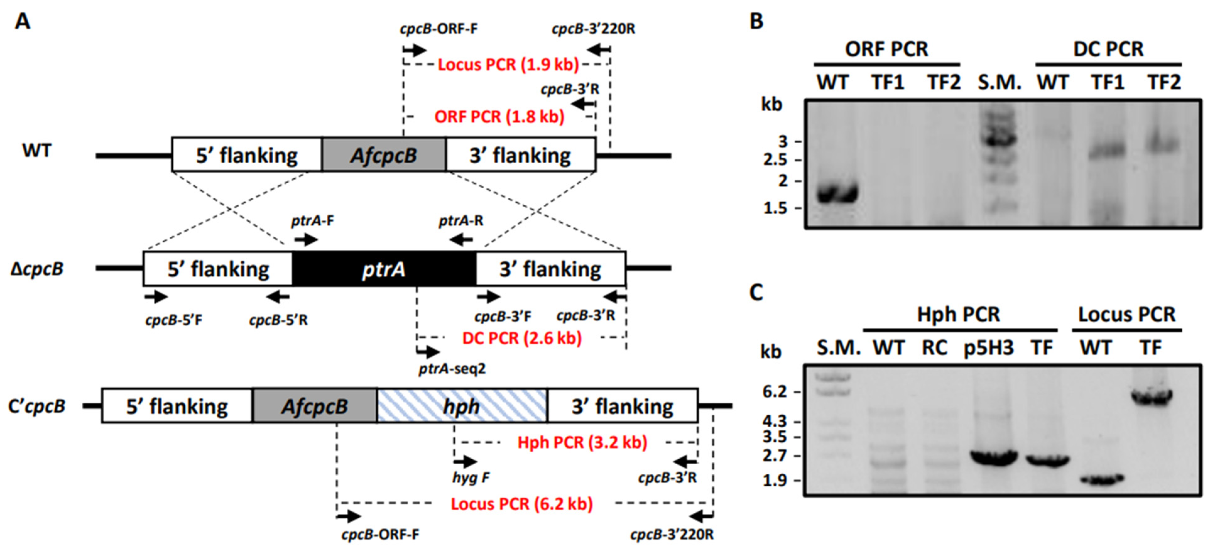
3.1. Targeted Deletion of AfcpcB

In A. fumigatus, cpcB-deletion strains were already generated in Af293.1 (pyrG1) and A1160 (Δku80; pyrG; veA1), termed Af293.1-7 and CZ01, respectively [9,11]. However, the Af293 strain is medium fertility, which produces low numbers of cleistothecia [13]. Ku70 and Ku80 form a heterodimer and play an important role in the non-homologous end joining (NHEJ) process in DNA repair [45]. The akuBKu80 mutant showed an increased rate of homologous recombination, which resulted in the majority of transformed progenies with insertion mutagenesis [45,46]. While the Δku80 strain is a useful tool to improve transformation efficiency, it is not suitable for the study of the sexual development required for the recombinational repair process [47]. To investigate the functional roles of AfCpcB in the sexual development in A. fumigatus, we generated the ΔAfcpcB strain in the supermater parental AFB62, which can produce a high number of cleistothecia [31]. The deletion construct was amplified using modified fusion PCR and employed for protoplast transformation. Putative transformants were grown on GMM agar containing pyrithiamine, and two candidates were selected based on PCR screening using cpcB ORF-F/cpcB 3’R and ptrA-seq2/cpcB-3’R primer sets for the open reading frame (ORF) PCR and disruption cassette (DC) PCR, respectively (Supplementary Table S1). As illustrated in Figure 1A, the genomic DNA of the transformants was not amplified using the ORF PCR primer set but amplified using the DC PCR primer set. Results from the PCR revealed a 1.8-kb band in the wild-type (WT) by ORF PCR and a 2.6-kb band in knock-out mutants based on DC PCR (Figure 1B). To complement the mutation, PCR fragments carrying a wild-type copy of cpcB, flanked by a hygromcyin resistance gene were used to transform ΔAfcpcB protoplasts. Complementation of the cpcB gene in the ΔAfcpcB strain background was confirmed by PCR using hyg F/cpcB 3’R and cpcB-ORF-F/cpcB-3’220R primer sets for Hph PCR and Locus PCR, respectively (Figure 1C).

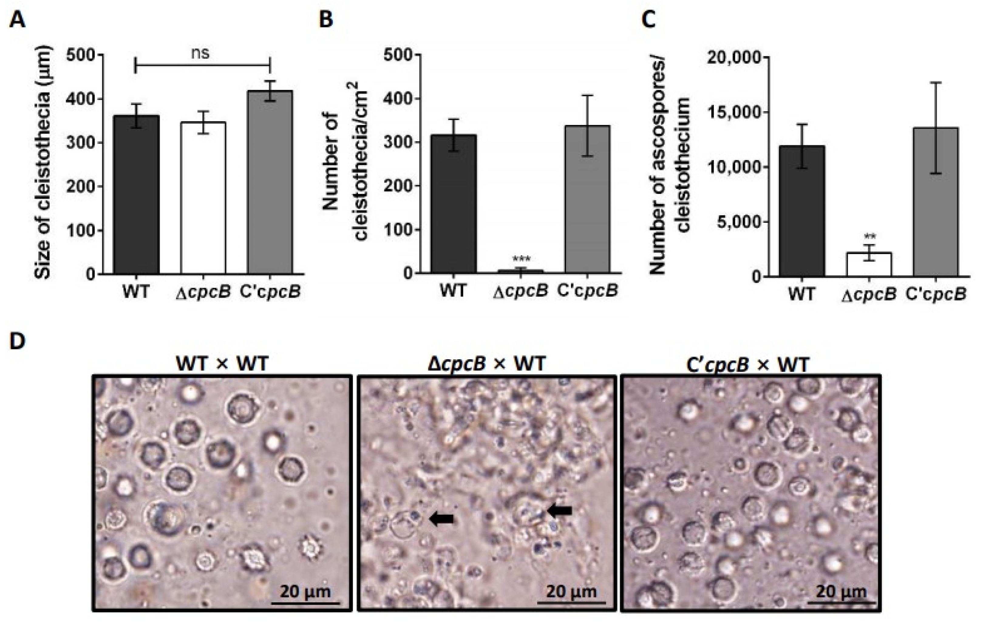
3.2. AfCpcB Is Required for Complete Sexual Development

To elucidate the role of AfCpcB in cleistothecia formation, the ΔAfcpcB strain (MAT1-1) was crossed with the supermater AFIR928 strain (MAT1-2) using the VeM method [13,14]. In oatmeal agar, A. fumigatus produces both asexual (green conidia) and sexual (white cleistothecia) reproductive organs [13]. Crossing the ΔAfcpcB strain with the AFIR928 strain (see Figure 2A, ΔcpcB) formed cleistothecia similar to the cleistothecia formed by crossing of the AFB62 with the AFIR928 strain (see Figure 2A, WT) and the C’AfcpcB strain with the AFIR928 strain (see Figure 2A, C’cpcB) (Figure 2A). However, the number of cleistothecia produced in the ΔAfcpcB × AFIR928 cross (6.3 ± 6.3 cleistothecia/cm2) was significantly lower than that in the WT pair (315.8 ± 36.4 cleistothecia/cm2) and C’cpcB pair (337.2 ± 69.6 cleistothecia/cm2) (Figure 2B). To investigate the formation of ascospores, which are final sexual reproductive organs, we picked up cleistothecia and ruptured them on glass slides. A cloud of ascospores with two equatorial crests was released from a cleistothecium formed by crossing the WT pair (Figure 2D, left) and the C’AfcpcB × WT pair (Figure 2D, right). In the ΔAfcpcB × AFIR928 cross, few or no ascospores were formed in cleistothecia. Instead, a mass of irregularly shaped ascogenous cells (black arrows) can be seen within the cleistothecia (Figure 2D, center). In agreement with phenotypic data, statistical analysis revealed that the cleistothecia in the ΔAfcpcB × AFIR928 cross contained only about 16~18% of the ascospores (2181 ± 706 ascospores) compared to that in the WT pair cross (11,891 ± 2006 ascospores) and the C’AfcpcB × WT pair (13,556 ± 4129 ascospores) (Figure 2C). Taken together, these data suggest that AfCpcB is required for the complete sexual development of A. fumigatus.

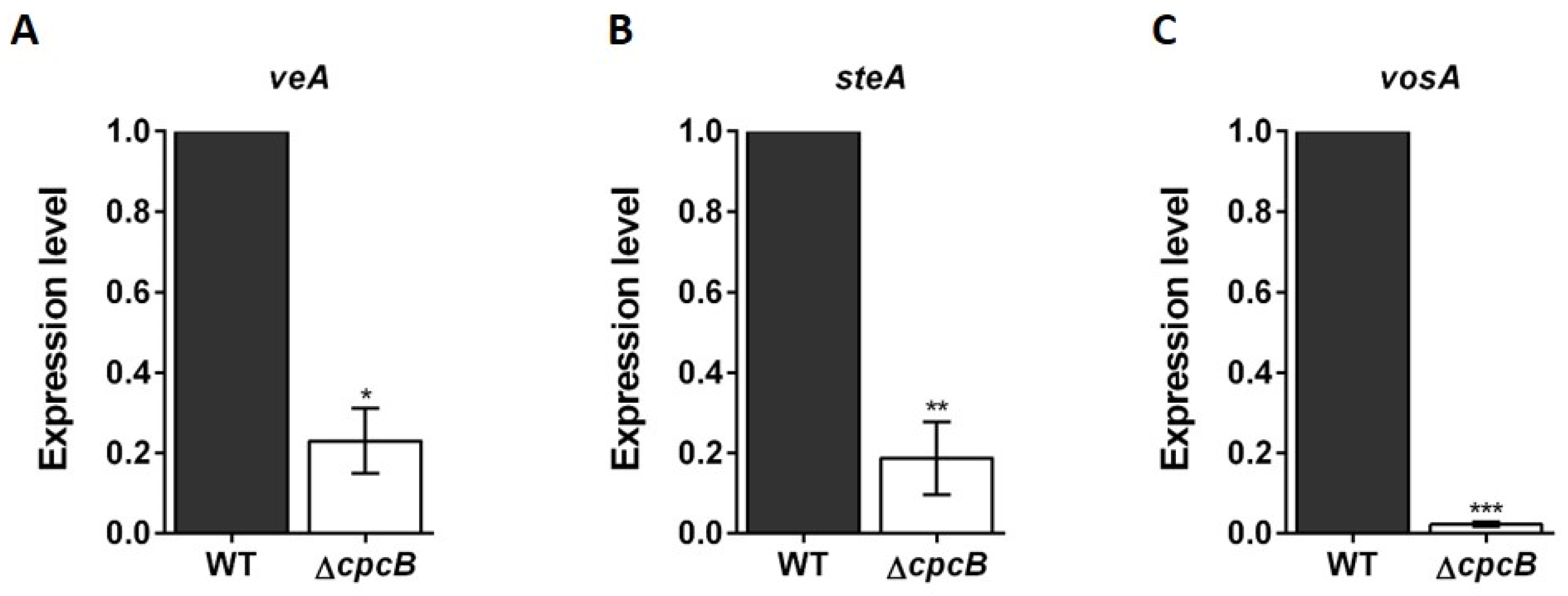
3.3. Transcript Expression Patterns in Cleistothecia

Mating induced by the VeM method on oatmeal agar produced a high number of both conidia and cleistothecia [14]. To investigate transcript expression patterns in only sexual reproductive organs containing ascus and ascospores, we performed the amplification of cDNA using total RNA extracted from cleistothecia.
To investigate the effect of AfcpcB deletion on the transcription levels of genes involved in sexual development, veA, steA, and vosA expression levels were detected in cleistothecia sorted from the AFB62 × AFIR928 and ΔAfcpcB × AFIR928 mated cultures. In A. fumigatus, VeA is required for conidiation, gliotoxin production, and protease activity [48]. In A. nidulans, VeA activates sexual development, and controlled gene expression is required during sexual development [11,37,49]. SteA (sterile 12-like) is a homeodomain-C2/H2-Zn+2 finger transcription factor, and the steA-deletion strain does not differentiate cleistothecia, ascogenous hyphae, asci, or ascospores, thereby suggesting that SteAp functions very early in the sexual cycle [50]. VosA (viability of spores) is required for spore viability, and the vosA-deletion strain produces defective cleistothecia containing a very small number of ascospores [51]. The steA and vosA genes are expressed in the early and late sexual stages, respectively [37]. When the transcription of sexual development-associated genes veA, steA, and vosA were analyzed in A. fumigatus, transcript levels of veA, steA, vosA genes were found to be significantly decreased by 4.3-fold, 5.3-fold, and 44.4-fold, respectively, in the cleistothecia that originated from the ΔAfcpcB × AFIR928 cross (Figure 3). These results indicate that AfCpcB is essential for the completion of sexual development by regulating the transcription of genes associated with early and late sexual development in A. fumigatus.

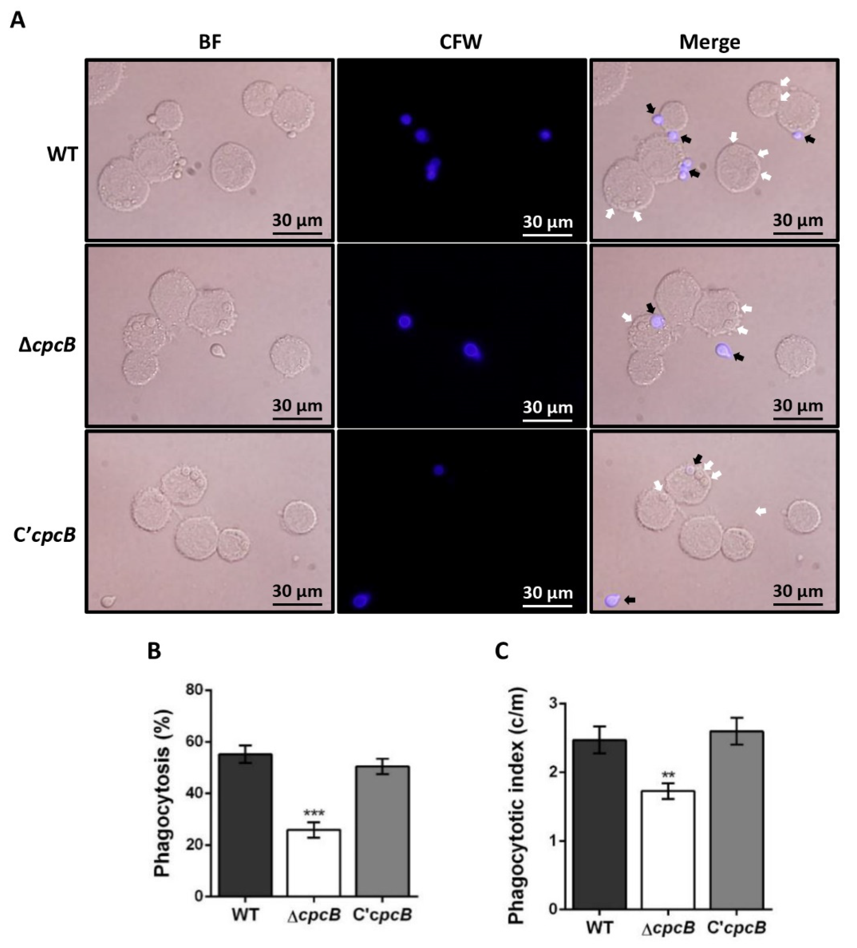
3.4. The ΔAfcpcB Mutant Is More Resistant to Phagocytosis by Alveolar Macrophages

To investigate the interaction between conidia and murine alveolar macrophages, adherent macrophages were challenged with conidia and co-cultured for 4 h. A significant decrease in the number of macrophages that ingested ΔAfcpcB conidia (36.1 ± 2.0%), compared to WT conidia (57.8 ± 1.9%) and C’AfcpcB conidia (56.8 ± 2.2%), was observed (Figure 4A,B). The phagocytic index, which represents the average number of conidia per macrophage, was also lower in macrophages containing ΔAfcpcB conidia (1.4 vs. 2.0 c/m) (Figure 4C). These in vitro data suggest that AfcpcB-deletion influences phagocytosis of conidia by possible changes in the cell wall components of the conidia surface.

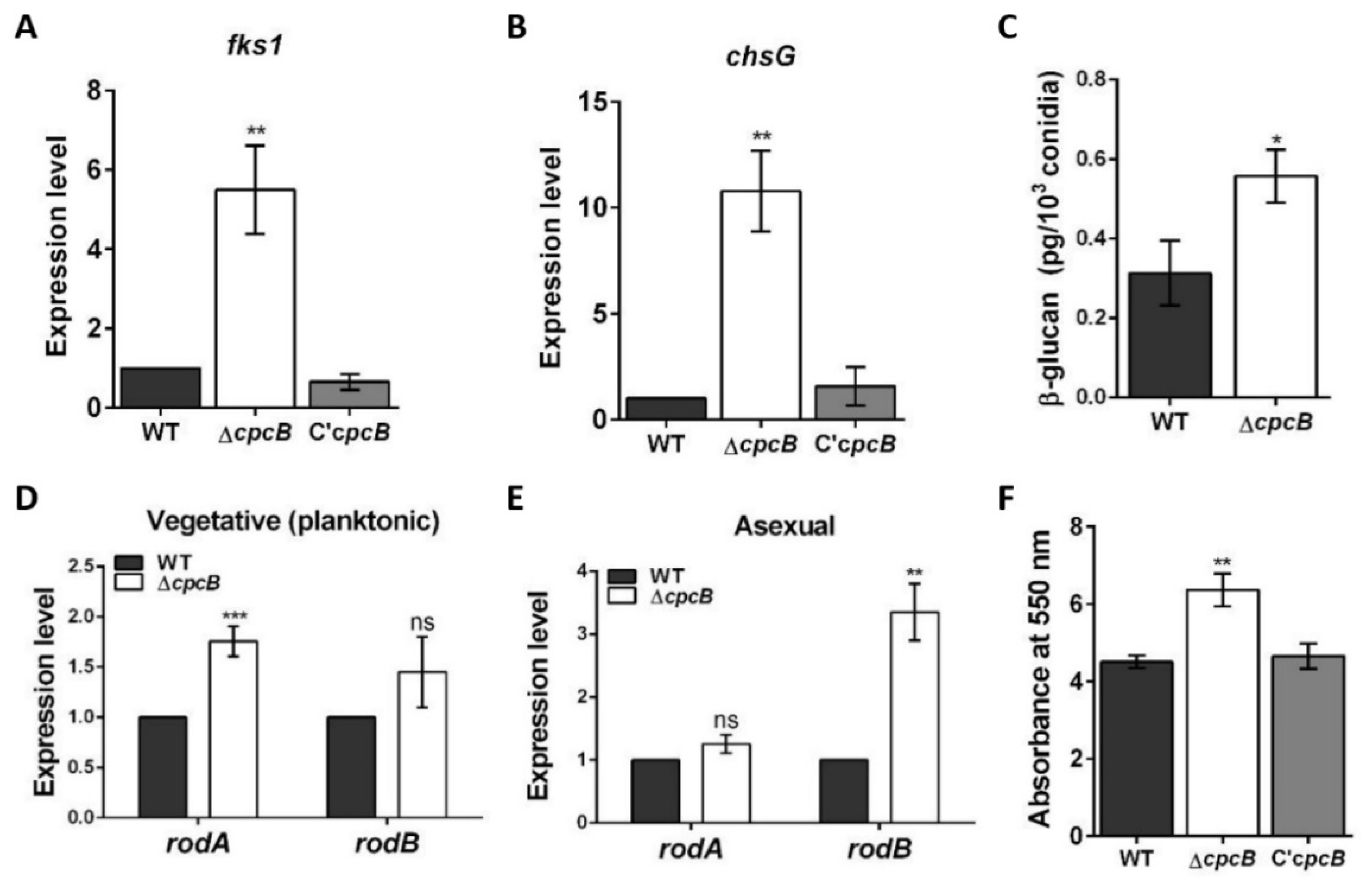
3.5. The AfcpcB Is Required for Biosynthesis of Cell Wall Components and Hydrophobin

To determine why the ΔAfcpcB conidia can deter phagocytosis by alveolar macrophages, we investigated the expression of cell wall biosynthesis genes. The fks1 and chsG genes encode β-1,3-glucan synthase and chitin synthase, respectively [52]. The fks1 and chsG mRNA were upregulated in the ΔAfcpcB strain (Figure 5A,B). In accordance with the fks1 expression pattern, the amount of β-1,3-glucan content was higher than that of WT (Figure 5C). These data suggested that AfCpcB is involved in the A. fumigatus cell wall biosynthesis.
Next, the expression of hydrophobin genes rodA and rodB was evaluated. Conidia are covered by outer layers, composed of the hydrophobins and melanin layers [17]. These layers protect conidia against host defense by masking the polysaccharides on the conidial surface, such as glucan and chitin, and hindering PRR-dependent recognition. We hypothesized that a change in hydrophobin composition in the ΔAfcpcB strain affects the PAMP–PRR interaction. The ΔAfcpcB strain showed a 1.8-fold increase in expression of the rodA gene during the vegetative stage (Figure 5D) and a 3.4-fold increase in the expression of the rodB gene during the asexual stage (Figure 5E). The extracellular matrix (ECM) of fungi is involved in biofilm formation [53]. In A. fumigatus biofilm condition, rodA and rodB were highly expressed [54]. To evaluate the ability of each strain to form biofilms on a solid surface, biofilms grown in 12-well plates were quantified using the crystal violet assay. Biofilm formation was increased in the ΔAfcpcB strain compared to WT and C’AfcpcB strain (Figure 5F). These results suggested that the AfcpcB strain causes a possible change in both inner and outer cell wall layers, thus affecting the capacity of PRRs in host immune cells to react with the A. fumigatus conidia surface.

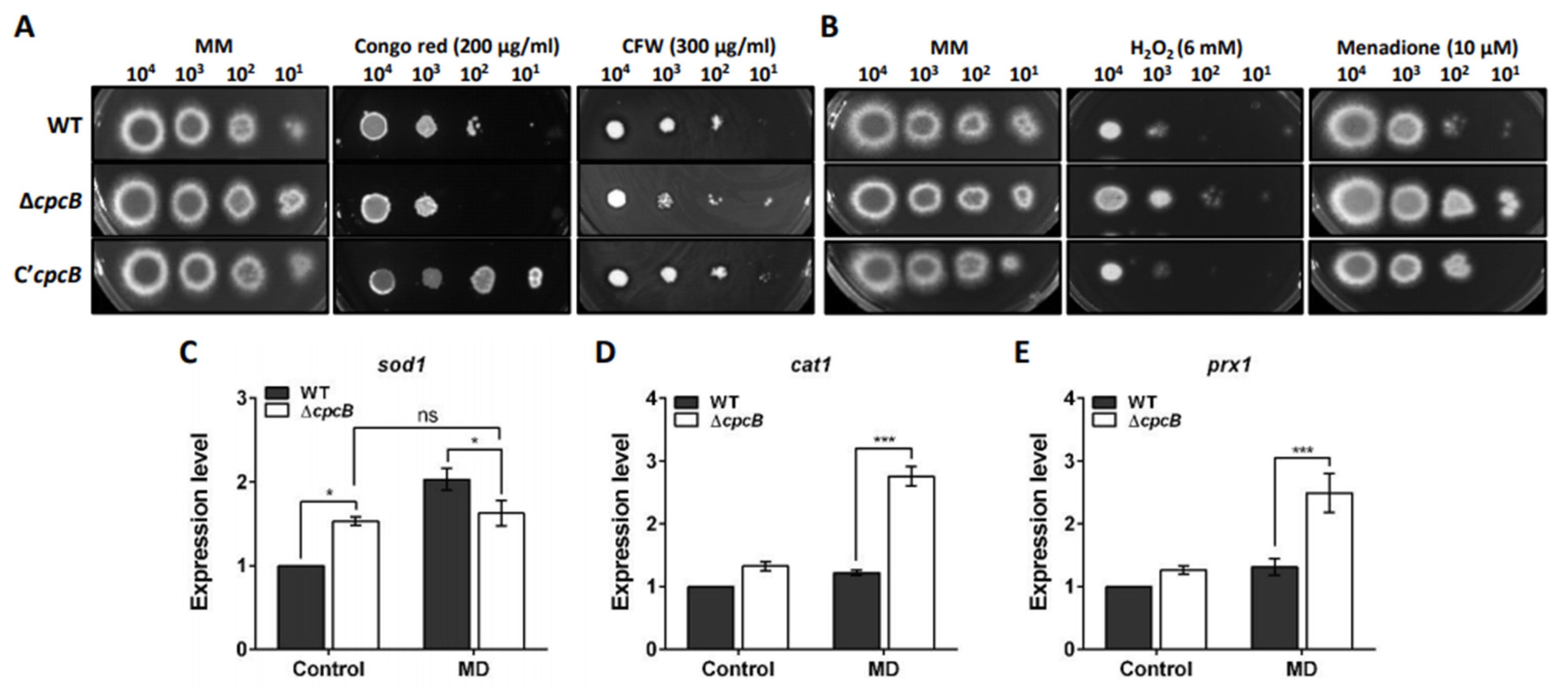
3.6. AfCpcB Functions in Response to Cell Wall and Oxidative Stress

To elucidate the cellular functions of CpcB in A. fumigatus, the effect of AfcpcB deletion on the response to external stress was investigated. Understanding cell wall integrity signaling is important for the study of drug response and virulence. Many fungal mutants with defective cell walls are sensitive to antifungal reagents, such as CFW and CR [55,56]. CFW binds to β-1,3 and β-1,4 polysaccharides in chitin [57]. The fungus responds to CR, which is considered to bind to polysaccharides and form covalent links between chitin chains and β-glucan, by modifying its cell wall composition and permeability [58]. The ΔAfcpcB strain was sensitive to CR compared to WT (Figure 6A). When the response to osmotic stress (NaCl and sorbitol) and DNA replicative stress (hydroxyurea, methyl methanesulfonate, and benomyl) was examined, no significant abnormalities were observed in the ΔAfcpcB strain (Supplementary Figure S1A,B).
The capacity of scavenging ROS produced by host phagocytes is regarded as a putative virulence factor in pathogenic fungi [59]. While ROS are byproducts of normal aerobic metabolism that participate in the signaling pathway [60], ROS generated by host defense mechanisms can kill conidia and induce hyphal defects in A. fumigatus [61]. To characterize the role of AfCpcB in response to oxidative stress, we investigated the resistance of the ΔAfcpcB strain to H2O2 and MD. The ΔAfcpcB strain showed increased resistance to H2O2 and MD compared to WT (Figure 6B). We investigated the mRNA expression of several genes known to be involved in the oxidative response in A. fumigatus, and particularly in the presence of MD (10 μM): sod1 encoding a copper-dependent superoxide dismutase (SOD), cat1 encoding a mycelial catalase, and prx1 encoding Cys-based peroxidase [62,63,64]. Upon MD treatment, the WT strain showed increased expression of sod1 (about 2-fold); however, the ΔAfcpcB strain showed no remarkable change in expression patterns and continuous expression of sod1 (Figure 6C). The expression levels of cat1 and prx1 were increased in the ΔAfcpcB strain in the presence of MD, compared to that in the WT strain (approximately 1.9-fold and 2.1-fold, respectively) (Figure 6D,E). Taken together, these results indicate that AfCpcB contributes to the transcriptional regulation of ROS scavenging enzymes, which are encoded by cat1 and prx1, and upregulation of these genes in the ΔAfcpcB strain may contribute to resistance against oxidative stress.

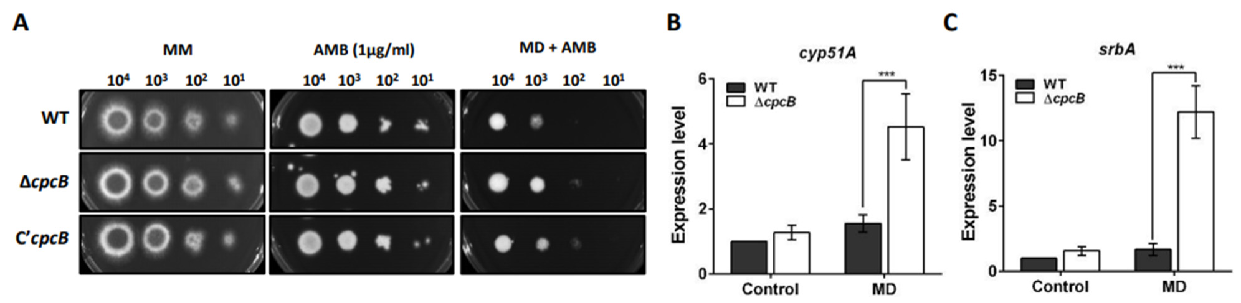
3.7. The ΔAfcpcB Strain under Oxidative Stress Showed Increased Expression of Ergosterol Biosynthesis-Related Genes

Azoles inhibit fungal sterol 14-α demethylase (Cyp51/Erg11A), which is required for ergosterol biosynthesis [65]. Polyenes, such as AMB and nystatin, bind to ergosterol, which kills cells by allowing membrane permeabilization and extracting ergosterol in the membrane [66]. Cai et al. revealed that the ΔAfcpcB strain in a veA1 and Δku80 background—which is not a suitable model for sexual development—demonstrated increased resistance against antifungal drugs, including voriconazole, bifonazole, and AMB [9]. To verify whether the supermater ΔAfcpcB strain exhibited resistance against antifungal drugs, conidia were serially diluted and spotted onto GMM with AMB. The ΔAfcpcB strain exhibited no significant change in resistance against AMB (Figure 7A, AMB). To examine the function of CpcB in ergosterol biosynthesis, the expression of cyp51A and srbA encoding sterol regulatory element-binding protein (SREBP) was analyzed using RT-qPCR. SrbA is a transcription factor containing a basic helix-loop-helix (bHLH) leucine zipper DNA binding domain and is required for cell polarity, hypoxia response, and azole drug resistance [25]. When overnight-cultured mycelia were transferred onto GMM agar, the expression pattern of both cyp51A and srbA genes showed no remarkable change between the WT and ΔAfcpcB strains (Figure 7B,C, control). To adapt to oxidative stress caused by MD, ergosterol biosynthesis is repressed in yeast [67]. We next investigated whether oxidative stress would cause a transcriptional adaptation of ergosterol biosynthesis. The ΔAfcpcB strain exhibited slightly increased resistance when MD was added to the AMB-containing medium (Figure 7A, MD + AMB). Under oxidative stress, the ΔAfcpcB strain showed increased expression of both the cyp51A and srbA genes (approximately 2.9-fold and 7.3-fold, respectively) (Figure 7B,C, MD). These data suggested that deletion of the AfcpcB gene causes dysregulation of the cyp51A and srbA expression under oxidative stress conditions.

4. Discussion

Although CpcB was found to be required for vegetative growth, germination, asexual development, and gliotoxin production in A. fumigatus [8] and growth and development of A. nidulans [11], the cpcB deletion in the A. fumigatus supermater strain showed no significant change in vegetative growth and asexual development (data not shown). Further investigation is required to explain the strain-dependent variation, however, it is noteworthy that a number of studies demonstrate significant interstrain variability with respect to phonotypes, immune responses, and virulence in various models of infection [68].
While mature cleistothecia of A. nidulans are black spheres with numerous red-purple ascospores [37,69], the ΔAncpcB strain produces small immature bright-red cleistothecia with no ascospores and a reduced number of cleistothecia and ascospores [11]. Based on the fact that AnCpcB is important in sexual development in A. nidulans, we investigated whether AfCpcB plays a crucial role in the sexual development in A. fumigatus. To investigate the role of AfCpcB in sexual development, the use of the supermater strain as a recipient for the generation of deletion-strain was indispensable. Therefore, we constructed an AfcpcB-deletion mutant using the supermater strain AFB62 as a recipient strain and crossed it with the supermater strain AFIR928 with the opposite mating type. The ΔAfcpcB × AFIR928 cross did not affect the size of cleistothecia, but rather the production of cleistothecia and the formation of ascospores (Figure 2). This is the first report to demonstrate that AfCpcB is involved in sexual development in A. fumigatus.
Unlike sexual induction in A. nidulans, the VeM method in A. fumigatus on oatmeal agar produced both asexual (conidia) and sexual (cleistothecia) reproductive organs [14]. Given the difficulties of molecular analysis using sexual developmental cultures in A. fumigatus, we investigated transcription patterns in cleistothecia that contain many asci and ascospores. The ΔAfcpcB × AFIR928 cross showed decreased veA, steA, and vosA transcription (Figure 3). Studies using ΔAncpcB and ΔAnsvfA strains have previously shown that proper expression of the veA gene is required for sexual development in A. nidulans [11,37]. These data suggest that CpcB is involved in the regulation of veA gene expression during sexual development in A. nidulans and A. fumigatus. In some filamentous fungi including A. nidulans, STE12 mutants showed impaired sexual reproduction [50,70,71,72]. The absence of cleistothecial primordia or foci indicates that SteA is required for an early sexual cycle. The vosA gene is expressed in conidia and ascospores for viability in A. nidulans [51]. Immature cleistothecia that contain no ascospores from the ΔAfcpcB × AFIR928 cross showed significantly reduced vosA expression, suggesting that VosA is expressed in ascospores in A. fumigatus. Although transcription patterns were not investigated at different developmental stages, these data indicate that AfCpcB is required for cleistothecia maturation by controlling the expression of genes such as steA and vosA.
Fungi engage in transcriptional, post-translational, and enzymatic strategies against oxidative stress, and such strategies include SOD, catalase, and peroxidase activities [73]. The ΔAfcpcB strain showed increased resistance to both H2O2 and MD (Figure 6B). MD generates intracellular ROS through the accumulation of superoxide anions and mitochondrial depolarization, which induces apoptosis [73,74,75]. As superoxide anions are unstable, they are converted to H2O2 either spontaneously or by SOD. Moreover, while the WT strain responds to MD treatment by inducing sod1 expression, the ΔAfcpcB strain continuously expresses the sod1 gene both under normal and oxidative stress conditions (Figure 6C). H2O2 is detoxified by various ROS scavenging enzymes, such as catalase, glutathione peroxidase, and thioredoxins. Our data showed upregulated expression of cat1 and prx1 genes in the ΔAfcpcB strain against MD treatment (Figure 6D,E), which can explain the increased resistance against oxidative stress in the spotting assay. These data suggest that CpcB negatively regulates the expression of cat1 and prx1 genes under oxidative stress conditions.
Oxidative stress represses the transcription of the ergosterol biosynthesis gene (ERG) and causes decreased levels of cellular ergosterol in the budding yeast, Saccharomyces cerevisiae [67]. In the fission yeast Schizosaccharomyces pombe, ergosterol levels are repressed during oxidative stress [76]. Transcriptomic and proteomic analyses revealed that hypoxia affects metabolic changes, such as ergosterol biosynthesis, in A. fumigatus [77]. A Zn2-Cys6 transcription factor AtrR positively regulates sterol biosynthesis [78]. The CCAAT-binding domain complex (CBC), a heterotrimer comprising HapB, HapC, and HapE, is a negative regulator of sterol biosynthesis [79]. Binding of the CBC at the cyp51A promoter is facilitated by another transcriptional regulator, an iron-responsive bZIP transcription factor HapX. SrbA directly regulates ergosterol transcription during hypoxia [80]. These studies suggest that regulation of ergosterol biosynthesis is required for an adaptive response to extracellular or intracellular stimuli, including ROS. Understanding the Aspergillus ergosterol biosynthesis pathway is also important to facilitate the development of antifungal drugs [81]. The ΔAfcpcB strain, generated in this study using the supermater AFB62, exhibited enhanced resistance in GMM containing both MD and AMB (Figure 7A). While WT showed no remarkable change in srbA and cyp51A gene expression under oxidative stress conditions, the ΔAfcpcB strain induced cyp51A and srbA expression under oxidative stress conditions (Figure 7B,C). These data indicate that, unlike yeast, transcriptional repression of ERG is not always necessary under MD-treated conditions in A. fumigatus. There is limited information on the mechanisms for ergosterol biosynthesis under oxidative stress conditions in A. fumigatus; however, our results suggest that AfCpcB plays a role in the negative regulation of cyp51A and srbA gene expression under oxidative stress conditions.
In this study, we report the involvement of AfCpcB in phagocytosis by alveolar macrophages for the first time. When we tested phagocytosis of murine alveolar macrophages challenged with conidia, the phagocytosis percentage and indigested number of ΔAfcpcB conidia by macrophages as values were reduced (Figure 4). These data suggest that AfCpcB affects the mechanisms of fungal recognition and internalization by macrophages. The inner cell wall components of conidia, such as glucan, galactomannan, and chitin, are recognized by PRRs in host cells [15,61]. The ΔAfcpcB strain decreased the resistance of A. fumigatus to CR (Figure 6A), and increased expression of cell wall genes and amounts of β-glucan (Figure 5), suggesting that AfCpcB modifies the properties of the cell wall surface. Cai et al. showed that the electron-dense pigment layer of the cell wall surface in both cpcB and gpaB (encoding Gα subunit)-deletion strains showed disrupted phenotypes in cell wall integrity in A. fumigatus [8]. In addition, the fungal cell wall features an outer layer composed of hydrophobins (rodlet proteins) and melanin, which mask cell wall PAMPs in conidia [82]. Among the seven rodlet proteins (RodA–RodG) in A. fumigatus, RodA is required for the formation of a rodlet layer and protection from phagocytosis by alveolar macrophages [21]. The rodA gene is not differentially expressed in planktonic culture but highly expressed in sporulating culture [54]. Although RodB is not required for rodlet formation and ΔrodB did not show any phenotype in biofilm conditions, the rodB gene is highly expressed in biofilm and in mice invasive aspergillosis model [21,54,82]. While the rodB gene is not differentially expressed in asexual culture, the ΔAfcpcB showed an increase in rodA and rodB genes in planktonic and asexual culture, respectively (Figure 5D,E), suggesting that AfCpcB is involved in the regulation of expression of hydrophobin genes. Taken together, these results suggest that AfCpcB might affect the phagocytosis by alveolar macrophages by regulating cell wall integrity and the formation of hydrophobins.
Interestingly, Cai et al. showed that the virulence of the ΔAfcpcB strain in a veA1 and Δku80 background is attenuated in an immunosuppressed mouse model (survival rate of 60%) [8]. While the ΔAfcpcB strain in a veA1 and Δku80 background showed a decreased growth rate, defects in asexual reproductive organs, and increased resistance against antifungal drugs, our ΔAfcpcB strain showed a similar growth rate with WT [8], defects in sexual reproductive organs, and increased resistance against oxidative stress. It is necessary to confirm the virulence of our ΔAfcpcB strain using an in vivo model system.
To summarize, we found that AfCpcB plays important roles in sexual development and the phagocytosis by alveolar macrophages. Based on our data together with the results of previous studies, we conclude that CpcB, a Gβ-like protein, might have functions that are involved in oxidative stress response and maintenance of cell wall properties.

Supplementary Materials

The following supporting information can be downloaded at: https://www.mdpi.com/article/10.3390/jof8010056/s1, Figure S1: Effects of AfcpcBdisruption on sensitivity to osmotic and DNA replicative stresses. Conidia with 10-fold serial dilutions were spotted onto media and then, the plates were cultured at 37 °C for 2 days. (A) Sensitivity of the strains to osmotic stresses (1.5 M NaCland 2.5 M sorbitol). (B) Sensitivity of the strains to DNA replicative stresses (15 mM HU, 10 μMMMS and 0.6 μg/mL of benomyl). HU, hydroxyurea; MMS, methyl methanesulfonate. Table S1. Primers used in this study.

Author Contributions

Conceptualization, H.-M.P.; funding acquisition, H.-M.P.; investigation, J.-Y.L. and Y.-J.K.; methodology, J.-Y.L., Y.-J.K. and H.-M.P.; project administration, H.-M.P.; supervision, H.-M.P.; visualization, J.-Y.L.; writing—original draft, J.-Y.L.; writing—review and editing, J.-Y.L. and H.-M.P. All authors have read and agreed to the published version of the manuscript.

Funding

This research was funded by the Basic Science Research Program through the National Research Foundation of Korea, grant number 2021R1A2C1009229.

Institutional Review Board Statement

Not applicable.

Informed Consent Statement

Not applicable.

Data Availability Statements

Not applicable.

Conflicts of Interest

The authors report no conflict of interest. The authors alone are responsible for the content and the writing of the paper.

References

  1. Richardson, M.; Bowyer, P.; Sabino, R. The human lung and Aspergillus: You are what you breathe in? Med. Mycol. 2019, 57, S145–S154. [Google Scholar] [CrossRef] [Green Version]
  2. Sakai, K.; Hiemori, K.; Tateno, H.; Hirabayashi, J.; Gonoi, T. Fucose-specific lectin of Aspergillus fumigatus: Binding properties and effects on immune response stimulation. Med. Mycol. 2019, 57, 71–83. [Google Scholar] [CrossRef]
  3. Abad, A.; Victoria Fernández-Molina, J.; Bikandi, J.; Ramírez, A.; Margareto, J.; Sendino, J.; Luis Hernando, F.; Pontón, J.; Garaizar, J.; Rementeria, A. What makes Aspergillus fumigatus a successful pathogen? Genes and molecules involved in invasive aspergillosis. Rev. Iberoam. Micol. 2010, 27, 155–182. [Google Scholar] [CrossRef]
  4. Pappas, P.G.; Alexander, B.D.; Andes, D.R.; Hadley, S.; Kauffman, C.A.; Freifeld, A.; Anaissie, E.J.; Brumble, L.M.; Herwaldt, L.; Ito, J.; et al. Invasive fungal infections among organ transplant recipients: Results of the Transplant-Associated Infection Surveillance Network (TRANSNET). Clin. Infect. Dis. 2010, 50, 1101–1111. [Google Scholar] [CrossRef]
  5. Koehler, P.; Cornely, O.A.; Böttiger, B.W.; Dusse, F.; Eichenauer, D.A.; Fuchs, F.; Hallek, M.; Jung, N.; Klein, F.; Persigehl, T.; et al. COVID-19 associated pulmonary aspergillosis. Mycoses 2020, 63, 528–534. [Google Scholar] [CrossRef]
  6. Chan, J.F.; Lau, S.K.; Wong, S.C.; To, K.K.; So, S.Y.; Leung, S.S.; Chan, S.; Pang, C.; Xiao, C.; Hung, I.F.; et al. A 10-year study reveals clinical and laboratory evidence for the ‘semi-invasive’ properties of chronic pulmonary aspergillosis. Emerg. Microbes Infect. 2016, 5, 1–7. [Google Scholar] [CrossRef] [Green Version]
  7. Lafon, A.; Han, K.; Seo, J.; Yu, J. G-protein and cAMP-mediated signaling in aspergilli: A genomic perspective. Fungal Genet. Biol. 2019, 43, 490–502. [Google Scholar] [CrossRef]
  8. Cai, Z.D.; Chai, Y.F.; Zhang, C.Y.; Qiao, W.R.; Sang, H.; Lu, L. The Gβ-like protein CpcB is required for hyphal growth, conidiophore morphology and pathogenicity in Aspergillus fumigatus. Fungal Genet. Biol. 2015, 81, 120–131. [Google Scholar] [CrossRef]
  9. Cai, Z.; Chai, Y.; Zhang, C.; Feng, R.; Sang, H.; Lu, L. Molecular characterization of Gβ-like protein CpcB involved in antifungal drug susceptibility and virulence in A. fumigatus. Front. Microbiol. 2016, 7, 106. [Google Scholar] [CrossRef]
  10. Hoffmann, B.; Wanke, C.; LaPaglia, S.K.; Braus, G.H. c-Jun and RACK1 homologues regulate a control point for sexual development in Aspergillus nidulans. Mol. Microbiol. 2000, 37, 28–41. [Google Scholar] [CrossRef] [Green Version]
  11. Kong, Q.; Wang, L.; Liu, Z.; Kwon, N.; Kim, S.C.; Yu, J.H. Gβ-Like CpcB plays a crucial role for growth and development of Aspergillus nidulans and Aspergillus fumigatus. PLoS ONE 2013, 8, e70355. [Google Scholar] [CrossRef] [Green Version]
  12. O’Gorman, C.M.; Fuller, H.T.; Dyer, P.S. Discovery of a sexual cycle in the opportunistic fungal pathogen Aspergillus fumigatus. Nature 2009, 457, 471–474. [Google Scholar] [CrossRef]
  13. Sugui, J.A.; Losada, L.; Wang, W. Identification and characterization of an Aspergillus fumigatus supermater pair. MBio 2011, 2, e00234-11. [Google Scholar] [CrossRef] [Green Version]
  14. Lim, J.Y.; Park, H.M. Expression of sexual genes in Aspergillus fumigatus homogeneous culture produced by vegetative mass mating. J. Microbiol. 2019, 57, 1–6. [Google Scholar] [CrossRef]
  15. Bellocchio, S.; Bozza, S.; Montagnoli, C.; Perruccio, K.; Gaziano, R.; Pitzurra, L.; Romani, L. Immunity to Aspergillus fumigatus: The basis for immunotherapy and vaccination. Med. Mycol. 2005, 43, 181–188. [Google Scholar] [CrossRef] [Green Version]
  16. Akoumianaki, T.; Kyrmizi, I.; Valsecchi, I.; Beauvais, A.; Latge, J.P.; Chamilos, G. Aspergillus cell wall melanin blocks LC3-associated phagocytosis to promote pathogenicity. Cell Host Microbe 2016, 19, 79–90. [Google Scholar] [CrossRef] [Green Version]
  17. Hatinguais, R.; Willment, J.A.; Brown, G.D. PAMPs of the fungal cell wall and mammalian PRRs. Curr. Top. Microbiol. Immunol. 2020, 425, 187–223. [Google Scholar] [CrossRef]
  18. Heinekamp, T.; Thywißen, A.; Macheleidt, J.; Keller, S.; Valiante, V.; Brakhage, A.A. Aspergillus fumigatus melanins: Interference with the host endocytosis pathway and impact on virulence. Front. Microbiol. 2012, 3, 440. [Google Scholar] [CrossRef] [Green Version]
  19. Langfelder, K.; Streibel, M.; Jahn, B.; Haase, G.; Brakhage, A.A. Biosynthesis of fungal melanins and their importance for human pathogenic fungi. Fungal Genet. Biol. 2003, 38, 143–158. [Google Scholar] [CrossRef]
  20. Dagenais, T.R.T.T.; Keller, N.P. Pathogenesis of Aspergillus fumigatus in invasive aspergillosis. Clin. Microbiol. Rev. 2009, 22, 447–465. [Google Scholar] [CrossRef] [Green Version]
  21. Beauvais, A.; Latgé, J.P. Aspergillus biofilm in vitro and in vivo. Microbiol. Spectr. 2015, 3, 1–10. [Google Scholar] [CrossRef] [Green Version]
  22. Aimanianda, V.; Latgé, J.P. Fungal hydrophobins form a sheath preventing immune recognition of airborne conidia. Virulence 2010, 1, 185–187. [Google Scholar] [CrossRef]
  23. Matherne, G.P.; Headrick, J.P.; Coleman, S.D.; Berne, R.M. Interstitial transudate purines in normoxic and hypoxic immature and mature rabbit hearts. Pediatr. Res. 1990, 28, 348–353. [Google Scholar] [CrossRef] [Green Version]
  24. Fröhlich, S.; Boylan, J.; Mcloughlin, P. Hypoxia-induced inflammation in the lung: A potential therapeutic target in acute lung injury? Am. J. Respir. Cell Mol. Biol. 2013, 48, 271–279. [Google Scholar] [CrossRef]
  25. Willger, S.D.; Puttikamonkul, S.; Kim, K.H.; Burritt, J.B.; Grahl, N.; Metzler, L.J.; Barbuch, R.; Bard, M.; Lawrence, C.B.; Cramer, R.A. A sterol-regulatory element binding protein is required for cell polarity, hypoxia adaptation, azole drug resistance, and virulence in Aspergillus fumigatus. PLoS Pathog. 2008, 4, e1000200. [Google Scholar] [CrossRef] [Green Version]
  26. Han, K.H.; Lee, D.B.; Kim, J.H.; Kim, M.S.; Han, K.Y.; Kim, W.S.; Park, Y.S.; Kim, H.B.; Han, D.M. Environmental factors affecting development of Aspergillus nidulans. J. Microbiol. 2003, 41, 34–40. [Google Scholar]
  27. Zhang, J.; Snelders, E.E.; Zwaan, B.J.; Schoustra, S.E.; Kuijper, E.J.; Arendrup, M.C.; Melchers, W.J.G.; Verweij, P.E.; Debets, A.J.M. Relevance of heterokaryosis for adaptation and azole-resistance development in Aspergillus fumigatus. Proc. Biol. Sci. 2019, 286, 20182886. [Google Scholar] [CrossRef] [Green Version]
  28. Gintjee, T.J.; Donnelley, M.A.; Thompson, G.R. Aspiring antifungals: Review of current antifungal pipeline developments. J. Fungi 2020, 6, 28. [Google Scholar] [CrossRef] [Green Version]
  29. Mellado, E.; Diaz-Guerra, T.M.; Cuenca-Estrella, M.; Rodriguez-Tudela, J.L. Identification of two different 14-α sterol demethylase- related genes (cyp51A and cyp51B) in Aspergillus fumigatus and other Aspergillus species. J. Clin. Microbiol. 2001, 39, 2431–2438. [Google Scholar] [CrossRef] [Green Version]
  30. Van Rhijn, N.; Bromley, M.; Richardson, M.; Bowyer, P. CYP51 paralogue structure is associated with intrinsic azole resistance in fungi. MBio 2021, 12, e01945-21. [Google Scholar] [CrossRef]
  31. Zhang, J.; Snelders, E.; Zwaan, B.J.; Schoustra, S.E.; Meis, J.F.; Van Dijk, K.; Hagen, F.; Van Der Beek, M.T.; Kampinga, G.A.; Zoll, J.; et al. A novel environmental azole resistance mutation in Aspergillus fumigatus and a possible role of sexual reproduction in its emergence. MBio 2017, 8, e00791-17. [Google Scholar] [CrossRef] [Green Version]
  32. Verweij, P.E.; Zhang, J.; Debets, A.J.M.; Meis, J.F.; van de Veerdonk, F.L.; Schoustra, S.E.; Zwaan, B.J.; Melchers, W.J.G. In-host adaptation and acquired triazole resistance in Aspergillus fumigatus: A dilemma for clinical management. Lancet Infect. Dis. 2016, 16, e251–e260. [Google Scholar] [CrossRef]
  33. Käfer, E. Meiotic and mitotic recombination in Aspergillus and its chromosomal aberrations. Adv. Genet. 1977, 19, 33–131. [Google Scholar] [CrossRef]
  34. Oakley, B.R.; Szewczyk, E.; Nayak, T.; Oakley, C.E.; Edgerton, H.; Xiong, Y.; Taheri-Talesh, N.; Osmani, S.A. Fusion PCR and gene targeting in Aspergillus nidulans. Nat. Protoc. 2006, 1, 3111–3120. [Google Scholar] [CrossRef]
  35. Kubodera, T.; Yamashita, N.; Nishimura, A. Pyrithiamine resistance gene (ptrA) of Aspergillus oryzae: Cloning, characterization and application as a dominant selectable marker for transformation. Biosci. Biotechnol. Biochem. 2000, 64, 1416–1421. [Google Scholar] [CrossRef]
  36. Park, H.S.; Yu, J.H. Multi-copy genetic screen in Aspergillus nidulans. Methods Mol. Biol. 2012, 944, 183–190. [Google Scholar] [CrossRef]
  37. Lim, J.Y.; Kang, E.H.; Park, Y.H.; Kook, J.H.; Park, H.M. Survival factor SvfA plays multiple roles in differentiation and is essential for completion of sexual development in Aspergillus nidulans. Sci. Rep. 2020, 10, 5586. [Google Scholar] [CrossRef]
  38. Geoghegan, I.A.; Emes, R.D.; Archer, D.B.; Avery, S.V. Method for RNA extraction and transcriptomic analysis of single fungal spores. MethodsX 2020, 7, 50–55. [Google Scholar] [CrossRef]
  39. Park, D.S.; Yu, Y.M.; Kim, Y.J.; Maeng, P.J. Negative regulation of the vacuole-mediated resistance to K+ stress by a novel C2H2 zinc finger transcription factor encoded by aslA in Aspergillus nidulans. J. Microbiol. 2015, 53, 100–110. [Google Scholar] [CrossRef]
  40. Matsunaga, K.; Klein, T.W.; Friedman, H.; Yamamoto, Y. Alveolar macrophage cell line MH-S is valuable as an in vitro model for Legionella pneumophila infection. Am. J. Respir. Cell Mol. Biol. 2001, 24, 326–331. [Google Scholar] [CrossRef]
  41. Philippe, B.; Boleti, H.; Grenet, D.; Stern, M.; Latgé, J.P.; Latge, J.P. Phagocytosis and intracellular fate of Aspergillus fumigatus conidia in alveolar macrophages. Infect. Immun. 2003, 71, 891–903. [Google Scholar] [CrossRef]
  42. Rocha, M.C.; Fabri, J.H.T.M.; Franco de Godoy, K.; Alves de Castro, P.; Hori, J.I.; Ferreira da Cunha, A.; Arentshorst, M.; Ram, A.F.J.; van den Hondel, C.A.; Goldman, G.H.; et al. Aspergillus fumigatus MADS-Box transcription factor rlmA is required for regulation of the cell wall integrity and virulence. G3 2016, 6, 2983–3002. [Google Scholar] [CrossRef] [Green Version]
  43. Lim, J.Y.; Kim, Y.J.; Woo, S.A.; Jeong, J.W.; Lee, Y.R.; Kim, C.H.; Park, H.M. The LAMMER kinase, LkhA, affects Aspergillus fumigatus pathogenicity by modulating reproduction and biosynthesis of cell wall PAMPs. Front. Cell. Infect. Microbiol. 2021, 11, 756206. [Google Scholar] [CrossRef]
  44. Lohmar, J.M.; Puel, O.; Cary, J.W.; Calvo, A.M. The Aspergillus flavus rtfA gene regulates plant and animal pathogenesis and secondary metabolism. Appl. Environ. Microbiol. 2019, 85, e02446-18. [Google Scholar] [CrossRef] [Green Version]
  45. da Silva Ferreira, M.E.; Kress, M.; Savoldi, M.; Goldman, M.H.S.; Härtl, A.; Heinekamp, T.; Brakhage, A.A.; Goldman, G.H. The akuBKU80 mutant deficient for nonhomologous end joining is a powerful tool for analyzing pathogenicity in Aspergillus fumigatus. Eukaryot. Cell 2006, 5, 207–211. [Google Scholar] [CrossRef] [Green Version]
  46. Zhang, C.; Meng, X.; Wei, X.; Lu, L. Highly efficient CRISPR mutagenesis by microhomology-mediated end joining in Aspergillus fumigatus. Fungal Genet. Biol. 2016, 86, 47–57. [Google Scholar] [CrossRef]
  47. Bernstein, H.; Byers, G.S.; Michod, R.E. Evolution of sexual reproduction: Importance of DNA Repair, complementation, and variation. Am. Nat. 1981, 117, 537–549. [Google Scholar] [CrossRef]
  48. Dhingra, S.; Andes, D.; Calvoa, A.M. VeA regulates conidiation, gliotoxin production, and protease activity in the opportunistic human pathogen Aspergillus fumigatus. Eukaryot. Cell 2012, 11, 1531–1543. [Google Scholar] [CrossRef] [Green Version]
  49. Kim, H.S.; Han, K.Y.; Kim, K.J.; Han, D.M.; Jahng, K.Y.; Chae, K.S. The veA gene activates sexual development in Aspergillus nidulans. Fungal Genet. Biol. 2002, 37, 72–80. [Google Scholar] [CrossRef]
  50. Vallim, M.A.; Miller, K.Y.; Miller, B.L. Aspergillus SteA (sterile12-like) is a homeodomain-C2/H2-Zn+2 finger transcription factor required for sexual reproduction. Mol. Microbiol. 2000, 36, 290–301. [Google Scholar] [CrossRef]
  51. Ni, M.; Yu, J.H. A novel regulator couples sporogenesis and trehalose biogenesis in Aspergillus nidulans. PLoS ONE 2007, 2, e970. [Google Scholar] [CrossRef] [Green Version]
  52. Mellado, E.; Aufauvre-Brown, A.; Gow, N.A.R.; Holden, D.W. The Aspergillus fumigatus chsC and chsG genes encode Class III chitin synthases with different functions. Mol. Microbiol. 1996, 20, 667–679. [Google Scholar] [CrossRef]
  53. Gibbons, J.G.; Beauvais, A.; Beau, R.; McGary, K.L.; Latgé, J.P.; Rokas, A. Global transcriptome changes underlying colony growth in the opportunistic human pathogen Aspergillus fumigatus. Eukaryot. Cell 2012, 11, 68–78. [Google Scholar] [CrossRef] [Green Version]
  54. Valsecchi, I.; Dupres, V.; Stephen-Victor, E.; Guijarro, J.I.; Gibbons, J.; Beau, R.; Bayry, J.; Coppee, J.Y.; Lafont, F.; Latgé, J.P.; et al. Role of hydrophobins in Aspergillus fumigatus. J. Fungi 2018, 4, 2. [Google Scholar] [CrossRef] [Green Version]
  55. He, X.; Li, S.; Kaminskyj, S.G.W. Using Aspergillus nidulans to identify antifungal drug resistance mutations. Eukaryot. Cell 2014, 13, 288–294. [Google Scholar] [CrossRef] [Green Version]
  56. Valiante, V.; Macheleidt, J.; Föge, M.; Brakhage, A.A. The Aspergillus fumigatus cell wall integrity signalling pathway: Drug target, compensatory pathways and virulence. Front. Microbiol. 2015, 6, 1–12. [Google Scholar] [CrossRef] [Green Version]
  57. Roncero, C.; Durán, A. Effect of Calcofluor white and Congo red on fungal cell wall morphogenesis: In vivo activation of chitin polymerization. J. Bacteriol. 1985, 163, 1180–1185. [Google Scholar] [CrossRef] [Green Version]
  58. Liu, Z.; Raj, S.; van Rhijn, N.; Fraczek, M.; Michel, J.P.; Sismeiro, O.; Legendre, R.; Varet, H.; Fontaine, T.; Bromley, M.; et al. Functional genomic and biochemical analysis reveals pleiotropic effect of congo red on Aspergillus fumigatus. MBio 2021, 12, e00863-21. [Google Scholar] [CrossRef]
  59. Shin, K.S.; Yu, J.H. Expression and activity of catalases is differentially affected by GpaA (Ga) and FlbA (Regulator of G Protein Signaling) in Aspergillus fumigatus. Mycobiology 2013, 41, 145–148. [Google Scholar] [CrossRef] [Green Version]
  60. Chi, M.H.; Craven, K.D. RacA-mediated ROS signaling is required for polarized cell differentiation in conidiogenesis of Aspergillus fumigatus. PLoS ONE 2016, 11, e0149548. [Google Scholar] [CrossRef] [Green Version]
  61. Van De Veerdonk, F.L.; Gresnigt, M.S.; Romani, L.; Netea, M.G.; Latgé, J.P. Aspergillus fumigatus morphology and dynamic host interactions. Nat. Rev. Microbiol. 2017, 15, 661–674. [Google Scholar] [CrossRef]
  62. Calera, J.A.; Paris, S.; Monod, M.; Hamilton, A.J.; Debeaupuis, J.P.; Diaquin, M.; López-Medrano, R.; Leal, F.; Latgé, J.P. Cloning and disruption of the antigenic catalase gene of Aspergillus fumigatus. Infect. Immun. 1997, 65, 4718–4724. [Google Scholar] [CrossRef] [PubMed] [Green Version]
  63. Rocha, M.C.; de Godoy, K.F.; Bannitz-Fernandes, R.; Fabri, J.H.T.M.; Barbosa, M.M.F.; de Castro, P.A.; Almeida, F.; Goldman, G.H.; da Cunha, A.F.; Netto, L.E.S.; et al. Analyses of the three 1-Cys peroxiredoxins from Aspergillus fumigatus reveal that cytosolic Prx1 is central to H2O2 metabolism and virulence. Sci. Rep. 2018, 8, 12314. [Google Scholar] [CrossRef] [PubMed]
  64. Lambou, K.; Lamarre, C.; Beau, R.; Dufour, N.; Latge, J.P. Functional analysis of the superoxide dismutase family in Aspergillus fumigatus. Mol. Microbiol. 2010, 75, 910–923. [Google Scholar] [CrossRef] [PubMed]
  65. Da Silva Ferreira, M.E.; Colombo, A.L.; Paulsen, I.; Ren, Q.; Wortman, J.; Huang, J.; Goldman, M.H.S.; Goldman, G.H. The ergosterol biosynthesis pathway, transporter genes, and azole resistance in Aspergillus fumigatus. Med. Mycol. 2005, 43, 313–319. [Google Scholar] [CrossRef] [PubMed] [Green Version]
  66. Perlin, D.S.; Rautemaa-Richardson, R.; Alastruey-Izquierdo, A. The global problem of antifungal resistance: Prevalence, mechanisms, and management. Lancet Infect. Dis. 2017, 17, e383–e392. [Google Scholar] [CrossRef]
  67. Montañés, F.M.; Pascual-Ahuir, A.; Proft, M. Repression of ergosterol biosynthesis is essential for stress resistance and is mediated by the Hog1 MAP kinase and the Mot3 and Rox1 transcription factors. Mol. Microbiol. 2011, 79, 1008–1023. [Google Scholar] [CrossRef]
  68. Bertuzzi, M.; van Rhijn, N.; Krappmann, S.; Bowyer, P.; Bromley, M.J.; Bignell, E.M. On the lineage of Aspergillus fumigatus isolates in common laboratory use. Med. Mycol. 2021, 59, 7–13. [Google Scholar] [CrossRef]
  69. Paoletti, M.; Seymour, F.A.; Alcocer, M.J.C.; Kaur, N.; Calvo, A.M.; Archer, D.B.; Dyer, P.S. Mating type and the genetic basis of self-fertility in the model fungus Aspergillus nidulans. Curr. Biol. 2007, 17, 1384–1389. [Google Scholar] [CrossRef] [Green Version]
  70. Hoi, J.W.S.; Dumas, B. Ste12 and Ste12-like proteins, fungal transcription factors regulating development and pathogenicity. Eukaryot. Cell 2010, 9, 480–485. [Google Scholar]
  71. Li, D.; Bobrowicz, P.; Wilkinson, H.H.; Ebbole, D.J. A mitogen-activated protein kinase pathway essential for mating and contributing to vegetative growth in Neurospora crassa. Genetics 2005, 170, 1091–1104. [Google Scholar] [CrossRef] [Green Version]
  72. Nolting, N.; Pöggeler, S. A STE12 homologue of the homothallic ascomycete Sordaria macrospora interacts with the MADS box protein MCM1 and is required for ascosporogenesis. Mol. Microbiol. 2006, 62, 853–868. [Google Scholar] [CrossRef] [PubMed]
  73. Warris, A.; Ballou, E.R. Oxidative responses and fungal infection biology. Semin. Cell Dev. Biol. 2019, 89, 34–46. [Google Scholar] [CrossRef] [PubMed]
  74. Loor, G.; Kondapalli, J.; Schriewer, J.M.; Chandel, N.S.; Vanden Hoek, T.L.; Schumacker, P.T. Menadione triggers cell death through ROS-dependent mechanisms involving PARP activation without requiring apoptosis. Free Radic. Biol. Med. 2010, 49, 1925–1936. [Google Scholar] [CrossRef] [PubMed] [Green Version]
  75. Zaccaria, M.; Ludovici, M.; Sanzani, S.M.; Ippolito, A.; Cigliano, R.A.; Sanseverino, W.; Scarpari, M.; Scala, V.; Fanelli, C.; Reverberi, M. Menadione-induced oxidative stress re-shapes the oxylipin profile of Aspergillus flavus and its lifestyle. Toxins 2015, 7, 4315–4329. [Google Scholar] [CrossRef] [PubMed]
  76. Tafforeau, L.; Le Blastier, S.; Bamps, S.; Dewez, M.; Vandenhaute, J.; Hermand, D. Repression of ergosterol level during oxidative stress by fission yeast F-box protein Pof14 independently of SCF. EMBO J. 2006, 25, 4547–4556. [Google Scholar] [CrossRef]
  77. Barker, B.M.; Kroll, K.; Vödisch, M.; Mazurie, A.; Kniemeyer, O.; Cramer, R.A. Transcriptomic and proteomic analyses of the Aspergillus fumigatus hypoxia response using an oxygen-controlled fermenter. BMC Genom. 2012, 13, 62. [Google Scholar] [CrossRef] [Green Version]
  78. Hagiwara, D.; Miura, D.; Shimizu, K.; Paul, S.; Ohba, A.; Gonoi, T.; Watanabe, A.; Kamei, K.; Shintani, T.; Moye-Rowley, W.S.; et al. A novel Zn2-Cys6 transcription factor AtrR plays a key role in an azole resistance mechanism of Aspergillus fumigatus by co-regulating cyp51A and cdr1B expressions. PLoS Pathog. 2017, 13, e1005775. [Google Scholar] [CrossRef] [PubMed]
  79. Gsaller, F.; Hortschansky, P.; Furukawa, T.; Carr, P.D.; Rash, B.; Capilla, J.; Müller, C.; Bracher, F.; Bowyer, P.; Haas, H.; et al. Sterol biosynthesis and azole tolerance is governed by the opposing actions of SrbA and the CCAAT binding complex. PLoS Pathog. 2016, 12, e1006096. [Google Scholar] [CrossRef]
  80. Chung, D.; Barker, B.M.; Carey, C.C.; Merriman, B.; Werner, E.R.; Lechner, B.E.; Dhingra, S.; Cheng, C.; Xu, W.; Blosser, S.J.; et al. ChIP-seq and in vivo transcriptome analyses of the Aspergillus fumigatus SREBP SrbA reveals a new regulator of the fungal hypoxia response and virulence. PLoS Pathog. 2014, 10, e1004487. [Google Scholar] [CrossRef] [Green Version]
  81. Dhingra, S.; Cramer, R.A. Regulation of sterol biosynthesis in the human fungal pathogen Aspergillus fumigatus: Opportunities for therapeutic development. Front. Microbiol. 2017, 8, 92. [Google Scholar] [CrossRef] [PubMed] [Green Version]
  82. Paris, S.; Debeaupuis, J.P.; Crameri, R.; Carey, M.; Charlès, F.; Prévost, M.C.; Schmitt, C.; Philippe, B.; Latgé, J.P. Conidial hydrophobins of Aspergillus fumigatus. Appl. Environ. Microbiol. 2003, 69, 1581–1588. [Google Scholar] [CrossRef] [PubMed] [Green Version]
Figure 1. Generation of A. fumigatus cpcB-deletion and complementation stains. (A) Schematic illustration of the gene deletion and complementation method. (B) PCR confirmation of gene deletion. Genomic DNAs of WT, transformants 1 and 2 were extracted and used as a template for PCR reactions. Open reading frame (ORF) PCR products (1.8 kb) were amplified using AfcpcB-ORF-F and cpcB-3’R. Successful deletion was confirmed by 2.6 kb of disruption cassette (DC) PCR reaction using ptrA-seq2 and cpcB-3’R. Amplified PCR products were separated in a 0.8% agarose gel. (C) PCR confirmation of gene complementation. Hph PCR products (3.2 kb) were amplified using hyg F and cpcB-3’R. Locus PCR products (1.9 kb for WT and 6.2 kb for C’cpcB strain) were amplified using cpcB-ORF-F and cpcB-3’220R. TF, transformant; S.M., size marker; p5H3, plasmid carrying predicted promoter and ORF of cpcB and hygromycin resistance gene.
Jof 08 00056 g001
Figure 2. Effects of AfCpcB on sexual development. WT × WT pair (AFB62 and AFIR928 strains), ΔcpcB × WT pair (ΔAfcpcB and AFIR928 strains) and C’AfcpcB × WT pair (C’AfcpcB and AFIR928 strains) were used for sexual development induction by the vegetative mass mating method. (A) Size of cleistothecia. Diameter of randomly selected cleistothecia was measured using ImageJ software (n = 8). “ns” indicates not significantly different. (B) Quantification of the number of cleistothecia (n = 4). Statistical analysis was performed using Student’s t-test. *** p < 0.001. (C) Number of ascospores in a cleistothecium. Each cleistothecium was ruptured using 0.05% tween80 and counted using a hemocytometer (n = 8). Statistical analysis was performed using Student’s t-test. ** p < 0.01. (D) Differential interference contrast microscopic images of ruptured cleistothecia of 14-day-old cultures. Black arrow indicates irregular-shaped ascogenous cells.
Jof 08 00056 g002
Figure 3. Expression pattern of the genes associated with sexual development, veA (A), steA (B), and vosA (C) genes, in cleistothecia (n = 4). In 14-day cultures containing both cleistothecia and conidia, a cleistothecium was collected and rolled onto a new agar plate using a stereomicroscope. The cleistothecium was transferred into a centrifuge tube with beads, RNase inhibitor, and DEPC-treated water and ground. Synthesized cDNA from this solution was used for RT-qPCR with primers binding to an exon–exon boundary. The RT-qPCR expression ratios were normalized using the 18S rRNA gene. Statistical analysis was performed using Student’s t-test. * p < 0.05, ** p < 0.01, *** p < 0.001.
Jof 08 00056 g003
Figure 4. Alveolar macrophage response to WT, ΔAfcpcB and C’AfcpcB conidia. MH-S murine alveolar macrophages were challenged with three-fold WT and ΔafcpcB conidia. Cultures were then incubated for 4 h at 37 °C in an atmosphere of 5% CO2. (A) Differential interference contrast microscopic views of adherent macrophages containing conidia. External conidia (black arrows) were stained by CFW (1 mg/mL in DPBS). White arrows indicate conidia endocytosed by macrophage cells. BF, bright-field microscopy; CFW, calcofluor white. (B) Phagocytosis indicates percentage of macrophages containing one or more ingested conidia (n = 21). Statistical analysis was performed using Student’s t-test. *** p < 0.001. (C) Phagocytosis index represents the average number of indigested conidia per macrophage (c/m) (n = 70). Statistical analysis was performed using Student’s t-test. ** p < 0.01.
Jof 08 00056 g004
Figure 5. Role of AfCpcB in biosynthesis of cell wall components. Expression pattern of the (A) fks1 and (B) chsG genes during vegetative growth (n = 4–8). Mycelia were cultured in liquid broth for 16 h. The RT-qPCR expression ratios were normalized using the 18S rRNA gene. (C) Amount of β-glucan (pg) per 103 conidia in WT and ΔAfcpcB strains. (D) Expression pattern of the rod genes during vegetative growth. Mycelia were cultured in liquid broth for 16 h. (E) Expression patterns of the rod genes during asexual development. Mycelia were transferred onto GMM agar and incubated for 48 h. The RT-qPCR expression ratios were normalized using the 18S rRNA gene. (F) Biofilm formation. The biofilms were stained with 0.01% crystal violet and dissolved in 30% acetic acid solution. The amounts of dye were measured by spectrophotometry at 550 nm. Statistical analysis was performed using Student’s t-test. * p < 0.05, ** p < 0.01, *** p < 0.001. “ns” indicates not significant.
Jof 08 00056 g005
Figure 6. Cell wall and oxidative stress response mediated by AfCpcB. Conidia with 10-fold serial dilutions were spotted onto media and cultured at 37 °C for 2 days. (A) Sensitivity of the strains to cell wall stresses (200 μg/mL of Congo red and 300 μg/mL of CFW). CFW, Calcofluor-white. (B) Sensitivity of the strains to oxidative stresses (6 mM H2O2 and 10 μM MD). MD, menadione. Expression patterns of genes associated with oxidative stress response, (C) sod1, (D) cat1, and (E) prx1 under MD treatment conditions (n = 4–8). Mycelia were cultured in liquid broth, transferred onto GMM (Control) and GMM containing 10 μM MD, and incubated for 6 hr. The RT-qPCR expression ratios were normalized using 18S rRNA gene. Statistical analysis was performed using Student’s t-test. * p < 0.05, *** p < 0.001. “ns” indicates not significantly different.
Jof 08 00056 g006
Figure 7. Resistance to AMB and expression pattern of cyp51A and srbA genes under oxidative stress conditions. (A) Sensitivity of the WT, ΔAfcpcB, and C’AfcpcB strains to 1 μg/mL of AMB in the presence and absence of 10 μM MD. Conidia with 10-fold serial dilutions were spotted onto media and the plates were then cultured at 37°C for 2–3 days. Expression pattern of genes associated with ergosterol biosynthesis, (B) cyp51A, and (C) srbA (n = 4). Mycelia were cultured in liquid broth, transferred onto GMM (Control) and GMM containing 10 μM MD (MD), and incubated for 6 h. The RT-qPCR expression ratios were normalized using the 18S rRNA gene. Statistical analysis was performed using Student’s t-test. *** p < 0.001. AMB, amphotericin B; MD, menadione.
Jof 08 00056 g007
Publisher’s Note: MDPI stays neutral with regard to jurisdictional claims in published maps and institutional affiliations.

