Article

Differential Physiological Prerequisites and Gene Expression Profiles of Conidial Anastomosis Tube and Germ Tube Formation in Colletotrichum gloeosporioides

1
National Fungal Culture Collection of India (NFCCI), Biodiversity and Palaeobiology Group, MACS-Agharkar Research Institute, G.G. Agarkar Road, Pune 411004, India
2
Department of Microbiology, Savitribai Phule Pune University, Pune 411007, India
3
Genetics and Plant Breeding Group, MACS-Agharkar Research Institute, G.G. Agarkar Road, Pune 411004, India
*
Author to whom correspondence should be addressed.
J. Fungi 2021, 7(7), 509; https://doi.org/10.3390/jof7070509
Submission received: 11 June 2021 / Revised: 21 June 2021 / Accepted: 22 June 2021 / Published: 25 June 2021
(This article belongs to the Special Issue Emergent Fungal Models for Genetics and Cell Biology)

Abstract

:
The conidia of a hemibiotrophic fungus, Colletotrichum gloeosporioides, can conventionally form a germ tube (GT) and develop into a fungal colony. Under certain conditions, they tend to get connected through a conidial anastomosis tube (CAT) to share the nutrients. CAT fusion is believed to be responsible for the generation of genetic variations in few asexual fungi, which appears problematic for effective fungal disease management. The physiological and molecular requirements underlying the GT formation versus CAT fusion remained underexplored. In the present study, we have deciphered the physiological prerequisites for GT formation versus CAT fusion in C. gloeosporioides. GT formation occurred at a high frequency in the presence of nutrients, while CAT fusion was found to be higher in the absence of nutrients. Younger conidia were found to form GT efficiently, while older conidia preferentially formed CAT. Whole transcriptome analysis of GT and CAT revealed highly differential gene expression profiles, wherein 11,050 and 9786 genes were differentially expressed during GT formation and CAT fusion, respectively. A total of 1567 effector candidates were identified; out of them, 102 and 100 were uniquely expressed during GT formation and CAT fusion, respectively. Genes coding for cell wall degrading enzymes, germination, hyphal growth, host-fungus interaction, and virulence were highly upregulated during GT formation. Meanwhile, genes involved in stress response, cell wall remodeling, membrane transport, cytoskeleton, cell cycle, and cell rescue were highly upregulated during CAT fusion. To conclude, the GT formation and CAT fusion were found to be mutually exclusive processes, requiring differential physiological conditions and sets of DEGs in C. gloeosporioides. This study will help in understanding the basic CAT biology in emerging fungal model species of the genus Colletotrichum.

1. Introduction

The asexual spore or conidium is essential in the life cycle of many fungi because it is the key for dispersal and serves as a safehouse for the fungi in unfavorable environmental conditions. Under favorable conditions, the conidia germinate to form hyphae and finally a fully grown fungal colony. The mechanics of conidial germination are diverse and vary between different fungal species [1]. In general, the first morphological change in conidial germination is isotropic growth, also called the swelling of conidium. The next stage is polarized growth that results in forming a germ tube (GT) that extends and successively branches to establish the fungal colony [2]. However, under certain conditions such as starvation, a conidium will form specialized hypha, called conidial anastomosis tubes (CATs), instead of forming GTs. The CAT fusion between conidia results in an interconnected germling network, allowing genetic exchange and the sharing of nutrients, water, and cell organelles between conidia [3], which has been theorized to improve survival chances in an adverse environment.
The CAT has shown to be morphologically and physiologically distinct from GT in Colletotrichum lindemuthianum, Neurospora crassa, and Fusarium oxysporum [4,5,6]. In general, CATs are short, thin, and usually unbranched compare to GTs. CATs grow towards each other, while GTs avoid each other [7]. It has also been shown that GT formation and CAT fusion are antagonistic in Colletotrichum fructicola, Colletotrichum nymphaeae, and Colletotrichum theobromicola [8]. The CAT fusion versus GT formation were independent of conidial age in N. crassa and F. oxysporum [4,5,9]. Interestingly, older conidia were optimal for CAT fusion while younger conidia were suitable for GT formation in C. lindemuthianum, C. gloeosporioides, and C. siamense [10,11]. The CAT fusion might play a vital role in the life cycle of fungi by improving the chances of colony establishment during nutrient starvation [7]. However, there are contrasting reports on the relationship between CAT induction and nutrient availability. CAT fusion is inhibited in high nutrient conditions in C. lindemuthianum and C. graminicola, which only occurs in water/water agar media [6,12]. In N. crassa and F. oxysporum, CAT induction requires some nutrients to occur [4,5]. Therefore, it is not clear how conidial age and the availability of nutrients can influence the conidial propensity to either undergo CAT fusion or GT formation.
CAT fusion was shown to play a role in the generation of phenotypic and genetic variations in few fungi. The heterokaryotic progenies have been generated through CAT fusion with distinct phenotypes compared to parental strains in F. oxysporum and C. lindemuthianum [9,13]. Inter-specific CAT fusion between C. lindemuthianum and C. gossypii resulted in the formation of some hybrids, which exhibited phenotypic variations; one hybrid was highly pathogenic [14] Furthermore, we have demonstrated that an inter-specific CAT fusion between C. gloeosporioides and C. siamense generated genetic and phenotypic diversity in these fungal pathogens [11]. Pathogen populations with higher genetic variations tend to show increased mean fitness, resilience, expansion of host range, and fungicide resistance. Therefore, it is imperative to decipher the unique and specific physiological and molecular requirements for CAT fusion and GT formation, which will add to the understanding of the basic biology of CAT fusion.
C. gloeosporioides starts its usual life cycle from conidial germination to the formation of melanized infection structures, appressoria [15]. The formation of GTs and CATs in C. gloeosporioides were reported in both in vitro and in vivo conditions [11,16]. Despite having legitimate morphological variations between GT and CAT, their whole transcriptome comparison has never been reported. Therefore, C. gloeosporioides can be considered as an alternative model organism and as a proxy to understand the underlying physiological and molecular requirements for GT formation and CAT fusion. In the present study, we systematically investigated the in vitro dynamics and the nutritional and physiological requirements of GT formation and CAT fusion in C. gloeosporioides. Furthermore, a comparative transcriptome analysis of CAT fusion and GT formation was done to decipher the underlying differential molecular prerequisites for these two processes in C. gloeosporioides.

2. Materials and Methods

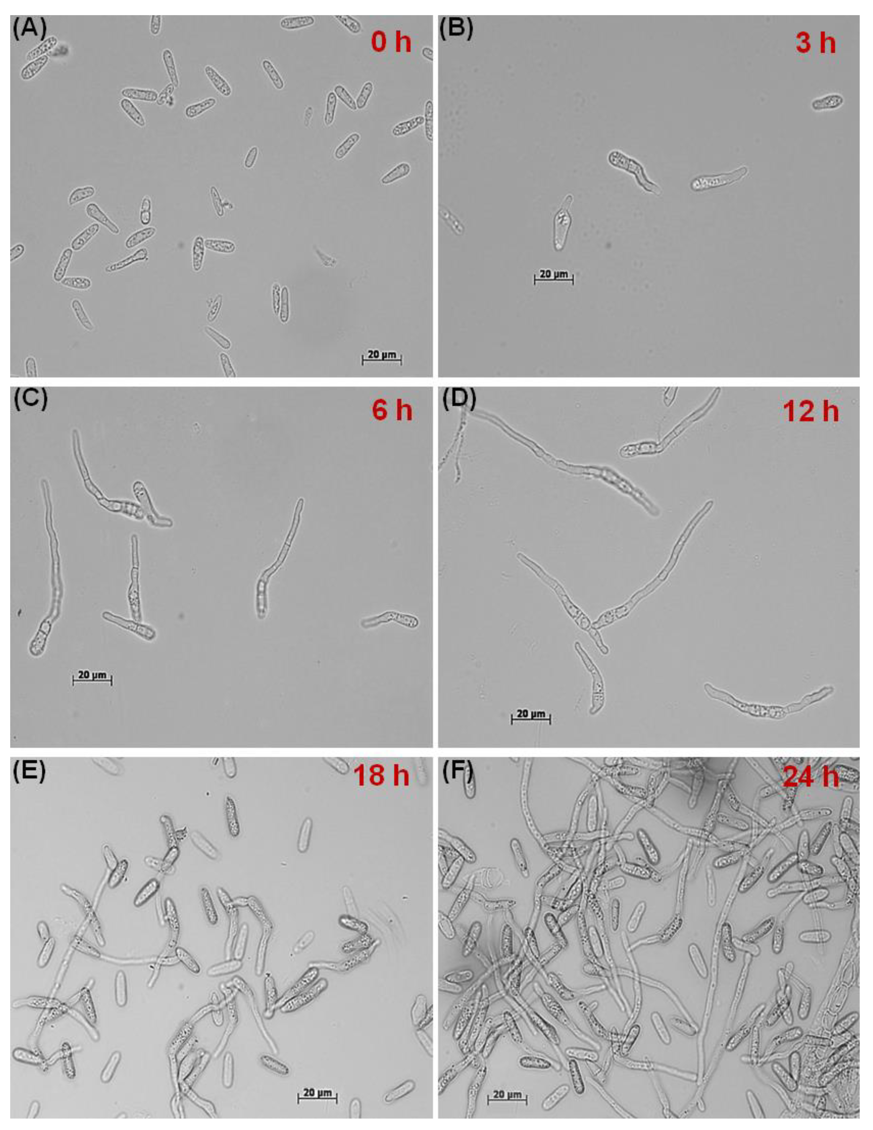
2.1. In Vitro Dynamics of GT Formation and CAT Fusion

To study the effect of conidial age on GT formation and CAT fusion and their mutual exclusiveness in C. gloeosporioides, the GT and CAT induction assays were carried out by following a protocol developed in our lab [11]. The C. gloeosporioides (CBS 953.97) culture was inoculated on bean pod agar medium (autoclaved French bean pods submerged in 2% water agar) and was incubated in the dark at 25 °C to induce conidiation. Post-inoculation, the conidia were harvested at different time points, viz., 6, 10, 13, and 17 days. Conidial cells were counted using Neubauer’s chamber and conidial suspension of 4 × 10⁵ per ml was prepared in distilled water and potato dextrose broth (PDB) for CAT and GT induction, respectively. One ml of such conidial suspensions was placed in a 24-well tissue culture plate (Tarsons, Kolkata, West Bengal, India) and incubated in the dark at 25 °C for different time points, viz., 3, 6, 12, 18, and 24 h for GT formation and 24, 48, 72, and 96 h for CAT fusion. They were then examined under a microscope (Olympus BX53 with Olympus DP73 camera, Cellsens 1.13 imaging software) using differential interference contrast (DIC) optics. The GT formation and CAT fusion were quantified as the percentage of conidia (n = 300) involved in GT formation or CAT fusion [6]. Different stages of GT formation such as initiation, adhesion, and elongation, and various stages of CAT formation such as induction, homing, and fusion were examined at different time points.
Different conidial numbers were tested to determine the threshold of conidial concentrations required for CAT fusion and GT formation in C. gloeosporioides. Different concentrations of 6-day-old conidia were incubated in PDB for 18 h in the dark for GT induction. In contrast, for CAT induction, different concentrations of 17-day-old conidia were incubated in dH2O for 72 h in the dark. The concentrations of conidia tested were 4 × 10², 4 × 10³, 4 × 10⁴, 4 × 10⁵, and 4 × 10⁶ per mL [11].

2.2. Effects of Availability of Nutrients on GT and CAT Induction

To study whether CAT fusion and GT formation in C. gloeosporioides are dependent on the availability of nutrients and starvation stress conditions, CAT fusion (in 17-day-old conidia) and GT formation (in 6-day-old conidia) were induced in nutrient-limiting conditions (water), and high nutrient conditions such as PDB, 2% glucose. Furthermore, to study whether the CAT induction or GT formation in C. gloeosporioides is mediated through MAP kinase (MEK) pathway, CAT fusion and GT formation percentage was also determined in the presence of an MEK inhibitor InSolution PD98059 (5 µM).

2.3. Whole Transcriptome Analysis of Resting Conidia, GT and CAT

2.3.1. RNA Extraction, Quantification, and Qualification

GT formation was induced in 6-day-old conidia at 4 × 10⁵ conidia/mL density in PDB incubated for 18 h in the dark, and the CAT induction was carried out in 17-day-old conidia at 4 × 10⁵ conidia/mL density in water incubated for 72 h in the dark. After that, total RNA was isolated from resting conidia (6-day-old conidia grown on bean pod agar media), germinating conidia (GTs), and fused conidia via CAT (CATs) (3 replicates of each sample) of C. gloeosporioides using NucleoSpin RNA Plant kit (Macherey-Nagel, Düren, Germany) according to manufacturer’s guidelines. DNAse treatment was carried out using the Amplification grade DNase I kit (Sigma Aldrich, St. Louis, MI, USA). Isolated total RNA was quantified and qualified using a Nanodrop ND-1000 spectrophotometer and Qubit fluorometer. RNA quality was also assessed using formaldehyde agarose gel electrophoresis. RNA integrity was checked using a Bioanalyzer chip and Agilent RNA TapeStation (Agilent Technologies, Santa Clara, CA, USA).

2.3.2. Library Preparation and Transcriptome Sequencing

Three µg of total RNA from the three technical replicates of each life stage, namely, resting conidia, GTs, and CATs of C. gloeosporioides, were pooled, and independent library preparations were carried out for these three different life stages. Pooling of the RNA samples for library preparation was done due to economic constraints. RNA-seq library preparation was performed as per Illumina-compatible NEBNext UltraTM Directional RNA Library Prep. Sequencing for 150 bp length paired-end (PE) reads was performed in an Illumina HiSeq sequencer.

2.3.3. Quality Control, De Novo Assembly, and Sequence Clustering

Reads were processed for quality assessment and low-quality filtering before the assembly. The raw data generated was checked for the quality using FastQC, and preprocessing of the data, which includes removing the adapter sequences and low-quality bases (<q30), was done with Cutadapt [17].
Processed reads were assembled using a graph-based approach by the rnaSPAdes program [18]. rnaSPAdes is a tool for de novo transcriptome assembly from RNA-Seq data and is suitable for all kinds of organisms. First, the characteristic properties, including N50 length, average length, maximum length, and the minimum length of the assembled contigs, were calculated. Then, in the second step of the assembly procedure, clustering of the assembled transcripts based on sequence similarity is performed using CD-HIT-EST [19] with 95% similarity between the sequences, which reduces the redundancy without exclusion of sequence diversity that is used for further transcript annotation and differential expression analysis.

2.3.4. Read Mapping to the Reference Genome and Differential Counting

To assess the quality of the assembly, an evaluation of the read content approach was used. Processed reads from all three libraries were aligned back to the final assembly using Bowtie2 [20] with end-to-end parameters. C. gloeosporioides Nara gc5 isolate (ftp://ftp.ncbi.nlm.nih.gov/genomes/genbank/fungi/Colletotrichum_gloeosporioides/ (accessed on 8 April 2019) was used as a reference genome. For each sample, the count of mapped reads was derived and normalized to RPKM (reads per kilobase of exon model per million mapped reads). The count data was further used to identify differentially expressed genes at each time point using the DESeq package [21]. Sequencing (uneven library size/depth (accessed on 8 April 2019) bias among the samples was removed by library normalization using size factor calculation in DESeq. Fold changes were determined according to the formula “Expression of treated sample/Expression of control sample”. The raw P values were adjusted for multiple testing with the procedure described by [22] to control the FDR (false discovery rate). Finally, the regulation for each transcript was assigned based on their log2fold change.

2.3.5. Gene Ontology Enrichment, Differential Expression Analysis, and Functional Annotation

Assembled transcripts were functionally enriched and categorized based on blast sequence homologies and gene ontology (GO) annotations describing biological processes and molecular functions using Blast2GO software (p < 0.05), selecting the NCBI blast Fungi as a taxonomy filter and the default parameters [23]. Out of 121,260 total, 88,491 transcripts (72%) were functionally annotated against all fungal protein sequences from the Uniprot Protein database. Those transcripts with more than 30% identity as a cutoff were taken for further analysis.
Multiple databases (Uniprot, NCBI, KEGG pathway, and Pfam) were used to annotate the transcripts and determine the possible roles of the annotated genes/proteins. Clustered transcripts were annotated using the homology approach to assign functional annotation using BLAST tool [24] against “Fungi” data from the Uniprot database. Transcripts were assigned with a homolog protein from other organisms if the match was found at e-value less than e−5 and minimum similarity greater than 30%. The top 50 highly upregulated transcripts of each GT and CAT were further manually annotated using the Ensembl Fungi database (https://fungi.ensembl.org/Colletotrichum_gloeosporioides_cg_14_gca_000446055/Info/Index (accessed on 14 June 2019) for C. gloeosporioides to find out more accurate molecular and biological functions.

2.3.6. Transcription Factors and Secreted Protein Analysis

Assembled transcripts were translated into protein using TransDecoder. The putative transcription factors (TFs) were predicted using the Fungal Transcription Factors Database (http://ftfd.snu.ac.kr (accessed on 4 October 2019). Transcription factor analysis was performed based on homology approach using NCBI-blast program.
For effector protein identification, proteins with signal peptides were identified using SignalP [25]. Proteins with a transmembrane helix predicted using TMHMM [26] were excluded from the analysis. Candidate effector proteins were identified by subcellular localization prediction using TargetP [27] and WolfPsort [28]. All putative proteins with a SignalP D-score = Y were considered. These proteins were then scanned for transmembrane spanning regions using TMHMM, and proteins with 0 transmembrane domains (TM) were retained. Finally, proteins with location predicted as Loc = S [25] using TargetP and proteins predicted as extracellular (Ext >17) using WolfPSort were retained in the final candidate effector protein dataset. The Pfam search (EMBL-EBI) was performed for functional annotation and to find out the possible biological roles of secreted proteins during GT formation and CAT fusion.

2.3.7. Data Availability

RNA-seq data were submitted to the NCBI SRA database under accession numbers SRR12245378, SRR12245377, and SRR12245376.

2.4. Real-Time qRT-PCR Validation

Validation of differential gene expression obtained from the transcriptome analysis was performed using qRT-PCR assay on total cDNA samples from the three life stages, viz., resting conidia, GT, and CAT of C. gloeosporioides. For this, 27 differentially expressed genes from resting conidia, GT, and CAT detected by RNA-seq were randomly chosen for validation by qPCR. The Beta-tubulin gene was used as a reference gene for normalization. Specific primer pairs were designed for these 27 selected genes using the Primer3 Software to generate final amplicon sizes of each gene between 50 to 150 bp with 60 °C melting temperature. All primers used in this study are listed in Table S8. Quantitative real-time PCR (qRT-PCR) was carried out as previously described [29]. In brief, total RNA (1 μg) from resting conidia, GTs, and CATs were reverse transcribed to cDNA with an oligo (dT) primer using SuperScript III (Invitrogen, Carlsbad, CA, USA) according to the manufacturer’s instructions. A regular PCR amplification for each primer pair was performed to optimize the annealing temperature. The qRT-PCR was performed in PCRmax Eco 48 Real-Time Cycler using 50 ng of cDNA, 5 μL of SYBR™ Green PCR Master Mix (Applied Biosystems), 10 pmol of sense primers, and 10 pmol of antisense primers in a final volume of 10 μL. The PCR cycle conditions were set as follows: 95 °C for 30 s, followed by 40 cycles of 95 °C for 10 s, 55 °C for 30 s, and 60 °C for 15 s. Lastly, in order to check the amplification of a single targeted amplicon, melting curves were analyzed for all the primer pairs. Expression of genes was evaluated according to their relative quantification using the 2−∆∆Ct method [30]. Each sample was run in triplicates for all selected and reference genes. Data were analyzed using the EcoStudy software v5.2. A correlation between expression levels of DEGs by qRT-PCR and RNA seq was evaluated using log2fold expression values.

2.5. Statistical Analyses

The GT formation and CAT fusion data were analyzed by Analysis of Variance (ANOVA) with Tukey’s post hoc test using GraphPad Prism 5 Statistics Software. Differences with a p-value < 0.05 were considered statistically significant. All GT formation and CAT fusion assays were performed in triplicates and with 300 conidial numbers.

3. Results

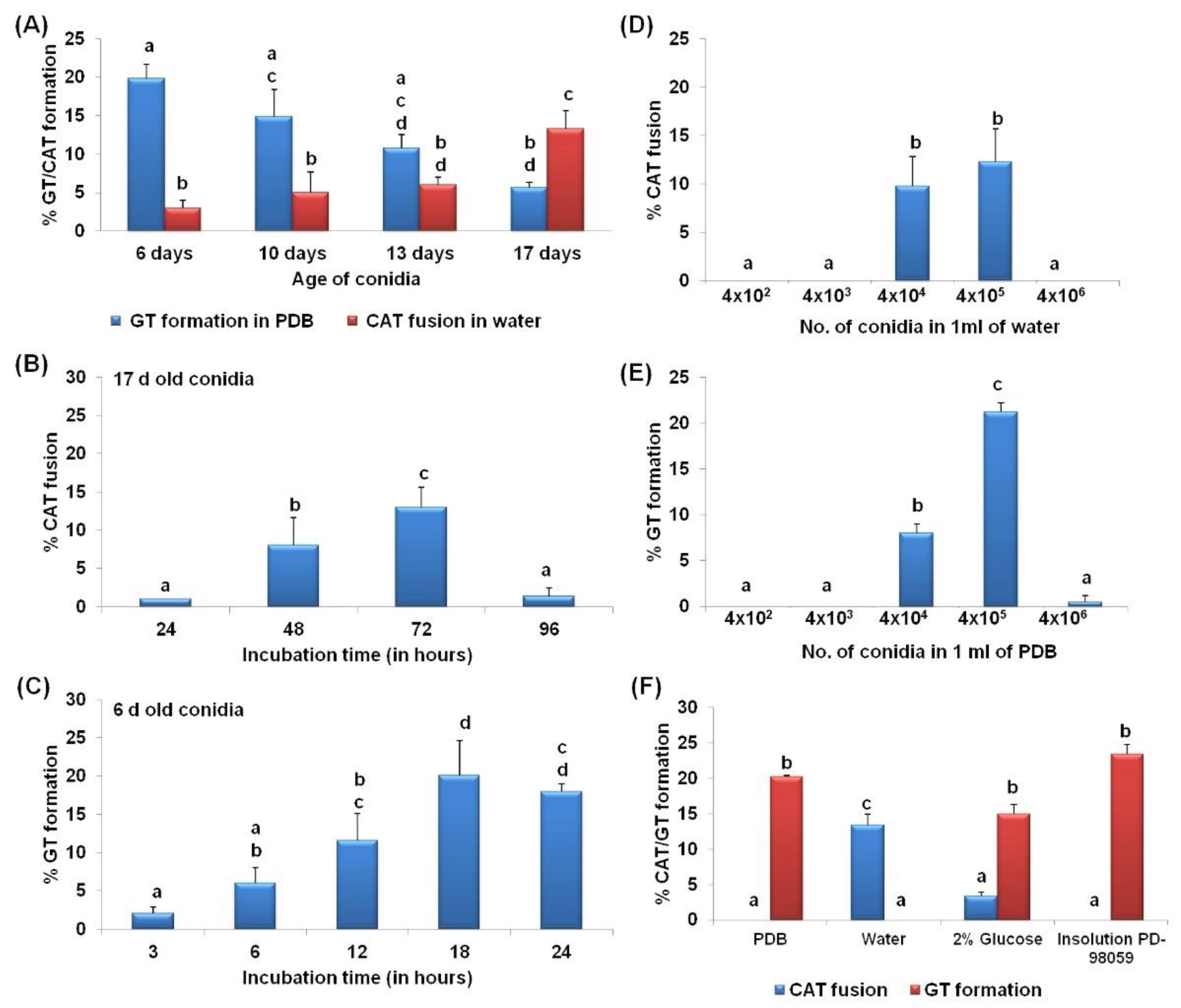
3.1. CAT Fusion and GT Formation Are Mutually Exclusive in C. gloeosporioides

The different stages of CAT fusion were demonstrated in C. gloeosporioides. Initially, conidia become primed for CAT fusion as a result of CAT induction (Figure 1A). Subsequently, CATs home towards each other, which is known as CAT homing (Figure 1B). Finally, the CATs were fused to each other, constituting the third stage, i.e., CAT fusion (Figure 1C). Later on, with increasing, incubation time, CAT connections expanded and formed a CAT network (Figure 1D) through 72 h of incubation. The different stages of the GT formation were also determined in C. gloeosporioides, which could be divided into three stages, namely, conidial swelling and adhesion at 3 h post-incubation (hpi) (Figure 2A,B), GT initiation including polarized growth at 6 hpi (Figure 2B,C), GT elongation with hyphal growth and formation of septa 12–18 hpi (Figure 2D,E), and finally, GT network formation at and after 24 hpi (Figure 2F).
When CAT fusion and GT formation was studied in differently aged conidia, it was observed that young conidia (6 days) in PDB were efficient in forming GT, while older conidia (17 days) in sterile distilled water were good at forming CAT (Figure 3A). The younger (6 days) and older (17 days) conidia in PDB do not form CAT at all, while in water, they showed 3 ± 1% and 13.3 ± 2.3% CAT fusion, respectively. As the age of the conidia increases, the GT formation percentage decreases. In contrast, the CAT fusion percentage increased with the increasing age of conidia. The young conidia (6 days) in PDB showed 19.8 ± 1.8% GT formation and no CAT fusion, while the same aged conidia in water showed 3 ± 1% CAT fusion and no GT formation (Figure 3A). The old conidia (17 days), when incubated in water, showed 13.3 ± 2.3% CAT fusion and no GT formation, while the same aged conidia in PDB showed 5.7 ± 0.6% GT formation and no CAT fusion (Figure 3A). To determine the optimal incubation for GT formation, 6-day-old conidia were incubated in PDB for 3, 6, 12, 18, and 24 h; the maximum GT formation (20.1 ± 4.5%) was seen at 18 hpi (Figure 3B). When 17-day-old conidia were incubated in distilled water for 24, 48, 72, and 96 h, the maximum CAT fusion frequency (13.5 ± 2.6%) was observed at 72 hpi (Figure 3C). Therefore, most of the further experiments were conducted using these optimized parameters for in vitro GT and CAT induction.

3.2. CAT Fusion and GT Formation Are Dependent on the Conidial Density

A density of 4 × 10⁴ and 4 × 10⁵ conidia per ml of water was found to be optimal for CAT fusion in C. gloeosporioides. The CAT fusion percentages were 9.8 ± 3% and 12.3 ± 3.4% in 4 × 10⁴ and 4 × 10⁵ conidia per ml of water, respectively (Figure 3D). However, for GT formation 4 × 10⁵ conidial density was optimal, resulted in 21.2 ± 1% GT formation (Figure 3E). GT formation decreased to 8 ± 1% when the conidial density reduced to 10-fold, that is 4 × 10⁴ conidia per ml of PDB (Figure 3E). Much lower and higher conidial densities, viz., 4 × 10², 4 × 10³, and 4 × 10⁶ were not favorable for GT as well as CAT formation. These results suggest that CAT fusion and GT formation were dependent on the conidial concentration.

3.3. Differential Nutritional Requirements for CAT versus GT Formation

The conidia of C. gloeosporioides failed to undergo CAT fusion in the presence of nutrient-rich medium such as PDB (Figure 3F). As shown in the previous section, a high percentage of CAT fusion (13.4 ± 1.5%) was observed in sterile distilled water. However, when the conidia were incubated in water with 2% glucose, the CAT fusion frequency decreased to 3.4 ± 0.5% (Figure 3F). Thus, it shows that CAT fusion occurred predominantly during nutrient starvation conditions and was inhibited in high nutrient conditions. Furthermore, the conidia failed to induce CAT fusion in the presence of InSolution PD98059, an inhibitor of MEK, suggesting the involvement of the MAPK pathway in CAT fusion in C. gloeosporioides (Figure 3F).
On the other hand, GT formation in C. gloeosporioides conidia was observed in the presence of nutrients such as PDB and 2% glucose at the frequencies of 20.3 ± 0.2% and 15 ± 1.4%, respectively (Figure 3F). However, GT formation was not seen in nutrient limiting conditions such as water as a medium, indicating that GT formation occurs predominantly in nutrient-rich environments but not in nutrient limiting conditions. Surprisingly, GT formation frequency increased up to 25 ± 1.3% in the InSolution™ PD98059, an MEK inhibitor (Figure 3F).

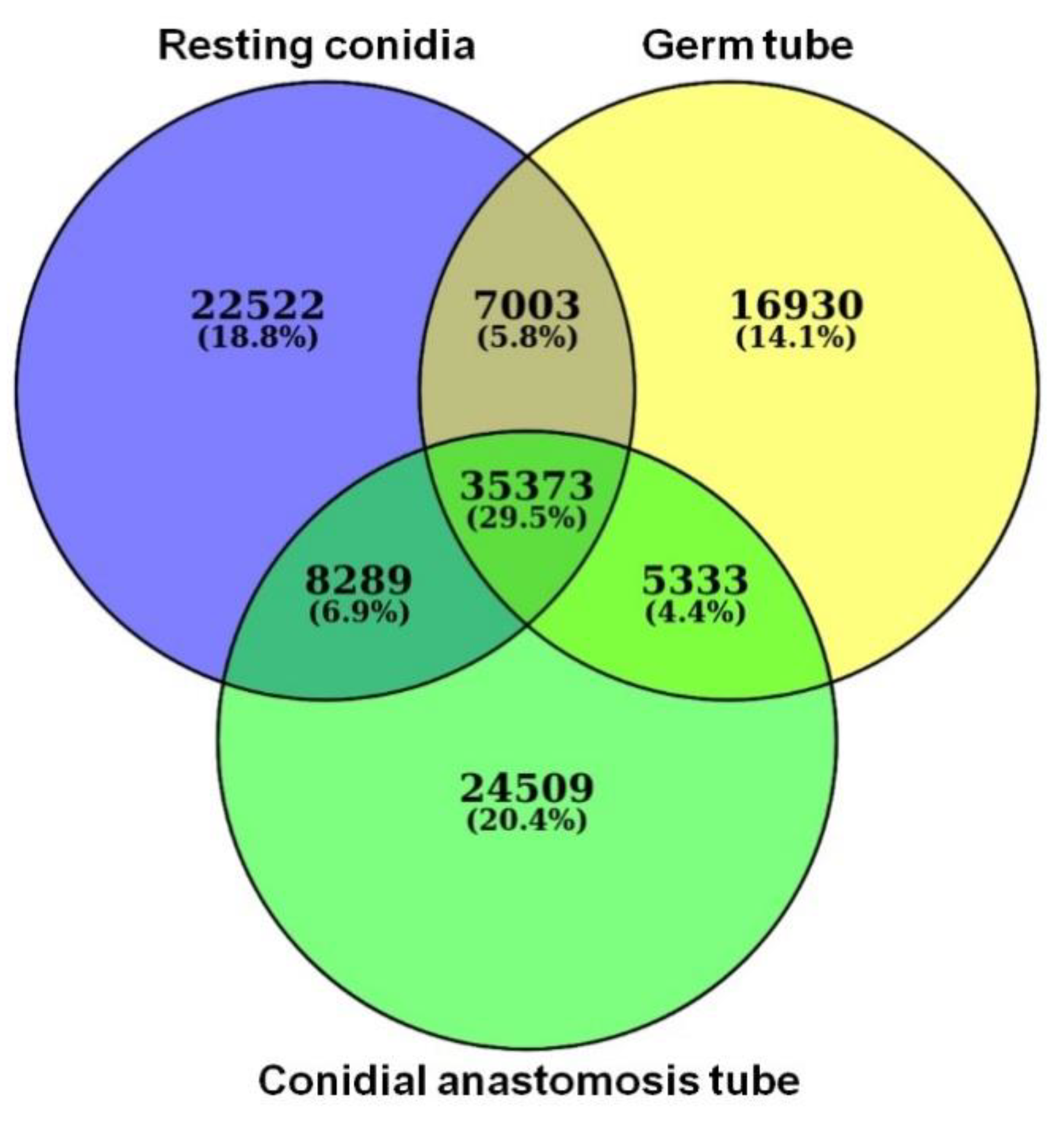
3.4. RNA-Seq Data Analysis

All RNA samples had an RNA integrity number (RIN) greater than 9.0. An average of 41.62 million raw sequencing PE reads were produced in total for three samples, out of which an average of 39 million reads were used for the downstream analysis after preprocessing. Thus, for every sample on an average of 95% of high-quality data was retained and used for analysis (Table S1). A total of 211,330 transcripts were obtained, which consisted of 73,187, 64,639, and 73,504 transcripts of resting conidia, GT, and CAT, respectively. Among these transcripts, 35,373 were present in all three life stages, while 22,522, 16,930, and 24,509 transcripts were uniquely expressed in resting conidia, GT, and CAT, respectively (Figure 4).
The assembly of high-quality reads was performed separately for resting conidia, GT, and CAT. Sample wise transcripts were further clustered into 121,260 transcripts with an average length of 603 bp and N50 of 1849 bp (Table S2). The length of the final assembly ranged from 37 to 31,011 bps.

3.5. Gene Ontology Enrichment Analysis of GT and CAT

The assembled transcripts for GT and CAT were individually grouped into ten molecular functions (MF) and ten biological processes (BP) (Figure S1). The transcripts, which were significantly enriched and variable in numbers under the biological processes, included fungal type (GO:0031505), carbohydrate (GO:0005975), and transmembrane (GO:0055085); the molecular functions included chitin (GO:0008061), serine-type (GO:0004252), iron (GO:0005506), oxidoreductase (GO:0016491), and hydrolase (GO:0004553) during both GT formation and CAT fusion (Figure S1). The comparison of the biological processes of GT and CAT indicated that fungal type (GO:0031505) GO terms were higher in CAT than in GT. In contrast, the carbohydrate (GO:0005975) GO terms were more significant in GT than in CAT (Figure S1A,B). However, when the molecular functions were compared, it was observed that the serine-type (GO:0004252) and iron (GO:0005506) GO terms were higher in CAT than in GT (Figure S1C,D).

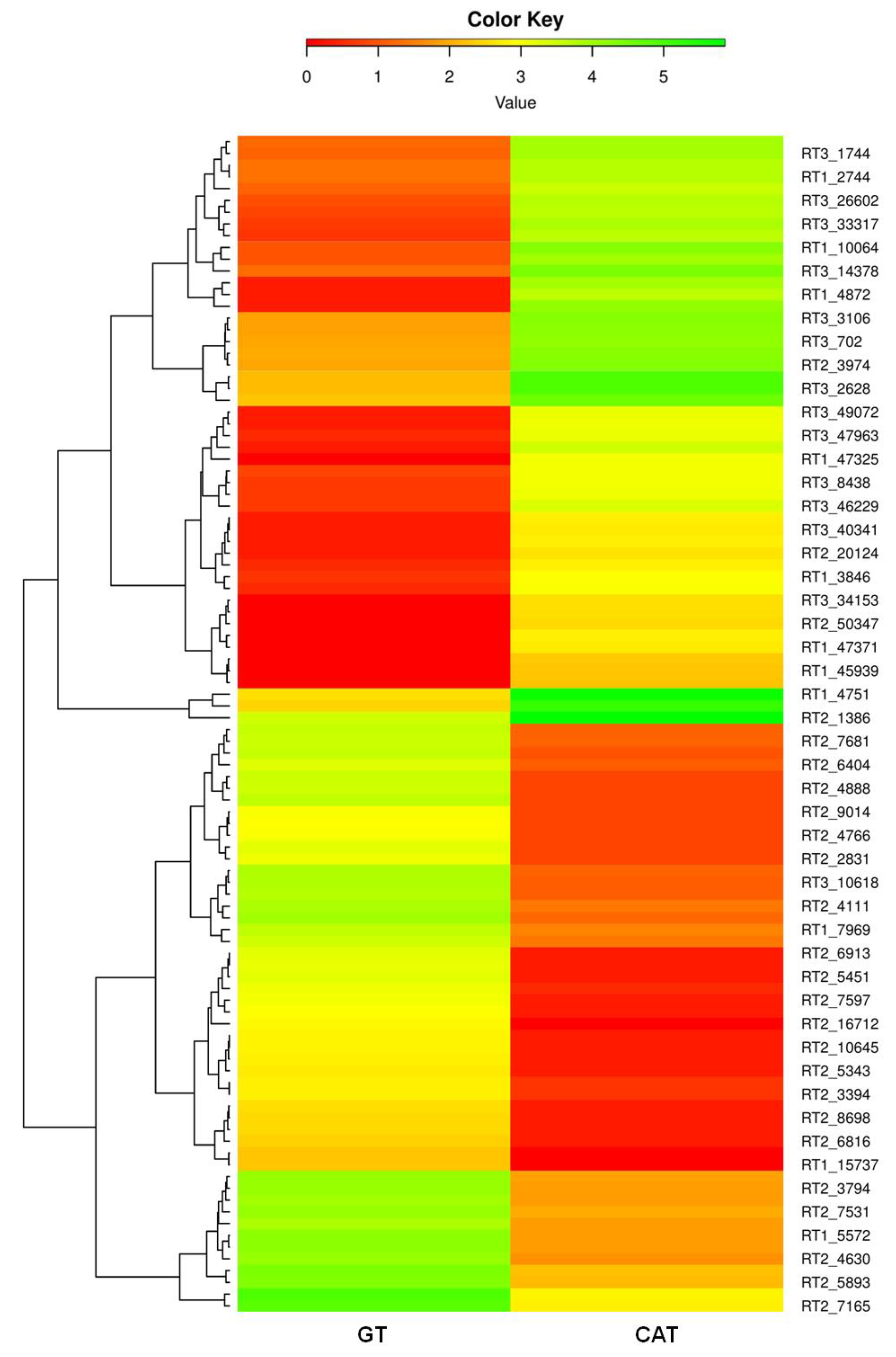
3.6. Identification and Functional Annotation of DEGs in GT and CAT

The DEGs were further analyzed between GT and CAT. The transcripts which show a log2fold change less than −1 are represented as downregulated, the values greater than 1 are defined as upregulated, and between −1 to 1 are termed as neutrally regulated.
Heatmaps of the top 50 differentially expressed genes in GT and CAT showed a significant variation in their expression profiles. The top 25 upregulated transcripts of GT showed significant downregulation in CAT. On the other hand, the top 25 upregulated genes from CAT were significantly downregulated in GT (Figure 5). Heatmaps based on DEGs of resting conidia v/s GT and resting conidia v/s CAT also showed remarkable differential expression in these life stages of C. gloeosporioides (Figure S2). Manual functional annotation of the top 50 transcripts for both GT and CAT resulted in specific categories of proteins, which probably play an important role in the development of CAT and GTs in C. gloeosporioides (Tables S3 and S4). Out of these top 50 differentially expressed transcripts, we found four major groups of highly upregulated genes during GT formation, which were involved in cell wall degradation, host-fungus interaction, germination, transport, signaling, and virulence and whose possible functions and expression values in GT and CAT are given in Table 1. Likewise, out of those top 50 DEGs, four major groups of highly upregulated genes involved in CAT fusion were stress response, cell wall, membrane transport, and signaling, cytoskeleton, cell cycle, and cell rescue and pathogenicity genes, whose possible functions and expression values in CAT and GT are given in Table 2.

3.7. Transcription Factor Candidates Involved in GT Formation and CAT Fusion

In this study, a total of 49 Transcription factor (TF) families and 1044 TF proteins were predicted using the Fungal Transcription Factors Database; out of theses, 38 TF families (Table 3) and 220 TF proteins (Table S5) were differentially expressed during GT and CAT induction. Twenty-four TF families, e.g., bZIP, C2H2 zinc finger, HMG, Zn2Cys6, etc., were upregulated during CAT fusion compared to GT formation, while 14 TF families such as AraC type, Homeobox, etc., were upregulated in GT formation (Table 3). Among the 220 transcription factor genes, 42 and 82 were uniquely expressed during GT formation and CAT fusion, respectively (Table S5).

3.8. Effector Candidates Secreted during GT Formation and CAT Fusion

GT formation and CAT fusion in C. gloeosporioides involve the deployment of secreted effector proteins. Effector candidates are often <200 amino acids in length and cysteine-rich [31]; therefore, we principally focused on small secreted proteins <250 amino acids in length. There were 1567 effector candidates; of these, 776 and 791 were secreted during GT and CAT formation, respectively. Out of 776 GT effector proteins, 102 were uniquely secreted during GT formation (Table S6), and out of 791 CAT effector proteins, 100 were uniquely secreted during CAT fusion (Table S7).
Differentially expressed secretory proteins were manually annotated and categorized in four and three major groups for GT formation and CAT fusion, respectively. Few selected uniquely expressed effector candidates during GT formation include hydrolytic enzymes, adhesion, germination, hyphal development associated proteins, transport, signaling-related proteins, and proteins responsible for virulence. Some examples of these groups of effector proteins uniquely secreted during GT formation and their possible functions are given in Table 4. Likewise, the major effector proteins uniquely secreted during CAT fusion included stress-associated proteins, cell wall, membrane transport, signaling-related proteins, cytoskeleton, cell cycle, and fungal development associated proteins. Some examples of these groups of effector proteins uniquely secreted during CAT fusion and their possible functions are given in Table 5.

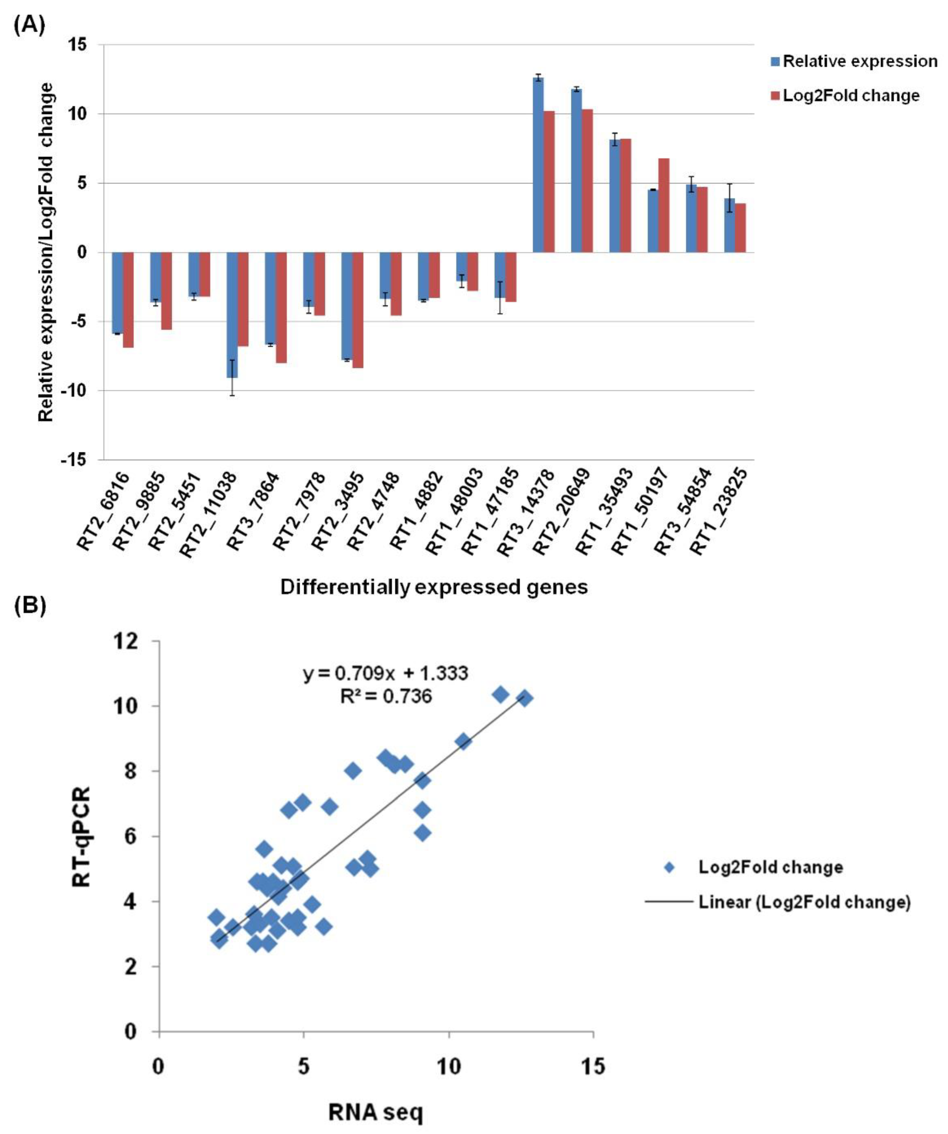
3.9. Real-Time qPCR Validation of Selected DEGs

Differentially expressed genes in resting conidia, GT, and CAT identified by RNA-seq were further verified through a quantitative real-time PCR (qRT-PCR). We observed a strong correlation between the qRT-PCR and RNA-Seq analysis of 17 DEGs in GT versus CAT formation (Figure 6A). Similar levels of correlations were also attained in the expression levels of 14 and 13 DEGs in resting conidia v/s GT and resting conidia v/s CAT, respectively (Figure S3). Log2fold expression values were used to evaluate the correlation between RNA-seq and qRT-PCR. The results obtained by qRT-PCR showed significant correlation (R2 = 0.736; p < 0.01) with transcriptome Log2fold data (Figure 6B). It showed that the RNA-seq experiment was conducted with accuracy and RNA-seq data is reliable to understand the molecular mechanism underlying CAT formation further.

4. Discussion

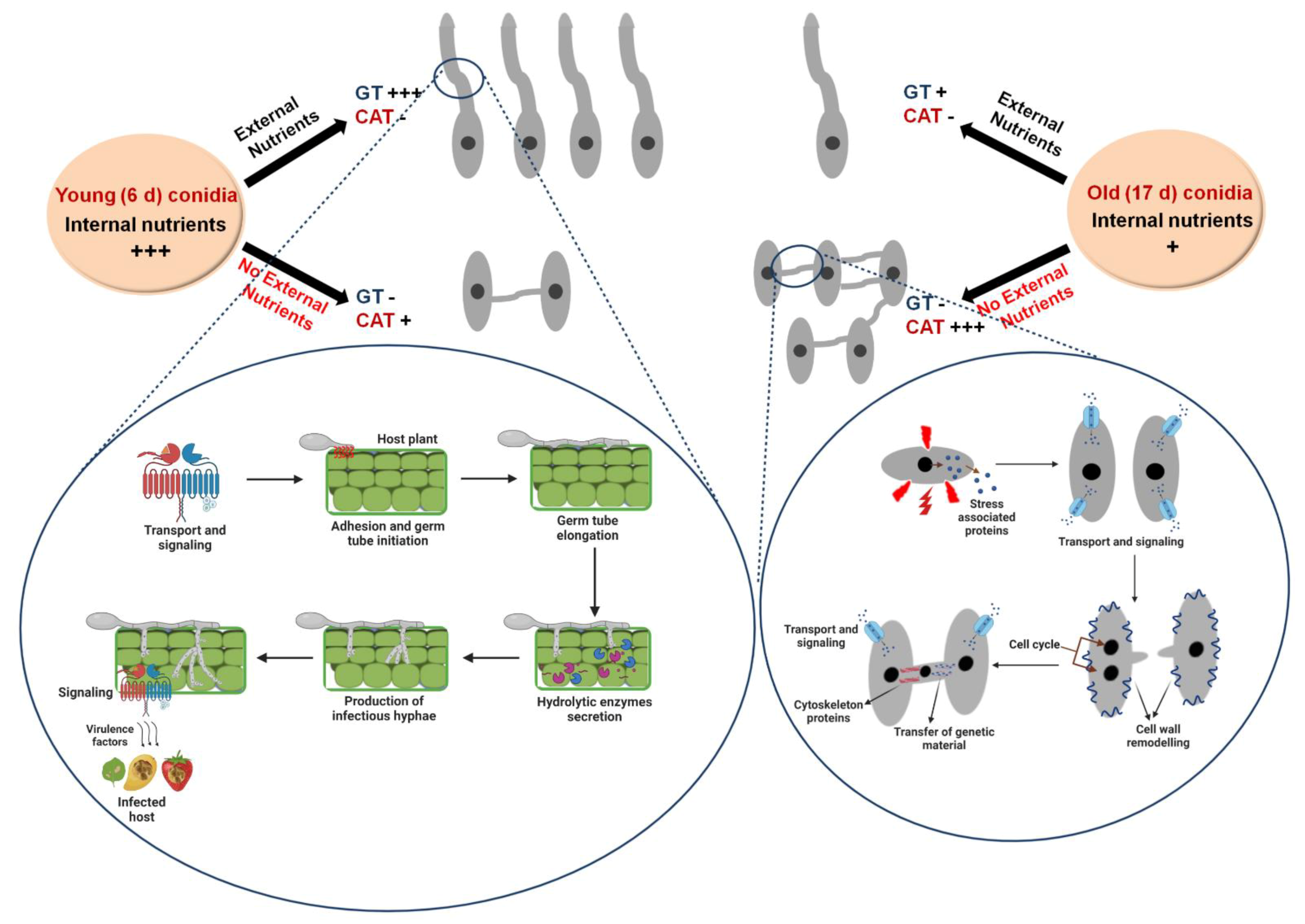
4.1. Differential Physiological Requirements for GT and CAT

In the present study, we have attempted to understand the required in vitro conditions for GT and CAT formation in C. gloeosporioides. We observed significant CAT fusion in 17-day-old conidia and GT formation in 6-day-old conidia (Figure 3A). Our results seem to fit with the conidial age requirement of the genus Colletotrichum; older conidia (17 d old) for CAT fusion and younger conidia (6 d old) for GT formation were preferred in C. gloeosporioides (Figure 3A). It has been reported that CAT fusion is dependent on conidial age for different species; 16-day-old conidia of C. lindemuthianum and C. gossypii were more appropriate for CAT fusion, while 20-day-old conidia of C. fructicola and C. nymphaeae were suitable for CAT fusion [6,8,14]. In contrast, the younger conidia (10 d old) of C. lindemuthianum were suitable for GT formation in nutrient-rich medium such as PDB [10]. However, the conidial age requirements for both CAT fusion and GT formation in F. oxysporum and N. crassa were 7–10 and 5–6 days, respectively [4,5,9]. The GT/CAT formation was shown to be conidial density-dependent in N. crassa and F. oxysporum [4,5]. In these fungi, the conidial number threshold requirement for CAT induction was more or less same, i.e., 1 × 10⁶ conidia/mL [4,5,6,8,9,32]. The GT induction has also been shown to depend on conidial density in F. oxysporum; the maximum GT formation was observed in 1 × 10⁵ conidia per mL [4]. The conidial number requirement for CAT fusion and GT formation in C. gloeosporioides was also found to be 4 × 10⁵ conidia/mL (Figure 3D,E). It indicates that a conidial threshold is a requisite for the CAT induction as well as GT formation (Figure 3D,E).
We observed that the conidia of C. gloeosporioides could efficiently form CAT in the absence of nutrients (water) compared to nutrient-rich conditions such as PDB. The presence of glucose in water also reduces CAT fusion percentage in this fungus (Figure 3F). On the other hand, significant GT formation occurred only in the presence of nutrients such as PDB, and nutrient limiting conditions negatively affected GT formation (Figure 3F). These observations indicate that the CAT fusion occurs in nutrient limiting conditions; conversely, GT formation occurs only in the nutrient-rich conditions in C. gloeosporioides. It has been shown previously that nutrient limiting conditions induce CAT fusion, and nutrient-rich conditions promote GT formation in few fungi [4,10]. In another species of the genus Colletotrichum, i.e., C. lindemuthianum, and C. graminicola, CAT fusion does not occur in nutrient-rich conditions such as PDB/oatmeal agar (OMA) and even in nutrient-poor Vogel’s medium, while it occurs only in water media [10,12]. Meanwhile, in other CAT-forming fungi such as N. crassa and F. oxysporum, CAT fusion occurs in the presence of nutrients, e.g., Vogel’s medium for N. crassa and YNB+KNO3/1% PDB +NaNO3 for F. oxysporum [4,5,9,33,34]. We have also shown that CAT fusion in C. gloeosporioides is inhibited by a MAP kinase inhibitor InSolution PD98059 (Figure 3F), suggesting a possible role of MAP kinase pathway in CAT fusion in C. gloeosporioides. Interestingly, in the presence of the MAP kinase pathway inhibitor InSolution PD98059, the frequency of GT formation increased considerably compared to PDB (Figure 3F), thereby suggesting that this pathway may not be a significant player in early GT formation. Since there are different MAPK pathways in fungal pathogens, it might be possible that a different MAPK pathway is operational during GT formation. However, the MAPK pathway was found to be essential in appressorium formation in the same fungus [35].
We propose that the choice of conidia to either form GT or CAT depends upon multiple factors, viz., conidial age (internal nutrients), availability of external nutrients, and/or starvation stress conditions. We have shown that the older conidia were superlative for CAT fusion while younger conidia were ideal for GT formation. We believe that increased CAT fusion reduces the internal resources for GT formation; however, the conidia’s decision to either form CAT or GT does not only rely solely upon the internal resources. We had shown that when the older conidia (17 days) were incubated in water (no nutrients), they could undergo extensive CAT fusion and no GT formation. However, when the same aged older conidia were incubated in PDB, they could also form GT but at a reduced rate compared to younger conidia (6 and 10 days old) (Figure 3A). In C. graminicola, the older conidia were shown to undergo autophagic activity due to nutrient stress [36]. Therefore, we hypothesize that as the age of conidia increases, the internal resources get depleted; in this scenario, if the external resources are provided, then conidia could still form GTs to a very lesser extent. On the contrary, in the absence of external nutritional resources, the older conidia undergo extensive CAT fusion to exchange their nutrients for survival.
After understanding the differential physiological requirements of CAT and GT formation, we attempted to decipher the differential molecular prerequisites of these two processes in C. gloeosporioides by undertaking RNA-sequencing and analysis. As a result, we observed significant differences in transcriptomes of GT formation and CAT fusion. Furthermore, we identified many differentially expressed genes, transcription factors, and secretory effector proteins during GT formation and CAT fusion in C. gloeosporioides.

4.2. Transcriptome Analysis of GT

Genes involved in cell wall degradation, host-fungus interaction, germination, transport, signaling, and virulence were found to be upregulated during GT formation. Previously, it has been shown that genes responsible for plant cell wall degradation, secondary metabolism, and detoxification were upregulated during GT formation in C. fructicola [37]. We also observed that some hydrolytic enzymes involved in plant cell wall degradation such as pectin lyase, glycosyl hydrolase families, mandelate racemase/muconate lactonizing enzyme, and fungal cellulose-binding domain-containing protein were upregulated during GT formation (Table 1). In the present study, Hydrophobin 2 and HsbA genes were also upregulated during GT formation in C. gloeosporioides, which are probably involved in conidial adhesion. Fungal hydrophobins are small, secreted hydrophobic proteins involved in the adhesion of conidia to the host surface prior to GT formation [38]. Fungal hydrophobin, HsbA-like protein were shown to be upregulated during GT and appressorium development in A. oryzae and C. fructicola [37,39]. Other significant DEGs include GPI-anchored cell wall beta-1,3-endoglucanase Egl, which probably plays a role in germination, and hydrophobic surface binding protein A, which increases the hydrophobicity of conidia, aerial hyphae, and fruiting bodies.
Fungal transporter proteins play a broad range of biological functions and an essential role in the pathogenicity of phytopathogens [40,41,42]. In the present study, few transporters and signaling genes such as Fg-gap repeat protein, phosphate-repressible phosphate transporter, transmembrane amino acid transporter, MFS monosaccharide transporter, high-affinity methionine permease, and Uso1/p115-like vesicle tethering protein were upregulated during GT formation (Table 1). These transports and associated signaling genes are probably important to sense the favorable conditions for germination, which results in GT formation. However, in previous studies, it has been demonstrated that an ABC protein CgABCF2 was required for appressorium formation and plant infection in C. gloeosporioides [35,43].
Some secondary metabolites and virulence associated genes were shown to add to pathogenicity in C. gloeosporioides [44]. We have also found that various secondary metabolite backbone genes such as peptidase S41 family protein, nrps-like protein, FAD-dependent oxidoreductase, and cyanide hydratase were upregulated during GT formation, which may contribute to pathogenicity [45,46]. During GT formation, few other highly upregulated genes such as polysaccharide deacetylase, FGGY-family pentulose kinase, acetylornithine aminotransferase, GMC oxidoreductase, GNAT family acetyltransferase, CAP-22 protein, and NmrA family transcriptional regulator may also be implicated for pathogenicity in C. gloeosporioides (Table 1).
Transcription factors play important roles in various biological processes. Out of 220 annotated TF genes, 42 were uniquely expressed during GT formation, and these genes are probably involved in the regulation of GT formation in C. gloeosporioides (Table S5). Effector proteins are usually secretory proteins, which can play an important role in plant infection. For example, one study showed that effectors are secreted from appressorium before host invasion in C. higginsianum, a close relative of C. gloeosporioides [47]. In our study, we have identified uniquely expressed effector candidate genes during GT formation. Interestingly, the annotated effectors could also be categorized in the same categories, which were based on DGE analysis of GT formation. The uniquely expressed effector gene candidates include hydrolytic enzymes, adhesion, germination, hyphal development associated proteins, transport, signaling related proteins, and proteins responsible for virulence (Table 4). Transcripts of gene glucanases that degrade glucan in the fungal cell wall for cell wall modulation were observed during germination in Aspergillus niger [2]. Various transcripts and secretory effectors of glucanases were observed in our study during GT formation, which might be involved in the cell wall morphogenesis leading to germination. Trehalose accumulates in dormant conidia of A. niger and is degraded during germination [2]. We detected trehalase as a secretory effector during GT formation, which probably provides energy by degrading trehalose in C. gloeosporioides. Other noteworthy uniquely expressed effector gene candidates include carbonic anhydrase and manganese lipoxygenase, which might be important for fruiting body development, ascospore germination, and accelerating programmed spore germination, respectively (Table 4).
The evidences for the involvement of MAPK in conidial germination are conflicting and equally diverse between the species. The deletion of MAPK gene CMK1 in C. lagenarium prevents germination, whereas deletion of MAPK gene pmk1 in Magnaporthe grisea blocks appressorium development but not germination [1]. The MAPK pathway was shown to be essential for appressorium development in C. gloeosporioides [35]. However, in our case, GT formation was not inhibited in the presence of MAPK pathway inhibitor Insolution™ PD98059, and no transcripts related to MAPK pathway could be detected in GT transcriptome, thereby indicating that MAPK pathway may not be essential for early GT formation in C. gloeosporioides.

4.3. Transcriptome Analyses of CAT

The CAT fusion seems to involve a different set of genes compared to GT formation while retaining few common transcripts in both of these processes. We observed that the major groups of highly upregulated genes involved in CAT fusion belong to stress response, cell wall integrity, membrane transport, signaling, cytoskeleton, cell cycle, and cell rescue processes (Table 2).
In this study, we found many stress-associated genes that were upregulated during CAT fusion such as pyridine nucleotide-disulfide, formate dehydrogenase, NADH dehydrogenase, oxalate decarboxylase family bicupin, maleylacetate reductase, O-acetyl homoserine amino carboxypropyl transferase, iron-sulfur cluster assembly protein 1, bZIP transcription factor, HhH-GPD superfamily base excision DNA repair protein, ribulose-phosphate 3-epimerase, and dipeptidase A. All these genes are believed to be involved in oxidative stress response. Reactive oxygen species (ROS) are known to play an important role in redox signaling pathways, including cell differentiation, development, and cytoskeleton remodeling [48]. NADPH-oxidases (NOX) are responsible for the production of superoxide (ROS) by oxidizing NADPH and reducing molecular oxygen, which is believed to be involved during communication, cell fusion, and CAT fusion in several fungi, e.g., N. crassa, F. oxysporum, and V. dahliae [48,49,50,51]. It has been shown that NADPH oxidase BcNoxA or Nox regulator BcNoxR gene deletion mutant of Grey mold Botrytis cinerea were defective in CAT fusion [52]. In our study, NADPH oxidase cytochrome p450 and stress response regulator SrrA, which probably participate in redox signaling and stress signal transduction, were also upregulated during CAT fusion (Table 2). It has been previously documented that starvation response is correlated with the expression of genes encoding oxidative stress response in S. cerevisiae [53]. Further oxidative stress was shown during the aging of stationary cultures of S. cerevisiae [54]. Our study demonstrated that the older conidia under nutritional starvation exhibit efficient CAT fusion, and our transcriptome data analysis also revealed high expression of oxidative stress related genes. This suggests that starvation in C. gloeosporioides conidia induced a strong oxidative response, and the genes involved in this response might play important roles in CAT fusion in this fungus.
The CAT fusion involves sensing of quorum through signaling and transport followed by cell wall and cell membrane remodeling. Many membrane transporters and cell wall modulation genes, including MFS transporter, transmembrane amino acid transporter, peptidoglycan-N-acetylglucosamine deacetylase, and Glycosyltransferase family 2 protein, were found to be upregulated during CAT fusion. In addition, other important transcripts, viz., alcohol dehydrogenase GroES-like domain, flippase-like domain-containing protein, and acetyl-CoA carboxylase, were upregulated during CAT fusion, which probably play roles in protein folding, pheromone response, cytoskeletal dynamics, cell division, lipid metabolism, and fatty acid synthesis in C. gloeosporioides.
The cytoskeleton actin is important for several functions, including cell polarity, exocytosis, endocytosis, septation, movement of organelle, and chemotropic growth of fungi [48]. In N. crassa, the actin cables and patches are significantly increased at CAT tips during chemotropic interactions between germlings or conidia [55]. However, microtubules are dispensable for germling communication, while actin is critically important in N. crassa [55]. We observed that the MreB/Mbl proteins, which are constituents of the eukaryotic actin cytoskeleton, were highly upregulated during CAT fusion in C. gloeosporioides, suggesting that actin is critically essential for polarized growth and CAT fusion. We have previously shown that an inter-specific CAT fusion between C. gloeosporioides and C. siamense involved nuclear transfer [11]. Therefore, we assume that even in the present study, CAT fusion in C. gloeosporioides also involves nuclear exchange. Corroborating to this belief, we have observed that several cell cycle and cell rescue genes were highly upregulated during CAT fusion in C. gloeosporioides, which include the F-box domain gene, which is thought to be involved in mitosis or cell division cycle; DNA repair; and the MutS domain V gene which is probably involved in the protection of cells from DNA damage-induced genome instability by repairing the DNA mismatch.
Transcription factors play important roles in various biological processes. Out of the total 220 annotated TF genes, 82 were uniquely expressed during CAT fusion, and these genes are probably involved in the regulation of CAT fusion in C. gloeosporioides (Table S5). Two such transcription factors, adv-1 and pp-1, a C2H2-Zn2C transcription factor, were previously shown to be necessary for germling communication and fusion in N. crassa [56,57]. Similarly, we have also observed that few transcription factors, including C2H2-Zn2C, Zn 2cys6, bZIP transcription factors, were upregulated during CAT fusion in C. gloeosporioides.
We have also found various unique effector genes specifically secreted during CAT fusion, and interestingly the major transcripts could be grouped in similar categories such as general DEGs, which included stress-associated proteins, cell wall, membrane transport, signaling related proteins, cytoskeleton, cell cycle, and fungal development-associated proteins. We observed a few stress-associated effector genes, including Acetyl-CoA acetyl transferase, Calcineurin-like phosphoesterase, LipA, NB-ARC domain-containing protein, and WSC domain-containing protein-coding gene, which might be involved in the physiological stress response and nutrient availability signals during CAT fusion in C. gloeosporioides. The WSC domain-containing protein-coding gene has previously been shown to be important in germling and hyphal fusion in N. crassa [58]. We observed few MAPK pathway-related transcripts as secretory effectors during CAT fusion, which included Hkr1p, which is known to regulate the high-osmolarity glycerol (HOG) and filamentous growth (FG) MAPK pathway; the PAN domain, which is essential for RasA-mediated morphogenetic signaling; and Pmp3, which is involved in mating and cell wall integrity MAPK pathway. The upregulation of MAP kinase pathway genes corroborates well with our physiological data on inhibition of CAT fusion in the presence of InSolution™ PD 98059, an inhibitor of MEK. These data together indicate that the MAPK pathway is probably involved in CAT fusion in C. gloeosporioides. Calcium and some calcium-dependent genes have been shown to play a critical role during the membrane fusion in germling communication in various fungi [48]. Calcium is also essential for polarized hyphal growth and fusion in N. crassa [59,60]. We detected some calcium-dependent genes as secretory effectors, which included calcium influx-promoting protein ehs1, calcium-related spray gene, SOCE-associated regulatory factor of calcium homoeostasis gene, which are probably involved in maintaining cell wall integrity, calcium signaling, calcium stress response, growth, and virulence during CAT fusion in C. gloeosporioides. We also detected some transcripts of cell division cycle genes, which uniquely secreted during CAT fusion, viz., coronin, which is known to play a role in the organization and dynamics of actin and F-actin remodeling, and Wlm domain-containing genes, which is associated with sister chromatid separation and segregation during mitosis.

5. Conclusions

We propose a model to explain the mutual exclusiveness of GT formation and CAT fusion and their dependency on conidial age, availability of external nutrients, and differential RNA profiles (Figure 7). The younger conidia (6 days) are expected to have substantial internal resources; therefore, when such young conidia are incubated in a nutrient-rich medium, they tend to form GT extensively, and when these young conidia are incubated in a nutrient-poor medium, they do not form GT and negligible CAT. On the other hand, when the older conidia (17 days) are believed to be exhausted of internal nutrients, and therefore, when incubated in a nutrient-rich medium, they could form GT with lower frequency. However, when such older conidia are incubated in water (no nutrients), they tend to form extensive CATs to exchange the nutrients for survival (Figure 7). During the GT formation, genes responsible for adhesion, GT elongation, and germination were highly upregulated, which signifies that they tend to undergo GT formation under suitable conditions when the host plant is available for the conidia by infectious hyphae. The transcriptome data also revealed high expression of genes coding for hydrolytic enzymes, infectious hyphae, and virulence factors during GT formation, which again signifies that eventually, conidia will have to degrade the plant cell wall and establish an infection in the plant, which also requires certain virulence factors. On the contrary, during the CAT fusion, stress response genes were expressed to cope with the stress. Further, for CAT fusion, extensive transport, signaling, and cell wall remodeling are required; therefore, genes coding for these processes were found to be highly expressed during CAT fusion. Since CAT fusion involved forming a bridging tube and transferring cell organelles, including nuclei, many gene coding for cytoskeleton and cell cycle regulation are expressed uniquely during CAT fusion. To conclude, we have systematically addressed the relationship between availability of nutrients, and conidial age with respect to CAT and GT formation, and deciphered that these two processes are mutually exclusive. The first ever whole transcriptome comparison of CAT and GT revealed significant differences in their gene expression profiles in C. gloeosporioides. It would be interesting to know whether other fungal species capable of forming CAT also exhibit similar transcriptome profiles.

Supplementary Materials

The following are available online at https://www.mdpi.com/article/10.3390/jof7070509/s1. Figure S1: Gene ontology enrichment of biological processes and molecular functions during GT and CAT fusion in C. gloeosporioides. (A,B) GO counts of highly enriched GO biological processes terms in GT and CAT, respectively. (C,D) GO counts of highly enriched GO molecular function terms in GT and CAT, respectively. Figure S2: Heatmaps of the differential expression profile of selected genes between (A) Resting conidia versus GT formation. (B) Resting conidia versus CAT fusion. Figure S3: A graph of the level of correlation between relative expression obtained by qRT-PCR and RNA-Seq log2fold change of (A) 14 DEGs in resting conidia versus GT formation. (B) 13 DEGs in resting conidia versus CAT fusion. RT1: transcripts of resting conidia, RT2: transcripts of GT, and RT3: transcripts of CAT. Table S1: Read statistics of prepared libraries of resting conidia, GT, and CAT. Table S2: Assembly statistics of prepared libraries of resting conidia, GT, and CAT. Table S3: Top 50 upregulated genes during GT formation. Table S4: Top 50 upregulated genes during CAT fusion. Table S5: Differentially expressed transcription factor proteins involved in GT formation and CAT fusion. Table S6: Uniquely expressed effector candidate proteins during GT formation. Table S7: Uniquely expressed effector candidate proteins during CAT fusion. Table S8: List of primers used in the study for real time qRT-PCR.

Author Contributions

N.M. and A.B. designed the research; N.M. performed the experiments; N.M., R.P. and A.B. analyzed and interpreted the experiments and results. They also wrote the manuscript; All authors have read and agreed to the published version of the manuscript.

Funding

This study was supported by grants from the Science and Engineering Research Board (SERB)—Department of Science and Technology, New Delhi, India. The project grant number is CRG/2018/001786.

Institutional Review Board Statement

Not applicable.

Informed Consent Statement

Not applicable.

Data Availability Statement

RNA-seq data were available at NCBI SRA database under accession numbers SRR12245378, SRR12245377, and SRR12245376.

Acknowledgments

AB is thankful to SERB-DST for the core research grant (CRG/2018/001786). NM acknowledges Council for Scientific and Industrial Research (CSIR) New Delhi for the senior research fellowship.

Conflicts of Interest

The authors declared no conflict of interest.

References

  1. Osherov, N.; May, G.S. The molecular mechanisms of conidial germination. FEMS Microbiol. Lett. 2001, 199, 153–160. [Google Scholar] [CrossRef] [PubMed]
  2. Van Leeuwen, M.R.; Krijgsheld, P.; Bleichrodt, R.; Menke, H.; Stam, H.; Stark, J.; Wösten, H.A.B.; Dijksterhuis, J. Germination of conidia of Aspergillus niger is accompanied by major changes in RNA profiles. Stud. Mycol. 2013, 74, 59–70. [Google Scholar] [CrossRef] [PubMed]
  3. Read, N.D.; Lichius, A.; Shoji, J.Y.; Goryachev, A.B. Self-signalling and self-fusion in filamentous fungi. Curr. Opin. Microbiol. 2009, 12, 608–615. [Google Scholar] [CrossRef]
  4. Kurian, S.M.; Di Pietro, A.; Read, N.D. Live-cell imaging of conidial anastomosis tube fusion during colony initiation in Fusarium oxysporum. PLoS ONE 2018, 13, e0195634. [Google Scholar] [CrossRef] [PubMed] [Green Version]
  5. Roca, M.G.; Arlt, J.; Jeffree, C.E.; Read, N.D. Cell biology of conidial anastomosis tubes in Neurospora crassa. Eukaryot. Cell 2005, 4, 911–919. [Google Scholar] [CrossRef] [PubMed] [Green Version]
  6. Roca, M.G.; Davide, L.C.; Mendes-Costa, M.C.; Wheals, A. Conidial anastomosis tubes in Colletotrichum. Fungal Genet. Biol. 2003, 40, 138–145. [Google Scholar] [CrossRef]
  7. Roca, M.G.; Read, N.D.; Wheals, A.E. Conidial anastomosis tubes in filamentous fungi. FEMS Microbiol. Lett. 2005, 249, 191–198. [Google Scholar] [CrossRef] [PubMed] [Green Version]
  8. Gonçalves, A.E.; Velho, A.C.; Stadnik, M.J. Formation of conidial anastomosis tubes and melanization of appressoria are antagonistic processes in Colletotrichum spp. from apple. Eur. J. Plant Pathol. 2016, 146, 497–506. [Google Scholar] [CrossRef]
  9. Shahi, S.; Beerens, B.; Bosch, M.; Linmans, J.; Rep, M. Nuclear dynamics and genetic rearrangement in heterokaryotic colonies of Fusarium oxysporum. Fungal Genet. Biol. 2016, 91, 20–31. [Google Scholar] [CrossRef] [PubMed] [Green Version]
  10. Ishikawa, F.H.; Souza, E.A.; Read, N.D.; Roca, M.G. Live-cell imaging of conidial fusion in the bean pathogen, Colletotrichum lindemuthianum. Fungal Biol. 2010, 114, 2–9. [Google Scholar] [CrossRef] [PubMed]
  11. Mehta, N.; Baghela, A. Quorum sensing-mediated inter-specific conidial anastomosis tube fusion between Colletotrichum gloeosporioides and C. siamense. IMA Fungus 2021, 12, 1–33. [Google Scholar] [CrossRef]
  12. Nordzieke, D.E.; Sanken, A.; Antelo, L.; Raschke, A.; Deising, H.B.; Pöggeler, S. Specialized infection strategies of falcate and oval conidia of Colletotrichum graminicola. Fungal Genet. Biol. 2019, 133, 103276. [Google Scholar] [CrossRef]
  13. Ishikawa, F.H.; Souza, E.A.; Shoji, J.Y.; Connolly, L.; Freitag, M.; Read, N.D.; Roca, M.G. Heterokaryon incompatibility is suppressed following conidial anastomosis tube fusion in a fungal plant pathogen. PLoS ONE 2012, 7, e31175. [Google Scholar] [CrossRef] [Green Version]
  14. Roca, M.G.; Davide, L.C.; Davide, L.M.C.; Mendes-Costa, M.C.; Schwan, R.F.; Wheals, A.E. Conidial anastomosis fusion between Colletotrichum species. Mycol. Res. 2004, 108, 1320–1326. [Google Scholar] [CrossRef]
  15. De Silva, D.D.; Crous, P.W.; Ades, P.K.; Hyde, K.D.; Taylor, P.W.J. Life styles of Colletotrichum species and implications for plant biosecurity. Fungal Biol. Rev. 2017, 31, 155–168. [Google Scholar] [CrossRef]
  16. Araujo, L.; Stadnik, M.J. Multiple appressoria and conidial anastomoses tubes in the infectious process of Colletotrichum gloeosporioides in apple trees. Bragantia 2013, 72, 180–183. [Google Scholar] [CrossRef] [Green Version]
  17. Martin, M. Cutadapt removes adapter sequences from high-throughput sequencing reads. EMBnet J. 2011, 17, 10–12. [Google Scholar] [CrossRef]
  18. Bankevich, A.; Nurk, S.; Antipov, D.; Gurevich, A.A.; Dvorkin, M.; Kulikov, A.S.; Lesin, V.M.; Nikolenko, S.I.; Pham, S.; Prjibelski, A.D.; et al. SPAdes: A new genome assembly algorithm and its applications to single-cell sequencing. J. Comput. Biol. 2012, 19, 455–477. [Google Scholar] [CrossRef] [Green Version]
  19. Fu, L.; Niu, B.; Zhu, Z.; Wu, S.; Li, W. CD-HIT: Accelerated for clustering the next-generation sequencing data. Bioinformatics 2012, 28, 3150–3152. [Google Scholar] [CrossRef] [PubMed]
  20. Langmead, B.; Salzberg, S.L. Fast gapped-read alignment with Bowtie 2. Nat. Methods 2012, 9, 357. [Google Scholar] [CrossRef] [PubMed] [Green Version]
  21. Anders, S.; Huber, W. Differential expression analysis for sequence count data. Genome Biol. 2010, 11, R106. [Google Scholar] [CrossRef] [Green Version]
  22. Benjamini, Y.; Hochberg, Y. Controlling the False Discovery Rate: A Practical and Powerful Approach to Multiple Testing. J. R. Stat. Soc. Ser. B 1995, 57, 289–300. [Google Scholar] [CrossRef]
  23. Götz, S.; García-Gómez, J.M.; Terol, J.; Williams, T.D.; Nagaraj, S.H.; Nueda, M.J.; Robles, M.; Talón, M.; Dopazo, J.; Conesa, A. High-throughput functional annotation and data mining with the Blast2GO suite. Nucleic Acids Res. 2008, 36, 3420–3435. [Google Scholar] [CrossRef]
  24. Altschul, S.F.; Gish, W.; Miller, W.; Myers, E.W.; Lipman, D.J. Basic local alignment search tool. J. Mol. Biol. 1990, 215, 403–410. [Google Scholar] [CrossRef]
  25. Petersen, T.N.; Brunak, S.; Von Heijne, G.; Nielsen, H. SignalP 4.0: Discriminating signal peptides from transmembrane regions. Nat. Methods 2011, 8, 785–786. [Google Scholar] [CrossRef]
  26. Emanuelsson, O.; Brunak, S.; von Heijne, G.; Nielsen, H. Locating proteins in the cell using TargetP, SignalP and related tools. Nat. Protoc. 2007, 2, 953. [Google Scholar] [CrossRef]
  27. Möller, S.; Croning, M.D.R.; Apweiler, R. Evaluation of methods for the prediction of membrane spanning regions. Bioinformatics 2001, 17, 646–653. [Google Scholar] [CrossRef] [PubMed] [Green Version]
  28. Horton, P.; Park, K.J.; Obayashi, T.; Fujita, N.; Harada, H.; Adams-Collier, C.J.; Nakai, K. WoLF PSORT: Protein localization predictor. Nucleic Acids Res. 2007, 35, W585–W587. [Google Scholar] [CrossRef] [Green Version]
  29. Rudd, J.J.; Kanyuka, K.; Hassani-Pak, K.; Derbyshire, M.; Andongabo, A.; Devonshire, J.; Lysenko, A.; Saqi, M.; Desai, N.M.; Powers, S.J.; et al. Transcriptome and metabolite profiling of the infection cycle of Zymoseptoria tritici on wheat reveals a biphasic interaction with plant immunity involving differential pathogen chromosomal contributions and a variation on the hemibiotrophic lifestyle def. Plant Physiol. 2015, 167, 1158–1185. [Google Scholar] [CrossRef] [Green Version]
  30. Schefe, J.H.; Lehmann, K.E.; Buschmann, I.R.; Unger, T.; Funke-Kaiser, H. Quantitative real-time RT-PCR data analysis: Current concepts and the novel “gene expression’s C T difference” formula. J. Mol. Med. 2006, 84, 901–910. [Google Scholar] [CrossRef] [PubMed]
  31. Oliva, R.; Win, J.; Raffaele, S.; Boutemy, L.; Bozkurt, T.O.; Chaparro-Garcia, A.; Segretin, M.E.; Stam, R.; Schornack, S.; Cano, L.M.; et al. Recent developments in effector biology of filamentous plant pathogens. Cell. Microbiol. 2010, 12, 705–715. [Google Scholar] [CrossRef] [PubMed]
  32. Leu, L.S. Anastomosis in Venturia Inaequalis; University of Wisconsin: Madison, WI, USA, 1967. [Google Scholar]
  33. Fischer-Harman, V.; Jackson, K.J.; Muñoz, A.; Shoji, J.Y.; Read, N.D. Evidence for tryptophan being a signal molecule that inhibits conidial anastomosis tube fusion during colony initiation in Neurospora crassa. Fungal Genet. Biol. 2012, 49, 896–902. [Google Scholar] [CrossRef] [PubMed]
  34. Palma-Guerrero, J.; Huang, I.C.; Jansson, H.B.; Salinas, J.; Lopez-Llorca, L.V.; Read, N.D. Chitosan permeabilizes the plasma membrane and kills cells of Neurospora crassa in an energy dependent manner. Fungal Genet. Biol. 2009, 46, 585–594. [Google Scholar] [CrossRef] [PubMed]
  35. Wang, M.; Zhou, Z.; Wu, J.; Ji, Z.; Zhang, J. Comparative transcriptome analysis reveals significant differences in gene expression between appressoria and hyphae in Colletotrichum gloeosporioides. Gene 2018, 670, 63–69. [Google Scholar] [CrossRef]
  36. Mims, C.W.; Richardson, E.A.; Clay, R.P.; Nicholson, R.L. Ultrastructure of conidia and the conidium aging process in the plant pathogenic fungus Colletotrichum graminicola. Int. J. Plant Sci. 1995, 156, 9–18. [Google Scholar] [CrossRef]
  37. Zhang, L.; Huang, X.; He, C.; Zhang, Q.Y.; Zou, X.; Duan, K.; Gao, Q. Novel fungal pathogenicity and leaf defense strategies are revealed by simultaneous transcriptome analysis of Colletotrichum fructicola and strawberry infected by this fungus. Front. Plant Sci. 2018, 9, 434. [Google Scholar] [CrossRef] [PubMed] [Green Version]
  38. Tucker, S.L.; Talbot, N.J. Surface attachment and pre-penetration stage development by plant pathogenic fungi. Annu. Rev. Phytopathol. 2001, 39, 385–417. [Google Scholar] [CrossRef] [PubMed]
  39. Ohtaki, S.; Maeda, H.; Takahashi, T.; Yamagata, Y.; Hasegawa, F.; Gomi, K.; Nakajima, T.; Abe, K. Novel hydrophobic surface binding protein, HsbA, produced by Aspergillus oryzae. Appl. Environ. Microbiol. 2006, 72, 2407–2413. [Google Scholar] [CrossRef] [Green Version]
  40. Sun, C.B.; Suresh, A.; Deng, Y.Z.; Naqvi, N.I. A multidrug resistance transporter in Magnaporthe is required for host penetration and for survival during oxidative stress. Plant Cell 2006, 18, 3686–3705. [Google Scholar] [CrossRef] [Green Version]
  41. Gupta, A.; Chattoo, B.B. Functional analysis of a novel ABC transporter ABC4 from Magnaporthe grisea. FEMS Microbiol. Lett. 2008, 278, 22–28. [Google Scholar] [CrossRef] [PubMed] [Green Version]
  42. Stefanato, F.L.; Abou-Mansour, E.; Buchala, A.; Kretschmer, M.; Mosbach, A.; Hahn, M.; Bochet, C.G.; Métraux, J.P.; Schoonbeek, H.J. The ABC transporter BcatrB from Botrytis cinerea exports camalexin and is a virulence factor on Arabidopsis thaliana. Plant J. 2009, 58, 499–510. [Google Scholar] [CrossRef]
  43. Zhou, Z.; Wu, J.; Wang, M.; Zhang, J. ABC protein CgABCF2 is required for asexual and sexual development, appressorial formation and plant infection in Colletotrichum gloeosporioides. Microb. Pathog. 2017, 110, 85–92. [Google Scholar] [CrossRef] [PubMed]
  44. Gan, P.; Ikeda, K.; Irieda, H.; Narusaka, M.; O’Connell, R.J.; Narusaka, Y.; Takano, Y.; Kubo, Y.; Shirasu, K. Comparative genomic and transcriptomic analyses reveal the hemibiotrophic stage shift of Colletotrichum fungi. New Phytol. 2013, 197, 1236–1249. [Google Scholar] [CrossRef]
  45. Muszewska, A.; Stepniewska-Dziubinska, M.M.; Steczkiewicz, K.; Pawlowska, J.; Dziedzic, A.; Ginalski, K. Fungal lifestyle reflected in serine protease repertoire. Sci. Rep. 2017, 7, 1–2. [Google Scholar] [CrossRef] [Green Version]
  46. Le Govic, Y.; Papon, N.; Le Gal, S.; Bouchara, J.P.; Vandeputte, P. Non-ribosomal Peptide Synthetase Gene Clusters in the Human Pathogenic Fungus Scedosporium apiospermum. Front. Microbiol. 2019, 10, 2062. [Google Scholar] [CrossRef]
  47. Kleemann, J.; Rincon-Rivera, L.J.; Takahara, H.; Neumann, U.; van Themaat, E.V.L.; van der Does, H.C.; Hacquard, S.; Stüber, K.; Will, I.; Schmalenbach, W.; et al. Sequential delivery of host-induced virulence effectors by appressoria and intracellular hyphae of the phytopathogen Colletotrichum higginsianum. PLoS Pathog. 2012, 8, e1002643. [Google Scholar] [CrossRef]
  48. Fischer, M.S.; Glass, N.L. Communicate and fuse: How filamentous fungi establish and maintain an interconnected mycelial network. Front. Microbiol. 2019, 10, 619. [Google Scholar] [CrossRef]
  49. Cano-Domínguez, N.; Bowman, B.; Peraza-Reyes, L.; Aguirre, J. Neurospora crassa NADPH Oxidase NOX-1 Is Localized in the Vacuolar System and the Plasma Membrane. Front. Microbiol. 2019, 10, 1–14. [Google Scholar] [CrossRef] [Green Version]
  50. Nordzieke, D.E.; Fernandes, T.R.; El Ghalid, M.; Turrà, D.; Di Pietro, A. NADPH oxidase regulates chemotropic growth of the fungal pathogen Fusarium oxysporum towards the host plant. New Phytol. 2019, 224, 1600–1612. [Google Scholar] [CrossRef]
  51. Vangalis, V.; Knop, M.; Typas, M.A.; Papaioannou, I.A. Establishment of conidial fusion in the asexual fungus Verticillium dahliae as a useful system for the study of non-sexual genetic interactions. Curr. Genet. 2021, 67, 471–485. [Google Scholar] [CrossRef] [PubMed]
  52. Roca, G.M.; Weichert, M.; Siegmund, U.; Tudzynski, P.; Fleißner, A. Germling fusion via conidial anastomosis tubes in the grey mould Botrytis cinerea requires NADPH oxidase activity. Fungal Biol. 2012, 116, 379–387. [Google Scholar] [CrossRef]
  53. Petti, A.A.; Crutchfield, C.A.; Rabinowitz, J.D.; Botstein, D. Survival of starving yeast is correlated with oxidative stress response and nonrespiratory mitochondrial function. Proc. Natl. Acad. Sci. USA 2011, 108, E1089–E1098. [Google Scholar] [CrossRef] [Green Version]
  54. Jakubowski, W.; Biliński, T.; Bartosz, G. Oxidative stress during aging of stationary cultures of the yeast Saccharomyces cerevisiae. Free Radic. Biol. Med. 2000, 28, 659–664. [Google Scholar] [CrossRef]
  55. Roca, M.G.; Kuo, H.C.; Lichius, A.; Freitag, M.; Read, N.D. Nuclear dynamics, mitosis, and the cytoskeleton during the early stages of colony initiation in Neurospora crassa. Eukaryot. Cell 2010, 9, 1171–1183. [Google Scholar] [CrossRef] [Green Version]
  56. Leeder, A.C.; Jonkers, W.; Li, J.; Louise Glass, N. Early colony establishment in Neurospora crassa requires a MAP kinase regulatory network. Genetics 2013, 195, 883–898. [Google Scholar] [CrossRef] [PubMed] [Green Version]
  57. Dekhang, R.; Wu, C.; Smith, K.M.; Lamb, T.M.; Peterson, M.; Bredeweg, E.L.; Ibarra, O.; Emerson, J.M.; Karunarathna, N.; Lyubetskaya, A.; et al. The Neurospora transcription factor ADV-1 transduces light signals and temporal information to control rhythmic expression of genes involved in cell fusion. G3 Genes Genomes Genet. 2017, 7, 129–142. [Google Scholar] [CrossRef] [PubMed] [Green Version]
  58. Maddi, A.; Dettman, A.; Fu, C.; Seiler, S.; Free, S.J. WSC-1 and HAM-7 are MAK-1 MAP kinase pathway sensors required for cell wall integrity and hyphal fusion in Neurospora crassa. PLoS ONE 2012, 7, e42374. [Google Scholar] [CrossRef] [PubMed] [Green Version]
  59. Palma-Guerrero, J.; Hall, C.R.; Kowbel, D.; Welch, J.; Taylor, J.W.; Brem, R.B.; Glass, N.L. Genome Wide Association Identifies Novel Loci Involved in Fungal Communication. PLoS Genet. 2013, 9, e1003669. [Google Scholar] [CrossRef] [Green Version]
  60. Takeshita, N.; Evangelinos, M.; Zhou, L.; Serizawa, T.; Somera-Fajardo, R.A.; Lu, L.; Takaya, N.; Nienhaus, G.U.; Fischer, R. Pulses of Ca2+ coordinate actin assembly and exocytosis for stepwise cell extension. Proc. Natl. Acad. Sci. USA 2017, 114, 5701–5706. [Google Scholar] [CrossRef] [Green Version]
Figure 1. Different stages of CAT fusion in C. gloeosporioides in vitro. (A) CAT induction. (B) CAT homing. (C) CAT fusion. (D) CAT network. Arrow indicates CAT fusion. Scale Bar = 20 μm.
Jof 07 00509 g001
Figure 2. Different stages of GT formation at different time points in C. gloeosporioides in vitro. (A) Swelling and adhesion of conidia. (B) GT initiation. (CE) GT elongation. (F) Hyphal network. Scale Bar = 20 μm.
Jof 07 00509 g002
Figure 3. Germ tube formation and conidial anastomosis tube fusion dynamics in C. gloeosporioides. (A) Percentage GT/CAT formation in different aged conidia, viz., 6, 10, 13, and 17 days of C. gloeosporioides. (B) Percentage CAT fusion in 17 days old conidia of C. gloeosporioides incubated for different incubation times, viz., 24, 48, 72, and 96 h. (C) Percentage GT formation in 6 days old conidia of C. gloeosporioides incubated for different incubation times, viz., 3, 6, 12, 18, and 24 h. (D) Percentage CAT fusion in different conidial densities, viz., 4 × 10², 4 × 10³, 4 × 10⁴, 4 × 10⁵, and 4 × 10⁶ of 17-day-old conidia incubated in 1 mL of water. (E) Percentage GT formation in different conidial densities, viz., 4 × 10², 4 × 10³, 4 × 10⁴, 4 × 10⁵, and 4 × 10⁶ of 6-day-old conidia incubated in 1 mL of PDB. (F) CAT and GT formation percentage in PDB, water, glucose, and Insolution PD98059 (inhibitor of MEK), CAT fusion induced in water supplemented with glucose and Insolution PD98059; GT formation was assayed in PDB supplemented with glucose and Insolution PD98059. GT/CAT formation was quantified as the percentage of conidia involved in GT/CAT formation. Average from three replicates (n = 3), and 300 conidial numbers were counted per replicate. Bar indicates standard deviation. Statistical significance of differences was analyzed by one-way ANOVA with Tukey’s multiple comparison post-hoc test (bars with the same letter are not significantly different; p ≤ 0.05).
Jof 07 00509 g003
Figure 4. Venn diagram showing the number of transcripts in resting conidia, germ tube, and conidial anastomosis tube.
Jof 07 00509 g004
Figure 5. Heatmap depicting differential expression profile of selected genes involved during GT formation versus CAT fusion in C. gloeosporioides.
Jof 07 00509 g005
Figure 6. Relative expression of DEGs detected by qPCR and RNA seq. (A) Comparison of differential expression of DEGs obtained by qRT–PCR and RNA Seq in GT formation versus CAT fusion in C. gloeosporioides. RT1: transcripts of resting conidia, RT2: transcripts of GT, and RT3: transcripts of CAT. (B) Scatter plot showing the correlation between expression levels of DEGs detected by qRT–PCR and RNA seq in GT and CAT in C. gloeosporioides.
Jof 07 00509 g006
Figure 7. Model representing the differential physiological and molecular requirements of GT formation and CAT fusion in C. gloeosporioides (Model created with BioRender.com).
Jof 07 00509 g007
Table 1. Selected upregulated genes during GT formation showing their possible functions and respective expression values in GT and CAT in C. gloeosporioides.
Transcript IDGene NamePossible FunctionsGTCAT
Cell wall degrading enzymes
RT2_5451Pectin lyaseDegrades pectin17122
RT2_4111Glycosyl hydrolase family 76Degrades cellulose, hemicellulose, and lignin found in plant cell wall792024
RT2_6404Mandelate racemase/muconate lactonizing enzyme domain-containing proteinInvolved in the breakdown of lignin-derived aromatics184813
RT2_6816Fungal cellulose binding domain-containing proteinActive in plant cell-wall hydrolysis2542
Host-fungus interaction and germination
RT2_16712Quinone reductaseHost-fungus interaction & infection7611
RT1_10305Hydrophobin 2Involved in adhesion956316
RT1_4917Cerato-plataninRole during fungus-plant interactions and infection16,86762
RT2_5893GPI-anchored cell wall beta-1,3-endoglucanase EglCRole in germination21,332143
RT3_7789Hydrophobic surface binding protein AIncrease the hydrophobicity of conidia, aerial hyphae, and fruiting bodies86,745621
Transport and signaling
RT2_6913Fg-gap repeat proteinImportant for ligand binding15712
RT2_4144Phosphate-repressible phosphate permease/transporterMembrane transport proteins15562
RT2_4587Transmembrane amino acid transporterUbiquitin dependent endocytosis and signaling responses34066
RT1_15737MFS monosaccharide transporterMembrane transport proteins1821
RT2_4766High affinity methionine permeaseCysteine and methionine transport across plasma membrane10276
RT3_9669Uso1/p115 like vesicle tethering proteinIntracellular protein transport24,227173
Virulence
RT2_3663Peptidase S41 family proteinInvolved in pathogenesis44306
RT2_27038nrps-like proteinInvolved in virulence12993
RT2_3495Polysaccharide deacetylaseInvolved in virulence356110
RT2_10908FGGY-family pentulose kinaseInvolved in virulence6252
RT1_5572Acetylornithine aminotransferaseRequired for growth, conidiogenesis, and pathogenicity17,45664
RT2_5240GMC oxidoreductaseRole in pathogenicity3242
RT2_2131GNAT family acetyltransferaseRole in chitin metabolism of fungi development and pathogenicity10,43368
RT2_7531CAP-22 proteinExpressed in the conidium during appressorium formation13,700100
RT1_7969NmrA family transcriptional regulatorRequired for the invasive virulence422533
RT1_7368Cyanide hydrataseCatalyzes the hydration of cyanide to formamide. It may be necessary for plant pathogenic fungi in infection of cyanogenic plants290223
Different shades of colors (white, grey and black) represent expression of GT and CAT Jof 07 00509 i001.
Table 2. Selected upregulated genes during CAT fusion, their possible functions, and respective expression values in CAT and GT in C. gloeosporioides.
Transcript IDGene NamePossible FunctionCATGT
Stress associated genes
RT1_6087Taurine catabolism dioxygenaseUtilization of taurine under sulfate starvation83762
RT3_33317Pyridine nucleotide-disulfideInvolved in cellular oxidative stress response70935
RT3_14378Formate dehydrogenaseRole under hypoxia stress response24,05720
RT3_26602NADH dehydrogenaseRole in oxidative stress813214
RT1_5411Oxalate decarboxylase family bicupinRole in stress response891317
RT3_3106Maleylacetate reductaseHas an oxidoreductase activity19,97175
RT1_51991Proline dehydrogenaseRole in osmotic, drought, and salinity stress2131
RT1_52730O-acetyl homo serine amino carboxypropyl transferaseInvolved in oxidative stress77314
RT1_51711Iron sulfur cluster assembly protein 1Involved in oxidative stress792
RT1_51144bZIP transcription factorResponses to oxidative stress26621
RT1_59783HhH-GPD superfamily base excision DNA repair proteinInvolve in stress and hormone signaling242
RT1_47091Ribulose-phosphate 3-epimeraseCellular response to oxidative stress343
RT3_1744NADPH cytochrome p450Role in redox signaling58058
RT1_54531Stress response regulator SrrAInvolved in stress signal transduction622
RT1_20193Dipeptidase ARole in fungal growth and heat shock stress132
Cell wall, membrane transport, and signaling
RT1_11804MFS transporterMembrane transport proteins14,5222
RT1_871Transmembrane amino acid transporterPromoting endocytosis and triggering signaling responses19,65495
RT3_59479Peptidoglycan-N-acetylglucosamine deacetylaseInvolved in cell wall morphogenesis and remodeling2016
RT1_49642Alcohol dehydrogenase GroES-like domainFunctions in protein folding and intercellular signaling592
RT3_50435Flippase-like domain-containing proteinPheromone response, cytoskeletal dynamics, cell division, lipid metabolism, and lipid signaling763
RT3_62264Acetyl-CoA carboxylase biotin carboxylase subunitFatty acid synthesis pathway201
RT3_59549Glycosyltransferase family 2 proteinCell wall synthesis and other functions795
Cytoskeleton, cell cycle, and cell rescue
RT1_7001MreB/Mbl proteinConstituents of the eukaryotic cytoskeleton (tubulin, actin)29072
RT3_59288F-box domain proteinInvolved in cell division cycle, glucose sensing, and stress response13322
RT3_43400DNA repair proteinProtects cells from DNA damage-induced genome instability5971
RT1_35493Zn 2cys6 transcription factorInvolved in meiosis, stress response, and pleiotropic drug resistance5822
RT3_50458MutS domain VDNA mismatch repair protein5623
RT1_51248ActinMajor components of the cytoskeleton4419131
Pathogenicity associated proteins
RT1_2872Tetraspanin 10Small integral membrane proteins, required for pathogenicity17,77271
RT3_8438Integral membrane proteinRequired for pathogenicity10465
Different shades of colors (white, grey and black) represent expression of GT and CAT Jof 07 00509 i001.
Table 3. Transcription factors families and their expression values involved in GT formation and CAT fusion in C. gloeosporioides.
TF Family NameNumber of TF Families
GT FormationCAT Fusion
APSES1614
AraC type509443
AT-rich interaction region813
BED-type predicted42
bHLH3538
Bromodomain transcription factor 98
bZIP109130
C2H2 zinc finger590665
CCHC-type142179
DHHC-type3443
Forkhead915
GATA type zinc finger5453
Grainyhead/CP254
GRF-type127
Helix-turn-helix type 334
HMG228264
Homeobox2416
Homeodomain-like221226
HTH12
LSD1-type1318
MADS-box3127
MIZ-type1310
Myb6062
NDT80/PhoG like DNA-binding45
Negative transcriptional regulator78
NF-X1-type817
Not1710
OB-fold500540
p53-like transcription factor912
Psq64
Rad18-type putative2830
ssDNA-binding transcriptional regulator1620
TEA/ATTS2024
Transcription factor jumonji912
Transcription factor TFIIS1311
Tubby transcription factors109
UAF complex subunit Rrn1034
Zn2Cys620572134
Different shades of colors (white, grey and black) represent expression of GT and CAT Jof 07 00509 i001.
Table 4. Uniquely expressed secretory effector proteins during GT formation in C. gloeosporioides.
Transcript IDProtein NamesPossible Functions
Hydrolytic enzymes
RT2_4955.p1Acetyl esteraseDegradation of hemicelluloses and pectin
RT2_12928.p2Alkaline phosphataseRole in P mobilization from organic substrates under P starvation conditions
RT2_9649.p1AmidohydrolaseType of hydrolase that acts upon amide bonds
RT2_4824.p1Berberine bridge enzymeIt is a cellobiose oxidase that degrades the carboxymethylcellulose (CMC), xylan, and lignin
RT2_14227.p1Beta-1,3-endoglucanaseRole in cell wall softening
RT2_10729.p1Endo-chitosanaseRole in degradation of the deacetylated portion of chitin in the fungal cell wall
RT2_9596.p1Extracellular cellulaseDegrades cellulose and some other related polysaccharides
RT2_1942.p1Glycosyl hydrolase family 15Degrades cellulose, hemicellulose, and lignin found in plant cell wall
RT2_15512.p2Glycosyl hydrolase family 28Degrades cellulose, hemicellulose, and lignin found in plant cell wall
RT2_618.p1Glycosyl hydrolase family 31Degrades cellulose, hemicellulose, and lignin found in plant cell wall
RT2_12327.p1Xylosidase arabinofuranosidaseRole in xylan degradation
Adhesion, germination, and hyphal development
RT2_1963.p1Acid trehalaseProvides energy during conidial germination
RT2_6726.p1Carbonic anhydraseRole in fruiting body development and ascospore germination
RT2_2892.p2Endoglucanase 3Role in germination
RT2_10852.p2Fungal hydrophobinRole in adhesion
RT2_5434.p1Glycosyltransferase sugar-binding region containing DXD motifRole in pathogenesis of plants by enabling hyphal growth
RT2_4975.p1Manganese lipoxygenaseAccelerates programmed spore germination
RT2_13809.p1Pollen proteins Ole e I likePotentially implicated in pollen germination
RT2_18566.p1Spindle poison sensitivity proteinRole as spindle machinery for chromosome segregation and cytokinesis
RT2_10806.p1WD40 domain protein beta propellerRegulates fungal cell differentiation
RT2_11269.p2Acid phosphatase, putativeInvolved in fungal growth
RT2_4368.p1Class III aminotransferase, putativeRegulate the fungus-host interaction
Transport and signaling
RT2_12723.p2Bacterial extracellular solute-binding protein, family 3Act as chemoreceptors, recognition constituents of transport systems, and initiators of signal transduction pathways
RT2_18223.p1Eukaryotic porinIt is an important regulator of Ca2+ transport in and out of the mitochondria
RT2_1739.p1WSC domain-containing proteinResponses to stress cues and metal ions
Virulence
RT2_4851.p1Calcineurin-like phosphoesteraseRole in stress survival, sexual differentiation, and virulence
RT2_11269.p2Phosphoinositide phospholipase C, Ca2+-dependentRoles in growth, stress tolerance, sexual development, and virulence
RT2_3804.p1Secretory lipase, putativePotential virulence factors
RT2_5435.p1Versicolorin b synthaseRole in the biosynthesis of aflatoxins
RT2_15735.p1Zinc-binding dehydrogenaseRole in infection
Table 5. Uniquely expressed secretory effector proteins during CAT fusion in C. gloeosporioides.
Transcript IDProtein NamesPossible Functions
Stress associated
RT3_6147_m.31990Acetyl-CoA acetyltransferaseRole in oxidative and cell wall stresses
RT3_8826_m.37297Calcineurin-like phosphoesteraseInvolved in stress survival and fungal virulence
RT3_10144_m.39239Chitin synthesis regulation, resistance to Congo redRole in cell wall stress, noxious chemicals, and osmotic pressure changes
RT3_12971_m.42487Cytochrome oxidase assembly proteinCox11p is an integral protein of the inner mitochondrial membrane that is essential for cytochrome c oxidase assembly. Role in response to hydrogen peroxide exposure
RT3_12332_m.41840LipA and NB-ARC domain-containing proteinFunction as key integrators of stress and nutrient availability signals
RT3_571_m.6736Pyridoxamine 5’-phosphate oxidaseRole in oxidative stress
RT3_15296_m.44520Spherulation-specific family 4Required for spherulation under starvation conditions
RT3_2386_m.18522SWIM zinc fingerRoles in the stress response and virulence
RT3_10404_m.39608WSC domain-containing proteinResponses to stress cues and metal ions
Cell wall, membrane transport, and signaling
RT3_17141_m.45950Ankyrin-3-like protein 3Roles in cell motility, activation, proliferation, contact, and the maintenance of specialized membrane domains
RT3_870_m.9203Aspartic endopeptidaseRole in cell-wall assembly, remodeling, and cell wall integrity
RT3_2825_m.20686Blastomyces yeast-phase-specific proteinRequired for cell wall morphogenesis and pathogenesis
RT3_100_m.1747Cytochrome p450Role in redox signaling
RT3_6853_m.33623Endo-beta-1,6-glucanaseCell wall morphogenesis and remodeling
RT3_3953_m.25310Gamma-glutamyltranspeptidaseRole in the vacuolar transport and metabolism of glutathione
RT3_2987_m.21396GPI biosynthesis protein family Pig-FRequired for Cell Wall Biogenesis and Normal Hyphal Growth
RT3_25_m.564Hkr1pRegulate the filamentation and mitogen-activated protein kinase pathway
RT3_2892_m.20978Mid2 like cell wall stress sensorRequired for stress-induced nuclear to cytoplasmic translocation of Cyclin C and cell wall integrity signaling pathway
RT3_18063_m.46545MIP family channel proteinInvolved in the transport of water and neutral solutes across the membranes
RT3_268_m.3768NCS1 nucleoside transporterResponsible for uptake of purines, pyrimidines, or related metabolites
RT3_704_m.7811PAN domainEssential for RasA-mediated morphogenetic signaling
RT3_5718_m.30849Pectin esteraseFacilitate plant cell wall modification and subsequent breakdown
RT3_18612_m.46914PQ loop repeatFunction as cargo receptors in vesicle transport
RT3_11944_m.41457Probable lipid transferInvolved in lipid transfer
RT3_21269_m.48652Protein serine/threonine kinaseRoles in cellular function such as regulation of signaling pathways
RT3_3541_m.23752Protein YOP1Involved in membrane/vesicle trafficking
RT3_543_m.6478Proteolipid membrane potential modulatorPmp3 is transcriptionally regulated by the Spc1 stress MAPK (mitogen-activated protein kinases) pathway
RT3_2326_m.18214SOCE-associated regulatory factor of calcium homoeostasisNegative regulator of intracellular calcium signaling
RT3_2348_m.18321Stretch-activated Ca2+-permeable channel componentRequired for Ca2+ influx induced by the mating pheromone, alpha-factor
Cytoskeleton, cell cycle, and fungal development
RT3_12912_m.42432Alkaline proteinaseRole in fungal physiology and development
RT3_20705_m.48270CoroninRole in the organization and dynamics of actin, F-actin remodeling
RT3_13797_m.43265Frequency clock proteinRegulates various aspects of the circadian clock in Neurospora crassa
RT3_16749_m.45643Galactoside-binding lectinRole in fruiting body development
RT3_15066_m.44317Nucleoside diphosphate kinaseRegulation of spore and sclerotia development
RT3_1_m.4WD40 domain-containing proteinRegulates fungal cell differentiation
RT3_3897_m.25109Wlm domain-containing protein (Fragment)Related to the DNA damage response proteins WSS1 involved in sister chromatid separation and segregation
Publisher’s Note: MDPI stays neutral with regard to jurisdictional claims in published maps and institutional affiliations.

