Morphological and Molecular Identification of Three New Macrofungal Species from Shenyang and Adjacent Areas, Northeast China

Abstract
1. Introduction
2. Materials and Methods
2.1. Specimen Collections
2.2. Morphological Studies
2.3. DNA Extraction, Amplification, and Sequencing
2.4. Phylogenetic Analyses
3. Results
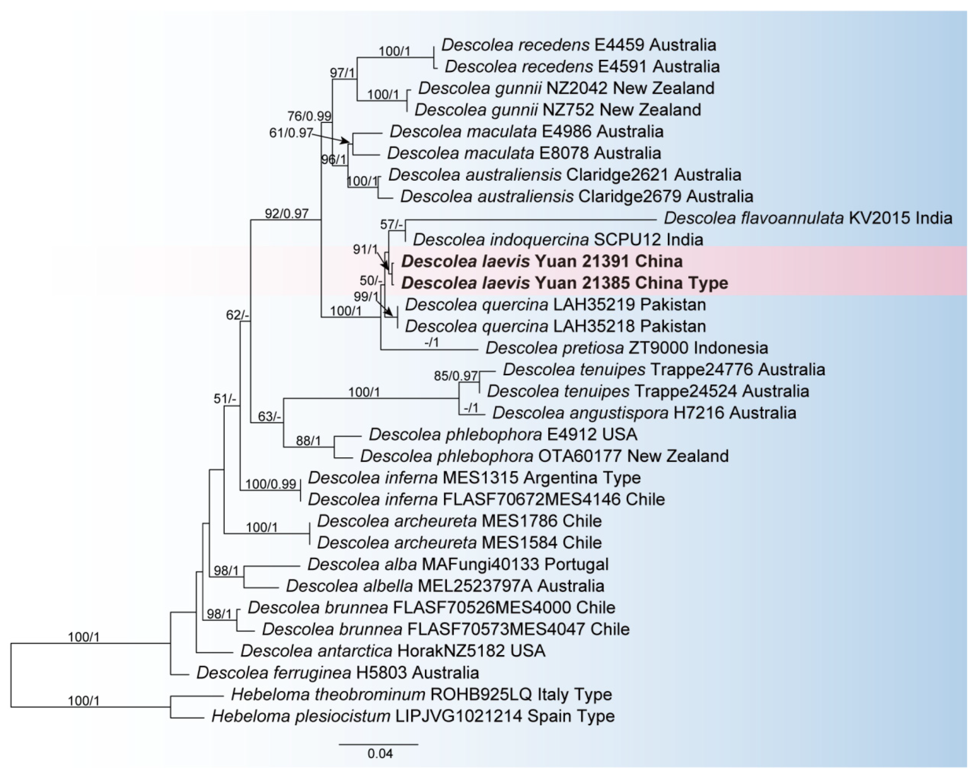
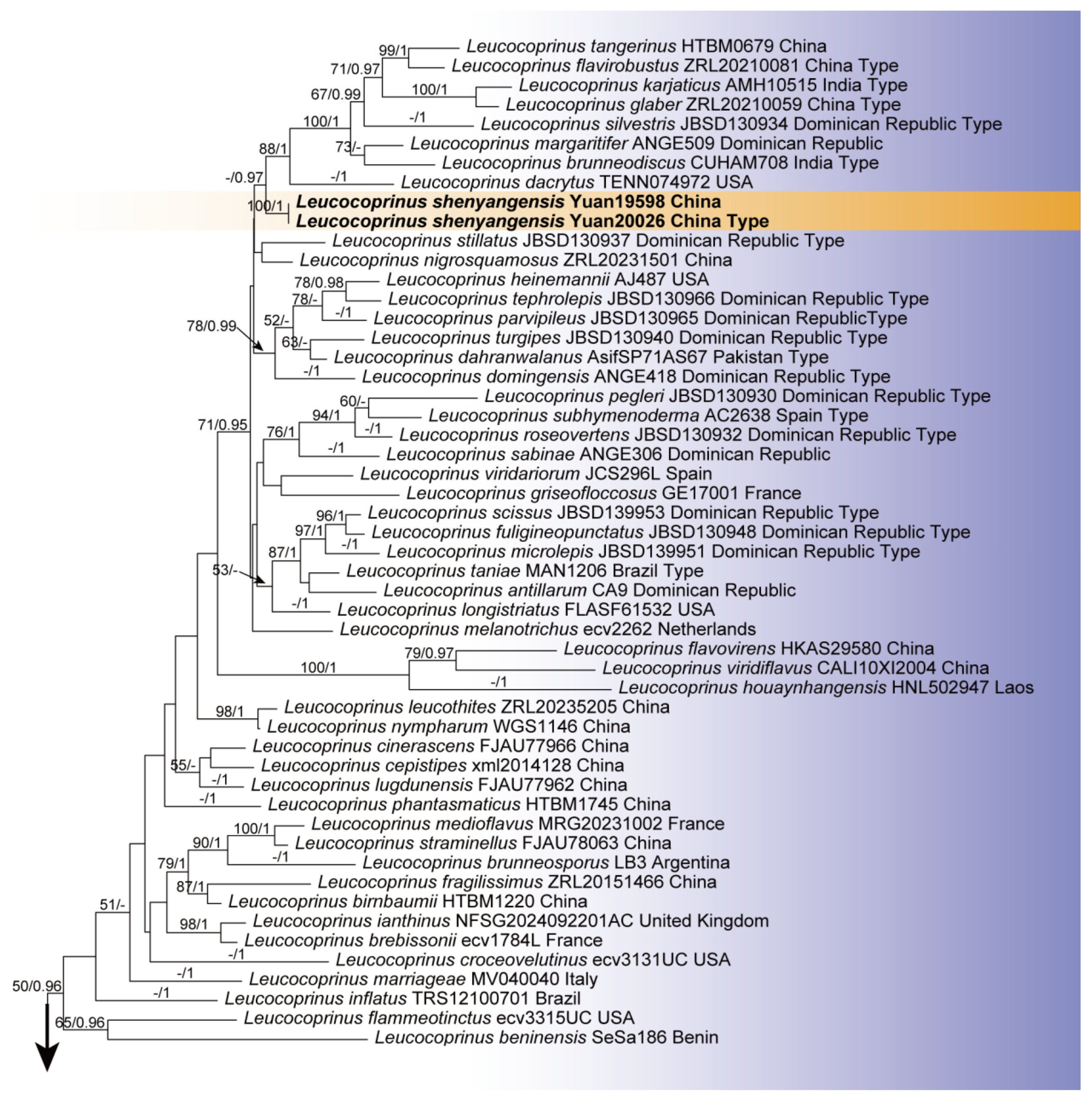
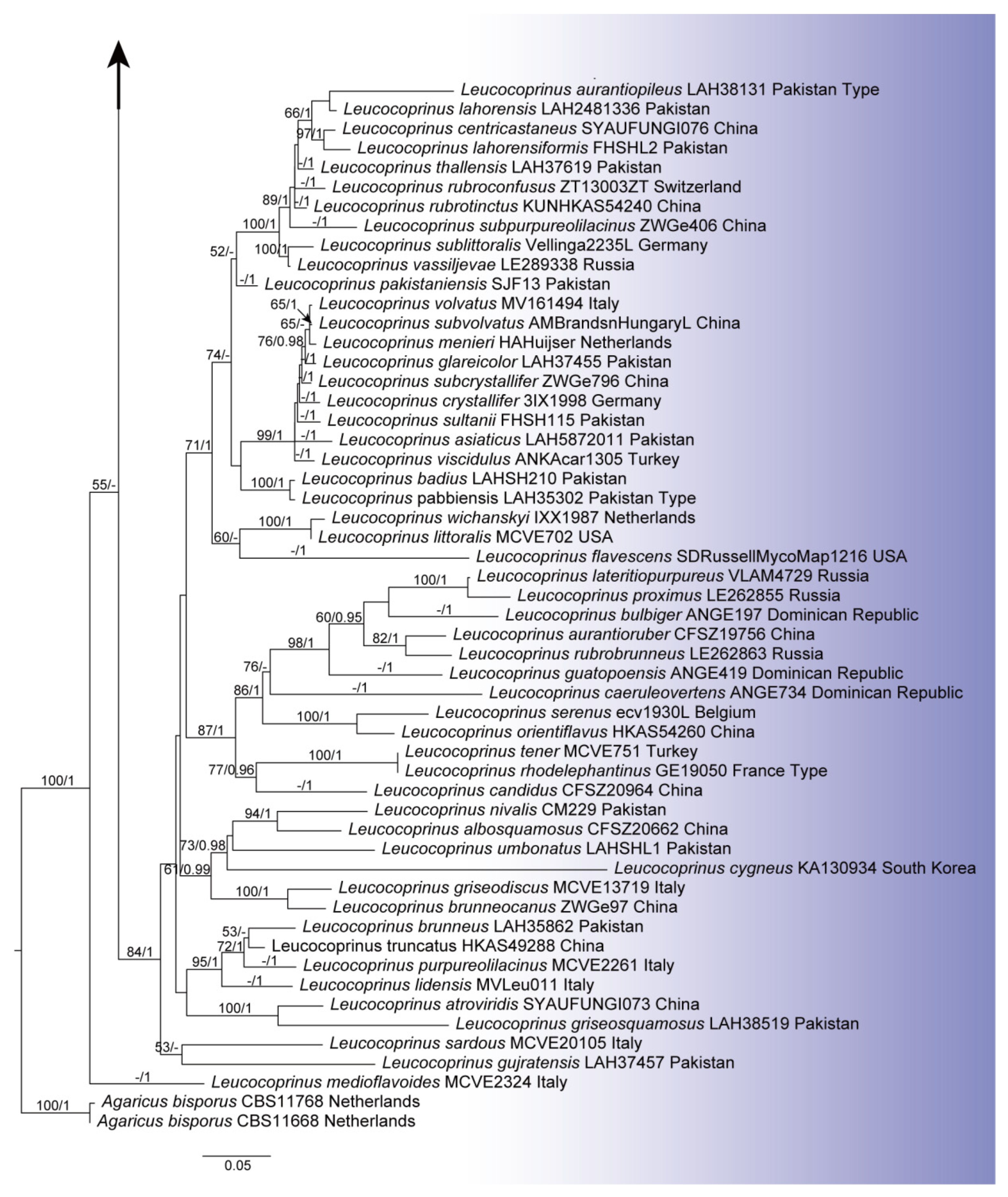
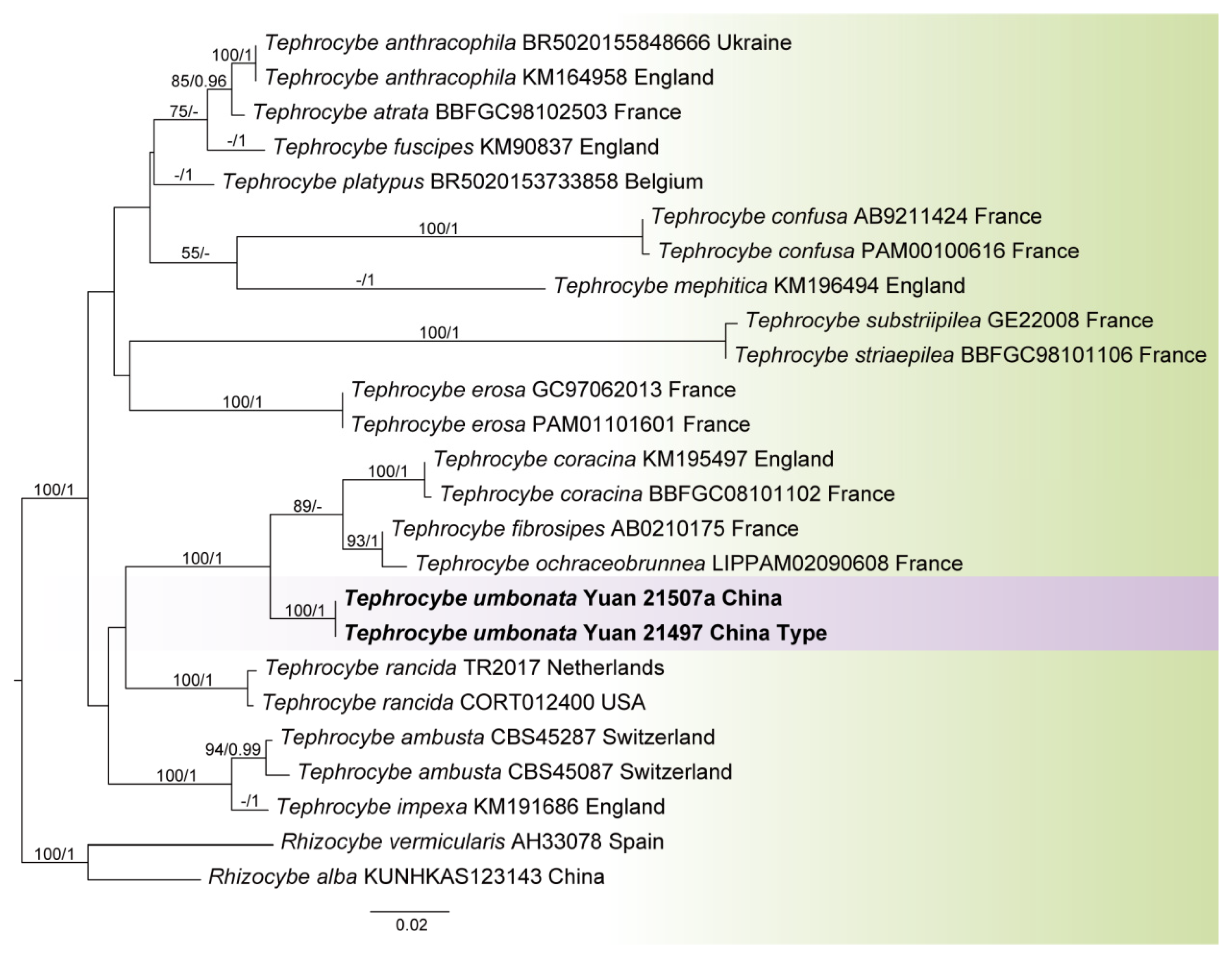
3.1. Phylogeny
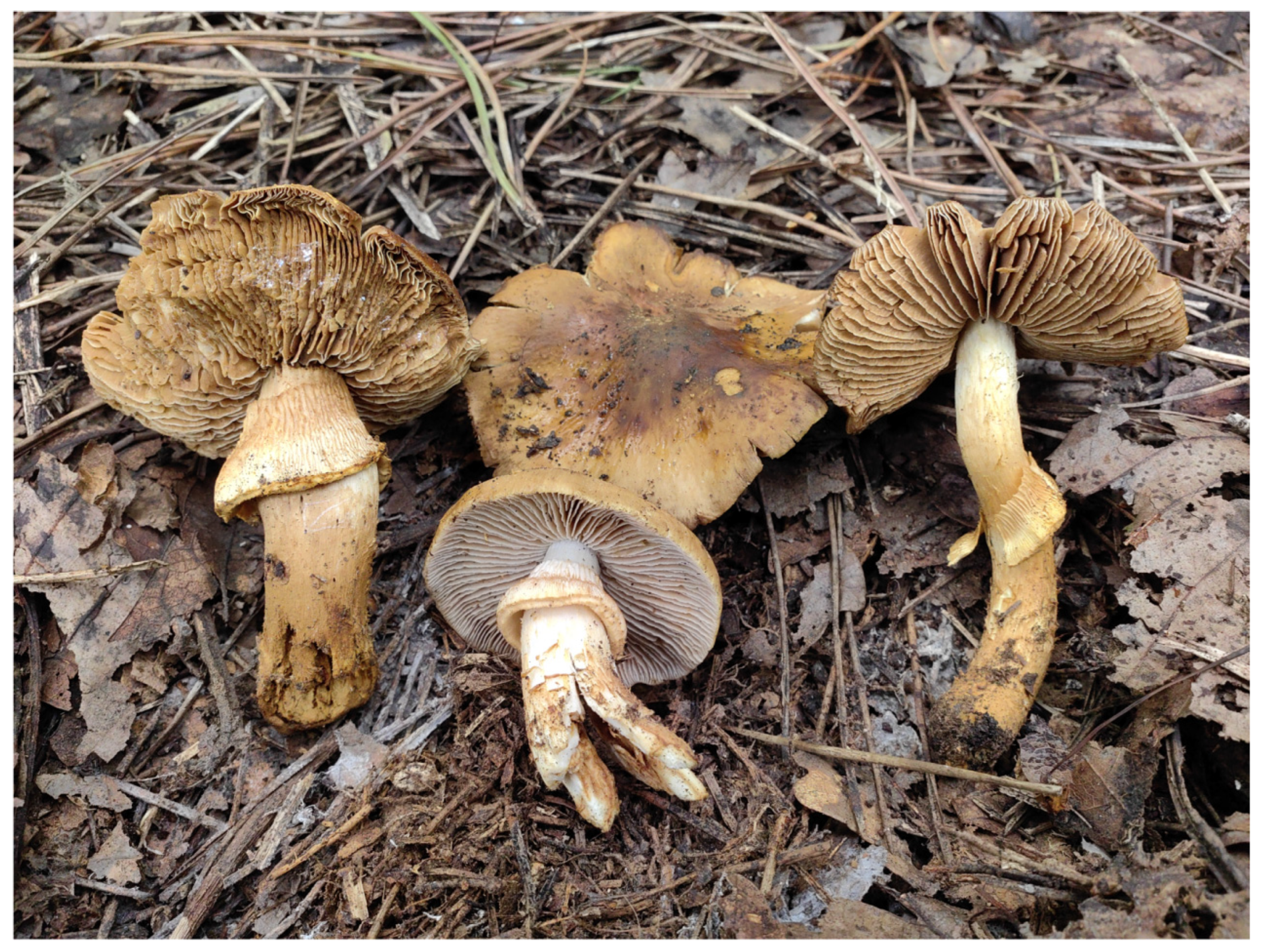
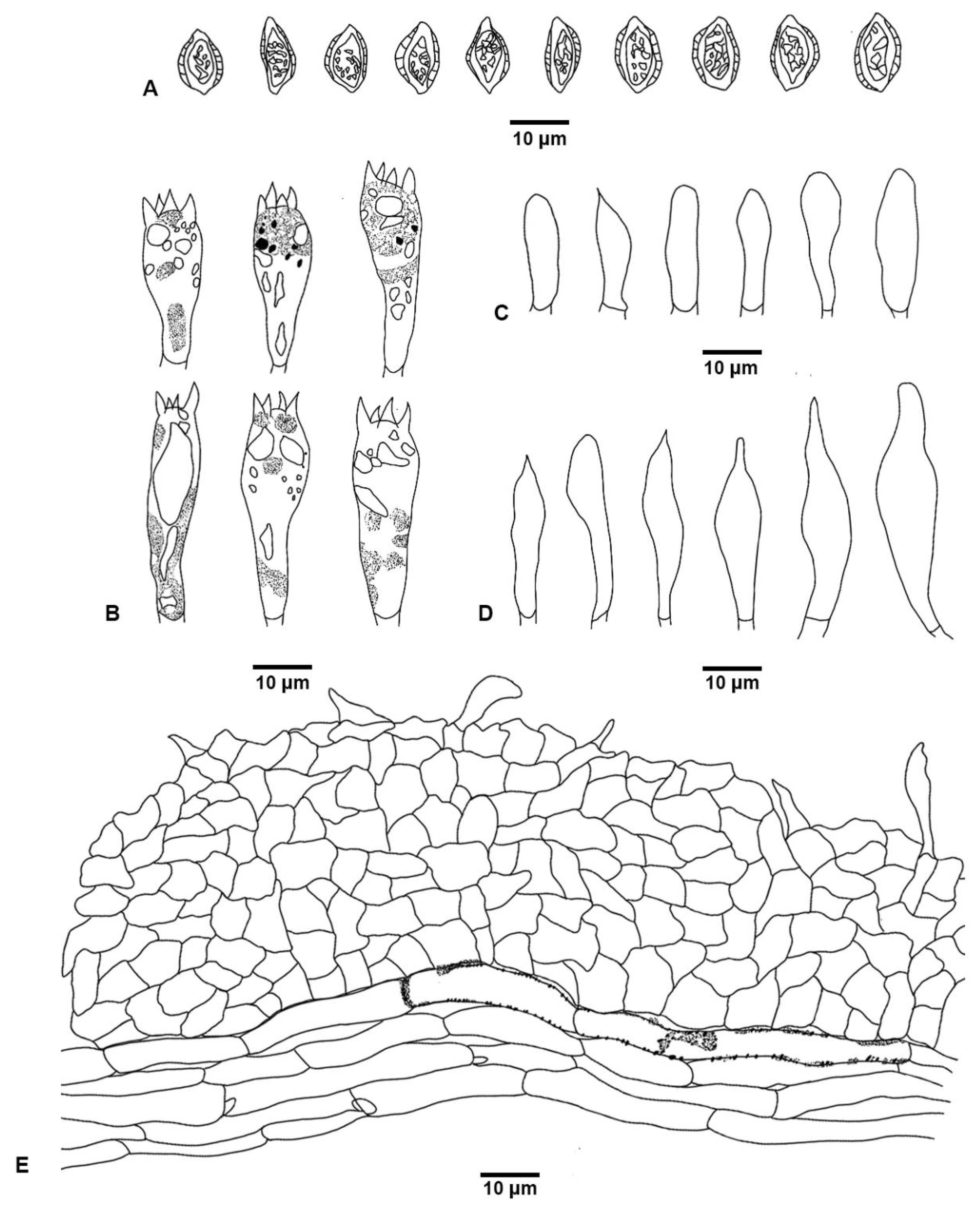
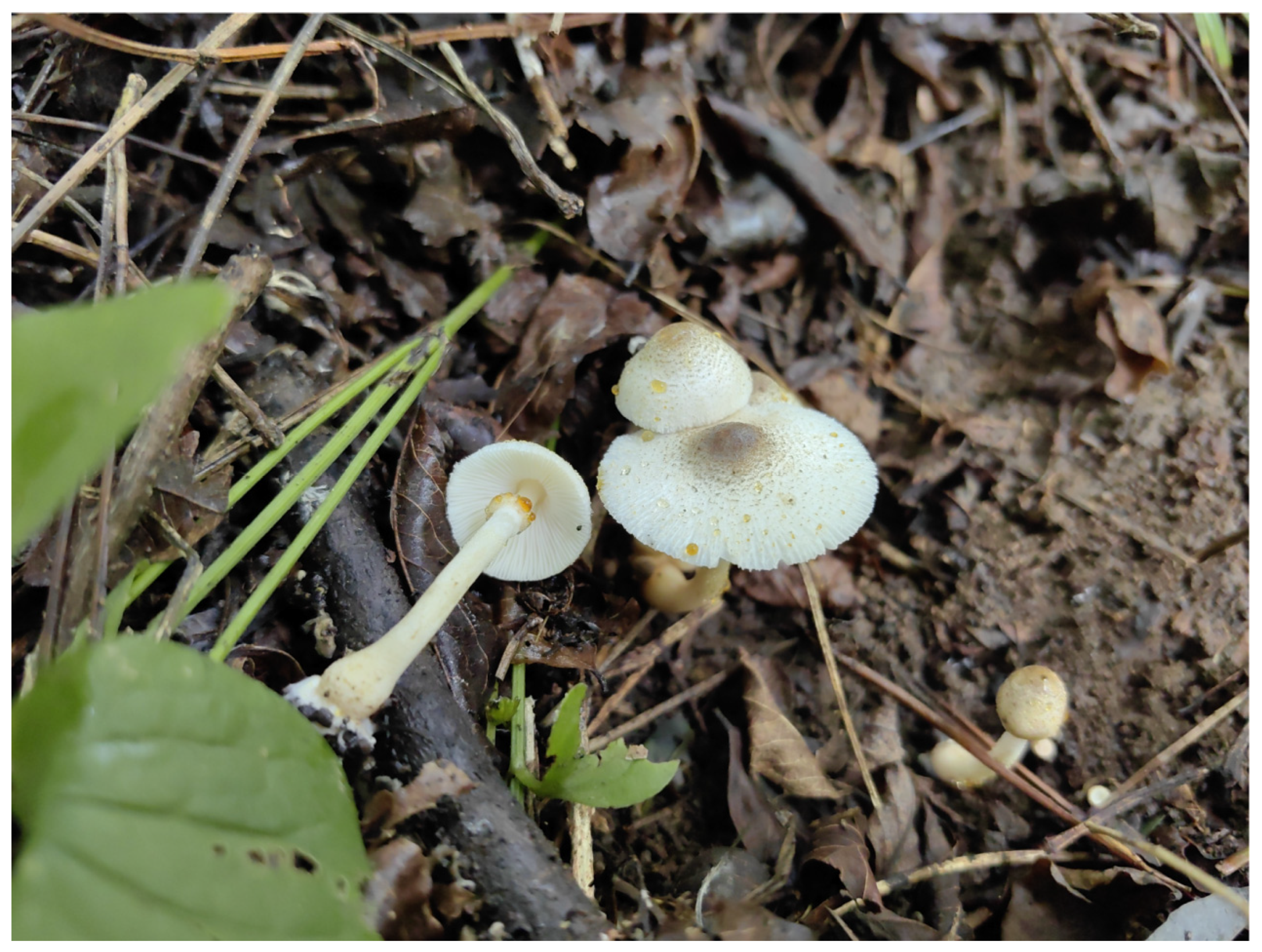
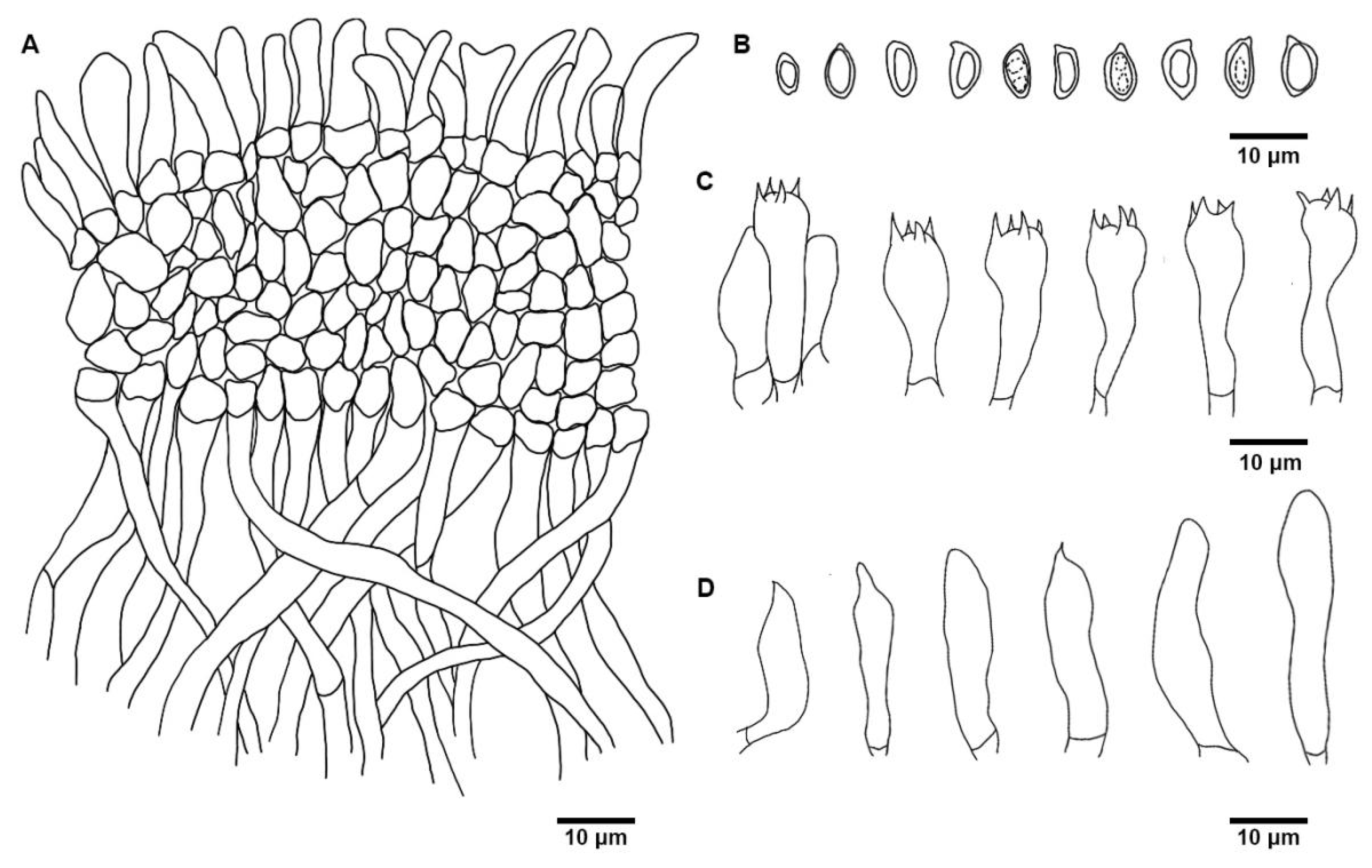
3.2. Taxonomy
4. Discussion
Author Contributions
Funding
Institutional Review Board Statement
Informed Consent Statement
Data Availability Statement
Conflicts of Interest
References
- Horak, E. Studies on the genus Descolea Sing. Persoonia 1971, 6, 231–248. [Google Scholar]
- Kuhar, F.; Smith, M.E.; Mujic, A.B.; Truong, C.; Nouhra, E. A systematic overview of Descolea (Agaricales) in the Nothofagaceae forests of Patagonia. Fungal Biol. 2017, 121, 876–889. [Google Scholar] [CrossRef] [PubMed]
- Khan, J.; Sher, H.; Naseer, A.; Khalid, A.N. Descolea quercina (Bolbitiaceae), a new species from moist temperate forests in Pakistan. MycoKeys 2017, 27, 65–77. [Google Scholar] [CrossRef] [PubMed]
- Yang, Z.L. Revision of the genera Rozites and Descolea from China. Fungal Sci. 1998, 13, 61–74. [Google Scholar] [CrossRef]
- Vellinga, E.C. Genera in the family Agaricaceae: Evidence from nrITS and nrLSU sequences. Mycol. Res. 2004, 108, 354–377. [Google Scholar] [CrossRef] [PubMed]
- Ijaz, H.; Mureed, M.; Yasmeen, A.; Naseer, A.; Hussain, A.; Jabeen, S. Molecular phylogeny and morphological characterization of Leucocoprinus birnbaumii from Punjab, Pakistan. Adv. Life Sci. 2023, 10, 148–151. [Google Scholar] [CrossRef]
- Muñoz, G.; Caballero, A.; Salom, J.C.; Ercole, E.; Vizzini, A. Leucoagaricus viridariorum (Agaricaceae, Agaricales), a new species from Spain. Phytotaxa 2015, 236, 226–236. [Google Scholar] [CrossRef]
- Ma, Y.; Liu, T.; Yu, X.; Wei, T.; Ge, Z.W. Six new species of Leucoagaricus (Agaricaceae) from northeastern China. Diversity 2022, 14, 314. [Google Scholar] [CrossRef]
- Asif, M.; Saba, M.; Raza, M.; Vellinga, E.C. Molecular insights into fungal diversity reveal three novel species of Leucocoprinus from southern Punjab, Pakistan. Mycologia 2024, 116, 601–620. [Google Scholar] [CrossRef] [PubMed]
- Yang, K.L.; Lin, J.Y.; Li, G.M.; Li, T.; Yang, Z.L. Rediscovering Leucoagaricus sinicus, with the recognition of Leucoagaricus and Leucocoprinus as separate genera, and two new genera in Agaricaceae (Basidiomycota). Phytotaxa 2024, 676, 199–255. [Google Scholar] [CrossRef]
- Yang, K.L.; Lin, J.Y.; Li, G.M. Stray studies on mushrooms from China. Phytotaxa 2026, 746, 1–119. [Google Scholar] [CrossRef]
- Ferretti-Cisneros, N.U.; Silva-Filho, A.G.; Wartchow, F. Leucocoprinus rhodolepis (Agaricaceae: Basidiomycota), a new species from Brazilian semi-arid region. Rev. Mex. Biodivers. 2022, 93, e933821. [Google Scholar] [CrossRef]
- Vellinga, E.C. Ecology and distribution of lepiotaceous fungi (Agaricaceae)-a review. Nova Hedwig. 2004, 78, 273–299. [Google Scholar] [CrossRef]
- Szczepkowski, A.; Gierczyk, B.; Kujawa, A. Greenhouses of botanical gardens as a habitat of alien and native macrofungi: A case study in Poland. Cent. Eur. J. Biol. 2014, 9, 777–795. [Google Scholar] [CrossRef]
- Zhang, A.Q.; Jin, R.; Zhang, X.C.; Tarafder, E.; Xu, J.Z. Two new species of genus Leucoagaricus and Leucocoprinus (Agaricaceae, Agaricales) from China. MycoKeys 2025, 125, 33–49. [Google Scholar] [CrossRef] [PubMed]
- Vizzini, A.; Alvarado, P.; Consiglio, G.; Marchetti, M.; Xu, J. Family matters inside the order Agaricales: Systematic reorganization and classification of incertae sedis clitocyboid, pleurotoid and tricholomatoid taxa based on an updated 6-gene phylogeny. Stud. Mycol. 2024, 107, 67–148. [Google Scholar] [CrossRef] [PubMed]
- He, M.Q.; Zhao, R.L.; Hyde, K.D.; Begerow, D.; Kemler, M.; Yurkov, A.; McKenzie, E.H.C.; Raspé, O.; Kakishima, M.; Sánchez-Ramírez, S.; et al. Notes, outline and divergence times of Basidiomycota. Fungal Divers. 2019, 99, 105–367. [Google Scholar] [CrossRef]
- Wei, S.W.; Lu, B.Y.; Wang, Y.; Dou, W.J.; Wang, Q.; Li, Y. Morphology and phylogeny of lyophylloid mushrooms in China with description of four new species. J. Fungi 2023, 9, 77. [Google Scholar] [CrossRef] [PubMed]
- Hofstetter, V.; Redhead, S.A.; Kauff, F.; Moncalvo, J.M.; Matheny, P.B.; Vilgalys, R. Taxonomic revision and examination of ecological transitions of the Lyophyllaceae (Basidiomycota, Agaricales) based on a multigene phylogeny. Cryptogam. Mycol. 2014, 35, 399–425. [Google Scholar] [CrossRef]
- van de Peppel, L.J.J.; Aime, M.C.; Læssøe, T.; Pedersen, O.S.; Coimbra, V.R.M.; Kuyper, T.W.; Stubbe, D.; Aanen, D.K.; Baroni, T.J. Four new genera and six new species of lyophylloid agarics (Agaricales, Basidiomycota) from three different continents. Mycol. Prog. 2022, 21, 85. [Google Scholar] [CrossRef]
- Orton, P.D. The genus Tephrocybe Donk in Britain. Bull. Br. Mycol. Soc. 1984, 18, 114–120. [Google Scholar] [CrossRef]
- Kornerup, A.; Wanscher, J.H. Methuen Handbook of Colour, 2nd ed.; Methuen: London, UK, 1967. [Google Scholar]
- Kotlaba, F.; Pouzar, Z. Preliminary results on the staining of spores and other structures of Homobasidiomycetes in cotton blue and its importance for taxonomy. Feddes Repert. 1964, 69, 131–142. [Google Scholar] [CrossRef]
- Singer, R. The Agaricales in Modern Taxonomy, 4th ed.; Koeltz Scientific Books: Koenigstein, Germany, 1986. [Google Scholar]
- White, T.J.; Bruns, T.; Lee, S.; Taylor, J. Amplification and direct sequencing of fungal ribosomal RNA genes for phylogenetics. In PCR Protocols: A Guide to Methods and Applications; Innis, M.A., Gelfand, D.H., Sninsky, J.J., White, T.J., Eds.; Academic Press: San Diego, CA, USA, 1990; pp. 315–322. [Google Scholar] [CrossRef]
- Schoch, C.L.; Seifert, K.A.; Huhndorf, S.; Robert, V.; Spouge, J.L.; Levesque, C.A.; Chen, W.; Fungal Barcoding Consortium. Nuclear ribosomal internal transcribed spacer (ITS) region as a universal DNA barcode marker for fungi. Proc. Natl. Acad. Sci. USA 2012, 109, 6241–6246. [Google Scholar] [CrossRef] [PubMed]
- Vilgalys, R.; Hester, M. Rapid genetic identification and mapping of enzymatically amplified ribosomal DNA from several Cryptococcus species. J. Bacteriol. 1990, 172, 4238–4246. [Google Scholar] [CrossRef] [PubMed]
- Sayers, E.W.; Cavanaugh, M.; Frisse, L.; Pruitt, K.D.; Schneider, V.A.; Underwood, B.A.; Yankie, L.; Karsch-Mizrachi, I. GenBank 2025 update. Nucleic Acids Res. 2025, 53, D56–D61. [Google Scholar] [CrossRef] [PubMed]
- Katoh, K.; Standley, D.M. MAFFT multiple sequence alignment software version 7: Improvements in performance and usability. Mol. Biol. Evol. 2013, 30, 772–780. [Google Scholar] [CrossRef] [PubMed]
- Kumar, S.; Stecher, G.; Tamura, K. MEGA7: Molecular evolutionary genetics analysis version 7.0 for bigger datasets. Mol. Biol. Evol. 2016, 33, 1870–1874. [Google Scholar] [CrossRef] [PubMed]
- Khan, J.; Sher, H.; Ali, H. Tricholyophyllum (Agaricales, Lyophyllaceae), an addition to the fungal flora of Pakistan with the description of a new species, T. rubrum. Nord. J. Bot. 2024, 2024, e04484. [Google Scholar] [CrossRef]
- Ronquist, F.; Teslenko, M.; van der Mark, P.; Ayres, D.L.; Darling, A.; Höhna, S.; Larget, B.; Liu, L.; Suchard, M.A.; Huelsenbeck, J.P. MrBayes 3.2: Efficient Bayesian phylogenetic inference and model choice across a large model space. Syst. Biol. 2012, 61, 539–542. [Google Scholar] [CrossRef] [PubMed]
- Stamatakis, A. RAxML version 8: A tool for phylogenetic analysis and post-analysis of large phylogenies. Bioinformatics 2014, 30, 1312–1313. [Google Scholar] [CrossRef] [PubMed]
- Rambaut, A. FigTree v.1.4.4. Available online: http://tree.bio.ed.ac.uk/software/figtree/ (accessed on 10 March 2026).
- Vu, D.; Groenewald, M.; de Vries, M.; Gehrmann, T.; Stielow, B.; Eberhardt, U.; Al-Hatmi, A.; Groenewald, J.Z.; Cardinali, G.; Houbraken, J.; et al. Large-scale generation and analysis of filamentous fungal DNA barcodes boosts coverage for kingdom fungi and reveals thresholds for fungal species and higher taxon delimitation. Stud. Mycol. 2019, 92, 135–154. [Google Scholar] [CrossRef] [PubMed]
- Martín, M.P.; Moreno, G. Molecular data confirm Setchelliogaster tenuipes and S. rheophyllus as Cortinariales. Mycotaxon 2001, 78, 257–263. [Google Scholar] [CrossRef]
- Francis, A.A.; Bougher, N.L. Cortinarioid sequestrate (truffle-like) fungi of Western Australia. Australas. Mycol. 2004, 23, 1–26. [Google Scholar]
- Peintner, U.; Bougher, N.L.; Castellano, M.A.; Moncalvo, J.M.; Moser, M.M.; Trappe, J.M.; Vilgalys, R. Multiple origins of sequestrate fungi related to Cortinarius (Cortinariaceae). Am. J. Bot. 2001, 88, 2168–2179. [Google Scholar] [CrossRef]
- Matheny, P.B.; Curtis, J.M.; Hofstetter, V.; Aime, M.C.; Moncalvo, J.-M.; Ge, Z.-W.; Yang, Z.-L.; Slot, J.C.; Ammirati, J.F.; Baroni, T.J.; et al. Major clades of Agaricales: A multilocus phylogenetic overview. Mycologia 2006, 98, 982–995. [Google Scholar] [CrossRef] [PubMed]
- Teasdale, S.E.; Beulke, A.K.; Guy, P.L.; Orlovich, D.A. Environmental barcoding of the ectomycorrhizal fungal genus Cortinarius. Fungal Divers. 2013, 58, 299–310. [Google Scholar] [CrossRef]
- Eberhardt, U.; Beker, H.J.; Vila, J.; Vesterholt, J.; Llimona, X.; Gadjieva, R. Hebeloma species associated with Cistus. Mycol. Res. 2009, 113, 153–162. [Google Scholar] [CrossRef] [PubMed]
- Eberhardt, U.; Beker, H.J.; Vesterholt, J.; Dukić, K.; Walther, G.; Vila, J.; Fernández-Brime, S. European species of Hebeloma section Theobromina. Fungal Divers. 2013, 58, 103–126. [Google Scholar] [CrossRef]
- Justo, A.; Angelini, C.; Bizzi, A. The genera Leucoagaricus and Leucocoprinus in the Dominican Republic. Mycologia 2021, 113, 348–389. [Google Scholar] [CrossRef] [PubMed]
- Ge, Z.W.; Yang, Z.L.; Qasim, T.; Nawaz, R.; Khalid, A.N.; Vellinga, E.C. Four new species in Leucoagaricus (Agaricaceae, Basidiomycota) from Asia. Mycologia 2015, 107, 1033–1044. [Google Scholar] [CrossRef] [PubMed]
- Maula, F.; Saba, M.; Asif, M.; Rani, A.K.; Akram, W.; Ullah, F.; Batool, S.; Bakhsh, K.; Bibi, A.; Izhar, A.; et al. Leucocoprinus aurantiopileus (Agaricaceae), a new species from Pakistan. N. Z. J. Bot. 2026, 64, e70026. [Google Scholar] [CrossRef]
- Hussain, S.; Jabeen, S.; Khalid, A.N.; Ahmad, H.; Afshan, N.U.S.; Sher, H.; Pfister, D.H. Underexplored regions of Pakistan yield five new species of Leucoagaricus. Mycologia 2018, 110, 387–400. [Google Scholar] [CrossRef] [PubMed]
- Sarawi, S.; Yorou, N.S.; Piepenbring, M. Leucocoprinus beninensis, a new species of Agaricaceae from Benin. Cryptogam. Mycol. 2026, 47, 1–9. [Google Scholar] [CrossRef]
- Vellinga, E.C.; de Kok, R.P.; Bruns, T.D. Phylogeny and taxonomy of Macrolepiota (Agaricaceae). Mycologia 2003, 95, 442–456. [Google Scholar] [CrossRef]
- Dutta, A.K.; Stallman, J.K.; Bera, S.; Hoque, E.; Paloi, S.; Acharya, K. Lepiotaceous fungi of West Bengal, India: Two new species of Leucoagaricus. Mycol. Prog. 2021, 20, 493–507. [Google Scholar] [CrossRef]
- Suarez, J.M.; Gottlieb, A.M.; Lechner, B.E. A new and intriguing brown-spored Leucocoprinus species. Phytotaxa 2021, 479, 44–54. [Google Scholar] [CrossRef]
- Ullah, Z.; Jabeen, S.; Faisal, M.; Ahmad, H.; Khalid, A.N. Leucoagaricus brunneus sp. nov. from Khyber Pakhtunkhwa, Pakistan. Mycotaxon 2019, 134, 601–611. [Google Scholar] [CrossRef]
- Li, J.X.; Zhao, R.L.; Phurbu, D.; Xing, R.; Liu, D.M.; He, M.Q.; Ling, Z.L.; Han, X.X.; Feng, H.Y.; Zhu, X.Y.; et al. Disentangling taxonomic chaos in Agaricaceae s.l.: An integrative phylogenomic framework with divergence dating reconstructs classification. Fungal Divers. 2025, 135, 745–881. [Google Scholar] [CrossRef]
- Vellinga, E.C.; Sundberg, W.J. Lepiotaceous fungi of California, USA. 6. Lepiota castanescens. Mycotaxon 2008, 103, 97–108. [Google Scholar] [CrossRef]
- Kim, C.S.; Jo, J.W.; Kwag, Y.N.; Sung, G.H.; Lee, S.G.; Kim, S.Y.; Shin, C.H.; Han, S.K. Mushroom flora of Ulleung-gun and a newly recorded Bovista species in the Republic of Korea. Mycobiology 2015, 43, 239–257. [Google Scholar] [CrossRef] [PubMed]
- Swenie, R.A.; Matheny, P.B. New reports, new species, and high diversity of Cantharellus in the southern Appalachians. Mycologia 2023, 115, 44–68. [Google Scholar] [CrossRef] [PubMed]
- Vellinga, E.C. Lepiotaceous fungi in California, USA. Leucoagaricus sect. Piloselli. Mycotaxon 2010, 112, 393–444. [Google Scholar] [CrossRef]
- Liang, J.F.; Yang, Z.L.; Xu, J.; Ge, Z.W. Two new unusual Leucoagaricus species (Agaricaceae) from tropical China with blue-green staining reactions. Mycologia 2010, 102, 1141–1152. [Google Scholar] [CrossRef] [PubMed]
- Zhao, R.L.; Li, G.J.; Sánchez-Ramírez, S.; Stata, M.; Yang, Z.L.; Wu, G.; Dai, Y.C.; He, S.H.; Cui, B.K.; Zhou, J.L.; et al. A six-gene phylogenetic overview of Basidiomycota and allied phyla with estimated divergence times of higher taxa and a phyloproteomics perspective. Fungal Divers. 2017, 84, 43–74. [Google Scholar] [CrossRef]
- Ashraf, S.; Naseer, A.; Usman, M.; Khalid, A.N. Two new species of genus Leucoagaricus (Agaricaceae, Agaricales) from Pakistan. MycoKeys 2023, 96, 159–171. [Google Scholar] [CrossRef] [PubMed]
- Osmundson, T.W.; Robert, V.A.; Schoch, C.L.; Baker, L.J.; Smith, A.; Robich, G.; Mizzan, L.; Garbelotto, M.M. Filling gaps in biodiversity knowledge for macrofungi: Contributions and assessment of an herbarium collection DNA barcode sequencing project. PLoS ONE 2013, 8, e62419. [Google Scholar] [CrossRef] [PubMed]
- Lagardère, V.; Eyssartier, G. Leucocoprinus griseofloccosus sp. nov. une nouvelle espèce de la section Denudati. Bull. Soc. Mycol. Fr. 2016, 132, 105–114. [Google Scholar]
- Rehman, A.; Usman, M.; Afshan, N.U.S.; Khalid, A.N. Leucoagaricus gujratensis sp. nov. (Agaricaceae, Agaricales) from Pakistan. Phytotaxa 2023, 589, 39–50. [Google Scholar] [CrossRef]
- Sysouphanthong, P.; Bouamanivong, S.; Salichanh, T.; Xaybouangeun, N.; Sucharitakul, P.; Osathanunkul, M.; Suwannapoom, C. Leucoagaricus houaynhangensis (Agaricaceae), a new yellowish-green species from Lao People’s Democratic Republic. Chiang Mai J. Sci. 2018, 45, 1287–1295. [Google Scholar]
- Solomon, S.E.; Rabeling, C.; Sosa-Calvo, J.; Lopes, C.T.; Rodrigues, A.; Vasconcelos, H.L.; Bacci, M.J.; Mueller, U.G.; Schultz, T.R. The molecular phylogenetics of Trachymyrmex Forel ants and their fungal cultivars provide insights into the origin and coevolutionary history of higher-attine ant agriculture. Syst. Entomol. 2019, 44, 939–956. [Google Scholar] [CrossRef]
- Patil, P.B.; Patil, N.P.; Chahar, S.; Maurya, S. Leucoagaricus karjaticus (Agaricaceae), a new species from Maharashtra, India. Mycoscience 2024, 65, 244–252. [Google Scholar] [CrossRef] [PubMed]
- Qasim, T.; Amir, T.; Nawaz, R.; Niazi, A.R.; Khalid, A.N. Leucoagaricus lahorensis, a new species of L. sect. Rubrotincti. Mycotaxon 2015, 130, 533–541. [Google Scholar] [CrossRef]
- Malysheva, E.F.; Svetasheva, T.Y.; Bulakh, E.M. Fungi in the Russian Far East. I. Leucoagaricus lateritiopurpureus and new species of Leucoagaricus (Agaricaceae) with reddish brown basidiocarps. Mikol. Fitopatol. 2013, 47, 169–179. [Google Scholar]
- Migliozzi, V.; Alvarado, P. Nomenclatural novelties: Leucoagaricus lidensis. Index Fungorum 2021, 478, 1. [Google Scholar]
- Migliozzi, V.; Donato, G. Nuevas combinaciones en el género Leucocoprinus. 2. Errotari 2025, 22, 80–83. [Google Scholar]
- Jabeen, S.; Waseem, B.; Tuba; Hamid, M.; Yasmeen, A. First record of Leucoagaricus nivalis from Pakistan. Bangladesh J. Plant Taxon. 2020, 27, 453–459. [Google Scholar] [CrossRef]
- Ge, Z.W. Leucoagaricus orientiflavus, a new yellow lepiotoid species from southwestern China. Mycotaxon 2010, 111, 121–126. [Google Scholar] [CrossRef]
- Usman, M.; Khalid, A.N. Leucoagaricus pabbiensis sp. nov. from Punjab, Pakistan. Mycotaxon 2018, 133, 355–364. [Google Scholar] [CrossRef]
- Yang, K.L.; Lin, J.Y.; Li, G.M.; Yang, Z.L. New and interesting fungi from Guangzhou City, South China. Phytotaxa 2025, 707, 1–95. [Google Scholar] [CrossRef]
- Boisselet, P.; Eyssartier, G. Un Leucoagaric décrit du littoral Morbihannais: Leucoagaricus rhodelephantinus sp. nov. Bull. Trimest. Soc. Mycol. Fr. 2020, 133, 263–272. [Google Scholar]
- Justo, A.; Angelini, C.; Bizzi, A.; Vizzini, A. Leucoagaricus sabinae (Agaricaceae), a new species from the Dominican Republic. N. Am. Fungi 2015, 10, 1–15. [Google Scholar] [CrossRef]
- Heisecke, C.; Lima, D.F.; Mueller, G.M.; Neves, M.A. Leucoagaricus taniae sp. nov. (Agaricaceae), a sand-dwelling mushroom from Brazil. Brittonia 2022, 74, 18–29. [Google Scholar] [CrossRef]
- Khan, Z.; Izhar, A.; Khalid, A.N. Leucoagaricus thallensis (Agaricaceae; Basidiomycota), a new species from Punjab, Pakistan. Phytotaxa 2023, 594, 163–177. [Google Scholar] [CrossRef]
- Migliozzi, V.; Donato, G. Alcuni Leucocoprinus della sezione Sericeomyces: Descrizione di Leucocoprinus amylosporus, Leucocoprinus crystallifer, Leucocoprinus cfr. viscidulus e Leucocoprinus volvatus s.l. Bol. Soc. Micol. Madr. 2025, 49, 51–68. [Google Scholar]
- He, Z.M.; Chen, Z.H.; Bau, T.; Wang, G.S.; Yang, Z.L. Systematic arrangement within the family Clitocybaceae (Tricholomatineae, Agaricales): Phylogenetic and phylogenomic evidence, morphological data and muscarine-producing innovation. Fungal Divers. 2023, 123, 1–47. [Google Scholar] [CrossRef]
- Alvarado, P.; Moreno, G.; Vizzini, A.; Consiglio, G.; Manjón, J.L.; Setti, L. Atractosporocybe, Leucocybe and Rhizocybe: Three new clitocyboid genera in the Tricholomatoid clade (Agaricales) with notes on Clitocybe and Lepista. Mycologia 2015, 107, 123–136. [Google Scholar] [CrossRef] [PubMed]
- Hofstetter, V.; Clémençon, H.; Vilgalys, R.; Moncalvo, J.M. Phylogenetic analyses of the Lyophylleae (Agaricales, Basidiomycota) based on nuclear and mitochondrial rDNA sequences. Mycol. Res. 2002, 106, 1043–1059. [Google Scholar] [CrossRef]
- Bellanger, J.M.; Moreau, P.A.; Corriol, G.; Bidaud, A.; Chalange, R.; Dudova, Z.; Richard, F. Plunging hands into the mushroom jar: A phylogenetic framework for Lyophyllaceae (Agaricales, Basidiomycota). Genetica 2015, 143, 169–194. [Google Scholar] [CrossRef] [PubMed]
- Eyssartier, G. Tephrocybe substriipilea, une espèce plus méconnue que rare? Bull. Mycol. Bot. Dauphiné-Savoie 2025, 259, 17–21. [Google Scholar]
- Choudhary, S.; Verma, K.; Sharma, Y.P.; Uniyal, P. A novel species and a new record of genus Descolea from Indian Himalaya. Taiwania 2024, 69, 356–365. [Google Scholar] [CrossRef]
- Yuan, Y.; Li, Y.K.; Liang, J.F. Leucoagaricus tangerinus, a new species with drops from southern China. Mycol. Prog. 2014, 13, 893–898. [Google Scholar] [CrossRef]
- Consiglio, G.; Contu, M. Taxons nouveaux dans le genre Lyophyllum P. Karst. Doc. Mycol. 2001, 30, 43–46. [Google Scholar]
- Bon, M. Agaricomycètes rares ou intéressants de Nord-Picardie. Doc. Mycol. 1993, 22, 41–51. [Google Scholar]
- Knudsen, H.; Vesterholt, J. Funga Nordica: Agaricoid, Boletoid, and Cyphelloid Genera; Nordsvamp: Copenhagen, Denmark, 2008. [Google Scholar]
- Vellinga, E.C.; Balsley, R.B. Leucoagaricus dacrytus-a new species from New Jersey, USA. Mycotaxon 2010, 113, 73–80. [Google Scholar] [CrossRef]
- Hughes, K.W.; Matheny, P.B.; Miller, A.N.; Petersen, R.H.; Iturriaga, T.M.; Johnson, K.D.; Methven, A.S.; Raudabaugh, D.B.; Swenie, R.A.; Bruns, T.D. Pyrophilous fungi detected after wildfires in the Great Smoky Mountains National Park expand known species ranges and biodiversity estimates. Mycologia 2020, 112, 677–698. [Google Scholar] [CrossRef] [PubMed]
- Yamanaka, T. Fruit-body production and mycelial growth of Tephrocybe tesquorum in urea-treated forest soil. Mycoscience 2001, 42, 333–338. [Google Scholar] [CrossRef]
- Matheny, P.B.; Norvell, L.L.; Giles, E.C. A common new species of Inocybe in the Pacific Northwest with a diagnostic PDAB reaction. Mycologia 2013, 105, 436–446. [Google Scholar] [CrossRef] [PubMed]
- Burke, D.J.; López-Gutiérrez, J.C.; Smemo, K.A.; Chan, C.R. Vegetation and soil environment influence the spatial distribution of root-associated fungi in a mature beech-maple forest. Appl. Environ. Microbiol. 2009, 75, 7639–7648. [Google Scholar] [CrossRef] [PubMed]



| Species Name | ITS | LSU | Specimen No. | Country | References |
|---|---|---|---|---|---|
| Agaricus bisporus (J.E. Lange) Imbach | MH859080 | MH870797 | CBS11668 | The Netherlands | Vu et al., 2019 [35] |
| A. bisporus | MH859081 | MH870798 | CBS11768 | The Netherlands | Vu et al., 2019 [35] |
| Descolea alba (Bull.) Kuhar, Nouhra & M.E. Sm. | AJ296296 | / | MA-Fungi40133 | Portugal | Martín and Moreno 2001 [36] |
| D. albella (Massee & Rodway) Kuhar, Nouhra & M.E. Sm. | PV650409 | / | MEL2523797A | Australia | NCBI Database |
| D. angustispora (A.A. Francis & Bougher) Kuhar, Nouhra & M.E. Sm. | DQ328058 | / | H7216 | Australia | Francis and Bougher 2004 [37] |
| D. antarctica Singer | AF325647 | / | HorakNZ5182 | New Zealand | Peintner et al., 2001 [38] |
| D. archeureta (Halling) Kuhar, Nouhra & M.E. Sm. | KY523096 | / | MES-1786 | Chile | Kuhar et al., 2017 [2] |
| D. archeureta | KY523092 | / | MES-1584 | Chile | Kuhar et al., 2017 [2] |
| D. australiensis (G.W. Beaton, Pegler & T.W.K. Young) Kuhar, Nouhra & M.E. Sm. | AF325627 | / | Claridge2679 | Australia | Peintner et al., 2001 [38] |
| D. australiensis | AF325628 | / | Claridge2621 | Australia | Peintner et al., 2001 [38] |
| D. brunnea (E. Horak) Kuhar, Nouhra & M.E. Sm. | OP339609 | / | FLAS:F-70573-MES-4047 | Chile | NCBI Database |
| D. brunnea | OP339580 | / | FLAS:F-70526-MES-4000 | Chile | NCBI Database |
| D. ferruginea (J.W. Cribb) Kuhar, Nouhra & M.E. Sm. | DQ328083 | / | H5803 | Australia | Kuhar et al., 2017 [2] |
| D. flavoannulata (Lj.N. Vassiljeva) E. Horak | PP345433 | / | KV-20-15 | India | NCBI Database |
| D. gunnii (Berk. ex Massee) E. Horak | AF325652 | / | NZ752 | New Zealand | Peintner et al., 2001 [38] |
| D. gunnii | AF325653 | / | NZ2042 | New Zealand | Peintner et al., 2001 [38] |
| D. indoquercina S. Choudhary, P. Uniyal & Y.P. Sharma | OR979479 | / | SC/PU/12 | India | NCBI Database |
| D. inferna Kuhar, Nouhra & M.E. Sm. | NR154032 | / | CORD:MES1315 (T) | Argentina | Kuhar et al., 2017 [2] |
| D. inferna | OP339680 | / | FLAS:F70672MES-4146 | Chile | NCBI Database |
| D. laevisZ.Q. You & H.S. Yuan | PZ235489 | PZ235510 | Yuan21385 (T) | China | Present study |
| D. laevis | PZ235490 | PZ235511 | Yuan21391 | China | Present study |
| D. maculata Bougher | DQ192181 | DQ457664 | E8078(PERTH) | Australia | Matheny et al., 2006 [39] |
| D. maculata | AF325651 | / | E4986 | Australia | Peintner et al., 2001 [38] |
| D. phlebophora E. Horak | AF325657 | / | E4912 | Australia | Peintner et al., 2001 [38] |
| D. phlebophora | JX178627 | / | OTA:60177 | New Zealand | Teasdale et al., 2013 [40] |
| D. pretiosa E. Horak | MN267170 | / | ZT9000 | Indonesia | NCBI Database |
| D. quercina J. Khan & Naseer | MF966638 | / | LAH35219 | Pakistan | Khan et al., 2017 [3] |
| D. quercina | MF966637 | MF966635 | LAH35218 | Pakistan | Khan et al., 2017 [3] |
| D. recedens (Sacc.) Singer | AF325648 | / | E4591 | Australia | Peintner et al., 2001 [38] |
| D. recedens | AF325649 | / | E4459 | Australia | Peintner et al., 2001 [38] |
| D. tenuipes (Setch.) Neville & Poumarat | AF325624 | / | Trappe24776 | Australia | Peintner et al., 2001 [38] |
| D. tenuipes | AF325623 | / | Trappe24524 | Australia | Peintner et al., 2001 [38] |
| Hebeloma plesiocistum Beker, U. Eberh. & Vila | NR119686 | / | LIP:JVG1021214 (T) | Spain | Eberhardt et al., 2009 [41] |
| H. theobrominum Quadr. | NR120177 | / | ROHB:925LQ (T) | Italy | Eberhardt et al., 2013 [42] |
| Leucocoprinus albosquamosus (Y.R. Ma, Z.W. Ge & T.Z. Liu) M. Asif, Saba & Vellinga | OM976879 | OM976865 | CFSZ20662 | China | Ma et al., 2022 [8] |
| L. antillarum Justo, Bizzi & Angelini | MN482989 | / | CA9 | Dominican Republic | Justo et al., 2021 [43] |
| L. asiaticus (Qasim, Nawaz & Khalid) M. Asif, Saba & Vellinga | KP164972 | / | LAH5872011 | Pakistan | Ge et al., 2015 [44] |
| L. atroviridis (Y.R. Ma, Z.W. Ge & T.Z. Liu) M. Asif, Saba & Vellinga | OM976852 | OM976868 | SYAUFUNGI073 | China | Ma et al., 2022 [8] |
| L. aurantiopileus Maula, Asif, A.K. Rani & Afshan | PP383877 | PP583799 | LAH38131 (T) | Pakistan | Maula et al., 2026 [45] |
| L. aurantioruber (Y.R. Ma, Z.W. Ge & T.Z. Liu) M. Asif, Saba & Vellinga | OM976875 | OM976863 | CFSZ19756 | China | Ma et al., 2022 [8] |
| L. badius (S. Hussain, Pfister, Afshan & Khalid) M. Asif, Saba & Vellinga | KU647734 | / | LAHSH210 | Pakistan | Hussain et al., 2018 [46] |
| L. beninensis Sarawi | PX634061 | / | SeSa186 | Benin | Sarawi et al., 2026 [47] |
| L. birnbaumii (Corda) Singer | PQ321881 | PQ319807 | HTBM1220 | China | Yang et al., 2024 [10] |
| L. brebissonii (Godey) Locq. | AF482859 | AY176446 | ecv1784L | France | Vellinga et al., 2003 [48] |
| L. brunneocanus (Fei Yu, Jun F. Liang & Z.W. Ge) M. Asif, Saba & Vellinga | KP096237 | / | ZWGe97 | China | Ge et al., 2015 [44] |
| L. brunneodiscus (A.K. Dutta & K. Acharya) Kun L. Yang, Jia Y. Lin & Zhu L. Yang | NR198110 | NG244261 | CUHAM708 (T) | India | Dutta et al., 2021 [49] |
| L. brunneosporus B.E. Lechner & J.M. Suárez | MT796198 | MT796197 | LB3 | Argentina | Suarez et al., 2021 [50] |
| L. brunneus (Zia Ullah, Jabeen & Khalid) M. Asif, Saba & Vellinga | MH990662 | / | LAH35862 | Pakistan | Ullah et al., 2019 [51] |
| L. bulbiger (Justo, Bizzi & Angelini) M. Asif, Saba & Vellinga | MN483028 | / | ANGE197 | Dominican Republic | Justo et al., 2021 [43] |
| L. caeruleovertens (Justo, Bizzi & Angelini) M. Asif, Saba & Vellinga | MN483032 | / | ANGE734 | Dominican Republic | Justo et al., 2021 [43] |
| L. candidus (Y.R. Ma, Z.W. Ge & T.Z. Liu) M. Asif & Saba | OM976877 | OM976864 | CFSZ20964 | China | Ma et al., 2022 [8] |
| L. centricastaneus (Y.R. Ma, Z.W. Ge & T.Z. Liu) M. Asif, Saba & Vellinga | OM976855 | OM976871 | SYAUFUNGI076 | China | Ma et al., 2022 [8] |
| L. cepistipes (Sowerby) Pat. | PV470803 | PV476031 | xml2014128 | China | Li et al., 2025 [52] |
| L. cinerascens (Quél.) Locq. | PX527069 | PX527070 | FJAU77966 | China | NCBI Database |
| L. croceovelutinus Bon & Boiffard | EU166351 | / | ecv3131UC | USA | Vellinga and Sundberg 2008 [53] |
| L. crystallifer (Vellinga) Migl. & Donato | AF482863 | AY176412 | 3IX1998 | Germany | Vellinga et al., 2003 [48] |
| L. cygneus (J.E. Lange) Bon | KR673661 | / | KA130934 | Republic of Korea | Kim et al., 2015 [54] |
| L. dacrytus (Vellinga) Kun L. Yang, Jia Y. Lin & Zhu L. Yang | MT196954 | / | TENN:074972 | USA | Swenie and Matheny 2023 [55] |
| L. dahranwalanus Asif, Saba & Raza | OQ947827 | OQ947833 | AsifSP71AS67 (T) | Pakistan | Asif et al., 2024 [9] |
| L. domingensis Justo, Bizzi, Angelini &Vizzini | MN483016 | / | ANGE418 (T) | Dominican Republic | Justo et al., 2021 [43] |
| L. flammeotinctus (Kauffman) Redhead | GU136165 | / | ecv3315UC | USA | Vellinga 2010 [56] |
| L. flavescens (Morgan) H.V. Sm. | MW567852 | / | SDRussellMycoMa-p1216 | USA | NCBI Database |
| L. flavirobustus R.L. Zhao & J.X. Li | PV470825 | PV475985 | ZRL20210081 (T) | China | Li et al., 2025 [52] |
| L. flavovirens (Jun F. Liang, Zhu L. Yang & J. Xu) Kun L. Yang, Jia Y. Lin & Zhu L. Yang | EU416293 | EU416294 | HKAS29580 | China | Liang et al., 2010 [57] |
| L. fragilissimus (Ravenel ex Berk. & M.A. Curtis) Pat. | LT716029 | KY418844 | ZRL20151466 | China | Zhao et al., 2017 [58] |
| L. fuligineopunctatus Justo, Bizzi & Angelini | NR173872 | / | JBSD130948 (T) | Dominican Republic | Justo et al., 2021 [43] |
| L. glaber R.L. Zhao & J.X. Li | PV470824 | PV475895 | ZRL20210059 (T) | China | Li et al., 2025 [52] |
| L. glareicolor (S. Ashraf, Naseer & Khalid) M. Asif, Saba & Vellinga | OP605604 | OP782028 | LAH37455 | Pakistan | Ashraf et al., 2023 [59] |
| L. griseodiscus (Bon) Migl. & Donato | GQ329059 | / | MCVE13719 | Italy | Osmundson et al., 2013 [60] |
| L. griseofloccosus Lagardère & Eyssart. | MH257568 | / | GE17001 | France | Lagardère and Eyssartier 2016 [61] |
| L. griseosquamosus (Sysouph. & Thongkl.) Kun L. Yang, Jia Y. Lin & Zhu L. Yang | PQ871415 | / | LAH38519 | Pakistan | NCBI Database |
| L. guatopoensis (Dennis) M. Asif, Saba & Vellinga | MN483031 | / | ANGE419 | Dominican Republic | Justo et al., 2021 [43] |
| L. gujratensis (A. Rehman, Usman, Afshan & Khalid) M. Asif, Saba & Vellinga | OP526420 | OP885328 | LAH37457 | Pakistan | Rehman et al., 2023 [62] |
| L. heinemannii Migl. | MN483010 | / | AJ487 | USA | Justo et al., 2021 [43] |
| L. houaynhangensis (Sysouph.) Kun L. Yang, Jia Y. Lin & Zhu L. Yang | KX640915 | / | HNL502947 | Laos | Sysouphan-thong et al., 2018 [63] |
| L. ianthinus (Sacc.) P. Mohr | PQ796880 | / | NFSG2024092201-AC | United Kingdom | NCBI Database |
| L. inflatus Raithelh. | MK685764 | / | TRS12100701 | Brazil | Solomon et al., 2019 [64] |
| L. karjaticus (P.B. Patil, N.P. Patil, S. Chahar & S. Maurya) R.L. Zhao & J.X. Li | NR198656 | NG243935 | AMH10515 (T) | India | Patil et al., 2024 [65] |
| L. lahorensiformis (S. Hussain, H. Ahmad, Afshan & Khalid) M. Asif, Saba & Vellinga | KU647730 | KU900516 | FHSHL2 | Pakistan | Hussain et al., 2018 [46] |
| L. lahorensis (Qasim, T. Amir & Nawaz) M. Asif, Saba & Vellinga | KJ701796 | / | LAH2481336 | Pakistan | Qasim et al., 2015 [66] |
| L. lateritiopurpureus (Lj.N. Vassiljeva) M. Asif, Saba & Vellinga | JX133174 | / | VLAM4729 | Russia | Malysheva et al., 2013 [67] |
| L. leucothites (Vittad.) Redhead | PV470789 | PV476017 | ZRL20235205 | China | Li et al., 2025 [52] |
| L. lidensis (Migl. & P. Alvarado) Migl. & Donato | MT416130 | / | MVLeu011 | Italy | Migliozzi and Alvarado 2021 [68] |
| L. littoralis (Ménier) M. Asif, Saba & Vellinga | GQ329041 | / | MCVE702 | Italy | Osmundson et al., 2013 [60] |
| L. longistriatus (Peck) H.V. Sm. & N.S. Weber | MH211956 | / | FLASF61532 | USA | NCBI Database |
| L. lugdunensis S. Basso & N. Schwab | PX527021 | PX527063 | FJAU77962 | China | NCBI Database |
| L. margaritifer (Justo, Bizzi & Angelini) M. Asif, Saba & Vellinga | MN482997 | / | ANGE509 | Dominican Republic | Justo et al., 2021 [43] |
| L. marriageae (D.A. Reid) Migl. & Donato | PV026219 | / | MV040040 | Italy | Migliozzi and Donato 2025 [69] |
| L. medioflavoides (Bon) Migl. & Donato | GQ329055 | / | MCVE2324 | Italy | Osmundson et al., 2013 [60] |
| L. medioflavus (Boud.) Bon | PQ570805 | / | MRG20231002 | France | NCBI Database |
| L. melanotrichus (Malençon & Bertault) Migl. & Donato | AY176417 | / | ecv2262 | Netherlands | Vellinga 2004 [5] |
| L. menieri (Sacc.) Migl. & Donato | KP300879 | / | HAHuijser | Netherlands | Ge et al., 2015 [44] |
| L. microlepis Justo, Bizzi & Angelini | NR173874 | / | JBSD139951 (T) | Dominican Republic | Justo et al., 2021 [43] |
| L. nigrosquamosus R.L. Zhao & J.X. Li | PV470838 | PV476015 | ZRL20231501 | China | Li et al., 2025 [52] |
| L. nivalis (W.F. Chiu) M. Asif, Saba & Vellinga | MK106151 | / | CM229 | Pakistan | Jabeen et al., 2020 [70] |
| L. nympharum (Kalchbr.) M. Asif, Saba & Vellinga | OM974312 | OM967233 | WGS1146 | China | Ma et al., 2022 [8] |
| L. orientiflavus (Z.W. Ge) M. Asif, Saba & Vellinga | GU084262 | JN940290 | HKAS54260 | China | Ge 2010 [71] |
| L. pabbiensis (Usman & Khalid) M. Asif, Saba & Vellinga | MG973423 | / | LAH35302 (T) | Pakistan | Usman and Khalid 2018 [72] |
| L. pakistaniensis (Jabeen & Khalid) Asif & Saba | KU647727 | KU900515 | SJF13 | Pakistan | Hussain et al., 2018 [46] |
| L. parvipileus Justo, Bizzi, Angelini & Vizzini | NR200351 | / | JBSD130965 (T) | Dominican Republic | Justo et al., 2021 [43] |
| L. pegleri (Justo, Bizzi & Angelini) M. Asif, Saba & Vellinga | NR173878 | / | JBSD130930 (T) | Dominican Republic | Justo et al., 2021 [43] |
| L. phantasmaticus Kun L. Yang, Jia Y. Lin & Zhu L. Yang | PV620895 | PV616973 | HTBM1745 | China | Yang et al., 2025 [73] |
| L. proximus (E.F. Malysheva, Svetash. & E.M. Bulakh) M. Asif, Saba & Vellinga | JX133171 | / | LE262855 | Russia | Malysheva et al., 2013 [67] |
| L. purpureolilacinus (Huijsman) M. Asif, Saba & Vellinga | GQ329053 | / | MCVE2261 | Italy | Osmundson et al., 2013 [60] |
| L. rhodelephantinus (Boisselet & Eyssart.) Migl. & Donato | MT984270 | / | GE19050 (T) | France | Boisselet and Eyssartier 2020 [74] |
| L. roseovertens (Justo, Bizzi & Angelini) M. Asif, Saba & Vellinga | NR173879 | / | JBSD130932 (T) | Dominican Republic | Justo et al., 2021 [43] |
| L. rubrobrunneus (E.F. Malysheva, Svetash. & E.M. Bulakh) M. Asif, Saba & Vellinga | JX133168 | / | LE262863 | Russia | Malysheva et al., 2013 [67] |
| L. rubroconfusus (Migl. & Coccia) Redhead | KP300875 | / | ZT13003ZT | Switzerland | Ge et al., 2015 [44] |
| L. rubrotinctus (Peck) Redhead | JN944081 | JN940295 | KUNHKAS54240 | China | NCBI Database |
| L. sabinae (Angelini, Justo & Vizzini) Kun L. Yang, Jia Y. Lin & Zhu L. Yang | KM983667 | KM983669 | ANGE306 | Dominican Republic | Justo et al., 2015 [75] |
| L. sardous (Zecchin & Migl.) Migl. & Donato | KU041693 | / | MCVE20105 | Italy | Muñoz et al., 2015 [7] |
| L. scissus Justo, Bizzi & Angelini | NR173873 | / | JBSD139953 (T) | Dominican Republic | Justo et al., 2021 [43] |
| L. serenus (Fr.) M. Asif, Saba & Vellinga | AF482871 | AY176421 | ecv1930L | Belgium | Vellinga et al., 2003 [48] |
| L. shenyangensis Z.Q. You & H.S. Yuan | PV460238 | PZ235509 | Yuan20026 (T) | China | Present study |
| L. shenyangensis | PV460239 | PZ235508 | Yuan19598 | China | Present study |
| L. silvestris (Justo, Bizzi & Angelini) M. Asif, Saba & Vellinga | NR173876 | / | JBSD130934 (T) | Dominican Republic | Justo et al., 2021 [43] |
| L. stillatus (Justo, Bizzi & Angelini) M. Asif, Saba & Vellinga | NR173877 | / | JBSD130937 (T) | Dominican Republic | Justo et al., 2021 [43] |
| L. straminellus (Bagl.) Narducci & Caroti | PX527025 | PX527066 | FJAU78063 | China | NCBI Database |
| L. subcrystallifer (Z.W. Ge & Zhu L. Yang) M. Asif, Saba & Vellinga | KP205399 | / | ZWGe796 | China | Ge et al., 2015 [44] |
| L. subhymenoderma (Bon & A. Caball.) Kun L. Yang, Jia Y. Lin & Zhu L. Yang | KT992146 | / | AC2638 (T) | Spain | NCBI Database |
| L. sublittoralis (Kühner ex Hora) Migl. & Donato | AY176442 | AY176443 | Vellinga2235L | Netherlands | Vellinga 2004 [5] |
| L. subpurpureolilacinus (Z.W. Ge & Zhu L. Yang) M. Asif, Saba & Vellinga | KP096233 | / | ZWGe406 | China | Ge et al., 2015 [44] |
| L. subvolvatus (Malençon & Bertault) M. Asif, Saba & Vellinga | KP300878 | / | AMBrandsnHunga-ryL | Hungary | Ge et al., 2015 [44] |
| L. sultanii (S. Hussain, H. Ahmad & Khalid) M. Asif, Saba & Vellinga | KU647732 | KU900519 | FHSH115 | Pakistan | Hussain et al., 2018 [46] |
| L. tangerinus (Y. Yuan & Jun F. Liang) Kun L. Yang, Jia Y. Lin & Zhu L. Yang | PQ321878 | PQ319804 | HTBM0679 | China | Yang et al., 2024 [10] |
| L. taniae (C. Heisecke & M.A. Neves) Kun L. Yang, Jia Y. Lin & Zhu L. Yang | MT952879 | / | MAN1206 (T) | Brazil | Heisecke et al., 2022 [76] |
| L. tener (P.D. Orton) M. Asif, Saba & Vellinga | GQ329043 | / | MCVE751 | Italy | Osmundson et al., 2013 [60] |
| L. tephrolepis Justo, Bizzi, Angelini & Vizzini | NR200350 | / | JBSD130966 (T) | Dominican Republic | Justo et al., 2021 [43] |
| L. thallensis Z. Khan, Izhar & Khalid | OP972578 | OQ568217 | LAH37619 | Pakistan | Khan et al., 2023 [77] |
| L. truncatus (Z.W. Ge & Zhu L. Yang) M. Asif, Saba & Vellinga | NR155319 | / | HKAS49288 | China | Ge et al., 2015 [44] |
| L. turgipes (Justo, Bizzi & Angelini) M. Asif, Saba & Vellinga | NR173880 | / | JBSD130940 (T) | Dominican Republic | Justo et al., 2021 [43] |
| L. umbonatus (S. Hussain, H. Ahmad & Afshan) M. Asif, Saba & Vellinga | KU647737 | KU900521 | LAHSHL1 | Pakistan | Hussain et al., 2018 [46] |
| L. vassiljevae (E.F. Malysheva, Svetash. & E.M. Bulakh) M. Asif, Saba & Vellinga | JX133170 | / | LE289338 | Russia | Malysheva et al., 2013 [67] |
| L. viridariorum (G. Muñoz, A. Caball., Salom & Vizzini) Kun L. Yang, Jia Y. Lin & Zhu L. Yang | KU041692 | / | JCS296L | Spain | Muñoz et al., 2015 [7] |
| L. viridiflavus (Petch) E. Ludw. | GU574745 | / | CALI10XI2004 | China | Liang et al., 2010 [57] |
| L. viscidulus (Heinem.) M. Asif, Saba & Vellinga | PP756657 | / | ANKAcar1305 | Turkey | NCBI Database |
| L. volvatus (Bon & A. Caball.) Migl. & Donato | PV026213 | / | MV161494 | Italy | Migliozzi and Donato 2025 [78] |
| L. wichanskyi (Pilát) Migl. & Donato | AF482874 | / | IXX1987 | The Netherlands | Vellinga et al., 2003 [48] |
| Rhizocybe alba Y.X. Ding & E.J. Tian | OP626999 | OP646425 | KUN-HKAS123143 | China | He et al., 2023 [79] |
| R. vermicularis (Fr.) Vizzini, P. Alvarado, G. Moreno &Consiglio | KJ681034 | KJ681040 | AH44080 | Spain | Alvarado et al., 2015 [80] |
| Tephrocybe ambusta (Fr.) Donk | AF357058 | / | CBS450.87 | France | Hofstetter et al., 2002 [81] |
| T. ambusta | AF357057 | / | CBS452.87 | Switzerland | Hofstetter et al., 2002 [81] |
| T. anthracophila (Lasch) P.D. Orton | OM905952 | / | BR5020155848666 | Ukraine | van de Peppel et al., 2022 [20] |
| T. anthracophila | OM905953 | / | K(M):164958 | England | van de Peppel et al., 2022 [20] |
| T. atrata (Fr.) Donk | KP192645 | / | BBF:GC98102503 | France | Bellanger et al., 2015 [82] |
| T. confusa (P.D. Orton) P.D. Orton | KP192548 | / | PAM00100616 | France | Bellanger et al., 2015 [82] |
| T. confusa | KP192611 | / | AB92-11-424 | France | Bellanger et al., 2015 [82] |
| T. coracina (Fr.) M.M. Moser | OM905954 | / | K(M):195497 | England | van de Peppel et al., 2022 [20] |
| T. coracina | KP192632 | / | BBF:GC08101102 | France | Bellanger et al., 2015 [82] |
| T. erosa (Fr.) Bon | KP192541 | / | PAM01101601 | France | Bellanger et al., 2015 [82] |
| T. erosa | KP192634 | / | GC97062013 | France | Bellanger et al., 2015 [82] |
| T. fibrosipes Métrod ex Bon | KP192576 | / | AB02-10-175 | France | Bellanger et al., 2015 [82] |
| T. fuscipes P.D. Orton | OM905955 | / | K(M):90837 | England | van de Peppel et al., 2022 [20] |
| T. impexa (P. Karst.) M.M. Moser | OM905956 | / | K(M):191686 | England | van de Peppel et al., 2022 [20] |
| T. mephitica (Fr.) M.M. Moser | OM905958 | / | K(M):196494 | England | van de Peppel et al., 2022 [20] |
| T. ochraceobrunnea (Métrod ex Consiglio & Contu) P.-A. Moreau & Courtec. | KP192549 | / | LIP:PAM02090608 | France | Bellanger et al., 2015 [82] |
| T. platypus (Kühner) M.M. Moser | OM905961 | / | BR5020153733858 | Belgium | van de Peppel et al., 2022 [20] |
| T. rancida (Fr.) Donk | OM905966 | OM906004 | CORT012400 | USA | van de Peppel et al., 2022 [20] |
| T. rancida | OM905967 | OM906005 | TR2017 | The Netherlands | van de Peppel et al., 2022 [20] |
| T. striaepilea (Fr.) Donk | KP192647 | / | BBF:GC98101106 | France | Bellanger et al., 2015 [82] |
| T. substriipilea Contu & Vizzini | PX632623 | / | GE22008 | France | Eyssartier 2025 [83] |
| T. umbonata Z.Q. You & H.S. Yuan | PZ235487 | PZ235512 | Yuan21497 (T) | China | Present study |
| T. umbonata | PZ235488 | PZ235513 | Yuan21507a | China | Present study |
Disclaimer/Publisher’s Note: The statements, opinions and data contained in all publications are solely those of the individual author(s) and contributor(s) and not of MDPI and/or the editor(s). MDPI and/or the editor(s) disclaim responsibility for any injury to people or property resulting from any ideas, methods, instructions or products referred to in the content. |
© 2026 by the authors. Licensee MDPI, Basel, Switzerland. This article is an open access article distributed under the terms and conditions of the Creative Commons Attribution (CC BY) license.
Share and Cite
You, Z.-Q.; Zhou, L.-J.; Yuan, H.-S.; Lee, H.B. Morphological and Molecular Identification of Three New Macrofungal Species from Shenyang and Adjacent Areas, Northeast China. J. Fungi 2026, 12, 491. https://doi.org/10.3390/jof12070491
You Z-Q, Zhou L-J, Yuan H-S, Lee HB. Morphological and Molecular Identification of Three New Macrofungal Species from Shenyang and Adjacent Areas, Northeast China. Journal of Fungi. 2026; 12(7):491. https://doi.org/10.3390/jof12070491
Chicago/Turabian StyleYou, Zi-Qi, Lin-Jiang Zhou, Hai-Sheng Yuan, and Hyang Burm Lee. 2026. "Morphological and Molecular Identification of Three New Macrofungal Species from Shenyang and Adjacent Areas, Northeast China" Journal of Fungi 12, no. 7: 491. https://doi.org/10.3390/jof12070491
APA StyleYou, Z.-Q., Zhou, L.-J., Yuan, H.-S., & Lee, H. B. (2026). Morphological and Molecular Identification of Three New Macrofungal Species from Shenyang and Adjacent Areas, Northeast China. Journal of Fungi, 12(7), 491. https://doi.org/10.3390/jof12070491