1. Introduction
Candidozyma auris (
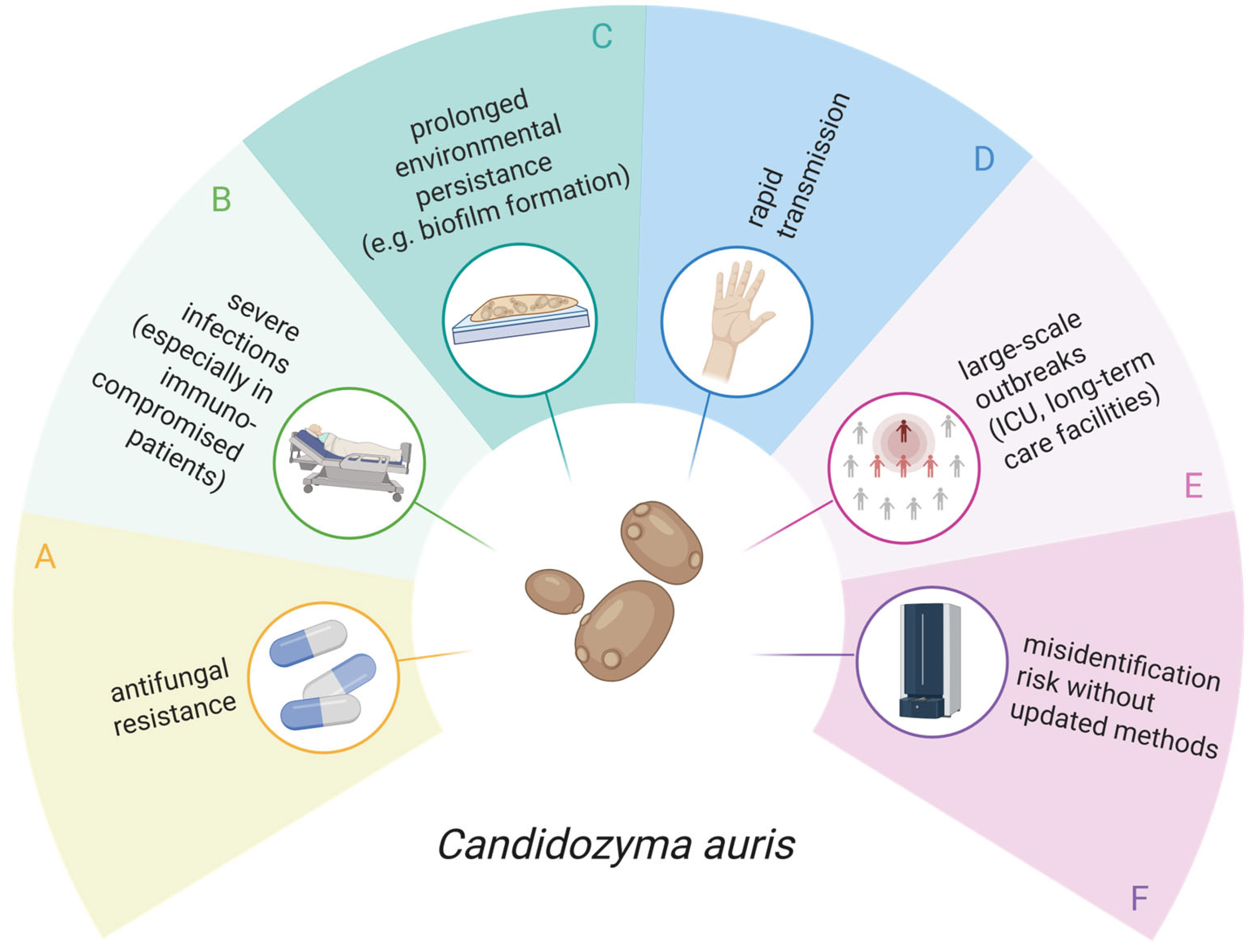
Candida auris) has emerged as a healthcare-associated yeast of major concern because of its persistent colonisation of human skin and medical devices, frequent multidrug resistance, and strong association with difficult-to-control outbreaks in healthcare settings, particularly among immunocompromised patients with severe infections (
Figure 1) [
1]. Clinical manifestations range from candidaemia to device-associated infections, as well as wound and soft tissue, urinary tract, and ear infections, depending on the patient population. Importantly, asymptomatic carriage with prolonged skin colonisation, predominantly involving the nares, axilla, and groin, is common. This represents a major risk factor for transmission and subsequent invasive disease [
2,
3].
2. Materials and Methods
A C. auris isolate (internal laboratory ID: CAU18) was recovered in May 2025 from a used face mask worn by a dental student during clinical activities at the Medical University of Innsbruck. The isolate originated from a blinded, anonymised quality-control programme assessing microbial contamination in dental working environments. Thirty used face masks collected in the dental clinic were screened for the presence of Candida spp.
Detection was performed by contact sampling on CHROMagarTM Candida Plus agar (Mast Diagnostica, Reinfeld, Germany). Species-level identification was subsequently confirmed by matrix-assisted laser desorption time-of-flight mass-spectrometry (MALDI-TOF MS), as described below.
All patient-related Candida spp. isolates analysed at the Institute of Hygiene and Medical Microbiology, Medical University of Innsbruck, between 2017 and 2025 were included retrospectively. Collected data comprised diagnostic findings and patient sex as a demographic variable. Samples were categorised as invasive, non-invasive, or of unknown origin. Invasive samples underwent species-level identification in almost all cases, whereas non-invasive samples were identified to the species level less consistently in routine diagnostics. In routine laboratory practice, samples that did not undergo species-level identification were reported as Candida spp. based on microscopy of suspected colonies grown on Sabouraud agar or Columbia blood agar, or recovered from Sabouraud broth. These findings were always supported by microscopy of the original specimen. Where microscopy was inconclusive, samples were cultured on CHROMID® Candida agar (bioMérieux, Marcy-l’Etoile, France) to enable presumptive differentiation by colony colour. MALDI-TOF MS was then used to support identification.
Samples positive for Candida spp. were cultured on CHROMID® Candida agar to enable preliminary differentiation based on colony colour. Definitive species identification was then performed by MALDI-TOF MS using the MALDI Biotyper Smart system with the Biotyper library v.4.1 (Bruker Daltonik GmbH, Bremen, Germany). Identification scores of ≥2.0 were interpreted as reliable for species-level identification.
For rapid clade assignment, an allele-specific PCR for
C. auris [
5] was performed at the Medical University of Innsbruck. Genomic DNA was extracted using the Yeast DNA Extraction Kit (Thermo Fisher Scientific, Waltham, MA, USA) according to the manufacturer’s instructions, and PCR was performed as described previously [
5]. In addition, the
ERG11 gene was sequenced to identify clade-associated mutations. To confirm these findings further, WGS was performed at the Austrian National Reference Centre for Yeasts and Moulds (Vienna), as previously reported [
6]. WGS data for the
C. auris isolate CAU18 are available under NCBI BioSample SAMN55402253.
Antifungal susceptibility testing was performed by broth microdilution in accordance with the recommendations of the European Committee on Antimicrobial Susceptibility Testing [
7]. Minimum inhibitory concentrations (MICs) were determined for the following antifungal agents at the indicated concentration ranges: anidulafungin (0.008–16 mg/L), micafungin (0.008–16 mg/L), caspofungin (0.008–16 mg/L), fluconazole (0.125–256 mg/L), posaconazole (0.016–32 mg/L), voriconazole (0.008–16 mg/L), 5-flucytosine (0.032–64 mg/L), amphotericin B (0.032–16 mg/L), and manogepix (0.002–16 mg/L).
3. Results
3.1. Environmental Detection and Characterisation of the C. auris Isolate
In May 2025, a single C. auris isolate was recovered from a used face mask worn by a dental student during clinical activities in Innsbruck, Austria. The sample originated from a structured environmental quality-control programme designed to assess microbial contamination in the dental working environment. Of the thirty used masks evaluated, one (3.3%) tested positive for C. auris, five (16.7%) for C. albicans, and five (16.7%) for C. parapsilosis. No other Candida species were detected. The program was not performed in response to a suspected outbreak or a known C. auris case. Because the programme was conducted in a blinded and anonymised manner, the wearer could not be identified retrospectively. Consequently, no follow-up sampling, including colonisation screening, could be performed, and no exposure history, such as recent travel or prior healthcare contact, could be obtained.
The isolate was identified as
C. auris and assigned to clade III. Sequence analysis of the
ERG11 gene revealed the V125A/F126L amino acid substitutions previously associated with azole resistance [
8]. The isolate demonstrated a high MIC to fluconazole (256 mg/L), whereas MICs for voriconazole and posaconazole were lower at 1 mg/L and 0.25 mg/L, respectively. MICs for the echinocandins were low (anidulafungin 0.25 mg/L, micafungin 0.125 mg/L, caspofungin 0.25 mg/L). The MIC was 2 mg/L for amphotericin B, 0.125 mg/L for flucytosine, and 0.008 mg/L for manogepix.
At the time of the investigation, no additional C. auris isolates were recovered from environmental samples collected by the hospital hygiene team. This argues against widespread environmental contamination during that period.
3.2. Local Laboratory Observations and Intensified Yeast Species Identification (2017–2025)
To provide longitudinal context around the first documented human
C. auris case in Austria in 2018 [
6], we retrospectively reviewed all
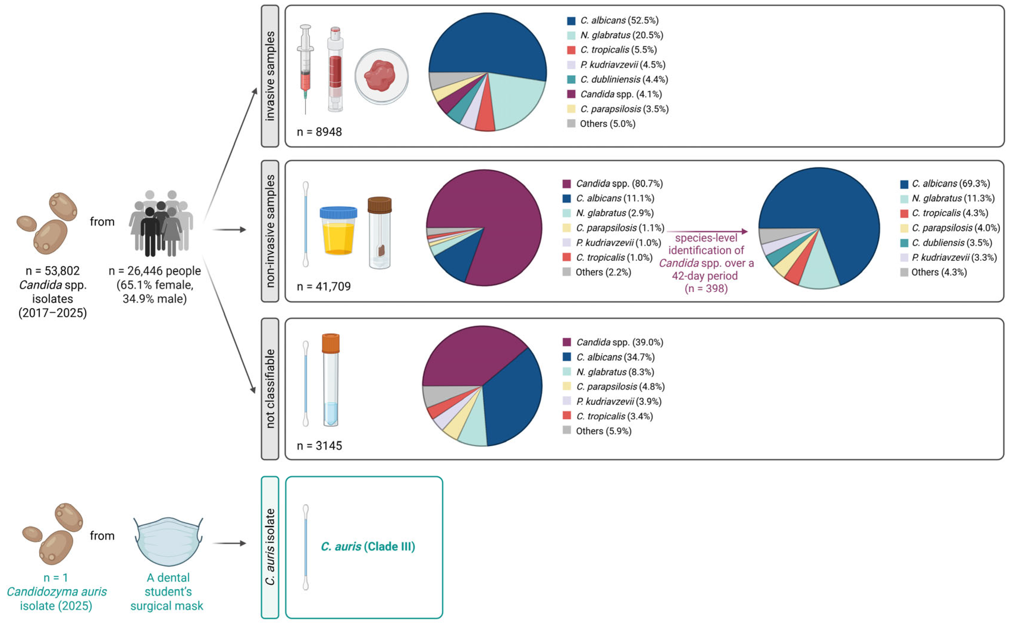
Candida spp. detections recorded in our routine diagnostic laboratory from 2017 onwards. Over the 9-year study period (2017–2025), 8948 invasive specimens and 41,709 non-invasive specimens were processed for yeast growth as part of routine clinical microbiology diagnostics (
Figure 2,
Table 1). In addition, 3145 specimens of unknown origin were included. Overall, 53,802 specimens from 26,446 individuals were analysed; 65.1% of individuals were female, largely because of the high number of vaginal swab submissions.
Across the study period, C. albicans was the most frequently detected species, accounting for 54.7% of invasive samples and 57.4% of non-invasive samples that underwent species-level identification. This was followed by Nakaseomyces glabratus, identified in 21.3% of invasive samples and 15.2% of non-invasive samples. In routine practice, species-level identification was performed for nearly all invasive specimens, whereas non-invasive and specimens of unknown origin were usually reported as Candida spp. unless further identification was specifically requested. Consequently, only a small portion of non-invasive samples underwent species-level identification. Because this approach could theoretically have missed sporadic C. auris detections in non-invasive material, species-level identification was intensified after the environmental finding. During an additional 42-day period, 398 yeast-positive non-invasive specimens were further analysed by CHROMID® Candida agar and MALDI-TOF MS. No non-classifiable samples were submitted to the routine laboratory during this period. Accordingly, intensified diagnostics were limited to non-invasive specimens. This was likely due to improvements in pre-analytical procedures over the years, including a revised laboratory request form completed by the submitting clinicians or institutions and mandatory follow-up calls by the routine microbiology laboratory when non-classifiable samples are received. This short-term analysis showed a similar species distribution, with C. albicans remaining the dominant species and accounting for 69.3% of non-invasive samples. No C. auris was detected in any clinical specimens during the observation period.
4. Discussion
The observation of a single mask-positive isolate in the absence of positive routine clinical specimens may be explained by several non-mutually exclusive factors. However, these explanations remain speculative and should be regarded as hypothesis-generating only, as the study was based on a single environmental isolate and the anonymised design precluded epidemiological follow-up. Importantly, this finding does not provide evidence of transmission or the presence of C. auris in the underlying population.
First, routine diagnostics should not be equated with colonisation screening. In our setting, the laboratory primarily processes blood cultures, urine, respiratory specimens, and wound swabs. The absence of
C. auris in these materials does not exclude unrecognised asymptomatic carriage, particularly at typical colonisation sites that are not routinely screened but are central to transmission [
2,
3].
Second, the source of the mask-associated isolate remains uncertain. It could not be determined retrospectively whether the isolate originated from the inner or outer surface of the mask. In addition, the anonymised quality-control design prevented re-identification of the wearer and confirmatory follow-up. The finding may therefore reflect nasal colonisation; transient hand-to-mask transfer after contact with contaminated surfaces, equipment, or gloves; or contamination of the outer mask surface within the clinical environment. This interpretation is compatible with the prolonged viability of
C. auris under dry conditions [
2]. Nonetheless, because colonisation cannot be distinguished from contamination, any further interpretation remains speculative.
Third, the isolate belonged to clade III, which has been reported in multiple regions worldwide, including South Africa, the United Kingdom, China, Saudi Arabia, Spain, Australia, Canada, Germany, and Austria [
4,
6]. However, possible routes of introduction could not be assessed because of the blinded and anonymised sampling design. Moreover, the
C. auris isolate showed no close genetic relationship to any previously detected
C. auris isolate in Austria [
6].
Although no
C. auris outbreak was detected at the Medical University of Innsbruck during the study period, even an isolated detection warrants attention, as sporadic introductions may precede transmission under favourable conditions. The fact that the isolate was identified through a routine, blinded environmental quality control programme, rather than a case-driven investigation, highlights the potential value of such monitoring for the early detection of unusual or emerging pathogens in healthcare-associated settings, including dental medicine. Given the well-documented ability of
C. auris to persist on dry, frequently touched surfaces, consistent environmental cleaning with agents active against this pathogen remains essential. Additionally, strict adherence to hand hygiene must complement the environmental cleaning and disinfection measures [
9].
5. Conclusions
In summary, we recovered a single clade III C. auris isolate from a used face mask collected during a dental environmental quality-control programme in May 2025. By contrast, C. auris was not detected in routine patient specimens processed between 2017 and 2025, including during an intensified 42-day period of expanded species-level identification. Although these findings are consistent with the absence of recognised clinical transmission in our setting, the lack of systematic colonisation screening and the anonymised sampling design limit conclusions regarding silent carriage and the route of introduction. Continued vigilance and laboratory preparedness therefore remain essential, as even a single introduction may become epidemiologically relevant if infection-prevention measures are applied inconsistently.
Author Contributions
Conceptualization, A.B. and C.L.-F.; methodology, A.B., A.M., S.T., K.S., B.W., R.K., C.L.-F.; formal analysis, C.L.-F. and A.B.; investigation, A.B., A.M., S.T., K.S., B.W., R.K., C.L.-F.; writing—original draft preparation, A.B. and C.L.-F.; writing—review and editing, A.B., A.M., S.T., K.S., B.W., R.K., C.L.-F.; visualization, A.B. All authors have read and agreed to the published version of the manuscript.
Funding
This study has been funded by MUI grant D-155110-017-016.
Institutional Review Board Statement
Not applicable. This was a retrospective analysis of routinely collected clinical specimens. All samples/data were fully anonymised prior to analysis, and no identifiable patient information was accessed or recorded. According to local/institutional policy, this study did not require ethical committee approval.
Informed Consent Statement
Not applicable.
Data Availability Statement
The original contributions presented in this study are included in the article. WGS data for the C. auris isolate CAU18 are available under NCBI BioSample SAMN55402253. Further inquiries can be directed to the corresponding author.
Conflicts of Interest
The authors declare no conflicts of interest.
Abbreviations
The following abbreviations are used in this manuscript:
| MALDI-TOF MS | Matrix-assisted laser desorption time-of-flight mass-spectrometry |
| WGS | Whole-genome sequencing |
| MIC | Minimum inhibitory concentration |
References
- Lionakis, M.S.; Chowdhary, A. Candida auris Infections. N. Engl. J. Med. 2024, 391, 1924–1935. [Google Scholar] [CrossRef]
- Horton, M.V.; Holt, A.M.; Nett, J.E. Mechanisms of pathogenicity for the emerging fungus Candida auris. PLoS Pathog. 2023, 19, e1011843. [Google Scholar] [CrossRef]
- Chowdhary, A.; Lionakis, M.S.; Chauhan, N. Candida auris: Host interactions, antifungal drug resistance, and diagnostics. Microbiol. Mol. Biol. Rev. 2025, 90, e0018722. [Google Scholar] [CrossRef]
- de Gaetano, S.; Midiri, A.; Mancuso, G.; Avola, M.G.; Biondo, C. Candida auris Outbreaks: Current Status and Future Perspectives. Microorganisms 2024, 12, 927. [Google Scholar] [CrossRef] [PubMed]
- Carolus, H.; Jacobs, S.; Lobo Romero, C.; Deparis, Q.; Cuomo, C.A.; Meis, J.F.; van Dijck, P. Diagnostic Allele-Specific PCR for the Identification of Candida auris Clades. J. Fungi 2021, 7, 754. [Google Scholar] [CrossRef] [PubMed]
- Spettel, K.; Kriz, R.; Wu, C.; Achter, L.; Schmid, S.; Galazka, S.; Selitsch, B.; Camp, I.; Makristathis, A.; Lagler, H.; et al. Candida auris in Austria—What Is New and What Is Different. J. Fungi 2023, 9, 129. [Google Scholar] [CrossRef] [PubMed]
- Guinea, J.; Meletiadis, J.; Arikan-Akdagli, S.; Giske, C.; Muehlethaler, K.; Arendrup, M.C.; Subcommittee on Antifungal Susceptibility Testing (AFST) of the ESCMID European Committee for Antimicrobial Susceptibility Testing. Method for the determination of broth dilution minimum inhibitory concentrations of antifungal agents for yeasts. In EUCAST Definitive Document E.Def 7.4; European Committee for Antimicrobial Susceptibility Testing: Växjö, Sweden, 2023. [Google Scholar]
- Williamson, B.; Wilk, A.; Guerrero, K.D.; Mikulski, T.D.; Elias, T.N.; Sawh, I.; Cancino-Prado, G.; Gardam, D.; Heath, C.H.; Govender, N.P.; et al. Impact of Erg11 Amino Acid Substitutions Identified in Candida auris Clade III Isolates on Triazole Drug Susceptibility. Antimicrob. Agents Chemother. 2022, 66, e0162421. [Google Scholar] [CrossRef] [PubMed]
- Steixner, S.; Bauer, A.; Grubwieser, P.; Lass-Flörl, C. Can we prevent azole-resistant Candida parapsilosis from becoming the next Candidozyma auris? Expert Rev. Anti-Infect. Ther. 2025, 23, 1047–1049. [Google Scholar] [CrossRef] [PubMed]
| Disclaimer/Publisher’s Note: The statements, opinions and data contained in all publications are solely those of the individual author(s) and contributor(s) and not of MDPI and/or the editor(s). MDPI and/or the editor(s) disclaim responsibility for any injury to people or property resulting from any ideas, methods, instructions or products referred to in the content. |




 ,
,