1. Introduction
Invasive fungal infections (IFIs) predominantly affect immunocompromised or critically ill patients and remain an important cause of morbidity and mortality in children [
1,
2,
3]. Yeast-related fungemia represents a substantial proportion of IFIs, with
Candida species accounting for most pediatric cases [
4,
5,
6]. In contrast, fungemia caused by non-
Candida yeasts is uncommon; however, recent reports indicate an increasing frequency, particularly among patients with hematologic malignancies, prolonged neutropenia, intensive care hospitalization, or exposure to broad-spectrum antimicrobials [
1,
2,
3,
5]. Non-
Candida yeasts are widely distributed in the environment and may colonize skin and mucosal surfaces. Although sometimes regarded as contaminants, these organisms can cause life-threatening infections in susceptible hosts [
7,
8].
Several non-
Candida yeast genera have been reported as opportunistic human pathogens, including
Trichosporon,
Rhodotorula,
Saprochaete/Magnusiomyces,
Malassezia,
Saccharomyces,
Cryptococcus, and
Geotrichum [
2,
3,
4]. Although these organisms are less frequently encountered non-
Candida species, they can cause severe bloodstream infections in immunocompromised hosts and are often associated with distinct antifungal susceptibility patterns. Their emergence has been attributed to several factors, including improved survival of immunocompromised children, extensive use of antifungal agents, central venous catheter dependence, broad-spectrum antibiotic exposure, and intensive chemotherapy or immunosuppressive therapies [
3,
4,
5,
6,
7,
8]. Despite growing recognition, epidemiological data on these rare pathogens, particularly in pediatric populations, remain limited. Clinical management is further complicated by diagnostic delays, heterogeneous antifungal susceptibility profiles, absence of standardized clinical breakpoints, and uncertainty regarding optimal therapeutic strategies [
1,
3]. For these reasons, non-
Candida yeast fungemia warrants focused clinical attention.
Given the rarity of these infections in children, the present study was designed as a retrospective descriptive observational study aiming to characterize the clinical features, microbiological findings, antifungal management, and outcomes of rare non-Candida yeast fungemia episodes observed at our tertiary pediatric center over a six-year period.
3. Results
During the study period, non-
Candida yeast fungemia was identified in five patients (three
Trichosporon spp., one
Magnusiomyces [
Saprochaete]
clavatus, and one
Rhodotorula mucilaginosa). Among 139 yeast-related fungemia episodes recorded at our center, rare non-
Candida yeasts accounted for 3.6%. Demographic characteristics, underlying conditions, antifungal therapy, and clinical outcomes are summarized in
Table 1.
Case 1: A 9-year-old girl diagnosed with unilateral sporadic retinoblastoma presented on the 12th day of the sixth cycle of ifosfamide, carboplatin, and etoposide (ICE) therapy with a fever reaching 39 °C in the axillary region. Physical examination findings were normal. The whole blood count revealed a hemoglobin level of 10.9 g/dL, a white blood cell (WBC) count of 260/mm3, a neutrophil count of 40/mm3, and a platelet count of 157.000/mm3. The C-reactive protein (CRP) was 7.2 mg/L. Blood cultures were obtained from catheter and peripheral blood, and urine cultures were obtained, and intravenous (IV) piperacillin-tazobactam treatment was initiated for febrile neutropenia. The patient tested positive for SARS-CoV-2 in a respiratory multiplex polymerase chain reaction (PCR) and was placed in isolation.
During follow-up, yeast signals were detected in the peripheral blood culture at the 32nd hour, prompting control cultures to be taken and intravenous (IV) caspofungin treatment to be initiated. However, on the sixth day of hospitalization, T. asahii was detected in the peripheral blood culture, and IV voriconazole therapy was initiated instead of caspofungin. Piperacillin-tazobactam therapy was also discontinued on the same day. In response to treatment, the patient’s fever subsided, her clinical condition stabilized, and neutropenia recovered gradually during antifungal therapy. The patient was discharged on the sixth day of IV therapy with oral voriconazole, and the treatment period was completed in three weeks.
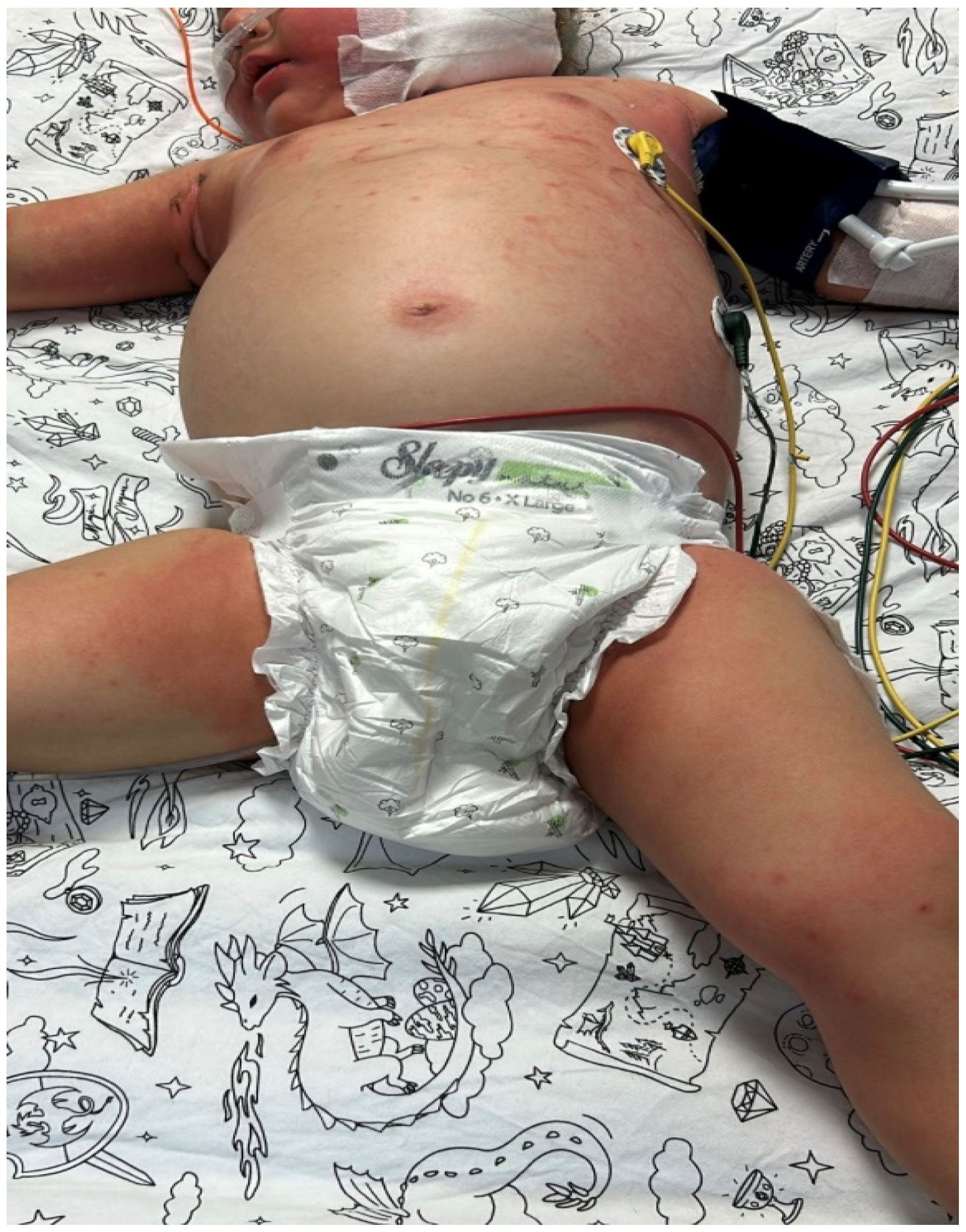
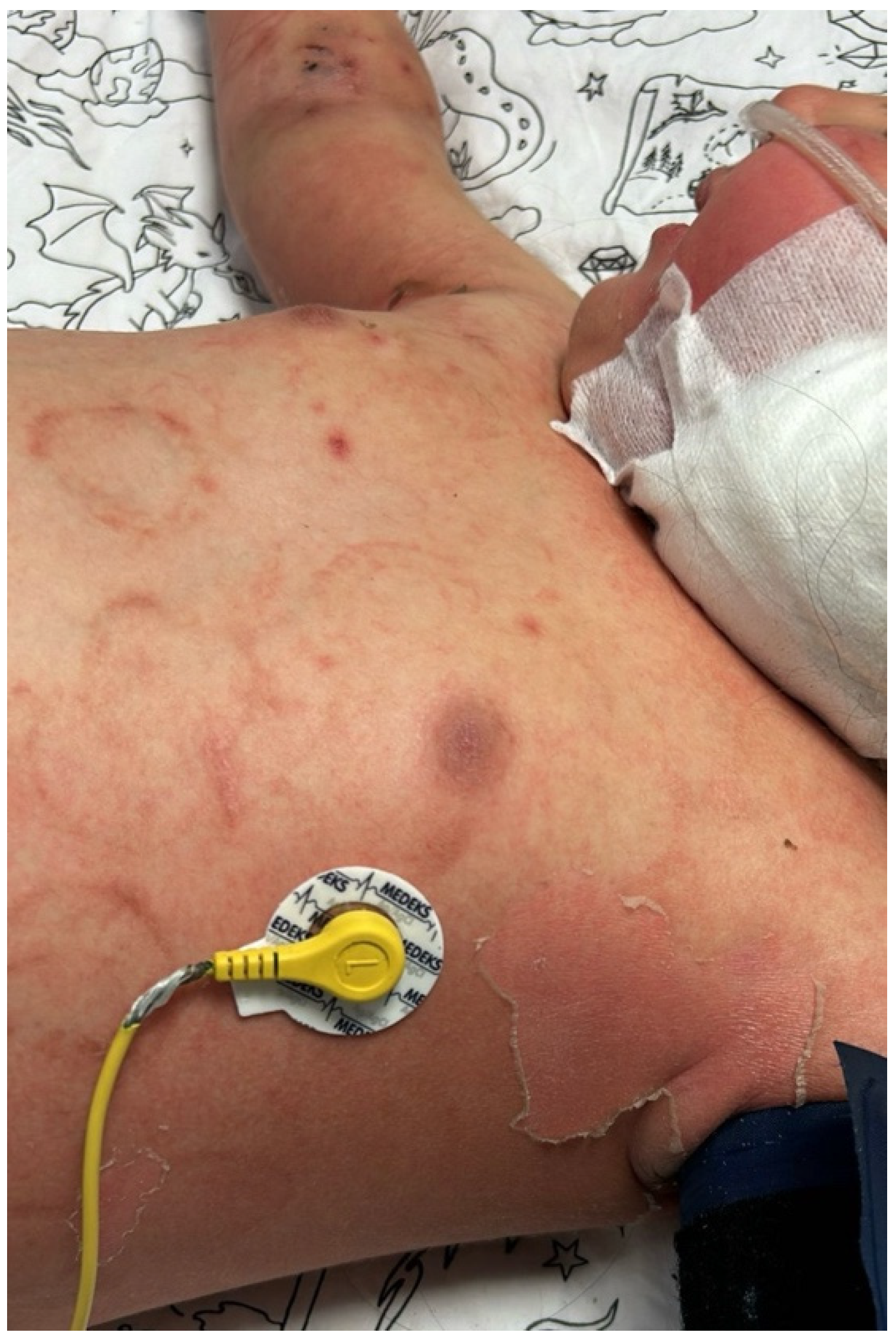
Case 2: A 23-month-old girl with newly diagnosed acute myeloid leukemia (AML) developed febrile neutropenia on the 10th day of induction chemotherapy. Piperacillin-tazobactam therapy was initiated. As the fever persisted, antibiotic therapy was gradually expanded and revised to include IV meropenem, teicoplanin, liposomal amphotericin B, levofloxacin, and colistin. On the sixth day of fever, she was admitted to the pediatric intensive care unit with a diagnosis of sepsis, where high-flow oxygen support was initiated and the central catheter was removed. On the seventh day of fever, macular rashes developed with widespread erythema on the trunk, extremities, and scalp (
Figure 1 and
Figure 2). Viral serology and PCR tests were performed, and intravenous immunoglobulin (IVIG) therapy was initiated. A biopsy was taken due to persistent skin lesions. A yeast signal was detected in the blood culture at 29 h, and caspofungin was started in addition to the existing liposomal amphotericin B therapy.
Plasmapheresis (PEX) was performed on the tenth day of fever due to the emergence of hyperinflammatory findings. With the development of renal failure, continuous renal replacement therapy (CRRT) was initiated. On the eleventh day of fever, the patient was intubated, and inotropic support and hydrocortisone therapy were added. On the 12th day of fever, the fifth day of blood culture, T. asahii was reported as the causative agent. Hence, caspofungin treatment was discontinued, and voriconazole treatment was added to liposomal amphotericin B (3 mg/kg/d) treatment. During this process, a thoracoabdominal computed tomography (CT) revealed diffuse peribronchovascular reticular infiltration and ground-glass opacities in both lungs, along with diffuse infiltration in the spleen and newly developed widespread millimetric hypodense foci. The findings were considered consistent with the underlying malignancy and, in particular, systemic fungal infection. No focus of infection was detected on echocardiography. During follow-up, peritoneal sampling was performed due to the development of abdominal distension and suspicion of perforation. T. asahii growth was detected in peritoneal fluid culture, but the causative agent could not be isolated in tissue culture. The skin biopsy was considered nonspecific; immunohistochemical staining did not reveal findings suggestive of leukemic infiltration or fungal infection. Although the cause of the rash could not be definitively established, the temporal association with fungemia suggested potential clinical relevance.
Despite dual antifungal therapy, intermittent growth in blood cultures persisted for approximately 20 days. No new growth was detected during the following 45-day period, and abdominal imaging revealed regression of splenic involvement. During follow-up, thrombocytopenia secondary to the underlying malignancy and recurrent septic episodes led to intracranial hemorrhage, resulting in hydrocephalus. Therefore, a ventriculoperitoneal shunt was placed. The patient’s neurological condition subsequently deteriorated, and she developed brain death and died.
Case 3: A 27-year-old mother gave birth to a male neonate weighing 3450 g at 39 weeks via cesarean section. The infant presented to an external center on the second postnatal day with complaints of failure to feed and lethargy. The patient was evaluated with preliminary diagnoses of early neonatal sepsis or metabolic disease, and ornithine transcarbamylase (OTC) deficiency was considered the most likely cause. He was transferred to our neonatal intensive care unit on the fourth day.
Upon admission, he was in poor general condition, intubated, with widespread edema and abdominal distension. There was a necrotic decubitus ulcer 3 cm in diameter on his back. Blood ammonia level was measured as 1199 µmol/L. A hemodialysis catheter was inserted, and CRRT was initiated. Blood and urine cultures were obtained, and treatment with IV meropenem and vancomycin, which had been started at the external center, was continued.
On the third day of follow-up, IV amikacin and liposomal amphotericin B (3 mg/kg/d) were added to the treatment due to clinical deterioration. A paracentesis was performed on the patient, who had widespread ascites; leukocytes were detected at 67,161/mm3 and erythrocytes at 15/mm3 in the peritoneal fluid. Treatment was readjusted to IV meropenem infusion, colistin, and vancomycin. However, despite all inotropic, antibiotic, and supportive treatments, the patient died on the seventh day of hospitalization (the fourth day of liposomal amphotericin B treatment).
After death, yeast signals were reported in the blood culture taken 95 h before starting liposomal amphotericin B treatment. The causative agent was identified as Trichosporon spp. on the sixth day of typing. Concurrent peritoneal cultures grew Klebsiella pneumoniae (susceptible to carbapenems and amikacin), Stenotrophomonas maltophilia, and Enterococcus faecalis. Due to the patient’s death, advanced species identification and antifungal susceptibility testing could not be performed on the Trichosporon isolate. Trichosporon was also detected in postmortem blood, catheter, and cerebrospinal fluid samples.
Case 4: A 1.5-month-old boy was transferred to our hospital’s pediatric intensive care unit with preliminary diagnoses of subdural hematoma, subarachnoid hemorrhage, and pulmonary contusion following a car accident. The patient was intubated and empirically treated with IV ceftriaxone, which was discontinued on the 7th day. On the 10th day of hospitalization, peripheral blood, urine, and catheter blood cultures were obtained due to the development of fever, and empirical piperacillin-tazobactam treatment was initiated. As the fever persisted, Pseudomonas aeruginosa growth was detected in the daily peripheral and catheter blood cultures. Consequently, treatment was revised to IV meropenem and amikacin, and the catheter was removed. Empirically, IV fluconazole therapy was added due to persistent fever. A yeast signal was reported at the 96th hour of the blood culture. Subsequently, on the 7th day, R. mucilaginosa was reported in the blood culture, and fluconazole was discontinued. Treatment was changed to IV liposomal amphotericin B. Meropenem and amikacin treatments were discontinued on day 12. Liposomal amphotericin B treatment was completed on day 14. No pathology was detected in the cardiology evaluation, and there was no growth in the follow-up cultures. The patient was discharged in a clinically stable condition.
Case 5: An 11-year-old girl diagnosed with relapsed B-acute lymphoblastic leukemia (ALL) presented to the emergency department with febrile neutropenia. Piperacillin-tazobactam therapy was initially started. Upon physical examination, tenderness was detected in the right lower quadrant, and IV metronidazole and caspofungin were added to the treatment with a preliminary diagnosis of typhlitis. Abdominal ultrasound showed hepatosplenomegaly, minimal free fluid in the pelvis, moderate wall thickening, and subserosal edema in the cecum and terminal ileum. Due to persistent fever, piperacillin-tazobactam was replaced with meropenem, and teicoplanin was added to the treatment due to the presence of oral mucosal lesions. The patient’s fever persisted during follow-up, and Salmonella spp. growth was detected in the catheter blood culture. Intravenous ciprofloxacin and catheter lock therapy were initiated. Clinical improvement was achieved after this treatment, and the fever subsided. However, on the 10th day of treatment, the fever rose again, and tachypnea and desaturation developed. Physical examination revealed decreased bilateral breath sounds, particularly prominent in the lower right lung zone. An emergency chest CT scan showed ground-glass opacities in the bilateral lower zones. The catheter was removed; IV liposomal amphotericin B (3 mg/kg/d) and trimethoprim-sulfamethoxazole were added to the treatment, and caspofungin and metronidazole were discontinued. Serum galactomannan assay and Pneumocystis jirovecii PCR tests were negative. A minimal amount of pericardial fluid was detected on cardiac examination.
On the fourth day of liposomal amphotericin B therapy, yeast signals were reported in peripheral and catheter blood cultures. Upon identification of the causative agent as M. clavatus, IV voriconazole was added to the treatment on the fifth day. On the 10th day of amphotericin therapy and the sixth day of voriconazole, the patient developed nonspecific, petechiae-like erythematous rashes. A biopsy could not be performed on this patient, who had severe thrombocytopenia and was receiving palliative care. Because of persistent growth in blood cultures, the dosage of liposomal amphotericin B was increased to 5 mg/kg/day. On the second day of the dosage increase (11th day of amphotericin and 7th day of voriconazole), the fever subsided, and the rash completely disappeared within seven days. After 72 h without fever, antibiotics were discontinued; the patient was monitored under liposomal amphotericin B and voriconazole treatment. On the 11th day of voriconazole treatment, the patient developed blurred vision. No papilledema was detected on fundus examination, and no pathology was observed on cranial imaging. However, as the complaints persisted and no other cause was found, treatment was switched to posaconazole on day 15, considering that voriconazole might be a common side effect. Antifungal treatment was completed in 21 days. Liposomal amphotericin B treatment was continued for another week and was discontinued on day 35, after the patient recovered from neutropenia and a thoracic CT showed regression of the lesions.
General Findings
Of the five patients included in the study, three (60%) had underlying hematologic or oncologic diseases and one (20%) had suspected metabolic disease. Sixty percent of patients had a history of neutropenia lasting a median of 12 days (range 4–13) prior to the onset of fungemia. Sixty percent of patients were monitored in the neonatal or pediatric intensive care unit, and 80% had a central venous catheter. No patient had a history of surgical intervention within the last month. The median time between hospital admission and onset of fungemia was 12 days (range 5–21).
Yeast growth was detected in blood cultures in all cases. The median time to a positive fungal signal was 32 h (range 29–96). Species were identified in four patients; in one fatal case (Case 3), identification was completed postmortem, and susceptibility testing was not performed. Across the cohort, identified species comprised Trichosporon spp. (n = 3), R. mucilaginosa (n = 1), and M. clavatus (n = 1).
Antifungal susceptibility testing could only be performed for two isolates (1 T. asahii, 1 M. clavatus). In the remaining cases, susceptibility testing could not be performed because viable colonies were not available for further testing or because the patient died shortly after culture positivity. Since clinical breakpoints have not yet been established for these species, only minimum inhibitory concentration (MIC) distributions were reported. Voriconazole and itraconazole showed in vitro activity against both isolates with low MIC values in the range of 0.03–0.25 µg/mL, while relatively high MIC values (2–4 µg/mL) were detected for fluconazole. Although amphotericin B showed a low MIC value (0.5 µg/mL) for T. asahii, clinical efficacy could not be achieved, and voriconazole was added to the treatment regimen in the relevant case.
Upon initial yeast positivity—before species identification and susceptibility results—empirical therapy was initiated or adjusted according to each patient’s on-treatment status: fluconazole (n = 1), liposomal amphotericin B (n = 1), and echinocandin (n = 2); one patient died on the day culture positivity was reported. After species identification and/or susceptibility data became available, antifungal regimens were modified in all surviving cases.
Breakthrough fungemia occurred under liposomal amphotericin B (
n = 1) and under echinocandin therapy (
n = 1). Species distribution, minimum inhibitory concentration (MIC) values, preferred antifungal therapies, and clinical outcomes are summarized in
Table 2.
Overall mortality was 40%, and attributable mortality was 20%. In the attributable death, the interval from fungemia onset to death was 4 days.
4. Discussion
In this retrospective descriptive study, rare non-
Candida yeasts were infrequently encountered over a six-year period (five cases), yet these infections remained clinically significant because of delayed species-level identification and limited antifungal susceptibility data. Given the rarity of these pathogens and the small number of cases, the findings of our study should be interpreted primarily as descriptive clinical observations rather than definitive epidemiological conclusions. Similarly to our experience, most available data on rare non-
Candida yeast infections in children derive from small case series or individual case reports due to the low incidence of these pathogens in pediatric populations [
10,
11,
12,
13,
14]. Nevertheless, our experience highlights important diagnostic and therapeutic challenges associated with these infections in vulnerable pediatric patients.
In our series, empirical antifungal regimens frequently required modification after species identification, underscoring the gap between empiric strategies primarily targeting
Candida spp. [
15] and the intrinsic resistance or reduced susceptibilities typical of non-
Candida yeasts [
16]. Timely and appropriate antifungal therapy is pivotal in fungemia, as therapeutic delay or suboptimal initial choices are associated with increased mortality [
8,
16]. In our cohort, the median time from blood culture positivity to species-level identification was six days. Although MALDI-TOF MS allows rapid species identification once an adequate colony is available, the reported identification time reflects the entire diagnostic workflow rather than the MALDI-TOF procedure itself. This interval includes the time required for blood culture positivity and subculture growth before MALDI-TOF analysis can be performed. In addition, routine laboratory workflow factors such as processing schedules, weekends, and laboratory staffing patterns may occasionally contribute to delays between culture positivity and final species identification. Therefore, clinicians should be aware of predisposing risk factors and maintain a high index of suspicion for these uncommon pathogens [
8]. In our cohort, central venous catheter use and recent antibiotic exposure were each present in 80% of cases, while neutropenia was observed in 60%. Consistent with prior reports, well-recognized risk factors—including hematologic malignancy, prolonged neutropenia, intensive care unit admission, central venous catheter use, and recent broad-spectrum antibiotic exposure—were prevalent in our cohort [
7,
16]. Similar predisposing factors have been described in pediatric studies of rare yeast infections, where central venous catheter use, prolonged hospitalization, exposure to broad-spectrum antibiotics, and underlying immunocompromising conditions are frequently observed [
13]. Overall, the epidemiology of rare non-
Candida yeast fungemia in children appears to mirror that of invasive candidiasis, predominantly affecting immunocompromised patients, particularly those with hematologic malignancies, prolonged neutropenia, and central venous catheter dependence.
After assessing risk factors, it is necessary to focus on the clinical significance of specific yeast species.
Trichosporon spp. are rare yeast species that can colonize the skin, gastrointestinal, and respiratory systems, causing superficial and invasive infections.
T. asahii is the primary causative agent of invasive trichosporonosis, and hematologic malignancy patients and premature newborns are particularly at risk. Azoles are the first-line agents for treatment [
2].
Trichosporon spp. are naturally resistant to echinocandins and show variable susceptibility to amphotericin B [
7]. Our series included three cases of
Trichosporon spp.: In one case, despite a low MIC value, breakthrough infection developed under amphotericin B, and a response was achieved with the addition of voriconazole (Case 2). In a fatal case, species identification was only possible postmortem, and susceptibility testing could not be performed (Case 3). In another case, although susceptibility testing could not be performed, clinical improvement was achieved with empirical voriconazole (Case 1). Metastatic skin lesions, pneumonia, and spleen and liver abscesses are frequently reported clinical findings in the literature [
8,
17]. In our series, skin and spleen involvement with pneumonia was observed in one patient, while peritonitis and central nervous system involvement with simultaneous cerebrospinal fluid culture growth were observed in a newborn. The prognosis for trichosporonosis is generally poor; mortality has been reported to be 28.5–30% in children and 80–87.5% in adults [
10]. Pediatric studies further highlight the clinical severity of invasive trichosporonosis. In a recent 10-year single-center pediatric study, 12 cases of invasive
Trichosporon infection were reported, with an overall mortality rate of 41.7%, emphasizing the significant morbidity and mortality associated with these infections in immunocompromised children [
13]. Mortality in our cases was 66.7%, which appears higher than the pediatric rates reported in the literature; however, this finding should be interpreted cautiously, given the very small sample size and the severity of underlying conditions in our patients.
Another rare yeast species is
Rhodotorula. These yeasts are commonly found in the environment and can colonize the skin and mucous membranes. The most frequently isolated species is
R. mucilaginosa, which is a causative agent of opportunistic infections, particularly in immunocompromised patients. The most important predisposing factors are CVC and malignancy.
Rhodotorula species are naturally resistant to echinocandins and fluconazole. Liposomal amphotericin B and/or flucytosine are recommended for treatment [
2]. Although susceptibility testing could not be performed in this case, the patient responded well to liposomal amphotericin B treatment.
Another important group of factors is
Saprochaete/Magnusiomyces species. These pathogens have been identified as causative agents of infection, particularly in cases of hematological malignancy, chemotherapy, prolonged neutropenia, and CVC [
7,
8]. Chemotherapies that compromise the mucosal barrier’s integrity can lead to gastrointestinal translocation and fungemia development [
11]. Pediatric data on these infections remain extremely limited, and only a small number of cases have been reported in children. A pediatric case of
M. clavatus fungemia occurring after hematopoietic stem cell transplantation highlighted prolonged neutropenia and severe immunosuppression as key predisposing factors and emphasized the importance of antifungal therapy combined with immune reconstitution for successful infection control [
14]. In this case, the previous
Salmonella bacteremia and gastrointestinal involvement were thought to pave the way for fungemia. Clinically, it usually presents with a disseminated infection pattern. Most isolates appear resistant to echinocandins and fluconazole but sensitive to amphotericin B, flucytosine, and newer azoles [
11]. Therefore, a combination of voriconazole, posaconazole, amphotericin B, and flucytosine is recommended for treatment. However, despite these treatments, mortality rates as high as 60 to 85% have been reported in some studies [
11,
12].
Implications for practice. Given the mismatch between standard empiric regimens for candidemia and the susceptibility profiles of non-Candida yeasts, our findings also have important implications for empirical antifungal therapy in high-risk pediatric patients. Because standard empiric regimens frequently rely on echinocandins or fluconazole, they may not provide adequate coverage for certain rare yeasts such as Trichosporon or Rhodotorula species, which may exhibit intrinsic resistance or reduced susceptibility to these agents. Where susceptibility testing is limited or delayed, risk-factor assessment and species-directed empiric adjustments may improve outcomes. Empiric antifungal algorithms should be tailored to local species and susceptibility patterns, and laboratories should immediately alert clinicians of any yeast-positive blood culture.
Limitations: The primary limitation of this study is the small cohort size (n = 5), which restricts the generalizability of the findings and prevents robust statistical analysis. The retrospective, single-center design introduces potential selection biases. Additionally, antifungal susceptibility testing could not be performed for all isolates, limiting assessment of resistance patterns and their relationship to clinical outcomes. Larger, multicenter studies with standardized diagnostic workflows are needed to better define the epidemiology and optimize treatment strategies for these rare pathogens.
In conclusion, although non-Candida yeasts account for only a small proportion of fungal infections, they are clinically significant due to their antifungal resistance and high mortality rates. In settings where species-level identification and susceptibility testing take days, initial empiric therapy should be guided by local epidemiology and patient risk factors, then promptly individualized once organism- and susceptibility-level data become available. Strengthening rapid diagnostic capacity and surveillance will improve both empiric decision-making and patient-level outcomes, recognizing the broad clinical spectrum of these infections and the need for tailored care.



 ,
,