Article

Diversity of Culturable Fungi in Two-Phase Olive Mill Waste, a Preliminary Evaluation of Their Enzymatic Potential, and Two New Trichoderma Species

1
Laboratory of General and Agricultural Microbiology, Agricultural University of Athens, Iera Odos 75, 11855 Athens, Greece
2
Section of Genetics and Biotechnology, Department of Biology, National and Kapodistrian University of Athens, Panepistemiopolis, 15701 Athens, Greece
*
Author to whom correspondence should be addressed.
J. Fungi 2025, 11(9), 687; https://doi.org/10.3390/jof11090687
Submission received: 19 July 2025 / Revised: 6 September 2025 / Accepted: 18 September 2025 / Published: 22 September 2025
(This article belongs to the Section Fungal Evolution, Biodiversity and Systematics)

Abstract

This study investigates the diversity and provides a preliminary evaluation of the enzymatic potential of culturable fungi present in two-phase olive mill waste (TPOMW), a lignocellulose- and phenolic-rich agro-industrial by-product generated in large quantities in olive oil-producing countries. Ninety-four isolates, representing 31 species of the phyla Ascomycota, Basidiomycota, and Mucoromycota, were obtained and identified by using ITS, 28S, tef1-α, tub2, rpb2, act, and/or cal sequences. Among the identified taxa, two new Trichoderma species within the Harzianum clade, namely Trichoderma amurcicola (phylogenetically related to T. simile and T. guizhouense) and Trichoderma olivarum (phylogenetically related to T. simmonsii), were described following a multilocus phylogenetic analysis combined with a study of their morphoanatomical features. A rather high phylogenetic divergence was detected in Candida boidinii, Pleurostoma richardsiae, and Mucor circinelloides, while Cladosporium limoniforme, Mucor pseudolusitanicus, Stagonosporopsis ailanthicola, and Talaromyces nanjingensis were recorded for the first time in TPOMW. A preliminary screening revealed 29 species with cellulolytic and/or xylanolytic activities; 26 species displayed dye decolorization capacity, while ligninolytic and laccase activities were restricted to a few taxa. The most promising degraders of lignocellulosics included strains of Cladosporium limoniforme, C. ramotenellum, Fuscoporia ferrea, Peniophora lycii, and Pseudophlebia setulosa. Fungi detected in TPOMW are promising biotechnological tools to be exploited in the frame of circular economy applications.

1. Introduction

Olive cultivation and olive oil are cornerstones of Mediterranean agriculture, economy, and nutrition. However, the olive oil extraction process is linked with the generation of huge quantities of olive mill wastewater, a by-product with significant environmental repercussions due to its high organic load and chemical composition [1,2]. Disposal of improperly managed olive mill wastewater can lead to contamination of soil and water receptors, disruption of ecosystem functions, and public health risks [3,4]. Consequently, sustainable waste management strategies have become a priority, aiming to align with circular economy principles while mitigating both ecological and socio-economic impacts [5,6].
Towards this end, the relatively recent transition leading to the wide use of two-phase olive oil extraction technology has significantly reduced the volume of waste generated; the latter is a sludge-like effluent widely known as ‘alpeorujo’ or two-phase olive mill waste (TPOMW) (Figure 1). It is rich in polyphenols, fatty acids, and proteins [7,8], while it is also characterized by high chemical oxygen demand (COD) and biological oxygen demand (BOD) values, which render it as a pollutant with severe adverse environmental impact. Furthermore, TPOMW’s seasonal production and composition heterogeneity further complicate its effective management. Therefore, innovative strategies are essential for mitigating these risks while unlocking the exploitation potential of this waste stream.
In general, olive mill waste (OMW) presents significant opportunities for sustainable applications [9]; recent advances in biotechnology have enabled the recovery of bioactive compounds for use in agriculture (e.g., biostimulants, biopesticides), and in the food and pharmaceutical industries. In particular, the lignocellulosic fraction of TPOMW could be transformed through the use of microbial extracellular enzymes (cellulases, hemicellulases, lignin-modifying enzymes) into fermentable sugars, biofuels, and value-added molecules [10,11]. For example, fungal taxa within Ascomycota (e.g., Trichoderma, Aspergillus), Basidiomycota (e.g., white-rot species), and Mucoromycota (e.g., Mucor, Rhizopus) secrete a broad spectrum of hydrolytic and oxidative enzymes that target both polysaccharidic and aromatic fractions of OMW [12,13]. Concurrently, bacterial phyla—such as Firmicutes (e.g., Bacillus spp.), Proteobacteria (e.g., Pseudomonas, Acinetobacter), and Actinobacteria (e.g., Streptomyces, Arthrobacter)—contribute complementary hydrolytic and oxidative capabilities, particularly in anaerobic or microaerophilic microenvironments [1,14]. Furthermore, microbial communities, which are adapted to the toxic and recalcitrant nature of OMW, exhibit enhanced efficiency in degrading complex organic substrates [15,16,17]. However, the diversity and enzymatic potential of these communities remain largely underexplored [4,18,19], particularly of those existing in TPOMW [20,21,22], thus leaving significant opportunities untapped for developing its sustainable valorization. This approach aligns with circular economy principles, transforming waste into high-value products while addressing environmental concerns [23].
Fungal communities present in OMW have been extensively investigated over the past two decades, with a gradual methodological shift from classical morphology-based identification to molecular and metagenomic approaches. Early culture-based surveys identified yeast species, including Candida boidinii and Geotrichum candidum, in untreated TPOMW [20]. Subsequent studies reported filamentous taxa, such as Penicillium roqueforti, Cladosporium spp., and yeasts of the genera Pichia and Candida, in both raw and composted TPOMW [19,21]. Similar taxonomic profiles were observed in OMW and other related substrates, where members of Ascomycota, mainly Eurotiales and Hypocreales, prevail, whereas Basidiomycota are infrequently reported and occurrences of Chytridiomycota (e.g., Rhizophydium spp.) remain rare and sporadic [1,4,15,17,19,20,21,22,24].
This study aims to contribute to the knowledge of the diversity of culturable fungi present in TPOMW. A large variety of members from this particular fungal community were obtained using diverse selective solid media, inoculation techniques, and culture conditions. In addition, by integrating molecular and biochemical approaches, we aimed (a) to elucidate the taxonomic diversity of fungi existing in TPOMW through multilocus phylogenetic analyses and (b) to perform a preliminary/initial evaluation of the strains’ lignocellulolytic and dye-decolorization activities aiming at their future exploitation.

2. Materials and Methods

2.1. Fungal Isolation from TPOMW, Selection, and Cultivation Conditions

TPOMW samples were collected from an olive mill equipped with two-phase centrifugal decanters in Kalamata, Greece, during three production seasons. Following samples homogenization, 10 g from each were suspended in 100 mL sterile saline solution (0.9% NaCl), blended for 2 min, and passed through sterile gauze to remove coarse solids. The resulting homogenate was used for the screening and selection of culturable fungi on the following media: (a) carboxymethyl cellulose-enriched agar (CEA), (b) lignin-enriched agar (LEA), (c) yeast peptone dextrose agar (YPDA), and (d) potato dextrose agar (PDA, Condalab, Madrid, Spain) (Supplementary Materials, Table S1). All media were sterilized at 121 °C for 20 min. To suppress fast-growing mitosporic fungi, the ergosterol synthesis inhibitor econazole nitrate (Spectazole®, Janssen Pharmaceuticals, Beerse, Belgium) was added to the above media at concentrations of 10 and 25 mg L−1. Additionally, PDA supplemented with 0.04% (v/v) Remazol Brilliant Blue R (RBBR; Sigma-Aldrich, St. Louis, MO, USA) was used to enrich phenol-degrading fungi and to limit bacterial growth.
Three complementary isolation strategies were adopted: (a) serial dilutions: ten-fold serial dilutions (10−1 to 10−6) were prepared from the homogenate; (b) pre-enrichment cultures: 1 g of TPOMW was incubated in 50 mL of LEM (lignin-enriched medium), CEM (cellulose-enriched medium), or YPDB (yeast peptone dextrose broth) at 25 °C, 130 rpm for 48 h, and the resulting biomass was diluted and plated as above; (c) modified Warcup plate method: approximately 1.5 g of TPOMW was evenly distributed in 90 mm sterile Petri dishes and overlaid with 20 mL of liquefied, cooled agar medium (CEA, LEA, or YPDA; with or without econazole), followed by gentle swirling to ensure contact with the underlying substrate.
In all cases, aliquots of 100 μL from 10−3 to 10−5 suspensions were spread on agar plates (prepared in triplicate) to ensure that single colonies could develop for subsequent isolation. All plates were incubated in the dark under mesophilic (25 °C) and thermophilic (45 °C) temperature regimes for 14 days. The latter temperature was selected for isolating thermophilic fungi possibly present in TPOMW; it should be noted that during the extraction process, it is common practice to add hot water during milling and malaxation of olives to increase oil extraction efficiency. Emerging colonies were transferred for subculturing in PDA and grouped into morphological operational taxonomic units (MOTUs) based on their macro- and micromorphological characteristics, including colony morphology (e.g., color, growth rate, pigment production), microscopic features (e.g., hyphal morphology, spore-bearing structures), and isolation conditions for fungi lacking distinct morphological features (e.g., yeasts).
A total of 56 distinct MOTUs were retained, and representative isolates from each one were selected for molecular identification and biodegradation assays; pertinent information, including the marker(s) used to identify species for each genus represented by the MOTUs, are detailed in Table 1. All isolates were preserved in 30% (v/v) glycerol prepared with 0.85% (w/v) NaCl, and stored at −80 °C.

2.2. DNA Extraction, PCR, and Sequencing

Genomic DNA was extracted from colonies grown on PDA (3–5 days at 25 °C) for filamentous fungi and in YPDB (1–2 days at 30 °C and 120 rpm) for yeasts using a cetyltrimethylammonium bromide (CTAB)-based protocol provided by the NucleoSpin® Plant II kit (Macherey-Nagel GmbH, Düren, Germany). DNA quality and concentration were assessed spectrophotometrically (NanoDrop ND-1000, Thermo Fisher Scientific, Waltham, MA, USA), and integrity was confirmed by 1% agarose gel electrophoresis.
Seven nuclear loci were targeted for amplification: the internal transcribed spacer region (ITS), the large subunit ribosomal RNA gene (28S; domains D1 and D2), and partial fragments of translation elongation factor 1-α (tef1-α), β-tubulin (tub2), RNA polymerase II second largest subunit (rpb2), actin (act), and calmodulin (cal). ITS served as the primary molecular marker for the identification of fungi, while additional markers were employed when ITS was not sufficient alone to provide the necessary phylogenetic resolution and taxonomic accuracy for determining the identity of strains under study. The selection of additional markers was based on pertinent literature and on the availability of the respective reference sequences in public databases. Details on the markers used for each fungal genus are provided in Table 1.
PCRs were performed in a 25 µL reaction volume containing 1 × PCR buffer, 2.5 mM MgCl2, 0.25 mM of each dNTP, 0.25 µM of each primer, 1 U Platinum® Taq DNA Polymerase (Invitrogen, Carlsbad, CA, USA), and 10–50 ng of genomic DNA. Thermal cycling was performed in a MiniAmp Plus Thermal Cycler (Applied Biosystems, Foster City, CA, USA) under the following conditions: initial denaturation at 94 °C for 3 min; 35 cycles of 94 °C for 30 s, annealing temperature (as described in Supplementary Materials, Table S2) for 30 s, and 72 °C for 1 min; and final extension at 72 °C for 7 min. Amplification products were visualized on 1% agarose gels stained with ethidium bromide (EtBr; Sigma-Aldrich, St. Louis, MO, USA) and photographed under UV illumination. The primer sequences, expected amplicon sizes, and pertinent references are also listed in Table S2.
Amplicons of expected sizes were excised and purified using the PureLink® PCR Purification Kit (Thermo Fisher Scientific, Waltham, MA, USA). Sanger sequencing was performed in both directions by CEMIA Genomics Services (Larissa, Greece) using the same primer pairs as those used for amplification. Chromatograms were inspected and edited manually in Chromas Lite v2.6 (Technelysium Pty Ltd., Tewantin, Australia), and bidirectional reads were assembled into consensus sequences using MEGA v11 [25]. All assembled sequences were queried against the NCBI GenBank database using BLASTn (https://blast.ncbi.nlm.nih.gov/Blast.cgi, accessed on 10 March 2025) to obtain preliminary identifications. Verified sequences were deposited in GenBank, and accession numbers are listed in Table 1.

2.3. Phylogenetic Analyses

Phylogenetic analyses were conducted to resolve the taxonomic identity of the fungal strains. Nine datasets were assembled comprising both single-locus (ITS, 28S, tef1-α, tub2, rpb2, act, cal) and/or multilocus alignments (Supplementary Materials, Table S3). Multiple sequence alignments were generated using MAFFT v7.3 [26], https://mafft.cbrc.jp/alignment/server/, accessed on 25 March 2025, under default settings and manually adjusted in MEGA v11 to minimize gaps and trim the end of the markers. Both maximum likelihood (ML) and Bayesian inference (BI) approaches were employed to reconstruct phylogenetic relationships for each dataset. ML analyses were performed using IQ-TREE v2.2.7 [27] via the CIPRES Science Gateway [28], https://www.phylo.org, accessed on 2 April 2025. Node support was assessed using ultrafast bootstrap approximation (UFBoot) with 10,000 replicates. BI analyses were conducted in MrBayes v3.2.7a [29]. The best-fit models of nucleotide substitution were estimated using jModelTest2 v2.1.6 [30] under the Bayesian information criterion (BIC). Two independent runs of four Markov Chain Monte Carlo (MCMC) chains were performed, with sampling every 1000 generations until the average standard deviation of split frequencies dropped below 0.01. The first 25% of the trees were discarded as burn-in, and a 50% majority-rule consensus tree was constructed.
Phylogenetic trees were visualized and annotated using iTOL v5 [31]. The ML topology is presented in the corresponding figures, with both maximum likelihood bootstrap (MLBS ≥ 65%) and Bayesian posterior probabilities (BPP ≥ 0.95) values displayed at the nodes. All sequence alignments and resulting phylograms were deposited in TreeBASE (www.treebase.org, accessed on 3 July 2025) under accession number 32208.

2.4. Morphological Characterization of Isolated Fungi

Microscopic structures were examined on semi-permanent mounts prepared in 3% KOH. Observations were carried out using an Olympus BX53F2 (Olympus Corporation, Tokyo, Japan) or a Zeiss AxioImager A2 (Carl Zeiss Microscopy, Oberkochen, Germany) microscope and were photographed with an Olympus DP74 camera using the cellSens Entry software (Olympus Life Science, Waltham, MA, USA) and with an Axiocam 305 color camera using the ZEN v2.3 lite software (Carl Zeiss Microscopy, Oberkochen, Germany). Photographs were taken under bright field, phase contrast, and/or differential interference contrast (DIC) microscopy.
For the new species of the genus Trichoderma, quantitative morphological traits included measurements of phialides (length, width, basal width), supporting cells (width), conidia (length, width), and chlamydospores (length and width, when present). Approximately 70, 50, and 10 measurements from three different biological replicates (cultures per strain) were obtained from phialides, conidia, and chlamydospores, respectively. Τhe following ratios were calculated: (1) l/w = length-to-width ratio for each spore type and phialides; (2) phialide length-to-supporting hyphal width; (3) phialide width-to-supporting hyphal width. Images were edited, and measurements (n) were recorded using Fiji v2.7.03 [32]. The results are presented as means and ranges corresponding to 95% of measured values, and the observed minimum and maximum values are placed in parentheses.
Colony growth characteristics were evaluated on two nutrient media: PDA and synthetic nutrient-poor agar (SNA) (Table S1). Agar plugs (5 mm diameter) were excised from actively growing 3-day-old PDA cultures and placed 0.5 cm from the edge of 90 mm Petri dishes. Cultures were incubated at 25 °C, 30 °C, and 35 °C in the dark, and colony diameters were recorded daily until full colonization of the substrate. Each treatment was performed in triplicate.
The microscopic features assessed included colony appearance, radial growth, texture, color, aerial mycelium development, conidial pustule formation, and production of diffusible pigments and/or odor. Stereomicroscopic observations were conducted with a Nikon SMZ18 stereoscope (Nikon Corporation, Tokyo, Japan) with OCULAR v2.0 software (Teledyne Photometrics, Tucson, AZ, USA).

2.5. Enzyme Activity and Biodegradation Potential

A subset of 66 representative fungal isolates was screened for enzymatic activity on solid media enriched with various lignocellulosic or chromogenic substrates. The tested parameters included (1) cellulase, xylanase, and ligninase activity on CEA (cellulose), XEA (xylan), and LEA (lignin) media, respectively; (2) laccase activity on PDA supplemented with 0.04% guaiacol (PDA-G); and (3) dye decolorization ability on minimal salt agar (MSA) supplemented with 0.04% Remazol Brilliant Blue R (MS-RB). All media compositions are detailed in Table S1. Assays were conducted in 90 mm Petri dishes incubated at 25 °C in the dark, by means of 5 mm mycelial plugs placed near the edge of the Petri dish. Each test was performed in triplicate.
The enzyme index (EI) was calculated as a proxy for hydrolytic, oxidative, or dye-decolorizing capacity.
For filamentous fungi,
EIm = Rh/Rc.
For yeasts,
EIm = Rh/T,
where Rh is the radius (mm) of the halo zone, Rc the colony radius (mm), and T the number of incubation days [33,34].
Cellulase activity was revealed by halo formation after staining CEA plates with Lugol’s iodine solution (5% iodine) for 3–5 min. Xylan and lignin degradation were inferred from the clear halos formed around fungal colonies developing on the XEA and LEA media, respectively. RBBR decolorization was detected by loss of blue coloration around the colonies. Laccase activity on PDA-G was visualized as reddish-brown pigmentation around the growing mycelium, indicating guaiacol oxidation.

3. Results

3.1. Diversity and Phylogeny of Fungi from TPOMW

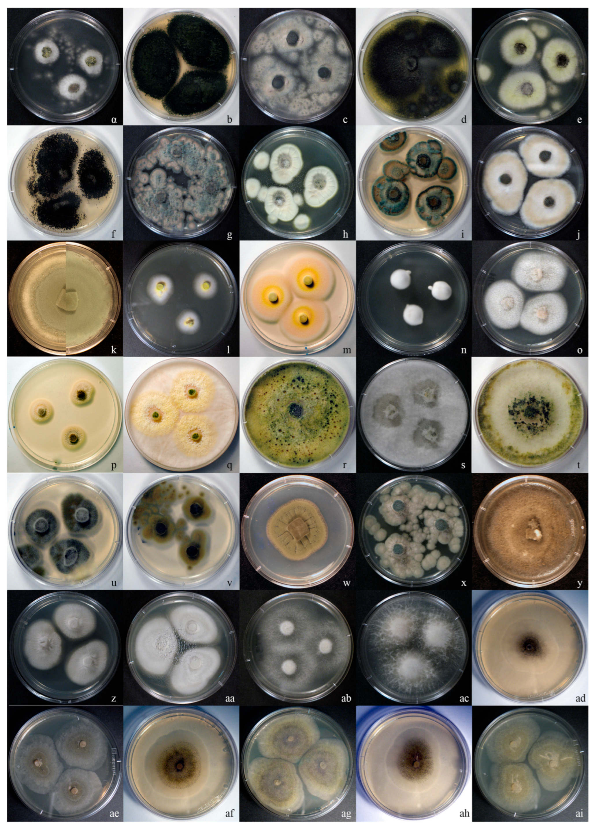
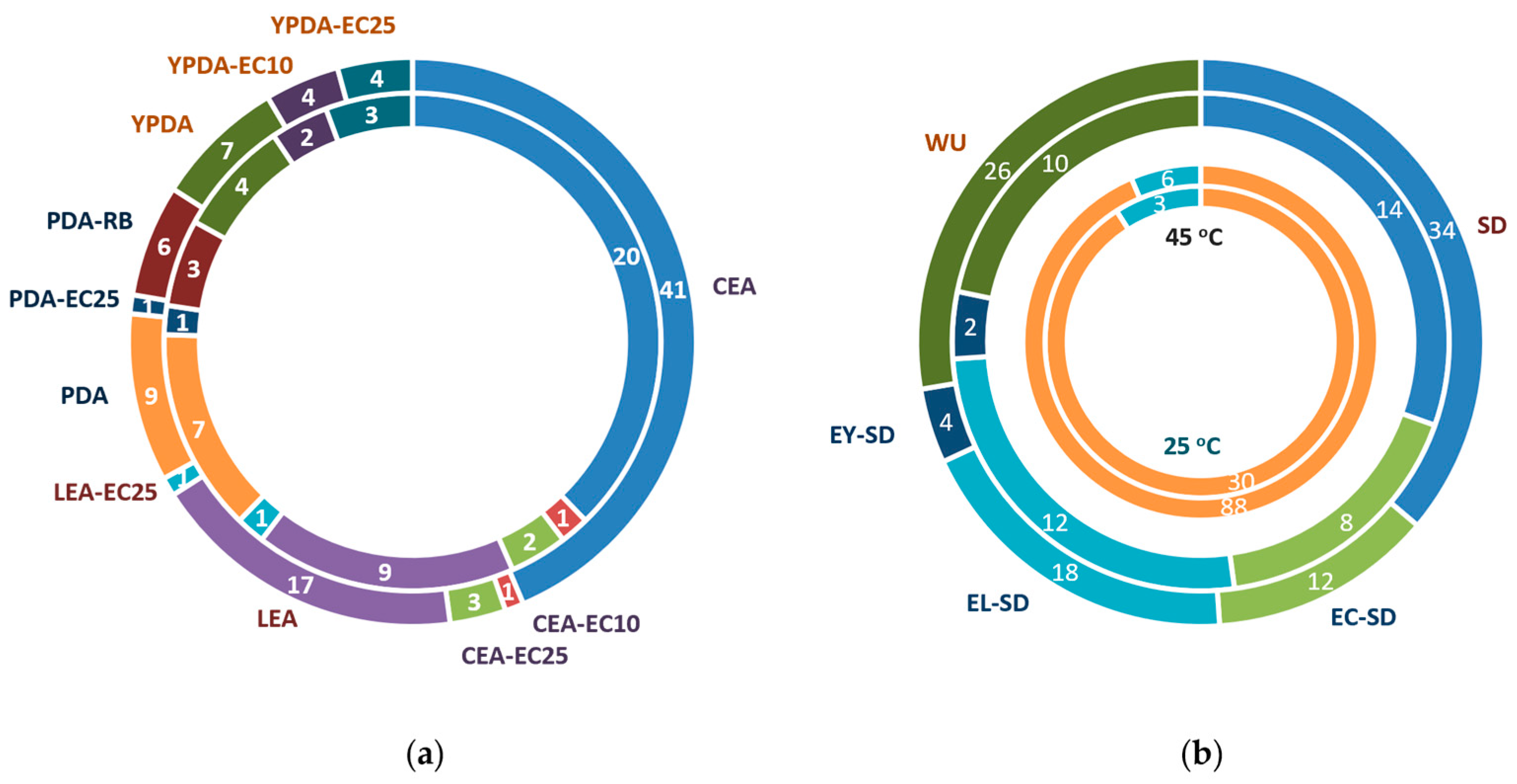
By using a combination of various substrates, inoculation strategies, and incubation regimes, 94 fungal strains were isolated from TPOMW and grouped into 56 MOTUs (indicative colony morphologies appear in Figure 2). The majority of isolates were recovered from cellulose-enriched media (CEA; 44%) and via serial dilution methods (36%) (Table 2 and Figure 3). Lignin-enriched media (LEA) yielded 19% of the strains, predominantly through the modified Warcup method (72% of them); species like Aspergillus fumigatus, Penicillium paneum, Fuscoporia ferrea, and Peniophora lycii were isolated only through this method. Incubation under mesophilic conditions (25 °C) led to the isolation of a considerably higher number of strains than under thermophilic conditions (45 °C) (94% vs. 6%, respectively). Among dominant genera, Aspergillus filifer, A. sydowii, A. westerdijkiae, Cladosporium cladosporioides, and C. limoniforme were each represented by one isolate. In contrast, Penicillium roqueforti and Candida boidinii were isolated multiple times, possibly reflecting overestimation of MOTU richness and/or their broader ecological amplitude due to their morphological variability and sampling through different isolation treatments.
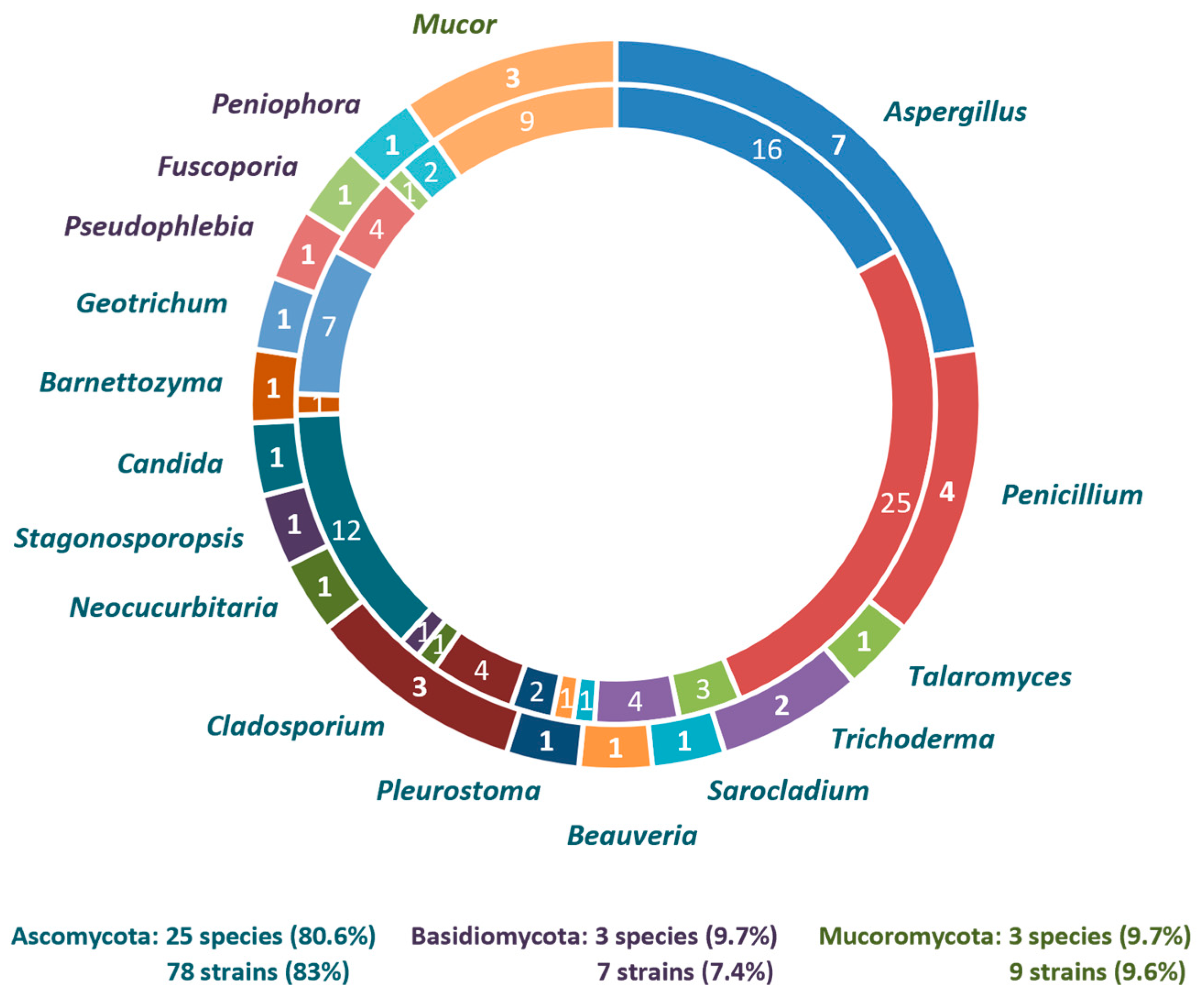
Molecular identification of MOTUs was carried out by using one to five markers (out of a total number of seven markers examined), depending on the fungal group under examination; hence, a total of 93 ITS, 1 28S, 22 tef1-α, 18 tub2, 7 act, 6 rpb2, and 4 cal sequences were generated for the first time (Table 1). In addition, nine datasets were analyzed for several fungal groups in order to resolve taxonomic uncertainties through single or multilocus phylogenetic inference using suitable reference sequences (Figure 4 and Figure 5; Supplementary Materials, Figures S1 and S2, Tables S3–S9). Subsequently, a total of 31 species assigned to 17 genera and 11 orders were identified.
The phylum Ascomycota dominated the culturable mycobiota, representing 83% of the strains isolated, followed by Basidiomycota and Mucoromycota, each accounting for 7% and 10% of the strains, respectively. Ascomycota were represented by 25 species of 13 genera distributed across eight orders: Calosphaeriales, Cladosporiales, Dipodascales, Eurotiales, Hypocreales, Phaffomycetales, Pichiales, and Pleosporales. In Basidiomycota, three species of different genera (i.e., Fuscoporia, Peniophora, and Pseudophlebia) were identified representing the orders Hymenochaetales and Polyporales. All Mucoromycota isolates were grouped into the genus Mucor (Mucorales).
More specifically, three species of Mucor were recovered: M. racemosus, M. circinelloides, and M. pseudolusitanicus (related to M. circinelloides) (Supplementary Materials, Figure S1). The M. circinelloides strain was grouped together with several publicly available strains submitted under this name [35], yet it was placed separately from the neotype of the species (CBS 195.68), sharing 99.0% ITS sequence identity. The distinct clustering may reflect ecological adaptation or taxonomic divergence, warranting future research on this taxonomic group. In contrast, strains corresponding to M. racemosus and M. pseudolusitanicus exhibited >99.8% ITS sequence identity to type-derived sequences and formed well-supported clades.
Three species of Basidiomycota were identified using ITS sequences: Peniophora lycii (Peniophoraceae), Pseudophlebia setulosa (Meruliaceae), and Fuscoporia ferrea (Hymenochaetaceae). Although sequences from type specimens were not available, the isolated strains matched with high identity (>99.5%) to numerous published sequences, e.g., CBS:264.56 [36], AH31879 [37], and CBS:460.86 [36], respectively.
As regards Ascomycota, the family Aspergillaceae (Eurotiales) dominated the culturable fungal diversity in TPOMW, accounting for nearly half of all isolates (44%) and comprising 11 species from the genera Aspergillus and Penicillium. Species-level identification was achieved primarily through the use of ITS and tub2, while tef1-α was additionally employed to improve species delimitation in certain cases.
The genus Aspergillus was represented by seven species across five sections: Circumdati (A. westerdijkiae), Flavi (A. novoparasiticus), Fumigati (A. fumigatus), Nidulantes (A. sydowii, A. filifer), and Nigri (A. tubingensis, A. welwitschiae) (Supplementary Materials, Figure S2a). Species delimitation within sections Nigri and Flavi was challenging due to high sequence identity among closely related taxa (e.g., A. welwitschiae vs. A. niger: ITS 99.5%, tub2 99.6%; A. welwitschiae vs. A. foetidus: ITS 100%, tub2 99.5%; A. tubingensis vs. A. neoniger: ITS 100%, tub2 99.6%; A. novoparasiticus vs. A. toxicarius: ITS 99.0%, tub2 100%). In contrast, taxa in sections Nudilantes, Fumigati, and Circumdati were rather clearly resolved, e.g., A. filifer (including its synonym A. chinensis) vs. A. stellatus (ITS 100%, tub2 93.0%), A. sydowii vs. A. griseoaurantiacus (ITS 98.4%, tub2 96.1%), A. fumigatus (including var. ellipticus) vs. A. oerlinghausenensis (ITS 99.4%, tub2 95.9%), and A. westerdijkiae vs. A. ochraceus (ITS 98.7%, tub2 94.7%).
The genus Penicillium was represented by 27 strains corresponding to four species (Supplementary Materials, Figure S2b): P. crustosum (section Fasciculata), P. kongii (section Brevicompacta), and P. roqueforti and P. paneum (section Roquefortorum). Species boundaries were primarily resolved through the use of tub2, given the high ITS identity among closely related taxa (99.6–99.8%). Clear separation was achieved between P. roqueforti and P. mediterraneum, P. crustosum and P. solitum, and P. kongii and P. brevicompactum (tub2, 97.0–99.1%).
The genus Cladosporium (Cladosporiaceae) was represented by four strains identified to three species (Supplementary Materials, Figure S2c), i.e., C. cladosporioides (C. cladosporioides complex) and C. ramotenellum and C. limoniforme (both members of the C. herbarum complex). Since ITS sequences were nearly identical among closely related taxa, species delimitation relied primarily on the use of act (pairwise identities 88.8–90.4%), with additional support from tef1-α and/or tub2.
Candida boidinii (Debaryomycetaceae) was the most frequently isolated yeast, since it was recovered from the majority of isolation treatments (Table 2). In the ITS + tef1-α phylogeny (Supplementary Materials, Figure S2d), the obtained strains grouped with the type strain (CBS 2428; [36]); however, they displayed a notable divergence (ITS, 96.8%; tef1-α, 95.2%), suggesting potential cryptic diversity. Multigene phylogeny in Saccharomycotina demonstrated that the genus Candida is an artificial and polyphyletic assemblage; this led to the transfer of C. boidinii and several taxa related to the genus Ogataea [38]. Nevertheless, this reclassification has not been universally adopted, and C. boidinii remains the most frequently cited name in applied microbiology, biotechnology, and environmental studies. For consistency with the prevailing literature and existing databases, we retain the traditional name C. boidinii in the present work.
Seven strains of Geotrichum (Dipodascaceae) were identified as G. candidum, despite demonstrating high ITS heterogeneity (92.9–99.5%). This taxon is currently treated as a single phylogenetic species, including strains identified as G. bryndzae and G. silvicola, which are considered synonyms (Supplementary Materials, Figure S2e) [39]. In addition, the ex-type strain CBS 772.71, formerly attributed to G. candidum, has been reassigned to G. galactomycetum, which also formed a distinct clade in our phylogenetic analysis (G. galactomycetum vs. G. candidum: 89.2–93.3% ITS identity). The recent taxonomic revision of the genus Geotrichum confirmed the complexity of G. candidum sensu lato, further highlighting the need for an exact assessment of the species boundaries.
Two strains were identified as Pleurostoma richardsiae (Pleurostomataceae) by using a five-marker concatenated phylogeny (ITS, 28S, rpb2, tef1-α, and tub2; Supplementary Materials, Figure S2f); one strain grouped with the isotype (CBS:270.33) and the ex-type of the synonym Phialophora calyciformis (strain A177 from type CBS 302.62), while the other clustered with a strain isolated from olive trees in Italy (P2BA), forming a well-supported subgroup. Despite high identities in ITS and 28S (>99.0%), the divergence noted in tub2 (95.2%), rpb2 (98.0%), and tef1-α (98.2%) suggests possible cryptic differentiation within the genus.
Three strains were assigned to Talaromyces nanjingensis (Trichocomaceae) based on ITS+tub2 phylogeny (Supplementary Materials, Figure S2g), while the tef1-α shows 99.7% identity to the respective part of the holotype’s (JP-NJ4) genome. This species, recently described from Pinus rhizosphere soil in China [40], was distinguished from its closest phylogenetic relatives (T. lianis, T. brevis) primarily by the tub2 marker (98.7% and 98.0% identity, respectively), while the ITS sequences were identical. This is the first report on the presence of T. nanjingensis in Europe and in a new habitat.
Several other taxa in TPOMW were each represented by a single isolate, i.e., Barnettozyma californica (Phaffomycetaceae), Beauveria pseudobassiana (Cordycipitaceae), Sarocladium kiliense (Sarocladiaceae), Neocucurbitaria keratinophila (Cucurbitariaceae), and Stagonosporopsis ailanthicola (Didymellaceae). Identification was mainly based on their ITS sequences, which were identical to the respective type material, i.e., CBS 252 (Supplementary Materials, Figure S2d), ARSEF 3405, CBS 122.29, MFLUCC 16-1439, and CBS 121759, respectively, while tef1-α sequences were used to further confirm their identity.
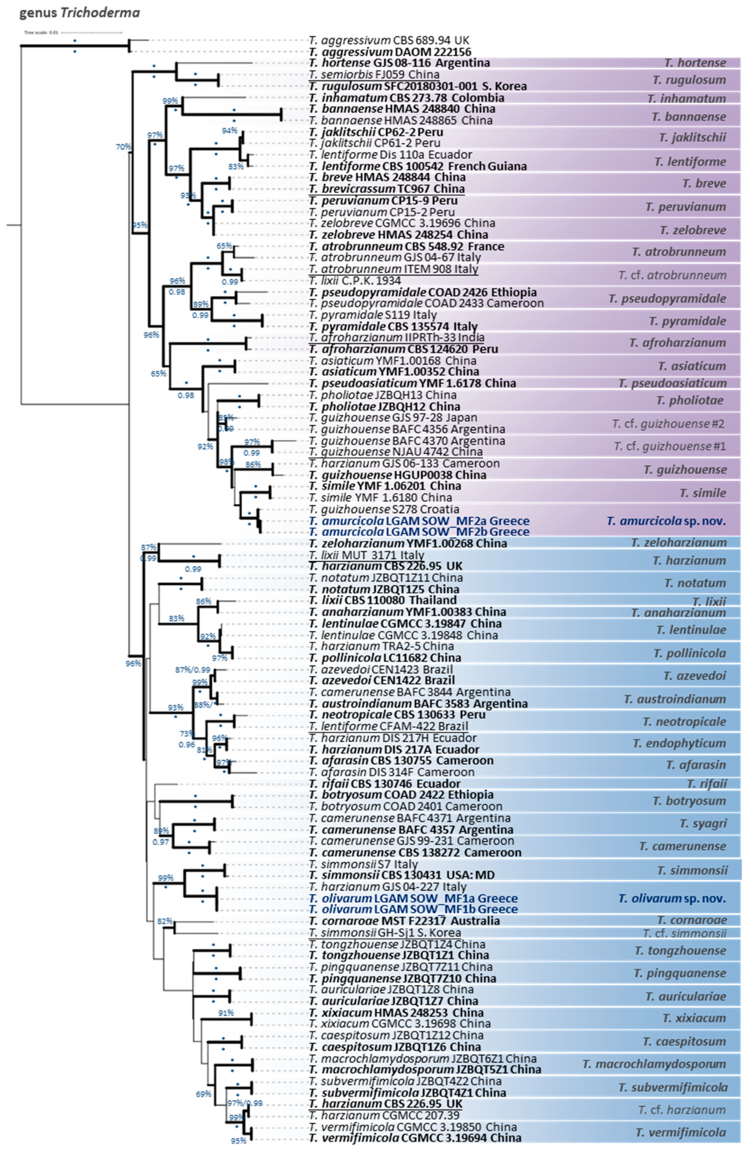
Especially as regards the genus Trichoderma (Hypocreaceae), two species new to science in the Harzianum clade were delineated and described herein as Trichoderma amurcicola and T. olivarum. Their delimitation was evidenced through the outcome of a multilocus phylogeny combining sequence data from ITS, rpb2, tef1-α, cal, and act markers (Figure 5), and was further supported by distinct morphological and cultural characters.
T. amurcicola formed a moderately supported clade (MLBS: 66%) with T. simile [41], nested within a broader, strongly supported group (MLBS: 98%; BPP: 1.00) also including T. guizhouense [42], T. pholiotae [43], and two undescribed lineages (T. cf. guizhouense #1, #2). Despite high ITS (99.6% vs. T. simile, 98.9% vs. T. guizhouense) and tef1-α (≥99.6%) identities with closely related strains, clear separation was provided by rpb2, which showed 1.6–3.5% divergence (98.3–98.4% vs. T. simile, 97.9–98.0% vs. T. guizhouense, 96.5–96.7% vs. T. cf. guizhouense #1). Additional support was provided through the use of the cal (99.1% vs. T. guizhouense, 99.1–99.3% vs. T. cf. guizhouense #1) and act markers (98.8% vs. T. guizhouense, 98.9–99.7% vs. T. cf. guizhouense #1). These values are below commonly accepted species-level thresholds in the genus Trichoderma [44,45].
It should be noted that several strains previously identified as T. guizhouense, including genome-sequenced or reference strains such as NJAU 4742 and GJS 97-28 from Japan [46], grouped outside the clade, which included the species holotype HGUP0038 from China [42,47], indicating taxonomic discrepancies. These strains were recovered within distinct, well-supported clades corresponding to T. cf. guizhouense #1 and #2, with BAFC 4370 and BAFC 4356 [46] serving as representative vouchers, respectively. In the aforementioned study, GJS 97-28 was erroneously designated as an ex-type strain of T. guizhouense, despite its clear phylogenetic incongruence and distant geographic origin from the species’ type locality. Furthermore, one Croatian isolate (S278) [44], formerly misassigned to T. guizhouense, was grouped into T. amurcicola. Additional isolates annotated as “T. guizhouense”, including both published (e.g., S628 from Greece; [44]) and unpublished sequences (e.g., Tr49 and Tr118 from Turkey; SZMC:1630, SZMC:1633 from Hungary; SZMC 25242, SZMC 25749 from Serbia; SZMC:12334, SZMC:12384 from India; PARC1022, PARC1023, PARC1025, and PARC1026 possibly from southern Italy) also grouped outside the true T. guizhouense clade and within T. amurcicola (according to rpb2 BLASTn results and phylogenetic analysis), thus suggesting a wider Eurasian distribution of the T. amurcicola. Although these strains were often considered to represent a single taxonomic entity (T. guizhouense), previous phylogenetic analyses frequently failed to group them within a monophyletic, well-supported clade [48], highlighting the need for multigene approaches in species delimitation within the Harzianum clade and the critical role of holotype-based validation/identification.
T. olivarum was resolved as a sister to T. simmonsii (MLBS: 99%, BPP: 1.00). Although the two species exhibited high ITS and tef1-α identity (100% and 99.6%, respectively), clear separation was evident through the use of the other markers used, i.e., rpb2 (97.8%), cal (95.7%), and act (98.9%). These values are below established species-level thresholds, and when combined, T. olivarum is clearly separated as a distinct lineage within the Harzianum clade. Interestingly, despite their phylogenetic distance, T. olivarum and T. subvermifimicola shared identical rpb2 sequences, demonstrating the limitations of single-locus resolution in species delimitation within the genus Trichoderma. In addition, the sequence from the genome of an isolate from South Korea (GH-Sj1, initially labeled T. simmonsii), grouped outside the T. simmonsii clade, which includes the holotype, thus representing a different taxon, which is provisionally named T. cf. simmonsii.

Taxonomy

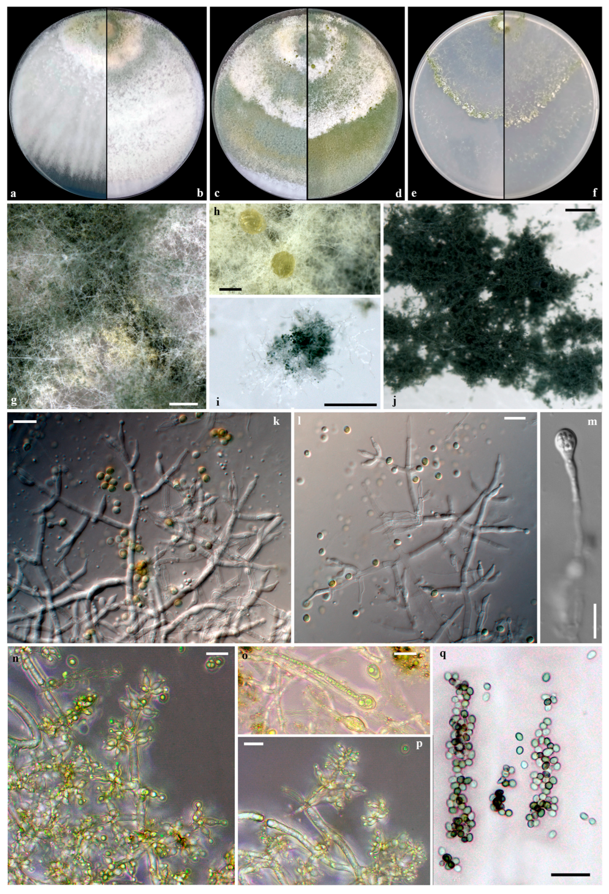
Trichoderma amurcicola V. Fryssouli, I. Kefalogianni, E. Polemis & G.I. Zervakis, sp. nov. (Figure 6)
Mycobank: MB860058.
Etymology: “amurcicola” from the Latin ‘amurca’ (deriving from Greek ἄμουργα), meaning the olive pomace or the sediment/residue remaining after olive oil extraction), and from the suffix ‘-cola’ (meaning residing/existing in Latin), referring to the substrate from which the fungus was isolated.
Diagnosis: Trichoderma amurcicola is distinguished within the Harzianum clade by the multilocus phylogeny (ITS, tef1-α, rpb2, cal, act), the relatively narrow phialides, the presence of rare terminal chlamydospores, and the presence of diffusible pigments.
Type: GREECE, Peloponnese, from TPOMW, November 2017. Holotype LGAM SOW_MF2a, preserved in 30% (v/v) glycerol at −80 °C in the Laboratory of General and Agricultural Microbiology, Agricultural University of Athens (Greece). GenBank accession numbers: ITS = PP766637, cal = PP768730, act = PP768723, rpb2 = PP768717, tef1-α = PP768704.
Conidiophores hyaline, smooth-walled, pyramidal to tree-like, typically with 1–3 levels of verticillate branching, forming a densely intricate reticulum. Main axis rebranch, producing paired, opposing, or singly side-branches that emerge predominantly asymmetrically and perpendicular to the axis or slightly oriented toward the terminus. Branch intervals measure (2.4-)7.0-25.1(-57.5) μm, mean 15.2 μm; septa conspicuous. Branches terminating in cruciate whorls of 2-4 (-5) phialides, occasionally solitary. Phialides predominately ampulliform to lageniform, straight to curved or inequilateral, distinctly constricted below the apex, forming symmetrical or slightly curved narrow necks, (4.6-)5.0-10.2(-11.7) × (2.1-)2.3-3.4(-3.6) μm, mean 7.3 × 2.9 μm, l/w (1.5-)1.7-3.8(-4.5), distinct base (0.9-)1.2-2.2(-2.4) μm wide; supporting cell (1.8-)1.9-4.4(-5.5) μm wide (mean 2.4 μm); ratio of phialide length to supporting cell width (1.8-)2.0-4.5 and phialide width to supporting cell width (0.8-)0.9-1.6(-1.9). Conidia globose to subglobose, occasionally ellipsoid, smooth, thin-walled, hyaline with yellow-green to dark green hue, (2.5-)2.8-3.3(-3.5) × (2.1-)2.3-2.9(-3.1) μm, mean 3.0 × 2.5 μm, l/w (1.0-)1.1-1.3(-1.5). Chlamydospores occasionally observed in PDA, terminal, ellipsoid to globose, 5.4–6.0 × 3.4–5.8 μm, l/w 1.0–1.7.
On PDA after 72 h, colonies attain radii of 64–70 mm at 25 °C, 67–77 mm at 30 °C, and 38–41 mm at 35 °C, covering the plate in 3 days at 25 °C. Colonies exhibit radial growth with dense, continuous cottony to floccose aerial mycelium, especially near inoculum; margin rather flat and well-defined, initially white, developing broad concentric, not well-defined zones altering in color from yellowish green, greyish green to olive-green, darkening with age; dark green in the center. Conidiation initiates within 48 h on aerial hypha, and minute pustules are formed around the inoculum and margin, transitioning from white to dark green in the center. Reverse yellow to orange diffuse pigmentation. Odor faintly fruity.
On SNA after 72 h, colony radii measure 37–43 mm at 25 °C, 30–43 mm at 30 °C, and 13–15 mm at 35 °C, covering the plate in 5 days at 25 °C. Colonies translucent, thin, and radially structured, with sparse, arachnoid aerial hyphae in concentric zones; margin not well-defined. Conidiation occurs on both aerial hyphae and pulvinate pustules form within 48 h; pustules distributed either diffusely or in concentric arrangements, transitioning from white to dark green in the center. No diffuse pigmentation on the reverse side. Odor indistinct.
Additional material examined. GREECE, Peloponnese, from TPOMW, November 2017. LGAM SOW_MF2b, preserved in 30% (v/v) glycerol at −80 °C in the Laboratory of General and Agricultural Microbiology, Agricultural University of Athens (Greece). GenBank accession numbers: ITS = PP766638, cal = PP768731, act = PP768724, rpb2 = PP768718, tef1-α = PP768705.
Notes. Trichoderma amurcicola is phylogenetically closely related to T. simile [41] and T. guizhouense [42,47], yet it is readily distinguishable based on consistent morphological and cultural characteristics. All three species exhibit pyramidal to tree-like conidiophores; however, they differ diagnostically in phialide morphology. The phialides of T. amurcicola (5.0-10.2 × 2.3-3.4 μm; l/w 1.7-3.8) are narrower and less variable than those of T. simile (4.3-11.9 × 2.7-3.9 μm; l/w 1.3-4.4). In contrast, T. guizhouense displays broader and shorter phialides (4.7-7.5 × 3.0-3.7 μm; l/w 1.4-2.4) according to Chaverri et al. [47], or rather narrow phialides (4.5-10 × 2-3 μm) according to Li et al. [42], since the two available descriptions differ notably in measurements. Conidial morphology (size and shape) does not provide reliable differentiation among these species. T. amurcicola produces globose to subglobose conidia (2.8-3.3 × 2.3-2.9 μm; l/w 1.1-1.3), overlapping with the slightly smaller oval conidia of T. simile (2.6-3.2 × 2.2-2.8 μm; l/w 1.0-1.2), whose low l/w ratio values more closely resemble globose forms. T. guizhouense produces primarily globose conidia (2-3 μm in diameter; 42; or 2.5-3.2 × 2.5-3.0 μm; l/w 1.0-1.2; 47). Chlamydospores in T. amurcicola are rare, exclusively terminal, and measure 5.4-6.0 × 3.4-5.8 μm (l/w 1.0-1.7). In contrast, T. simile forms abundant chlamydospores, both terminal and intercalary, and ranges in size (4.2-7.8 × 4.0-7.2 μm), whereas T. guizhouense lacks chlamydospores [42,47].
In culture, T. amurcicola exhibits faster growth at 35 °C (when measured for 72 h), particularly on PDA (38–41 mm), compared to T. simile (10 mm) and T. guizhouense (16–20 mm; 42), and similarly on SNA (13–15 mm vs. T. simile: 3 mm; T. guizhouense: 5–8 mm; 42). However, at 25 °C (for 72 h) on SNA, T. amurcicola grows more slowly (37–43 mm) than T. simile (47 mm) and T. guizhouense (58–61 mm; 42). Colonies of T. amurcicola produce yellow to orange diffusible pigments on PDA and emit a faint fruity odor, i.e., traits shared with T. guizhouense [42,47], but absent in T. simile.
In comparisons with more distantly related taxa, T. pholiotae [43] is readily differentiated by its larger and broader phialides (4.9-10.9 × 2.4-4.2 μm; l/w 1.4-3.4) and conidia (2.6-3.8 × 2.4-3.3 μm; l/w 1.0-1.3), as well as the frequent occurrence of larger terminal and intercalary chlamydospores (5.0-7.4 × 4.9-7.0 μm). T. asiaticum [41] can be distinguished by its considerably shorter phialides (4.0-6.0 × 2.0-3.0 μm; l/w 1.3-3.0), smaller conidia (2.4-3.0 × 2.1-2.7 μm; l/w 1.1-1.3), and the absence of chlamydospores. T. pseudoasiaticum [41] also forms slightly broader phialides (6.1-9.0 × 2.6-3.6 μm; l/w 1.5-3.6) and nearly spherical conidia (2.4-3.2 × 2.4-3.0 μm; l/w 1.0–1.1), while terminal chlamydospores are frequently observed (4.7-7.7 × 4.0-7.6 μm). None of these three species produce diffusible pigments, and none exhibit fast growth under thermotolerant conditions (35 °C for 72 h) on SNA (8–10 mm for T. pholiotae, 7 mm for T. asiaticum, and 2 mm for T. pseudoasiaticum).
Trichoderma olivarum V. Fryssouli, E. Polemis, Μ.A. Typas & G.I. Zervakis, sp. nov. (Figure 7)
Mycobank: MB860059.
Etymology. “olivarum” from the Latin ‘olivarum’ (genitive plural of ‘olivarium’, meaning olive grove), denoting the olive-associated habitat from which this fungus originated.
DiagnosisTrichoderma olivarum is distinguished within the Harzianum clade by the multilocus phylogeny (ITS, tef1-α, rpb2, cal, act), the relatively long and narrow phialides, the often ovoid to oblong conidia, and the frequently present chlamydospores.
Type. GREECE, Peloponnese, from TPOMW, Nov. 2017. Holotype LGAM SOW_MF1a, preserved in 30% (v/v) glycerol at −80 °C in the Laboratory of General and Agricultural Microbiology, Agricultural University of Athens (Greece). GenBank accession numbers: ITS = PP766635, cal = PP768728, act = PP768721, rpb2 = PP768715, tef1-α = PP768702.
Conidiophores hyaline, smooth-walled, pyramidal to tree-like, typically with 1–2(–3) perpendicular rebranching levels, forming a densely intricate reticulum where the main axis is often unrecognizable. Main axis producing single or paired opposing side-branches that emerge asymmetrically. Branch intervals measure (2.5-)4.2-27.8(-30.3) μm, mean 15.2 μm; septa conspicuous. Branches terminating in whorls of 2–4 phialides or solitary. Phialides ampulliform to lageniform, straight to curved or inequilateral, distinctly constricted below the apex, forming symmetrical or slightly bent narrow necks usually elongated, especially in the terminal solitary phialides, (5.4-)5.7-12.4(-13.1) × (1.9-)2.1-3.1(-3.3) μm, mean 8.7 × 2.6 μm, l/w 1.7-6.0, distinct base, 0.9–2.4 μm wide (mean 1.7 μm); supporting cells 1.9–3.1 μm wide (mean 2.4 μm); ratio of phialide length to supporting cell width 1.9–5.7 (mean 3.6), and phialide width to supporting cell width 0.7–1.4 (mean 1.1). Conidia globose, subglobose, ovoid to oblong, smooth, thin-walled, hyaline with pale green, yellow-green to dark green hue, (2.8-)2.9-3.9(-4.2) × (2.1-)2.3-2.9(-3.0) μm, mean 3.4 × 2.6 μm, l/w (1.0-)1.1-1.5(-1.9). Chlamydospores frequently present, terminal to intercalary, globose to ellipsoid, 5.6-9.6 × 5.1-6.5 μm, l/w 1.0-1.5.
On PDA after 72 h, colonies attain radii of 69–73 mm at 25 °C, 73–74 at 30 °C, 17–28 mm at 35 °C, covering the plate in 3 days at 25 °C. Colonies radially structured, with dense, continuous cottony to floccose aerial mycelium, especially near inoculum, initially white transitioning to dark green with age, developing broad concentric zones, white to olivaceus; margin rather flat and inconspicuous with faint mycelium. Conidiation initiates within 48 h on aerial hypha; pustules are formed around the inoculum and margin, transitioning from white to dark green in the center. No diffuse pigmentation on the reverse side. Odor faintly fruity.
On SNA after 72 h, colony radii measure 47–49 mm at 25 °C, 51–55 mm at 30 °C, and 5–7 mm at 35 °C, covering the plate in 5 days at 25 °C. Colonies translucent, thin, and radially structured, with sparse, arachnoid aerial hyphae concentrated on concentric zones; margin not well-defined. Conidiation occurs on pulvinate, compact pustules within 48 h, with pustules distributed near the inoculum and in concentric arrangements, transitioning from white to dark green in the center. No diffuse pigmentation on the reverse side. No distinctive odor is noted.
Additional material examined. GREECE, Peloponnese, from TPOMW, Nov. 2017. Holotype LGAM SOW_MF1b, preserved in 30% (v/v) glycerol at −80 °C in the Laboratory of General and Agricultural Microbiology, Agricultural University of Athens (Greece). GenBank accession numbers: ITS = PP766636, cal = PP768729, act = PP768722, rpb2 = PP768716, tef1-α = PP768703.
Notes. Detailed comparisons of morphological, physiological, and culture features reveal clear species-level distinction of T. olivarum from its sister species, T. simmonsii [47], and also from T. subvermifimicola [48], which present identical rpb2 sequences despite their relative distant topology and divergence demonstrated when using other markers. The three species share broadly similar conidiophore architecture (pyramidal, asymmetrically branched at 1–3 levels) similar to T. simmonsii, whereas T. subvermifimicola shows more symmetrical pyramidal arrangements. Phialide morphology is a key diagnostic character. In T. olivarum, phialides are longer and narrower (5.7-12.4 × 2.1-3.1 μm; l/w 1.7-6.0), contrasting with the shorter, broader phialides of T. simmonsii (5.2-6.5 × 3.0-3.7 μm; l/w 1.5-2.4) and of T. subvermifimicola (4.7-9.4 × 2.3-3.6 μm; l/w 1.4-3.9). Conidial dimensions also support species delimitation: T. olivarum forms variable, globose to oblong conidia (2.9-3.9 × 2.3-2.9 μm; l/w 1.1-.5), larger and more elongate than those of T. simmonsii (2.7-3.2 × 2.5-3.0 μm; l/w 1.0-1.1) and T. subvermifimicola (2.7-3.3 × 2.5-3.0 μm; l/w 1.0-1.2). In addition, T. olivarum forms frequent, large terminal and intercalary chlamydospores (5.6-9.6 × 5.1-6.5 μm), whereas T. simmonsii produces them rarely, and T. subvermifimicola forms smaller (4.0-6.9 × 3.5-6.4 μm), also intercalary and terminal chlamydospores.
Colony growth characteristics further distinguish T. olivarum from related species. On PDA at 25 °C, it reaches 69–73 mm in 72 h, outgrowing T. simmonsii (55–65 mm) and T. vermifimicola (60–64 mm), while T. simmonsii exhibits better growth at 35 °C (25–55 mm vs. 17–28 mm in T. olivarum, and 13–17 mm in T. subvermifimicola). Colonies do not produce diffuse pigmentation; however, T. simmonsii and T. subvermifimicola sometimes produce yellow pigmentation on the reverse surface.

3.2. Preliminary Evaluation of Biodegradation Efficacies of Isolated Fungi

In the frame of performing a preliminary evaluation of the biodegradation potential of the isolated fungi, 66 strains corresponding to 31 species were tested with respect to their hydrolytic, oxidative, and dye-decolorizing efficacies. The enzymatic index (EI) was assessed on cellulose, xylan, lignin, guaiacol, and RBBR substrates (Table 2; Supplementary Materials, Table S10). Τhe tested strains exhibited high degradation potential; 96% of them displayed cellulolytic activity, and 82% were capable of degrading xylan (22% and 19% were evaluated as significant degraders). In contrast, ligninolytic activity and laccase production were observed in fewer species (i.e., 13% and 17%, respectively), suggesting specialization in oxidative mechanisms for certain taxa. In addition, 72% of the strains displayed the ability to decolorize RBBR (5% were evaluated with strong activity), indicating a significant prevalence of dye-degrading capabilities within the collection. Despite variable enzymatic activity, all strains were able to grow well on lignocellulosic media.
As regards cellulose degradation, the highest cellulolytic activities were exhibited by Aspergillus sydowii, Fuscoporia ferrea, Penicillium paneum, P. crustosum, and Beauveria pseudobassiana strains. In addition, Aspergillus novoparasiticus, Penicillium kongii, Pleurostoma richardsiae, and Trichoderma amurcicola, as well as all strains of Mucorales (M. circinelloides, M. pseudolusitanicus, M. racemosus) consistently displayed strong cellulolytic profiles. As concerns xylan degradation, the highest xylanolytic activity was detected in Beauveria pseudobassiana, Geotrichum candidum, Peniophora lycii, and Neocucurbitaria keratinophila. Notable xylan-degrading capabilities were also detected in Mucor circinelloides and M. pseudolusitanicus, the two new Trichoderma species, and in several Aspergillus and Penicillium strains. Ligninolytic activity was most pronounced in the basidiomycetes Pseudophlebia setulosa, Fuscoporia ferrea, and Peniophora lycii. Among Ascomycota, Cladosporium ramotenellum and C. limoniforme exhibited the strongest lignin-degrading and laccase activities, while Pleurostoma richardsiae and Beauveria pseudobassiana were also efficient in this respect. In contrast, Aspergillus fumigatus and A. filifer grew well on lignin-containing media but did not exhibit detectable ligninolytic activity. Strong RBBR decolorization, indicative of oxidative potential, was observed in Beauveria pseudobassiana and Candida boidinii. Additional high decolorization activities were noted in Geotrichum candidum, Barnettozyma californica, Cladosporium limoniforme, and C. ramotenellum. Moderate dye-degrading capacity was widespread across species of Basidiomycota, and of the genera Aspergillus, Penicillium, and Mucor.
More importantly, a subset of isolates exhibited broad enzymatic repertoires across all substrates tested. These included Cladosporium ramotenellum, C. limoniforme, Fuscoporia ferrea, and Pseudophlebia setulosa, while significant activities were also detected in Beauveria pseudobassiana, Trichoderma amurcicola, Pleurostoma richardsiae, and Peniophora lycii. In addition, several taxa already known from the literature for their broad biotechnological relevance—including Aspergillus sydowii, Barnettozyma californica, Candida boidinii, Geotrichum candidum, and Mucor circinelloides—also displayed strong and versatile enzymatic profiles.

4. Discussion

4.1. Fungal Diversity in TPOMW

The culturable fungal community in TPOMW was dominated by Ascomycota, particularly Eurotiales and Hypocreales, with Basidiomycota and Mucoromycota recovering at lower numbers, which is in accordance with previous findings [4,15,22]. This pattern highlights the selective ecological pressures imposed by such types of substrates, i.e., with acidic pH, high content in lipids, phenolics, and other chemical stressors [1]. TPOMW exerts a strong selective effect on fungal communities, explaining the frequent recovery of Aspergillus, Cladosporium, Mucor, and Penicillium strains as previously noted [15,16,21]. These taxa share common ecological strategies favoring their growth and persistence under environmentally challenging conditions, including rapid growth, tolerance to phenolic compounds, and the capacity to exploit complex carbon sources. In addition to filamentous taxa, Candida and yeast-like Geotrichum strains were abundant, reflecting their ability to thrive in sugar- and lipid-rich niches and to colonize substrates at early stages of decomposition [15,19,20]. Their recurrent presence across diverse agro-wastes suggests convergent ecological strategies shaped by nutrient-rich but chemically restrictive environments. Furthermore, two new Trichoderma species (T. amurcicola and T. olivarum) were also found in TPOMW, indicating that this substrate could serve as a reservoir of unexplored fungal diversity.
Basidiomycetes are less frequently isolated from OMW-based substrates; the genera Pseudophlebia, Fuscoporia, and Peniophora, which are typically associated with wood degradation, are recorded for the first time in TPOMW. This was also the case for Beauveria, Neocucurbitaria, Pleurostoma, and Stagonosporopsis, which are mostly known as pathogens of plants, insects, or animals [49,50,51]; their presence in TPOMW reflects their wide adaptation in different ecological niches. In addition, Barnettozyma californica, Cladosporium limoniforme, Mucor pseudolusitanicus, and Talaromyces nanjingensis were recorded for the first time in olive mill wastes.
These patterns underline the strong selection effect of TPOMW on fungal communities and reveal a structured mycobiota composed of stress-tolerant filamentous fungi (mainly Ascomycota and Mucoromycota), opportunistic yeasts, and (occasionally) filamentous basidiomycetes [4,15,22]. This diversity provides an ecological basis for understanding fungal succession in phenolic- and lipid-rich agro-industrial residues.

4.2. General Taxonomic and Phylogenetic Remarks on the Genus Trichoderma

Our analyses outlined the widespread taxonomic issues related to type collections and genome-linked strains in the genus Trichoderma. Beyond the case of the type material of T. guizhouense, which was previously discussed, additional issues were identified in the course of our investigation. One of them relates to T. afarasin collections reported as type material in different studies, i.e., CBS 130755 by Chaverri et al. [47] and Dis 314f by Barrera et al. [46]. Further confusion arises when the identity of a strain has changed over time without the respective necessary updates in the INSDC databases. For example, sequences derived from the holotypes of T. inhamatum (CBS 273.78; FJ577683) and T. endophyticum (CBS 130729/DIS 217A; FJ442243, FJ463319, FJ442292) remain deposited under the name “T. harzianum”, while the sequences derived from the holotype of T. syagri are still labeled as T. camerunense (BAFC 4357; MG822711, MG822714, MG822717). Moreover, some strains are linked to multiple collection codes, complicating accurate traceability and material management (Table S10). An indicative example is the type of T. austroindianum (i.e., strain BAFC 3583 [46]), which in NCBI appears under VAB-T050 without any reference to the former code and without mentioning that it corresponds to the type material.
Inconsistencies were also detected as regards the association of genome sequences with validated species concepts. Several genome sequences were found to be misassigned, appearing in clades not related to their taxonomic identity. For example, strain CFAM-422 (submitted as T. lentiforme) clusters within the T. neotropicale lineage, strain MUT 3171 labeled as T. lixii groups within the T. harzianum clade, and strain FJ059 identified as T. semiorbis is phylogenetically placed within the T. rugulosum group. Another notable case involves the genome of the type strain of T. brevicrassum (TC967) [52], which in our work nested within the clade, which includes the T. breve holotype. This is in contrast with their phylogenetic placement in the study where both species were originally described [53]; the former was placed in the Chlorosporum clade, whereas the latter forms part of the Harzianum clade. This discordance indicates a probable misidentification and calls for genomic re-evaluation of the deposited sequence. Furthermore, the type strain CBS 226.95 of T. harzianum also displays conflicting placements. Based on sequence data from Chaverri et al. [47], it clusters—as expected—within the T. harzianum clade, and this placement is congruent with published phylogenies. However, the genome assigned to CBS 226.95 (unpublished) falls into a separate clade, herein provisionally designated as T. cf. harzianum.
These findings reveal a series of pressing issues in the Trichoderma taxonomy and nomenclature. Persistent taxonomic gaps such as misassigned genomes and ambiguous type designations continue to undermine species-level resolution. Furthermore, our results confirm that rpb2 and cal were the most informative markers for species delimitation within the Harzianum clade, offering higher resolution compared to ITS and tef1-α. In particular, rpb2 showed consistency in delimiting among species (with some exceptions like the low resolution between T. olivarum and T. subvermifimicola) and a clear barcoding gap across comparisons, confirming previous findings on its discriminatory power in Trichoderma [45]. Despite their usefulness, cal and act were less broadly available in public databases, limiting their comparative value. tef1-α resolution was rather inconsistent, which is mostly due to the different target regions used among studies [47,54].
Consistent morphological differences (e.g., phialide and conidial dimensions, chlamydospore morphology) and physiological traits (e.g., growth at higher temperatures) further contribute to species separation. These findings support the necessity of a polyphasic taxonomic approach, especially for cryptic taxa.

4.3. Biodegradation Potential of Fungal Strains

The enzymatic profiles of TPOMW-associated fungi highlight the coexistence of distinct functional guilds that play complementary roles in substrate degradation. Filamentous Ascomycota, including Aspergillus, Cladosporium, Penicillium, Talaromyces, and Trichoderma, exhibited broad hydrolytic and, in several cases, ligninolytic capacities, as previously demonstrated [12,15,21]. These features are consistent with their frequent dominance in lipid-, phenolic-, and lignocellulose-rich environments, where rapid growth, metabolic versatility, and tolerance to chemical stressors provide a competitive advantage [55,56,57,58,59,60,61,62,63]. Within this group, thermotolerant isolates such as Aspergillus fumigatus and Trichoderma amurcicola are of particular interest since their ability to function at elevated temperatures aligns with potential applications in biomass pretreatment and other industrial and biotechnological processes [11]. This is also reported in the past for Aspergillus fumigatus [64,65,66,67], A. tubingensis [68], and A. welwitschiae [69,70,71], although the respective strains isolated from TPOMW did not show a high enzymatic potential. Species of Cladosporium, though less frequently reported in pertinent studies [61,72], demonstrated significant ligninolytic activities, indicating their potential exploitation in the degradation of recalcitrant substrates.
The presence of Barnettozyma, Candida, and Geotrichum yeasts reflects their capacity to colonize sugar- and lipid-rich substrates, and to initiate early stages of substrate decomposition [20]. Their enzymatic versatility, including hydrolytic and oxidative dye-degrading activities, is consistent with their reported applications in bioethanol production, wastewater detoxification, value-added metabolite synthesis, or broader interest [73,74,75,76,77,78,79]. White-rot basidiomycetes (Pseudophlebia, Fuscoporia, Peniophora), though isolated less frequently, exhibited high ligninolytic and laccase activities, confirming their ecological relevance and biotechnological potential in the degradation of lignocellulosics [10,80,81,82,83].
Members of Mucorales, especially Mucor circinelloides, displayed notable hydrolytic activity, dye-decolorization capacity and rapid substrate colonization. These features underline their ecological role as early colonizers in shaping fungal succession and driving initial biomass conversion. Furthermore, several strains already examined in applications related to bioethanol production, lipid accumulation, and enzyme secretion [84,85,86,87,88].
As regards other less common—but enzymatically active—fungi isolated from TPOMW, Beauveria pseudobassiana had been primarily studied for use in agriculture as an insect pathogen; however, it revealed strong hydrolytic and ligninolytic activities, suggesting broader biotechnological potential [89,90]. In addition, Pleurostoma, Neocucurbitaria, and Sarocladium, usually investigated as plant or human pathogens [49,50], exhibited hydrolytic and dye-decolorizing activities that expand their ecological significance and exploitation potential [22,60].
Preliminary assays revealed widespread cellulolytic and xylanolytic activity, with substantial dye decolorization and targeted ligninolytic potential in Basidiomycota and Dothideomycetes. Strains from several species (i.e., Beauveria pseudobassiana, Cladosporium ramotenellum, C. limoniforme, Fuscoporia ferrea, and Pseudophlebia setulosa) exhibited a broad spectrum of enzymatic activities across all tested substrates. Additional ‘multifunctional’ strains were those of Mucor circinelloides, Aspergillus sydowii, Candida boidinii, and Geotrichum candidum with potential applications in biofuel production, enzyme biocatalysis, and agricultural biotechnology, as was previously evidenced for these particular species [55,73,74,77,78,79,84,86]. The importance of targeting culturable fungi, which remain accessible for downstream applications, is apparent. Further research should focus on optimizing microbial/fungal consortia (e.g., thermotolerant species/strains, yeasts) [14,59,65,68,76], enzyme production conditions, and scaling up of bioprocesses to fully harness their potential in sustainable waste management and circular bioeconomy systems.

5. Conclusions

This study contributes to the assessment of the taxonomically and functionally diverse culturable mycobiota existing in TPOMW, which is mainly shaped by the selective pressure of this chemically complex substrate. Beyond recovering well-known genera such as Aspergillus, Penicillium, and Candida, we documented previously unreported taxa in olive mill wastes, including two new species of Trichoderma (i.e., T. amurcicola and T. olivarum). Preliminary enzymatic screening revealed broad hydrolytic, ligninolytic, and dye-decolorizing activities, highlighting the presence of multifunctional strains with potential applications in biorefineries, biofuel production, wastewater treatment, and sustainable agriculture. By combining multigene phylogenetics with functional assays, this work evidences that TPOMW—albeit overlooked—could serve as a valuable reservoir of fungal diversity, including strains of particular biotechnological relevance. While culture-dependent methods and preliminary enzymatic assays do not reveal the full potential of the organisms present in TPOMW, they provide an effective entry point for identifying promising strains. Future research could focus on determining the enzymatic potential of isolated strains through quantitative assays, the establishment of potent fungal consortia, and their exploitation in biotechnological applications.

Supplementary Materials

The following supporting information can be downloaded at https://www.mdpi.com/article/10.3390/jof11090687/s1: Figure S1. Phylogenetic tree for the genus Mucor, Mucoromycota (ITS; DSMUC, Supplementary Materials, Table S3), including strains recovered from TPOMW. Species identified are presented in colored boxes. Type strains are shown in bold black, while strains obtained in this study are marked in bold blue. Branch support values are shown where MLBS ≥ 65% and BPP ≥ 0.95; asterisks (*) denote MLBS = 100% and BPP = 1.00. Figure S2. Phylogenetic trees for fungal genera/species corresponding to strains (representing MOTUs) of Ascomycota recovered from TPOMW. The molecular markers and the respective datasets used appear in Table 1 and in Supplementary Materials, Table S3. Trees are presented in alphabetical order on the basis of the family name (Table 1): (a) Aspergillus (ITS and tub2; dataset DSASP); (b) Penicillium (ITS and tub2; DSPEN); (c) Cladosporium (ITS and act; DSCLA); (d) Candida boidinii and Barnettozyma californica (ITS and tef1-α; DSCA-BA); (e) Geotrichum (ITS; DSGEO); (f) Pleurostoma (ITS, 28S, tub2, tef1-α and rpb2; DSPLE); (g) Talaromyces (ITS, tub2; DSTAL). Species identified are presented in colored boxes. Type strains are shown in bold black, while strains obtained in this study are marked in bold blue. Intrageneric sections are indicated along major branches. Branch support values are shown where MLBS ≥ 65% and BPP ≥ 0.95; asterisks (*) denote MLBS = 100% and BPP = 1.00. Table S1. Composition of solid and liquid media used for the isolation of fungi and enzymatic assays. For each medium, the respective concentrations (g L−1) are provided. Table S2. Primer pairs used for the amplification of fungal DNA markers. Includes targeted locus, primer names and sequences (5′–3′), expected amplicon size (bp), annealing temperature (°C), and corresponding references. Table S3. Characteristics of the sequence datasets used in the phylogenetic analyses of each fungal genus. Table S4. Reference sequences used in the phylogenetic analysis of the genus Mucor. For each strain, the sequence identifier, voucher code, and GenBank accession numbers. Superscripts next to voucher codes denote type status: H = holotype, T = type, I = isotype, EX-N = ex-neotype. Table S5. Reference sequences used in the phylogenetic analyses of strains in the genera Aspergillus, Penicillium, and Talaromyces. Listed for each strain are the sequence identifier, voucher code, and GenBank accession numbers for the examined loci. Superscripts next to voucher codes denote type status: H = holotype, T = type, N = neotype, E = epitype, S = syntype, EX = ex-type. Table S6. Reference sequences used in the phylogenetic analysis of the genus Cladosporium. For each strain, the sequence identifier, voucher code, and GenBank accession numbers for the examined loci. Superscripts next to voucher codes denote type status: H = holotype, N = neotype, E = epitype. Table S7. Reference sequences used in the phylogenetic analysis of the genera Candida, Barnettozyma, and Geotrichum. For each strain, the sequence identifier, voucher code, and GenBank accession numbers for the examined loci. Superscripts next to voucher codes denote type status: T = type, H = holotype. Table S8. Reference sequences used in the phylogenetic analysis of the genus Pleurostoma. For each strain, the sequence identifier, voucher code, and GenBank accession numbers for the examined loci. Superscripts next to voucher codes denote type status: H = holotype, T = type, I = isotype, and P = paratype. Table S9. Reference sequences used in the phylogenetic analysis of the genus Trichoderma. For each strain, the species name (as deposited in GenBank, if different from the accepted name), voucher code, and GenBank accession numbers for the examined loci. Superscripts next to voucher codes denote type status: H = holotype, T = type, E = epitype, EX = ex-type. Genome-derived sequences are marked with (G). Table S10. Enzymatic profiles of fungal strains isolated from TPOMW, with detailed results of enzyme indices (EI), laccase activity, and RBBR decolorization efficiency. Values represent means ± standard deviations based on three replicates. Strains with the highest enzymatic efficiency per category are highlighted in bold. “n.d.” denotes no detected enzymatic activity. References [91,92,93,94,95,96,97,98,99] are cited in the supplementary materials.

Author Contributions

Conceptualization, V.F., M.A.T., and G.I.Z.; methodology, V.F., I.K., E.P., M.A.T., and G.I.Z.; software, V.F. and G.I.Z.; validation, V.F., I.K., E.P., and G.I.Z.; formal analysis, V.F., I.K., and G.I.Z.; investigation, V.F., I.K., E.P., and G.I.Z.; resources, M.A.T. and G.I.Z.; data curation, V.F., I.K., and G.I.Z.; writing—original draft preparation, V.F. and G.I.Z.; writing—review and editing, V.F., I.K., E.P., M.A.T., and G.I.Z.; visualization, V.F. and E.P.; supervision, M.A.T. and G.I.Z.; project administration, G.I.Z.; funding acquisition, M.A.T. and G.I.Z. All authors have read and agreed to the published version of the manuscript.

Funding

This research was partly co-financed by the European Union (European Social Fund—ESF) and Greek national funds through the Operational Program “Education and Lifelong Learning” of the National Strategic Reference Framework (NSRF)—Research Funding Program titled “Metagenomics of ligninolytic microorganisms—Bioconversion of plant by-products into high-added value products” (THALIS—UOA—MIS 377062).

Institutional Review Board Statement

Not applicable.

Informed Consent Statement

Not applicable.

Data Availability Statement

The data presented in the manuscript are available on request from the corresponding author. In addition, sequences generated by this study are deposited in GenBank, and phylogenetic trees and pertinent data are deposited in TreeBASE at www.treebase.org, reference number 32208.

Acknowledgments

The authors would like to thank the owners of the olive oil mill in the Kalamata area (Peloponnese) for providing the TPOMW.

Conflicts of Interest

The authors declare no conflicts of interest.

References

  1. Ntougias, S.; Bourtzis, K.; Tsiamis, G. The Microbiology of Olive Mill Wastes. BioMed Res. Int. 2013, 2013, 784591. [Google Scholar] [CrossRef]
  2. Souilem, S.; El-Abbassi, A.; Kiai, H.; Hafidi, A.; Sayadi, S.; Galanakis, C.M. Olive Oil Production Sector: Environmental Effects and Sustainability Challenges. In Olive Mill Waste; Galanakis, C.M., Ed.; Academic Press: London, UK, 2017; pp. 1–28. [Google Scholar] [CrossRef]
  3. Justino, C.I.; Pereira, R.; Freitas, A.C.; Rocha-Santos, T.A.; Panteleitchouk, T.S.; Duarte, A.C. Olive oil mill wastewaters before and after treatment: A critical review from the ecotoxicological point of view. Ecotoxicology 2012, 21, 615–629. [Google Scholar] [CrossRef]
  4. Martínez-Gallardo, M.R.; López, M.J.; López-González, J.A.; Jurado, M.M.; Suárez-Estrella, F.; Pérez-Murcia, M.D.; Sáez, J.A.; Moral, R.; Moreno, J. Microbial communities of the olive mill wastewater sludge stored in evaporation ponds: The resource for sustainable bioremediation. J. Environ. Manag. 2021, 279, 111810. [Google Scholar] [CrossRef] [PubMed]
  5. Morillo, J.A.; Antizar-Ladislao, B.; Monteoliva-Sánchez, M.; Ramos-Cormenzana, A.; Russell, N.J. Bioremediation and biovalorisation of olive-mill wastes. Appl. Microbiol. Biotechnol. 2009, 82, 25–39. [Google Scholar] [CrossRef] [PubMed]
  6. Romero-García, J.M.; Niño, L.; Martínez-Patiño, C.; Álvarez, C.; Castro, E.; Negro, M.J. Biorefinery based on olive biomass: State of the art and future trends. Bioresour. Technol. 2014, 159, 421–432. [Google Scholar] [CrossRef] [PubMed]
  7. Sánchez-Sánchez, C.; González-González, A.; Cuadros-Salcedo, F.; Cuadros-Blázquez, F. Two-phase olive mill waste: A circular economy solution to an imminent problem in Southern Europe. J. Clean. Prod. 2020, 274, 122789. [Google Scholar] [CrossRef]
  8. Podgornik, M.; Bučar-Miklavčič, M.; Levart, A.; Salobir, J.; Rezar, V.; Butinar, B. Chemical characteristics of two-phase olive-mill waste and evaluation of their direct soil application in humid Mediterranean regions. Agronomy 2022, 12, 1621. [Google Scholar] [CrossRef]
  9. Roig, A.; Cayuela, M.L.; Sánchez-Monedero, M.A. An overview on olive mill wastes and their valorisation methods. Waste Manag. 2006, 26, 960–969. [Google Scholar] [CrossRef] [PubMed]
  10. Ntougias, S.; Baldrian, P.; Ehaliotis, C.; Nerud, F.; Merhautová, V.; Zervakis, G.I. Olive mill wastewater biodegradation potential of white-rot fungi–Mode of action of fungal culture extracts and effects of ligninolytic enzymes. Bioresour. Technol. 2015, 189, 121–130. [Google Scholar] [CrossRef]
  11. Basak, B.; Saha, S.; Chatterjee, P.K.; Ganguly, A.; Chang, S.W.; Jeon, B.H. Pretreatment of polysaccharidic wastes with cellulolytic Aspergillus fumigatus for enhanced production of biohythane in a dual-stage process. Bioresour. Technol. 2020, 299, 122592. [Google Scholar] [CrossRef]
  12. Öngen, G.; Güngör, G.; Kanberoglu, B. Decolourisation and dephenolisation potential of selected Aspergillus section Nigri strains–Aspergillus tubingensis in olive mill wastewater. World J. Microbiol. Biotechnol. 2007, 23, 519–524. [Google Scholar] [CrossRef]
  13. Rodríguez Márquez, M.; Rodríguez Gutiérrez, G.; Giménez, M.; Rizzo, P.F.; Bueno, L.; Deiana, C.; Monetta, P. Obtaining phenolic-enriched liquid fractions and compostable pomace for agriculture from alperujo using standard two-phase olive oil mill equipment. Agriculture 2024, 14, 1427. [Google Scholar] [CrossRef]
  14. Campaniello, D.; Speranza, B.; Altieri, C.; Sinigaglia, M.; Bevilacqua, A.; Corbo, M.R. Removal of phenols in table olive processing wastewater by using a mixed inoculum of Candida boidinii and Bacillus pumilus: Effects of inoculation dynamics, temperature, pH, and effluent age on the abatement efficiency. Microorganisms 2021, 9, 1783. [Google Scholar] [CrossRef]
  15. Baffi, M.A.; Romo-Sánchez, S.; Úbeda-Iranzo, J.; Briones-Pérez, A.I. Fungi isolated from olive ecosystems and screening of their potential biotechnological use. New Biotechnol. 2012, 29, 451–456. [Google Scholar] [CrossRef]
  16. Bavaro, S.L.; Susca, A.; Frisvad, J.C.; Tufariello, M.; Chytiri, A.; Perrone, G.; Mita, G.; Logrieco, A.F.; Bleve, G. Isolation, characterization, and selection of molds associated to fermented black table olives. Front. Microbiol. 2017, 8, 1356. [Google Scholar] [CrossRef]
  17. Slama, H.B.; Chenari Bouket, A.; Alenezi, F.N.; Khardani, A.; Luptakova, L.; Vallat, A.; Oszako, T.; Rateb, M.E.; Belbahri, L. Olive mill and olive pomace evaporation pond’s by-products: Toxic level determination and role of indigenous microbiota in toxicity alleviation. Appl. Sci. 2021, 11, 5131. [Google Scholar] [CrossRef]
  18. Lamrani, K.; Lakhtar, H.; Ismaili-Alaoui, M.; Ettalibi, M.; Boiron, P.; Augur, C.; Gaime-Perraud, I.; Roussos, S. Production of fumagillin by Aspergillus fumigatus isolated from traditional trituration units, “Maasra”, in Morocco. Micol. Apl. Int. 2008, 20, 35–41. [Google Scholar]
  19. Bouhia, Y.; Hafidi, M.; Ouhdouch, Y.; El Boukhari, M.E.M.; El Fels, L.; Zeroual, Y.; Lyamlouli, K. Microbial community succession and organic pollutants removal during olive mill waste sludge and green waste co-composting. Front. Microbiol. 2022, 12, 814553. [Google Scholar] [CrossRef]
  20. Giannoutsou, E.P.; Meintanis, C.; Karagouni, A.D. Identification of yeast strains isolated from a two-phase decanter system olive oil waste and investigation of their ability for its fermentation. Bioresour. Technol. 2004, 93, 301–306. [Google Scholar] [CrossRef]
  21. Morillo, J.A.; Aguilera, M.; Antízar-Ladislao, B.; Fuentes, S.; Ramos-Cormenzana, A.; Russell, N.J.; Monteoliva-Sánchez, M. Molecular microbial and chemical investigation of the bioremediation of two-phase olive mill waste using laboratory-scale bioreactors. Appl. Microbiol. Biotechnol. 2008, 79, 309–317. [Google Scholar] [CrossRef]
  22. Tortosa, G.; Torralbo, F.; Maza-Márquez, P.; Aranda, E.; Calvo, C.; González-Murua, C.; Bedmar, E.J. Assessment of the diversity and abundance of the total and active fungal population and its correlation with humification during two-phase olive mill waste (“alperujo”) composting. Bioresour. Technol. 2020, 295, 122267. [Google Scholar] [CrossRef]
  23. Kee, S.H.; Chiongson, J.B.V.; Saludes, J.P.; Vigneswari, S.; Ramakrishna, S.; Bhubalan, K. Bioconversion of agro-industry sourced biowaste into biomaterials via microbial factories–A viable domain of circular economy. Environ. Pollut. 2021, 271, 116311. [Google Scholar] [CrossRef]
  24. Mann, J.; Markham, J.L.; Peiris, P.; Nair, N.; Spooner-Hart, R.N.; Holford, P. Screening and selection of fungi for bioremediation of olive mill wastewater. World J. Microbiol. Biotechnol. 2010, 26, 567–571. [Google Scholar] [CrossRef]
  25. Tamura, K.; Stecher, G.; Kumar, S. MEGA11: Molecular evolutionary genetics analysis version 11. Mol. Biol. Evol. 2021, 38, 3022–3027. [Google Scholar] [CrossRef]
  26. Katoh, K.; Rozewicki, J.; Yamada, K.D. MAFFT online service: Multiple sequence alignment, interactive sequence choice and visualization. Brief. Bioinform. 2019, 20, 1160–1166. [Google Scholar] [CrossRef]
  27. Minh, B.Q.; Lanfear, R.; Ly-Trong, N.; Trifinopoulos, J.; Schrempf, D.; Schmidt, H.A. IQ-TREE version 2.2.0: Tutorials and manual phylogenomic software by maximum likelihood. Nucleic Acids Res. 2022, 44, W232–W235. [Google Scholar] [CrossRef]
  28. Miller, M.A.; Schwartz, T.; Pickett, B.E.; He, S.; Klem, E.B.; Scheuermann, R.H.; Passarotti, M.; Kaufman, S.; O’Leary, M.A. A RESTful API for access to phylogenetic tools via the CIPRES science gateway. Evol. Bioinform. 2015, 11, EBO-S21501. [Google Scholar] [CrossRef] [PubMed]
  29. Ronquist, F.; Teslenko, M.; van der Mark, P.; Ayres, D.L.; Darling, A.; Höhna, S.; Larget, B.; Liu, L.; Suchard, M.A.; Huelsenbeck, J.P. MrBayes 3.2: Efficient Bayesian phylogenetic inference and model choice across a large model space. Syst. Biol. 2012, 61, 539–542. [Google Scholar] [CrossRef] [PubMed]
  30. Darriba, D.; Taboada, G.L.; Doallo, R.; Posada, D. jModelTest 2: More models, new heuristics and parallel computing. Nat. Methods 2012, 9, 772. [Google Scholar] [CrossRef]
  31. Letunic, I.; Bork, P. Interactive Tree Of Life (iTOL) v5: An online tool for phylogenetic tree display and annotation. Nucleic Acids Res. 2021, 49, W293–W296. [Google Scholar] [CrossRef]
  32. Schindelin, J.; Arganda-Carreras, I.; Frise, E.; Kaynig, V.; Longair, M.; Pietzsch, T.; Preibisch, S.; Rueden, C.; Saalfeld, S.; Schmid, B.; et al. Fiji: An open-source platform for biological-image analysis. Nat. Methods 2012, 9, 676–682. [Google Scholar] [CrossRef]
  33. Saroj, P.; Narasimhulu, K. Characterization of thermophilic fungi producing extracellular lignocellulolytic enzymes for lignocellulosic hydrolysis under solid-state fermentation. Bioresour. Bioprocess. 2018, 5, 31. [Google Scholar] [CrossRef]
  34. Eichlerová, I.; Baldrian, P. Ligninolytic Enzyme Production and Decolorization Capacity of Synthetic Dyes by Saprotrophic White Rot, Brown Rot, and Litter Decomposing Basidiomycetes. J. Fungi 2020, 6, 301. [Google Scholar] [CrossRef]
  35. Walther, G.; Pawłowska, J.; Alastruey-Izquierdo, A.; Wrzosek, M.; Rodriguez-Tudela, J.L.; Dolatabadi, S.; Chakrabarti, A.; De Hoog, G.S. DNA barcoding in Mucorales: An inventory of biodiversity. Persoonia 2013, 30, 11–47. [Google Scholar] [CrossRef]
  36. Vu, D.; Groenewald, M.; de Vries, M.; Gehrmann, T.; Stielow, B.; Eberhardt, U.; Al-Hatmi, A.; Groenewald, J.Z.; Cardinali, G.; Houbraken, J.; et al. Large-scale generation and analysis of filamentous fungal DNA barcodes boosts coverage for kingdom fungi and reveals thresholds for fungal species and higher taxon delimitation. Stud. Mycol. 2019, 92, 135–154. [Google Scholar] [CrossRef] [PubMed]
  37. Moreno, G.; Blanco, M.N.; Checa, J.; Platas, G.; Peláez, F. Taxonomic and phylogenetic revision of three rare irpicoid species within the Meruliaceae. Mycol. Prog. 2011, 10, 481–491. [Google Scholar] [CrossRef]
  38. Kurtzman, C.P.; Robnett, C.J. Relationships among genera of the Saccharomycotina (Ascomycota) from multigene phylogenetic analysis of type species. FEMS Yeast Res. 2013, 13, 23–33. [Google Scholar] [CrossRef] [PubMed]
  39. Zhu, H.Y.; Shang, Y.J.; Wei, X.Y.; Groenewald, M.; Robert, V.; Zhang, R.P.; Li, A.H.; Han, P.J.; Ji, F.; Li, J.N.; et al. Taxonomic revision of Geotrichum and Magnusiomyces, with the descriptions of five new Geotrichum species from China. Mycology 2024, 15, 400–423. [Google Scholar] [CrossRef]
  40. Sun, X.R.; Xu, M.Y.; Kong, W.L.; Wu, F.; Zhang, Y.; Xie, X.L.; Li, D.W.; Wu, X.Q. Fine identification and classification of a novel beneficial Talaromyces fungal species from Masson pine rhizosphere soil. J. Fungi 2022, 8, 155. [Google Scholar] [CrossRef] [PubMed]
  41. Zheng, H.; Qiao, M.; Lv, Y.; Du, X.; Zhang, K.Q.; Yu, Z. New species of Trichoderma isolated as endophytes and saprobes from Southwest China. J. Fungi 2021, 7, 467. [Google Scholar] [CrossRef]
  42. Li, Q.R.; Tan, P.; Jiang, Y.L.; Hyde, K.D.; Mckenzie, E.H.; Bahkali, A.H.; Kang, J.C.; Wang, Y. A novel Trichoderma species isolated from soil in Guizhou, T. guizhouense. Mycol. Prog. 2013, 12, 167–172. [Google Scholar] [CrossRef]
  43. Cao, Z.J.; Qin, W.T.; Zhao, J.; Liu, Y.; Wang, S.X.; Zheng, S.Y. Three new Trichoderma species in Harzianum clade associated with the contaminated substrates of edible fungi. J. Fungi 2022, 8, 1154. [Google Scholar] [CrossRef]
  44. Jaklitsch, W.M.; Voglmayr, H. Biodiversity of Trichoderma (Hypocreaceae) in southern Europe and Macaronesia. Stud. Mycol. 2015, 80, 1–87. [Google Scholar] [CrossRef]
  45. Robbertse, B.; Strope, P.K.; Chaverri, P.; Gazis, R.; Ciufo, S.; Domrachev, M.; Schoch, C.L. Improving taxonomic accuracy for fungi in public sequence databases: Applying ‘one name one species’ in well-defined genera with Trichoderma/Hypocrea as a test case. Database 2017, 2017, bax072. [Google Scholar] [CrossRef]
  46. Barrera, V.A.; Iannone, L.; Romero, A.I.; Chaverri, P. Expanding the Trichoderma harzianum species complex: Three new species from Argentine natural and cultivated ecosystems. Mycologia 2021, 113, 1136–1155. [Google Scholar] [CrossRef]
  47. Chaverri, P.; Branco-Rocha, F.; Jaklitsch, W.; Gazis, R.; Degenkolb, T.; Samuels, G.J. Systematics of the Trichoderma harzianum species complex and the re-identification of commercial biocontrol strains. Mycologia 2015, 107, 558–590. [Google Scholar] [CrossRef] [PubMed]
  48. Cao, Z.J.; Zhao, J.; Liu, Y.; Wang, S.X.; Zheng, S.Y.; Qin, W.T. Diversity of Trichoderma species associated with green mold contaminating substrates of Lentinula edodes and their interaction. Front. Microbiol. 2024, 14, 1288585. [Google Scholar] [CrossRef] [PubMed]
  49. Zhang, Y.; Zhuang, W.Y. Trichoderma brevicrassum strain TC967 with capacities of diminishing cucumber disease caused by Rhizoctonia solani and promoting plant growth. Biol. Control 2020, 142, 104151. [Google Scholar] [CrossRef]
  50. Valenzuela-Lopez, N.; Cano-Lira, J.F.; Stchigel, A.M.; Rivero-Menendez, O.; Alastruey-Izquierdo, A.; Guarro, J. Neocucurbitaria keratinophila: An emerging opportunistic fungus causing superficial mycosis in Spain. Med. Mycol. 2019, 57, 733–738. [Google Scholar] [CrossRef] [PubMed]
  51. Carlucci, A.; Raimondo, M.L.; Cibelli, F.; Phillips, A.J.; Lops, F. Pleurostomophora richardsiae, Neofusicoccum parvum and Phaeoacremonium aleophilum associated with a decline of olives in southern Italy. Phytopathol. Mediterr. 2013, 52, 517–527. [Google Scholar] [CrossRef]
  52. Garibaldi, A.; Tabone, G.; Luongo, I.; Gullino, M.L. First report of Stagonosporopsis ailanthicola causing leaf spot on Delphinium consolida in Italy. J. Plant Pathol. 2022, 104, 1553. [Google Scholar] [CrossRef]
  53. Chen, K.; Zhuang, W.Y. Discovery from a large-scaled survey of Trichoderma in soil of China. Sci. Rep. 2017, 7, 9090. [Google Scholar] [CrossRef]
  54. Cai, F.; Druzhinina, I.S. In honor of John Bissett: Authoritative guidelines on molecular identification of Trichoderma. Fungal Divers. 2021, 107, 1–69. [Google Scholar] [CrossRef]
  55. Ibrahim, S.R.M.; Mohamed, S.G.A.; Alsaadi, B.H.; Althubyani, M.M.; Awari, Z.I.; Hussein, H.G.A.; Aljohani, A.A.; Albasri, J.F.; Faraj, S.A.; Mohamed, G.A. Secondary metabolites, biological activities, and industrial and biotechnological importance of Aspergillus sydowii. Mar. Drugs 2023, 21, 441. [Google Scholar] [CrossRef]
  56. Yang, J.K.; Xiong, W.; Chen, F.Y.; Xu, L.; Han, Z.G. Aromatic amino acids in the cellulose binding domain of Penicillium crustosum endoglucanase EGL1 differentially contribute to the cellulose affinity of the enzyme. PLoS ONE 2017, 12, e0176444. [Google Scholar] [CrossRef]
  57. Marques, G.L.; dos Santos Reis, N.; Silva, T.P.; Ferreira, M.L.O.; Aguiar-Oliveira, E.; de Oliveira, J.R.; Franco, M. Production and characterisation of xylanase and endoglucanases produced by Penicillium roqueforti ATCC 10110 through the solid-state fermentation of rice husk residue. Waste Biomass Valorization 2018, 9, 2061–2069. [Google Scholar] [CrossRef]
  58. He, R.; Bai, X.; Cai, P.; Sun, C.; Zhang, D.; Chen, S. Genome sequence of Talaromyces piceus 9-3 provides insights into lignocellulose degradation. 3 Biotech 2017, 7, 368. [Google Scholar] [CrossRef]
  59. Zhang, X.; Kong, D.; Liu, X.; Xie, H.; Lou, X.; Zeng, C. Combined microbial degradation of crude oil under alkaline conditions by Acinetobacter baumannii and Talaromyces sp. Chemosphere 2021, 273, 129666. Chemosphere 2021, 273, 129666. [Google Scholar] [CrossRef] [PubMed]
  60. Tarayre, C.; Bauwens, J.; Brasseur, C.; Mattéotti, C.; Millet, C.; Guiot, P.A.; Destain, J.; Vandenbol, M.; Portetelle, D.; De Pauw, E.; et al. Isolation and cultivation of xylanolytic and cellulolytic Sarocladium kiliense and Trichoderma virens from the gut of the termite Reticulitermes santonensis. Environ. Sci. Pollut. Res. Int. 2015, 22, 4369–4382. [Google Scholar] [CrossRef] [PubMed]
  61. Isaie, M.; Padmavathi, T. Optimization of process parameters for biosynthesis of cellulase by Cladosporium cladosporioides using agro wastes. Int. J. Pharma Bio Sci. 2013, 4, B1129–B1138. [Google Scholar]
  62. Grujić, M.; Dojnov, B.; Potočnik, I.; Atanasova, L.; Duduk, B.; Srebotnik, E.; Druzhinina, I.S.; Kubicek, C.P.; Vujčić, Z. Superior cellulolytic activity of Trichoderma guizhouense on raw wheat straw. World J. Microbiol. Biotechnol. 2019, 35, 194. [Google Scholar] [CrossRef]
  63. Xia, Y.; Wang, J.; Guo, C.; Xu, H.; Wang, W.; Yang, M.; Shen, Q.; Zhang, R.; Miao, Y. Exploring the multi-level regulation of lignocellulases in the filamentous fungus Trichoderma guizhouense NJAU4742 from an omics perspective. Microb. Cell Factories 2022, 21, 144. [Google Scholar] [CrossRef]
  64. Liu, D.; Li, J.; Zhao, S.; Zhang, R.; Wang, M.; Miao, Y.; Shen, Y.; Shen, Q. Secretome diversity and quantitative analysis of cellulolytic Aspergillus fumigatus Z5 in the presence of different carbon sources. Biotechnol. Biofuels 2013, 6, 149. [Google Scholar] [CrossRef]
  65. Dias, L.M.; Dos Santos, B.V.; Albuquerque, C.J.B.; Baeta, B.E.L.; Pasquini, D.; Baffi, M.A. Biomass sorghum as a novel substrate in solid-state fermentation for the production of hemicellulases and cellulases by Aspergillus niger and A. fumigatus. J. Appl. Microbiol. 2018, 124, 708–718. [Google Scholar] [CrossRef]
  66. Vivekanand, V.; Dwivedi, P.; Sharma, A.; Sabharwal, N.; Singh, R.P. Enhanced delignification of mixed wood pulp by Aspergillus fumigatus laccase mediator system. World J. Microbiol. Biotechnol. 2008, 24, 2799. [Google Scholar] [CrossRef]
  67. Ye, J.S.; Yin, H.; Qiang, J.; Peng, H.; Qin, H.M.; Zhang, N.; He, B.Y. Biodegradation of anthracene by Aspergillus fumigatus. J. Hazard. Mater. 2011, 185, 174–181. [Google Scholar] [CrossRef] [PubMed]
  68. Thakor, R.; Mistry, H.; Tapodhan, K.; Bariya, H. Efficient biodegradation of Congo red dye using fungal consortium incorporated with Penicillium oxalicum and Aspergillus tubingensis. Folia Microbiol. 2022, 67, 33–43. [Google Scholar] [CrossRef]
  69. El-Naggar, N.E.A.; Haroun, S.A.; El-Weshy, E.M.; Metwally, E.A.; Sherief, A.A. Mathematical modeling for bioprocess optimization of a protein drug, uricase, production by Aspergillus welwitschiae strain 1–4. Sci. Rep. 2019, 9, 12971. [Google Scholar] [CrossRef] [PubMed]
  70. Hussain, A.; Shah, M.; Hamayun, M.; Qadir, M.; Iqbal, A. Heavy metal tolerant endophytic fungi Aspergillus welwitschiae improves growth, ceasing metal uptake and strengthening antioxidant system in Glycine max L. Environ. Sci. Pollut. Res. 2022, 29, 15501–15515. [Google Scholar] [CrossRef]
  71. Lawal, O.T.; Sanni, D.M.; Olajuyigbe, F.M. Characterization of thermotolerant and thermostable rhodanese from high cyanide-degrading and agricultural wastes-utilizing Aspergillus welwitschiae LOT1 isolated from battery-effluent contaminated soil. Biocatal. Agric. Biotechnol. 2023, 52, 102807. [Google Scholar] [CrossRef]
  72. Patil, S.M.; Chandanshive, V.V.; Rane, N.R.; Khandare, R.V.; Watharkar, A.D.; Govindwar, S.P. Bioreactor with Ipomoea hederifolia adventitious roots and its endophyte Cladosporium cladosporioides for textile dye degradation. Environ. Res. 2016, 146, 340–349. [Google Scholar] [CrossRef] [PubMed]
  73. Santana, N.B.; Dias, J.C.T.; Rezende, R.P.; Franco, M.; Oliveira, L.K.S.; Souza, L.O. Production of xylitol and bio-detoxification of cocoa pod husk hemicellulose hydrolysate by Candida boidinii XM02G. PLoS ONE 2018, 13, e0195206. [Google Scholar] [CrossRef] [PubMed]
  74. Demiray, E.; Açıkel, E.; Ertuğrul Karatay, S.; Dönmez, G. Saccharomyces cerevisiae and newly isolated Candida boidinii co-fermentation of industrial tea waste for improved bioethanol production. Energy Sources Part A 2022, 44, 1160–1172. [Google Scholar] [CrossRef]
  75. Nouri, H.; Azin, M.; Mousavi, S.L. Enhanced ethanol production from sugarcane bagasse hydrolysate with high content of inhibitors by an adapted Barnettozyma californica. Environ. Prog. Sustain. Energy 2018, 37, 1169–1175. [Google Scholar] [CrossRef]
  76. Ali, S.S.; Sun, J.; Koutra, E.; El-Zawawy, N.; Elsamahy, T.; El-Shetehy, M. Construction of a novel cold-adapted oleaginous yeast consortium valued for textile azo dye wastewater processing and biorefinery. Fuel 2021, 285, 119050. [Google Scholar] [CrossRef]
  77. Asses, N.; Ayed, L.; Bouallagui, H.; Sayadi, S.; Hamdi, M. Biodegradation of different molecular-mass polyphenols derived from olive mill wastewaters by Geotrichum candidum. Int. Biodeterior. Biodegrad. 2009, 63, 407–413. [Google Scholar] [CrossRef]
  78. Maldonado, R.R.; Lopes, D.B.; Aguiar-Oliveira, E.; Kamimura, E.S.; Macedo, G.A. A review on Geotrichum lipases: Production, purification, immobilization and applications. Chem. Biochem. Eng. Q. 2016, 30, 439–454. [Google Scholar] [CrossRef]
  79. Bourret, T.B.; Kramer, E.K.; Rogers, J.D.; Glawe, D.A. Isolation of Geotrichum candidum pathogenic to tomato (Solanum lycopersicum) in Washington State. North Am. Fungi 2013, 8, 1–7. [Google Scholar] [CrossRef]
  80. Xiao, P.; Mori, T.; Kamei, I.; Kondo, R. Metabolism of organochlorine pesticide heptachlor and its metabolite heptachlor epoxide by white rot fungi, belonging to genus Phlebia. FEMS Microbiol. Lett. 2011, 314, 140–146. [Google Scholar] [CrossRef]
  81. Tri, C.L.; Khuong, L.D.; Kamei, I. The improvement of sodium hydroxide pretreatment in bioethanol production from Japanese bamboo Phyllostachys edulis using the white rot fungus Phlebia sp. MG-60. Int. Biodeterior. Biodegrad. 2018, 133, 86–92. [Google Scholar] [CrossRef]
  82. Catto, A.L.; Montagna, L.S.; Almeida, S.H.; Silveira, R.M.; Santana, R.M. Wood plastic composites weathering: Effects of compatibilization on biodegradation in soil and fungal decay. Int. Biodeterior. Biodegrad. 2016, 109, 11–22. [Google Scholar] [CrossRef]
  83. Brebu, M. Environmental degradation of plastic composites with natural fillers—A review. Polymers 2020, 12, 166. [Google Scholar] [CrossRef]
  84. Wei, H.; Wang, W.; Yarbrough, J.M.; Baker, J.O.; Laurens, L.; Van Wychen, S.; Chen, X.; Taylor, L.E., II; Xu, Q.; Himmel, M.E.; et al. Genomic, proteomic, and biochemical analyses of oleaginous Mucor circinelloides: Evaluating its capability in utilizing cellulolytic substrates for lipid production. PLoS ONE 2013, 8, e71068. [Google Scholar] [CrossRef]
  85. Yamazaki, K.I.; Takagi, K.; Kataoka, R.; Kotake, M.; Yamada, T.; Kiyota, H. Novel phosphorylation of aldrin-trans-diol by dieldrin-degrading fungus Mucor racemosus strain DDF. Int. Biodeterior. Biodegrad. 2014, 92, 36–40. [Google Scholar] [CrossRef]
  86. Rodrigues Reis, C.E.; Bento, H.B.; Carvalho, A.K.; Rajendran, A.; Hu, B.; De Castro, H.F. Critical applications of Mucor circinelloides within a biorefinery context. Crit. Rev. Biotechnol. 2019, 39, 555–570. [Google Scholar] [CrossRef]
  87. Ule, O.; Ogbonna, D.N.; Okparama, R.N.; Nrior, R.R. Myco-enhanced bioremediation in open field crude oil contaminated soil using Mucor racemosus and Aspergillus niger. Curr. J. Appl. Sci. Technol. 2021, 40, 119–141. [Google Scholar] [CrossRef]
  88. Almeida, R.R.; Pinto, N.A.R.; Soares, I.C.; Ferreira, L.B.C.; Lima, L.L.; Leitão, A.A.; de Lima Guimarães, L.G. Production and physicochemical properties of fungal chitosans with efficacy to inhibit mycelial growth activity of pathogenic fungi. Carbohydr. Res. 2023, 525, 108762. [Google Scholar] [CrossRef]
  89. Amobonye, A.; Bhagwat, P.; Pandey, A.; Singh, S.; Pillai, S. Biotechnological potential of Beauveria bassiana as a source of novel biocatalysts and metabolites. Crit. Rev. Biotechnol. 2020, 40, 1019–1034. [Google Scholar] [CrossRef]
  90. Amobonye, A.; Bhagwat, P.; Singh, S.; Pillai, S. Enhanced xylanase and endoglucanase production from Beauveria bassiana SAN01, an entomopathogenic fungal endophyte. Fungal Biology 2021, 125, 39–48. [Google Scholar] [CrossRef]
  91. Martínez, Á.T.; Speranza, M.; Ruiz-Dueñas, F.J.; Ferreira, P.; Camarero, S.; Guillén, F.; Martínez, M.J.; Gutiérrez Suárez, A.; Río Andrade, J.C.D. Biodegradation of lignocellulosics: Microbial, chemical, and enzymatic aspects of the fungal attack of lignin. Int. Microbiol. 2005, 8, 195–204. [Google Scholar] [PubMed]
  92. White, T.J.; Bruns, T.; Lee, S.; Taylor, J. Amplification and Direct Sequencing of Fungal Ribosomal RNA Genes for Phylogenetics. In PCR Protocols: A Guide to Methods and Applications; Innis, M.A., Gelfand, D.H., Sninsky, J.J., White, T.J., Eds.; Academic Press: San Diego, CA, USA, 1990; pp. 315–322. [Google Scholar]
  93. Vilgalys, R.; Hester, M. Rapid genetic identification and mapping of enzymatically amplified ribosomal DNA from several Cryptococcus species. J. Bacteriol. 1990, 172, 4238–4246. [Google Scholar] [CrossRef]
  94. Woudenberg, J.H.C.; Groenewald, J.Z.; Binder, M.; Crous, P.W. Alternaria redefined. Stud. Mycol. 2009, 64, 171–212. [Google Scholar] [CrossRef]
  95. Liu, Y.J.; Whelen, S.; Hall, B.D. Phylogenetic relationships among ascomycetes: Evidence from an RNA polymerase II subunit. Mol. Biol. Evol. 1999, 16, 1799–1808. [Google Scholar] [CrossRef] [PubMed]
  96. Rehner, S.A.; Buckley, E. A Beauveria phylogeny inferred from nuclear ITS and EF1-α sequences: Evidence for cryptic diversification and links to Cordyceps teleomorphs. Mycologia 2005, 97, 84–98. [Google Scholar] [CrossRef] [PubMed]
  97. Carbone, I.; Kohn, L.M. A method for designing primer sets for speciation studies in filamentous ascomycetes. Mycologia 1999, 91, 553–556. [Google Scholar] [CrossRef]
  98. Jaklitsch, W.M.; Komon, M.; Kubicek, C.P.; Druzhinina, I.S. Hypocrea voglmayrii sp. nov. from the Austrian Alps represents a new phylogenetic clade in Hypocrea/Trichoderma. Mycologia 2006, 98, 106–115. [Google Scholar] [CrossRef]
  99. Samuels, G.J.; Dodd, S.L.; Lu, B.S.; Petrini, O.; Schroers , H.-J.; Druzhinina , I.S. The Trichoderma koningii aggregate species. Stud. Mycol. 2006, 56, 67–133. [Google Scholar] [CrossRef]
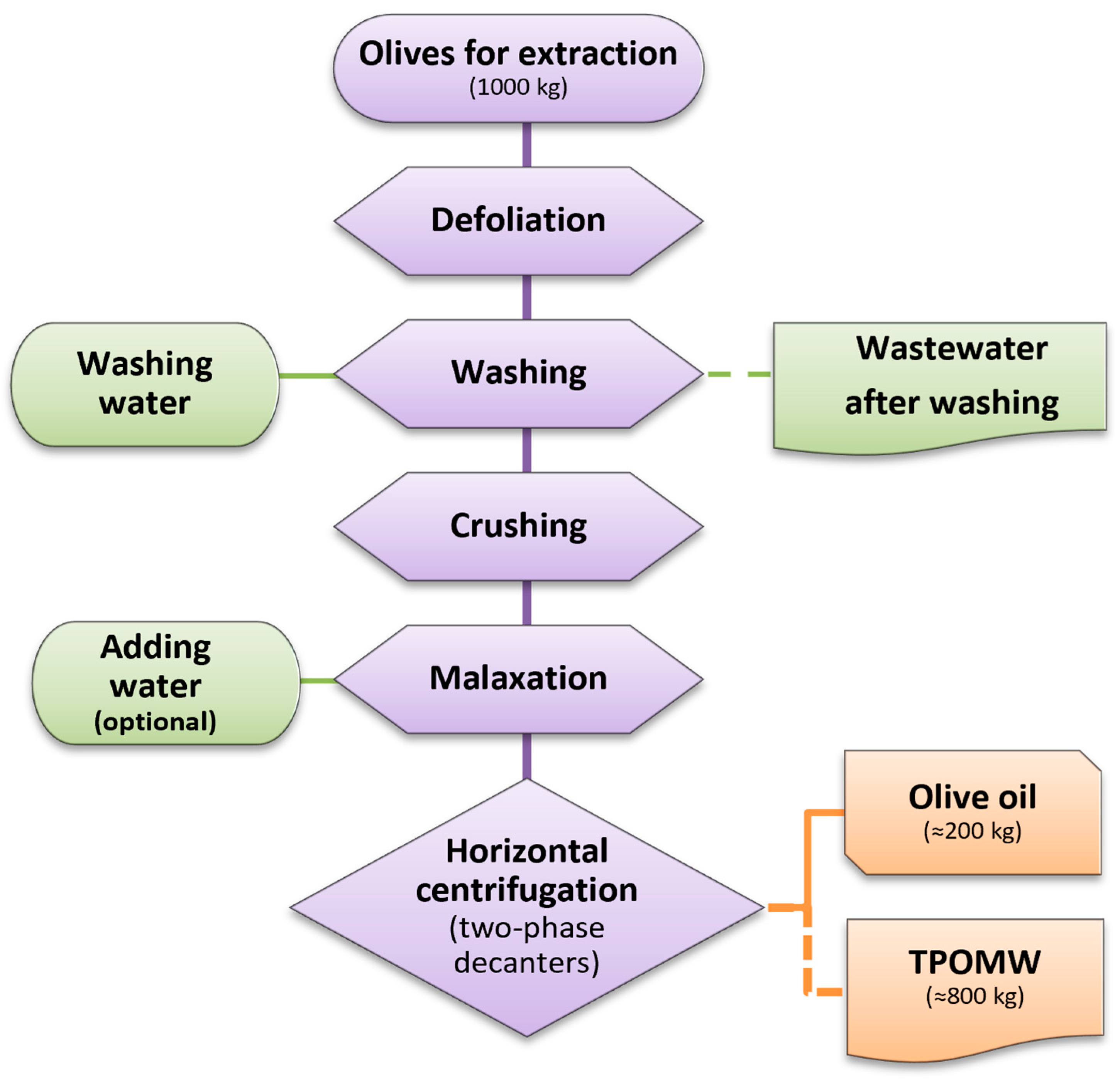
Figure 1. Schematic representation of the two-phase olive oil extraction process and the generation of two-phase olive mill waste (TPOMW). Waste streams, including wastewater from olive washing and TPOMW, are depicted by dashed lines.
Jof 11 00687 g001
Figure 2. Representative fungal colonies (growing on PDA at 25 °C for 2 to 5 days) derived from isolates obtained from TPOMW. Strain codes are given in parentheses by omitting the common-to-all prefix “LGAM SOW_”. (a,b) Aspergillus novoparasiticus (T2, T1); (c,d) A. fumigatus (A3); (e,f) A. welwitschiae (A4, A8); (g) A. tubingensis (A6); (h) Penicillium crustosum (P3); (i) P. kongii (PT5); (j) P. paneum (M15); (k) P. roqueforti (composition of cultures OM122 and OM132); (l,m) Talaromyces nanjingensis (M13); (n) Beauveria pseudobassiana (BF1); (o) Sarocladium kiliense (M10); (p) Pleurostoma richardsiae (M3); (q,r) Trichoderma olivarum (MF1a); (s,t) T. amurcicola (MF2a); (u) Cladosporium cladosporioides (M4); (v) C. ramotenellum (PT4); (w) C. limoniforme (OM52); (x) Neocucurbitaria keratinophila (M4a); (y) Stagonosporopsis ailanthicola (OM34); (z,aa) Geotrichum candidum (OM5, GZ3); (ab) Pseudophlebia setulosa (PT3N); (ac) Fuscoporia ferrea (M9a); (ad,ae) Mucor racemosus (Z1); (af,ag) M. circinelloides (Z3); (ah,ai) M. pseudolusitanicus (Z7).
Jof 11 00687 g002
Figure 3. Isolation conditions of the culturable fungal diversity in TPOMW: (a) origin of isolates across various substrates (CEA, XEA, LEA, PDA-RB, YPDA), including selective factors (RB: RBBR; EC10/EC25; Table 1). (b) Origin of isolates based on inoculation techniques (SD: serial dilution; EC-SD, EL-SD, EY-SD: pre-enrichment in CEA, LEA, YPDB; WU: Warcup method), and incubation temperatures (25 °C, 45 °C). The outer circle represents the number of isolates, while the inner circle indicates the number of identified species.
Jof 11 00687 g003
Figure 4. Composition of culturable fungal diversity in TPOMW: number of identified species per fungal genus (outer circle), an number of isolates per genus (inner circle). Distribution of fungal species per phylum is presented by different colors: Ascomycota (blue), Basidiomycota (purple), and Mucoromycota (green).
Jof 11 00687 g004
Figure 5. Multilocus phylogenetic tree (ITS, rpb2, tef1-α, cal, act) showing the placement of Trichoderma amurcicola sp. nov. and T. olivarum sp. nov. within the Harzianum clade (in bold blue). Type strains are depicted in bold black, and genome-sequenced strains are underlined. Branch support values are shown, where MLBS ≥ 65% and BPP ≥ 0.95; asterisks (*) denote MLBS = 100% and BPP = 1.00.
Jof 11 00687 g005
Figure 6. Trichoderma amurcicola sp. nov. (LGAM SOW_MF2a). Cultures on PDA at 25 °C after 3, 5, and 7 days ((ac), respectively), at 30 °C after 7 days (d), and on SNA at 25 °C after 7 days (e,f); conidiation: aerial hyphae and pustules on PDA (gi) and on SNA (j); conidiophores and phialides (k,l,np); chlamydospores (l,m); conidia (q). Scale bars: (gj) 0.25 mm, (kq) 10 μm.
Jof 11 00687 g006
Figure 7. Trichoderma olivarum sp. nov. (LGAM SOW_MF1a). Cultures on PDA at 25 °C after 3, 5, and 7 days ((ac), respectively), at 30 °C after 7 days (d), and on SNA after 7 days at 25 °C and 30 °C ((e,f), respectively); conidiation: aerial hyphae and pustules on PDA (g,h) and on SNA (i,j); conidiophores and phialides (k,l,n,p); chlamydospores (m,o); conidia (q). Scale bars: (gj) 0.25 mm, (kq) 10 μm.
Jof 11 00687 g007
Table 1. Identity of fungal isolates recovered from TPOMW, including species names, strain codes, number of MOTUs, and GenBank accession numbers for ITS, tef1-α, tub2, rpb2, act, and cal sequences produced in this work. The only 28S sequence generated in this study had the accession number PP748919 and was obtained from Pleurostoma richardsiae LGAM SOW_M3. Species are presented in alphabetical order per phylum, then family, and finally genus. Strain codes are provided by omitting the common-to-all prefix “LGAM SOW_”.
SpeciesStrain CodeNo. of
MOTUs
ITStef1-αtub2rpb2actcal
ASCOMYCOTA         
Aspergillus
(Aspergillaceae)
        
A. filiferA55PP766592 PP768675   
A. fumigatusA13PP766593 PP768676   
A23PP766594 PP768677   
A33PP766595PP768693PP768678   
A. novoparasiticusT11PP766596 PP768679   
T21PP766597     
A. sydowiiPT1b4 PP768694PP768680   
A. tubingensisA67PP766598     
A77PP766599 PP768681   
PT4a7PP766600     
A. welwitschiaeA46PP766601     
A86PP766602 PP768682   
PT16PP766603PP768695    
A. westerdijkiaeA92PP766604     
Penicillium
(Aspergillaceae)
        
P. crustosumM1b9PP766605PP768696    
M210PP766606     
M710PP766607PP768697    
M611PP766608 PP768683   
P111PP766609     
P212PP766610     
P312PP766611     
P413PP766612     
P513PP766613     
P. kongiiPT28PP766614     
PT58PP766615     
P. paneumM814PP766616PP768698    
M1514PP766617     
P. roquefortiM515PP766618PP768699PP768684   
M1215PP766619PP768700    
M1616PP766620     
M16a16PP766621     
M16b16PP766622     
M16c16PP766623     
OM2717PP766624     
OM3517PP766625 PP768685   
OM5918PP766626     
OM7318PP766627     
OM12219PP766628     
OM12519PP766629     
OM13220PP766630     
OM14720PP766631     
Cladosporium
(Cladosporiaceae)
        
C. cladosporioidesM427PP766643PP768710  PP768725 
C. limoniformeOM5228PP766644 PP768690 PP768726 
C. ramotenellumPT1a29PP766645 PP768691   
PT429PP766646PP768711PP768692 PP768727 
Beauveria
(Cordycipitaceae)
        
B. pseudobassianaBF124PP766639PP768706    
Neocucurbitaria
(Cucurbitariaceae)
        
N. keratinophilaM4a30PP766647PP768712    
Candida
(Debaryomycetaceae)
        
C. boidiniiY1a32PP766649     
Y233PP766650     
Y333PP766651     
Y834PP766652     
Y934PP766653     
Y535PP766654     
Y636PP766655     
Y737PP766656     
Y1038PP766657PP768713    
Y1139PP766658     
Y1240PP766659     
Y1441PP766660     
Stagonosporopsis
(Didymellaceae)
        
S. ailanthicolaOM3431PP766648     
Geotrichum
(Dipodascaceae)
        
G. candidumOM543PP766662     
OM5843PP766663     
OM6243PP766664     
OM8443PP766665     
GZ344PP766666     
G145PP766667     
G245PP766668     
Trichoderma
(Hypocreaceae)
        
T. amurcicola sp. nov.MF2a23PP766637PP768704 PP768717PP768723PP768730
MF2b23PP766638PP768705 PP768718PP768724PP768731
T. olivarum sp. nov.MF1a22PP766635PP768702 PP768715PP768721PP768728
MF1b22PP766636PP768703 PP768716PP768722PP768729
Pleurostoma
(Pleurostomataceae)
        
P. richardsiaeM326PP766641PP768708PP768688PP768719  
M3a26PP766642PP768709PP768689PP768720  
Barnettozyma
(Phaffomycetaceae)
        
B. californicaY442PP766661PP768714    
Sarocladium
(Sarocladiaceae)
        
S. kilienseM1025PP766640PP768707    
Talaromyces
(Trichocomaceae)
        
T. nanjingensisM1321PP766632PP768701PP768686   
Μ13a21PP766633 PP768687   
Μ13c21PP766634     
BASIDIOMYCOTA        
Fuscoporia
(Hymenochaetaceae)
        
F. ferreaM9a48PP766673     
Pseudophlebia
(Meruliaceae)
        
P. setulosaOM10646PP766669     
OM14146PP766670     
PT3N47PP766671     
PT5N47PP766672     
Peniophora
(Peniophoraceae)
        
P. lyciiΜ1149PP766676     
M1449PP766677     
MUCOROMYCOTA        
Mucor
(Mucoraceae)
        
M. circinelloidesZ354PP766678     
M. pseudolusitanicusZ455PP766679     
Z655PP766680     
Z756PP766681     
Z950PP766682     
M. racemosusZ151PP766683     
Z551PP766684     
Z252PP766685     
Z853PP766686     
Table 2. Enzyme indices (EIs) of selected fungal strains isolated from TPOMW on various substrates. Enzymatic activities were evaluated on cellulose-enriched agar (CEA), xylan-enriched agar (XEA), lignin-enriched agar (LEA), potato dextrose agar supplemented with guaiacol (PDA-G), and minimal salts agar supplemented with Remazol Brilliant Blue R (MS-RB), and were expressed in terms of semi-quantitative value categories ranging from no activity [depicted as ‘0’] to high activity [depicted as ‘3’]; value categories are placed at the base of this table. Columns indicate the fungal species identified, the number of strains tested vs. the number of strains isolated per species, the isolation conditions, e.g., substrate type [CEA, LEA, PDA, YPDA, with or without RBBR (-RB) or Econazole at 10 and 25 mg L−1 (-EC10 or -EC25)], inoculation method (SD: serial dilution; EC-SD, EL-SD, EY-SD: pre-enrichment with CEM, LEM, or YPDB followed by serial dilution; WU: modified Warcup plating), temperature (25 °C or 45 °C), and EI values on the five tested media. Strains with activity in all examined assays are presented in bold; species are presented in alphabetical order per phylum, then family, and finally genus (as in Table 1).
SpeciesStrain (Tested/Total)Isolation ConditionsCEAXEALEAPDA-GMS-RB
Ascomycota        
Aspergillus filifer1/1PDA/EC-SD/2511001
Aspergillus fumigatus2/3CEA, LEA/WU/4511000
Aspergillus novoparasiticus2/2PDA/EL-SD/25, 451–22000–1
Aspergillus sydowii1/1CEA/SD/2531010
Aspergillus tubingensis2/3PDA, CEA/EC-SD, EL-SD, SD/2521001
Aspergillus welwitschiae2/3PDA-RB/EC-SD, EL-SD, SD/25, 4501001
Aspergillus westerdijkiae1/1PDA/EL-SD/2512001
Penicillium crustosum5/9CEA, CEA-EC25, LEA/WU, EL-SD, SD/251–31–2000–1
Penicillium kongii2/2CEA/SD/252–31001
Penicillium paneum2/2YPDA-EC25, LEA/WU/2532001
Penicillium roqueforti9/14LEA, CEA, PDA/SD, EL-SD, EY-SD, WU/251–31000–2
Cladosporium cladosporioides1/1LEA/EL-SD/2522001
Cladosporium limoniforme1/1CEA/SD/2521322
Cladosporium ramotenellum2/2CEA/SD/252–31–2332
Beauveria pseudobassiana1/1CEA/SD/2533033
Neocucurbitaria keratinophila1/1YPDA/SD/2523001
Candida boidinii2/12YPDA, YPDA-EC10, YPDA-EC25, CEA, CEA-EC10, LEA-EC25, PDA-EC25/EC-SD, EL-SD, EY-SD, WU/2532–3003
Stagonosporopsis ailanthicola1/1CEA/SD/2500000
Geotrichum candidum6/7CEA, YPDA, PDA-RB/SD, EC-SD, EL-SD/251–33000–1
Trichoderma amurcicola sp. nov.1/2CEA/SD/2511011
Trichoderma olivarum sp. nov.1/2CEA/SD/2511000
Pleurostoma richardsiae2/2CEA, PDA/EC-SD/251–21–2011–2
Barnettozyma californica1/1CEA/EL-SD/2533002
Sarocladium kiliense1/1CEA/EL-SD/2512001
Talaromyces nanjingensis2/3YPDA-EC10, YPDA-EC25/WU, EL-SD/2511000
Basidiomycota       
Fuscoporia ferrea1/1LEA/WU/2531111
Pseudophlebia setulosa2/4CEA/SD/251–211–22–31–2
Peniophora lycii2/2CEA-EC25/WU/2513202
Mucoromycota       
Mucor circinelloides1/1LEA/SD/2512001
Mucor pseudolusitanicus4/4PDA-RB, LEA, CEA/WU, EC-SD/2511–2001
Mucor racemosus4/4CEA, LEA, PDA/WU, EC-SD/2510000–1
EI value categories are as follows: on CEA, ‘1’ for ≤1.8, ‘2’ for 1.8–2.8, and ‘3’ for ≥2.8; on XEA, ‘1’ for ≤1.5, ‘2’ for 1.5–2.0, and ‘3’ for ≥2.0; on LEA, ‘1’ for ≤1.2, ‘2’ for 1.2–1.7, and ‘3’ for ≥1.7; on PDA-G, ‘1’ for ≤1.0, ‘2’ for 1.0–1.9, and ‘3’ for ≥1.9; and on PDA-RB, ‘1’ for ≤1.3, ‘2’ for 1.3–1.9, and ‘3’ for ≥1.9.
Disclaimer/Publisher’s Note: The statements, opinions and data contained in all publications are solely those of the individual author(s) and contributor(s) and not of MDPI and/or the editor(s). MDPI and/or the editor(s) disclaim responsibility for any injury to people or property resulting from any ideas, methods, instructions or products referred to in the content.