Article

New Contributions to the Species Diversity of the Genus Hydnum (Hydnaceae, Cantharellales) in China: Four New Taxa and Newly Recorded Species

1
Engineering Research Center of Edible and Medicinal Fungi Ministry of Education, Jilin Agricultural University, Changchun 130118, China
2
College of Life Sciences, Zhejiang Normal University, Jinhua 321000, China
3
Sanjiang Fungal Industry Collaborative Innovation Center, Jilin Agricultural University, Changchun 130118, China
4
Industrial Development Institute for Plants, Animals and Fungi Integration of Biyang County, Biyang 463799, China
*
Authors to whom correspondence should be addressed.
J. Fungi 2025, 11(6), 431; https://doi.org/10.3390/jof11060431
Submission received: 16 April 2025 / Revised: 27 May 2025 / Accepted: 2 June 2025 / Published: 4 June 2025
(This article belongs to the Section Fungal Evolution, Biodiversity and Systematics)

Abstract

:
Hydnum, a well-defined genus in the family Hydnaceae (order Cantharellales), is characterized by its distinctive spine-bearing hymenophores. In this study, we performed a multi-locus phylogenetic analysis (ITS-nrLSU-tef1) of Hydnum species. Integrating morphological examinations and phylogenetic evidence, we identified and delineated five Hydnum species in China, which include four novel species (Hydnum crassipedum, H. albomarginatum, H. fulvostriatum, and H. bifurcatum) and the first record (H. orientalbidum) in Anhui Province. This study provides a comprehensive morphological description (including macroscopic morphology and microscopic structure), hand-drawn illustrations (encompassing basidiocarps, basidiospores, basidia, and pileipellis hyphae), morphological comparative analysis with similar species, and comparative phylogenetic analysis with related taxa. Furthermore, we developed a dichotomous key for identifying Hydnum species distributed in China.

1. Introduction

The genus Hydnum L. (Hydnaceae and Cantharellales), typified by H. repandum L. [1], is microscopically characterized by a monomitic hyphal system, nodose-septate hyphae, 2–6-sterigmate basidia, and subglobose to obovoid-elliptical, smooth, subhyaline, thin-walled basidiospores [2,3]. With transcontinental distribution spanning Asia, Australia, Europe, and North America, species within this genus have been both collected and utilized by humans for centuries [2,4]. Notably, beyond serving as a culinary resource, they establish ectomycorrhizal symbioses with plants in Fagales and Pinaceae, contributing significantly to forest ecosystem stabilization [2].
Linnaeus established the genus Hydnum in 1753, which initially encompassed all fungi with hydnoid hymenophores [5]. Molecular phylogenetic analyses later revealed that the hydnoid hymenophore evolved independently in distantly related lineages [6], leading to the transfer of many former Hydnum species to other genera such as Hericium, Phellodon, and Sarcodon [5,6,7]. Early morphology-based studies underestimated Hydnum diversity [8,9,10,11] due to extensive intraspecific morphological overlaps, particularly within the H. rufescens complex [1,7]. Advancements in molecular systematic studies, however, have progressively revealed greater species richness in this genus: Feng et al. [12] identified 31 Hydnum species through multilocus analyses (ITS, RPB1, and TEF1α), delineating four major evolutionary lineages (Albomagnum, Vesterholtii, Rufescens, and Repandum). Subsequently, Niskanen et al. [1] elevated the species count to 49 via integrative taxonomy, constructing a hierarchical framework with four subgenera (Alba, Hydnum, Pallida, and Rufescentia), four sections (Hydnum, Olympica, Magnorufescentia, and Rufescentia), and three subsections (Mulsicoloria, Rufescentia, and Tenuiformia). Building on this, Cao et al. [6] proposed six phylogenetically distinct clades within Hydnum: Rufescentia, Hydnum, Pallida, Brevispina, Alba, and Alba sensu lato (s.l.). Collectively, these advances highlight the pivotal role of integrative taxonomy—systematically combining molecular phylogenetics with morphological traits—in resolving taxonomic ambiguities and uncovering hidden biodiversity across Hydnum and its related genera.
The genus Hydnum exhibits remarkable species diversity, with approximately 60 species recognized worldwide [8,13,14,15,16,17], among which 32 have been described as novel species from Europe and North America. In recent years, research on Hydnum diversity in China has progressed rapidly. Prior to 2016, only three species—H. repandum, H. repandum var. album, and H. rufescens—had been reported in China [18,19]. In 2016, Feng et al. [12] conducted molecular phylogenetic studies on Chinese Hydnum species, which revealed substantial diversity within this genus. Subsequently, Cao et al. [6] performed a phylogenetic investigation of Cantharellales in China and described 10 new Hydnum species. To date, 29 Hydnum species have been confirmed to be distributed in China [6,13,20,21,22]. Previous studies have demonstrated that species within the genus Hydnum are predominantly distributed in temperate regions globally, while subtropical areas remain significantly underrepresented in taxonomic records [1,2,7,12,14,23]. This disparity is particularly evident in the Dabie Mountains, a critical transitional zone between subtropical and warm-temperate ecosystems in China [24], which supports exceptional species diversity and constitutes a key biodiversity conservation area [25]. Despite its ecological significance, Hydnum species distribution data in this region remain critically deficient, with no comprehensive taxonomic documentation available to date. Continued intensive field investigations in this area are expected to yield new Hydnum species discoveries, underscoring the urgency of systematic taxonomic research. This study aims to carry out the following: (i) refine the taxonomic framework of the genus Hydnum with particular emphasis on understudied subtropical ecosystems such as the Dabie Mountains and (ii) develop a diagnostic key for Chinese Hydnum species to facilitate accurate field identification and inform biodiversity conservation strategies.

2. Materials and Methods

2.1. Specimen Collecting

Fifteen specimens of Hydnum were collected during the rainy season (July to September) in Anhui (114°54′–119°37′ E, 29°41′–34°38′ N) and Jilin Province (121°38′–131°19′ E, 40°50′–46°19′ N), China, from 2019 to 2023. The photographs of the specimens were taken in situ. After thoroughly documenting the fresh macroscopic features, the specimens were placed in a drying oven at 40–45 °C. The dried specimens were deposited in the Mycology Herbarium of Jilin Agricultural University (HMJAU).

2.2. Morphological Studies

Micromorphological observations were conducted using a Carl Zeiss Axio Lab A1 compound microscope (Carl Zeiss AG, Jena, Germany). Specimens (collected from each basidiocarp: five spines, approximately 0.3 cm2 of pileipellis and 0.3 cm2 of stipitipellis) were mounted in 3% (w/v) KOH solution containing 1% (w/v) Congo Red for staining. Amyloid reactions were tested with Melzer’s reagent [1.5 g KI, 5 g I2, 20 g CCl3CH(OH)2 (dissolved in 20 mL glycerol)]. Spore dimensions (n = 30 per spine) were recorded as (a) b–c (d) (98% values within b–c), with a mean length (avg. L) and width (avg. W) calculated excluding extremes; Q denotes L/W ratio (L: spore length, W: spore width). For ultrastructural analysis [26,27,28], spores were imaged using a JEOL JSM-7900F scanning electron microscope (JEOL Ltd., Tokyo, Japan) at an accelerating voltage of 5.00 kV. Specimens were prepared via silica gel desiccation followed by gold sputter coating (45 s deposition time under a chamber pressure of <1.0 × 10³ Pa). Colorimetric parameters were calibrated according to the Methuen Handbook [29].

2.3. DNA Extraction, PCR, and Sequencing

The total genomic DNA was extracted using a NuClear Plant Genomic DNA Kit (CW0531M, CoWin Biosciences, Taizhou, China) according to the manufacturer’s instructions. The primer pairs ITS1F/ITS4 [30,31], LROR/LR7 [32], and tef1F/tef1R [12] were employed to amplify and sequence the internal transcribed spacer regions (ITSs), large subunit nuclear ribosomal RNA (nrLSU), and translation elongation factor 1 (tef1), respectively. Each PCR reaction (15 μL final volume) contained 1 μL of genomic DNA, 7.5 μL of SanTaq® PCR Master Mix (B532061-0040, Sangon Biotech, Shanghai, China), 4.5 μL of ddH2O, and 1 μL each of forward and reverse primer (10 μM). The thermal cycling protocol included an initial denaturation at 95 °C for 3 min, followed by 36 cycles at 95 °C for 40 s, 56 °C for 45 s, and 72 °C for 1.5 min, with a final extension at 72 °C for 8 min (10° C/s ramp rate). PCR products were visualized using UV transillumination after electrophoresis on 1.0% agarose gels stained with ethidium bromide and purified using a Genview High-Efficiency Agarose Gels DNA Purification Kit (Gen-View Scientific Inc., Galveston, TX, USA). Purified products were Sanger sequenced by Sangon Biotech Limited Company (Shanghai, China). Sequences were assembled using Seqman (Lasergene v.7.1, DNASTAR), and the consensus sequences were deposited in GenBank (https://www.ncbi.nlm.nih.gov/genbank/, accessed on 16 March 2025).

2.4. Phylogenetic Analyses

A multilocus (ITS-nrLSU-tef1) phylogenetic tree was constructed by integrating newly sequenced data and GenBank-derived sequences using PhyloSuite v1.2.3 [33] (Table 1). The workflow included the following: (1) sequence alignment via MAFFT implemented in PhyloSuite; (2) optimal substitution model selection through ModelFinder under the Akaike Information Criterion (HKY + I + G + F for Bayesian Inference; G4 + I + G + F for Maximum Likelihood); (3) Bayesian analysis with 15 million generations (sampling every 100 generations, 25% burn-in, and split frequency < 0.001); (4) Maximum Likelihood analysis with 1000 bootstrap replicates; and (5) topology visualization using the ITOL platform.

3. Results

3.1. Taxonomy

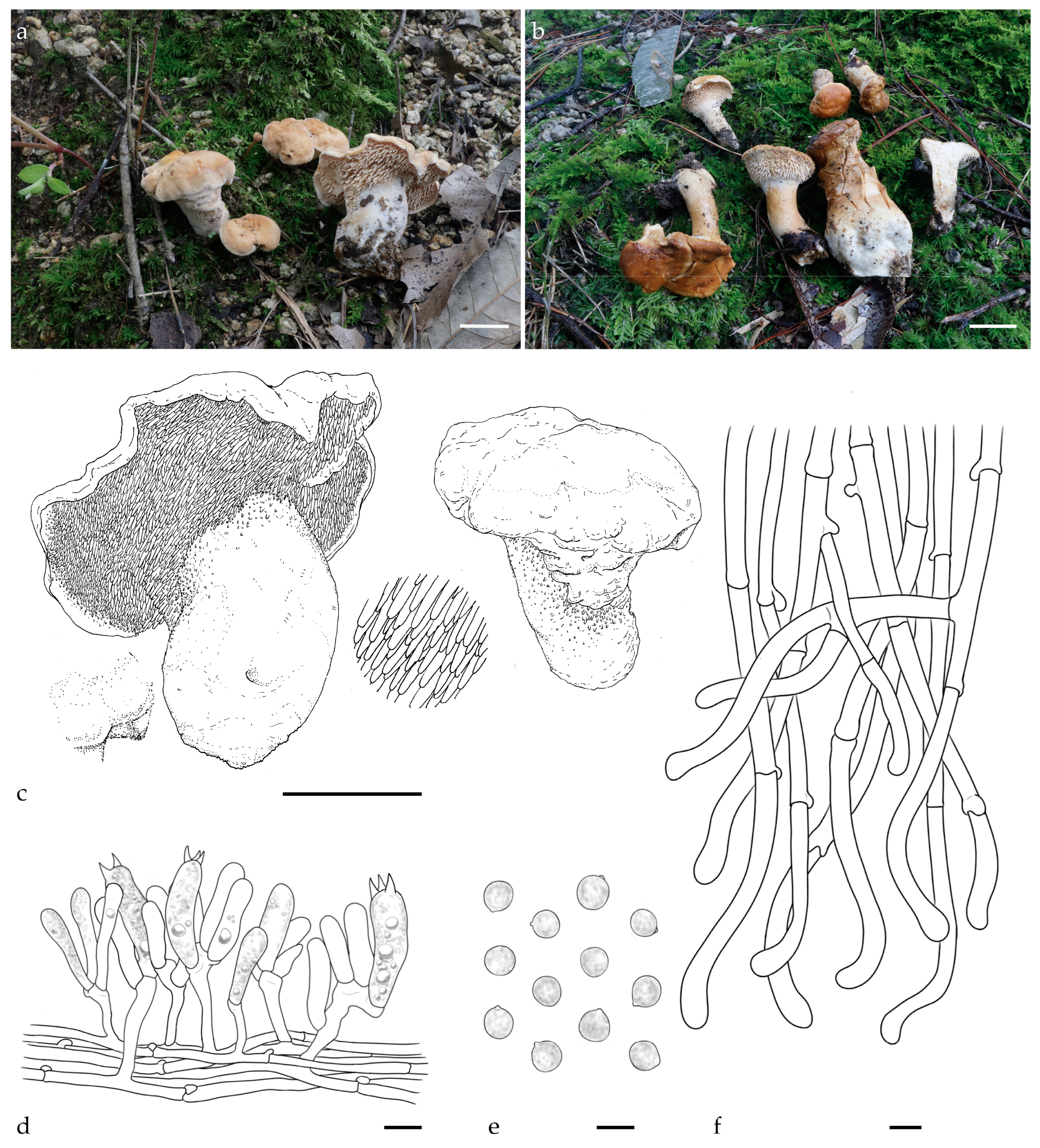
Hydnum fulvostriatum Y.L.Tuo, B. Zhang & Y. Li sp. nov.; Figure 1a–f, Figure A1 and Figure A2.
Fungal Name: FN 572487.
Etymology: The specific epithet “fulvostriatum” is derived from the Latin words fulvus (yellowish-brown) and striatus (striped), referring to the characteristic yellowish-brown zonate pattern observed on the pileus margin of this species.
Holotype: CHINA. Anhui Province, Lu’an city, Tianma National Nature Reserve, 31°13′16.34″ N, 115°49′59.49″ E, elev. 629.4 m. on soil in Quercus glauca Thunb. Forest, 6 August 2023, Yonglan Tuo (FJAU66566). GenBank accession numbers: ITS = PV329849 and LSU = PV356807.
Diagnosis: H. fulvostriatum is distinguished from other Hydnum species by a yellowish-brown zonate pattern on the pileus margin and globose to subglobose basidiospores (7.0–7.2 × 6.8–7.0 μm).
Description: Basidiocarps medium to large, 26–45 mm in height, solitary to scattered. Pileus 44–65 mm wide, yellowish white to pale orange (4A2–5A3), irregularly round, shallowly depressed in the center, covered with white to yellowish white (4A1–4A2) tomentum, surface dry; margin incurved, thin, pronounced yellow-brown zonate, discoloration not observed. Hymenophore hydnoid, adnate, not decurrent, surface orange–white (4A3); spines conical, brittle, 2–7 mm long, 0.6–1.2 mm diameter, crowded, 2–3 per mm2. Stipe central, white (4A1), cylindrical, 30–45 mm long, 8–11 mm wide, solid, surface covered with white (4A1) tomentum. Context fleshy, 5–10 mm thick, dry, discoloration not observed. Odor mild or fruity.
Basidiospores (6.8) 7.0–7.2 (8.0) × (6.5) 6.8–7.0 (7.5) μm, avg. L = 7.2 μm, avg. W = 6.9 μm, Q = 1.00–1.08 (1.17) (n = 60/4), avg. Q = 1.04, globose to subglobose, thin-walled, smooth, hyaline in 3% KOH, some with granular contents or hyaline oily droplets, hilar appendix 0.5–1.0 μm long. Basidia fusiform to suburniform, (35.0) 40.0–46.0 (50.0) × (7.5) 8.0–9.5 (10.5) μm, some with granular contents or hyaline oily droplets; sterigmata 2–4, 4.5–6.0 × 0.5–1.0 µm, conical, thin-walled, smooth. Basidioles numerous, subclavate, smaller than basidia, (20.5) 25.0–30.0 × 7.0–8.0 (9.0) μm, some with granular contents. Cystidia absent. Subhymenium trama filamentous, hyphae 3.5–4.0 μm wide, thin-walled, olive in 3% KOH. Hyphae of spines 5.0–7.0 μm, thin-walled, apex cylindrical. Pileipellis composed of cylindrical hyphae, 6.0–8.5 μm wide, subparallel, occasionally branched; cells 57.0–145.5 × 6.0–8.5 μm, yellowish in 3% KOH, terminal elements dilated at apex. Stipitipellis composed of subcylindrical hyphae, thin-walled hyphae, 6.5–9.0 μm wide. Clamps present in all tissues.
Habitat and distribution: Solitary to gregarious in Q. glauca forests; currently known only from the Dabie Mountains, China.
Additional specimens examined: CHINA. Anhui Province, Lu’an city, Tianma National Nature Reserve, 31°14′10.97″ N, 115°50′3.08″ E, elev. 688.5 m. on soil in Q. glauca forest, 16 August 2023, Yonglan Tuo (FJAU66567). GenBank accession numbers: ITS = PV329850 and LSU = PV356808.
Notes: Morphologically, H. fulvostriatum is closely related to H. subrufescens, with medium to large basidiocarps, a pale yellowish-white pileal surface, white to pale cream spines, and a white tomentum stipe. However, it can be distinguished from H. subrufescens by its smaller basidiospores (average size: 7.2 × 6.9 vs. 8.1 × 7.0 μm) and its distinctive yellowish-brown striations along the margin of the pileus.
Multigene phylogenetic analysis revealed that the sequences of H. fulvostriatum clustered together, forming a distinct lineage. This lineage was identified as a sister clade to H. roseotangerinum, but it exhibits a more distant phylogenetic relationship with H. subrufescens. Based on the morphological characteristics provided above and the phylogenetic results, H. fulvostriatum should be classified as a member of the subsect. Rufescentia.
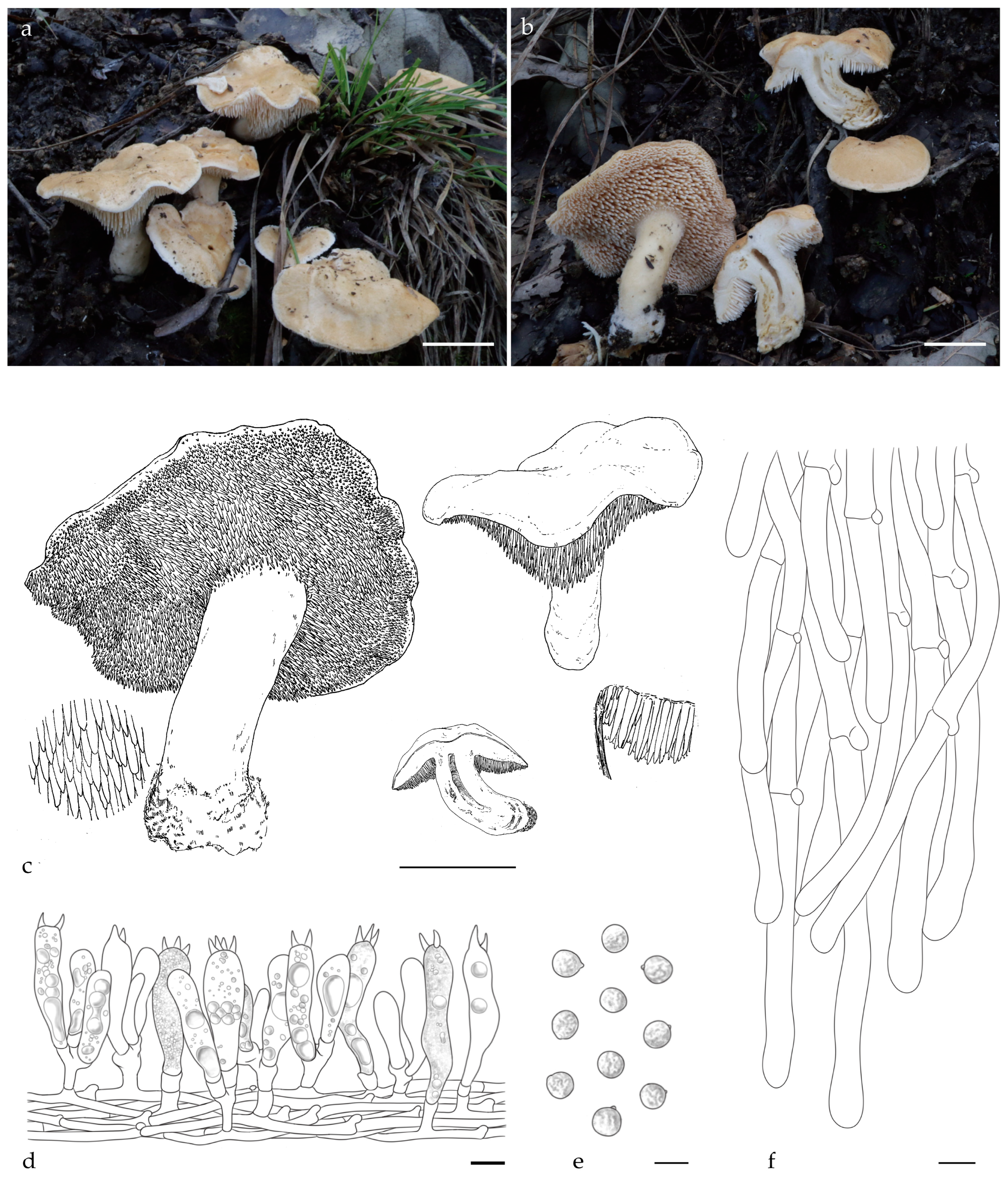
Hydnum crassipedum Y.L.Tuo, B. Zhang & Y. Li sp. nov. Figure 2a–f, Figure A1 and Figure A2.
Fungal Name: FN 572486.
Etymology: The species epithet “crassipedum” is derived from Latin crassus (thick) and pes (foot), referring to the characteristically robust stipe of this species.
Holotype: CHINA. Anhui Province, Lu’an city, Tianma National Nature Reserve, 31°20′1.13″ N, 115°54′6.53″ E, elev. 636.1 m. on the ground of Quercus variabilis Blume forest, 28 September 2023, Yonglan Tuo (FJAU66572). GenBank accession numbers: ITS = PV329853, tef1 = PP357260, and LSU = PV356811.
Diagnosis: H. crassipedum can be distinguished from other Hydnum species by its thicker stipe (15–25 mm), brownish-yellow tomentum pileus surface, and subglobose to broadly ellipsoid basidiospores measuring 8.0–8.5 × 7.0–7.5 µm.
Description: Basidiocarps medium, 32–45 mm in height, fleshy, and usually gregarious. Pileus 24–56 mm wide, yellowish-white to pale orange (4A2–5A3) when fresh, irregularly round, plano-convex when young, center shallowly depressed in age, discoloration not observed. Margin incurved, covered with white to pale orange (4A1–5A3) tomentum when young. Context 6–12 mm thick, white (4A1), odor mild or slightly sweet, discoloration not observed. Hymenophore spinose, decurrent, surface white to yellowish white (4A1–4A2), shorter near the pileus margin, cylindrical, slightly pointed tip, 2–5 mm long, 0.5–1 mm diameter, sparse, 1–2 per mm2. Stipe central or eccentric, white (4A1), 21–45 mm long, 15–25 mm wide, subcylindrical, slightly enlarged downwards, solid, covered with white (4A1) tomentum, discoloration not observed. Odor mild or fruity.
Basidiospores (7.0) 8.0–8.5 (9.0) × (6.5) 7.0–7.5 (8.0) µm (n = 60/2), avg. L = 8.0 µm, avg. W = 6.99 µm, Q = 1.11–1.17 (1.23), avg. Q = 1.15, subglobose to broadly ellipsoid, smooth, thin-walled, hyaline in 5% KOH, with finely granulose contents, hilar appendix 0.5–1 μm long. Basidia subcylindric or subclavate, (36.0) 37.5–45.5 × 9.0–9.5 (11.0) µm, sometimes with finely granulose contents; sterigmata 2–4, up to 4.5–5.5 μm long. Basidioles numerous, subclavate, smaller than basidia, 32.0–35.0 × 8.0–10.0 μm. Cystidia absent. Subhymenium trama filamentous, 3.0–4.0 μm wide, cylindrical, thin-walled, subparallel, pale yellow in 3% KOH. Hyphae of spines 5.0–7.0 µm wide. Pileipellis composed of cylindrical hyphae, 6.0–9.0 μm wide, thin walled, subparallel, rarely branched; cells, 67.0–125.0 × 6.0–9.0 μm, terminal elements rounded at apex. Stipitipellis composed of cylindrical hyphae, thin walled, 4.0–4.5 μm wide, terminal elements rounded at apex. Clamps present in all tissues.
Additional specimens examined: CHINA, Anhui Province, Lu’an city, Tianma National Nature Reserve, 31°20′4.38″ N, 115°54′6.69″ E, elev. 627.5 m, on the ground of Q. variabilis forest, 15 September 2023, Yonglan Tuo (FJAU66573). GenBank accession numbers: ITS = PV329854, tef1 = PP357261, and LSU = PV356812.
Habitat and distribution: Growing gregariously in Q. variabilis forest. Currently known only from the Dabie Mountains, China.
Notes: Morphologically, H. crassipedum is similar to H. erectum. However, the stipe of H. crassipedum is thicker (15–25 vs. 1.2–1.5 mm), and its basidiospores are larger (average 8.0 × 6.99 vs. 7.67 × 7.09 µm).
Phylogenetic analysis revealed that H. crassipedum forms a distinct lineage and is closely related to H. roseotangerinum. Based on the morphological characteristics described above and the phylogenetic results, H. crassipedum should be classified as a member of subg. Rufescentia.
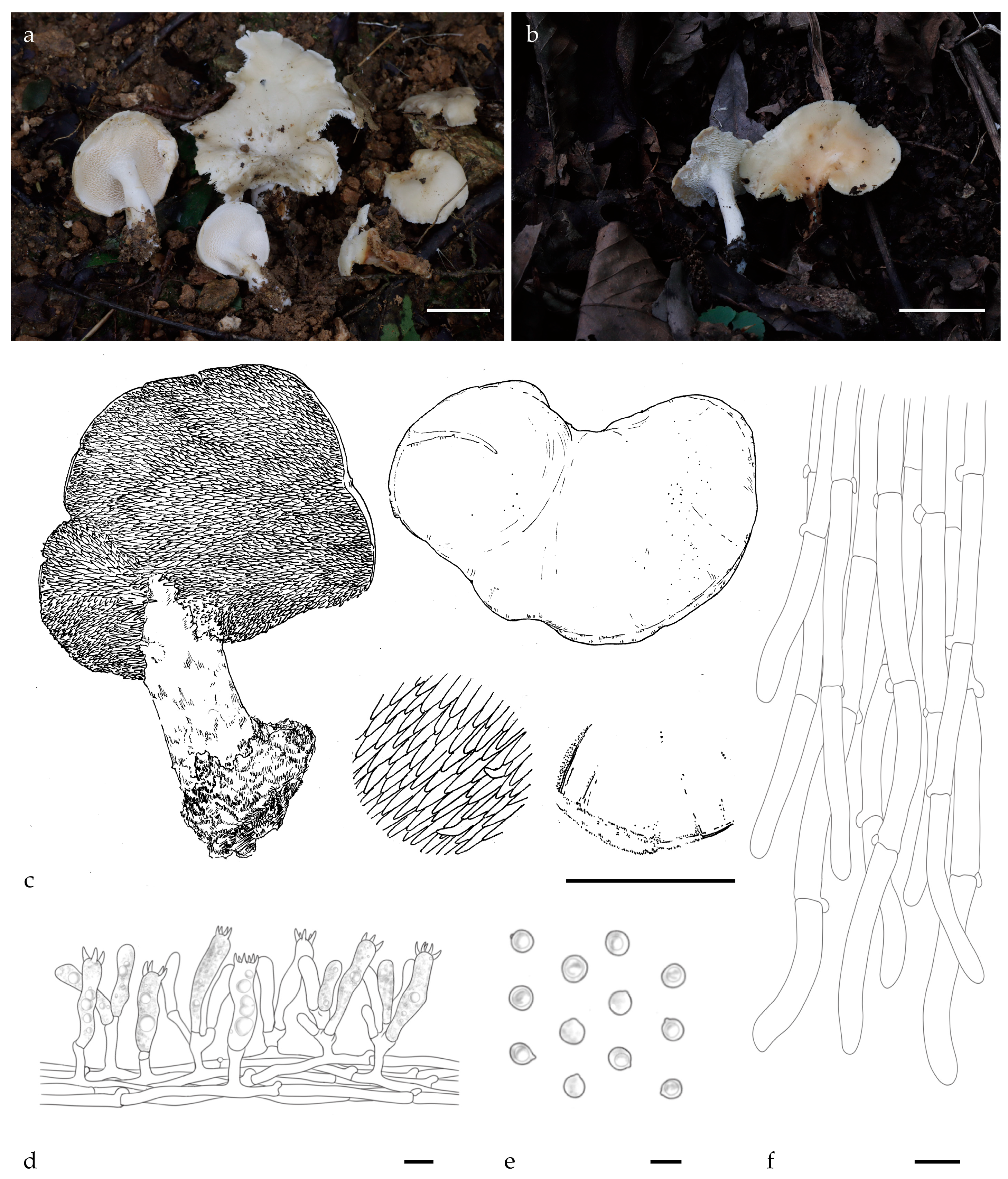
Hydnum albomarginatum Y.L.Tuo, B. Zhang & Y. Li sp. nov.; Figure 3a–f, Figure A1 and Figure A2.
Fungal Name: FN 572488.
Etymology: The specific epithet “albomarginatum” is derived from Latin albus (white) and marginatus (edged), referring to the distinctive white marginal zone characteristic of this species.
Holotype: CHINA. Anhui Province, Lu’an city, Tianma National Nature Reserve, 31°18′58.24″ N, 115°55′32.23″ E, elev. 751.2 m. on the ground of Quercus serrata Thunb. forest, 22 October 2023, Yonglan Tuo (FJAU66574). GenBank accession numbers: ITS = PV329855, tef1 = PP357262, and LSU = PV356813.
Diagnosis: H. albomarginatum differs from other Hydnum species by having a white tomentum along the margin of the pileus, forming characteristic white striations, and globose basidiospores measuring 8.0–8.5 × 7.5–8.0 μm.
Description: Basidiocarps small to medium, fleshy, 30–45 mm in height, usually gregarious. Pileus 23–66 mm wide, covered with yellowish white (4A2) tomentum, irregularly round, shallowly depressed in the center; margin incurved, and covered white (4A1) tomentum, forming characteristic white zonate, discoloration not observed. Hymenophore hydnoid, spines not decurrent, conic, orange–white (4A3), 2–6 mm long, 0.5–1.0 mm diameter, sparse, 1–2 per mm2. Stipe 26–50× 8–17 mm, central, cylindrical, hollow, slightly enlarged at the base, white to orange–white (4A1–4A3), surface covered white (4A1) tomentum. Context 5–10 mm thick, dry, fleshy. Odor mild or fruity. Discoloration not observed.
Basidiospores (7.0) 8.0–8.5 (9.0) × (7.0) 7.5–8.0 (8.5) μm (n = 60/2), avg. L =8.07 μm, avg. W = 7.92 μm, Q = 1.00–1.09 (1.18), avg. Q = 1.01, globose, thin-walled, smooth, hyaline in 3% KOH, some with granular contents or hyaline oily droplets; hilar appendix 0.2–0.5 μm long. Basidia clavate to suburniform, (30.5) 33.5–40.0 (42.5) × (9.0) 10.0–10.5 (11.0) μm, some with granular contents or hyaline oily droplets; sterigmata 2–4, 5.0–6.5 × 1.5–2.0 µm, conical, thin-walled, and smooth. Basidioles numerous, subclavate, smaller than basidia, (22.0) 25.0–30.0 (31.0) × (7.0) 8.0–9.0 (11.0) μm, some with granular contents. Cystidia absent. Subhymenium trama filamentous, hyphae 4.0–6.0 μm wide, thin-walled, olive in 3% KOH. Hyphae of spines 6.5–8.0 μm, thin-walled, apex cylindrical. Pileipellis, subparallel to slightly interwoven; cells 40.0–160.0 × 5.0–8.0 μm, terminal elements conical at apex. Stipitipellis composed of cylindrical hyphae, slightly interwoven, 6.0–7.0 μm wide, terminal elements rounded at apex. Clamp connections present.
Habitat and distribution: Growing solitarily or gregariously in Q. serrata forest. Currently known only from the Dabie Mountains, China.
Additional specimens examined: CHINA, Anhui Province, Lu’an city, Tianma National Nature Reserve, 31°18′50.13″ N, 115°55′32.96″ E, elev. 764.5 m, 11 October 2023 on the ground of Q. serrata forest, Yonglan Tuo (FJAU66575). GenBank accession numbers: ITS = PV329856, tef1 = PP357263, and LSU = PV356814.
Notes: Morphologically, although H. albomarginatum and H. berkeleyanum both exhibit smaller pileus diameters and globose basidiospores, they can be distinguished by the smaller basidiospore size of the former (8.07 × 7.92 vs. 8.5 × 7.95 µm). Additionally, H. albomarginatum features a yellowish pileus with distinct white striations along the margin, whereas H. berkeleyanum typically displays a pale orange to ochraceous-brown pileus lacking zonate patterns.
Phylogenetic analysis showed that H. albomarginatum forms a distinct lineage. This lineage exhibits a more distant phylogenetic divergence with H. berkeleyanum. Based on the morphological characteristics given above and the phylogenetic results, H. albomarginatum should be classified as a member of the subsect. Rufescentia.
Hydnum bifurcatum Y.L.Tuo, B. Zhang & Y. Li sp. nov.; Figure 4a–f, Figure A1 and Figure A2.
Fungal Name: FN572475.
Etymology: The specific epithet “bifurcatum” derives from Latin bi- (two) and furca (fork), referring to the species’ characteristic bifurcated hymenial spines.
Holotype: CHINA. Jilin Province, Ji’an city, Wunvfeng National Forest Park, 41°16′40″ N, 126°7′5″ E, elev. 810 m, on the ground of Quercus mongolica Fischer ex Ledebour forest, 12 September 2019, Yonglan Tuo (FJAU66562). GenBank accession numbers: ITS = PV329845 and tef1 = PP357252.
Diagnosis: H. bifurcatum differs from other Hydnum species by its larger pileus (65–135 mm), larger basidiospores (8.5–9.5 × 8–9.5 µm), and bifurcated spines.
Description: Basidiocarps medium to large, 70–110 mm in height, solitary or gregarious. Pileus 65–135 mm wide, orange–white to pale orange (4A3–5A3), irregularly round, surface convex to depressed, discoloration not observed; margin incurved, covered with white (4A1) tomentum when young. Context 6–15 mm thick, white (4A1), brittle, odor mild or slightly sweet, discoloration not observed. Hymenophore spinose, adnate, decurrent; spines, subulate, orange–white (4A3), shorter near the pileus margin, 2–7 mm long, 0.6–1.2 mm diameter, sparse, 1–2 per mm2. Stipe central, 30–50 mm long, 15–40 mm wide, subcylindrical, slightly enlarged downwards, solid; surface covered white (4A1) tomentum.
Basidiospores (8.0) 8.5–9.5 (10.0) × (7.5) 8.0–9.5 (10.0) µm, avg. L = 9.12 µm, avg. W = 8.97 µm, Q = 1.00–1.06 (1.11), avg. Q = 1.01, globose, smooth, thin-walled, hyaline, some with granular contents; hilar appendix 0.5–1.0 μm long. Basidia 50.0–60.0 × 10.0–11.0 µm, clavate, sometimes with finely granulose contents; sterigmata up to 4.0–5.0 µm long, 2–4 sterigmata. Basidioles numerous, subcylindrical or subclavate, smaller than basidia, 40.0–52.0 × 8.0–10.0 μm. Cystidia absent. Subhymenium trama filamentous, 3.0–4.0 μm wide, subcylindrical, thin-walled, subparallel, rarely branched hyphae, extend along the surface of the spines. Hyphae of spines 6.0–10.0 µm wide, thin-walled, hyaline in 5% KOH. Pileipellis composed of cylindrical hyphae, 8–10 μm wide, thin-walled, densely interwoven to subparallel, rarely branched; cells, 80.0–170.0 × 8.0–11.5 μm, terminal slightly dilated at apex. Stipitipellis composed of subcylindrical hyphae, thin-walled, 4.0–7.0 μm wide. Clamp connections present.
Additional specimens examined: CHINA. Jilin Province, Ji’an city, Wunvfeng National Forest Park, 41°16′33″ N, 126°7′3″ E, elev. 856 m, on the ground of Q. mongolica forest, 16 September 2020, Yonglan Tuo (FJAU66563). GenBank accession numbers: ITS = PV329846 and tef1 = PP357253.
Habitat and distribution: Occurs solitarily in Q. mongolica forest; currently known only from Jilin Province, China.
Notes: Morphologically, H. bifurcatum can easily be misidentified as H. tomaense. However, H. bifurcatum is distinguished by its bifurcated spines, larger basidiocarps (65–135 vs. 40–100 mm), and larger basidiospores (avg. = 9.12 × 8.97 vs. 7.5–8.5 × 7–8 µm). Notably, bifurcated spines are rare among Hydnum species.
The multilocus phylogenetic analyses revealed that the two Chinese H. bifurcatum specimens form a monophyletic clade, exhibiting close phylogenetic relationships with H. tomaense, H. treui, and H. zongolicense. Based on the morphological characteristics described earlier and these phylogenetic findings, we propose that H. bifurcatum should be classified within subg. Alba s.l.
Hydnum orientalbidum R. Sugaw. & N. Endo.; Figure 5a–f, Figure A1 and Figure A2.
Basidiocarps small to medium sized, solitary to scattered. Pileus 30–45 mm wide, convex to subapplanate, smooth, azonate, white to yellowish white (4A1–4A2), and covered white (4A1) tomentum. Context 2–3 mm thick, white (4A1), discoloration not observed when exposed. Hymenophore hydnoid, spines fleshy, non-decurrent, subulate, surface white (4A1), 1–3 mm long, 0.25–0.5 mm diameter, crowded, 2–4 pre mm2. Stipe central to slightly eccentric, white (4A1), 20–35 mm long, 8–12 mm wide, subcylindrical, solid, basal mycelium white.
Basidiospores (4.0) 4.5–5 (6.0) × (4.0) 4.5–5 (6.0) μm, avg. L = 4.6 μm, avg. W = 4.4 μm, Q = 1.0–1.11 (n = 60/2), avg. Q = 1.05, globose, thin-walled, smooth, hyaline in 3% KOH, some with granular contents; hilar appendix 0.5 μm long. Basidia clavate to suburniform, (30.0) 32.5–38.5 (44.5)× (6.0) 6.5–7.5 (9.0) μm, some with granular contents and hyaline oily droplets, sterigmata 2–6, 3.5–4.0 × 2.0–6.5 µm, conical, thin-walled, smooth. Basidioles numerous, smaller than basidia, (20.0) 25.0–30.0 (42.0) × (5.0) 6.0–7.0 (9.0) μm, some with granular contents. Cystidia absent. Subhymenium trama filamentous, hyphae 6.5–7.0 μm wide, thin-walled, hyaline in 3% KOH. Hyphae of spines 3–4 μm, thin-walled, apex cylindrical. Pileipellis composed of cylindrical hyphae, subparallel, terminal elements cylindrical at apex, 5.5–8.0 μm. Stipitipellis composed of cylindrical hyphae, slightly interwoven, 3.0–5.5 μm wide, terminal elements rounded at apex. Clamp connections present.
Habitat and distribution: The species grows either solitarily or gregariously in Q. serrata forest. Currently documented in Japan, the Province of Anhui, Sichuan, and Zhejiang provinces in China.
Specimens examined: China, Anhui Province, Lu’an city, Tianma National Nature Reserve, 31°15′39.37″ N, 115°41′47.35″ E, elev. 1129.6 m, 5 October 2023, Yonglan Tuo (FJAU66570, ITS = PV329857, tef1 = PP357258, LSU = PV356809). Anhui Province, Lu’an city, Tianma National Nature Reserve, 31°15′37.09″ N, 115°41′54.55″ E, elev. 1055.1 m, 15 October 2023, Yonglan Tuo (FJAU66571, ITS = PV329858, tef1 = PP357259, LSU = PV356810).
Notes: The Anhui specimens share consistent characteristics with previous descriptions of H. orientalbidum [7,22]; however, the habitat and distribution differ from those in the present study (Q. serrata vs. Picea glehnii forest).
Phylogenetic analyses based on multilocus datasets indicate that two specimens from Anhui are well nested within the H. orientalbidum clade.

3.2. Molecular Phylogeny

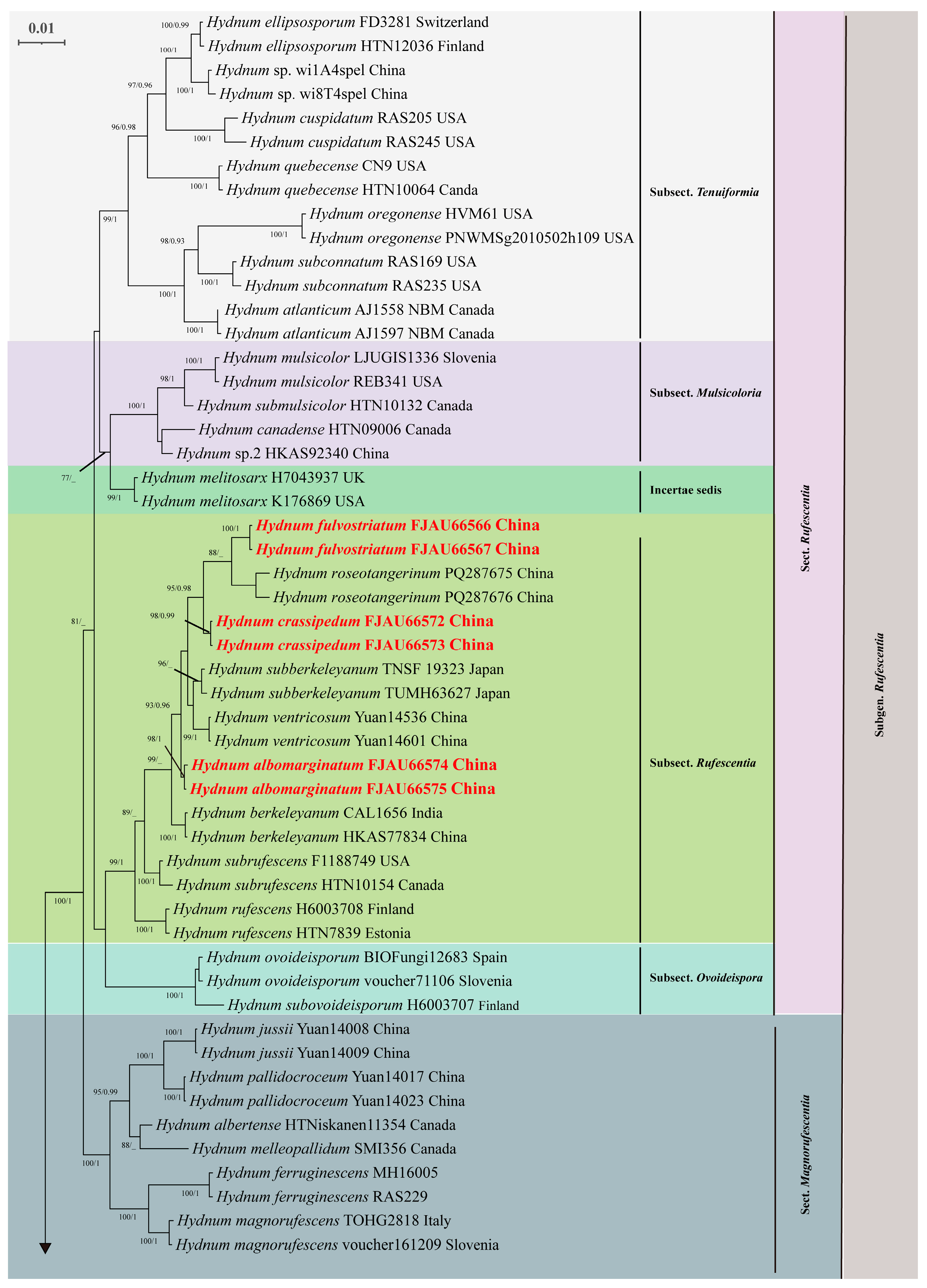
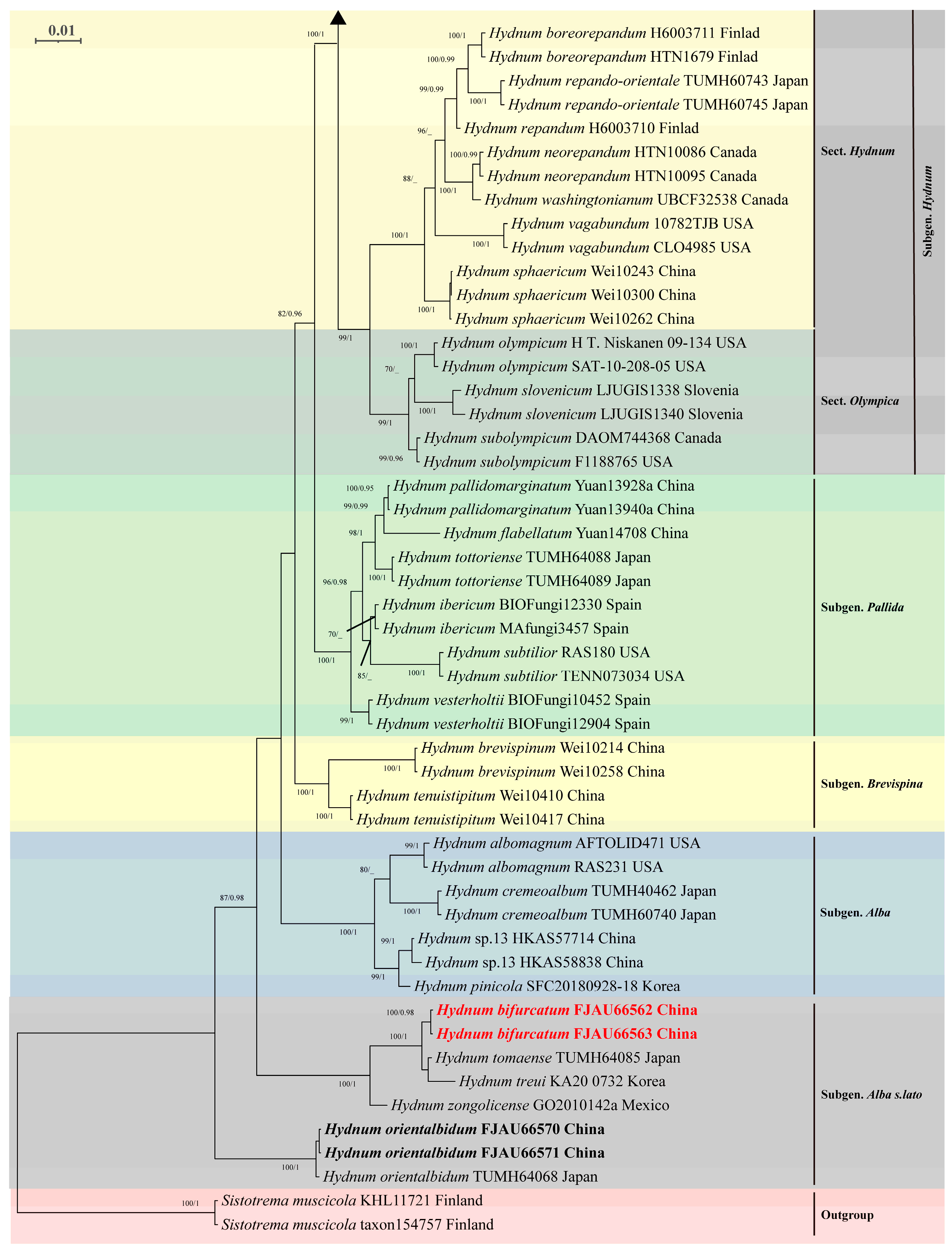
Phylogenetic analysis based on the concatenated ITS-nrLSU-tef1 dataset (comprising 170 sequences) revealed congruent topologies between Maximum Likelihood (ML) and Bayesian Inference (BI) methods (BI tree only). The resulting phylogeny was divided into six major clades: Subgenus Rufescentia, subgenus Hydnum, subgenus Pallida, subgenus Brevispina, subgenus Alba, and subgenus Alba s.l. (Figure 6 and Figure 7).
Within subg. Rufescentia, two sections were identified: sect. Magnorufescentia and sect. Rufescentia, the latter being further subdivided into four subsections (subsect. Tenuiformia, subsect. Mulsicoloria, subsect. Rufescentia, and subsect. Ovoideispora). Subg. Hydnum contained sect. Hydnum and sect. Olympica.
The newly generated sequences resolved five independent, well-supported species-level clades (PP = 0.95–1.0; BS = 98–100%). Notably, three distinct lineages (FJAU66566/FJAU66567; FJAU66572/FJAU66573; FJAU66574/FJAU66575) were resolved as subsect. Rufescentia (subg. Rufescentia). Two distinct lineages (FJAU66562/FJAU66563; FJAU66570/FJAU66571) were resolved as subg. Alba s.l.

4. Discussion

Through integrated morphological and molecular analyses, this study confirmed and characterized five Hydnum species in China, including four novel taxa (H. bifurcatum, H. crassipedum, H. albomarginatum, and H. fulvostriatum) and a newly recorded species (H. orientalbidum) from Anhui Province. Currently, 33 Hydnum species have been documented in China, 22 of which are only known so far in this country [6,12,13,21,22]. Table A1 (see Appendix B) provides a comparative summary of the key morphological characteristics and ecological information for the Hydnum species identified in China.
In our study, three Hydnum species—H. crassipedum, H. albomarginatum, and H. fulvostriatum—are characterized by small to medium-sized basidiomata that are ochraceous to orange-brown in color, with Q values ranging from 1.00 to 1.14. According to the taxonomic key established by Niskanen et al. [1] for the genus Hydnum, these species should be classified under the subsection Magnorufescentia (Q = 1.07–1.13). However, by integrating the latest phylogenetic findings from Niskanen et al. [1] and Cao et al. [6], along with newly generated sequences from this study, we demonstrate that H. crassipedum, H. albomarginatum, and H. fulvostriatum form well-supported monophyletic clades within the subsection Rufescentia. This incongruence between the morphological taxonomy and molecular phylogeny necessitates a re-evaluation of the diagnostic criteria for these subsections. Based on phylogenetic analysis and the morphological characterization of the subsection Rufescentia (including basidiocarp morphology, basidiospore shape, and size parameters), we propose that the Q-value boundary for basidiospores in the subsection Rufescentia should be expanded beyond the threshold (Q = 1.15–1.30) previously defined by Niskanen et al. [1].
Both H. bifurcatum and H. orientalbidum are classified within subg. Alba s.l., consistent with previous taxonomic studies [1,6]. However, they exhibit distinct morphological differences: H. bifurcatum possesses a comparatively larger, yellowish pileus (diameter 65–135 mm), whereas H. orientalbidum is characterized by a whitish pileus. Notably, the basidiospores of H. bifurcatum are approximately twice the size of those in H. orientalbidum (9.12 × 8.97 µm vs. 4.6 × 4.4 µm). Phylogenetic analyses further reveal that these two species form independent clades, indicating substantial genetic divergence. This evidence suggests that their current classification under the same subgenus (Alba s.l.) may require revision, potentially meriting their recognition as distinct subgenera. However, such taxonomic adjustments would require validation through comprehensive morphological comparisons.
Spore characteristics (shape, size, average length, average width, and Q value) are reliable indicators for interspecific identification among species within the genus Hydnum [8,9,10,11]. Based on these features, the new taxa described in this study can be clearly distinguished from similar species: H. bifurcatum vs. H. tomaense (average L × W = 9.12 × 8.97 vs. 7.5–8.5 × 7.0–8.0 µm); H. crassipedum vs. H. erectum (average L × W = 8.0 × 6.99 vs. 7.67 × 7.09 µm); H. fulvostriatum vs. H. subrufescens (average L × W = 7.2 × 6.9 vs. 8.1 × 7.0 µm), and H. albomarginatum vs. H. berkeleyanum (average L × W = 8.07 × 7.92 vs. 8.4 × 7.95 µm). Furthermore, the new taxa can be readily differentiated from their congeners when assessed alongside macro-morphological characteristics, including pileus size, coloration, surface texture, spine dimensions, and spine morphology [1]. For instance, H. bifurcatum can be readily distinguished from species exhibiting bifurcated spine structures through an analysis of spore morphology and dimensions. Similarly, H. fulvostriatum and H. albomarginatum exhibit distinctive brown and white tomentum annulations at the pileus margin, respectively, which serve as diagnostic characters that separate them from other species in the genus. In addition, under the scanning electron microscope (SEM), we observed that the spores of five Hydnum species exhibited nearly smooth surfaces with distinct fish scale-like ornamentations of varying depths (Appendix A: Figure A1). This feature has often been overlooked in previous studies, as nearly all prior research findings indicated that spore surfaces in the genus Hydnum are smooth [1,2,6,7,9,13,15,16,17,21,22,41]. This discrepancy is understandable, as the resolution limitations of optical microscopes may affect observation accuracy. Therefore, our findings suggest that spore ornamentation patterns could represent a significant taxonomic characteristic for species identification within the genus Hydnum. However, further validation is required to confirm their diagnostic utility.
The distribution of most Hydnum species within their host flora appears to be limited [12]. In the Northern Hemisphere, particularly in Europe and North America, these species primarily associate with plant hosts in the Pinaceae and Fagaceae families [23,42]. Our newly identified species are distributed in Quercus forest and typically emerge in early autumn in Northeast China and Anhui Province, suggesting a potential linkage to Quercus presence and seasonal factors [43,44]. Although 28 Hydnum species have been recorded in China, they are rarely reported in Quercus-dominated forests [1,6,45,46]. This scarcity may stem from two interrelated factors: on the one hand, most Quercus species have restricted distributions, especially those forming homogeneous forest communities [47,48,49]; on the other hand, recorded specimens primarily appeared 3–5 days post rainfall during autumn, likely representing their optimal growth phase [50,51,52]. Beyond this window, dry weather and soil moisture evaporation may suppress spore germination and mycelial growth [53], thereby reducing detection opportunities for ideal habitats and temporal windows.

5. Conclusions

Based on morphological observations (basidiocarps size, pileus color, basidiospores dimensions, etc.), H. crassipedum, H. albomarginatum, and H. fulvostriatum can be effectively assigned to sect. Rufescentia, whereas H. orientalbidum and H. bifurcatum are classified into subgenus Alba s.l. These taxonomic conclusions were consistently validated through molecular phylogenetic analyses. Comparative morphological analyses (basidiospores morphology and dimensions, spines characteristics, pileus zonate, etc.) further demonstrated that the five Hydnum species in this study could be clearly distinguished from related taxa within the genus Hydnum. Phylogenetic trees inferred from ITS, nrLSU, and TEF1α sequence data provided robust support for these classifications (BS = 98–100, PP = 0.98–1.0). However, inconsistencies arise in subsect. assignments: phylogenetically, H. crassipedum, H. albomarginatum, and H. fulvostriatum cluster within subsect. Rufescentia, whereas morphologically, their Q values (1.00–1.14) fall below the diagnostic threshold for the subsect. Rufescentia (1.15–1.30), but align closely with those of subsect. Magnorufescentia (Q = 1.07–1.13). To resolve this discrepancy, we propose revising the Q value criterion for subsect. Rufescentia. These findings increase the total number of Hydnum species in China to 33, 22 of which are only known so far in this country. This study expands the known species distribution in temperate regions, fills a taxonomic gap in the Dabie Mountains, and refines the classification system of Hydnum.

Key to Species of Hydnum in China

1.Basidiomata more or less white to cream yellow2
1.Basidiomata yellow to orange11
2.Pileus white3
2.Pileus cream yellow7
3.Pileus < 30 mm wideH. flavidocanum
3.Pileus > 30 mm wide4
4.Habitat in broad-leaved forests5
4.Habitat in Fagaceous forests6
5.Pileus > 60 mm wideH. cremeoalbum
5.Pileus < 60 mm wideH. orientalbidum
6.Basidiospores < 5 μm long on averageH. minus
6.Basidiospores > 5 μm long on averageH. treui
7.Pileus > 60 mm wide8
7.Pileus < 60 mm wide9
8.Habitat in broad-leaved forestsH. roseoalbum
8.Habitat in Quercus mongolica forestsH. bifurcatum
9. Spines < 3 mm long10
9. Spines > 3 mm longH. albomagnum
10.Spines pale orangeH. pinicola
10.Spines cream yellowH. minum
11.Basidiomata yellowish-white12
11.Basidiomata orange17
12.Basidiospores < 6 μm long on average13
12.Basidiospores > 6 μm long on average14
13.Habitat in angiosperm forestsH. brevispinum
13.Habitat in mixed forestsH. microcarpum
14.Basidiospores < 7.5 μm long on average15
14.Basidiospores > 7.5 μm long on average16
15.Spines 1–3 mm longH. tenuistipitum
15.Spines 2–7 mm longH. fulvostriatum
16.Habitat in Q. variabilis forestsH. crassipedum
16.Habitat in Q. serrata forestsH. albomarginatum
17.Basidiospores 7–8 μm long on average18
17.Basidiospores > 8 μm long on average19
18.Habitat in coniferous forestsH. jussii
18.Habitat in Fagaceous forestsH. erectum
19.Basidiospores 8–9 μm long on average20
19.Basidiospores > 9 μm long on average27
20.Spines white21
20.Spines orange–white or cream-yellow22
21.Spines < 3 mm longH. sphaericum
21.Spines > 3 mm longH. sinorepandum
22.Spines cream-yellowH. cremeum
22.Spines orange–white23
23.Basidiospores 8.1–8.5 μm long on average24
23. Basidiospores > 8.5 μm long on average26
24. Spines < 2 mm longH. vesterholtii
24.Spines > 2 mm long25
25.Pileus < 50 mm wideH. tangerinum
25.Pileus > 50 mm wideH. berkeleyanum
26.Basidiospores Q < 1.2H. ventricosum
26.Basidiospores Q > 1.2H. pallidomarginatum
27.Pileus < 50 mm wide28
27.Pileus > 50 mm wideH. roseotangerinum
28.Pileus orange–white29
28.Pileus light yellow31
29.Habitat in mixed forestsH. flabellatum
29.Habitat in broad forests30
30.Basidiospores > 9.5 μm long on averageH. longibasidium
30.Basidiospores < 9.5 μm long on averageH. longipes
31.Basidiospores Q > 1.3H. melitosarxm
31.Basidiospores Q < 1.332
32.Habitat in Pinus forestsH. pallidocroceum
32.Habitat in mixed forestsH. flavoquamosum.

Author Contributions

Y.-L.T.: Conceptualization, Data curation, Visualization, Writing—original draft, and Writing—review and editing. L.W.: Data curation and Software. X.-F.L.: Investigation and Software. H.C.: Data curation. M.L.: Validation and Investigation. J.H.: Conceptualization and Methodology. Z.-X.Q.: Conceptualization and Software. X.L.: Funding acquisition, review and editing. B.Z.: Conceptualization, Project administration, Resources, and Writing—review and editing; Y.L.: Funding acquisition and Project administration. All authors have read and agreed to the published version of the manuscript.

Funding

This work was supported by the National Key Research and Development Program of China (Project No 2024YFD1200204-4); The pilot selection projects in higher education institutions (No. 24GXYSZZ15); The Natural Science Foundation of Jilin Province (Project No 20240101236JC); Technology and demonstration of factory cultivation of Agaricus bisporus mushroom by high value utilization of straw and livestock manure (Project No 20230202121NC), and the “111” Program (No. D17014).

Institutional Review Board Statement

Not applicable.

Informed Consent Statement

Not applicable.

Data Availability Statement

The species registration names and gene accession numbers referenced in this study are publicly available in the following online databases: Fungal Names (https://nmdc.cn/fungalnames/) and NCBI GenBank (https://www.ncbi.nlm.nih.gov/genbank/), with the data accessed on 25 March 2025.

Acknowledgments

We gratefully acknowledge Professor Shuli Wang (Jilin Agricultural University) for providing scanning electron microscopy (SEM) imaging support. We would like to express our most sincere gratitude to the local authorities and native communities for their help in searching for pristine habitats: Jinzhai County Forestry Bureau and Tianma National Nature Reserve.

Conflicts of Interest

The authors declare that they have no known competing financial interests or personal relationships that could have appeared to influence the work reported in this paper.

Appendix A. Characteristics of Optical Microscopes and Scanning Electron Microscopes (SEMs)

Appendix A.1. Basidiospores
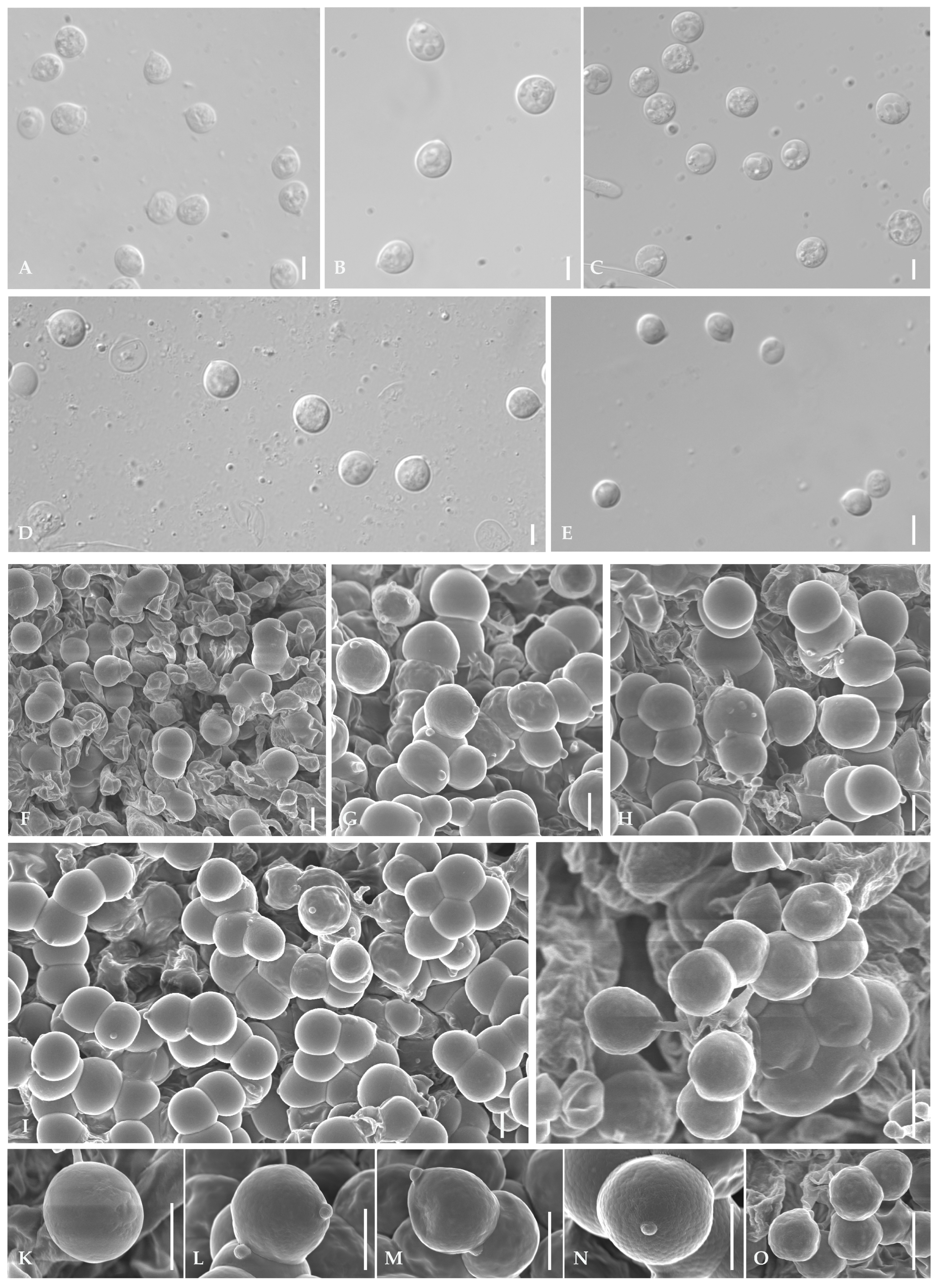
Figure A1. Microscopic features of basidiospores (AE), (A) = Hydnum fulvostriatum; (B) = H. crassipedum; (C) = H. albomarginatum; (D) = H. bifurcatum; and (E) = H. orientalbidum. Scanning Electron Microscopes features (FO), (F,K) = H. fulvostriatum; (G,L) = H. crassipedum; (H,M) = H. albomarginatum; (I,N) = H. bifurcatum; and (J,O) = H. orientalbidum. Scale bar: (AJ) = 5 μm.
Jof 11 00431 g0a1

Appendix A.2. Basidia

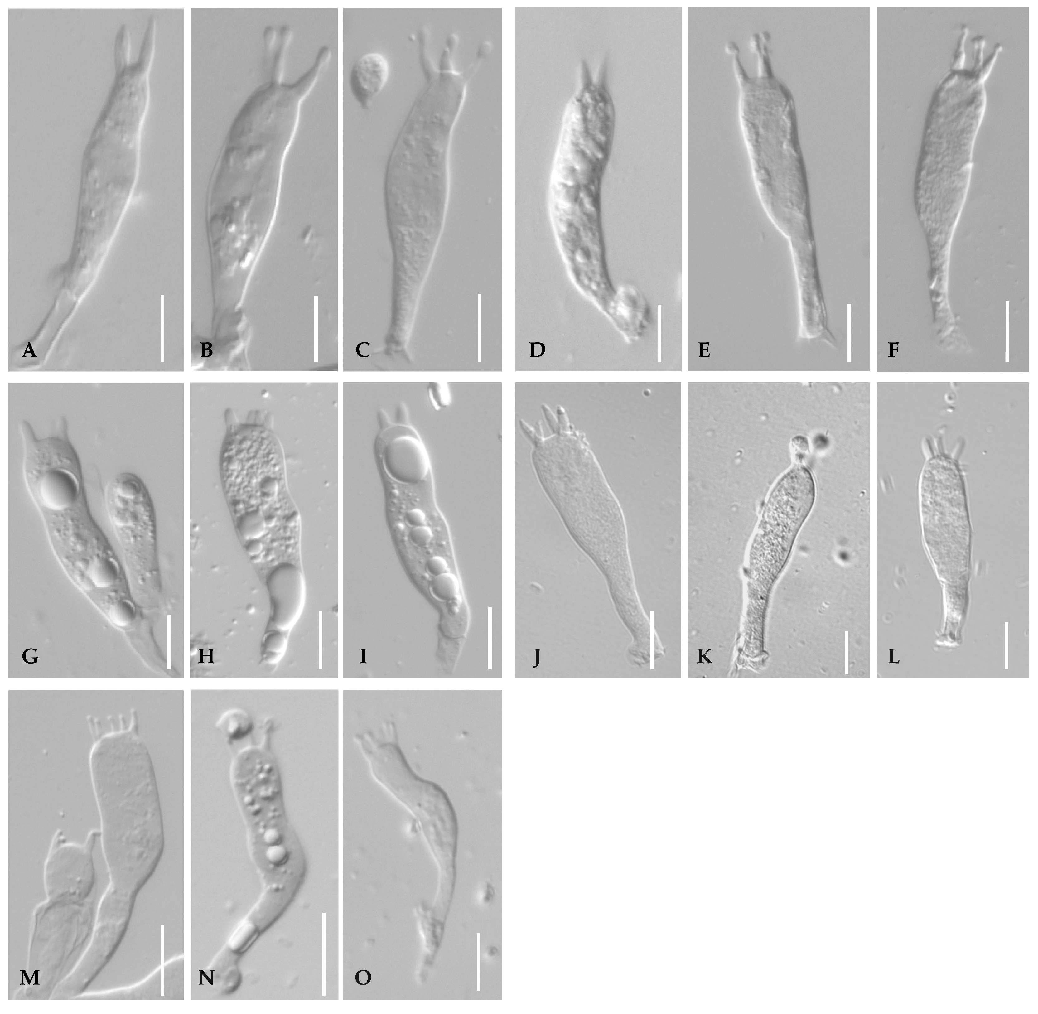
Figure A2. Microscopic features of basidia (AO), (AC) = Hydnum fulvostriatum; (DF) = H. crassipedum; (GI) = H. albomarginatum; (JL) = H. bifurcatum; and (MO) = H. orientalbidum. Scale bar: (AJ) = 5 μm.
Jof 11 00431 g0a2

Appendix B

Table A1. Morphological comparison of Hydnum species in China.
SpeciesPileusSpinesSporesHabitatReferencesDistribution
ColorSize (cm)ColorLength (mm)Size (µm)av. L×W (µm)Q
H. albomagnumCream-white to pale cream25–60Cream-white to pale cream6–12(4.6) 4.8–6.7 (7.1) × (3.5) 3.9–5.4 (5.6)avg. 5.0–6.5 × 4–5.5Q = 1.07–1.36Hardwood-oak forests[2]China
Japan
H. albomarginatumYellowish-white23–66Pale cream2–6(7.0) 8.0–8.5 (9.0) × (7.0) 8.0–8.2 (9.0)avg. 8.04 × 7.93Q = 1.00–1.09Quercus serrata forestsThis studyAnhui
China
H. berkeleyanumPale orange to light orange25–80Orange–white2–9(7.5) 8.4–8.5 (9.5) × (6.9) 7.9–8 (8.8)avg. 8.4 × 7.95Q = 1.01–1.17Mixed forests[3]India
China
H. bifurcatumPale cream65–135Yellowish-white2–7(8.0) 8.5–9.5 (10.0) × (7.5) 8.0–9.5 (10.0)avg. 9.12 × 8.97Q = 1.00–1.06Q. mongolica forestsThis studyJilin
China
H. brevispinumYellowish-white10–15Pure white0.2–0.8(4.5) 5.0–5.8 (6.0) × (3.5) 3.8–4.8 (5.0)avg. 5.28 × 4.16Q = 1.27–1.31Angiosperm forests[1]Hunan Province
H. crassipedumYellowish-white to orange–white24–56Yellowish2–5(7.0) 8.0–8.5 (9.0) × (6.5) 7.0–7.5 (6.0)avg. 8.0× 6.99Q = 1.11–1.17Q. variabilis forestsThis studyAnhui
China
H. cremeoalbumWhite to cream-white30–100Yellowish-white2–54.0–5.5 × 3.5–4.5avg. 4.73 × 3.88Q = 1.1–1.42Broad-leaved forests
Mixed forests
[4]China
Japan
H. cremeumWarm cream to yellowish-white25–37Cream-yellow4–58.1–9.5 × 7.1–9.0 (9.5)avg. 8.8 × 8.05Q = 1.00–1.11Mixed forests[5]Yunnan China
H. erectumCreamy yellow to pale salmon35–45White to pale salmon1–2(6.5) 7.33–8.0 × (5.5) 6.68–7.5 (8.0)avg. 7.67 × 7.09Q = 1.0–1.1Fagaceous forests[54]Zhejiang China
H. flabellatumYellow to grayish-yellow30–45Orange–white0.6–2(7.8) 8.5–9.5 (10.0) × (6.0) 6.5–7.8 (8.0)avg. 9.07 × 7.04Q = 1.26–1.29Mixed forests[1]Liaoning China
H. flavidocanumYellowish-white or yellowish-gray20–30Orange–white0.5–2(7.0) 7.2–8.8 (8.9) × (5.2) 5.5–6.5 (6.8)avg. 7.75 × 6.01Q = 1.29–1.31Mixed forests[1]Yunnan China
H. flavoquamosumLight yellow to light brownish-orange30–50Light yellow to light brownish-orange0.58.6–9.5 (10.0) × 7.6–8.6 (9.0)avg. 9.05 × 8.1Q = 1.05–1.23Mixed forests[5]Yunnan China
H. fulvostriatumYellowish-white44–65White2–7(6.8) 7–7.2 (8) × (6.5) 6.8–7.0 (7.5)avg. 7.2 × 6.9Q = 1.00–1.08Q. glauca forestsThis studyAnhui
China
H. jussiiMedium orange ocher35–60Whitish 7.2–8.0 × 6.6–7.5avg. 7.5 × 7.0Q = 1.03–1.18Coniferous forests[6]Finland
China
H. longibasidiumOrange–white to grayish orange10–15Orange–white to pale orange1–4(8.0) 8.5–11.0 (11.5) × (7.5) 7.8–9.8 (10.0)avg. 9.81 × 9.03Q = 1.09–1.13Angiosperm forests[1]Hunan China
H. longipesOrange, light orange20–30Orange–white to pale orange1–3(7.5) 8.0–10.0 (10.5) × (6.0) 7.0–8.5 (9.0)avg. 9.32 × 7.63Q = 1.05–1.43Q. aquifolioides forests
Pinus densata forests
[7]Yunnan China
H.melitosarxmLight orange to orange20–40Yellowish-white to pale orange2–58.0–11.0× 7.0–10.0avg. 9.37 × 8.71Q = 1.0–1.28Broad-leaved forests
Mixed forests
[6]Europe
Asia
H. microcarpumYellowish-white to orange–white10–20White to yellowish-white0.5–1.55.0–6.0 (6.5) × 5.0–5.5 (6.0)avg. 5.78 × 5.26Q = 1.0–1.2Mixed forests[7]Guangdong China
H. minumCream-yellow to pale buff10–25Cream yellow0.5–1.74.5–5.5 × 3.0–4.5avg. 4.8 × 3.8Q = 1.1–1.5Mixed forests[1]China
Japan
H. minusCream-white to cream15–25White to cream-white2–4(4.0) 4.72–5.0 × (3) 3.68–4.0avg. 4.0–5.0 × 3.0–4.0Q = 1.13–1.29Fagaceous forest[54]China
Japan
H. orientalbidumWhite30–45White1–3(4.0) 4.5–5 (6.0) × (4.0) 4.5–5.0 (6.0)avg. 4.6 × 4.4Q = 1.0–1.11Broad-leaved forests
Q. serrata forests
This studyJapan
China
H. pallidocroceumOrange–white to pale orange25–40Light yellow1–5(7.5) 7.8–9.5 (10.0) × (5.5) 6.0–7.5 (8.0)avg. 9.09 × 6.72Q = 1.32–1.35Pinus sp. forests[1]Xinjiang China
H. pallidomarginatumOrange–white to pale orange20–35Orange–white to pale orange0.5–2(8.0) 8.2–9.8 (10.0) × (6.0) 6.5–7.8 (8.2)avg. 8.75 × 6.99Q = 1.25–1.28Angiosperm forests[1]Yunnan China
H. pinicolaPale yellow to pale orange25–60Orange–white to pale orange1.5–34.0–6.0 × 3.5–5.0avg. 4.72 × 4.02Q = 1.0–1.25Mixed forests[2]China
Japan
H. roseoalbumCream to whitish65Pale orange 8.1–9.0 (9.5) × 8.1–9.0 Q = 1.00–1.06Broad-leaved forests[5]Yunnan
China
H. roseotangerinumPale brownish-orange65Yellowish-orange to pinkish-orange4–58.6–9.5 (10.0) × 7.6–9.0avg. 9.05 × 8.3Q = 1.00–1.18Mixed forests[5]Yunnan China
H. sinorepandumLight yellow to light orange40–120White to yellowish-white3–7(7.5) 8–9 (10) × 6.5–7.5avg. 8.16 × 7.12Q = 1.0–1.38Broad-leaved forests
Mixed forests
[7]Yunnan China
H. sphaericumOrange–white20–35White0.5–3(7.5) 8–8.8 (9) × (6) 6.5–7.5 (8)avg. 8.36 × 6.94Q = 1.2–1.23Angiosperm forests[1]Hunan China
H. tangerinumOrange to brownish-orange10–50Orange–white2–6(7) 7.2–8.8 (9) × (5.5)
5.8–7 (7.5)
avg. 8.11 × 6.19Q = 1.23–1.31Angiosperm forests[1]Hunan China
H. tenuistipitumYellow white to orange–white10–30Orange–white1–3(6.5) 6.8–7.2 (7.5) × (5.2) 5.5–6.5 (6.8)avg. 7.08 × 6.09Q = 1.07–1.16Angiosperm forests[1]Hunan China
H. treuiWhite to creamy white28–57White to cream-white1–4(6) 6.44–7 (7.5) × (5) 5.88–6.5 (7) Q = 1.0–1.2Fagaceous forests[54]China
Papua New Guinea
H. ventricosumOrange to brown28–35Orange–white1–5(7.5) 8.2–9 (9.5) × (7) 7.5–8.5 (9)avg. 8.64 × 8.17Q = 1.05–1.09Mixed forests[1]Liaoning China
H. vesterholtiiOcher to light ocher10–50Pale ocher1–1.7(7) 8–9 (9.5) × 6–7.5 (8)avg. 8.2–8.7 × 6.4–6.8Q = 1.27–1.3Abies alba forests
Fagus sylvatica forests
[12]China
France

References

  1. Niskanen, T.; Liimatainen, K.; Nuytinck, J.; Kirk, P.; Ibarguren, I.O.; Garibay-Orijel, R.; Norvell, L.; Huhtinen, S.; Kytövuori, I.; Ruotsalainen, J. Identifying and naming the currently known diversity of the genus Hydnum, with an emphasis on European and North American taxa. Mycologia 2018, 110, 890–918. [Google Scholar] [CrossRef]
  2. Swenie, R.A.; Baroni, T.J.; Matheny, P.B. Six new species and reports of Hydnum (Cantharellales) from eastern North America. MycoKeys 2018, 42, 35–72. [Google Scholar] [CrossRef] [PubMed]
  3. Hall, D.; Stuntz, D. Pileate hydnaceae of the puget sound area. I. White-spored genera: Auriscalpium, Hericium, Dentinum and Phellodon. Mycologia 1971, 63, 1099–1128. [Google Scholar] [CrossRef]
  4. Lv, Z.; Liu, X.; Jia, B.; Wang, H.; Wu, Y.; Lu, Z. Development of a novel high-entropy alloy with eminent efficiency of degrading azo dye solutions. Sci. Rep. 2016, 6, 34213. [Google Scholar] [CrossRef]
  5. Hibbett, D.S.; Bauer, R.; Binder, M.; Giachini, A.; Hosaka, K.; Justo, A.; Larsson, E.; Larsson, K.; Lawrey, J.D.; Miettinen, O. 14 Agaricomycetes. In Systematics and Evolution: Part A; Springer: Berlin/Heidelberg, Germany, 2014; pp. 373–429. [Google Scholar] [CrossRef]
  6. Cao, T.; Hu, Y.; Yu, J.; Wei, T.; Yuan, H. A phylogenetic overview of the Hydnaceae (Cantharellales, Basidiomycota) with new taxa from China. Stud. Mycol. 2021, 99, 100121. [Google Scholar] [CrossRef] [PubMed]
  7. Sugawara, R.; Maekawa, N.; Sotome, K.; Nakagiri, A.; Endo, N. Systematic revision of Hydnum species in Japan. Mycologia 2022, 114, 413–452. [Google Scholar] [CrossRef]
  8. Olariaga, I.; Grebenc, T.; Salcedo, I.; Martín, M.P. Two new species of Hydnum with ovoid basidiospores: H. ovoideisporum and H. vesterholtii. Mycologia 2012, 104, 1443–1455. [Google Scholar] [CrossRef]
  9. Ostrow, H.; Beenken, L. Hydnum ellipsosporum spec. nov. (Basidiomycetes, Cantharellales)–ein Doppelgänger von Hydnum rufescens Fr. Z. Fuer Mykol. 2004, 70, 137–156. [Google Scholar]
  10. Banker, H.J. A contribution to a revision of the North American Hydnaceae. Mem. Torrey Bot. Club 1906, 12, 99–194. [Google Scholar] [CrossRef]
  11. Huhtinen, S.; Ruotsalainen, J. Variability of Hydnum rufescens in Finland: Three taxa hidden under one name–and appearance. Karstenia 2006, 46, 17–24. [Google Scholar] [CrossRef]
  12. Feng, B.; Wang, X.; Ratkowsky, D.; Gates, G.; Lee, S.S.; Grebenc, T.; Yang, Z.L. Multilocus phylogenetic analyses reveal unexpected abundant diversity and significant disjunct distribution pattern of the Hedgehog Mushrooms (Hydnum L.). Sci. Rep. 2016, 6, 25586. [Google Scholar] [CrossRef] [PubMed]
  13. Qin, H.; Liao, Y.; Zhang, Y.; Lin, W.; Yang, X.; Zeng, N. A Contribution to the Knowledge of Hydnum (Hydnaceae, Cantharellales) in China, Introducing a New Taxon and Amending Descriptions of Five Known Species. Diversity 2024, 16, 166. [Google Scholar] [CrossRef]
  14. Grebenc, T.; Martín, M.P.; Kraigher, H. Ribosomal ITS diversity among the European species of the genus “Hydnum” (Hydnaceae). In Anales del Jardín Botánico de Madrid; Real Jardín Botánico: Madrid, España, 2009; pp. 121–132. [Google Scholar]
  15. Vizzini, A.; Picillo, B.; Ercole, E.; Voyron, S.; Contu, M. Detecting the variability of Hydnum ovoideisporum (Agaricomycetes, Cantharellales) on the basis of Italian collections, and H. magnorufescens sp. nov. Mycosphere 2013, 4, 32–44. [Google Scholar] [CrossRef]
  16. Baroni, T.J.; Swenie, R.A.; Lacey, L.; Ovrebo, C.L.; Cifuentes, J.; Corrales, A.; Smith, M.E.; Lodge, D.J. Hydnum (Cantharellales) of the neotropics: Four new species and new reports from Belize, Costa Rica, Dominican Republic, Mexico, and Panama. Mycol. Prog. 2025, 24, 13. [Google Scholar] [CrossRef]
  17. Márquez-Sanz, R.; Gorjón, S.P.; Salcedo, I.; Olariaga, I. Hydnum pallidum Raddi, the Correct Name for H. albidum Peck in the Sense of European Authors and the Recently Described H. reginae Kibby, Liimat. & Niskanen. J. Fungi 2023, 9, 1141. [Google Scholar] [CrossRef]
  18. Ying, J.; Zang, M. Economic Macrofungi from Southwestern China; Science Press: Beijing, China, 1994; pp. 1–398. [Google Scholar]
  19. Zang, M.; Li, B.; Xi, J.; Zhang, D. Fungi of Hengduan mountains; Science Press: Beijing, China, 1996. [Google Scholar]
  20. Zhang, M.; Wang, C.; Buyck, B.; Deng, W.; Li, T. Multigene phylogeny and morphology reveal unexpectedly high number of new species of Cantharellus subgenus Parvocantharellus (Hydnaceae, Cantharellales) in China. J. Fungi 2021, 7, 919. [Google Scholar] [CrossRef]
  21. Zhang, M.; Wang, C.; Bai, H.; Deng, W. A Contribution to the Phylogeny and Taxonomy of Hydnum (Cantharellales, Basidiomycota) from China. J. Fungi 2024, 10, 98. [Google Scholar] [CrossRef]
  22. Su, L.; Yu, T.; Xue, R.; Zhang, W.; Xu, C.; Xia, X.; Li, J.; Lei, H.; Dong, Y.; Zhang, G. New Contributions on Species Diversity of Genus Hydnum and Lentaria sl in China. J. Fungi 2024, 10, 824. [Google Scholar] [CrossRef]
  23. McNabb, R.F.R. Some new and revised taxa of New Zealand Basidiomycetes (Fungi). New Zealand J. Bot. 1971, 9, 355–370. [Google Scholar] [CrossRef]
  24. Xie, Y.; MacKinnon, J.; Li, D. Study on biogeographical divisions of China. Biodivers. Conserv. 2004, 13, 1391–1417. [Google Scholar] [CrossRef]
  25. Xu, B.; Pan, J. Spatial distribution characteristics of national protected areas in China. J. Geogr. Sci. 2019, 29, 2047–2068. [Google Scholar] [CrossRef]
  26. Khaund, P.; Joshi, S. Micromorphological characterization of wild edible mushroom spores using scanning electron microscopy. Natl. Acad. Sci. Lett. 2014, 37, 521–527. [Google Scholar] [CrossRef]
  27. Read, N.D.; Lord, K.M. Examination of living fungal spores by scanning electron microscopy. Exp. Mycol. 1991, 15, 132–139. [Google Scholar] [CrossRef]
  28. Umroong, P. Techniques for Preparing Spores and Hyphae of Schizophyllum commune for Morphological Observation. Microsc. Microanal. Res.–J. Microsc. Soc. Thail. 2020, 33, 22–27. [Google Scholar] [CrossRef]
  29. Kornerup, A.; Wanscher, J.H. Taschenlexikon der Farben: 1440 Farbnuancen und 600 Farbnamen; Musterschmidt: Göttingen, Germany, 1975. [Google Scholar]
  30. White, T.J.; Bruns, T.; Lee, S.; Taylor, J. Amplification and direct sequencing of fungal ribosomal RNA genes for phylogenetics. PCR Protoc. A Guide Methods Appl. 1990, 18, 315–322. [Google Scholar] [CrossRef]
  31. Gardes, M.; Bruns, T.D. ITS primers with enhanced specificity for basidiomycetes-application to the identification of mycorrhizae and rusts. Mol. Ecol. 1993, 2, 113–118. [Google Scholar] [CrossRef]
  32. Vilgalys, R.; Hester, M. Rapid genetic identification and mapping of enzymatically amplified ribosomal DNA from several Cryptococcus species. J. Bacteriol. 1990, 172, 4238–4246. [Google Scholar] [CrossRef]
  33. Xiang, C.; Gao, F.; Jakovlić, I.; Lei, H.P.; Hu, Y.; Zhang, H.; Zou, H.; Wang, G.; Zhang, D. Using PhyloSuite for molecular phylogeny and tree-based analyses. Imeta 2023, 2, e87. [Google Scholar] [CrossRef]
  34. Justo, A.; Hood, A.W.; Swenie, R.A.; Matheny, P.B. Hydnum atlanticum, a new species from Eastern North America. Fungal Syst. Evol. 2023, 11, 63–70. [Google Scholar] [CrossRef]
  35. Wang, X.-H.; Das, K.; Horman, J.; Antonin, V.; Baghela, A.; Chakraborty, D.; Hembrom, M.E.; Nakasone, K.; Ortiz-Santana, B.; Vizzini, A. Fungal biodiversity profiles 51–60. Cryptogam. Mycol. 2018, 39, 211–257. [Google Scholar] [CrossRef]
  36. Baird, R.; Wallace, L.E.; Baker, G.; Scruggs, M. Stipitate hydnoid fungi of the temperate southeastern United States. Fungal Divers. 2013, 62, 41–114. [Google Scholar] [CrossRef]
  37. Moeller, H.V.; Peay, K.G.; Fukami, T. Ectomycorrhizal fungal traits reflect environmental conditions along a coastal California edaphic gradient. FEMS Microbiol. Ecol. 2014, 87, 797–806. [Google Scholar] [CrossRef] [PubMed]
  38. Kim, J.S.; Lee, W.; Kim, C.; Park, H.; Kim, C.S.; Lim, Y.W. Unveiling the Diversity of Hydnum in the Republic of Korea with One New Species, Hydnum paucispinum. Mycobiology 2023, 51, 300–312. [Google Scholar] [CrossRef] [PubMed]
  39. Yanaga, K.; Sotome, K.; Ushijima, S.; Maekawa, N. Hydnum species producing whitish basidiomata in Japan. Mycoscience 2015, 56, 434–442. [Google Scholar] [CrossRef]
  40. Nilsson, R.H.; Larsson, K.-H.; Larsson, E.; Kõljalg, U. Fruiting body-guided molecular identification of root-tip mantle mycelia provides strong indications of ectomycorrhizal associations in two species of Sistotrema (Basidiomycota). Mycol. Res. 2006, 110, 1426–1432. [Google Scholar] [CrossRef]
  41. Ovon Ludwig Freiherrn, Hydnum Schiedermayeri Hflr: Ein neues Hydnum aus Oberösterreich. Österreichische botanische Zeitschrift. 1870, 20, 33–38.
  42. Lee, S.S.; Watling, R.; Sikin, Y.N. Fructification des basidiorcarpes ectomycorhiziens dans les forêts ombrophiles de faible altitude de la Malaisie péninsulaire. Bois For. Des Trop. 2002, 274, 33–43. [Google Scholar] [CrossRef]
  43. Arnolds, E. The fate of hydnoid fungi in The Netherlands and Northwestern Europe. Fungal Ecol. 2010, 3, 81–88. [Google Scholar] [CrossRef]
  44. Morera, A.; LeBlanc, H.; de Aragón, J.M.; Bonet, J.A.; de-Miguel, S. Analysis of climate change impacts on the biogeographical patterns of species-specific productivity of socioeconomically important edible fungi in Mediterranean forest ecosystems. Ecol. Inform. 2024, 81, 102557. [Google Scholar] [CrossRef]
  45. He, X.; Peng, W.; Wang, D. An Illustration of Important Wild Edible Fungi in Sichuan; Science Press: Beijing, China, 2021; pp. 1–206. [Google Scholar]
  46. Yang, Z.-L.; Wang, X.-H.; Wu, G. Mushrooms of Yunnan; Science Press: Beijing, China, 2022; pp. 1–378. [Google Scholar]
  47. Nixon, K.C. Global and neotropical distribution and diversity of oak (genus Quercus) and oak forests. In Ecology and Conservation of Neotropical Montane Oak Forests; Springer: Berlin/Heidelberg, Germany, 2006; pp. 3–13. [Google Scholar]
  48. Sun, S.; Zhang, Y.; Huang, D.; Wang, H.; Cao, Q.; Fan, P.; Yang, N.; Zheng, P.; Wang, R. The effect of climate change on the richness distribution pattern of oaks (Quercus L.) in China. Sci. Total Environ. 2020, 744, 140786. [Google Scholar] [CrossRef]
  49. Shen, Z.H.; Fang, J.Y.; Chiu, C.A.; Chen, T.Y. The geographical distribution and differentiation of Chinese beech forests and the association with Quercus. Appl. Veg. Sci. 2015, 18, 23–33. [Google Scholar] [CrossRef]
  50. Taniguchi, T.; Kitajima, K.; Douhan, G.W.; Yamanaka, N.; Allen, M.F. A pulse of summer precipitation after the dry season triggers changes in ectomycorrhizal formation, diversity, and community composition in a Mediterranean forest in California, USA. Mycorrhiza 2018, 28, 665–677. [Google Scholar] [CrossRef] [PubMed]
  51. Zhang, Y.; Zhou, D.Q.; Zhao, Q.; Zhou, T.X.; Hyde, K.D. Diversity and ecological distribution of macrofungi in the Laojun Mountain region, southwestern China. Biodivers. Conserv. 2010, 19, 3545–3563. [Google Scholar] [CrossRef]
  52. Setlow, P. Spore germination. Curr. Opin. Microbiol. 2003, 6, 550–556. [Google Scholar] [CrossRef]
  53. Ayerst, G. The effects of moisture and temperature on growth and spore germination in some fungi. J. Stored Prod. Res. 1969, 5, 127–141. [Google Scholar] [CrossRef]
  54. Hibbett, D.S.; Pine, E.M.; Langer, E.; Langer, G.; Donoghue, M.J. Evolution of gilled mushrooms and puffballs inferred from ribosomal DNA sequences. Proc. Natl. Acad. Sci. USA 1997, 94, 12002–12006. [Google Scholar] [CrossRef]
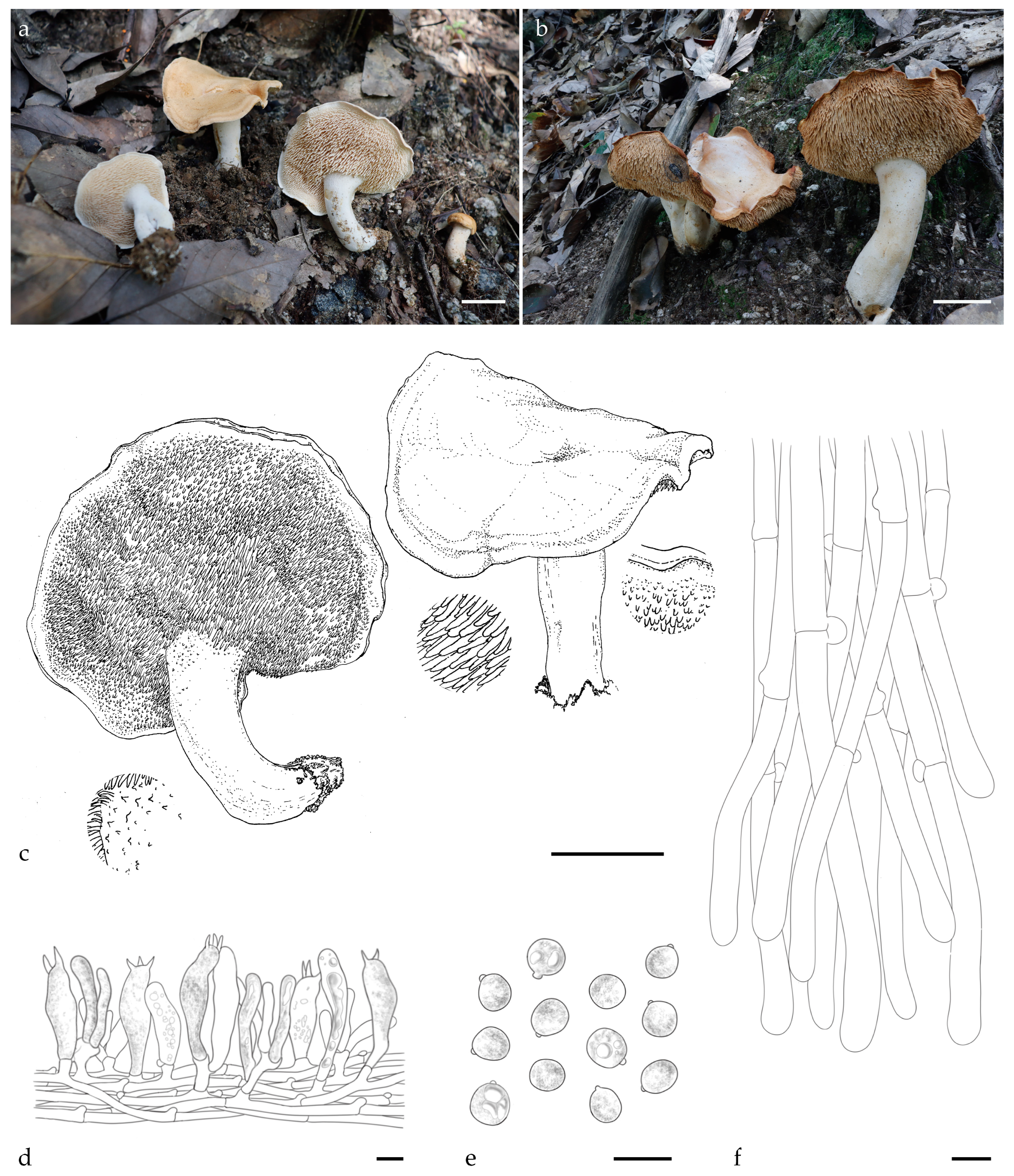
Figure 1. Macroscopic and microscopic features of H. fulvostriatum (FJAU66566, holotype). (ac) Basidiomata; (d) hymenium and subhymenium; (e) basidiospores; and (f) pileipellis. Scale bar: (ac) = 2 cm; (df) = 10 μm.
Jof 11 00431 g001
Figure 2. Macroscopic and microscopic features of H. crassipedum (FJAU66572, holotype). (ac) Basidiomata; (d) hymenium and subhymenium; (e) basidiospores; (f) and pileipellis. Scale bar: (ac) = 2 cm; (df) = 10 μm.
Jof 11 00431 g002
Figure 3. Macroscopic and microscopic features of H. albomarginatum (FJAU66574, holotype). (ac) Basidiomata; (d) hymenium and subhymenium; (e) basidiospores; (f) and pileipellis. Scale bar: (ac) = 2 cm; (df) = 10 μm.
Jof 11 00431 g003
Figure 4. Macroscopic and microscopic features of H. bifurcatum (FJAU66562, holotype). (ac) Basidiomata; (d) hymenium and subhymenium; (e)basidiospores; (f) and pileipellis. Scale bar: (ac) = 5cm; (df) = 10 μm.
Jof 11 00431 g004
Figure 5. Macroscopic and microscopic features of H. orientalbidum (FJAU66574). (ac) Basidiomata; (d) hymenium and subhymenium; (e) basidiospores; and (f) pileipellis. Scale bar: (ac) = 2 cm; (df) = 10 μm.
Jof 11 00431 g005
Figure 6. Phylogenetic tree of Hydnum inferred through Bayesian Inference and Maximum Likelihood analyses based on the combined nrLSU, ITS, and tef1 dataset. Node support is indicated as bootstrap (BS) > 70% and posterior probability (PP) > 0.95. Sequences generated in this study are in bold; new taxa are highlighted in red; and new records are indicated in black.
Jof 11 00431 g006
Figure 7. Phylogenetic tree of Hydnum inferred through Bayesian Inference and Maximum Likelihood analyses based on the combined nrLSU, ITS, and tef1 dataset. Node support is indicated as bootstrap (BS) > 70% and posterior probability (PP) > 0.95. Sequences generated in this study are in bold; new taxa are highlighted in red; and new records are indicated in black.
Jof 11 00431 g007
Table 1. Specimens and sequences used in this study.
SpeciesSpecimen VoucherGenBank No.CountryReferences
nrLSUITStef1
H. albertenseH T. Niskanen 11-354-KX388664-Canada[1]
H. albomagnumAFTOL-ID 471AY700199DQ218305DQ234568USA[2]
H. albomagnumRAS231-MH379943-USA[2]
H. albomarginatumFJAU66574PV356813PV329855PP357262ChinaThis study
H. albomarginatumFJAU66575PV356814PV329856PP357263ChinaThis study
H. atlanticumAJ1597-OQ235218OQ236553Canada[34]
H. atlanticumAJ1558-OQ235214OQ236551Canada[34]
H. berkeleyanumCAL 1656NG070500NR158533-India[35]
H. berkeleyanumHKAS77834KU612667KU612525-China[12]
H. bifurcatumFJAU66562-PV329845PP357252ChinaThis study
H. bifurcatumFJAU66563-PV329846PP357253ChinaThis study
H. boreorepandumH T. Niemela 1679-KX388658-Finland[1]
H. boreorepandumH 6003711-KX388657-Finland[1]
H. brevispinumIFP 019464MW979559MW980578-China[6]
H. brevispinumIFP 019465MW979560MW980579-China[6]
H. canadenseH T N 09-006-KX388681 -Canada[1]
H. crassipedumFJAU66572PV356811PV329853PP357260ChinaThis study
H. crassipedumFJAU66573PV356812PV329854PP357261ChinaThis study
H. cremeoalbumTUMH40462-AB906674-Japan[1]
H. cremeoalbumTUMH60740-AB906678-Japan[1]
H. cuspidatumRAS246-MH379944-USA[2]
H. cuspidatumRAS205-MH379936-USA[2]
H. ellipsosporumFD3281KX086217KX086215-Switzerland[9]
H. ellipsosporumH T. Niskanen 12-036-KX388671-Finland[1]
H. ferruginescensMH16005-MH379905-USA[2]
H. ferruginescensRAS229-MH379942-USA[2]
H. flabellatumIFP 019459MW979556MW980575-China[6]
H. fulvostriatumFJAU66566PV356807PV329849-ChinaThis study
H. fulvostriatumFJAU66567PV356808PV329850-ChinaThis study
H. ibericumBIO:Fungi:12330-HE611086-Spain[8]
H. ibericumMA-fungi 3457-AJ547879-Spain[14]
H. jussiiIFP 019485MW979539MW980553MW999436China[6]
H. jussiiIFP 019486MW979540MW980554MW999437China[6]
H. magnorufescensvoucher 161209KU612669KU612549-Slovenia[12]
H. magnorufescensTO HG2818-KC293545-Italy[15]
H. melitosarxH T. Niskanen 11-056-KX388683-USA[1]
H. melitosarxK 176869-KX388685-UK[1]
H. melleopallidumSMI356-FJ845406-Canada[15]
H. mulsicolorLJU GIS 1336-AJ547885 -Slovenia[14]
H. mulsicolorREB-341-JX093560-USA[36]
H. neorepandumH T. Niskanen 10-095-KX388659-Canada[1]
H. neorepandumH T. Niskanen 10-086-KX388660-Canada[1]
H. olympicumH T. Niskanen 09-134-KX388661-USA[1]
H. olympicumSAT-10-208-05-MT955159-USA[6]
H. oregonenseHVM61-KF879509-USA[37]
H. oregonensePNW-MS g2010502h1-09-AJ534972-USA[14]
H. orientalbidumFJAU66570PV356809PV329857PP357258ChinaThis study
H. orientalbidumFJAU66571PV356810PV329858PP357259ChinaThis study
H. orientalbidumTUMH:64068-LC621862-Japan[7]
H. ovoideisporumvoucher71106-KU612536-Slovenia[6]
H. ovoideisporumBIO Fungi 12683-NR119818-Spain[8]
H. pallidocroceumIFP 019466MW979554MW980568MW999449China[6]
H. pallidocroceumIFP 019467MW979555MW980569MW999450China[6]
H. pallidomarginatumIFP 019468MW979552MW980566MW999447China[6]
H. pallidomarginatumIFP 019469MW979553MW980567MW999448China[6]
H. pinicolaSFC20180928-18OR211401OR211383OR220059Korea[38]
H. quebecenseH T. Niskanen 10-064-KX388662-Canada[1]
H. quebecenseCN9-MH379881-USA[2]
H. repando-orientaleTUMH60745-AB906683-Japan[39]
H. repando-orientaleTUMH60743-AB906684-Japan[39]
H. repandumH 6003710-NR164553-Finland[1]
H. roseotangerinumMHKMU LP Tang 3458PQ287756PQ287675PQ295849China[22]
H. roseotangerinumMHKMU LP Tang 3458-1PQ287757PQ287676PQ295850China[22]
H. rufescensH 6003708-KX388688-Finland[1]
H. rufescensHTN7839-KX388656-Estonia[1]
H. slovenicumLJU GIS 1338-AJ547870-Slovenia[14]
H. slovenicumLJU GIS 1340-AJ547884-Slovenia[14]
H. sp.wi1A4spel-KC679833-China[6]
H. sp.wi8T4spel-KC679834-China[6]
H. sp.13HKAS57714KU612673KU612617-China[12]
H. sp.13HKAS58838KU612675KU612616-China[12]
H. sp.2HKAS92340KU612661KU612543-China[12]
H. sphaericumIFP 019470MW979549MW980563MW999444China[6]
H. sphaericumIFP 019472MW979550MW980564MW999445China[6]
H. sphaericumIFP 019471MW979551MW980565MW999446China[6]
H. subberkeleyanumTNS:F-19323-LC621879-Japan[7]
H. subberkeleyanumTUMH 63627-LC621880LC622505Japan[7]
H. subconnatumRAS235-MH379930-USA[2]
H. subconnatumRAS169-MH379916-USA[2]
H. submulsicolorH T. Niskanen 10-132-KX388682-Canada[1]
H. subolympicumF1188765KU612653KU612599-USA[12]
H. subolympicumDAOM744368-MH174257-Canada[1]
H. subovoideisporumH 6003707-NR158494-Finland[1]
H. subrufescensH T. Niskanen 10-154-KX388649-Canada[1]
H. subrufescensF1188749KU612663KU612535-USA[12]
H. subtiliorRAS180-MH379918-USA[2]
H. subtiliorTENN 073034-NR164029-USA[2]
H. tenuistipitumIFP 019476MW979557MW980576-China[6]
H. tenuistipitumIFP 019477MW979558MW980577-China[6]
H. tomaenseTUMH64085-LC622508-Japan[7]
H. tottorienseTUMH64088-LC621887LC622511Japan[7]
H. tottorienseTUMH64089-LC621888LC622512Japan[7]
H. treuiKA20-0732ON907772ON907793OR220061Korea[1]
H. vagabundum10782TJB-MH379949-USA[2]
H. vagabundumCLO4985-MH379909-USA[2]
H. ventricosumIFP 019478MW979547MW980561MW999442China[6]
H. ventricosumIFP 019479MW979548MW980562MW999443China[6]
H. vesterholtiiBIO:Fungi:10429-HE611084-Spain[8]
H. vesterholtiiBIO:Fungi:10452-HE611085-Spain[8]
H. washingtonianumUBC F-32538-MF954990-Canada[6]
H. zongolicenseGO-2010-142a-KC152121-Mexico[1]
Sistotrema. muscicolaKHL 11721-AJ606040-Sweden[40]
Sistotrema. muscicolataxon:154757-AJ606041-Sweden[40]
Newly generated sequences in this study are in bold.
Disclaimer/Publisher’s Note: The statements, opinions and data contained in all publications are solely those of the individual author(s) and contributor(s) and not of MDPI and/or the editor(s). MDPI and/or the editor(s) disclaim responsibility for any injury to people or property resulting from any ideas, methods, instructions or products referred to in the content.