
Unveiling Species Diversity Within Early-Diverging Fungi from China VII: Seven New Species of Cunninghamella (Mucoromycota)

 ,
,
Abstract
1. Introduction
2. Materials and Methods
2.1. Sample Collection and Strain Isolation
2.2. Morphology and Maximum Growth Temperature
2.3. DNA Extraction, PCR Amplification, and Sequencing
2.4. Phylogenetic Analyses
3. Results
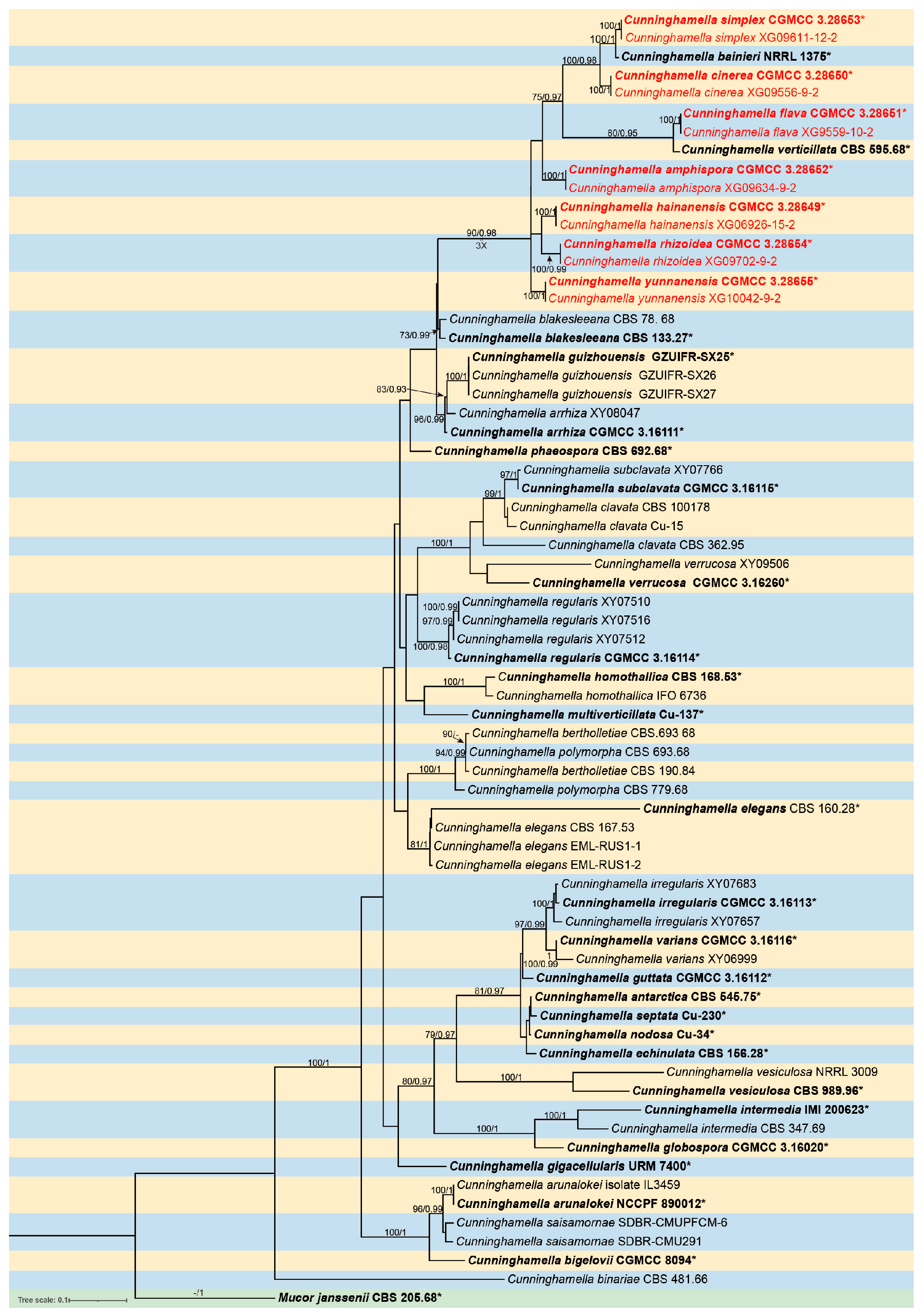
3.1. Phylogeny
3.2. Taxonomy
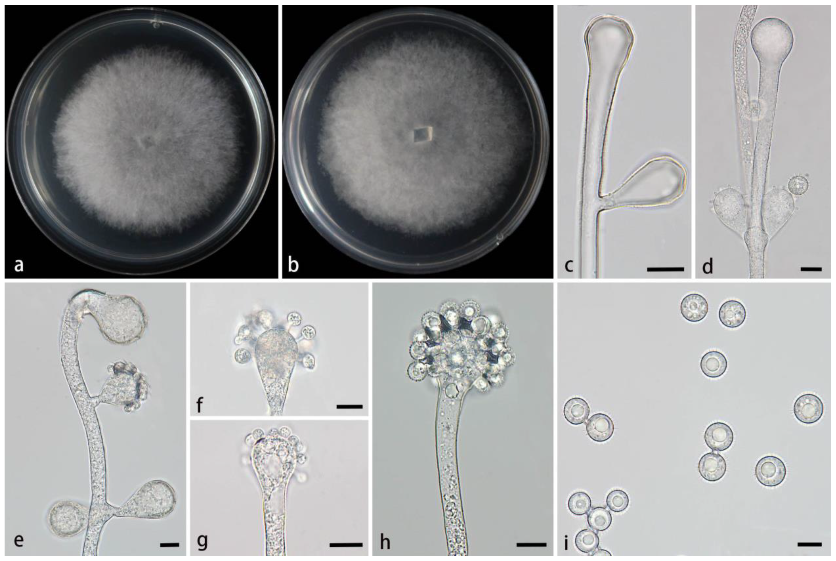
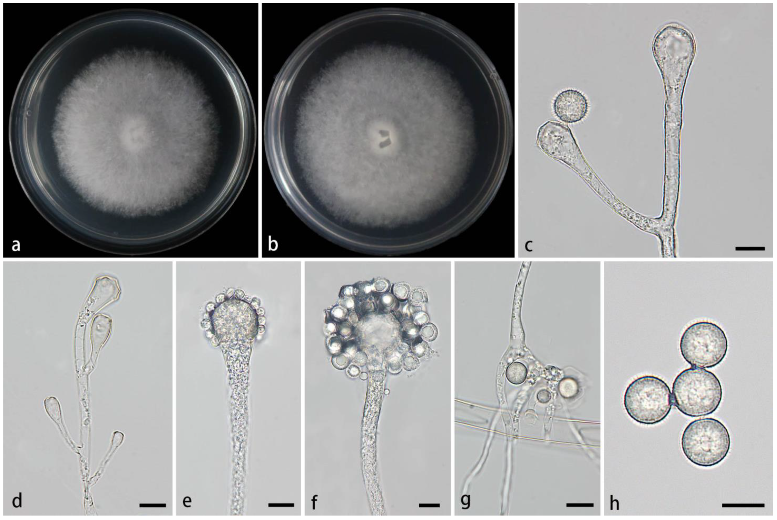
3.2.1. Cunninghamella amphispora Z.Y. Ding & X.Y. Liu, sp. nov., Figure 2
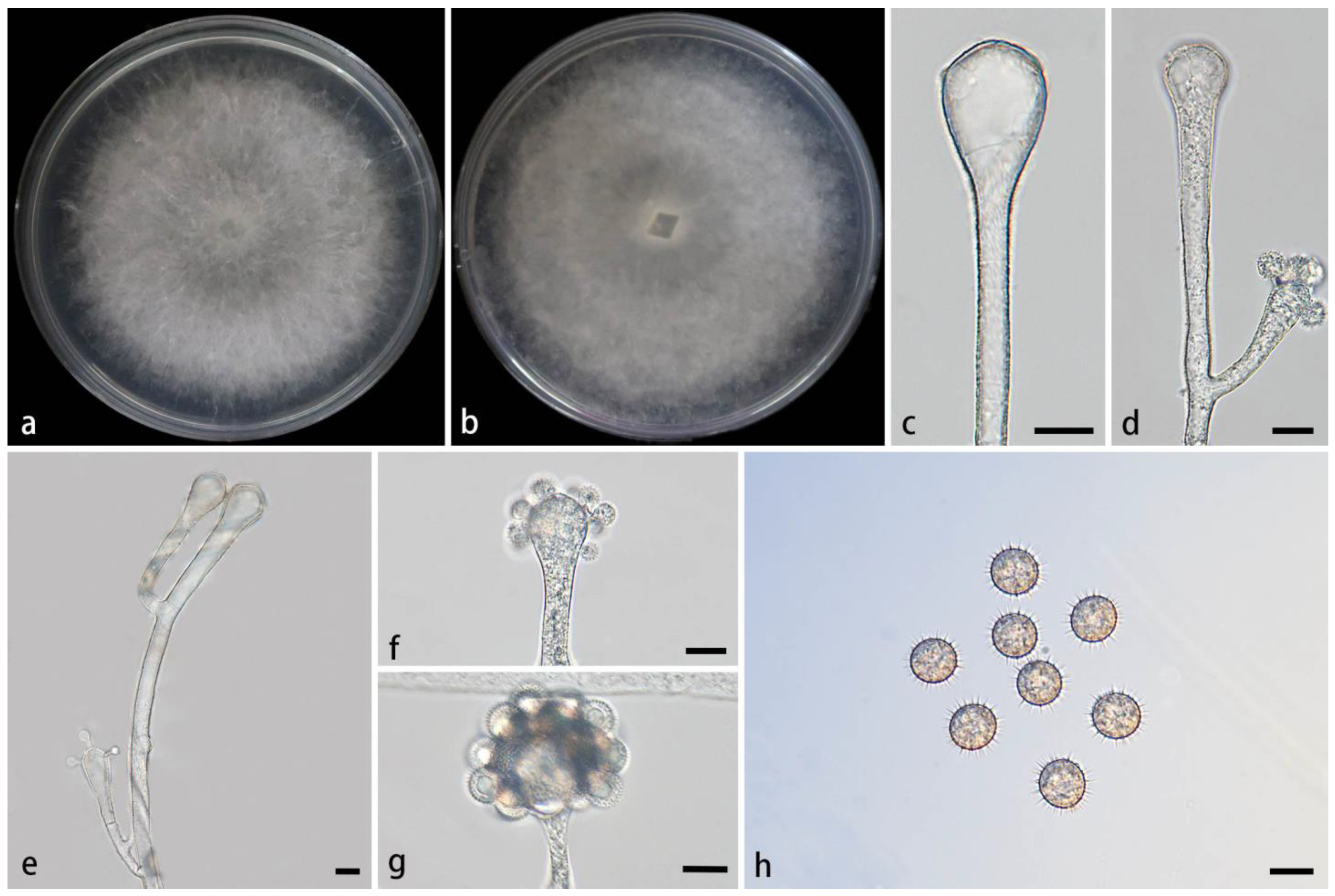
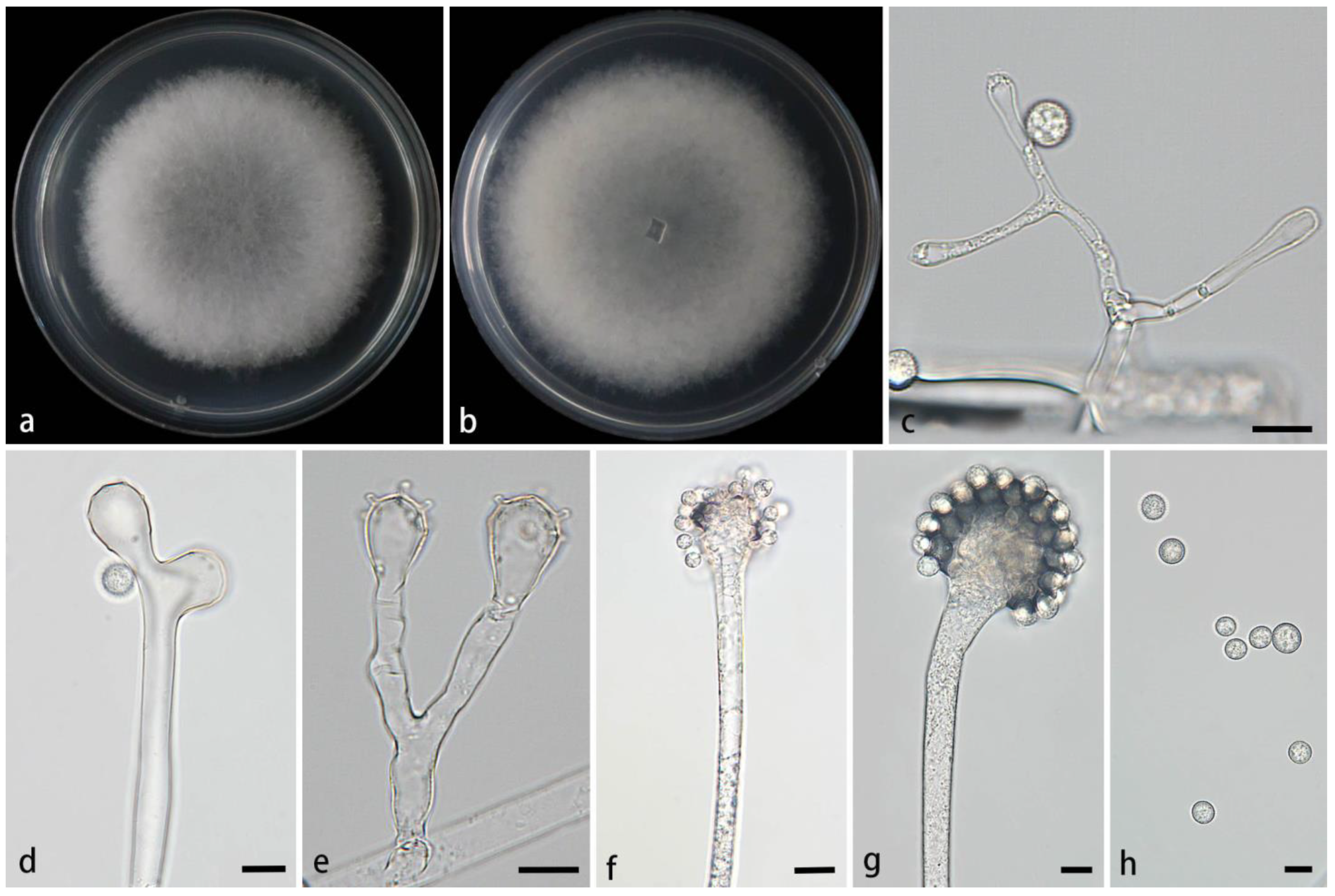
3.2.2. Cunninghamella cinerea Z.Y. Ding & X.Y. Liu, sp. nov., Figure 3
3.2.3. Cunninghamella flava Z.Y. Ding & X.Y. Liu, sp. nov., Figure 4
3.2.4. Cunninghamella hainanensis Z.Y. Ding & X.Y. Liu, sp. nov., Figure 5
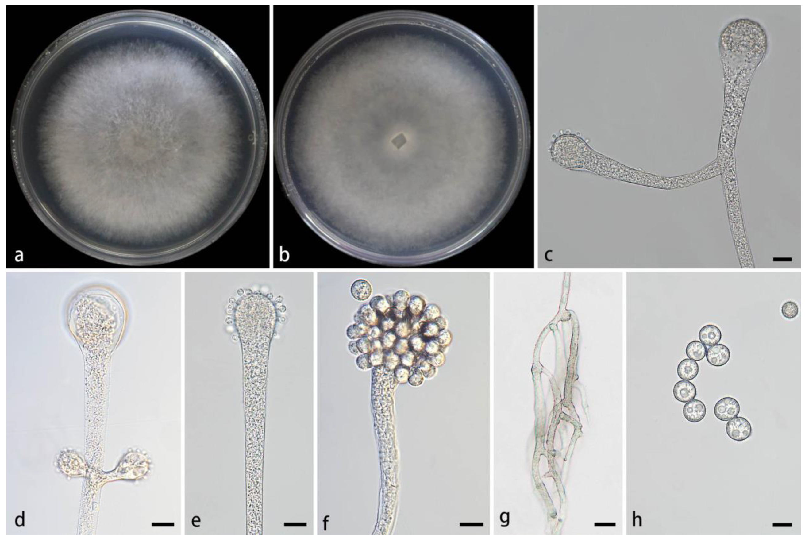
3.2.5. Cunninghamella rhizoidea Z.Y. Ding & X.Y. Liu, sp. nov., Figure 6

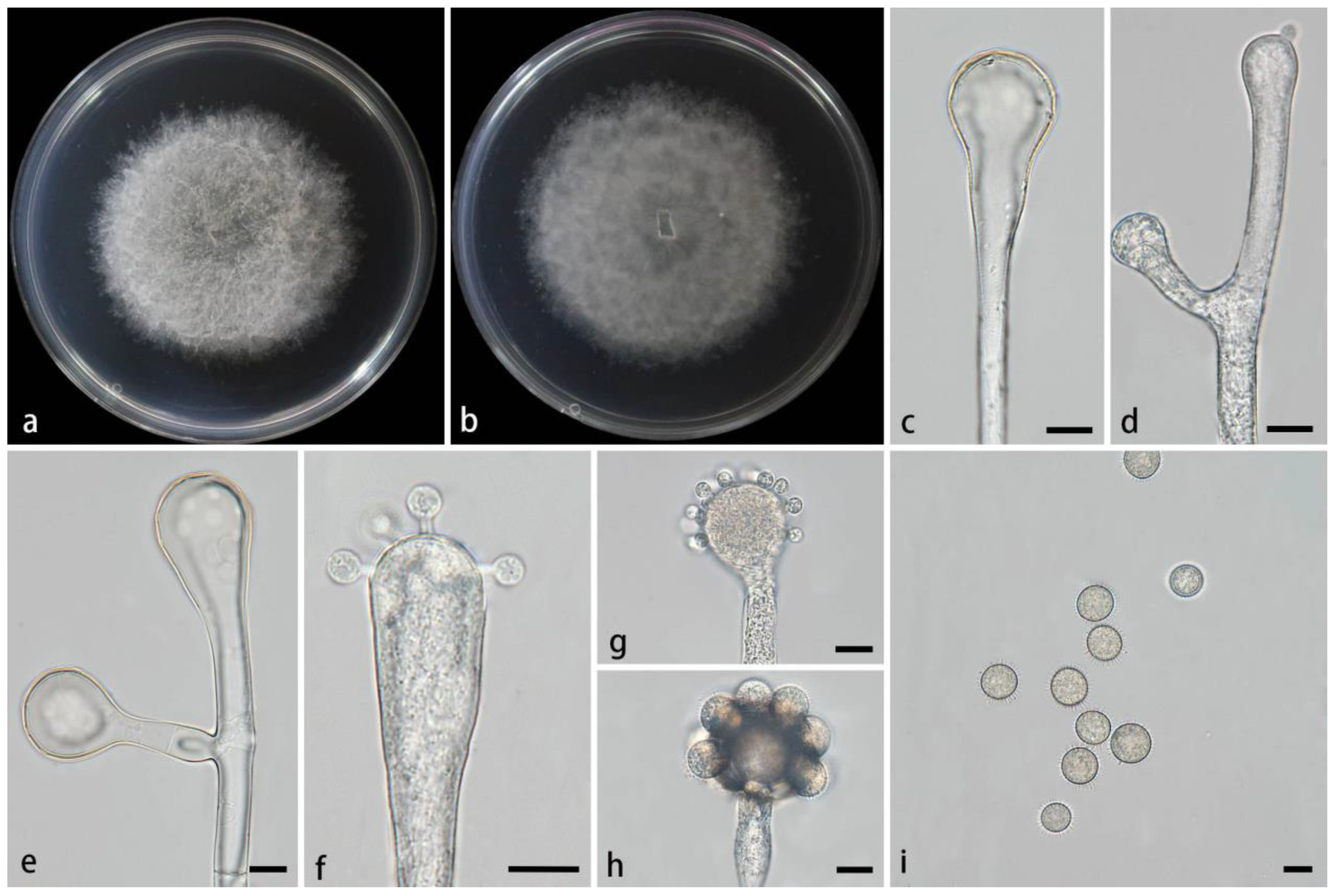
3.2.6. Cunninghamella simplex Z.Y. Ding & X.Y. Liu, sp. nov., Figure 7
3.2.7. Cunninghamella yunnanensis Z.Y. Ding & X.Y. Liu, sp. nov., Figure 8
4. Discussion
Supplementary Materials
Author Contributions
Funding
Institutional Review Board Statement
Informed Consent Statement
Data Availability Statement
Conflicts of Interest
References
- Spatafora, J.W.; Chang, Y.; Benny, G.L.; Lazarus, K.; Smith, M.E.; Berbee, M.L.; Bonito, G.; Corradi, N.; Grigoriev, I.; Gryganskyi, A.; et al. A phylum-level phylogenetic classification of zygomycete fungi based on genome-scale data. Mycologia 2016, 108, 1028–1046. [Google Scholar] [CrossRef] [PubMed]
- Zheng, R.Y.; Chen, G.Q. A monograph of Cunninghamella. Mycotaxon 2001, 80, 1–75. [Google Scholar]
- Walther, G.; Pawłowska, J.; Alastruey-Izquierdo, A.; Wrzosek, M.; Rodriguez-Tudela, J.L.; Dolatabadi, S.; Chakrabarti, A.; de Hoog, G.S. DNA barcoding in Mucorales: An inventory of biodiversity. Persoonia 2013, 30, 11–47. [Google Scholar] [CrossRef] [PubMed]
- Matruchot, L. Une Mucorinée purement conidienne, Cunninghamella africana. Annales Mycologici. 1903, 1, 45–60. [Google Scholar]
- Baijai, U.; Mehrotra, B.S. The genus Cunninghamella—A reassessment. Sydowia 1980, 33, 1–13. [Google Scholar]
- Reed, M.D.A.; Body, P.B.; Austin, M.D.M.B.; Frierson, M.D.H., Jr. Cunninghamella bertholletiae and Pneumocystis carinii pneumonia as a fatal complication of chronic lymphocytic leukemia. Hum. Pathol. 1988, 19, 1470–1472. [Google Scholar] [CrossRef]
- Nguyen, T.T.T.; Choi, Y.J.; Lee, H.B. Zygomycete fungi in Korea: Cunninghamella bertholletiae, Cunninghamella echinulata, and Cunninghamella elegans. Mycobiology 2017, 45, 318–326. [Google Scholar] [CrossRef]
- Gomes, M.Z.; Lewis, R.E.; Kontoyiannis, D.P. Mucormycosis caused by unusual mucormycetes, non-Rhizopus, -Mucor, and -Lichtheimia. Clin. Microbiol. Rev. 2011, 24, 411–445. [Google Scholar] [CrossRef]
- Kwon-Chung, K.J. Taxonomy of fungi causing mucormycosis and entomophthoramycosis (zygomycosis) and nomenclature of the disease: Molecular mycologic perspectives. Clin. Infect. Dis. 2012, 54 (Suppl. S1), S8–S15. [Google Scholar] [CrossRef]
- Yu, J.; Walther, G.; Van Diepeningen, A.D.; Gerrits Van Den Ende, A.H.G.; Li, R.Y.; Moussa, T.A.A.; Almaghrabi, O.A.; De Hoog, G.S. DNA barcoding of clinically relevant Cunninghamella species. Med. Mycol. 2015, 53, 99–106. [Google Scholar] [CrossRef]
- Fakas, S.; Papanikolaou, S.; Galiotou-Panayotou, M.; Komaitis, M.; Aggelis, G. Organic nitrogen of tomato waste hydrolysate enhances glucose uptake and lipid accumulation in Cunninghamella echinulata. J. Appl. Microbiol. 2008, 105, 1062–1070. [Google Scholar] [CrossRef]
- Alakhras, R.; Bellou, S.; Fotaki, G.; Stephanou, G.; Demopoulos, N.A.; Papanikolaou, S.; Aggelis, G. Fatty acid lithium salts from Cunninghamella echinulata have cytotoxic and genotoxic effects on HL-60 human leukemia cells. Eng. Life. Sci. 2015, 15, 243–253. [Google Scholar] [CrossRef]
- Zhao, H.; Lv, M.; Liu, Z.; Zhang, M.; Wang, Y.; Ju, X.; Song, Z.; Ren, L.; Jia, B.; Qiao, M.; et al. High-yield oleaginous fungi and high-value microbial lipid resources from Mucoromycota. Bioenerg. Res. 2020, 14, 1196–1206. [Google Scholar] [CrossRef]
- Liu, C.W.; Liou, G.Y.; Chien, C.Y. New records of the genus Cunninghamella (Mucorales) in Taiwan. Fungal Sci. 2005, 20, 1–9. [Google Scholar]
- Guo, J.; Wang, H.; Liu, D.; Zhang, J.N.; Zhao, Y.H.; Liu, T.X.; Xin, Z.H. Isolation of Cunninghamella bigelovii sp. nov. CGMCC 8094 as a new endophytic oleaginous fungus from Salicornia bigelovii. Mycol. Prog. 2015, 14, 11. [Google Scholar] [CrossRef]
- Alves, A.L.; de Souza, C.A.; de Oliveira, R.J.; Cordeiro, T.R.; de Santiago, A.L. 2017 Cunninghamella clavata from Brazil: A new record for the western hemisphere. Mycotaxon 2017, 132, 381–389. [Google Scholar] [CrossRef]
- Bragulat, M.R.; Castella, G.; Isidoro-Ayza, M.; Domingo, M.; Cabanes, F.J. Characterization and phylogenetic analysis of a Cunninghamella bertholletiae isolate from a bottlenose dolphin (Tursiops truncatus). Rev. Iberoam. Micol. 2017, 34, 215–219. [Google Scholar] [CrossRef]
- Weitzman, I.; Crist, M.Y. Studies with clinical isolates of Cunninghamella II. Physiological and morphological studies. Mycologia 1980, 72, 661–669. [Google Scholar] [CrossRef]
- Liu, X.Y.; Huang, H.; Zheng, R.Y. Relationships within Cunninghamella based on sequence analysis of ITS rDNA. Mycotaxon 2001, 80, 77–95. [Google Scholar]
- Hyde, K.D.; Hongsanan, S.; Jeewon, R.; Bhat, D.J.; McKenzie, E.H.C.; Jones, E.B.G.; Phookamsak, R.; Ariyawansa, H.A.; Boonmee, S.; Zhao, Q.; et al. Fungal diversity notes 367–490: Taxonomic and phylogenetic contributions to fungal taxa. Fungal Divers. 2016, 80, 1–270. [Google Scholar] [CrossRef]
- Suwannarach, N.; Kumla, J.; Supo, C.; Honda, Y.; Nakazawa, T.; Khanongnuch, C.; Wongputtisin, P. Cunninghamella saisamornae (Cunninghamellaceae, Mucorales), a new soil fungus from northern Thailand. Phytotaxa 2021, 509, 291–300. [Google Scholar] [CrossRef]
- Zhang, Z.Y.; Zhao, Y.X.; Shen, X.; Chen, W.H.; Han, Y.F.; Huang, J.Z.; Liang, Z.Q. Molecular phylogeny and morphology of Cunninghamella guizhouensis sp. nov. (Cunninghamellaceae, Mucorales), from soil in Guizhou, China. Phytotaxa 2020, 455, 31–39. [Google Scholar] [CrossRef]
- Wang, Y.J.; Zhao, T.; Wu, W.Y.; Wang, M.; Liu, X.Y. Cunninghamella verrucosa sp. nov. (Mucorales, Mucoromycota) from Guangdong province in China. Phytotaxa 2022, 560, 274–284. [Google Scholar] [CrossRef]
- Zhao, H.; Zhu, J.; Zong, T.K.; Liu, X.L.; Ren, L.Y.; Lin, Q.; Qiao, M.; Nie, Y.; Zhang, Z.D.; Liu, X.Y. Two new species in the family Cunninghamellaceae from China. Mycobiology 2021, 49, 142–150. [Google Scholar] [CrossRef] [PubMed]
- Zhao, H.; Nie, Y.; Zong, T.; Wang, K.; Lv, M.; Cui, Y.; Tohtirjap, A.; Chen, J.; Zhao, C.; Wu, F.; et al. Species diversity, updated classification and divergence times of the phylum Mucoromycota. Fungal Divers. 2023, 123, 49–157. [Google Scholar] [CrossRef]
- Zhao, H.; Nie, Y.; Huang, B.; Liu, X.Y. Unveiling species diversity within early-diverging fungi from China I: Three new species of Backusella (Backusellaceae, Mucoromycota). MycoKeys 2024, 109, 285–304. [Google Scholar] [CrossRef] [PubMed]
- Tao, M.F.; Ding, Z.Y.; Wang, Y.X.; Zhang, Z.X.; Zhao, H.; Meng, Z.; Liu, X.Y. Unveiling species diversity within early-diverging fungi from China II: Three new species of Absidia (Cunninghamellaceae, Mucoromycota) from Hainan Province. MycoKeys 2024, 110, 255–272. [Google Scholar] [CrossRef]
- Wang, Y.X.; Zhao, H.; Jiang, Y.; Liu, X.Y.; Tao, M.F.; Liu, X.Y. Unveiling species diversity within early-diverging fungi from China III: Six new species and a new record of Gongronella. (Cunninghamellaceae, Mucoromycota). MycoKeys 2024, 110, 287–317. [Google Scholar] [CrossRef]
- Li, W.X.; Wei, Y.H.; Zou, Y.; Liu, P.; Li, Z.; Gontcharov, A.A.; Stephenson, S.L.; Wang, Q.; Zhang, S.H.; Li, Y. Dictyostelid cellular slime molds from the Russian Far East. Protist 2020, 171, 125756. [Google Scholar] [CrossRef]
- Zou, Y.; Hou, J.G.; Guo, S.N.; Li, C.T.; Li, Z.; Stephenson, S.L.; Pavlov, I.N.; Liu, P.; Li, Y. Diversity of dictyostelid cellular slime molds, including two species new to science, in forest soils of Changbai Mountain, China. Microbiol. Spectr. 2022, 10, e0240222. [Google Scholar] [CrossRef]
- Zhao, H.; Nie, Y.; Zong, T.; Dai, Y.; Liu, X.Y. Three new species of Absidia (Mucoromycota) from China based on phylogeny, morphology and physiology. Diversity 2022, 14, 132. [Google Scholar] [CrossRef]
- Zhao, H.; Nie, Y.; Zong, T.; Wang, Y.; Wang, M.; Dai, Y.; Liu, X.Y. Species diversity and ecological habitat of Absidia (Cunninghamellaceae, Mucorales) with emphasis on five new species from forest and grassland soil in China. J. Fungi 2022, 8, 471. [Google Scholar] [CrossRef] [PubMed]
- Corry, J.E.L. Rose bengal chloramphenicol (RBC) agar. In Progress in Industrial Microbiology; Elsevier: Amsterdam, The Netherlands, 1995; pp. 431–433. [Google Scholar]
- Zheng, R.Y.; Chen, G.Q.; Huang, H.; Liu, X.Y. A monograph of Rhizopus. Sydowia 2007, 59, 273–372. [Google Scholar]
- Zheng, R.Y.; Liu, X.Y.; Li, R.Y. More Rhizomucor causing human mucormycosis from China: R. chlamydosporus sp. nov. Sydowia 2009, 61, 135–147. [Google Scholar]
- Zong, T.K.; Zha, H.; Liu, X.L.; Ren, L.Y.; Zhao, C.L.; Liu, X.Y. Taxonomy and phylogeny of four new species in Absidia (Cunninghamellaceae, Mucorales) from China. Front. Microbiol. 2021, 12, 677836. [Google Scholar] [CrossRef]
- Doyle, J.J.; Doyle, J.L. Isolation of plant DNA from fresh tissue. Focus 1990, 12, 13–15. [Google Scholar] [CrossRef]
- Guo, L.D.; Hyde, K.D.; Liew, E.C.Y. Identification of endophytic fungi from Livistona chinensis based on morphology and rDNA sequences. New Phytol. 2000, 147, 617–630. [Google Scholar] [CrossRef]
- White, T.J.; Bruns, T.; Lee, S.; Taylor, J. Amplification and direct sequencing of fungal ribosomal RNA genes for phylogenetics. In PCR Protocols: A Guide to Methods and Applications; Academic Press: New York, NY, USA, 1990; pp. 315–322. [Google Scholar]
- Vilgalys, R.; Hester, M. Rapid genetic identification and mapping of enzymatically amplified ribosomal DNA from several Cryptococcus species. J. Bacteriol. 1990, 172, 4238–4246. [Google Scholar] [CrossRef]
- O’Donnell, K.; Lutzoni, F.M.; Ward, T.J.; Benny, G.L. Evolutionary relationships among mucoralean fungi Zygomycota: Evidence for family polyphyly on a large scale. Mycologia 2001, 93, 286–297. [Google Scholar] [CrossRef]
- Zhang, Z.; Liu, R.; Liu, S.; Mu, T.; Zhang, X.; Xia, J. Morphological and phylogenetic analyses reveal two new species of Sporocadaceae from Hainan, China. MycoKeys 2022, 88, 171–192. [Google Scholar] [CrossRef]
- Kumar, S.; Stecher, G.; Tamura, K. MEGA7: Molecular evolutionary genetics analysis version 7.0 for bigger datasets. Mol. Biol. Evol. 2016, 33, 1870–1874. [Google Scholar] [CrossRef] [PubMed]
- Katoh, K.; Standley, D.M. MAFFT Multiple sequence alignment software version 7: Improvements in performance and usability. Mol. Biol. Evol. 2013, 30, 772–780. [Google Scholar] [CrossRef]
- Katoh, K.; Rozewicki, J.; Yamada, K.D. MAFFT online service: Multiple sequence alignment, interactive sequence choice and visualization. Brief. Bioinform. 2019, 20, 1160–1166. [Google Scholar] [CrossRef] [PubMed]
- Felsenstein, J. Confidence intervals on phylogenetics: An approach using bootstrap. Evolution 1985, 39, 783–791. [Google Scholar] [CrossRef] [PubMed]
- Stamatakis, A. RAxML-VI-HPC: Maximum likelihood-based phylogenetic analyses with thousands of taxa and mixed models. Bioinformatics 2006, 22, 2688–2690. [Google Scholar] [CrossRef]
- Minh, Q.; Nguyen, M.; Von Haeseler, A.A. Ultrafast approximation for phylogenetic bootstrap. Mol. Biol. Evol. 2013, 30, 1188–1195. [Google Scholar] [CrossRef]
- Stamatakis, A. RAxML version 8: A tool for phylogenetic analysis and post-analysis of large phylogenies. Bioinformatics 2014, 30, 1312–1313. [Google Scholar] [CrossRef]
- Huelsenbeck, J.P.; Ronquist, F. MRBAYES: Bayesian inference of phylogenetic trees. Bioinformatics 2001, 17, 754–755. [Google Scholar] [CrossRef]
- Nylander, J. MrModeltest V2: Program distributed by the author. Bioinformatics 2004, 24, 581–583. [Google Scholar] [CrossRef]
- Ronquist, F.; Teslenko, M.; Van der Mark, P.; Ayres, D.L.; Darling, A.; Höhna, S.; Larget, B.; Liu, L.; Suchard, M.A.; Huelsenbeck, J.P. MrBayes 3.2: Efficient Bayesian phylogenetic inference and model choice across a large model space. Syst. Biol. 2012, 61, 539–542. [Google Scholar] [CrossRef]
- Bauer, R.; Garnica, S.; Oberwinkler, F.; Riess, K.; Weiß, M.; Begerow, D. Entorrhizomycota: A new fungal phylum reveals new perspectives on the evolution of fungi. PLoS ONE 2015, 10, e0128183. [Google Scholar] [CrossRef] [PubMed]
- Jeewon, R.; Hyde, K.D. Establishing species boundaries and new taxa among fungi: Recommendations to resolve taxonomic ambiguities. Mycosphere 2016, 7, 1669–1677. [Google Scholar] [CrossRef]
- Maharachchikumbura, S.S.N.; Hyde, K.D.; Jones, E.B.G.; McKenzie, E.H.C.; Bhat, J.D.; Dayarathne, M.C.; Huang, S.K.; Norphanphoun, C.; Senanayake, I.C.; Perera, R.H.; et al. Families of Sordariomycetes. Fungal Divers. 2016, 79, 1–317. [Google Scholar] [CrossRef]
- Hu, D.M.; Wang, M.; Cai, L. Phylogenetic assessment and taxonomic revision of Mariannaea. Mycol. Prog. 2016, 16, 271–283. [Google Scholar] [CrossRef]

| Species | Colonies | Sporangiophores | Vesicles | Pedicels | Sporangiola | Reference |
|---|---|---|---|---|---|---|
| C. hainanensis | PDA: 26 °C 4 days, 85 mm, 21.25 mm/d, initially white, gradually becoming light gray, floccose | 2.9–10.6 µm wide, mostly erect, a few slightly bent, occasionally verticillate, unbranched or 1–3 branched, opposite, in pairs | Globose, subglobose, elliptic, terminal vesicles 15.1–27.8 × 11.7–26.8 µm and lateral vesicles 16.8–28.4 × 11.2–22.9 µm | 1.2–5.8 µm long | Mostly globose, 7.3–14 × 7.8–13.4 µm, with short spines, 0.7–1.9 µm long | This study |
| C. cinerea | PDA: 26 °C 4 days, 85 mm, 21.25 mm/d, initially white, gradually turning to smoky gray with age, floccose | 3.1–11.7 µm wide, erect or slightly bent, mainly unbranched or simply branched, mainly single or recumbent, never verticillate | Globose to elliptic, terminal vesicles 14.3–32.3 × 12.4–28.3 µm and lateral vesicles 11.9–20.3 × 9.9–17.3 µm | 1.2–2.2 µm long | Globose to ovoid, 5.9–16.7 × 5.9–15.5 µm, with short spines, 0.8–1.7 µm long | This study |
| C. flava | PDA: 26 °C 6 days, 72 mm, 12 mm/d, initially white, gradually turning to dry yellow with age, floccose | 3.8–25.6 µm wide, erect or slightly bent, unbranched or 1–7 branched, recumbent, opposite, in pairs, 1–3 verticillate | Globose, pear-shaped, elliptic, terminal vesicles 16.6–43.1 × 15.4–44.5 µm and lateral vesicles 9.3–28.1 × 9.4–25.3 µm | 1.6–2.2 µm long | Globose to ovoid, 8.9–18.6 × 8.8–18.7 µm wide, with short spines, 1.4–2.8 µm long | This study |
| C. amphispora | PDA: 26 °C 4 days, 85 mm, 21.25 mm/d, initially white, gradually becoming light gray, floccose | 2.3–19.1 µm wide, erect or few slightly bent, unbranched or simply branched, hyaline, single, no verticillate | Subglobose, globose, ovoid, pillar-shaped, terminal vesicles 12.9–56.6 × 11.6–46.7 µm and lateral vesicles 8–18.9 × 5.1–14.8 µm | 1.4–3.4 µm long | Globose, 6.3–16.7 × 6.3–15.8 µm, with short spines, 0.5–2.6 µm long | This study |
| C. simplex | PDA: 26 °C 4 days, 85 mm, 21.25 mm/d, initially white, gradually turning to dusky gray, floccose | 3.1–12.2 µm wide, erect or slightly bent, hyaline, unbranched or simply branched, in pairs, never verticillate | Spherical, ovoid, cylindrical, terminal vesicles 9.9–28.4 × 9.0–25.8 µm and lateral vesicles 6.7–17.0 × 3.8–10.8 µm | 1.1–2.9 µm long | Globose to ovoid, 5.9–10.2 × 5.6–9.7 µm, with very short spines, 0.3–0.8 µm long | This study |
| C. rhizoidea | PDA: 26 °C 4 days, 85 mm, 21.25 mm/d, initially white, gradually becoming gray with age, floccose | 2.8–11.4 µm wide, erect or slightly bent, simple branches or occasionally multiple branches, recumbent, opposite, occasionally verticillate | Globose, club-shaped, terminal vesicles 9.3–35.3 × 9.2–32.0 µm and lateral vesicles 2.7–26.6 × 2.4–27.1 µm | 1.5–5.4 µm long | Globose to ovoid, 8.2–13.4 × 7.9–12.9 µm, with short spines, 1.3–2.0 µm long | This study |
| C. yunnanensis | PDA: 26 °C 4 days, 85 mm, 21.25 mm/d, initially white, gradually becoming light gray with age, floccose | 1.8–14.6 µm wide, erect or few slightly bent, mainly unbranched or occasionally 1–7 branched, straight or recumbent, few verticillate | Subglobose, globose, ovoid, elliptic terminal vesicles 10.5–33.8 × 9.6–33.5 µm and lateral vesicles 5.5–13.9 × 4–12.4 µm | 1.1–4.1 µm long | Mainly globose, sometimes ovoid, 5.2–12.4 × 4.1–12.0 µm wide, with short spines, 0.7–2.0 µm long | This study |
| C. bainieri | SMA: 27 °C 4 days, 90 mm, at first white, soon becoming light gray, gray, to ‘Light Mouse Gray’, reverse cream, floccose | Erect, bent, or recumbent, main axes of sporangiophores (8–) 11–21 µm wide; primary branches (1–) 4–10 (–18) µm wide, monopodial, pseudoverticillate, or verticillate in 1–2 (–3) whorls of 3–8, typically in pairs | Globose, subglobose to ovoid, sometimes irregular, axial vesicles 18.5–32(–40) μm and lateral ones (8–)13.5–30 μm | 2.5–3.5 (–6) μm long | Ovoid to ellipsoid and 7–14.5 (–20) × 6.5–11 (–14.5) µm globose and 5.5–12.5 µm, lacrymoid and 9–20 (–32.5) × 7–14.5 (–20) µm | [2] |
| C. verticillata | SMA: 28 °C 5–6 days, 90 mm, at first white, from the sixth day near ‘Avellaneous’ to near ‘Colonial Buff’, reverse yellowish cream, floccose | Erect, straight, or recumbent, main axes of sporangiophores 7.5–17.5 (–25) µm; branches (0–) 4–15 (–25), verticillate to pseudoverticillate, rarely singly or in pairs, mostly simple, very rarely re-branched | Axial ones slightly depressed—globose, subglobose to globose, 225–50 (–70) μm and lateral ones usually globose to subglobose, sometimes broadly ovoid, 8.5–27.5 (–32.5) μm | 2.5–4 (–6.5) um long | Two kinds: globose, broadly ellipsoid ovoid and bluntly pointed at one end, 6–17.5 (–20) × 5.5–13.5 (–17.5) µm; dark giant sporangiola, globose, 11.5–17.5 (−25) µm | [2] |
Disclaimer/Publisher’s Note: The statements, opinions and data contained in all publications are solely those of the individual author(s) and contributor(s) and not of MDPI and/or the editor(s). MDPI and/or the editor(s) disclaim responsibility for any injury to people or property resulting from any ideas, methods, instructions or products referred to in the content. |
© 2025 by the authors. Licensee MDPI, Basel, Switzerland. This article is an open access article distributed under the terms and conditions of the Creative Commons Attribution (CC BY) license (https://creativecommons.org/licenses/by/4.0/).
Share and Cite
Ding, Z.-Y.; Tao, M.-F.; Ji, X.-Y.; Jiang, Y.; Wang, Y.-X.; Liu, W.-X.; Wang, S.; Liu, X.-Y. Unveiling Species Diversity Within Early-Diverging Fungi from China VII: Seven New Species of Cunninghamella (Mucoromycota). J. Fungi 2025, 11, 417. https://doi.org/10.3390/jof11060417
Ding Z-Y, Tao M-F, Ji X-Y, Jiang Y, Wang Y-X, Liu W-X, Wang S, Liu X-Y. Unveiling Species Diversity Within Early-Diverging Fungi from China VII: Seven New Species of Cunninghamella (Mucoromycota). Journal of Fungi. 2025; 11(6):417. https://doi.org/10.3390/jof11060417
Chicago/Turabian StyleDing, Zi-Ying, Meng-Fei Tao, Xin-Yu Ji, Yang Jiang, Yi-Xin Wang, Wen-Xiu Liu, Shi Wang, and Xiao-Yong Liu. 2025. "Unveiling Species Diversity Within Early-Diverging Fungi from China VII: Seven New Species of Cunninghamella (Mucoromycota)" Journal of Fungi 11, no. 6: 417. https://doi.org/10.3390/jof11060417
APA StyleDing, Z.-Y., Tao, M.-F., Ji, X.-Y., Jiang, Y., Wang, Y.-X., Liu, W.-X., Wang, S., & Liu, X.-Y. (2025). Unveiling Species Diversity Within Early-Diverging Fungi from China VII: Seven New Species of Cunninghamella (Mucoromycota). Journal of Fungi, 11(6), 417. https://doi.org/10.3390/jof11060417