Article

Silencing of MNT1 and PMT2 Shows the Importance of O-Linked Glycosylation During the Sporothrix schenckii–Host Interaction

by
Manuela Gómez-Gaviria
1,
José A. Martínez-Álvarez
1,
Iván Martínez-Duncker
2,
Andrea Regina de Souza Baptista
3 and
Héctor M. Mora-Montes
1,*
1
Departamento de Biología, División de Ciencias Naturales y Exactas, Campus Guanajuato, Universidad de Guanajuato, Noria Alta s/n, col. Noria Alta, C.P., Guanajuato 36050, Mexico
2
Laboratorio de Glicobiología Humana y Diagnóstico Molecular, Centro de Investigación en Dinámica Celular, Instituto de Investigación en Ciencias Básicas y Aplicadas, Universidad Autónoma del Estado de Morelos, Cuernavaca 62209, Mexico
3
Center for Microorganism’ Research, Biomedical Institute, Fluminense Federal University, Campus Valonguinho-Alameda Barros Terra, S/N, Niterói 24020-150, RJ, Brazil
*
Author to whom correspondence should be addressed.
J. Fungi 2025, 11(5), 352; https://doi.org/10.3390/jof11050352
Submission received: 30 March 2025 / Revised: 28 April 2025 / Accepted: 1 May 2025 / Published: 2 May 2025
(This article belongs to the Special Issue Protein Research in Pathogenic Fungi)

Abstract

:
Sporothrix schenckii is a pathogenic fungus of worldwide distribution and one of the etiological agents of sporotrichosis. The cell wall is the first point of contact with host cells; therefore, its composition has been widely studied. It has a cell wall composed of chitin, β-glucans, and glycoproteins modified with N-linked and O-linked glycans. Protein O-linked glycosylation is mediated by two gene families, PMT and MNT. Therefore, we evaluated the relevance of protein O-linked glycosylation during the interaction of S. schenckii with the host. Independent silencing of the MNT1 and PMT2 was accomplished by interference RNA. Morphological analyses revealed defects in cell morphology in both yeast and mycelial cells; however, these defects differed between MNT1 and PMT2 silencing. Subsequently, the cell wall was characterized, and the silencing of these genes markedly changed cell wall organization. When the silenced strains interacted with human peripheral blood mononuclear cells, a reduced ability to stimulate the proinflammatory cytokines IL-6 and TNFα was found. However, the PMT2-silenced mutants also stimulated higher levels of IL-10 and IL-1β. Interaction with macrophages and neutrophils was also altered, with increased phagocytosis and decreased extracellular trap formation in both sets of silenced strains. Survival assays in Galleria mellonella larvae showed that silencing of any of these genes reduced the ability of S. schenckii to kill the host. In addition, the mutant strains showed defects in the adhesion to extracellular matrix proteins. These data indicate that MNT1 and PMT2 are relevant for cell wall synthesis and interaction with the host.

1. Introduction

Sporotrichosis is an acute or chronic mycosis caused by thermodimorphic fungi of the Sporothrix genus, which can affect humans and other mammals [1,2]. This mycosis has a worldwide distribution; however, it is more prevalent in tropical and subtropical areas, with Latin America having reported the most cases in recent decades [3,4]. Sporothrix schenckii is considered one of the leading etiological agents, presenting the widest geographical distribution. Currently, among the Sporothrix species, this is the one where the cell wall has been thoroughly analyzed. Similar to other fungal species, it is thought that its cell wall is essential for the viability and interaction with the environment. A model of the S. schenckii cell wall proposes that the innermost layer is composed of chitin, followed by β-glucans, which represent a major component in the cell wall and are linked by β-1,3-, β-1,4-, and β-1,6- bonds, and finally, in the outermost layer of the wall, there is a fibrillar layer known as peptidorhamnomannan (PRM) [5]. The Sporothrix cell wall also has a high content of glycoproteins, which are an important component and contribute to virulence and recognition by the host immune effectors [5,6]. These glycoproteins may be modified with N-linked, O-linked, and C-linked glycans or GPI anchors.
The O-linked glycosylation in fungi is a highly regulated process, which occurs mainly in the Golgi apparatus and is important for the function of various proteins [6]. This pathway has been widely studied in detail in Saccharomyces cerevisiae and Candida albicans. In the latter, O-linked glycans are linear oligosaccharides of one to seven α-1,2-mannose residues [7]. The addition of α-linked mannose residues to Ser/Thr residues begins in the lumen of the endoplasmic reticulum, where dolichol–phosphate–mannose (Dol-P-Man) is the sugar donor in a reaction catalyzed by any of the members of the protein O-mannosyltransferase (PMT) gene family [8,9,10]. Subsequently, glycoproteins are transported to the Golgi apparatus to begin the addition of other mannose residues by the action of Golgi α-1,2-mannosyltransferases, which are encoded by MNT1 and MNT2 [7,11]. These are GDP-mannose-dependent mannosyltransferases that may have redundant functions, but have a preference for adding particular mannose residues to O-linked glycans [11]. In C. albicans, Mnt1 adds the second mannose, while Mnt2 adds the third and fourth mannose units during the elongation step [7]. This biosynthetic pathway is similar in other fungal species, such as Aspergillus fumigatus, Aspergillus nidulans, and Cryptococcus neoformans, but the size of PMT and MNT gene families vary among species [12,13,14,15,16].
In C. albicans, the loss of PMT2 leads to non-viable cells, indicating that this pathway is essential for cell growth and viability [15], but in the close relative Candida tropicalis, disruption of this gene did not compromise cell viability [17]. In addition, when O-linked glycans are incompletely elaborated, there are cell wall rearrangements, increased sensitivity to cell wall perturbing agents, defects in morphogenesis, and virulence attenuation [11,15,17]. Although PMT2 disruption in A. fumigatus altered the cell wall, no changes in virulence were detected, in comparison to the wild-type strain [18]. Moreover, the C. neoformans pmt2 null mutant did not show defects in cell growth or cell wall composition, but it had virulence attenuation [19]. Therefore, there is no obvious prediction of the PMT2 contribution to S. schenckii’s biology. In the case of MNT1, in C. albicans, A. fumigatus, and C. neoformans, disruption of this gene led to defects in the cell wall composition and virulence attenuation [11,19,20,21]. It is worth noting that in these organisms, Mnt1 has a function in extending both N-linked and O-linked glycans during maturation within the Golgi apparatus [21,22]; therefore, the association of these phenotypes with exclusively shorter O-linked glycans is imprecise.
In S. schenckii, the O-linked glycosylation has not been widely studied; however, recent investigations have shown that when defects occur in the production of N-linked glycans, the O-linked glycan content in the cell wall is positively affected [23,24]. This could indicate that the glycosylation pathways in S. schenckii are highly dynamic and that they adapt when cells are facing environmental changes. It is also known that N-linked and O-linked glycans are representative components of the cell wall, with N-linked and O-linked glycans accounting for 45% and 55% of total cell wall glycan content [24]. The structure of O-linked glycans has been studied in PRM. The main glycans found in this complex are tetra and pentasaccharides composed of an α-1,2-mannobiose core added with one glucuronic acid unit that can be either mono- or dirhamnosylated [25].
Since the contribution of both PMT2 and MNT1 to fungal biology seems to be species-specific, here, we silenced S. schenckii PMT2 and MNT1 genes and characterized the phenotype of the mutant cells, focusing on the cell wall composition. Furthermore, we analyzed the impact of these silenced strains on the interaction with distinct human innate immune cells, and on virulence in the invertebrate model Galleria mellonella.

2. Materials and Methods

2.1. Microorganisms, Strains, and Culture Conditions

The microorganisms used in this study are shown in Table 1. The fungal wild-type (WT) strain was from ATCC (https://www.atcc.org, accessed on 30 April 2025) and the bacteria were from Invitrogen (https://www.thermofisher.com, accessed on 30 April 2025). Both S. schenckii silencing mutants were generated in this work.
The Luria–Bertani (LB) medium [1% (w/v) casein peptone, 0.5% (w/v) NaCl, 0.5% (w/v) yeast extract] was used for the selection and maintenance of A. tumefaciens. This was supplemented with 2% (w/v) bacteriological agar when a solid medium was required. In addition, when necessary, ampicillin (100 µg mL−1) (GOLDBIO, St Louis, MO, USA) or kanamycin (150 µg mL−1) (GOLDBIO) was included in the medium. The A. tumefaciens growth was performed by incubating at 28 °C and shaking at 120 rpm. For the growing and selection of E. coli strains, LB broth plus ampicillin (100 µg mL−1) was used, and cells were incubated at 37 °C for 16 h and 120 rpm orbital shaking. A YPD medium [1% (w/v) yeast extract, 2% (w/v) gelatin peptone, and 3% (w/v) glucose] was used for the S. schenckii growth and propagation. The mycelial phase was obtained in YPD broth, pH 4.5 at 28 °C, for 4 days with orbital shaking at 120 rpm. Yeasts were obtained in YPD broth, pH 7.8, incubated at 37 °C for 4 days, and with orbital shaking at 120 rpm [24]. The fungal cells that had the binary vector were selected on YPD plates, pH 4.5, and the corresponding selection drug. For PMT2, we used hygromycin B (400 µg mL−1), and for MNT1, it was nourseothricin (25 µg mL−1). Yeast-like cell aggregates were disrupted by vortexing for 30 s or by incubating with 3 U β-glucanase from Trichoderma longibrachiatum for 1 h at 37 °C [26,27]. The former was used throughout the study for cell preparation.

2.2. PMT2 and MNT1 Silencing

For silencing, a 308 bp fragment of the MNT1 open reading frame (ORF) was amplified with the primer pair 5′ CTCGAGCGACTCGTCCAGCGACCC 3′ and 5′ AAGCTTATCATCGACCGAGCGACTTCCC 3′ (underlined sequences were added recognition sites for XhoI and HindIII). This fragment was cloned into the pSilent-1 XhoI and HindIII sites [28], generating pSilent-1-MNT1-sense. To obtain the antisense fragment, a second pair of primers was used, which had the same sequence, but with adapters for StuI and BglII. This was cloned into the corresponding sites of pSilent-1-MNT1-sense, generating the pSilent-1-MNT1-sense-antisense. For PMT2 silencing, a 213 bp fragment of the ORF was amplified with the primer pair 5′ CTCGAGGCAGCTGTTGTCGAGACTGA 3′ and 5′ AAGCTTAGCAACAAGAAGCGAAATGG 3′ (underlined sequences were added recognition sites for XhoI and HindIII). This fragment was cloned into the pSilent-1 XhoI and HindIII sites, generating pSilent-1-PMT2-sense. For antisense, a second pair of primers with the same sequence was used; however, the adapter sequences were for BglII and KpnI. This amplicon was cloned into the corresponding sites of pSilent-1-PMT2-sense, generating pSilent-1-PMT2-sense-antisense. Both pSilent-1-MNT1-sense-antisense and pSilent-1-PMT2-sense-antisense were used as templates to amplify a fragment of 2757 bp for MNT1 and 2567 bp for PMT2, respectively, spanning from the promoter (PtrpC) to (TtrpC) of pSilent-1. The primer pair used for this reaction was 5′ CTGCAGATGCCAGTTGTTGTTCCCAGTGATC 3′ and 3′ GAGCTCCCTCTAAACAAGTGTACCTGTGCATT 5′ (underlined sequences correspond to adapters for SacI and PstI, respectively). The resulting amplicons for each gene were cloned into the SacI and PstI sites of the binary vector pCambia-Nou for MNT1, and pBGgHg for PMT2 [24,29]. These constructs were used to transform A. tumefaciens AGL-1 and then S. schenckii, essentially as reported [30]. Monoconidial cultures were performed as previously described [30].
The insertion of the binary plasmid within the S. schenckii genome was confirmed by PCR, using primers amplifying a region of the gene conferring resistance to hygromycin B (5′ GGCGACCTCGTATTGGGAATC 3′ and 5′ CTATTCCTTTGCCCTCGGACGAG 3′) or the gene conferring resistance to nourseothricin (5′ TAAGAGAGGTCCGCAAGTAGATT 3′ and 5′ TTAGGGGGGCAGGCAGGGCATGC 3′) [24,31].

2.3. Analysis of Gene Expression and Insertional Events Within the Fungal Genome by qRT-PCR

Total RNA extraction and subsequent cDNA synthesis were performed as described [32], using an oligo (dT)20 primer. The RT-qPCR was performed on a Step One™ thermal cycler (Applied Biosystems, Waltham, MA, USA), using cDNA 200 ng µL−1 and following the instructions of the SYBR Green PCR Master Mix kit (Applied Biosystems). The relative expression levels were determined by calculating the 2−ΔΔΔCt [33]. Expression data were normalized using the gene encoding the ribosomal L6 protein (primer pair used for amplification 5′ ATTGCGACATCAGAGAAGG 3′ and 3′ TCGACCTTCTTCTTGATGTTGTTGG 5′) [34]. For MNT1, the primer pair used was 5′ AGCTCAGTATTCGGCAGCAC 3′ and 3′ GCTTGTCGTCGTTGAGGAAGACC 5′, which amplified a 257 bp fragment, and for PMT2, 5′ ACACAGACACCCTCCGTCAA 3′ and 3′ GAGAACTTGCTGCTGTGCCTCGT 5′, which amplified a 263 bp fragment. The amplification efficiency of these primer pairs was 100%, 104%, and 98% for MNT1, PMT2, and L6, respectively.
To calculate the number of binary plasmid insertional events within the S. schenckii genome, a similar approach was used but genomic DNA was used as template instead [23,24,31].

2.4. Growth Curves and Microscopic Analysis of MNT1 and PMT2 Mutants

Cultures of each strain were grown in YPD, pH 7.8, at 37 °C for four days. The optical density at 600 nm (OD600nm) was adjusted to 0.2 with fresh medium and was further incubated at 37 °C, measuring the OD600nm at 12 h intervals, for 72 h. To determine phenotype changes, hyphae and yeast-like cells were examined under brightfield microscopy, using a Zeiss Axioscope-40 microscope and an Axiocam MRc camera (both from Zeiss, Jena, Germany).

2.5. Cell Wall Analysis

Yeast-like cells were obtained from a 4-day culture, and were pelleted, washed three times with deionized water, and broken in a Braun homogenizer (Braun Biotech International GmbH, Melsungen, Germany) with 4 cycles of 3 min, with resting periods of 1 min on ice between cycles. To obtain the cell walls, they were centrifuged and subjected to a cleaning protocol with deionized water and NaCl, followed by incubation in boiled water with SDS, β-mercaptoethanol, EDTA, and Tris [35]. Subsequently, 5 mg of cell wall was hydrolyzed with 2M trifluoroacetic acid overnight. The acid was evaporated, this material was resuspended in deionized water, and the product was analyzed by high-performance anion-exchange chromatography with pulsed amperometric detection (HPAEC-PAD), as previously reported [35,36]. For protein quantification, walls were resuspended in 1N NaOH, boiled for 30 min, and neutralized with 1N HCl. The protein content was quantified using the Pierce BCA protein assay (Thermo-Fisher Scientific), as previously reported [35]. The exposure of β-1,3-glucan and chitin on the cell wall surface was also analyzed using fluorescently labeled lectins, as previously reported [37,38]. Cells were heat-killed (HK) by incubating at 60 °C for 2 h [36], and these were used to normalize labeling with lectins, as the fluorescence associated with HK cells was considered as 100% polysaccharide exposure at the cell wall surface.

2.6. Alcian Blue Binding Assay

To determine changes in net negative wall charge, yeast-like cells were stained with Alcian blue [39]. Four-day-grown yeast-like cells were pellet, washed with deionized water, and cell concentration was adjusted to DO600nm = 0.2. Aliquots containing one mL were pelleted, the supernatant discarded, and cells suspended in one mL of a 30 μg mL−1 Alcian blue solution in 0.02 M HCl (Sigma-Aldrich, San Luis, MO, USA) and incubated at room temperature for 10 min. Then, cells were centrifuged, the supernatant saved, and used to measure OD at 620 nm. The amount of dye bound to cells was calculated as reported [39].

2.7. Analysis of N-Linked and O-Linked Glycans

Aliquots containing 1 × 10⁹ cells mL−1 were used to remove cell wall glycans. For N-linked glycan trimming, cells were incubated with 25 U of endoglycosidase H (New England Biolabs, Ipswich, MA, USA) and were incubated for 18 h at 37 °C [40]. To remove O-linked glycans, the cell pellet was treated with 0.1 M NaOH and incubated overnight at room temperature with gentle agitation [7]. In both cases, cells were pellet, the supernatant was recovered, the pH neutralized, and were kept at −20 °C until use. Sugar quantification was performed by the phenol–sulfuric acid method and HPAEC-PAD as reported [41,42].

2.8. Adhesion Assays

Polystyrene microtiter plates (Maxisorp, Nunc, Sigma-Aldrich) were coated by passive adsorption overnight at 4 °C with 100 µL per well containing 1 µg of each extracellular matrix component (EMC) [43]. The plates were then washed with PBS containing 0.05% (v/v) Tween 20. Non-specific binding was blocked by incubating the plates for 2 h at 37 °C with 1% (w/v) bovine serum albumin in PBS. After an additional washing step with PBS-Tween, 1 × 10⁷ yeasts were added per well, followed by incubation for 1 h at 37 °C. Plates were then washed to remove unattached cells, and 100 µL of anti-S. schenckii Hsp60 antibody (1:3000) was added to each well [44]. Plates were incubated for 1 h at 37 °C, washed with PBS-Tween, and then incubated with a peroxidase-conjugated goat anti-rabbit IgG antibody (1:4000 in PBS-Tween). Plates were washed with PBS-Tween and the reaction was developed with o-phenylenediamine substrate [0.5 mg mL−1 and 0.005% (v/v) H2O2 in 0.01 M sodium citrate buffer, pH 5.6]. The reaction was stopped after 5 min with 0.2 M H2SO4, and the OD at 490 nm was measured using an automated plate reader. Each experiment was performed in triplicate. The ECM proteins assayed were bovine type II collagen (Sigma-Aldrich), human laminin, elastin, fibrinogen, recombinant fibronectin, recombinant thrombospondin-1, and type-I collagen (all from Sigma-Aldrich). ELISA-based assays showed that Hsp60 was similarly expressed at the cell surface of control and mutant strains.

2.9. Biofilm Formation

Biofilm formation was analyzed as previously reported [45]. Yeast-like cells were suspended in PBS, their concentration was adjusted to 1 × 10⁷ cells mL−1, and 100 µL were placed in flat-bottom Nunc polystyrene 96-microtiter plates (Thermo Fisher Scientific, Waltham, MA, USA). Cells were incubated for 4 h at 30 °C to stimulate adhesion to the plastic surface, and then, wells were washed three times with PBS to remove non-adherent cells, 100 μL RPMI-1640 medium supplemented with L-glutamine (Sigma-Aldrich) was added to each well, and plates were incubated for 24 h at 37 °C. The wells were washed five times with PBS, and 100 μL absolute methanol was added and incubated for 15 min at room temperature. Once the alcohol was removed, the plates were air-dried, 100 μL of 0.02% (w/v) crystal violet was added to each well, incubated for 20 min at room temperature, washed three times with deionized water, 150 μL of 33% (v/v) acetic acid was added per well, and the absorbance at 590 nm was measured.

2.10. Protease and Lipase Activity

Secreted protease activity was measured as described [46]. Yeast-like cells were incubated for 4 days at 37 °C in YPD, pH 7.8, the cells pellet by centrifuging, culture media dialyzed against PBS, and proteins were concentrated in an Amicon Ultra centrifugal filter with Ultracel-3K (Sigma-Aldrich). Aliquots containing 50 µg protein were added to microplates, then 300 μL 5.0% (w/v) BSA (Sigma-Aldrich) in 50 mM sodium citrate, pH 3.2, were added, and plates were incubated for 30 min at 37 °C. Then, 100 μL 2 M perchloric acid was added and incubated for 15 min at 4 °C. Plates were centrifuged to pellet-precipitated proteins, and the absorbance at 280 nm was measured from supernatants. Control wells with sodium citrate and perchloric acid were used to measure the basal absorbance at 280 nm. The change in absorbance between the test and control well per minute was defined as one enzyme unit. For lipase activity, 50 µg secreted protein was mixed with 100 μL of 40 mM 4-methylumbelliferyl palmitate (Sigma-Aldrich) in 50 mM MES-Tris buffer, pH 6.0, and plates were incubated for 30 min at 37 °C. Then, 200 μL 50 mM glycine-NaOH buffer, pH 11.0 was added, and the released 4-methylumbelliferone was measured in an LS-5B luminescence spectrofluorometer (Perkin- Elmer, Waltham, MA, USA) with excitation and emission set at 350 nm and 440 nm, respectively [47]. One nmole 4-methylumbelliferone per min was defined as one enzyme unit. Intracellular enzyme activities were measured with the same methodologies, but first, cell homogenates were prepared, as described in Section 2.5 [45].

2.11. Ethical Considerations

In this study, the use of human primary cells was approved by the Ethics Committee of the University of Guanajuato (CIBIUG-P52-2021). Human cells were collected from healthy adult volunteers after information about the study was provided and written informed consent was obtained. The procedures were carried out following the Declaration of Helsinki.

2.12. Human Peripheral Blood Mononuclear Cells Isolation and Cytokine Stimulation

Blood samples were collected from eight healthy volunteer donors by venipuncture into tubes containing EDTA, and mixed with Histopaque-1077 (Sigma-Aldrich). The cell suspension was subjected to density centrifugation as described [48]. The human peripheral blood mononuclear cell (PBMC)–yeast-like cell interactions were performed in sterile 96-well cell culture microtiter plates, containing 100 μL aliquots of 5 × 10⁶ PBMC mL−1 and 100 μL of 1 × 10⁵ yeast-like cells mL−1. The interactions were incubated for 24 h at 37 °C with 5% (v/v) CO2. Then, plates were centrifuged for 10 min at 874× g at 4 °C, and supernatants were collected and kept at –20 °C until used. TNF-α, IL-1β, IL-6, and IL-10 were quantified by sandwich ELISA, using standard ABTS ELISA Development kits (Peprotech, Cranbury, NJ, USA), following the manufacturer’s instructions. Mock wells, where only human PBMCs were incubated, were used as controls. To assess the contribution of some immune receptors during the Sporothrix-PBMC interaction, immune cells were preincubated for 1 h at 37 °C with 200 µg mL−1 laminarin (Sigma-Aldrich), 10 µg mL−1 anti-mannose receptor (MR) (Thermo-Fisher Scientific, MA5-44033), 10 µg mL−1 anti-TLR4 antibody (Santa Cruz Biotechnology, Dallas, TX, USA sc-293072), 10 µg mL−1 anti-TLR2 antibody (Thermo-Fisher Scientific, Waltham, MA, USA 16-9922-82), and 10 µg mL−1 anti-CD11b antibody (CR3, Thermo-Fisher Scientific, MA5-16528) [36,49]. All antibody solutions were supplemented with 5 µg mL−1 polymyxin B (Sigma-Aldrich), to have lipopolysaccharide-free preparations [50]. As controls, PBMCs were preincubated with isotype-matched antibodies before interacting with fungal cells. The antibodies used were IgG1 at a concentration of 10 µg mL−1 (Santa Cruz Biotechnology, Cat. No.sc-52003, to control experiments with anti-TLR4 and anti-MR antibodies), 10 µg mL−1 of IgG2ak antibody (Thermo-Fisher Scientific, 14-4724-85, to control experiments with anti-TLR2 antibody), and 10 µg mL−1 of IgG2 antibody (R&D, Minneapolis, MN, USA, Cat. No. MAB9794, to control experiments with the anti-CD11b antibody) [36,51].

2.13. Phagocytosis by Human Monocyte-Derived Macrophages

Human macrophages were obtained by incubating PBMCs with recombinant human granulocyte–macrophage colony-stimulating factor (Sigma-Aldrich), as reported [52]. Yeast-like cells were adjusted at 2 × 10⁷ cells mL−1 in PBS and labeled with 1 mg mL−1 acridine orange (Sigma-Aldrich). Cells were thoroughly washed with PBS to remove unbound dye, and cells were adjusted to 3 × 10⁷ yeast cells mL−1. Interactions were performed in 800 μL aliquots of DMEM (Sigma-Aldrich), in six-well plates with a macrophage–yeast ratio of 1:6. The plates were incubated for 2 h at 37 °C and 5% CO2 (v/v) [53]. Human cells were washed with cold PBS and resuspended in 1.25 mg mL−1 trypan blue before being analyzed by flow cytometry on a FACSCanto II system (Becton Dickinson, Franklin Lakes, NJ, USA), as described [53]. For each sample, 50,000 events were collected per sample. Fluorescent signals were obtained using the channels FL1 (green) and FL3 (red), previously compensated with human cells without any labeling [53]. Positive cells for the green channel were classified as in the early stage of phagocytosis, those positive for both channels in the intermediate stage of phagocytosis, while only positive cells for the red channel were grouped in the late stage of the phagocytic event [53].

2.14. Analysis of Neutrophils’ Extracellular Traps

Human granulocytes were isolated from human peripheral blood as described [54]. Aliquots containing 175 μL of human granulocytes at 4 × 10⁷ cells mL−1 in RPMI 1640 were placed in 96-well plates previously coated with 1% (w/v) bovine serum albumin, and incubated for 30 min at 37 °C and 5% (v/v) CO2. Then, 25 μL of yeast-like cells at 4 × 10⁸ cells mL−1 were added to the wells and further incubated for 4 h at 37 °C and 5% (v/v) CO2. Then, plates were centrifuged for 10 min at 1800× g and 4 °C, and the supernatant was saved and used to quantify nucleic acids by spectrophotometry at 260 nm in a NanoDrop One (Thermo Fisher Scientific). Interactions containing only human granulocytes and PBS were included as a control.

2.15. Analysis of Virulence

Virulence was analyzed in Galleria mellonella larvae, as previously described [55]. Animals were from an in-house colony already established and were fed on a diet based on corn bran and honey [55,56]. Animal groups contained 30 larvae, which were inoculated with the same fungal strain. Yeast-like cells were adjusted to 1 × 10⁷ cells mL−1, and 10 µL was injected in the last left pro-leg, with a Hamilton syringe and a 26-gauge needle [55]. Inoculated larvae were monitored daily for two weeks and were kept in Petri dishes at 37 °C during the observation period. Animal death was defined as extensive body melanization and loss of irritability to external stimuli. A control group inoculated with PBS was also included. Hemolymph was collected from dead animals or those alive at the end of the experiment, this was anticoagulated and used to quantify colony-forming units (CFUs), as reported [56]. Alternatively, groups of 10 larvae were inoculated and incubated at 37 °C. At 24 h post-inoculation, hemolymph was collected, anticoagulated, and used for hemocyte counting, cytotoxicity, and phenol oxidase activity [57]. Cytotoxicity and phenoloxidase activity were measured in cell-free hemolymph, using the Pierce LDH Cytotoxicity Assay (Thermo Fisher Scientific), and 20 mM 3,4-dihydroxyDL-phenylalanine (Sigma-Aldrich), respectively. The former was defined as the release of lactate dehydrogenase to the extracellular compartment [56].

2.16. Statistical Analyses

Statistical analyses were performed using GraphPad Prism 6 software. All data were assessed for normality using the Shapiro–Wilk test to determine whether the statistical tests to be used should be parametric or nonparametric. Cytokine stimulation and phagocytosis were performed in duplicate with samples from eight healthy donors, while the rest of the in vitro experiments were performed at least three times. Cytokine profiles and phagocytosis were analyzed with the Mann–Whitney U test. Survival experiments with G. mellonella larvae were performed with a total of 30 larvae per group, and data were analyzed using the log-rank test and are reported in Kaplan–Meier survival curves. Other results were analyzed with Student’s t test. Statistical significance in all experiments was set at p < 0.05. All data are represented with mean and standard deviation.

3. Results

3.1. Silencing of Sporothrix schenckii MNT1 and PMT2 and Morphology Abnormalities

MNT1 was previously characterized [58]. This encodes for a Golgi-resident α-1,2-mannosyl transferase belonging to the MNT1/KRE2 gene family, which in S. schenckii is composed of three members [59]. These enzymes are involved in the glycan maturation step within the Golgi complex, where the N-linked glycan core and the O-linked glycans are elongated with mannose residues [6,10,22]. Even though the family members have a wide range of acceptors, glycosylating both types of glycans, S. schenckii MNT1 is the sole family member that elongates O-linked glycans [58,59]. The PMT gene family is composed of three putative members in S. schenckii: SPSK_08548, SPSK_05892, and SPSK_08628 (gene symbols retrieved from https://www.ncbi.nlm.nih.gov/ accessed on 22 February 2024). Using C. albicans and S. cerevisiae proteomic information, organisms where this gene family has been experimentally characterized [12,15], bioinformatics analyses indicated that SPSK_08548 is the putative ortholog of Pmt2, and has a similarity of 65.4% and 50% for C. albicans and S. cerevisiae proteins, respectively; SPSK_05892, the putative Pmt1 ortholog, has a similarity of 44.1% and 45% for C. albicans and S. cerevisiae proteins, respectively; and SPSK_08628, the ortholog for Pmt4, has a similarity of 45.7% and 49% with C. albicans and S. cerevisiae proteins, respectively. Thus, SPSK_08548 is referred to hereafter as PMT2. The encoded polypeptide contains 758 amino acids, a putative transmembrane domain, and three MIR domains with beta-trefoil fold, characteristic of PMT family members [12].
Both genes were silenced using the A. tumefaciens-mediated Sporothrix transformation. The binary plasmids pCambia-Nou and pBGgHg were used to silence MNT1 and PMT2. Both vectors were previously used to silence genes in S. schenckii [23,29,31]. Transformed cells were selected in YPD supplemented with either 25 µg mL−1 nourseothricin or 400 µg mL−1 hygromycin B, and mononuclear cells were selected by monoconidial passages and induction of dimorphism [30]. Confirmation of the binary plasmid inserted within the fungal genome was performed by PCR, amplifying a fragment of the marker that confers resistance to the selective drug. As controls, the wild-type (WT) strain was transformed with the empty pCambia-Nou and pBGgHg vectors, and two randomly selected PCR-positive colonies were selected from each transformation (Table 1) and used to assess the contribution of binary plasmid to the phenotype shown by the silenced strains. After gene expression analysis, three MNT1-silenced strains were selected, HSS49, HSS50, and HSS51, which showed MNT1 expression levels of 14 ± 2.3%, 8.0 ± 1.1%, and 4.0 ± 0.9%, respectively, when compared to the MNT1 expression in the WT strain. The control strains HSS67 and HSS68 showed similar MNT1 expression levels to the WT strain (98.6 ± 0.6% and 99.2.0 ± 0.7%, respectively). Even though we analyzed dozens of PCR-positive strains transformed with pBGgHg-PMT2, we could not find strains showing high levels of gene silencing. The strains with the highest PMT2 silencing levels were HSS54, HSS55, and HSS56, with 34.0 ± 1.5%, 33.0 ± 1.1%, and 30.0 ± 1.5%, respectively. The control strains HSS39 and HSS40 showed similar PMT2 expression levels to those observed in the WT strain (99.2% ± 0.6% and 98.7% ± 0.9%, respectively).
The vectors used in this work are not site-directed, and it is not possible to control the number of integrative events within the genome. Thus, the number of insertional events was analyzed by qPCR, using the same primer pair used in expression analysis (see Section 2). Since the primer pair aligns in the sense and antisense regions used to generate the silencing constructions, it is expected to amplify two copies of this region when one insertional event has occurred within the S. schenckii genome. The S. schenckii genome is haploid [60]; therefore, a third copy of this region is expected to be quantified if the insertional event was ectopic. The six silenced mutants and the control strains showed one insertion event within the genome (the copy numbers for the selected regions were 3.0 ± 0.2, 2.8 ± 0.2, 2.9 ± 0.3, 3.0 ± 0.4, 2.9 ± 0.2, 2.8 ± 0.4, 3.1 ± 0.1, 3.0 ± 0.4, 3.2 ± 0.2, and 2.9 ± 0.2, for strains HSS49, HSS50, HSS51, HSS54, HSS55, HSS56, HSS67, HSS68, HSS39, and HSS40, respectively).
The colony morphology for the two groups of silenced mutants did not show significant changes when compared to the WT control strain, but cell morphology was aberrant (Figure 1). WT yeast-like cells showed the typical cigar-shaped morphology and hyphae were long septated filaments with small vacuoles and small and rounded conidia (Figure 1). The control strains showed a similar morphology. The three MNT1-silenced strains showed morphological changes in both stages: yeast-like cells were rounded and swollen with vacuolation inside the cytoplasm, whereas hyphae showed vacuolization and conidia were more abundant and swollen (Figure 1). In the case of the PMT2-silenced strains, yeast-like cells were rounded, vacuolated, and swollen; whistly hyphae were shorter and showed more branching (Figure 1). In both mutant series, yeast-like cells tended to aggregate (Figure 1), and this phenotype disappeared when cells were sonicated or treated with β-glucanase. Since yeast-like cells are the parasitic form obtained from infected tissues [61], we continued with the characterization of this cell morphology. When the length and wide of yeast-like cells were measured, both parameters were altered for the two mutant groups: the MNT1-silenced mutants showed 2.3 ± 0.4 µm and 1.4 ± 0.2 µm in length and width, respectively; whereas the PMT2-silenced strains showed 2.0 ± 0.5 µm and 1.35 ± 0.3 µm in average for length and width, respectively. These parameters are significantly different when compared to those measured in WT and control strains (3.3 ± 0.1 µm and 0.7 ± 0.3 µm on average, respectively; p < 0.05 when compared both parameters to the two mutant groups). The WT and control strains showed similar duplication times (average 8.6 ± 1.5 h), but for both groups, the MNT1- and PMT2-silenced mutants showed increased duplication times (12.6 ± 2.3 h on average for MNT1 mutants, and 16.0 ± 2.0 h in average for PMT2 mutants, p < 0.05 in both cases).

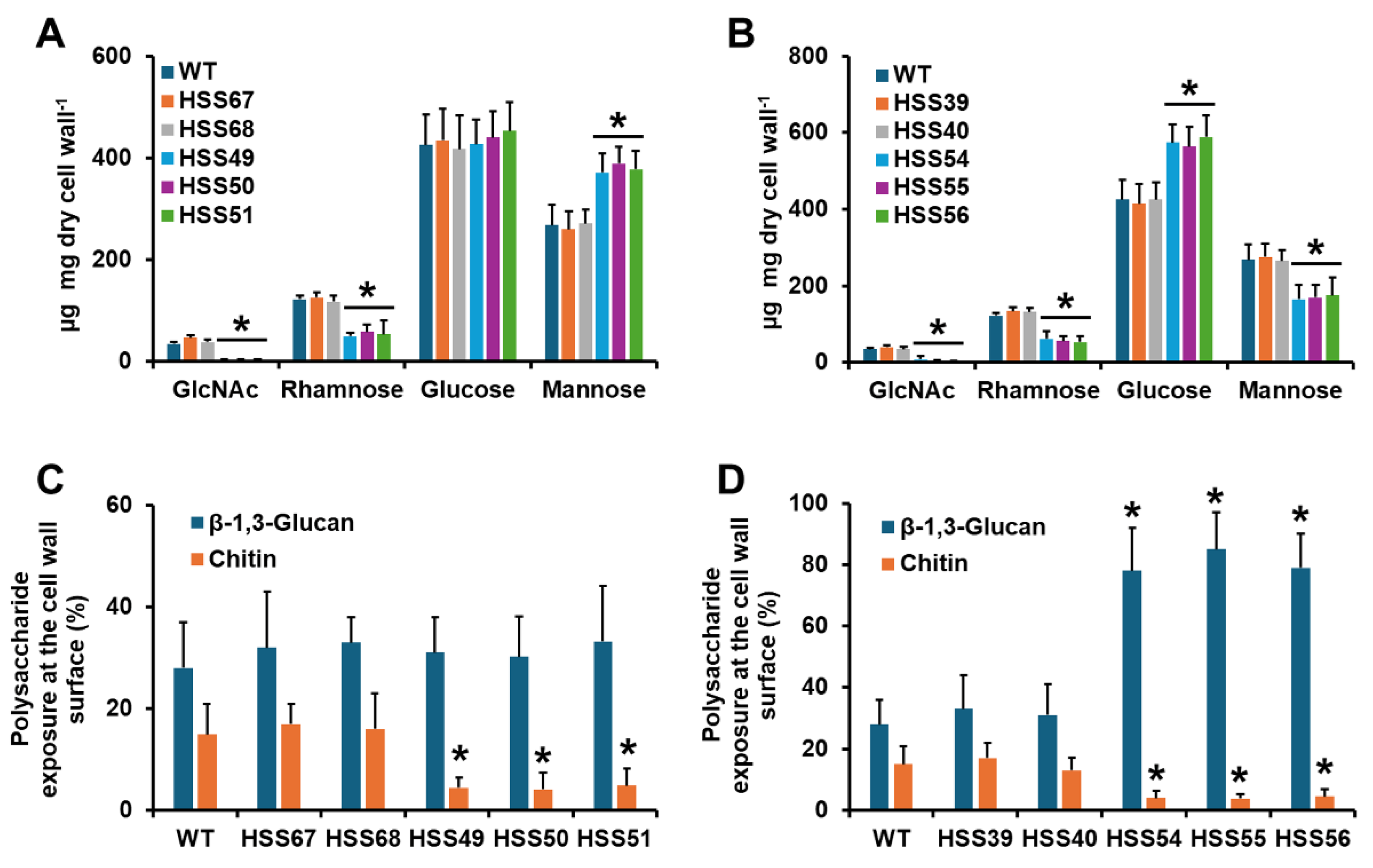
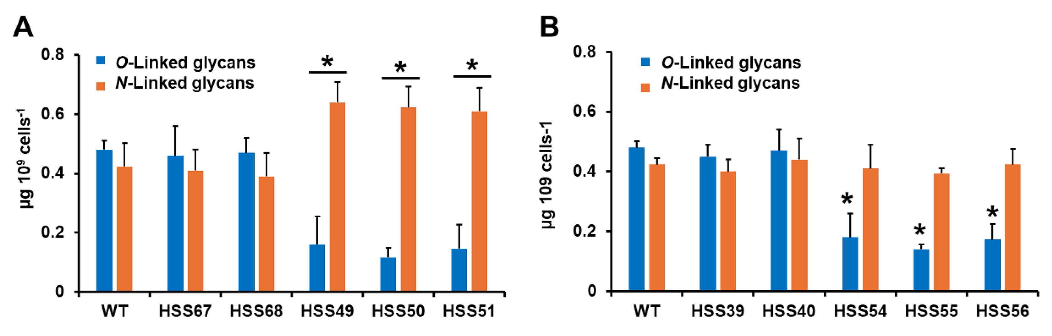
3.2. Silencing of Sporothrix schenckii MNT1 or PMT2 Affected the Cell Wall Composition and Protein Glycosylation

Since glycoproteins are part of the cell wall, and the genes under study are part of the O-linked glycosylation pathway, we next analyzed the impact of their silencing on the cell wall composition. Upon cell disruption, the cell walls were isolated and acid-hydrolyzed to break down polysaccharides and oligosaccharides into monosaccharides [35]. This treatment releases glucose and N-acetylglucosamine from glucans and chitin, respectively, and mannose and rhamnose from O-linked and N-linked glycans linked to cell wall glycoproteins [36]. The WT and control strains (HSS67, HSS68, HSS39, and HSS40) showed a similar content of monosaccharides, where glucose was the most abundant monosaccharide followed by mannose, rhamnose, and N-acetylglucosamine (Figure 2A,B). For the case of MNT1 silencing, the three mutant strains showed similar monosaccharide levels among them, and when compared with the WT or control strains, these contained lower N-acetylglucosamine and rhamnose levels (Figure 2A). Glucose levels were not affected, but mannose content was higher than the WT and control strains (Figure 2A). For the PMT2-silenced strains, N-acetylglucosamine, rhamnose, and mannose levels were lower than the WT and control strains, but glucose levels were higher (Figure 2B). The three PMT2-silenced strains did not show differences in the cell wall composition when compared among them. Next, we analyzed the distribution of the cell wall polysaccharides β-1,3-glucan and chitin, which are mainly located in the inner part of the cell wall, underneath glycoproteins [36]. We used bulky FITC-conjugated lectins that only have access to the polysaccharide located at the cell surface [37,38]. The WT and control strains showed similar levels of exposed β-1,3-glucan and chitin at the cell surface (Figure 2C,D). For MNT1-silenced strains, the three mutants did not show significant variations in β-1,3-glucan, but significantly lower chitin labeling (Figure 2C), which is in line with the N-acetylglucosamine content (Figure 2A). For the PMT2-silenced mutants, a similar trend was observed for chitin labeling (Figure 2D), which once again is in line with the monosaccharide content (Figure 2B). In addition, the three PMT2-silenced strains showed higher β-1,3-glucan content at the cell surface than the WT or control strains (Figure 2D).
The cell wall protein content was also quantified. For this purpose, walls were extensively washed with detergent, boiled, and treated with reductive agents to remove adsorbed or loosely attached proteins [35]. The WT and the four control strains showed similar cell wall protein content (Table 2), but the three MNT1-silenced strains and the three PMT2-silenced strains showed increased and similar levels of wall protein (Table 2). It was previously shown that the S. schenckii cell wall has a net-negative charge, as it can bind the cationic dye Alcian blue [24]. The WT and control strains showed similar ability to bind the dye, but for the case of the MNT1-silenced strains, the three mutants barely bound the dye (Table 2). The three PMT2-silenced strains also showed a reduction in the dye bound in the wall but this was not as severe as that observed in the MNT1-silenced strains. Collectively, these results suggest that the silencing of MNT1 or PMT2 affected the cell wall composition and organization.
Next, we analyzed the cell wall O-linked and N-linked glycan content. The walls were trimmed with either endoglycosidase H or β-eliminated to trim N-linked or O-linked glycans, respectively. The released glycans were quantified by HPAEC-PAD. WT and control strains (HSS67, HSS68, HSS39, and HSS40) showed similar levels of both types of glycans (Figure 3A,B). The three MNT1-silenced strains showed a significant reduction in O-linked glycans and an increment in N-linked glycans (Figure 3A). The three PMT2-silenced strains did not show changes in the N-linked glycan content, but a reduction in the O-linked glycan levels was observed (Figure 3B). These data suggest changes in the glycosylation pathways.

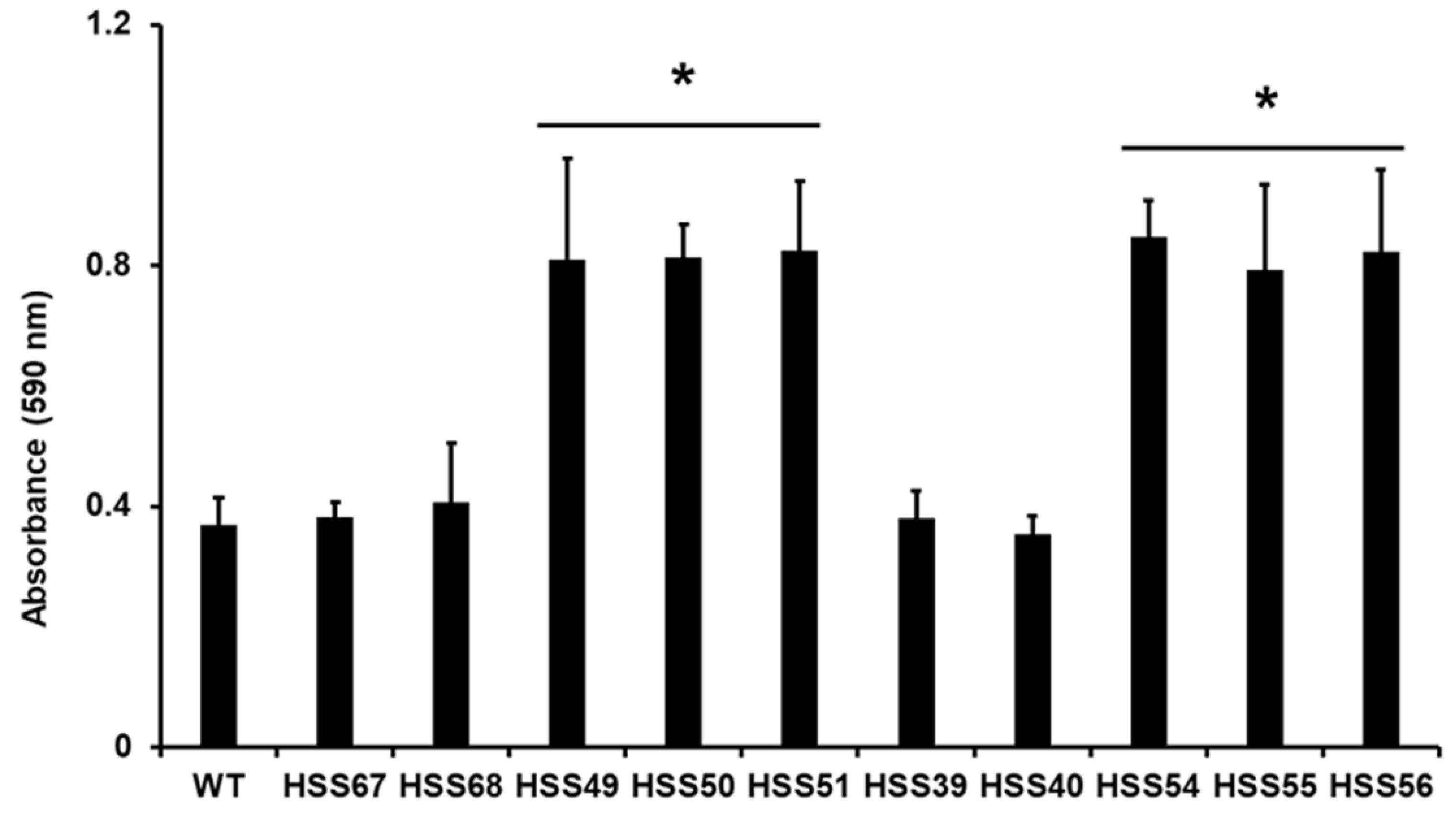
3.3. Silencing of Sporothrix schenckii MNT1 or PMT2 Affected Cell Adhesion, Biofilm Formation, and Secreted Protease and Lipase

The cell wall characterization of the mutants indicated changes in the wall composition and organization. Therefore, it is likely that biological functions associated with the cell wall, such as cell adhesion, biofilm formation, and hydrolytic enzymes [45,62] may be affected. The WT and control strains showed a similar adhesion profile to extracellular matrix components, having the highest binding ability to laminin and fibrinogen, followed by type-I and type-II collagen, elastin, and fibrinogen. This adhesion profile is similar to that previously reported for S. schenckii yeast-like cells [43,63,64] (Figure 4). Cells failed to adhere to thrombospondin-1, as previously documented for S. schenckii [43,63,64] (Figure 4). Control wells with no ECM component gave threshold readings, similar to those where thrombospondin was present (Figure 4). Both silenced mutant cells showed a similar defect in adhesion, with a significantly lower ability to bind to laminin, elastin, fibrinogen, fibronectin, and type-I and type-II collagens (Figure 4).
Next, the ability to form biofilms was investigated. We followed a well-standardized methodology to assess biofilm formation by biomass staining with crystal violet [40,65,66]. It is relevant to note that non-adherent cells were discarded after cell adhesion was stimulated, which ensures the establishment of biofilms [66]. The biomass within biofilms formed by the WT and control cells was similar among them, but those generated by the MNT1- and PMT2-silencing mutants were significantly higher and similar across them (Figure 5).
We also measured the levels of secreted protease and lipase activity of yeast-like cells. The secreted protease activity was similar among the WT and control strains (Table 3). This tended to be significantly lower in the MNT1-silenced mutants, and this was even lower in the PMT2-silenced mutants (Table 3). A similar trend was observed with the secreted lipase activity, with similar levels for the WT and control strains and lower levels for the mutant strains (Table 3). Once again, the PMT2-silenced strains showed lower enzyme levels than the MNT1-silenced strains (Table 3). The lower levels of secreted enzymes may be due to defects in the secretory pathway that transports these enzymes to the extracellular compartment. Thus, we measured the content of intracellular enzyme activities. For both protease and lipase, the WT, control, and MNT1-silenced strains showed similar levels of activity (Table 3). In the case of the PMT2-silenced mutants, both protease and lipase activities were significantly higher than the WT or control strains (Table 3). These data suggest that, at least in the PMT2-silenced mutant, the reduction in secreted hydrolytic activities may be linked to defects in their secretion to the extracellular compartment.

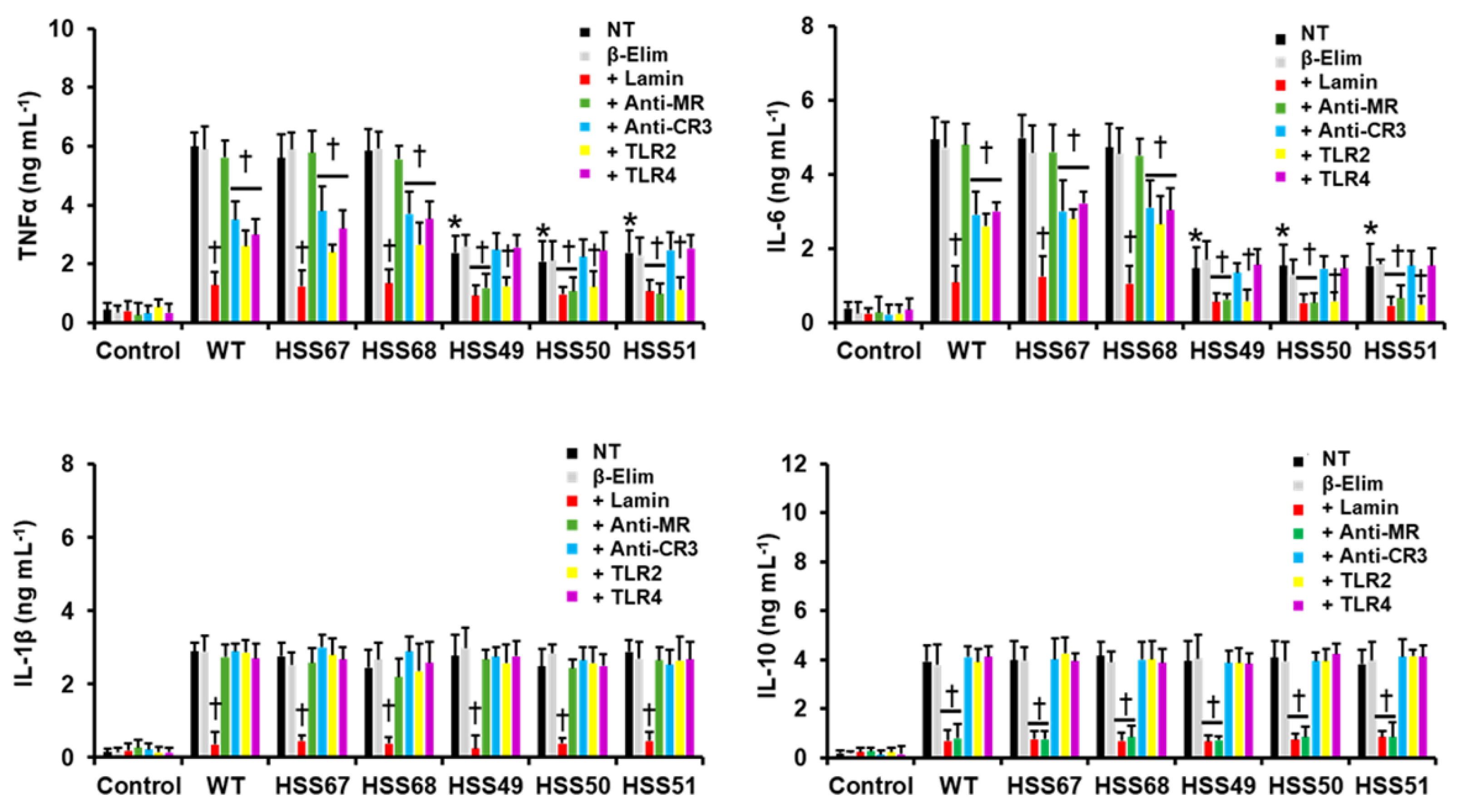
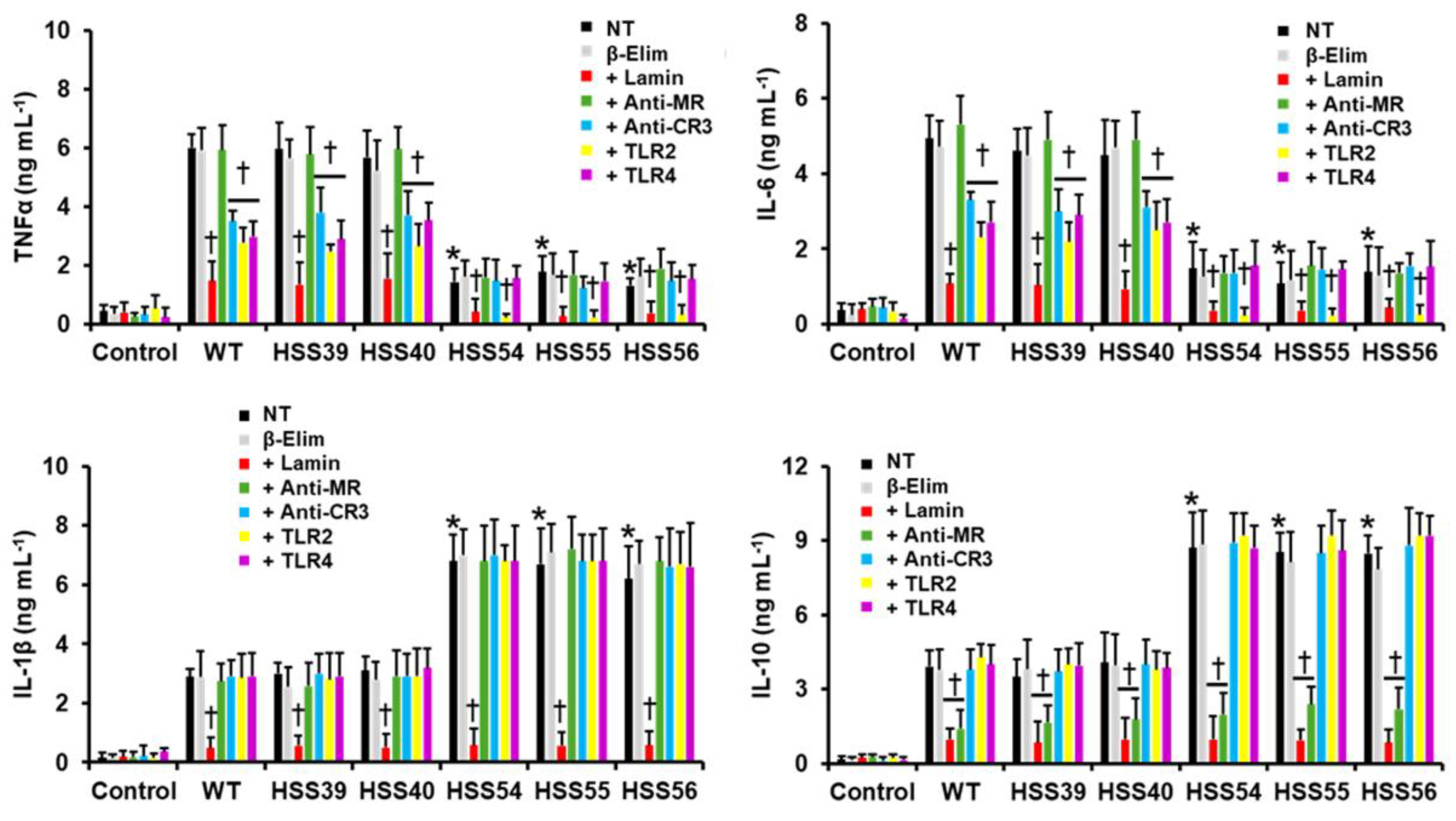
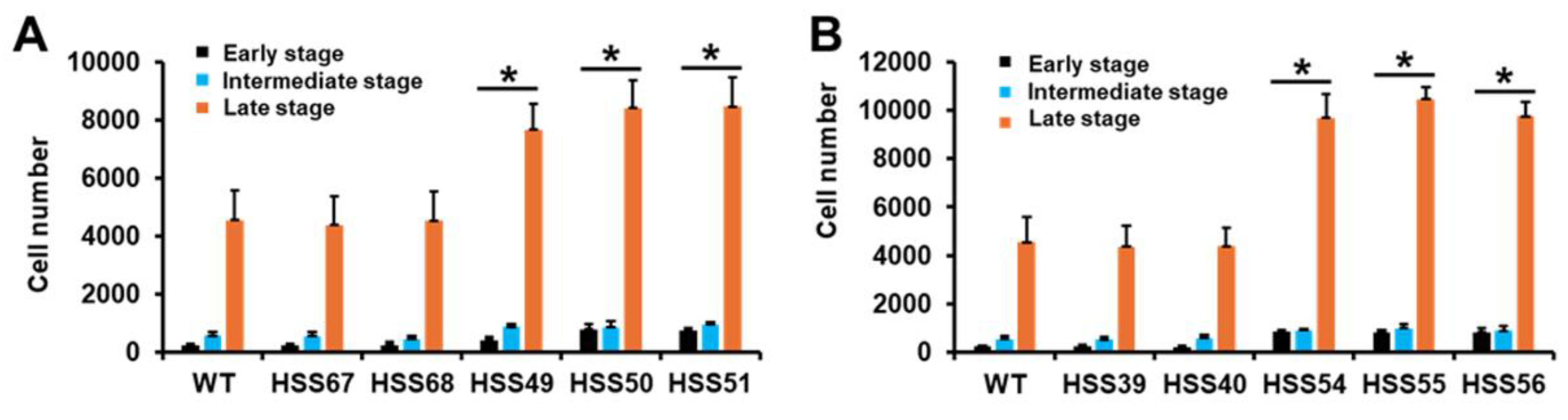
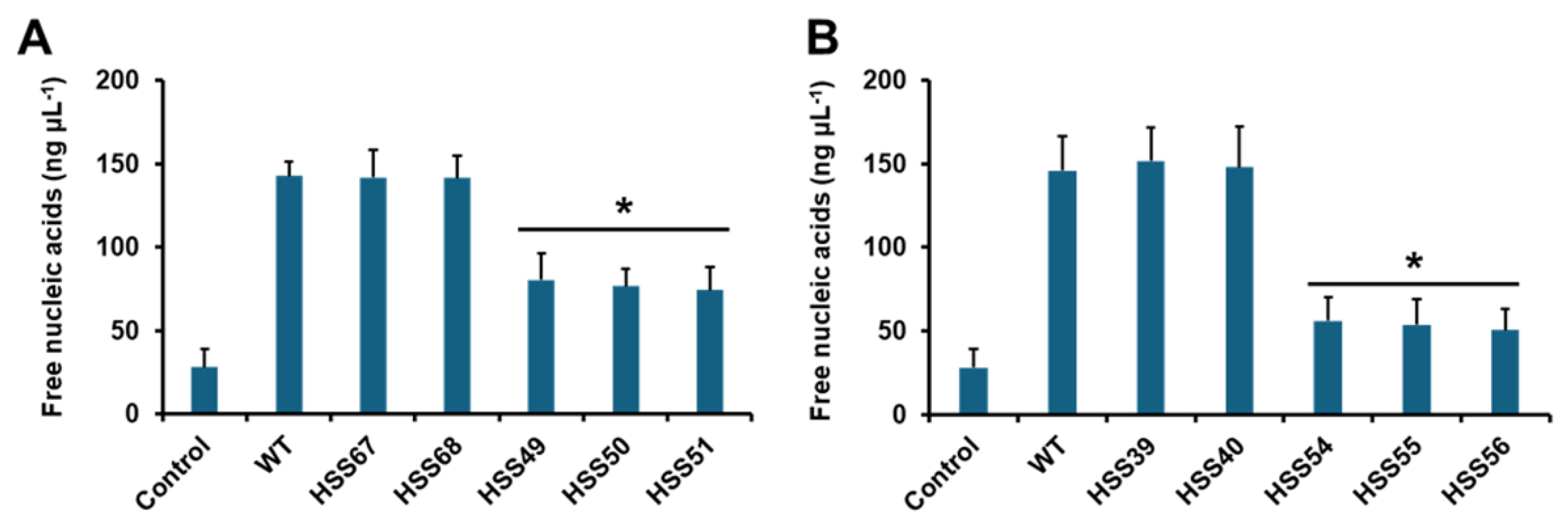
3.4. Human Innate Immune Cell–Sporothrix schenckii Interaction Is Affected by MNT1 or PMT2 Silencing

Next, we analyzed whether the MNT1 or PMT2 silencing affected the S. schenckii yeast-like cells’ interaction with human innate immune cells, particularly PBMCs. We measured the proinflammatory cytokines TNFα, IL-6, and IL-1β, and the anti-inflammatory cytokine IL-10, as these have shown to be good parameters to assess how cell wall changes affect the host–Sporothrix interaction [23,24,31,49,64,67]. The WT and control strains stimulated similar levels of the four cytokines (Figure 6 and Figure 7). In the case of the MNT1-silenced strains, these stimulated similar and reduced levels of TNFα and IL-6, whereas no changes were observed in the IL-1β and IL-10 stimulation. We repeated these experiments with β-eliminated cells, where O-linked glycans are chemically removed from the cell wall, and the results were similar to those generated with untreated cells, suggesting that the observations are, indeed, linked to the reduced O-linked glycan content (Figure 6). When the contribution of some pattern recognition receptors on this cell–cell interaction was analyzed, we found that TNFα and IL-6 production stimulated by the WT and control strains was dependent on recognition by dectin-1, complement receptor 3 (CR3), TLR2, and TLR4, while mannose receptor (MR) was dispensable for these two cytokines production (Figure 6 and Figure 7). However, MR was required for the TNFα and IL-6 stimulation by the MNT1-silenced strains (Figure 6). Different from the WT and control cells, the stimulation of these two cytokines by MNT1-silenced strains was not sensitive to the presence of anti-CR3 and anti-TLR4 antibodies, indicating that these receptors are not participating in cytokine stimulation by these mutant cells (Figure 6). The IL-1β production was dectin-1-dependent for WT, control, and MNT1-silenced strains, whereas IL-10 production was dectin-1- and MR-dependent. Control interactions with irrelevant isotype-matched antibodies gave similar results to interactions with specific antibodies.
For the PMT2-silenced mutants, TNFα and IL-6 stimulation was reduced, whereas IL-1β and IL-10 productions were significantly increased (Figure 7). Similarly to the MNT1-silenced mutants, β-elimination of the PMT2-silenced strains stimulated similar cytokine levels as the untreated cells (Figure 7). Both TNFα and IL-6 production were dependent on dectin-1 and TLR2, but contrasting with WT and control cells, CR3 and TLR4 were dispensable for these cytokines by the mutant strains (Figure 7). For IL-1β stimulation, this was dependent on dectin-1 for WT, control, and silencing strains (Figure 7); while dectin-1 and MR were required for IL-10 stimulation in a similar way for all the tested strains (Figure 7). Control interactions with irrelevant isotype-matched antibodies gave similar results to interactions with specific antibodies. Collectively, these data indicated that silencing of either MNT1 or PMT2 affected the PBMCs–S. schenckii interaction.
Next, we assessed the interaction of the silenced mutants with human monocyte-derived macrophages. We analyze the fungal uptake by cytometry, and depending on the fluorescent channel, cells can be grouped in the early, intermediate, and late stages of the phagocytic process [49,53]. After 2 hours of interaction, most of the human cells were in the late stage of phagocytosis when interacting with the WT strain, followed by cells in the intermediate and early stages (Figure 8). The control strains for both silenced genes showed similar uptake profiles to the WT strain (Figure 8A,B). Both the MNT1- and the PMT2-silenced strains showed an increment in the phagocytosis at the three different stages, having more PMT2-silenced cells phagocytosed than the MNT1-silenced mutants (Figure 8A,B). These results indicated that silencing of MNT1 or PMT2 positively affected the S. schenckii phagocytosis.
We also analyzed the interaction of S. schenckii yeast-like cells with human granulocytes, closely evaluating the ability of the silenced strains to stimulate neutrophil extracellular traps. Among the several components released by neutrophils during trap formation are the nucleic acids, which generate a sticky net that reduces the mobility of pathogens [68]. So, we measured the release of nucleic acids as an indirect parameter of the formation of extracellular traps. Control cells, with no fungal cells included, released low levels of nucleic acids, but this was significantly increased when human cells were incubated with WT cells (Figure 9A,B). The control strains for both silenced mutant sets showed similar ability to stimulate traps as the WT strain (Figure 9A,B). Both MNT1- and PMT2-silenced mutant strains showed a reduced ability to stimulate the extracellular traps when compared to the WT or control strains (Figure 9A,B). These data indicate that silencing of MNT1 or PMT2 also affected the interaction of S. schenckii with human granulocytes.

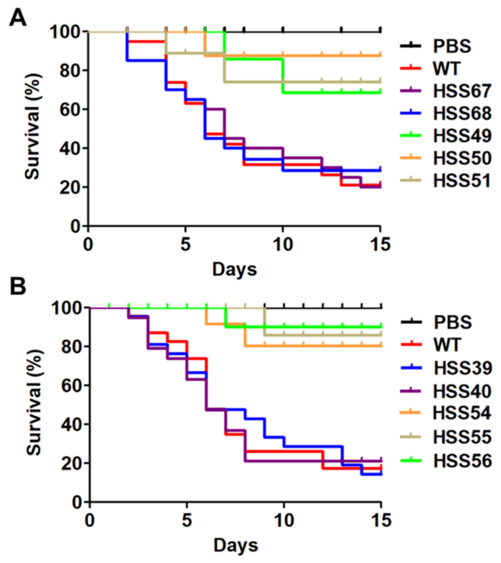
3.5. Virulence Is Attenuated in the Sporothrix schenckii MNT1- and PMT2-Silenced Mutans

Next, the virulence of the MNT1- and PMT2-silenced strains were analyzed in the alternative model of experimental sporotrichosis G. mellonella, since this generates similar results as the murine model of sporotrichosis, in terms of mortality rates [55,69,70,71,72]. The larvae infected with the WT and control strains showed a mortality rate of 80.9 ± 6.4%, with a median survival of 6.5 ± 0.9 days (Figure 10A,B). The larvae inoculated with MNT1-silenced strains showed similar survival curves, with a mortality rate of 12.2 ± 1.2% and a median survival of more than 15 days (Figure 10A). The PMT2-silenced mutants also showed virulence attenuation, generating a mortality rate of 6.7 ± 2.1% with a median survival of more than 15 days (Figure 10B). In both cases, the larvae infected with the silenced strains showed significantly different survival curves when compared to the WT or control strains (p < 0.05; Figure 10). The control group inoculated only with PBS did not show mortality during the two-week observation period (Figure 10). To associate larval death with the presence of fungal cells, hemolymph was collected, and the colony-forming units were quantified. A similar fungal burden was observed for the WT, control, and silenced strains (Table 4).
To further characterize the interaction between S. schenckii and G. mellonella, some hemolymph parameters were analyzed. Previous studies have shown that reduction in the hemocyte counts, cytotoxicity, melanin formation, and phenoloxidase activity are associated with virulence attenuation [23,31,57,64]. The larva groups inoculated with the WT or control strains showed similar levels in the four parameters under analysis (Table 4). However, both the MNT1- and the PMT2-silenced mutants showed significantly lower levels in the four investigated parameters when compared to either WT or control strains (Table 4). Collectively, these data indicate that silencing of either MNT1 or PMT2 negatively affected virulence in the model G. mellonella.

4. Discussion

S. schenckii belongs to the Sporothrix pathogenic clade and is the most studied species within this clade, especially in terms of its biology, genetics, and other fundamental aspects [61]. As in other pathogenic fungi, the cell wall plays an essential role in the interaction with the host. Therefore, it is relevant to analyze its synthesis, composition, organization, and dynamic changes [73,74]. The N- and O-glycans covalently bound to cell wall proteins play a crucial role in their structure and function, are species-specific, and show unique structures and compositions [8,75]. Such particularities generate distinctive molecular profiles that influence the interaction with the host immune response. In S. schenckii, the presence of these N- and O-glycans has been reported, most of them being part of the peptidorhamnomannan [25]. In addition, at least the N-linked glycans have been demonstrated to play a key role during the Sporothrix–host interaction [23,24,31,36].
In this study, we report the first silencing of two genes involved in the S. schenckii O-linked glycosylation pathway and evaluate its contribution to the biology of this organism and its role in pathogen–host interaction. To genetically manipulate this organism, we used the gene silencing strategy, which is well described, and although the insertion of the binary vector is random, several mutants with similar silencing levels were included to rule out the observed phenotypes being the product of insertional events [23,24,31,64]. The PMT2 silencing in S. schenckii represented a greater difficulty compared to MNT1. In the case of PMT2, the maximum level of silencing obtained was 70%. Considering studies in other organisms, such as C. neoformans and Schizosaccharomyces pombe [76,77,78], it is likely that PMT2 is an essential gene not only in the O-glycosylation pathway, but also in cell viability.
Silencing of S. schenckii MNT1 or PMT2 led to cell aggregation and relevant changes in growth and morphology. Since notable differences were observed between mutants of the different genes, it is possible to suggest that each of these genes contributes differently to the fungus’ biology. These results suggest that proper O-linked glycosylation is imperative for cell fitness and growth, as has been reported for mutants of these genes in C. albicans, C. tropicalis, S. cerevisiae, S. pombe, and C. neoformans [11,17,77,79,80,81]. It is hypothesized that the observed phenotypic changes could be related to alterations in the S. schenckii cell wall composition, as it has been demonstrated in C. albicans mutants in the O-linked glycosylation pathway [11,79]. In the case of MNT1-silenced mutants, a reduction in the percentage of N-acetylglucosamine and rhamnose was found, along with an increase in mannose, supporting this hypothesis. Furthermore, the results of Alcian blue binding assays and structural polysaccharide distribution analysis reinforce this idea. Different from the MNT1-silenced mutants, the PMT2-silenced strains showed an additional increase in glucan levels. These changes have been associated with the activation of regulatory mechanisms in response to stress, such as the cell wall integrity pathway [82,83]. This adaptive mechanism involves activation of the Pkc1-Mkc1 cascade and β-1,3-glucan synthase, which generates a compensatory increment in structural polysaccharides and their redistribution to avoid cell death [84]. Therefore, the increase in glucans observed in PMT2-silenced strains could be explained by the activation of the cell wall integrity pathway. This hypothesis is supported by the Alcian blue binding assay, in which the mutants showed a lower affinity for the dye, and the levels of polysaccharides exposed at the cell surface, which indicated modifications in protein glycosylation and cell wall rearrangement, respectively. Similar observations have been described in S. schenckii OCH1 and ROT2-silenced mutants [23,24]. The increase in cell wall protein content is consistent with the compensatory response of S. schenckii to maintain cell wall integrity in the presence of O-linked glycosylation defects. Alternatively, the increment in cell wall protein could be related to alterations in the secretory pathways, which could generate an abnormal protein accumulation in the cell wall. However, this explanation is not consistent with the results of protease and lipase activity assays, which revealed a decrease in the enzyme secretion, accompanied by an increase in their intracellular activity. These findings suggest that deficiencies in glycosylation not only affect cell wall composition, but also protein secretion. This could impact several key processes, including cell adhesion and biofilm formation. Protein glycosylation and the secretory pathway are two intimately linked pathways. In C. albicans and S. cerevisiae, defects in the secretory pathway affected protein glycosylation, probably because local stress in the Golgi complex affects glycosyltransferases [85,86]. Similarly, defects in protein glycosylation affected protein secretion in C. albicans and C. tropicalis [35,45]. Therefore, the observed defect in protein secretion in the silenced mutants analyzed in this study is not surprising.
Both sets of silencing mutants exhibited a reduced ability to adhere to extracellular matrix components, but retained their ability to form biofilms, raising several possible explanations. One hypothesis is that, despite the reduction in adhesion to the extracellular matrix components, the cells might be able to form biofilms due to an increase in the production of exopolysaccharides or other extracellular components, which facilitates cell adhesion to each other and favors the formation of a cohesive structure [87]. However, this is unlikely given the already-mentioned defect in protein secretion. Alternatively, changes in the regulation of genes related to biofilm biogenesis could contribute to this phenomenon, suggesting that key regulators of gene expression are decorated with O-linked glycans.
As expected, a decrease in O-linked glycan content was observed in both MNT1-silenced and PMT2-silenced mutants. However, in the case of the latter, no compensatory effect of N-glycan content was observed, unlike what was found in MNT1-silenced mutants. This phenomenon is particularly interesting because this kind of compensatory mechanism has not been reported in other strains that are deficient in this gene. One possible explanation is that other members of the MNT1 gene family may be upregulated in this genetic background. These gene products participate in the N-linked glycan extension [59]. This implies that in the MNT1-silenced mutants, the increment in N-linked glycans represents more sugar units per glycan rather than more glycosylated sites per protein.
MNT1 and PMT2 silencing resulted in a reduced ability to stimulate the cytokines IL-6 and TNFα when S. schenckii cells interacted with human PBMCs. However, PMT2 silencing induced higher levels of IL-10 and IL-1β, comparing to both the MNT1-silenced and WT strains. This increased cytokine production is likely linked to the higher levels of β-1,3-glucan, as this stimulation was blocked by laminarin, an antagonist of dectin-1, and anti-TLR2 antibodies, the two main immune receptors for β-1,3-glucans [88,89]. The changes in the cell wall were translated into changes in the contribution of pattern recognition receptors when the mutant cells were interacting with human PBMCs, underscoring the close relationship between fungal cell wall structure and immune sensing.
Even though both sets of mutants were readily phagocytosed by macrophages, this was more pronounced in PMT2-silenced mutants. This may be explained by the increment in β-1,3-glucan content, as mutants with similar cell wall defects tended to be more phagocytosed by human monocyte-derived macrophages [23,31,64].
The fact that both the MNT1- and PMT2-silenced mutants poorly stimulated neutrophil extracellular traps, along with the poor ability to stimulate proinflammatory cytokines by human PBMCs make us hypothesize that fungal O-linked glycans are key for the establishment of a proinflammatory response in the host when interacting with the pathogen. This observation contrasts with that reported in C. albicans, where these cell wall structures are a minor contributor to the stimulation of proinflammatory effectors [90]. This discrepancy is likely explained by the structure of O-linked glycans in these species. While C. albicans has O-linked glycans composed entirely of mannose units, S. schenckii has O-linked glycans composed of mannose, glucuronic acid, and rhamnose [25]. Rhamnose is a rare monosaccharide in the kingdom of fungi that has proinflammatory properties when included in S. schenckii glycans [31].
MNT1 and PMT2 silencing led to virulence attenuation in the G. mellonella model. These observations are consistent with MNT1 disruption in C. albicans and A. fumigatus and PMT2 deletion in C. albicans [11,20]. Considering that similar fungal loads were recovered from all MNT1 and PMT2 silencing strains, it is unlikely that the results are biased by the inability of the silencing mutants to adapt and grow in host tissues. Instead, our findings suggest that the observed defects are due to alterations in the repertoire of virulence factors, especially those that depend on proper glycosylation to perform their molecular function. Among the virulence factors identified in S. schenckii, adhesins and biofilm formation appear to be especially susceptible to defects in protein glycosylation [31], as some of them are highly glycosylated proteins, containing mannose- and rhamnose-based oligosaccharides [23,44]. It was clear that the mutant strains recovered the ability to grow within larvae hemolymph because our in vitro characterization indicated growth defects. The host milieu is likely to be a more stressful scenario than the culturing medium, and adaptation to cope with different stresses is mandatory for cell survival. Then, it is possible to speculate that this adaptation process is behind the discrepancy with the in vitro growth. The lower cytotoxicity observed in larvae inoculated with the silencing strains suggests a reduction in their ability to induce cell damage, in line with lower virulence. In addition, the low-hemocyte, melanin, and phenoloxidase levels indicated that insect immunity did not require a major upregulation to control the fungal cells, suggesting an immunological tolerance because of the low levels of cell damage in the host.
In conclusion, this study demonstrates that S. schenckii MNT1 and PMT2 are key for proper O-glycosylation, as well as for the organization and composition of the cell wall. The silencing of these genes affected the interaction of the fungus with human PBMCs, macrophages, and neutrophils, along with its virulence. This study paves the way for future research on O-linked glycosylation as a drug target candidate to control the infection caused by S. schenckii.

Author Contributions

Conceptualization, M.G.-G. and H.M.M.-M.; methodology, M.G.-G., J.A.M.-Á., A.R.d.S.B. and H.M.M.-M.; investigation, M.G.-G., I.M.-D. and H.M.M.-M.; and resources, H.M.M.-M.; data curation, H.M.M.-M., M.G.-G. and I.M.-D.; writing—original draft preparation, H.M.M.-M., A.R.d.S.B. and M.G.-G.; supervision, H.M.M.-M. and I.M.-D.; project administration, H.M.M.-M.; funding acquisition, H.M.M.-M. All authors have read and agreed to the published version of the manuscript.

Funding

This work was supported by the Secretaría de Ciencia, Humanidades, Tecnología e Innovación [Ciencia de Frontera 2019-6380 and CBF2023-2024-655], and Red Temática Glicociencia en Salud [CONACYT-México].

Institutional Review Board Statement

In this study, the use of human primary cells was approved by the Ethics Committee of the University of Guanajuato (CIBIUG-P52-2021). Human cells were collected from healthy adult volunteers after information about the study was provided and written informed consent was obtained. The procedures were carried out following the Declaration of Helsinki.

Informed Consent Statement

Not applicable.

Data Availability Statement

The original contributions presented in this study are included in the article. Further inquiries can be directed to the corresponding author(s).

Acknowledgments

We thank Gordon Brown from the University of Aberdeen for the donation of the IgG Fc-Dectin-1 chimera (current address is at the University of Exeter). We thank Luz A. López-Ramírez for the technical assistance in this project.

Conflicts of Interest

The authors declare no conflicts of interest. The funders had no role in the design of the study; in the collection, analyses, or interpretation of data; in the writing of the manuscript; or in the decision to publish the results.

References

  1. Rodrigues, A.M.; de Hoog, S.; de Camargo, Z.P. Emergence of pathogenicity in the Sporothrix schenckii complex. Med. Mycol. 2013, 51, 405–412. [Google Scholar] [CrossRef]
  2. Queiroz-Telles, F.; Fahal, A.H.; Falci, D.R.; Caceres, D.H.; Chiller, T.; Pasqualotto, A.C. Neglected endemic mycoses. Lancet Infect. Dis. 2017, 17, e367–e377. [Google Scholar] [CrossRef] [PubMed]
  3. de Lima Barros, M.B.; Schubach, A.O.; de-Vasconcellos Carvalhaes De-Oliveira, R.; Martins, E.B.; Teixeira, J.L.; Wanke, B. Treatment of cutaneous sporotrichosis with Itraconazole—Study of 645 patients. Clin. Infect. Dis. 2011, 52, e200–e206. [Google Scholar] [CrossRef]
  4. Rangel-Gamboa, L.; Martínez-Hernandez, F.; Maravilla, P.; Arenas-Guzmán, R.; Flisser, A. Update of phylogenetic and genetic diversity of Sporothrix schenckii sensu lato. Med. Mycol. 2016, 54, 248–255. [Google Scholar] [CrossRef] [PubMed]
  5. Lopes-Bezerra, L.M.; Walker, L.A.; Niño-Vega, G.; Mora-Montes, H.M.; Neves, G.W.P.; Villalobos-Duno, H.; Barreto, L.; Garcia, K.; Franco, B.; Martínez-Álvarez, J.A.; et al. Cell walls of the dimorphic fungal pathogens Sporothrix schenckii and Sporothrix brasiliensis exhibit bilaminate structures and sloughing of extensive and intact layers. PLoS Negl. Trop. Dis. 2018, 12, e0006169. [Google Scholar] [CrossRef]
  6. Gómez-Gaviria, M.; Vargas-Macías, A.P.; García-Carnero, L.C.; Martínez-Duncker, I.; Mora-Montes, H.M. Role of Protein Glycosylation in Interactions of Medically Relevant Fungi with the Host. J. Fungi 2021, 7, 875. [Google Scholar] [CrossRef]
  7. Diaz-Jimenez, D.F.; Mora-Montes, H.M.; Hernandez-Cervantes, A.; Luna-Arias, J.P.; Gow, N.A.; Flores-Carreon, A. Biochemical characterization of recombinant Candida albicans mannosyltransferases Mnt1, Mnt2 and Mnt5 reveals new functions in O- and N-mannan biosynthesis. Biochem. Biophys. Res. Commun. 2012, 419, 77–82. [Google Scholar] [CrossRef] [PubMed]
  8. Goto, M. Protein O-glycosylation in fungi: Diverse structures and multiple functions. Biosci. Biotechnol. Biochem. 2007, 71, 1415–1427. [Google Scholar] [CrossRef]
  9. Lommel, M.; Strahl, S. Protein O-mannosylation: Conserved from bacteria to humans. Glycobiology 2009, 19, 816–828. [Google Scholar] [CrossRef]
  10. Martínez-Duncker, I.; Díaz-Jímenez, D.F.; Mora-Montes, H.M. Comparative analysis of protein glycosylation pathways in humans and the fungal pathogen Candida albicans. Int. J. Microbiol. 2014, 2014, 267497. [Google Scholar] [CrossRef]
  11. Munro, C.A.; Bates, S.; Buurman, E.T.; Hughes, H.B.; Maccallum, D.M.; Bertram, G.; Atrih, A.; Ferguson, M.A.; Bain, J.M.; Brand, A.; et al. Mnt1p and Mnt2p of Candida albicans are partially redundant alpha-1,2-mannosyltransferases that participate in O-linked mannosylation and are required for adhesion and virulence. J. Biol. Chem. 2005, 280, 1051–1060. [Google Scholar] [CrossRef]
  12. Gentzsch, M.; Tanner, W. Protein-O-glycosylation in yeast: Protein-specific mannosyltransferases. Glycobiology 1997, 7, 481–486. [Google Scholar] [CrossRef]
  13. Timpel, C.; Strahl-Bolsinger, S.; Ziegelbauer, K.; Ernst, J.F. Multiple functions of Pmt1p-mediated protein O-mannosylation in the fungal pathogen Candida albicans. J. Biol. Chem. 1998, 273, 20837–20846. [Google Scholar] [CrossRef]
  14. Leitao, E.A.; Bittencourt, V.C.; Haido, R.M.; Valente, A.P.; Peter-Katalinic, J.; Letzel, M.; de Souza, L.M.; Barreto-Bergter, E. Beta-galactofuranose-containing O-linked oligosaccharides present in the cell wall peptidogalactomannan of Aspergillus fumigatus contain immunodominant epitopes. Glycobiology 2003, 13, 681–692. [Google Scholar] [CrossRef] [PubMed]
  15. Prill, S.K.; Klinkert, B.; Timpel, C.; Gale, C.A.; Schröppel, K.; Ernst, J.F. PMT family of Candida albicans: Five protein mannosyltransferase isoforms affect growth, morphogenesis and antifungal resistance. Mol. Microbiol. 2005, 55, 546–560. [Google Scholar] [CrossRef] [PubMed]
  16. Zhou, H.; Hu, H.; Zhang, L.; Li, R.; Ouyang, H.; Ming, J.; Jin, C. O-Mannosyltransferase 1 in Aspergillus fumigatus (AfPmt1p) is crucial for cell wall integrity and conidium morphology, especially at an elevated temperature. Eukaryot. Cell 2007, 6, 2260–2268. [Google Scholar] [CrossRef]
  17. Hernández-Chávez, M.J.; Martínez-Duncker, I.; Clavijo-Giraldo, D.M.; López-Ramirez, L.A.; Mora-Montes, H.M. Candida tropicalis PMT2 Is a dispensable gene for viability but required for proper interaction with the host. J. Fungi 2024, 10, 502. [Google Scholar] [CrossRef] [PubMed]
  18. Jin, C. Protein glycosylation in Aspergillus fumigatus is essential for cell wall synthesis and serves as a promising model of multicellular eukaryotic development. Int. J. Microbiol. 2012, 2012, 654251. [Google Scholar] [CrossRef]
  19. Lee, D.J.; Bahn, Y.S.; Kim, H.J.; Chung, S.Y.; Kang, H.A. Unraveling the novel structure and biosynthetic pathway of O-linked glycans in the Golgi apparatus of the human pathogenic yeast Cryptococcus neoformans. J. Biol. Chem. 2015, 290, 1861–1873. [Google Scholar] [CrossRef]
  20. Wagener, J.; Echtenacher, B.; Rohde, M.; Kotz, A.; Krappmann, S.; Heesemann, J.; Ebel, F. The putative alpha-1,2-mannosyltransferase AfMnt1 of the opportunistic fungal pathogen Aspergillus fumigatus is required for cell wall stability and full virulence. Eukaryot. Cell 2008, 7, 1661–1673. [Google Scholar] [CrossRef]
  21. Kadooka, C.; Hira, D.; Tanaka, Y.; Chihara, Y.; Goto, M.; Oka, T. Mnt1, an α-(1 → 2)-mannosyltransferase responsible for the elongation of N-glycans and O-glycans in Aspergillus fumigatus. Glycobiology 2022, 32, 1137–1152. [Google Scholar] [CrossRef] [PubMed]
  22. Mora-Montes, H.M.; Bates, S.; Netea, M.G.; Castillo, L.; Brand, A.; Buurman, E.T.; Diaz-Jimenez, D.F.; Jan Kullberg, B.; Brown, A.J.; Odds, F.C.; et al. A multifunctional mannosyltransferase family in Candida albicans determines cell wall mannan structure and host-fungus interactions. J. Biol. Chem. 2010, 285, 12087–12095. [Google Scholar] [CrossRef]
  23. López-Ramírez, L.A.; Martínez-Duncker, I.; Márquez-Márquez, A.; Vargas-Macías, A.P.; Mora-Montes, H.M. Silencing of ROT2, the encoding gene of the endoplasmic reticulum glucosidase II, affects the cell wall and the Sporothrix schenckii-host interaction. J. Fungi 2022, 8, 1220. [Google Scholar] [CrossRef] [PubMed]
  24. Lozoya-Pérez, N.E.; Casas-Flores, S.; de Almeida, J.R.F.; Martínez-Álvarez, J.A.; López-Ramírez, L.A.; Jannuzzi, G.P.; Trujillo-Esquivel, E.; Estrada-Mata, E.; Almeida, S.R.; Franco, B.; et al. Silencing of OCH1 unveils the role of Sporothrix schenckii N-linked glycans during the host-fungus interaction. Infect. Drug Resist. 2019, 12, 67–85. [Google Scholar] [CrossRef]
  25. Lopes-Bezerra, L.M. Sporothrix schenckii cell wall peptidorhamnomannans. Front. Microbiol. 2011, 2, 243. [Google Scholar] [CrossRef] [PubMed]
  26. Kanda, T.; Wakabayashi, K.; Nisizawa, K. Xylanase activity of an endo-cellulase of carboxymethyl-cellulase type from Irpex lacteus (Polyporus tulipiferae). J. Biochem. 1976, 79, 989–995. [Google Scholar] [CrossRef]
  27. Bauer, W.D.; Talmadge, K.W.; Keegstra, K.; Albersheim, P. The structure of plant cell walls: II. the hemicellulose of the walls of suspension-cultured Sycamore cells. Plant Physiol. 1973, 51, 174–187. [Google Scholar] [CrossRef]
  28. Nakayashiki, H.; Hanada, S.; Nguyen, B.Q.; Kadotani, N.; Tosa, Y.; Mayama, S. RNA silencing as a tool for exploring gene function in ascomycete fungi. Fungal Genet. Biol. 2005, 42, 275–283. [Google Scholar] [CrossRef]
  29. Tamez-Castrellón, A.K.; Romo-Lucio, R.; Martínez-Duncker, I.; Mora-Montes, H.M. Generation of a synthetic binary plasmid that confers resistance to nourseothricin for genetic engineering of Sporothrix schenckii. Plasmid 2018, 100, 1–5. [Google Scholar] [CrossRef]
  30. Lozoya-Pérez, N.E.; Casas-Flores, S.; Martínez-Álvarez, J.A.; López-Ramírez, L.A.; Lopes-Bezerra, L.M.; Franco, B.; Mora-Montes, H.M. Generation of Sporothrix schenckii mutants expressing the green fluorescent protein suitable for the study of host-fungus interactions. Fungal Biol. 2018, 122, 1023–1030. [Google Scholar] [CrossRef]
  31. Tamez-Castrellón, A.K.; van der Beek, S.L.; López-Ramírez, L.A.; Martínez-Duncker, I.; Lozoya-Pérez, N.E.; van Sorge, N.M.; Mora-Montes, H.M. Disruption of protein rhamnosylation affects the Sporothrix schenckii-host interaction. Cell Surf. 2021, 7, 100058. [Google Scholar] [CrossRef] [PubMed]
  32. Trujillo-Esquivel, E.; Franco, B.; Flores-Martínez, A.; Ponce-Noyola, P.; Mora-Montes, H.M. Purification of single-stranded cDNA based on RNA degradation treatment and adsorption chromatography. Nucleosides Nucleotides Nucleic Acids 2016, 35, 404–409. [Google Scholar] [CrossRef] [PubMed]
  33. Livak, K.J.; Schmittgen, T.D. Analysis of relative gene expression data using real-time quantitative PCR and the 2(-Delta Delta C(T)) Method. Methods 2001, 25, 402–408. [Google Scholar] [CrossRef]
  34. Trujillo-Esquivel, E.; Martínez-Álvarez, J.A.; Clavijo-Giraldo, D.M.; Hernández, N.V.; Flores-Martínez, A.; Ponce-Noyola, P.; Mora-Montes, H.M. The Sporothrix schenckii gene encoding for the ribosomal protein L6 has constitutive and stable expression and works as an endogenous control in gene expression analysis. Front. Microbiol. 2017, 8, 1676. [Google Scholar] [CrossRef] [PubMed]
  35. Mora-Montes, H.M.; Bates, S.; Netea, M.G.; Diaz-Jimenez, D.F.; Lopez-Romero, E.; Zinker, S.; Ponce-Noyola, P.; Kullberg, B.J.; Brown, A.J.; Odds, F.C.; et al. Endoplasmic reticulum alpha-glycosidases of Candida albicans are required for N glycosylation, cell wall integrity, and normal host-fungus interaction. Eukaryot. Cell 2007, 6, 2184–2193. [Google Scholar] [CrossRef]
  36. Martínez-Álvarez, J.A.; Pérez-García, L.A.; Mellado-Mojica, E.; López, M.G.; Martínez-Duncker, I.; Lópes-Bezerra, L.M.; Mora-Montes, H.M. Sporothrix schenckii sensu stricto and Sporothrix brasiliensis are differentially recognized by human peripheral blood mononuclear cells. Front. Microbiol. 2017, 8, 843. [Google Scholar] [CrossRef]
  37. Marakalala, M.J.; Vautier, S.; Potrykus, J.; Walker, L.A.; Shepardson, K.M.; Hopke, A.; Mora-Montes, H.M.; Kerrigan, A.; Netea, M.G.; Murray, G.I.; et al. Differential adaptation of Candida albicans in vivo modulates immune recognition by dectin-1. PLoS Pathog. 2013, 9, e1003315. [Google Scholar] [CrossRef]
  38. Mora-Montes, H.M.; Netea, M.G.; Ferwerda, G.; Lenardon, M.D.; Brown, G.D.; Mistry, A.R.; Kullberg, B.J.; O’Callaghan, C.A.; Sheth, C.C.; Odds, F.C.; et al. Recognition and blocking of innate immunity cells by Candida albicans chitin. Infect. Immun. 2011, 79, 1961–1970. [Google Scholar] [CrossRef]
  39. Hobson, R.P.; Munro, C.A.; Bates, S.; MacCallum, D.M.; Cutler, J.E.; Heinsbroek, S.E.; Brown, G.D.; Odds, F.C.; Gow, N.A. Loss of cell wall mannosylphosphate in Candida albicans does not influence macrophage recognition. J. Biol. Chem. 2004, 279, 39628–39635. [Google Scholar] [CrossRef]
  40. Navarro-Arias, M.J.; Defosse, T.A.; Dementhon, K.; Csonka, K.; Mellado-Mojica, E.; Dias Valério, A.; González-Hernández, R.J.; Courdavault, V.; Clastre, M.; Hernández, N.V.; et al. Disruption of protein mannosylation affects Candida guilliermondii cell wall, immune sensing, and virulence. Front. Microbiol. 2016, 7, 1951. [Google Scholar] [CrossRef]
  41. DuBois, M.; Gilles, K.A.; Hamilton, J.K.; Rebers, P.A.; Smith, F. Colorimetric method for determination of sugars and related substances. Anal. Chem. 1956, 28, 350–356. [Google Scholar] [CrossRef]
  42. Mora-Montes, H.M.; McKenzie, C.; Bain, J.M.; Lewis, L.E.; Erwig, L.P.; Gow, N.A. Interactions between macrophages and cell wall oligosaccharides of Candida albicans. Methods Mol. Biol. 2012, 845, 247–260. [Google Scholar]
  43. Lima, O.C.; Figueiredo, C.C.; Pereira, B.A.; Coelho, M.G.; Morandi, V.; Lopes-Bezerra, L.M. Adhesion of the human pathogen Sporothrix schenckii to several extracellular matrix proteins. Braz. J. Med. Biol. Res. 1999, 32, 651–657. [Google Scholar] [CrossRef]
  44. García-Carnero, L.C.; Salinas-Marín, R.; Lozoya-Pérez, N.E.; Wrobel, K.; Wrobel, K.; Martínez-Duncker, I.; Niño-Vega, G.A.; Mora-Montes, H.M. The Heat shock protein 60 and Pap1 participate in the Sporothrix schenckii-host interaction. J. Fungi 2021, 7, 960. [Google Scholar] [CrossRef] [PubMed]
  45. Clavijo-Giraldo, D.M.; Pérez-García, L.A.; Hernández-Chávez, M.J.; Martínez-Duncker, I.; Mora-Montes, H.M. Contribution of N-linked mannosylation pathway to Candida parapsilosis and Candida tropicalis biofilm formation. Infect. Drug Resist. 2023, 16, 6843–6857. [Google Scholar] [CrossRef]
  46. Smolenski, G.; Sullivan, P.A.; Cutfield, S.M.; Cutfield, J.F. Analysis of secreted aspartic proteinases from Candida albicans: Purification and characterization of individual Sap1, Sap2 and Sap3 isoenzymes. Microbiology 1997, 143, 349–356. [Google Scholar] [CrossRef] [PubMed]
  47. Mora-Montes, H.M.; López-Romero, E.; Zinker, S.; Ponce-Noyola, P.; Flores-Carreón, A. Hydrolysis of Man9GlcNAc2 and Man8GlcNAc2 oligosaccharides by a purified alpha-mannosidase from Candida albicans. Glycobiology 2004, 14, 593–598. [Google Scholar] [CrossRef]
  48. Endres, S.; Ghorbani, R.; Lonnemann, G.; van der Meer, J.W.; Dinarello, C.A. Measurement of immunoreactive interleukin-1 beta from human mononuclear cells: Optimization of recovery, intrasubject consistency, and comparison with interleukin-1 alpha and tumor necrosis factor. Clin. Immunol. Immunopathol. 1988, 49, 424–438. [Google Scholar] [CrossRef]
  49. Gómez-Gaviria, M.; Martínez-Duncker, I.; García-Carnero, L.C.; Mora-Montes, H.M. Differential recognition of Sporothrix schenckii, Sporothrix brasiliensis, and Sporothrix globosa by human monocyte-derived macrophages and dendritic cells. Infect. Drug Resist. 2023, 16, 4817–4834. [Google Scholar] [CrossRef]
  50. Schwartz, S.N.; Medoff, G.; Kobayashi, G.S.; Kwan, C.N.; Schlessinger, D. Antifungal properties of polymyxin B and its potentiation of tetracycline as an antifungal agent. Antimicrob. Agents Chemother. 1972, 2, 36–40. [Google Scholar] [CrossRef]
  51. Neves, G.W.P.; Wong, S.S.W.; Aimanianda, V.; Simenel, C.; Guijarro, J.I.; Walls, C.; Willment, J.A.; Gow, N.A.R.; Munro, C.A.; Brown, G.D.; et al. Complement-Mediated Differential Immune Response of Human Macrophages to Sporothrix Species Through Interaction With Their Cell Wall Peptidorhamnomannans. Front. Immunol. 2021, 12, 749074. [Google Scholar] [CrossRef] [PubMed]
  52. Perez-Garcia, L.A.; Csonka, K.; Flores-Carreon, A.; Estrada-Mata, E.; Mellado-Mojica, E.; Nemeth, T.; Lopez-Ramirez, L.A.; Toth, R.; Lopez, M.G.; Vizler, C.; et al. Role of protein glycosylation in Candida parapsilosis cell wall integrity and host interaction. Front. Microbiol. 2016, 7, 306. [Google Scholar] [CrossRef]
  53. Hernández-Chávez, M.J.; Franco, B.; Clavijo-Giraldo, D.M.; Hernández, N.V.; Estrada-Mata, E.; Mora-Montes, H.M. Role of protein phosphomannosylation in the Candida tropicalis–macrophage interaction. FEMS Yeast Res. 2018, 18, foy053. [Google Scholar] [CrossRef]
  54. Galván-Hernández, A.K.; Gómez-Gaviria, M.; Martínez-Duncker, I.; Martínez-Álvarez, J.A.; Mora-Montes, H.M. Differential recognition of clinically relevant Sporothrix species by human granulocytes. J. Fungi 2023, 9, 986. [Google Scholar] [CrossRef]
  55. Clavijo-Giraldo, D.M.; Matinez-Alvarez, J.A.; Lopes-Bezerra, L.M.; Ponce-Noyola, P.; Franco, B.; Almeida, R.S.; Mora-Montes, H.M. Analysis of Sporothrix schenckii sensu stricto and Sporothrix brasiliensis virulence in Galleria mellonella. J. Microbiol. Methods 2016, 122, 73–77. [Google Scholar] [CrossRef]
  56. García-Carnero, L.C.; Clavijo-Giraldo, D.M.; Gómez-Gaviria, M.; Lozoya-Pérez, N.E.; Tamez-Castrellón, A.K.; López-Ramírez, L.A.; Mora-Montes, H.M. Early virulence predictors during the Candida species-Galleria mellonella Interaction. J. Fungi 2020, 6, 152. [Google Scholar] [CrossRef] [PubMed]
  57. Lozoya-Pérez, N.E.; Clavijo-Giraldo, D.M.; Martínez-Duncker, I.; García-Carnero, L.C.; López-Ramírez, L.A.; Niño-Vega, G.A.; Mora-Montes, H.M. Influences of the culturing media in the virulence and cell wall of Sporothrix schenckii, Sporothrix brasiliensis, and Sporothrix globosa. J. Fungi 2020, 6, 323. [Google Scholar] [CrossRef]
  58. Hernández-Cervantes, A.; Mora-Montes, H.M.; Álvarez-Vargas, A.; Jiménez, D.F.D.; Robledo-Ortiz, C.I.; Flores-Carreón, A. Isolation of Sporothrix schenckii MNT1 and the biochemical and functional characterization of the encoded α1,2-mannosyltransferase activity. Microbiology 2012, 158, 2419–2427. [Google Scholar] [CrossRef] [PubMed]
  59. López-Ramírez, L.A.; Hernández, N.V.; Lozoya-Pérez, N.E.; Lopes-Bezerra, L.M.; Mora-Montes, H.M. Functional characterization of the Sporothrix schenckii Ktr4 and Ktr5, mannosyltransferases involved in the N-linked glycosylation pathway. Res. Microbiol. 2018, 169, 188–197. [Google Scholar] [CrossRef]
  60. Ferreira, B.H.; Ramírez-Prado, J.H.; Neves, G.W.P.; Torrado, E.; Sampaio, P.; Felipe, M.S.S.; Vasconcelos, A.T.; Goldman, G.H.; Carvalho, A.; Cunha, C.; et al. Ploidy determination in the pathogenic fungus Sporothrix spp. Front. Microbiol. 2019, 10, 284. [Google Scholar] [CrossRef]
  61. Lopes-Bezerra, L.M.; Mora-Montes, H.M.; Zhang, Y.; Nino-Vega, G.; Rodrigues, A.M.; de Camargo, Z.P.; de Hoog, S. Sporotrichosis between 1898 and 2017: The evolution of knowledge on a changeable disease and on emerging etiological agents. Med. Mycol. 2018, 56, S126–S143. [Google Scholar] [CrossRef]
  62. Tamez-Castrellón, A.K.; Romeo, O.; García-Carnero, L.C.; Lozoya-Pérez, N.E.; Mora-Montes, H.M. Virulence factors in Sporothrix schenckii, one of the causative agents of sporotrichosis. Curr. Protein Pept. Sci. 2020, 21, 295–312. [Google Scholar] [CrossRef]
  63. Lima, O.C.; Figueiredo, C.C.; Previato, J.O.; Mendonça-Previato, L.; Morandi, V.; Lopes Bezerra, L.M. Involvement of fungal cell wall components in adhesion of Sporothrix schenckii to human fibronectin. Infect. Immun. 2001, 69, 6874–6880. [Google Scholar] [CrossRef] [PubMed]
  64. López-Ramírez, L.A.; Martínez-Álvarez, J.A.; Martínez-Duncker, I.; Lozoya-Pérez, N.E.; Mora-Montes, H.M. Silencing of Sporothrix schenckii GP70 reveals its contribution to fungal adhesion, virulence, and the host-fungus interaction. J. Fungi 2024, 10, 302. [Google Scholar] [CrossRef]
  65. Pierce, C.G.; Thomas, D.P.; López-Ribot, J.L. Effect of tunicamycin on Candida albicans biofilm formation and maintenance. J. Antimicrob. Chemother. 2009, 63, 473–479. [Google Scholar] [CrossRef] [PubMed]
  66. Peeters, E.; Nelis, H.J.; Coenye, T. Comparison of multiple methods for quantification of microbial biofilms grown in microtiter plates. J. Microbiol. Methods 2008, 72, 157–165. [Google Scholar] [CrossRef] [PubMed]
  67. García-Carnero, L.C.; Martínez-Duncker, I.; Gómez-Gaviria, M.; Mora-Montes, H.M. Differential recognition of clinically relevant Sporothrix species by human mononuclear cells. J. Fungi 2023, 9, 448. [Google Scholar] [CrossRef]
  68. White, P.C.; Chicca, I.J.; Ling, M.R.; Wright, H.J.; Cooper, P.R.; Milward, M.R.; Chapple, I.L. Characterization, quantification, and visualization of neutrophil extracellular traps. Methods Mol. Biol. 2017, 1537, 481–497. [Google Scholar] [CrossRef]
  69. Reis, N.F.; de Jesus, M.C.S.; de Souza, L.C.d.S.V.; Alcântara, L.M.; Rodrigues, J.A.d.C.; Brito, S.C.P.; Penna, P.d.A.; Vieira, C.S.; Silva, J.R.S.; Penna, B.d.A.; et al. Sporothrix brasiliensis infection modulates antimicrobial peptides and stress management gene expression in the invertebrate biomodel Galleria mellonella. J. Fungi 2023, 9, 1053. [Google Scholar] [CrossRef]
  70. Aroonvuthiphong, V.; Bangphoomi, N. Therapeutic alternatives for sporotrichosis induced by wild-type and non-wild-type Sporothrix schenckii through in vitro and in vivo assessment of enilconazole, isavuconazole, posaconazole, and terbinafine. Sci. Rep. 2025, 15, 3230. [Google Scholar] [CrossRef]
  71. Li, S.; Tang, Z.; Liu, Z.; Lv, S.; Yao, C.; Wang, S.; Li, F. Antifungal activity of indolicidin-derived peptide In-58 against Sporothrix globosa in vitro and in vivo. Front. Med. 2024, 11, 1458951. [Google Scholar] [CrossRef] [PubMed]
  72. Borba-Santos, L.P.; Barreto, T.L.; Vila, T.; Chi, K.D.; Dos Santos Monti, F.; de Farias, M.R.; Alviano, D.S.; Alviano, C.S.; Futuro, D.O.; Ferreira, V.; et al. In vitro and in vivo antifungal activity of buparvaquone against Sporothrix brasiliensis. Antimicrob. Agents Chemother. 2021, 65, e0069921. [Google Scholar] [CrossRef]
  73. Díaz-Jiménez, D.F.; Pérez-García, L.A.; Martínez-Álvarez, J.A.; Mora-Montes, H.M. Role of the fungal cell wall in pathogenesis and antifungal resistance. Curr. Fungal Infect. Rep. 2012, 6, 275–282. [Google Scholar] [CrossRef]
  74. Latge, J.P.; Beauvais, A. Functional duality of the cell wall. Curr. Opin. Microbiol. 2014, 20, 111–117. [Google Scholar] [CrossRef] [PubMed]
  75. Toustou, C.; Walet-Balieu, M.-L.; Kiefer-Meyer, M.-C.; Houdou, M.; Lerouge, P.; Foulquier, F.; Bardor, M. Towards understanding the extensive diversity of protein -glycan structures in eukaryotes. Biol. Rev. 2022, 97, 732–748. [Google Scholar] [CrossRef]
  76. Mouyna, I.; Kniemeyer, O.; Jank, T.; Loussert, C.; Mellado, E.; Aimanianda, V.; Beauvais, A.; Wartenberg, D.; Sarfati, J.; Bayry, J.; et al. Members of protein O-mannosyltransferase family in Aspergillus fumigatus differentially affect growth, morphogenesis and viability. Mol. Microbiol. 2010, 76, 1205–1221. [Google Scholar] [CrossRef]
  77. Willer, T.; Brandl, M.; Sipiczki, M.; Strahl, S. Protein O-mannosylation is crucial for cell wall integrity, septation and viability in fission yeast. Mol. Microbiol. 2005, 57, 156–170. [Google Scholar] [CrossRef]
  78. Willger, S.D.; Ernst, J.F.; Alspaugh, J.A.; Lengeler, K.B. Characterization of the PMT gene family in Cryptococcus neoformans. PLoS ONE 2009, 4, e6321. [Google Scholar] [CrossRef]
  79. Buurman, E.T.; Westwater, C.; Hube, B.; Brown, A.J.; Odds, F.C.; Gow, N.A. Molecular analysis of CaMnt1p, a mannosyl transferase important for adhesion and virulence of Candida albicans. Proc. Natl. Acad. Sci. USA 1998, 95, 7670–7675. [Google Scholar] [CrossRef]
  80. Ernst, J.F.; Prill, S.K. O-glycosylation. Med. Mycol. 2001, 39 (Suppl. S1), 67–74. [Google Scholar] [CrossRef]
  81. Olson, G.M.; Fox, D.S.; Wang, P.; Alspaugh, J.A.; Buchanan, K.L. Role of protein O-mannosyltransferase Pmt4 in the morphogenesis and virulence of Cryptococcus neoformans. Eukaryot. Cell 2007, 6, 222–234. [Google Scholar] [CrossRef] [PubMed]
  82. Levin, D.E. Regulation of cell wall biogenesis in Saccharomyces cerevisiae: The cell wall integrity signaling pathway. Genetics 2011, 189, 1145–1175. [Google Scholar] [CrossRef] [PubMed]
  83. Dichtl, K.; Samantaray, S.; Wagener, J. Cell wall integrity signalling in human pathogenic fungi. Cell Microbiol. 2016, 18, 1228–1238. [Google Scholar] [CrossRef] [PubMed]
  84. de Nobel, H.; Ruiz, C.; Martin, H.; Morris, W.; Brul, S.; Molina, M.; Klis, F.M. Cell wall perturbation in yeast results in dual phosphorylation of the Slt2/Mpk1 MAP kinase and in an Slt2-mediated increase in FKS2-lacZ expression, glucanase resistance and thermotolerance. Microbiology 2000, 146 Pt 9, 2121–2132. [Google Scholar] [CrossRef]
  85. Cantero, P.D.; Ernst, J.F. Damage to the glycoshield activates PMT-directed O-mannosylation via the Msb2-Cek1 pathway in Candida albicans. Mol. Microbiol. 2011, 80, 715–725. [Google Scholar] [CrossRef]
  86. Neubert, P.; Strahl, S. Protein O-mannosylation in the early secretory pathway. Curr. Opin. Cell Biol. 2016, 41, 100–108. [Google Scholar] [CrossRef]
  87. Sánchez-Herrera, R.; Flores-Villavicencio, L.L.; Pichardo-Molina, J.L.; Castruita-Domínguez, J.P.; Aparicio-Fernández, X.; Sabanero López, M.; Villagómez-Castro, J.C. Analysis of biofilm formation by Sporothrix schenckii. Med. Mycol. 2021, 59, 31–40. [Google Scholar] [CrossRef]
  88. Brown, G.D.; Herre, J.; Williams, D.L.; Willment, J.A.; Marshall, A.S.; Gordon, S. Dectin-1 mediates the biological effects of beta-glucans. J. Exp. Med. 2003, 197, 1119–1124. [Google Scholar] [CrossRef]
  89. Reid, D.M.; Gow, N.A.; Brown, G.D. Pattern recognition: Recent insights from Dectin-1. Curr. Opin. Immunol. 2009, 21, 30–37. [Google Scholar] [CrossRef]
  90. Netea, M.G.; Gow, N.A.; Munro, C.A.; Bates, S.; Collins, C.; Ferwerda, G.; Hobson, R.P.; Bertram, G.; Hughes, H.B.; Jansen, T.; et al. Immune sensing of Candida albicans requires cooperative recognition of mannans and glucans by lectin and Toll-like receptors. J. Clin. Investig. 2006, 116, 1642–1650. [Google Scholar] [CrossRef]
Figure 1. Cell morphology of Sporothrix schenckii wild-type, MNT1-silenced and PMT2-silenced strains. Bright-field microscopy of yeast-like cells, hyphae, and conidia. WT, strain 1099-18 ATCC MYA 4821. MNT1 silencing, representative images of strains HSS49, HSS50, and HSS51. PMT2 silencing, representative images of strains HSS54, HSS55, and HSS56.
Jof 11 00352 g001
Figure 2. Cell wall analysis of Sporothrix schenckii wild-type, control, and silenced mutant strains. In (A,B), cells were disrupted, and walls were isolated and acid-hydrolyzed, breaking down oligosaccharides and polysaccharides into monosaccharides. These were separated and quantified by high-performance anion-exchange chromatography with pulsed amperometric detection. In (A), WT, control, and MNT1-silenced strains. In (B), WT, control, and PMT2-silenced strains. In (C,D), yeast-like cells were labeled with specific lectins for chitin (fluorescein isothiocyanate-conjugated wheat germ agglutinin) or β-1,3-glucan (IgG Fc-Dectin-1 chimera and anti-Fc IgG-fluorescein isothiocyanate), and the fluorescence associated with 300 cells was quantified. Data were normalized to the labeling obtained with heat-killed cells, which was considered 100%. In (C), WT, control, and MNT1-silenced strains. In (D), WT, control, and PMT2-silenced strains. For all panels, data are means ± SD of three biological replicates. Results were analyzed with Dunnett’s test and then the T-test. * p < 0.05 when compared to WT, HSS67, HSS68, HSS39 or HSS40 strains. WT strain was 1099-18 ATCC MYA 4821.
Jof 11 00352 g002
Figure 3. The cell wall O-linked and N-linked glycan content of Sporothrix schenckii wild-type, control, MNT1-silenced, and PMT2-silenced strains. In (A,B), yeast-like cells were incubated with endoglycosidase H or β-eliminated to remove N-linked or O-linked glycans, respectively. The released oligosaccharides were quantified by high-performance anion-exchange chromatography with pulsed amperometric detection and data normalized to 109 yeast-like cells. In (A), WT, control, and MNT1-silenced strains. In (B), WT, control, and PMT2-silenced strains. Data are means ± SD of three biological replicates. Results were analyzed with Dunnett’s test and then the T-test. * p < 0.05 when compared to WT, HSS67, HSS68, HSS39, or HSS40 strains. WT strain was 1099-18 ATCC MYA 4821.
Jof 11 00352 g003
Figure 4. Adhesion of Sporothrix schenckii wild-type, control, MNT1-silenced, and PMT2-silenced strains to extracellular matrix components. In (A,B), microplates were coated with the extracellular component, and yeast-like cells were added and incubated for 1 h at 37 °C. Fungal cells were labeled with anti-S. schenckii Hsp60 antibodies, and then with peroxidase-conjugated goat anti-rabbit IgG antibody. Antibody presence was evidenced by adding hydrogen peroxide and o-phenylenediamine. Control, wells were coated only with bovine serum albumin. Thrombos, thrombospondin-1; Type I col, type-I collagen; Type II col; type-II collagen. Data are means ± SD of three biological replicates. Results were analyzed with Dunnett’s test and then the T-test. * p < 0.05 when compared to WT, HSS67, HSS68, HSS39 or HSS40 strains. WT strain was 1099-18 ATCC MYA 4821.
Jof 11 00352 g004
Figure 5. Biofilm formation of Sporothrix schenckii wild-type, control, MNT1-silenced, and PMT2-silenced strains. Microplates were coated with yeast-like cells and incubated to allow cell adhesion. Non-adherent cells were removed and biofilms were allowed to mature for 24 h at 37 °C. Cell biomass was estimated by crystal violet staining. Data are means ± SD of three biological replicates. Results were analyzed with Dunnett’s test and then the T-test. * p < 0.05 when compared to WT, HSS67, HSS68, HSS39, or HSS40 strains. WT strain was 1099-18 ATCC MYA 4821.
Jof 11 00352 g005
Figure 6. Cytokine production by human peripheral blood mononuclear cells stimulated by Sporothrix schenckii wild-type, control, and MNT1-silenced strains. Yeast-like cells and immune cells were coincubated for 24 h at 37 °C, and the supernatants were collected and used to quantify cytokines by ELISA. NT, no treatment applied to fungal cells; β-Elim, β-eliminated yeast-like cells; + Lamin, human cells preincubated with laminarin; + Anti-MR, human cells preincubated with anti-mannose receptor antibody; anti-CR3; human cells preincubated with anti-complement receptor 3 antibody; + anti-TLR2, human cells preincubated with anti-TLR4 antibody; and + Anti-TLR4, human cells preincubated with anti-TLR4 antibody. Data are means ± SD obtained with samples from eight donors, assayed in duplicate wells. Results were analyzed with Dunnett’s test and then the Mann–Whitney U-test. * p < 0.05 when compared to wild-type (WT) or control cells (HSS67 and HSS68). † p < 0.05 when compared to NT cells.
Jof 11 00352 g006
Figure 7. Cytokine production by human peripheral blood mononuclear cells stimulated by Sporothrix schenckii wild-type, control, and PMT2-silenced strains. Yeast-like cells and immune cells were coincubated for 24 h at 37 °C, and the supernatants were collected and used to quantify cytokines by ELISA. NT, no treatment applied to fungal cells; β-Elim, β-eliminated yeast-like cells; + Lamin, human cells preincubated with laminarin; + Anti-MR, human cells preincubated with anti-mannose receptor antibody; anti-CR3; human cells preincubated with anti-complement receptor 3 antibody; + anti-TLR2, human cells preincubated with anti-TLR4 antibody; and + Anti-TLR4, human cells preincubated with anti-TLR4 antibody. Data are means ± SD obtained with samples from eight donors, assayed in duplicate wells. Results were analyzed with Dunnett’s test and then the Mann–Whitney U-test. * p < 0.05 when compared to wild-type (WT) or control cells (HSS39 and HSS41). † p < 0.05 when compared to NT cells.
Jof 11 00352 g007
Figure 8. Analysis of Sporothrix schenckii phagocytosis by human monocyte-derived macrophages. Yeast-like cells and human cells were co-incubated for 2 h at 37 °C, and 5% (v/v) CO2 and phagocytosis were analyzed by flow cytometry. In (A), yeast-like cells of the MNT1-silenced strains were used. In (B), yeast-like cells of the PMT2-silenced strains were used. Data are means ± SD obtained with samples from eight donors, assayed in duplicate wells. Results were analyzed with Dunnett’s test and then the Mann–Whitney U-test. * p < 0.05 when compared to wild-type (WT) or control cells (HSS67 and HSS68 in panel (A), or HSS39 and HSS41 in panel (B)).
Jof 11 00352 g008
Figure 9. Stimulation of neutrophils extracellular traps by Sporothrix schenckii wild-type, control, MNT1-silenced, and PMT2-silenced strains. Yeast-like cells and human cells were co-incubated for 4 h at 37 °C and 5% (v/v) CO2, plates were centrifuged, and supernatants were saved. The extracellular traps were measured by quantifying nucleic acids in supernatants. Control, human cells were incubated only with PBS. Data are means ± SD obtained with samples from eight donors, assayed in duplicate wells. Results were analyzed with Dunnett’s test and then the Mann–Whitney U-test. * p < 0.05 when compared to wild-type (WT) or control cells (HSS67 and HSS68 in panel (A), or HSS39 and HSS40 in panel (B)).
Jof 11 00352 g009
Figure 10. Virulence assays in Galleria mellonella. Experimental groups contained 30 larvae and were inoculated with the indicated Sporothrix schenckii strain, and mortality was recorded daily for two weeks. WT refers to the parental strain ATTCC MYA-4821. PBS, the control group that was inoculated only with phosphate-buffer saline (PBS). Data are shown in Kaplan–Meier plots. Panel (A) contains data on mortality associated with MNT1-silecenced mutants; while panel (B) contains results obtained with the PMT2-silenced mutants.
Jof 11 00352 g010
Table 1. Microorganisms used and obtained in this study.
MicroorganismsStrainGenotype
Escherichia coliDH5αF- Φ80lacZΔM15 Δ(lacZYAargF)
U169 recA1 endA1
hsdR17 (rk-, mk+) phoA supE44
λ-thi-1 gyrA96 relA1
Agrobacterium tumefaciensAGL1AGL0 (C58 pTiBo542) recA::bla, T-region deleted Mop (+) Cb(R)
S. schenckii1099-18 ATCC MYA 4821Wild-Type
S. schenckiiHSS49
HSS50
HSS51
Strain 1099-18 ATCC MYA 4821 transformed with pCambia-Nou-MNT1
S. schenckiiHSS54
HSS55
HSS56
Strain 1099-18 ATCC MYA 4821 transformed with pBGgHg-PMT2
S. schenckiiHSS39
HSS40
Strain 1099-18 ATCC MYA 4821 transformed with pBGgHg
S. schenckiiHSS67
HSS68
Strain 1099-18 ATCC MYA 4821 transformed with pCambia-Nou
Table 2. Cell wall protein content and ability to bind Alcian blue of Sporothrix schenckii wild-type, control, MNT1-silenced, and PMT2-silenced strains.
StrainCell Wall Protein Content
(µg mg Cell Wall−1) *
Alcian Blue Bound
(µg OD600nm = 1.0−1) *
WT186.5 ± 32.4113.5 ± 12.5
HSS67195.3 ± 28.4118.1 ± 11.2
HSS68188.6 ± 35.6116.8 ± 19.2
HSS49245.6 ± 48.2 †6.5 ± 5.3 †
HSS50239.8 ± 36.4 †12.4 ± 7.9 †
HSS51249.2 ± 41.1 †9.8 ± 5.6 †
HSS39188.5 ± 26.8115.9 ± 21.5
HSS40195.2 ± 35.5110.5 ± 18.5
HSS54268.5 ± 42.3 †40.7 ± 21.4 †
HSS55258.2 ± 39.5 †32.4 ± 12.5 †
HSS56271.3 ± 33.8 †35.7 ± 17.7 †
* Data are means ± SD of three biological replicates.Results were analyzed with the Dunnett’s test and then the T-test. † p < 0.05 when compared with the values obtained with the WT or control strains. WT, 1099-18 ATCC MYA 4821.
Table 3. Analysis of secreted protease activity, intracellular protease activity, secreted lipase activity, and intracellular lipase activity in MNT1- and PMT2-silenced strains.
StrainSecreted Protease Activity (U) *Intracellular Protease Activity (U)Secreted Lipase Activity (U)Intracellular Lipase Activity (U)
WT1280.1 ± 258.83956.1 ± 358.6412.5 ± 48.5386.4 ± 68.5
HSS671145.6 ± 285.73845.3 ± 324.5.435.6 ± 56.8378.5 ± 56.8
HSS681205.5 ± 305.04102.5 ± 389.6422.5 ± 42.1401.0 ± 48.1
HSS49656.2 ± 225.3 †4258.2 ± 412.598.5 ± 36.5 †423.4 ± 63.5
HSS50708.4 ± 306.5 †4125.3 ± 435.277.8 ± 45.5 †389.7 ± 45.7
HSS51777.5 ± 215.8 †4356.1 ± 386.4102.5 ± 48.7 †405.2 ± 66.5
HSS391178.3 ± 296.74025.4 ± 401.5435.6 ± 26.5398.4 ± 78.9
HSS401258.4 ± 298.53953.5 ± 385.7425.8 ± 29.8412.4 ± 85.7
HSS54325.1 ± 301.5 † ‡5199.2 ± 356.2 † ‡45.8 ± 45.6 † ‡658.4 ± 96.4 † ‡
HSS55268.1 ± 369.2 † ‡5258.3 ± 478.5 † ‡56.7 ± 52.4 † ‡703.1 ± 55.5 † ‡
HSS56298.5 ± 333.1 † ‡5124.5 ± 402.8 † ‡49.5 ± 47.8 † ‡688.7 ± 88.4 † ‡
* Data are means ± SD of three biological replicates. For protease activity, one enzyme unit (U) was defined as ∆280nm min−1. For lipase activity, one enzyme unit (U) was defined as one nmole 4-methylumbelliferone min−1. † Results were analyzed with the Dunnett’s test and then the T-test. p < 0.05 when compared with the values obtained with the WT or control strains. WT, 1099-18 ATCC MYA 4821. ‡ Results were analyzed with the Dunnett’s test and then the T-test. p < 0.05 when compared with the values obtained with strains HSS49, HSS50, or HSS51.
Table 4. Fungal burden, cytotoxicity, hemocyte, melanin, and phenoloxidase levels in larvae of Galleria mellonella infected with Sporothrix schenckii wild-type, control, MNT1- or PMT2-silenced strains.
StrainColony-Forming Units (×105) aCytotoxicity (%) b Hemocytes (×106) mL−1 Melanin cPhenoloxidase d
PBS e0.0 ± 0.011.8 ± 3.43.4 ± 0.61.4 ± 0.80.5 ± 0.3
WT f3.4 ± 0.796.1 ± 8.98.0 ± 0.45.6 ± 0.83.9 ± 0.8
HSS673.4 ± 0.691.2 ± 6.67.7 ± 0.95.8 ± 0.43.9 ± 0.5
HSS683.1 ± 0.498.0 ± 9.77.9 ±0.75.9 ± 0.93.4 ± 0.9
HSS493.2 ± 0.822.4 ± 5.5 *3.9 ± 0.6 *2.2 ± 0.6 *1.3 ± 0.4 *
HSS502.9 ± 1.030.5 ± 9.9 *4.0 ± 0.6 *2.4 ± 0.2 *1.0 ± 0.3 *
HSS513.1± 0.822.1 ± 7.7 *3.5 ± 0.8 *1.9 ± 0.6 *1.1 ± 0.9 *
HSS393.3 ± 0.894.9 ± 7.97.9 ± 0.45.3 ± 0.84.1 ± 0.9
HSS403.5 ± 0.597.5 ± 7.78.2 ± 0.95.4 ± 0.73.7 ± 0.8
HSS543.0 ± 0.918.4 ± 7.7 *3.4 ± 0.5 *1.5 ± 0.8 *0.9 ± 0.3 *
HSS553.3 ± 0.712.5 ± 6.8 *3.8 ± 0.7 *1.6 ± 0.2 *0.7 ± 0.2 *
HSS563.2 ± 0.917.5 ± 5.4 *3.9 ± 0.9 *1.9 ± 0.7 *1.1 ± 0.8 *
a Animals were decapitated and hemolymph was collected and used to calculate the colony-forming units by serial dilutions in YPD plates. b Lactate dehydrogenase activity was quantified in cell-free hemolymph. The 100% activity corresponds to data obtained with lysed hemocytes. c Calculated in the cell-free hemolymph as A405nm. d Enzyme activity defined as the Δ490nm min−1 μg protein −1. e Larvae inoculated only with PBS. f WT, strain 1099-18 ATCC MYA 4821. * p < 0.05 when compared with the values obtained in animals infected with the WT, or control strains.
Disclaimer/Publisher’s Note: The statements, opinions and data contained in all publications are solely those of the individual author(s) and contributor(s) and not of MDPI and/or the editor(s). MDPI and/or the editor(s) disclaim responsibility for any injury to people or property resulting from any ideas, methods, instructions or products referred to in the content.

