Article

Arthrobotrys mendozadegivensis sp. nov. (Fungi: Orbiliales) from Mexico: Predatory Activity and Nematocidal Activity of Its Liquid Culture Filtrates Against Haemonchus contortus (Nematoda: Trichostrongylidae)

by
Enrique Gutiérrez-Medina
1,2,
Pedro Mendoza-de Gives
1,*,
Gustavo Pérez-Anzúrez
1,2,
Antonio Colinas-Picazo
1,
Génesis Andrea Bautista-García
1,
Miguel Ángel Alonso-Díaz
3,
Elke von Son-de Fernex
3 and
María Eugenia López-Arellano
1
1
Laboratory of Helminthology, National Centre for Disciplinary Research in Animal Health and Innocuity (CENID-SAI), National Institute for Research in Forestry, Agriculture and Livestock (INIFAP-AGRICULTURA), Jiutepec 62550, Mexico
2
Production Sciences and Animal Health, Faculty of Veterinary Medicine and Zootechnics, National Autonomous University of Mexico (UNAM), Coyoacán, Mexico City 04510, Mexico
3
Tropical Livestock Center, Faculty of Veterinary Medicine and Zootechnics, National Autonomous University of Mexico, Martínez de la Torre 93600, Mexico
*
Author to whom correspondence should be addressed.
J. Fungi 2024, 10(12), 888; https://doi.org/10.3390/jof10120888
Submission received: 31 October 2024 / Revised: 9 December 2024 / Accepted: 19 December 2024 / Published: 22 December 2024
(This article belongs to the Special Issue Role of Nematophagous Fungi in Veterinary Parasitology)

Abstract

:
During the isolation, identification, and assessment of nematode-trapping fungi (NTF) against nematodes, we discovered an unusual fungus in decaying wood from Morelos State, Mexico. This isolate exhibited some characteristics similar to those of the Arthrobotrys genus; however, we found that it did not match any previously reported species within this genus after conducting morphological and phylogenetic analyses using the ITS, TEF, and RPB2 regions. This new species displays conidiophores with two or three stems emerging from the same initial site and conidiophores with only a single stem and aerial thickened hyphae from which single conidiophores emerge, forming 3D adhesive nets. The conidia, which have one or two septa, range from obovoid to ellipsoidal, crowned by four to six conidia. This report provides evidence that this species has not been described before, and we hereby introduce it as a new species, naming it Arthrobotrys mendozadegivensis. This species displayed a predatory activity of 76.92%, and its liquid culture filtrates in Sweet Potato Dextrose Broth and Czapek–Dox Broth were effective in killing 40.90% and 34.91% of Haemonchus contortus larvae, respectively. This study provides information about a previously unreported species of nematophagous fungus, which is important for systematics and has potential biotechnological applications against nematodes that affect the livestock industry.

1. Introduction

Nematode-trapping fungi (NTF) are a group of natural enemies of nematodes that inhabit soil, forming a regular part of the soil mycobiota. These fungi can be found in various environments worldwide, including tropical regions [1,2], high plateaus [3], forests [4], mangroves [5], deciduous woodlands [6], marine ecosystems [7], freshwater [8], and even the Antarctic [9]. This group of micro-fungi has existed for millions of years [10]. NTF are classified into three major groups: Zoopagomycota (Zoopagales), Basidiomycota (Polyporales and Agaricales), and Ascomycota (Orbiliomycetes). Within the Orbiliomycetes class, different species exhibit various strategies to capture, kill, and feed on nematodes. Some genera and species utilize adhesive and mechanical trapping methods, including three-dimensional structures, simple or constricting rings, and adhesive columns or buttons [11]. This important group of fungi plays a crucial ecological role in nature as decomposers of organic matter, contributes to nitrogen cycling in soil, is involved in food chains, and acts as a significant regulator of nematode populations [12]. The genus Arthrobotrys belongs to the Orbiliomycetes and was first described in 1839 [13]. Later, Zopf [14] referred to this fungus as a carnivorous species that preys on nematodes through the production of trapping devices [15]. As of now, more than a hundred species within the Arthrobotrys genus have been identified [16]. These fungi produce tiny, waxy, translucent, light-colored apothecia that can be sessile or sub-stipitate. Members of the Orbiliaceae family possess small ascospores that are asymmetrically globose to sub-fusoid [17]. The present study describes a new species of NTF isolated from decaying wood in Coatlán del Río Municipality, Morelos State, Mexico, based on its morphological characteristics and molecular analysis using the ITS, TEF, and RPB2 amplified regions via PCR, following protocols by White et al. [18], Stielow et al. [19], and Zhang et al. [16]. After thorough morphological and molecular analysis, this fungus exhibited characteristics similar to those of the Arthrobotrys genus. However, it did not fully align with any previously reported Arthrobotrys species, prompting the authors to designate this isolate as a new species, naming it Arthrobotrys mendozadegivensis. Additionally, this study evaluated the predatory activity of this fungus and the nematocidal activity of its liquid culture filtrates (LCF) against the sheep parasitic nematode Haemonchus contortus. This new species of NTF could have significant implications for future research as a potential biotechnological tool for controlling parasitic nematodes that are important in agriculture and livestock management.

2. Materials and Methods

2.1. Fungal Isolation

Fifty grams of decaying wood was collected from the Eco-Touristic Park “El Hoyanco” in the Municipality of Coatlán del Río, located in the State of Morelos, Mexico. Coatlán del Río is situated in the southwestern part of Morelos. This area is characterized by deciduous forests, with an average temperature of 24 °C and an annual rainfall of 1000 mm (Figure 1).
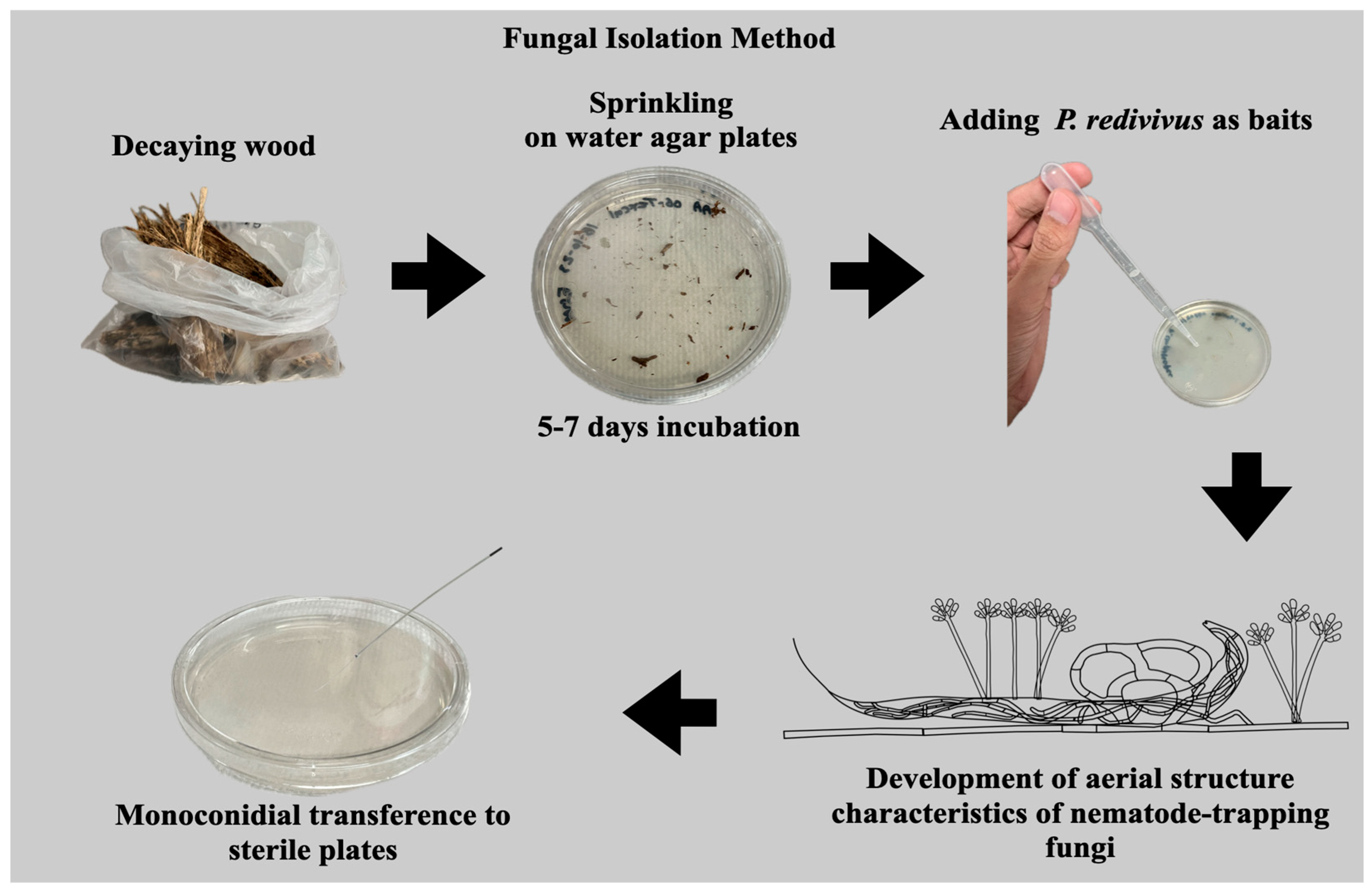
One gram of decaying wood was crushed into small particles and sprinkled on the surface of sterile water agar plates (90 × 15 mm). These plates were then incubated for 7 days at room temperature (18–25 °C). After this initial incubation, a few drops of an aqueous suspension containing an undetermined number of specimens of the free-living nematode Panagrellus redivivus were added to the agar surface as bait to stimulate the growth of aerial structures of nematode-trapping fungi [20]. Four days after introducing the nematodes, fungal aerial structures began to develop on the agar surface. These structures included conidiophores, conidia, and trapped nematodes. Aerial conidia were collected with a sterile needle and transferred to new sterile water agar plates. These fresh plates were incubated at the same temperature and examined under a microscope twice a week. To isolate the fungi, we propagated them on sterile water agar plates through successive monoconidial transfers until we obtained pure cultures [21] (Figure 2).

2.2. Morphological Identification

The microculture technique was carried out as follows: A small square block (1 cm2) was cut from a sterile potato–dextrose agar plate and placed on a slide [22]. This slide, along with the agar block, was positioned in a Petri dish (100 mm diameter). The agar block was inoculated on all four sides with the fungus and then covered with a coverslip. A thin film of sterile water was added to the bottom of the Petri dish to create a humid chamber [23]. The microcultures were incubated for 7 days at room temperature (18–25 °C). The Petri dishes were sealed with parafilm during this period. After 7 days, the coverslips containing the fungal growth were removed from the microcultures and placed over a water droplet on a slide. The fungal structures were then observed under a microscope at 40× and 100× magnifications. Additionally, water agar plates were inoculated with the mycelium of the isolate under study. Water droplets containing an undetermined number of specimens of P. redivivus were added, and the plates were allowed to incubate for another 7 days at the same room temperature. Free-living nematodes were commercially acquired as living fish food from a local pet store. Fungal structures including conidiophores, conidia, trapping devices, chlamydospores, and trapped nematodes were observed and analyzed. The lengths and widths of the conidia, as well as the lengths of the conidiophores, were measured using a Leica Zeiss DM6B microscope (Wetzlar, Germany). A total of 25 structures (i.e., conidia and conidiophores) were randomly selected for measurement. Descriptions of the main taxonomically important structures were recorded, and a series of microscopic images of these structures were captured using the camera of the same microscope, employing the LAS V4.9 program [23].

2.3. Molecular Identification

Genomic DNA Isolation

The genomic DNA from this isolate was extracted using the Wizard® Genomic DNA Purification Kit (PROMEGA, Madison, WI, USA). The concentration of the isolated genomic material was measured using a NanoPhotometer NP80 (IMPLEN, Munich, Germany). The extracted DNA was processed through endpoint PCR by following the protocols outlined by Tigano-Milani et al. (1995) [24], and these methods were standardized in the Helminthology Laboratory of INIFAP. The PCR reaction was performed in a final volume of 20 µL, which included 100 ng of genomic DNA, 10 µL of GoTaq® Green Master Mix 2X (PROMEGA, Madison, WI, USA), and 1.5 µL of each oligonucleotide at a concentration of 20 µM: ITS5-fw (5′-TCCTCCGCTTATTGATATGC-3′) and ITS4-rv (5′-GGAAGTAAAAGTCGTAACAAGG-3′) for the ITS region [18]; EF-1018F (GAYTTCATCAAGAACATGAT) and EF-1620R (GACGTTGAADCCRA-CRTTGTC) for the TEF1-α region [19]; and RPB2-6f (TGGGGKWTGGTYTGYCCTGC) and fRPB2-7f (CCCATRGCTTGYTTRCCCAT) for the RPB2 region [16]. Nuclease-free water was added to complete the reaction volume of 20 µL. The amplification conditions for the ITS region were as follows: an initial denaturation at 94 °C for 3 min; followed by 35 cycles of 94 °C for 60 s; 42 °C for 90 s of annealing; and 72 °C for 90 s of extension; concluding with a final extension at 72 °C for 90 s and cooling at 4 °C. The PCR conditions for the TEF1-α and RPB2 regions included an initial denaturation at 95 °C for 5 min; followed by 35 cycles at 95 °C for 1 min; annealing at 51 °C and 54 °C for 60 s, respectively; 72 °C for 120 s; followed by a final extension at 72 °C for 10 min. The PCR reactions were conducted using a C1000 Touch® Thermal Cycler (Bio-Rad, Hercules, CA, USA). Additionally, SDS-PAGE electrophoresis was performed on a 1.5% agarose gel for 80 min at 70 V to visualize the PCR products under UV light. Following this, the PCR products were purified using the Wizard® Genomic DNA Purification Kit (PROMEGA, Madison, WI, USA). DNA sequencing was carried out at the Biotechnology Institute of the National Autonomous University of Mexico (IBT-UNAM) in Cuernavaca City, Morelos State, Mexico.

2.4. Phylogenetic Analysis

The phylogenetic analysis was conducted using sequences obtained from the ITS, TEF1-α, and RPB2 regions. Alignments for each region were created using the NCBI-BLAST tool. All species reported in the Index Fungorum from the Arthrobotrys genus were searched for in the NCBI nucleotide database, and those with sequences from all three regions were included. Additionally, some species from the Dactylellina and Drechslerella genera, also with three reported regions in the NCBI database, were added to the analysis. In total, 39 species of Arthrobotrys, 5 of Dactylellina, and 4 of Drechslerella were included, with Vermispora fusarina (strain YXJ02-13-5) serving as the outgroup species (see Table 1). Multiple alignments for each region were performed using the CLUSTAL algorithm within MEGA Software (v11.0.13). BioEdit (v7.2.5) and MEGA software were utilized to edit and link the three alignments. The best substitution model was determined using jModelTest (v2.1.10), based on the Akaike Information Criterion (AIC). The optimal models were TIM2e + G4 for ITS and SYM + I + G4 for TEF1-α and RPB2. Maximum likelihood analysis was conducted using IQTREE software (v1.6.12), with statistical bootstrap support values obtained through ultrafast bootstrapping with 10,000 replicates. Additionally, Bayesian inference analysis was performed using MrBayes software (v3.2.6). The dataset was partitioned, and equivalent substitution models were applied. Four simultaneous Markov chains were run for 10,000,000 generations, with trees sampled every 1000 generations. The final 90% of the trees were used to estimate the posterior probabilities for the consensus tree. FigTree software (v1.4.4) was used to visualize the tree, while Photoshop CS6 (Adobe Systems, San Jose, CA, USA) and Microsoft PowerPoint (2016) were employed to edit the obtained consensus tree.

2.5. Predatory Activity Assessment

Water agar plates (2%) measuring 60 × 15 mm (n = 4) were inoculated with the isolate and maintained at a temperature of 25–30 °C for 10 days. A control series of plates, containing the same sample size and medium but without any fungus, was also prepared. After the 10-day incubation period, each plate from both groups (with and without fungus) received 50 mL of an aqueous suspension containing 200 infective larvae (L3) of Haemonchus contortus. All plates were incubated at the same temperature for an additional 12 days. The surface of each plate was examined under a stereomicroscope (Leitz, Wetzlar, Germany) on days 2, 7, and 9 to observe trap formation and the nematodes captured. On the 12th day of incubation, the entire content of each plate containing both fungus and captured/non-captured larvae was transferred to a Baermann funnel system. This system facilitated the descent of non-captured larvae from plates with fungus and the descent of all larvae from the control group. The contents remained in the Baermann funnel for 24 h at room temperature (25–30 °C). This technique enabled the recovery of larvae from both the treated and control series, allowing for a comparative analysis of the larvae recovered from each group. It is important to note that the mean number of larvae recovered from the control plates is considered to represent 100% of the initially deposited larvae, unaffected by the fungus. Counting of the recovered larvae was conducted by taking 10–50 µL aliquots, and the number of larvae in these aliquots was counted using an optical microscope (Leica Microsystems, Wetzlar, Germany) with a 10× objective. The entire experiment was performed in triplicate. The larval reduction percentage in the treated group, relative to the control group, attributed to the predatory effect of the fungus, was calculated using the following formula:
L a r v a l   r e d u c t i o n   ( % ) = M R L c M R L t M R L c 100
where
MRLc = mean of recovered larvae from the control group (without fungus);
MRLt = mean of recovered larvae from the treated group (fungus–nematode interaction).

2.6. Production of the Fungus in Liquid Media

The fungus was grown in two different media: (1) Sweet Potato Dextrose Broth (SPDB); and (2) Czapek–Dox Broth (CzDoxB). The media were prepared in three 250 mL Erlenmeyer flasks, each containing 100 mL of sterile liquid medium. From a 7-day-old culture of the fungus on a 2% water agar plate, three agar cylinders (1 cm diameter x 1 cm thickness) were added to each flask. Control flasks containing the same media without fungus were also prepared. All flasks were incubated at room temperature (25–30 °C) for 21 days under static conditions.

2.7. Obtaining Fungal Liquid Culture Filtrates

After incubation, the fungal biomass was separated by filtration to obtain the fungal liquid culture filtrate (LCF) for assessment. The process involved passing the entire content of the flasks through three different types of filters set on a funnel. First, a coffee filter was used for pre-filtration to separate the thick mycelial particles. The recovered liquid was then centrifuged at 3500 rpm for 10 min. The resulting liquid was further filtered through Whatman filter paper No. 4 (25 μm), followed by sterilization using two different syringe filters of 1.1 and 0.22 μm (Millipore, Merck KGaA, Darmstadt, Germany). This filtration process ensured the collection of a biomass-free and sterile LCF. The liquid was then concentrated using a rotary evaporator (Büchi R-300, Flawil, Switzerland) and finally dried by lyophilization (Labconco, Kansas, MO, USA), following the technique described by Pérez-Anzúrez et al. [23].

2.8. In Vitro Assessment of Nematocidal Activity of Liquid Culture Filtrates Against Haemonchus contortus Infective Larvae (L3)

The in vitro assessment of nematodes exposed to LCF was conducted using 96-well microtiter plates (n = 4). Fifty microliters of the prepared LCF (either from CzDoxB or SPDB) was added to each well, along with 50 μL of an aqueous suspension containing 150 H. contortus L3 larvae. Four wells received only sterile distilled water, CzDoxB, or SPDB as negative controls. Additionally, four wells containing 0.5% ivermectin with the nematodes were included as positive controls. Three different concentrations of LCF were tested: 25, 50, and 100 mg/mL. The nematocidal activity of the fungal LCF was assessed after 72 h. The total number of live and dead larvae in each treatment was counted, allowing for the calculation of the mortality percentage attributed to the LCF. The entire experiment was performed in triplicate.

2.9. Statistical Analysis

Data from the predation assay were analyzed by comparing the treated and control groups using Student’s t-test, with the mean number of recovered larvae considered as the dependent variable (p < 0.05). For the analysis of the nematocidal activity of LCF, the Kruskal–Wallis non-parametric method was employed. The statistical software used for these analyses was SPSS version 25 (IBM Corp., New York, NY, USA), with a significance set at (p < 0.05).

3. Results

3.1. Morphological Taxonomic Identification

The isolate has been deposited at the National Center of Genetic Resources (CM-CNRG) in Mexico, which is affiliated with the World Federation of Culture Collections (WFCC No. 1006) and the Latin American Federation for Culture Collections (FELACC No. 56). The deposit code for this sample is CM-CNRG 967. Sequences were uploaded to the NCBI GenBank under the following accession numbers: PQ655528 (TEF), PQ649538 (ITS), and PQ661202 (RPB2). Etymology: the species named “mendozadegivensis” is derived from the surname of its discoverer.
Materials examined: The specimen was collected in Coatlán del Río Municipality, State of Morelos, Mexico; the geographic coordinates are 18°45′5″ north latitude and 99°26′8″ west longitude. The specimen was obtained from decaying wood on 23 November 2022 by P. Mendoza de Gives. It is preserved in the fungal collection of the Laboratory of Helminthology at the National Center for Disciplinary Research in Animal Health and Innocuity, which is part of the National Institute of Research in Forestry, Agriculture, and Livestock in Mexico.

3.1.1. Macroscopic Characteristics

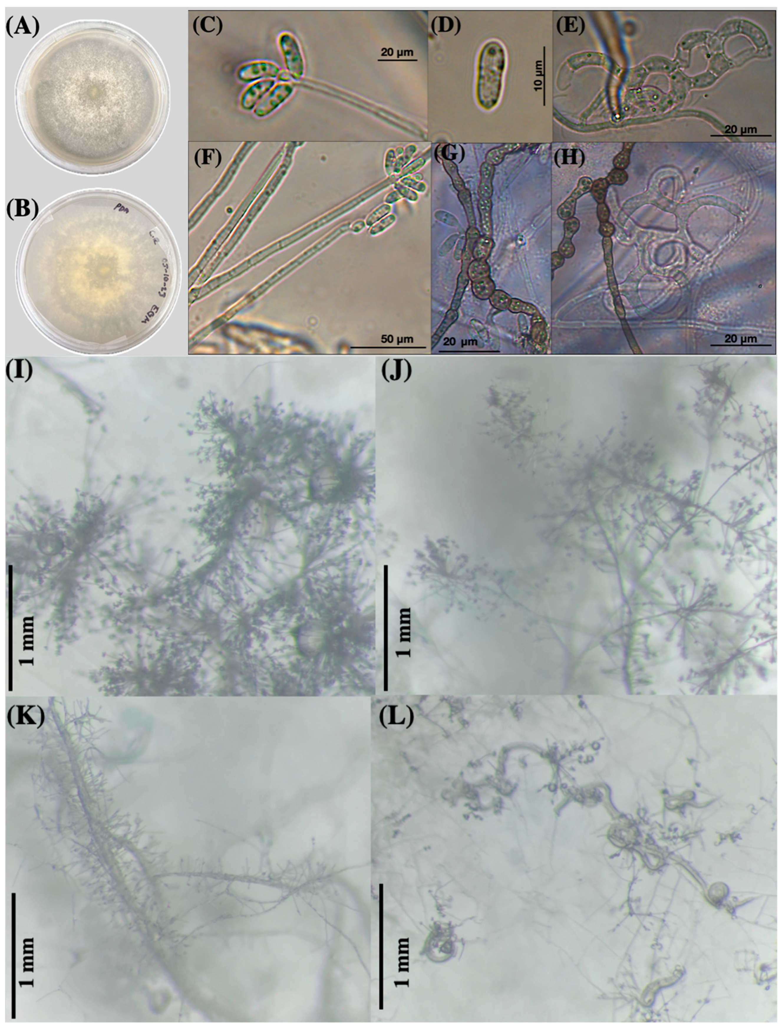
The main macroscopic characteristics of the fungus grown on potato dextrose agar (PDA) plates are illustrated in Figure 3A,B. After two weeks, the fungus displayed cotton-like, whitish, and radially scattered mycelial growth (as seen from above in Figure 3A and from below in Figure 3B). In water agar plates, the fungus of the same age exhibited a tiny, whitish, and transparent growth with scattered mycelia.
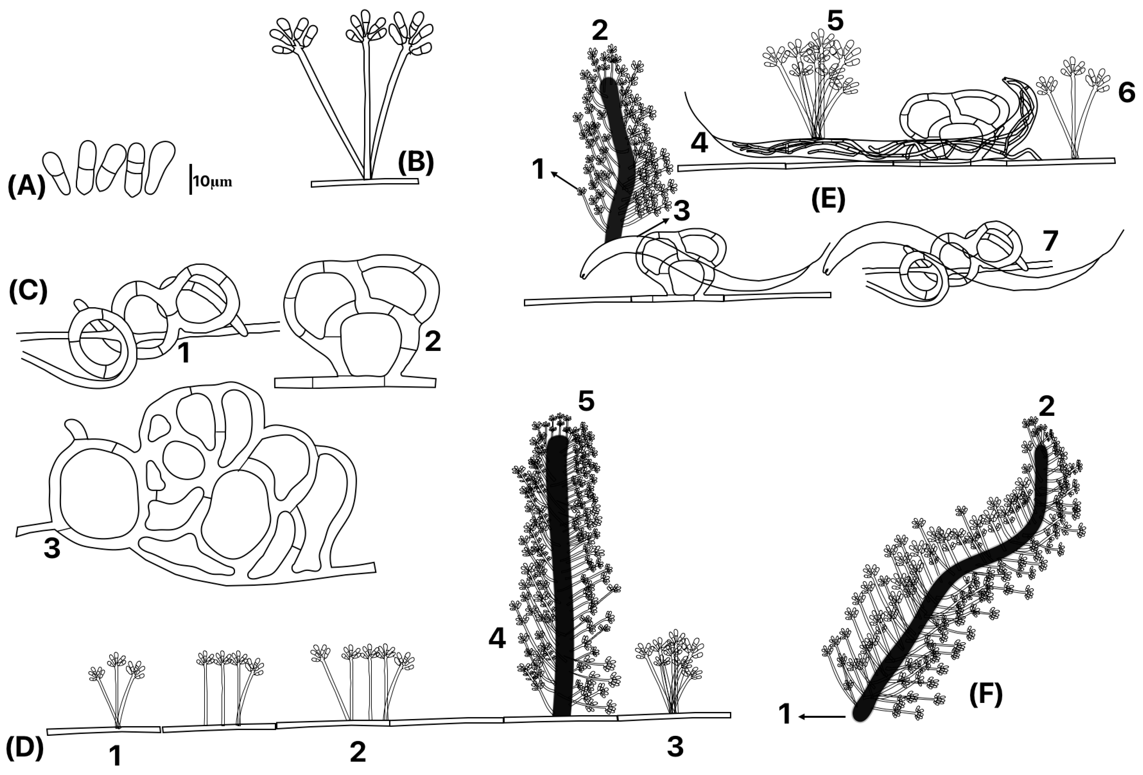
Likewise, in Figure 3C–H, a series of six microphotographs illustrate various aspects of conidia and conidiophores (C,F); one photo features a single conidium (D). Additionally, four microphotographs taken with a stereomicroscope depict the growth of A. mendozadegivensis after 15 days at room temperature (18–25 °C) on corn meal agar plates (I–L). Figure 3L shows P. redivivus trapped in three-dimensional adhesive nets on the surface of a water agar plate. Brown catenulated chlamydospores were produced in water agar and PDA cultures that were older than four weeks (Figure 4E,F).

3.1.2. Microscopic Fungal Characteristics

A microscopic photographic set of the main morphological characteristics of the fungus is shown in Figure 3. Additionally, a schematic drawing of the morphological details of the fungus is shown in Figure 4. The fungus produces an extensive net of thin hyphae growing on the agar surface where different kinds of conidiophores develop, i.e., two or three conidiophore stems emerging from the same initial site (bundled) or conidiophores with only a unique stem (Figure 4D(1–3)). The fungus also produces aerial thickened hyphae that act as “mother hyphae” or “major hyphae” (synnematous) where single (non-branched) conidiophores emerge (Figure 4(E1,D5,F2). Mother hyphae can be produced from the agar surface (D4), aerial hypha (F1) and from the nematode body (E1). This fungus produces three-dimensional adhesive nets (Figure 4C(1–3); Figure 4C) where nematodes are trapped (Figure 4E(3,4,7)). Synnematous hyphae are able to produce a large number of single-stem conidiophores along the hyphae (Figure 4E(1,2),F2)). Conidiophores’ lengths range between 31.66 and 109.33 μm. In the apical part of conidiophores, conidia from obovoid to ellipsoidal were produced and they form clusters from 4 to 6 conidia (Figure 4A,B,D). Most of the conidia showed one septum; however, scarce conidia—either non-septate or with the presence of two septa—were also found (Figure 4A). Conidia length ranged from 8.85 to 18.79 μm with a 3.27 to 5.97 μm width.

3.2. Molecular Identification

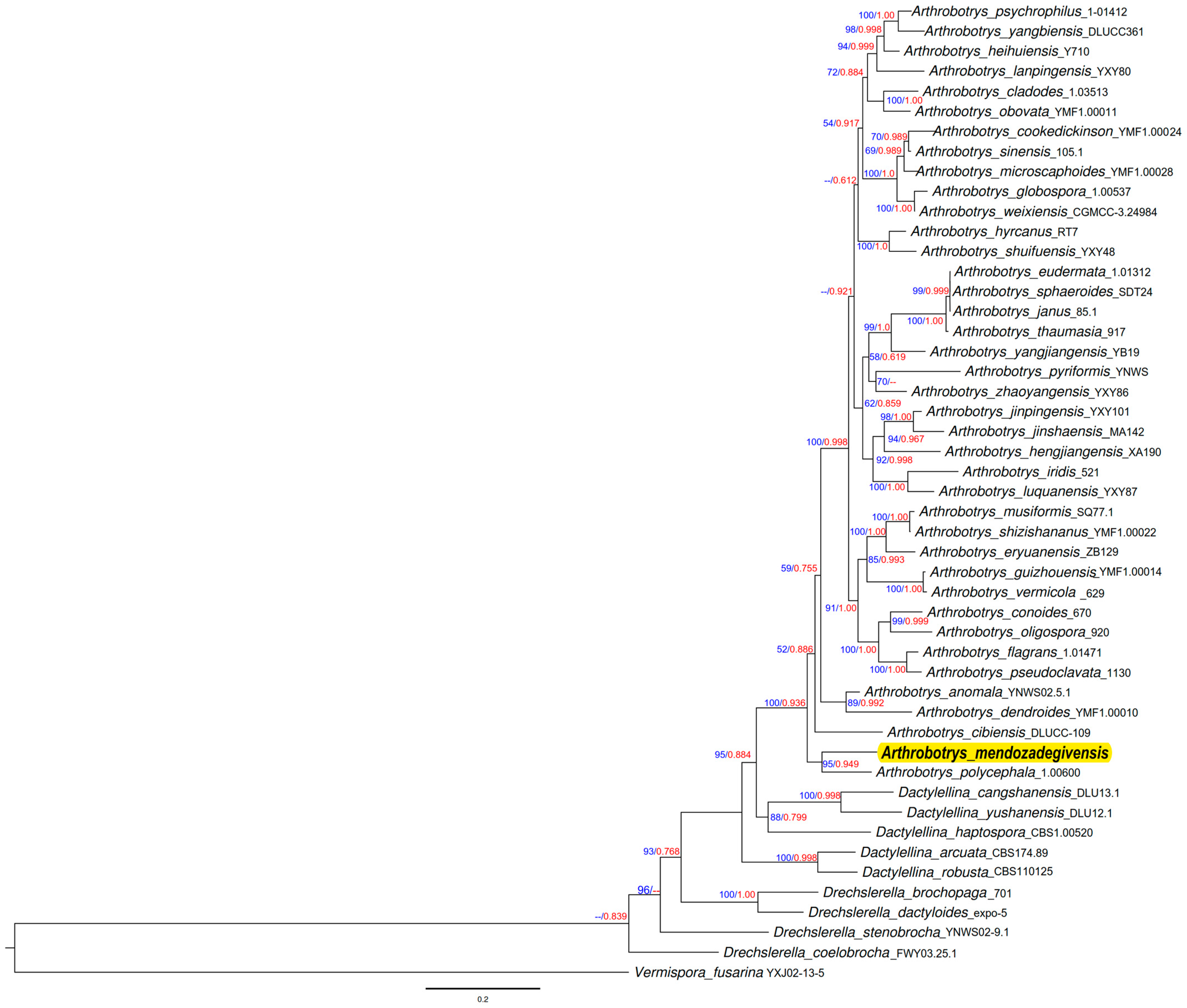
The analysis of the phylogenetic tree—which compared 48 species from nematode-trapping fungi of the Orbiliaceae family with our new isolate—indicated that the closest sequences corresponded to the species Arthrobotrys polycephala (strain 1.006). This phylogenetic tree suggested that our new isolate belongs to the genus Arthrobotrys; however, none of the closest species previously reported in the NCBI database matched this isolate (see Figure 5).
The DNA sequence analysis enabled us to generate a phylogenetic tree using both Maximum likelihood and Bayesian inference methods, as illustrated in Figure 5. Following morphological analysis, the authors of this study found no previously reported nematode-trapping fungus isolate that exhibited the characteristics of our isolate. Similarly, after conducting molecular analysis and comparing sequences with those of the closest matches in the NCBI database, we concluded that this isolate represents a new species of Arthrobotrys, which we propose to name Arthrobotrys mendozadegivensis.
After analyzing all the morphometric data, we were unable to achieve a conclusive classification for this isolate; therefore, we decided to support our morphological taxonomic identification with molecular analyses to clarify the species of our isolate. Once we received the sequence from the Institute of Biotechnology of the National Autonomous University of Mexico, we aligned it using the BLASTn tool and analyzed it by comparing it with closely related sequences from the NCBI database. The similarity and coverage percentages, along with the corresponding NCBI accession numbers, are presented in Table 2.

3.3. Predatory Activity Assessment

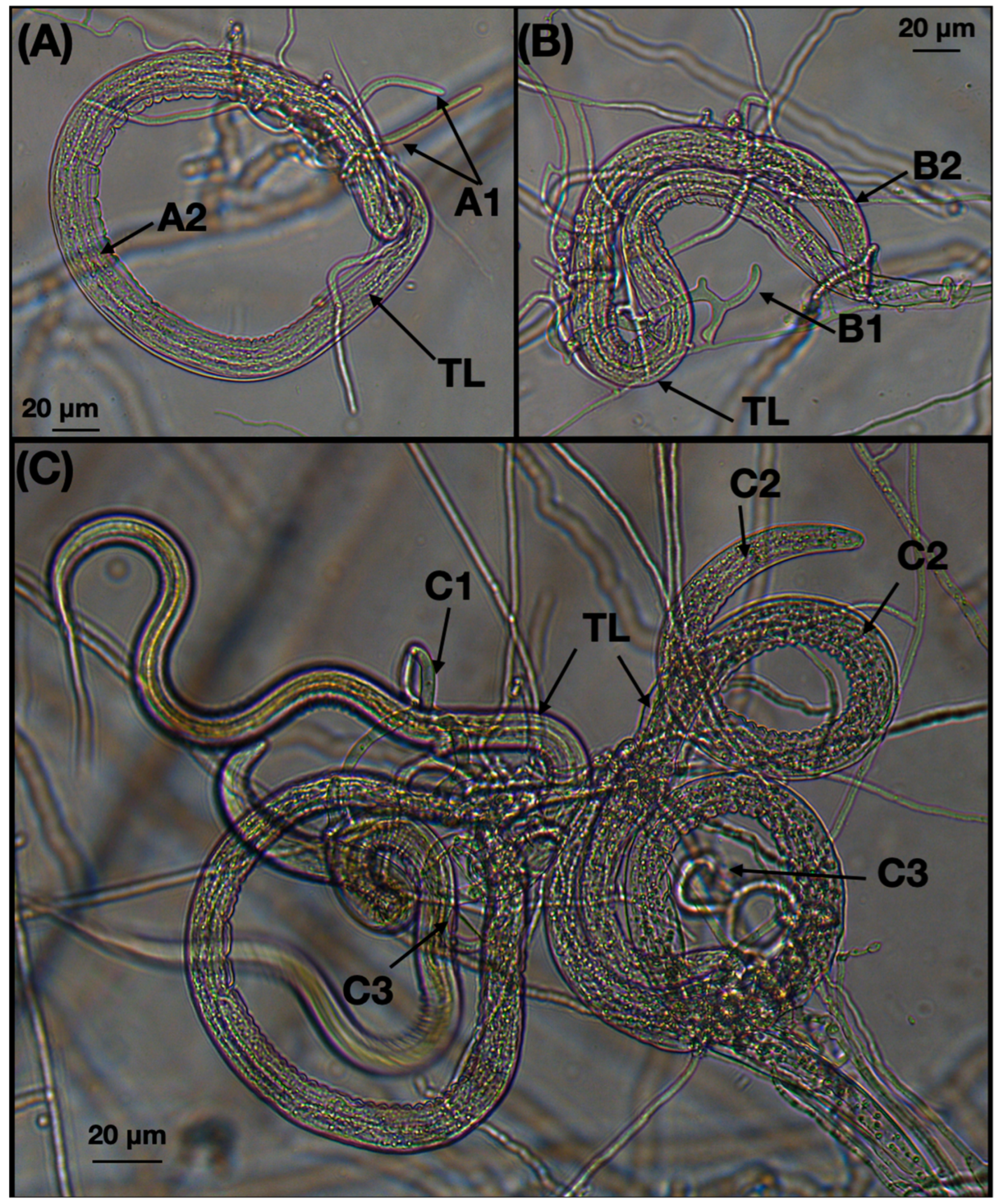
During the incubation process of the plates containing the interaction between fungus and nematodes, several images were captured to illustrate the formation of trapping devices and the nematodes that were trapped (see Figure 6). The analysis of the recovered larvae after the interaction between the fungus and nematodes revealed an average of 28 ± 25.01 larvae recovered from the treated group compared to 125.18 ± 39.84 larvae from the control group. By applying Abbott’s formula, we observed a larval reduction of 76.92%, attributed to the predatory effect of the fungus.

3.4. Nematocidal Activity of Liquid Culture Filtrates of Arthrobotrys Mendozadegivensis Against Haemonchus Contortus Infective Larvae (L3) Under In Vitro Conditions

Table 3 displays the mean number of dead larvae recovered from the plates containing the interaction of nematodes with the liquid culture filtrate (LCF) compared to the control plates, along with their standard deviation. The highest mortality rate was observed at the highest concentration of LCF (100 mg/mL).

4. Discussion

It is noteworthy that, from the initial microscopic observations conducted with a stereomicroscope on water agar plates sprinkled with decaying wood, we observed an unusual aerial growth at 2X magnification. The growth of mycelia and conidiophores resembled small bushes supporting numerous conidial agglomerations (Figure 3I–K). When we examined these structures at a higher magnification (5×), we noticed that conidiophores emerged either from a “mother” or “guide” hyphae. Additionally, some conidiophores appeared to originate from the same point, or bundle, within the corpses of invaded nematodes (Figure 4E(3,5)). Interestingly, although this isolate produces three-dimensional adhesive nets similar to various species within the genus Arthrobotrys, such as A. oligospora, A. arthrobotryoides, and A. robusta, we found that the conidia of our isolate were considerably smaller and distinctly different from those of the mentioned species. For instance, the conidia of A. oligospora are pyriform, whereas those of our isolate are elongated and very small, measuring between 8.85 and 18.79 × 3.27 and 5.97 μm. In contrast, the conidia of A. oligospora are nearly double the size: 17–35 × 8.5–16 μm. These growth characteristics are not commonly found in other species of nematode-trapping fungi, contributing to the challenges we faced in classifying our isolate. Moreover, when comparing the morphological traits of our new isolate with those documented in the literature, we found some similarities with the species A. dendroides. This species is characterized by a similar growth pattern of agglomerated conidiophores resembling bushes. However, A. dendroides produces geniculated conidia, which were not observed in our new species. Additionally, our isolate exhibited conidiophores emerging from a single hypha and we noted the presence of catenulated chlamydospores, while A. dendroides does not produce chlamydospores.
After analyzing the sequences and comparing them with those previously reported in the NCBI database, we discovered that one uncultured fungus (NCBI accession number: GU053870.1), uploaded by Frohlich et al. [32], exhibited the highest similarity percentage at 99.11%. Unfortunately, information regarding this isolate is incomplete, and it appears that it has not been published or cultured. Additionally, we found three species of nematode-trapping fungi—Arthrobotrys polycephala, A. dendroides, and A. ellipsospora—showing similarity percentages ranging from 91.55% to 93.12%. Specifically, the sequences with the highest similarities were A. dendroides at 91.55% (NR_159642.1), A. ellipsospora at 91.55% (LC146721.1), and A. polycephala at 93.12% (NR160072.1). These species are morphologically distinct from our new isolate. This study marks the first record of Arthrobotrys mendozadegivensis worldwide. This species demonstrated significant predatory activity against the blood-feeding nematode parasite of small ruminants, Haemonchus contortus, achieving rates close to 77%. This result aligns with the standard predatory activity observed in many Arthrobotrys species under in vitro conditions against gastrointestinal parasitic nematodes, such as H. contortus, which typically ranges from 70% to 99% (see Table 4). The predatory activity exhibited by this new species of Arthrobotrys positions it as a promising candidate for future research as a biological control agent against parasitic nematodes.
This is the first report of the nematocidal activity of the liquid culture filtrates (LCF) from Arthrobotrys mendozadegivensis grown in liquid media. After analyzing the results on the nematocidal activity of the LCF produced in SPDB and CzDoxB after 72 h, we observed mortality percentages of 40.90% and 34.91%, respectively. It is worthwhile to compare these results with other studies involving different Arthrobotrys species and the lethal activities of their LCF in the same liquid media. For instance, a recent study with A. musiformis showed that after growing in CzDoxB, its LCF caused 93.42% larval mortality against H. contortus; however, when the same fungus was grown in SPDB, it achieved only 26.80% mortality at its highest concentration of 100 mg/mL [23]. Additionally, another isolate of A. musiformis demonstrated 69.96% and 49.84% larval mortality in CzDoxB and potato dextrose broth, respectively [34]. Similarly, another species, A. sinensis, grown in a specific medium containing glucose, yeast extract, K2HPO4, MgSO4, ZnSO4, FeSO4, and CuSO4 showed a 64% larval mortality rate against Angiostrongylus vasorum, a parasite affecting dogs [39]. In another study, organic extracts from the mycelia of three Arthrobotrys species A. oligospora, A. conoides, and A. arthrobotryoides resulted in 72.1%, 81.6%, and 89.51% larval mortality against H. contortus, respectively [38]. Comparing our results with those from other species and isolates suggests that different species and isolates, even within the same genus, exhibit varying levels of nematocidal compound production. This variability is influenced by their biochemical and physiological states, which are affected by their microenvironments. This study provides insights into the potential biotechnological applications of these microorganisms for controlling one of the most pathogenic parasitic nematodes that affect small ruminants worldwide. Additionally, this research presents information about a previously unreported species of nematophagous fungus, which is important for systematics and offers potential biotechnological solutions for combating nematodes in the livestock industry.

Author Contributions

Conceptualization, E.G.-M., P.M.-d.G., and G.P.-A.; methodology, E.G.-M. and A.C.-P.; validation, E.v.S.-d.F., M.Á.A.-D., and M.E.L.-A.; formal analysis, G.A.B.-G.; investigation, E.G.-M.; resources, M.E.L.-A.; data curation, G.P.-A. and P.M.-d.G. writing—original draft preparation, P.M.-d.G. and E.G.-M.; writing—review and editing, G.P.-A. and E.G.-M.; visualization, E.G.-M.; supervision, P.M.-d.G., E.v.S.-d.F., and M.Á.A.-D.; project administration, P.M.-d.G.; funding acquisition, P.M.-d.G. and M.E.L.-A. All authors have read and agreed to the published version of the manuscript.

Funding

This study did not receive any budget to carry out the experiments; materials, reagents, and consumables were provided by the Laboratory of Helminthology of the National Centre for Disciplinary Research in Animal Health and Innocuity (CENID-SAI) of INIFAP-Mexico. DVM Enrique Gutiérrez-Medina received financial support from Consejo Nacional de Humanidades, Ciencia y Tecnología (CONAHCYT—the Mexican Government), scholarship number: 798579.

Informed Consent Statement

Data are contained within the article.

Data Availability Statement

The original contributions presented in the study are included in the article, further inquiries can be directed to the corresponding author.

Acknowledgments

The authors wish to greatly express our gratitude to Fa Zhang from the Institute of Eastern-Himalaya Biodiversity Research, Dali University, Dali, China, for his valuable comments and recommendations to prepare this paper. This study formed part of the Masters of Science Degree thesis of DVM Enrique Gutiérrez-Medina under the direction of Pedro Mendoza-de Gives and tutoring of Elke von Son-de Fernex and Miguel Angel Alonso-Díaz.

Conflicts of Interest

The authors declare no conflicts of interest.

References

  1. Crous, P.W.; Rong, I.H.; Wood, A.; Lee, S.; Glen, H.; Botha, W.; Slippers, B.; de Beer, W.Z.; Wingfield, M.J.; Hawksworth, D.L. How Many Species of Fungi Are There at the Tip of Africa? Stud. Mycol. 2006, 55, 13–33. [Google Scholar] [CrossRef] [PubMed]
  2. Ojeda-Robertos, N.F.; Aguilar-Marcelino, L.; Olmedo-Juárez, A.; Luna-Palomera, C.; Peralta-Torres, J.A.; López-Arellano, M.E.; Mendoza-de-Gives, P. In Vitro Predatory Activity of Nematophagous Fungi Isolated from Water Buffalo Feces and from Soil in the Mexican Southeastern. Rev. Bras. Parasitol. Vet. 2019, 28, 314–319. [Google Scholar] [CrossRef] [PubMed]
  3. Sánchez-Martínez, E.; Aguilar-Marcelino, L.; Hernández-Romano, J.; Castañeda-Ramírez, G.S.; Mendoza-de-Gives, P. Taxonomic and Biological Characterization and Predatory Activity of Four Nematophagous Fungi Isolates of Arthrobotrys Species from Tapachula, Chiapas, Mexico. Arch. Acker Pflanzenbau Bodenkd. 2023, 69, 327–343. [Google Scholar] [CrossRef]
  4. Quevedo, A.; Vera-Morales, M.; Espinoza-Lozano, F.; Castañeda-Ruiz, R.F.; Sosa del Castillo, D.; Magdama, F. Assessing the predatory activity of Arthrobotrys oligosporus strain C-2197 as biocontrol of the root-knot nematode Meloidogyne spp. Bionatura 2021, 6, 1586–1592. [Google Scholar] [CrossRef]
  5. Swe, A.; Jeewon, R.; Pointing, S.B.; Hyde, K.D. Diversity and Abundance of Nematode-Trapping Fungi from Decaying Litter in Terrestrial, Freshwater and Mangrove Habitats. Biodivers. Conserv. 2009, 18, 1695–1714. [Google Scholar] [CrossRef]
  6. Gray, N.F.; Bailey, F. Ecology of Nematophagous Fungi: Vertical Distribution in a Deciduous Woodland. Plant Soil 1985, 86, 217–223. [Google Scholar] [CrossRef]
  7. Tarigan, W.E.; Munir, E.; Hastuti, L.D.S.; Hartanto, A. Occurrence of nematophagous fungi in freshwater samples of Toba Lake, North Sumatra, Indonesia. J. Phys. Conf. Ser. 2020, 1462, 012052. [Google Scholar] [CrossRef]
  8. Hao, Y.; Mo, M.; Su, H.; Zhang, K. Ecology of aquatic nematode-trapping hyphomycetes in southwestern China. Aquat. Microb. Ecol. 2005, 40, 175–181. [Google Scholar] [CrossRef]
  9. Gray, N.F.; Lewis Smith, R.I. The Distribution of Nematophagous Fungi in the Maritime Antarctic. Mycopathologia 1984, 85, 81–92. [Google Scholar] [CrossRef]
  10. Jansson, H.-B.; Poinar, G.O., Jr. Some Possible Fossil Nematophagous Fungi. Trans. Br. Mycol. Soc. 1986, 87, 471–474. [Google Scholar] [CrossRef]
  11. Su, H.; Zhao, Y.; Zhou, J.; Feng, H.; Jiang, D.; Zhang, K.-Q.; Yang, J. Trapping Devices of Nematode-Trapping Fungi: Formation, Evolution, and Genomic Perspectives: Trapping Devices of Nematode-Trapping Fungi. Biol. Rev. Camb. Philos. Soc. 2017, 92, 357–368. [Google Scholar] [CrossRef]
  12. Mendoza-de Gives, P. Soil-Borne Nematodes: Impact in Agriculture and Livestock and Sustainable Strategies of Prevention and Control with Special Reference to the Use of Nematode Natural Enemies. Pathogens 2022, 11, 640. [Google Scholar] [CrossRef] [PubMed]
  13. Corda, A.K.J. Pracht-Flora Europaeischer Schimmelbildungen; G. Fleischer: Los Angeles, CA, USA, 1839. [Google Scholar]
  14. Zopf, W.F. Zur Kenntniss der Infections-Krankheiten niederer Thiere und Pflanzen. Acad. Nat. 1888, 52, 314–376. [Google Scholar]
  15. Jarowaja, N. The genus Arthrobotrys Corda. Acta Myc. 1970, 6, 337–406. [Google Scholar] [CrossRef]
  16. Zhang, F.; Boonmee, S.; Bhat, J.D.; Xiao, W.; Yang, X.-Y. New Arthrobotrys Nematode-Trapping Species (Orbiliaceae) from Terrestrial Soils and Freshwater Sediments in China. J. Fungi 2022, 8, 671. [Google Scholar] [CrossRef]
  17. Ekanayaka, A.H. Orbiliaceae from Thailand. Mycosphere 2018, 9, 155–168. [Google Scholar] [CrossRef]
  18. White, T.J.; Bruns, T.; Lee, S.; Taylor, J. Amplification and direct sequencing of fungal ribosomal RNA genes for phylogenetics. In En PCR Protocols; Elsevier: Amsterdam, The Netherlands, 1990; pp. 315–322. ISBN 9780123721808. [Google Scholar]
  19. Stielow, J.B.; Lévesque, C.A.; Seifert, K.A.; Meyer, W.; Iriny, L.; Smits, D.; Renfurm, R.; Verkley, G.J.M.; Groenewald, M.; Chaduli, D.; et al. One Fungus, Which Genes? Development and Assessment of Universal Primers for Potential Secondary Fungal DNA Barcodes. Persoonia 2015, 35, 242–263. [Google Scholar] [CrossRef]
  20. Barron, G.L. The Nematode Destroying Fungi; Editorial Lancaster Press, Inc.: Lancaster, PA, USA, 1977; 140p. [Google Scholar]
  21. Harirchi, S.; Rousta, N.; Varjani, S.; Taherzadeh, M.J. Sampling, Preservation, and Growth Monitoring of Filamentous Fungi. In En Current Developments in Biotechnology and Bioengineering; Elsevier: Amsterdam, The Netherlands, 2023; pp. 149–180. ISBN 9780323918725. [Google Scholar]
  22. Campbell, C.K.; Johnson, E.M. Identification of Pathogenic Fungi: Campbell/Identification of Pathogenic Fungi; Warnock, D.W., Ed.; Wiley-Blackwell: Chichester, UK, 2013; ISBN 9781444330700. [Google Scholar]
  23. Pérez-Anzúrez, G.; Olmedo-Juárez, A.; von-Son de Fernex, E.; Alonso-Díaz, M.Á.; Delgado-Núñez, E.J.; López-Arellano, M.E.; González-Cortázar, M.; Zamilpa, A.; Ocampo-Gutierrez, A.Y.; Paz-Silva, A.; et al. Arthrobotrys Musiformis (Orbiliales) Kills Haemonchus Contortus Infective Larvae (Trichostronylidae) through Its Predatory Activity and Its Fungal Culture Filtrates. Pathogens 2022, 11, 1068. [Google Scholar] [CrossRef] [PubMed]
  24. Tigano-Milani, M.S.; Honeycutt, R.J.; Lacey, L.A.; Assis, R.; McClelland, M.; Sobral, B.W.S. Genetic Variability of Paecilomyces Fumosoroseus Isolates Revealed by Molecular Markers. J. Invertebr. Pathol. 1995, 65, 274–282. [Google Scholar] [CrossRef] [PubMed]
  25. Yang, Y.; Yang, E.; An, Z.; Liu, X. Evolution of Nematode-Trapping Cells of Predatory Fungi of the Orbiliaceae Based on Evidence from rRNA-Encoding DNA and Multiprotein Sequences. Proc. Natl. Acad. Sci. USA 2007, 104, 8379–8384. [Google Scholar] [CrossRef] [PubMed]
  26. Zhang, F.; Yang, Y.-Q.; Zhou, F.-P.; Xiao, W.; Boonmee, S.; Yang, X.-Y. Multilocus Phylogeny and Characterization of Five Undescribed Aquatic Carnivorous Fungi (Orbiliomycetes). J. Fungi 2024, 10, 81. [Google Scholar] [CrossRef] [PubMed]
  27. Zhang, Y.R.; Zhang, Y.; Xu, J.P. Molecular Phylogeny and DNA Barcoding of Nematode-Trapping Hyphomycetes and Related Taxa; submitted; Yunnan University: Kunming, China, 2018. [Google Scholar]
  28. Qiao, M.; Zhang, Y.; Baral, H.; Zhang, K.Q.; Yu, Z.F. New Species and New Combination of Orbiliaceae from Yunnan, China; submitted; Yunnan University: Kunming, China, 2017. [Google Scholar]
  29. Zhang, F.; Yang, Y.-Q.; Zhou, F.-P.; Xiao, W.; Boonmee, S.; Yang, X.-Y. Morphological and Phylogenetic Characterization of Five Novel Nematode-Trapping Fungi (Orbiliomycetes) from Yunnan, China. J. Fungi 2023, 9, 735. [Google Scholar] [CrossRef] [PubMed]
  30. Masigol, H.; Rezakhani, F.; Pourmoghaddam, M.J.; Khodaparast, S.A.; Grossart, H.-P. The Introduction of Two New Species of Aquatic Fungi from Anzali Lagoon, Northern Iran. Diversity 2022, 14, 889. [Google Scholar] [CrossRef]
  31. Zhang, F.A.; Zhou, X.-J.; Monkai, J.; Li, F.-T.; Liu, S.-R.; Yang, X.-Y.; Wen, X.; Hyde, K.D. Two new species of nematode-trapping fungi (Dactylellina, Orbiliaceae) from burned forest in Yunnan, China. Phytotaxa 2020, 452, 65–74. [Google Scholar] [CrossRef]
  32. Frohlich-N, J.; Despres, V.R.; Poschl, U. Global Atmospheric Diversity of Fungi: Asco- and Basidiomycota in Continental and Marine Air; submitted; Max Planck Institute for Chemistry: Mainz, Germany, 2009. [Google Scholar]
  33. Wang, W.; Meng, Q.; Qiao, J.; Wang, J.; Yang, L.; Luo, J.; Zhu, X.; Wang, G.; Chen, C. Isolation of Arthrobotrys oligospora from soil of the Chinese Northern Tianshan Mountain slope pasture show predatory ability against Haemonchus contortus larvae. Biocontrol Sci. Technol. 2014, 24, 170–179. [Google Scholar] [CrossRef]
  34. Jaramillo-Tlalapango, J.; Mendoza-de Gives, P.; Isabel-Higuera-Piedrahita, R.; Ocampo-Gutiérrez, A.Y.; Eugenia-López-Arellano, M.; Pérez-Anzúrez, G.; Olmedo-Juárez, A.; Hernández-Romano, J.; Maza-Lopez, J.; Delgado-Núñez, E.J.; et al. Study of a Mexican Isolate of Arthrobotrys Musiformis (Orbiliales): Predatory Behavior and Nematocidal Activity of Liquid Culture Filtrates against Haemonchus Contortus (Trichostrongylidae), Protein Profile and Myco-Constituent Groups. Fungal Biol. 2023, 127, 1345–1361. [Google Scholar] [CrossRef]
  35. Campos, A.K.; Valadão, M.C.; Carvalho, L.M.; de Araújo, J.V.; Guimarães, M.P. In vitro nematophagous activity of predatory fungi on infective larvae of Strongyloides papillosus. Acta Vet. Bras. 2017, 11, 213–218. [Google Scholar] [CrossRef]
  36. Cai, K.-Z.; Wang, F.-H.; Wang, K.-Y.; Liu, J.-L.; Wang, B.-B.; Xu, Q.; Xue, Y.-J.; Wang, F.; Zhang, C.; Fang, W.-X.; et al. In Vitro Predatory Activity of Arthrobotrys Oligospora and after Passing through Gastrointestinal Tract of Small Ruminants on Infective Larvae of Trichostrongylides. Exp. Parasitol. 2017, 177, 104–111. [Google Scholar] [CrossRef] [PubMed]
  37. de Paula Santos, C.; Padilha, T.; de Lurdes de Azevedo Rodrigues, M. Predatory activity of Arthrobotrys oligospora and Duddingtonia flagrans on pre-parasitic larval stages of cyathostominae under different constant temperatures. Cienc. Rural 2001, 31, 839–842. [Google Scholar] [CrossRef]
  38. Ocampo-Gutiérrez, A.Y.; Hernández-Velázquez, V.M.; Aguilar-Marcelino, L.; Cardoso-Taketa, A.; Zamilpa, A.; López-Arellano, M.E.; González-Cortázar, M.; Hernández-Romano, J.; Reyes-Estebanez, M.; Mendoza-de Gives, P. Morphological and Molecular Characterization, Predatory Behaviour and Effect of Organic Extracts of Four Nematophagous Fungi from Mexico. Fungal Ecol. 2021, 49, 101004. [Google Scholar] [CrossRef]
  39. de Freitas Soares, F.E.; de Queiroz, J.H.; Braga, F.R.; Dos Santos Lima, W.; Zamprogno, T.T.; de Araújo, J.V. Proteolytic Activity of the Nematophagous Fungus Arthrobotrys Sinensis on Angiostrongylus Vasorum Larvae. BMC Res. Notes 2014, 7, 811. [Google Scholar] [CrossRef]
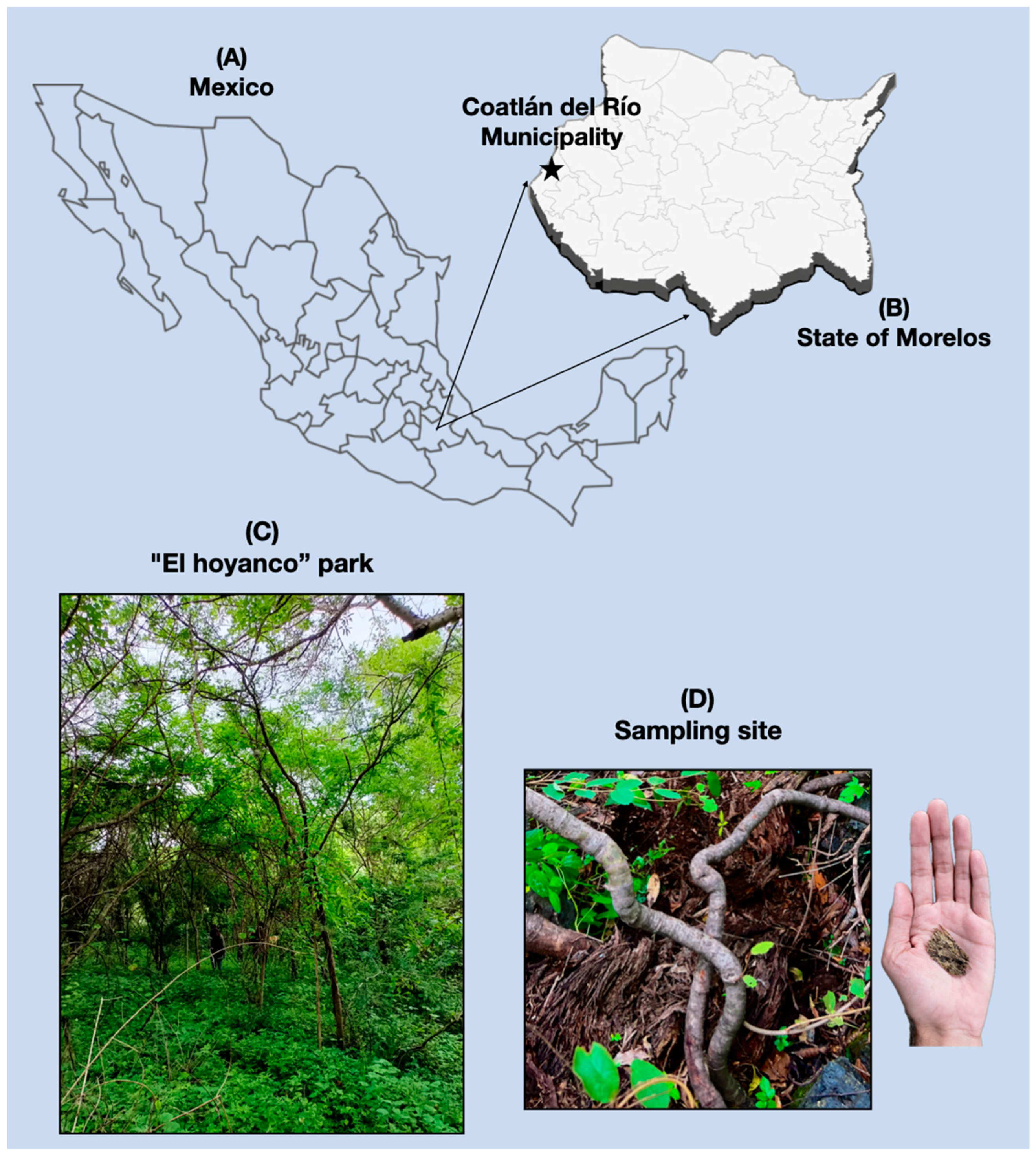
Figure 1. (A) Map of the Mexican Republic; (B) State of Morelos; (Star) Coatlán del Río Municipality; and (C) a picture of the site of sampling at the Eco-touristic park El Hoyanco; (D) sampling site.
Jof 10 00888 g001
Figure 2. Representative scheme of the nematophagous fungi isolation process from decaying wood.
Jof 10 00888 g002
Figure 3. Macroscopic view (A,B), microphotographs (from a compound microscope, (CH)), and views from a stereomicroscope (IL) of Potato Dextrose Agar plates of the nematode-trapping fungus Arthrobotrys mendozadegivensis; aspect of free-living nematodes of Panagrellus redivivus species captured by three-dimensional adhesive nets of the fungus on the water agar surface (L); aspect of three-dimensional adhesive nets (E,H); and catenulated chlamydospores (G,H).
Jof 10 00888 g003
Figure 4. Representative scheme of the development of aerial structures of the nematode-trapping fungus Arthrobotrys mendozadegivensis: (A) conidia; (B) hyphae-produced conidiophores from tillers; (C(13)) three-dimensional adhesive nets; (D) different kinds of conidiophores emerging from a hypha, (D1) three conidiophores emerging from same point from the hypha; (D2) individual and bundled conidiophores; (D3) bundled conidiophores; (D4) Synnematous hypha; (D5) conidiophores emerging from a synnematous hypha; (E(1,2)) conidiophores emerging from a synnematous hypha which in turn emerge from a trapped nematode; (E3) trapped nematode and a synnematous hypha emerging from its body; (E4) a nematode trapped and invaded by mycelia of the nematode-trapping fungus; (E5) bundled conidiophore emerging from the nematode body; (E6) a conidiophore growing from a hypha near to the trapped nematode; (E7) nematode trapped in a three-dimensional adhesive net; (F1) synnematous hypha; and (F2) a large amount of single-stem conidiophores emerging from synnematous hypha.
Jof 10 00888 g004
Figure 5. Maximum likelihood tree based on a combined ITS, TEF1-α, and RPB2 sequences from 48 species of Orbiliaceae nematode-trapping fungi. Bootstrap support values for ML greater than 50% and BI posterior probabilities values greater than 0.70 are indicated aside each node (in blue). The new isolate is in red. The tree is rooted by Vermispora fusarina YXJ-13-5.
Jof 10 00888 g005
Figure 6. Photographs taken from a Leica Digital Microscope showing Haemonchus contortus infective larvae trapped in the three-dimensional adhesive nets (AC). (TL) trapped larva; (A1) hyphae emerging from a trapped nematode body; (A2,B2,C2) invasive hyphae colonizing the intestinal tissues of larvae; (B1,C1) developing adhesive nets; (C3) three-dimensional adhesive nets.
Jof 10 00888 g006
Table 1. List of the strain sequences from ITS, TEF, and RPB2 regions reported at the NCBI database, used to construct the Maximum likelihood and Bayesian inference phylogenetic tree.
SpeciesStrain NumberGenBank Accession NumberReference
ITSTEFRPB2
Arthrobotrys anomalaYNWS02-5-1AY773451AY773393AY773422.1[25]
Arthrobotrys cibiensisDLUCC 109OR880379OR882792OR882797.1[26]
Arthrobotrys cladodes1.03513MH179792MH179615MH179892.1[27]
Arthrobotrys conoides670AY773455AY773397AY773426.1[25]
Arthrobotrys cookedickinsonYMF1.00024MF948393MF948550MF948474.1[28]
Arthrobotrys dendroidesYMF1.00010MF948388MF948545MF948469.1[28]
Arthrobotrys eryuanensisZB129MT612105 OM850307 OM850301[16]
Arthrobotrys eudermata1.01312MH179725MH179576MH179830.1[27]
Arthrobotrys flagrans1.01471MH179741MH179583MH179845.1[27]
Arthrobotrys globospora1.00537MH179706MH179562MH179814.1[27]
Arthrobotrys guizhouensisYMF1.00014MF948390MF948547MF948471.1[28]
Arthrobotrys heihuiensisY710OR902194OR882786OR882801.1[16]
Arthrobotrys hengjiangensisXA190OQ946586OQ989311OQ989301[29]
Arthrobotrys hyrcanusRT7MH367058OP351540___________[30]
Arthrobotrys iridis512AY773452AY773394AY773423.1[25]
Arthrobotrys janus85-1AY773459AY773401AY773430.1.[25]
Arthrobotrys jinpingensisYXY101ON808621ON809552OR882804.1[16]
Arthrobotrys jinshaensisMA142OR902197OR882789OR882804[26]
Arthrobotrys lanpingensisYXY80ON808618ON809549ON809555[16]
Arthrobotrys luquanensisYXY87ON808619ON809550ON809556[16]
Arthrobotrys microscaphoidesYMF1.00028MF948395MF948552MF948476.1[28]
Arthrobotrys musiformisSQ77-1 Y773469 AY773411AY773440.1[25]
Arthrobotrys obovataYMF1.00011MF948389MF948546MF948470.1[28]
Arthrobotrys oligospora920AY773404AY773462AY773433.1[25]
Arthrobotrys polycephala1.006MH179724MH179575MH179829.1[27]
Arthrobotrys pseudoclavata1130AY773446AY773388AY773417.1[25]
Arthrobotrys psychrophila1.01412MH179727MH179578MH179832.1[27]
Arthrobotrys pyriformisYNWS02-3-1AY773450AY773392AY773421.1[25]
Arthrobotrys shizishananusYMF1.00022MF948392MF948549MF948473.1[28]
Arthrobotrys shuifuensisYXY48ON808617ON809548ON809554.1.[16]
Arthrobotrys sinensis105-1AY773445AY773387AY773416.1[25]
Arthrobotrys sphaeroidesSDT24AY773465AY773407AY773436.1[25]
Arthrobotrys thaumasia917AY773461AY773403AY773432.1.[25]
Arthrobotrys vermicola629AY773454AY773396AY773425.1[25]
Arthrobotrys weixiensisCGMCC 3.24984OQ946585OQ989310OQ989300.1[29]
Arthrobotrys yangbiensisDLUCC 36-1OR880382OR882795OR882800.1[26]
Arthrobotrys yangjiangensisYB19OR902196OR882788OR882803.1[26]
Arthrobotrys zhaoyangensisYXY86ON808620ON809551ON809557[16]
Dactylellina arcuateCBS 174.89AF106527DQ999852DQ999799.1[25]
Dactylellina cangshanensisDLU 13-1MK372062MN915115MN915114.1[31]
Dactylellina haptosporaCBS 100520DQ999820DQ999850DQ999814.1[25]
Dactylellina robustaCBS 110125DQ999821DQ999851DQ999800.1[25]
Dactylellina yushanensisDLU12-1MK372061MN915113MN915112.1[31]
Drechslerella brochopaga701AY773456.1AY773398.1AY773427.1[25]
Drechslerella coelobrochaFWY03-25-1AY773464.1AY773406.1AY773435.1[25]
Drechslerella dactyloidesExpo-5AY773463.1AY773405.1AY773434.1[25]
Drechslerella stenobrochaYNWS02-9-1AY773460.1AY773402.1AY773431.1[25]
Dactylaria sp. YNWS02-7-1AY773457AY773399AY773428.1[25]
Vermispora fusarinaYXJ02-13-5AY773447AY773418AY773418.1[25]
Arthrobotrys
mendozadegivensis
INIFAP-EGM-01PQ649538PQ655528PQ661202Present study
Table 2. Similarity and coverage percentages of the sequence of a nematode-trapping fungus isolate after comparison with reported sequences from the GenBank–NCBI database, using the partial sequence of ITS1, 5.8S, and ITS2 regions.
FungusQuery Cover %Identity %GenBank Accession Number
Uncultured fungus 9899.11GU053870.1
Arthrobotrys polycephala9893.12NR_160072
A. polycephala9893.12MH855875.1
A. dendroides9891.55NR_159642.
A. dendroides9891.55MH861894.1
A. ellipsospora9891.55LC146721.1
Table 3. Mean of dead and live Haemonchus contortus infective larvae recovered from the wells of microtiter plates after 72-h interaction with Arthrobotrys mendozadegivensis culture filtrates and mortality percentages.
MediaConcentrationDead Larvae ± SDTotal Larvae ± SDMortality (%) ± SD
CzDoxBPBS1.92 ± 1.46101.24 ± 11.281.9 ± 1.68
NF4.10 ± 2.2395.03 ± 19.204.31 ± 3.41
254.31 ± 2.50103.65 ± 10.654.15 ± 2.43
507.45 ± 8.56100.09 ± 13.007.44 ± 8.68
10033.34 ± 15.4995.49 ± 14.4134.91 ± 16.67
SPDBPBS1.05 ± 0.6377.23 ± 17.491.36 ± 0.35
NF3.37 ± 2.597.25 ± 15.313.47 ± 2.07
252.23 ± 1.2192.66 ± 25.212.41 ± 1.82
502.51 ± 3.3992.73 ± 19.242.71 ± 2.62
10035.25 ± 16.5586.18 ± 8.3340.90 ± 18.66
CzDoxB = Czapek–Dox Broth; SPDB = Sweet Potato Dextrose Broth; PBS = phosphate buffer solution (pH = 7.2); NF = liquid culture filtrates without fungus.
Table 4. Trapping ability of different isolates of Arthrobotrys species under in vitro conditions against Haemonchus contortus infective larvae and other parasitic nematodes.
Arthrobotrys SpeciesBlank NematodePredatory
Activity
Author
Arthrobotrys oligosporaHaemonchus contortus76–79%[33]
A. musiformisH. contortus74.9%[34]
A. cladodesStrongyloidess papillosus99.5%[35]
A. oligosporaTrichostrongylus colubriformis
H. contortus
90–99.99%[36]
A. oligospora
A. flagrans
H. contortus47.5%
41.8%
[37]
A. conoidesH. contortus75%[38]
A. mendozadegivensisH. contortus76.92%(Present study)
Disclaimer/Publisher’s Note: The statements, opinions and data contained in all publications are solely those of the individual author(s) and contributor(s) and not of MDPI and/or the editor(s). MDPI and/or the editor(s) disclaim responsibility for any injury to people or property resulting from any ideas, methods, instructions or products referred to in the content.