


Abstract
Didymella contains numerous plant pathogenic and saprobic species associated with a wide range of hosts. Over the course of our mycological surveys of plant pathogens from terrestrial plants in Jiangxi Province, China, eight strains isolated from diseased leaves of four host genera represented three new species of Didymella, D. bischofiae sp. nov., D. clerodendri sp. nov., and D. pittospori sp. nov. Phylogenetic analyses of combined ITS, LSU, RPB2, and TUB2 sequence data, using maximum-likelihood (ML) and Bayesian inference (BI), revealed their taxonomic placement within Didymella. Both morphological examinations and molecular phylogenetic analyses supported D. bischofiae, D. clerodendri, and D. pittospori as three new taxa within Didymella. Illustrations and descriptions of these three taxa were provided, along with comparisons with closely related taxa in the genus.
1. Introduction
Didymella, the type genus of the family Didymellaceae, was introduced by Saccardo in 1880, with D. exigua as the type species, and later validated when a Latin diagnosis was provided [1,2]. The genus was recently emended by Chen et al. [3,4], who gave a very detailed account of generic concepts. The sexual morphs of Didymella are mainly characterized by solitary or confluent, ostiolate pseudothecial ascomata with multi-layered, pseudoparenchymatous ascomatal walls and cylindrical to clavate or saccate, 8-spored, bitunicate asci with hyaline or brownish uniseptate (symmetrical or asymmetrical) or multiseptate ascospores. The asexual morphs of Didymella are mainly characterized by solitary or confluent, ostiolate or poroid, pycnidial conidiomata with multi-layered, pseudoparenchymatous conidiomatal walls, and phialidic, hyaline conidiogenous cells that produce smooth conidia, which are generally aseptate, variable in shape, hyaline or occasionally pigmented, and larger or septate in at least one species in older cultures. Unicellular chlamydospores are often present in pure culture [2,3,4,5,6,7]. To date, 438 records of Didymella are listed in Species Fungorum [8], and most of them are usually found as saprobes from herbaceous and woody plants, but many are also important plant pathogens [3,4,9].
Didymella is an old, species-rich genus, but its early taxonomic placements are uncertain. The genus was originally described in the family Mycosphaerellaceae and later placed in Pleosporaceae, Phaeosphaeriaceae, Venturiaceae, or Pleosporales genera incertae sedis [2,4]. De Gruyter et al. [2] introduced a new family Didymellaceae with Didymella as the type genus to accommodate Ascochyta, Didymella, Phoma, and several related phoma-like genera based on evidence from phylogenetic analyses of combined LSU and SSU sequence data. Aveskamp et al. [5] indicated that Didymella appears to be polyphyletic, with some members mixed with other taxa of Leptosphaerulina, Macroventuria, Microsphaeropsis, Peyronellaea, and suggested that Didymella is in urgent need of taxonomic revision. Chen et al. [3] further clarified the generic delimitation in Didymellaceae using a morpho-molecular approach; Didymella was restricted to a monophyletic group and encompassed 37 species. Since then, 49 further species were added based on morphological and phylogenetic analyses [4,7,9,10,11,12,13,14,15,16,17,18,19,20,21,22,23,24,25,26], but D. acutilobae, D. erhaiensis, D. gongkaensis, D. hippuris, and D. myriophyllana were considered invalid species under the ICN Art. 40.8 or Art. F.5.1 [27].
Jiangxi Province is located on the south bank of the middle and lower reaches of the Yangtze Riverin southern China. It lies at 24°29′–30°04′ N and 113°34′–118°28′ E and covers a total area of 166,900 km2 with superior ecological environment, humid subtropical climate, and abundant plant resources, which provide favorable conditions for the survival and multiplication of various microbial species. During an investigation of the diversity of plant pathogens from terrestrial plants in Jiangxi Province, three interesting species of Didymella were collected from the symptomatic leaves of four host genera. Based on morphological and multi-loci (LSU, ITS, RPB2, and TUB2) phylogenetic analyses, they are proposed as new to science in the present study, and their names were registered in Index Fungorum [28].
2. Materials and Methods
2.1. Sample Collection and Fungal Isolation
Samples of diseased leaves were collected from botanical garden or conservation areas with rich plant resources in Jiangxi Province, China. Representative plants samples with leaf spots were placed in Ziploc™ bags, labeled, and returned to the laboratory. The strains from the collected diseased leaves were isolated and identified using a tissue separation method [29]. Before isolation, the collected leaf samples were rinsed with running water, and several tissue pieces (5 mm × 5 mm) from the junction of diseased and healthy parts were selected for surface disinfection. The tissue pieces were disinfected with 75% ethanol for 45 s and 5% sodium hypochlorite for 30 s, rinsed 3 times with sterile water, dried with sterilized filter paper, transferred to the potato dextrose agar (PDA; 20% potato + 2% dextrose + 2% agar, w/v) plates [30], and then incubated at 25 °C in darkness for 3–5 days. The growing hyphae at the edge of the colony was inoculated onto new PDA plates for purification and morphological studies.
2.2. Morphological and Cultural Characterization
Each fungal isolate was removed to the new PDA, MEA, and OA plates and incubated at 25 °C in darkness. Their morphological characters were recorded after 7 days. Morphological characteristics of conidia on PDA were observed using an Olympus BX 53 light microscope and captured using the Olympus DP 27 digital camera (Olympus Optical Co., Tokyo, Japan) with a 40 × objective at the same background color and scale, and the sizes of conidia were randomly selected for measurement. All fungal strains were stored in 10% sterilized glycerin at 4 °C for further studies. The studied specimens and cultures were deposited in the Herbarium of Jiangxi Agricultural University, Plant Pathology, Nanchang, China (HJAUP).
2.3. DNA Extraction, PCR Amplification, and Sequencing
Fungal isolates were incubated on PDA plates at 25 °C for 7–14 days. The hyphae were scraped from the surface of colonies and transferred into 2 mL microcentrifuge tubes for genomic DNA extraction. DNA extraction was carried out using the Solarbio Fungi Genomic DNA Extraction Kit (Beijing Solarbio Science & Technology Co., Ltd., Beijing, China). To confirm the species, the regions (ITS, LSU, RPB2, and TUB2) of all fungal isolates were sequenced. A portion of the internal transcribed spacer region (ITS), large ribosomal subunit (LSU), β-tubulin (TUB2) regions, and the second largest subunit of RNA polymerase II (RPB2) genes were amplified using primer pairs ITS5/ITS4 [31], LR0R/LR7 [32], Bt2a/Bt2b [33], and dRPB2-5f/dRPB2-7r [34], respectively. The corresponding primer pairs and PCR processes are listed in Table 1. The total volume of the PCR reaction was 20 μL, including 10 μL of 2 × Power Taq PCR Master Mix, 0.8 μL of each the forward and reverse primer, 7.4 μL of double-distilled water (ddH2O), and 1 μL of DNA template. The PCR products were visualized on a 1% agarose gel electrophoresis and stained with ethidium bromide. Sequencing was performed bidirectionally by Beijing Tsingke Biotechnology Co., Ltd. (Beijing, China). Newly obtained sequences in this study were deposited in NCBI GenBank (www.ncbi.nlm.nih.gov, accessed on 21 November 2023; Table 2).
Table 1.
Loci used in this study with the corresponding PCR primers and conditions.
Table 2.
Species and GenBank accession numbers of the sequences used in the phylogenetic analyses. New sequences are in bold.
2.4. Phylogenetic Analyses
The newly generated sequences from this study were analyzed using other related sequences obtained from GenBank (Table 2). Sequences of the individual loci were initially aligned using MAFFTv.7 [35] on the online server (http://maffTh.cbrc.jp/alignment/server/, accessed on 18 December 2023) using default settings and manually corrected where necessary. Phylogenetic analyses were first conducted individually for each locus, and then for a combined analyses of four loci (ITS, LSU, TUB2, and RPB2). The ITS, LSU, RPB2, and TUB2 sequence data were concatenated by using the “Concatenate Sequence” function in Phylosuite software v1.2.1 [36], and absent sequences data in the comparisons were treated using the question mark and “–” as missing data. The concatenated aligned dataset was analyzed separately using maximum-likelihood (ML) and Bayesian inference (BI). The best evolutionary model for each alignment dataset was selected using ModelFinder [37] and incorporated into the analyses. Maximum-likelihood phylogenies were inferred using IQ-TREE [38] under an edge-linked partition model for 10,000 ultrafast bootstraps [39]. The optima trees were inferred using the heuristic search option with 1000 random sequence additions. The best-fit model was TIM2e+I+G4 for ITS, LSU, TUB2, and RPB2 alignments. Based on the partition model (2 parallel runs, 2,000,000 generations), Bayesian inference phylogenies were inferred using MrBayes 3.2.6 [40], in which the initial 25% of sampled data was discarded as burn-in, and the best nucleotide substitution model for each locus was identified using ModelFinder of Phylosuite to be SYM+I+G4 for ITS and GTR+F+I+G4 for LSU, RPB2, and TUB2. The resulting trees were plotted using FigTree v.1.4.2 [36] (http://tree.bio.ed.ac.uk/software/figtree, accessed on 18 December 2023) and further edited in Adobe Illustrator 2021.
3. Results
3.1. Molecular Phylogeny
Based on the sequence data of ITS, LSU, RPB2, and TUB2, the phylogenetic relationships of the eight strains of Didymella were analyzed using the regions of four genes of 118 strains representing 96 species in Didymellaceae. The combined data set (ITS:1–462, LSU:463–1189, RPB2:1190–1630, and TUB2:1631–1915) was composed of 477 distinct patterns, 341 parsimony informative sites, 61 singleton sites, and 1513 constant sites. A total of four single-locus data sets, ITS, LSU, RPB2, and TUB2, contained 54, 19, 171, and 97 parsimony informative sites, respectively. Epicoccum nigrum (CBS 173.73) and E. poae (LC 8160) served as outgroups. The phylogenetic reconstructions obtained from the combined dataset of maximum-likelihood and Bayesian inference analyses support largely similar topologies, and the best-scoring ML consensus tree (lnL = –14164.265) is shown in Figure 1. The maximum-likelihood bootstrap support (MLBS) values above 80% and Bayesian posterior probability (BPP) greater than 0.80 are shown in the first and second position above the nodes. Our eight strains nested within the genus Didymella representing three new phylogenetic species, D. bischofiae, D. clerodendri, and D. pittospori. The strain of D. bischofiae (HJAUP C1776, HJAUP C1776b, and HJAUP C1776c) forms a distinct clade sister to D. nigricans (CBS 444.81) with strong statistical support (MLBS/BPP = 100/1.00); D. clerodendri (HJAUP C1698, HJAUP C1698band HJAUP C1698c) forms a high-support clade (MLBS/BPP = 100/0.99) with D. pittospori (HJAUP C1740 and HJAUP C1800), and they form a sister clade to D. bellidis (CBS 714.85) and D. segeticola (CGMCC 3.17489), with strong statistical support (MLBS/BPP = 90/0.98).
Figure 1.
Phylogram of Didymellaceae based on concatenated ITS, LSU, TUB2, and RPB2 sequence data. Significant MLBS/BPP support values above 80% and 0.80 are given at the nodes. The tree is rooted to Epicoccum nigrum (CBS 173.73) and E. poae (LC 8160). Strains from the present study are indicated in red. Two branches were shortened according to the indicated multipliers to fit the page size, and these are indicated by the symbol (//).
3.2. Taxonomy
Didymella bischofiae X.X. Luo, X.G. Zhang, and Jian Ma, sp. nov., Figure 2.
Figure 2.
Didymella bischofiae (HJAUP M1776, holotype): (A). Colony after 7 days on PDA (front and reverse); (B). Colony after 7 days on MEA (front and reverse); (C). Colony after 7 days on OA (front and reverse); (D,E). Leaf of host plant (front and reverse); (F). Pycnidia forming on PDA; (G). Pycnidium; (H). Chlamydospores; (I–K). Conidiogenous cells; (L). Conidia. Scale bars: (G) = 50 μm; (H–L) = 20 μm.
Index Fungorum number: IF901249.
Etymology: Referring to the host genus from which it was collected, Bischofia polycarpa.
Holotype: HJAUP M1776.
Description: Irregular leaf spots, yellow–brown in center, and pale red halos at margin. Asexual morph on PDA: Conidiomata are pycnidial, superficial, solitary or aggregated, subglobose, black, ostiolate, 195–292 × 131–232 μm (n = 20), with 1–2 papillate ostioles. Conidiogenous cells are phialidic, hyaline, smooth, ampulliform, 5.4–10.1 × 4.6–8.1 μm (n = 15). Conidia are ovoid or ellipsoidal, hyaline, smooth, thin-walled, aseptate, 4.1–7.1 × 1.9–3.1 μm ( = 5.5 × 2.4 μm, n = 40), mostly without guttules. Conidial matrix are pale white. Sexual morph not observed.
Culture characteristics: Colonies on PDA reaching 68–70 mm diam after 7 days at 25 °C, margin regular, aerial mycelium sparsely, flat, central pale olivaceous and white all around, reverse slightly pink, and abundant production of chlamydospores with growth. Colonies on MEA reaching 62–63 mm diam after 7 days at 25 °C, margin regular, the middle is pale brown and gradually becomes white around, covered with medium aerial mycelium, and reverse buff to white. Colonies on OA reaching 58–60 mm diam after 7 days at 25 °C, margin regular, pale olivaceous, and reverse concolorous.
Material examined: Xinyu Subtropical Forest Park, Jiangxi Province, China, on diseased leaves of Bischofia polycarpa (H.Lév.) Airy Shaw (Euphorbiaceae), 2 November 2022, X.X. Luo, HJAUP M1776 (holotype), ex-type living culture HJAUP C1776.
Notes: Strains HJAUP 1776, HJAUP 1776b, and HJAUP 1776c are similar in morphological characteristics and have identical DNA sequences; form a single, high support clade (MLBS/BPP = 100/1.00, Figure 1); and, therefore, are identified as the same new species, Didymella bischofiae. The phylogenetic tree showed that the strains of D. bischofiae formed a distinct lineage sister to D. nigricans (CBS 444.81) in a fully supported clade (MLBS/BPP = 100/1.00, Figure 1). Didymella bischofiae is closely related to D. nigricans and has 8 bp differences in four loci from the latter. Morphologically, D. bischofiae clearly differed from D. nigricans which produce fewer chlamydospores, smaller conidiogenous cells (4–8 × 5–8 μm vs. 5.4–10.1 × 4.6–8.1 μm), and allantoid to subcylindrical conidia mostly with 2–3 guttules [5,41].
Didymella clerodendri X.X. Luo, X.G. Zhang, and Jian Ma, sp. nov., Figure 3.
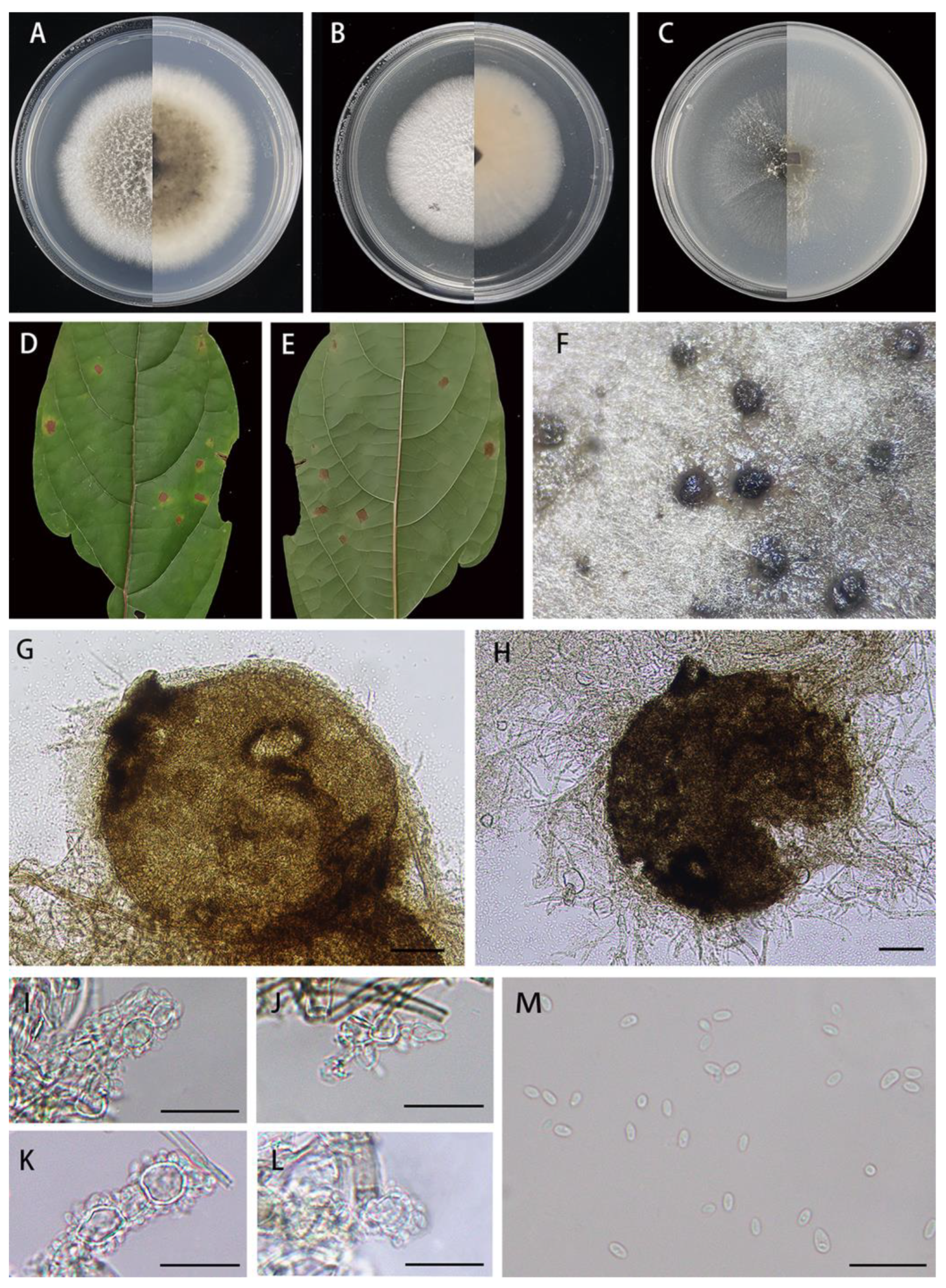
Figure 3.
Didymella clerodendri (HJAUP M1698, holotype): (A). Colony after 7 days on PDA (front and reverse); (B). Colony after 7 days on MEA (front and reverse); (C). Colony after 7 days on OA (front and reverse); (D,E). Leaf of host plant (front and reverse); (F). Pycnidia forming on PDA; (G,H). Pycnidium; (I–L). Conidiogenous cells; (M). Conidia. Scale bars: (G,H) = 50μm; (I–M) = 20 μm.
Index Fungorum number: IF901250.
Etymology: Referring to the host genus from which it was collected, Clerodendrum cyrtophyllum.
Holotype: HJAUP M1698.
Description: Irregular leaf spots, brown in center, and yellow to yellowish at margin. Asexual morph on PDA: Conidiomata are pycnidial, superficial, solitary or aggregated, mostly globose or subglobose, darker brown, with hyphal out growths, ostiolate, 206–330 × 190–290 μm (n = 20). Ostioles are single, central, and slightly papillate. Conidiogenous cells are hyaline, smooth, phialidic, subglobose, ampulliform to lageniform, 6.2–9.9 × 3.9–6.9 μm (n = 15). Conidia are ovoid or ellipsoidal, hyaline, smooth, thin walled, aseptate, 4.3–5.7 × 2.0–3.0 μm ( = 5.0 × 2.5 μm, n = 40), mostly with one or two minutes guttules. Conidial exudates buff. Sexual morph not observed.
Culture characteristics: Colonies on PDA reaching 68–70 mm diam after 7 days at 25 °C, margin regular, light brown in the middle, covered by white felt-like aerial hyphae, and white around; abundant production of pycnidia in the late growth stage. Colonies on MEA reaching 62–65 mm diam after 7 days at 25 °C, margin regular, covered by white felt-like aerial hyphae, and the back was buff. Colonies on OA reaching 57–59 mm diam after 7 days at 25 °C, margin regular, pale olivaceous, covered by a small amount of whitish aerial hyphae, and reverse pale olivaceous.
Material examined: Jingdezhen National Forest Park, Jiangxi Province, China, on diseased leaves of Clerodendrum cyrtophyllum Turcz. (Lamiaceae), 2 November 2022, X.X. Luo, HJAUP M1698 (holotype), ex-type living culture HJAUP C1698.
Notes: Strains HJAUP 1698, HJAUP 1698b, and HJAUP 1698c are similar in morphological characteristics and have identical DNA sequences; form a single, high support clade (MLBS/BPP = 100/0.98, Figure 1); and, therefore, are identified as the same new species, Didymella clerodendri. The phylogenetic tree showed that D. clerodendri clustered with D. pittospori in a high supported clade (MLBS/BPP = 100/0.99, Figure 1), and they form a sister clade to D. bellidis (CBS 714.85) and D. segeticola (CGMCC 3.17489) with a well-supported clade (MLBS/BPP = 90/0.98, Figure 1). Didymella clerodendriis closely related to D. bellidis, D. segeticola, and D. pittospori, and has, respectively, 26 bp, 22 bp, and 20 bp differences from D. bellidis, D. segeticola, and D. pittospori in four loci. Moreover, D. clerodendri morphologically differs from D. pittospori in having smaller pycnidia (206–330 × 190–290 μm vs. 290–496 × 151–323 μm), larger conidiogenous cells (6.2–9.9 × 3.9–6.9 μm vs. 4.7–7.2 × 3.6–5.6 μm), and larger conidia (4.3–5.7 × 2.0–3.0 μm vs. 3.1–5.2 × 1.6–2.5 μm). Didymella clerodendri is also morphologically similar to D. segeticola, but the latter has smaller pycnidia (90–105 × 75–95 μm vs. 206–330 × 190–290 μm), smaller conidiogenous cells (5–6.5 × 4–5.5 μm vs. 6.2–9.9 × 3.9–6.9 μm), and larger conidia (4.5–7 × 2.5–4 μm vs. 4.3–5.7 × 2.0–3.0 μm) with 1–6 polar guttules [12,42].
Didymella pittospori X.X. Luo, X.G. Zhang, and Jian Ma, sp. nov., Figure 4.
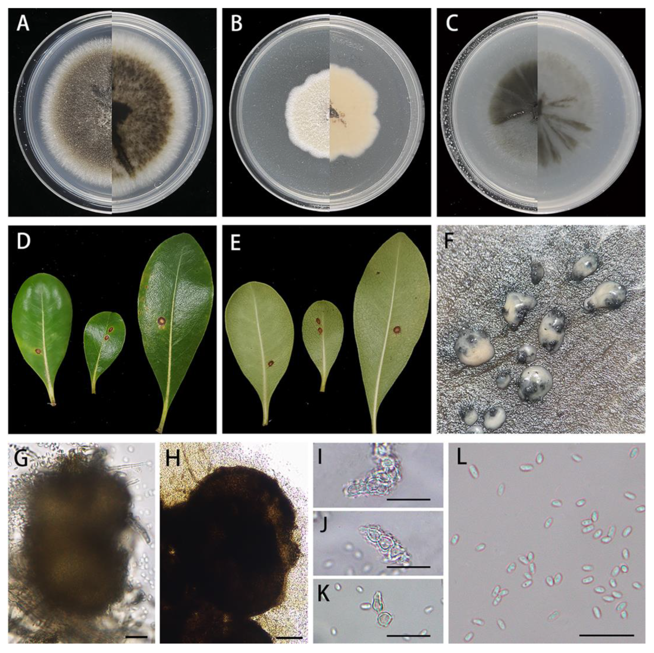
Figure 4.
Didymella pittospori (HJAUP M1740, holotype): (A). Colony after 7 days on PDA (front and reverse); (B). Colony after 7 days on MEA (front and reverse); (C). Colony after 7 days on OA (front and reverse); (D,E). Leaf of host plant (front and reverse); (F). Pycnidia forming on PDA; (G,H). Pycnidium; (I–K). Conidiogenous cells; (L). Conidia. Scale bars: (G,H) = 50μm; (I–L) = 20 μm.
Index Fungorum number: IF901251.
Etymology: Referring to the host genus from which it was collected, Pittosporum tobira.
Holotype: HJAUP M1740.
Description: Round leaf spots, brown to light-brown in center, and tan to black at margins with yellowish halos. Asexual morph on PDA: Conidiomata are pycnidial, superficial, solitary, solitary or aggregated, mostly globose or subglobose, black, glabrous, ostiolate, 290–496 × 151–323 μm (n = 20). Ostioles are single and slightly papillate. Conidiogenous cells are phialidic, hyaline, smooth, ampulliform to lageniform, 4.7–7.2 × 3.6–5.6 μm (n = 15). Conidia are oblong, ovoid or ellipsoidal, hyaline, smooth, thin-walled, aseptate, 3.1–5.2 × 1.6–2.5 μm ( = 4.1 × 2.0 μm, n = 40), and mostly with two minutes guttules. Conidial matrix is milky white. Sexual morph not observed.
Culture characteristics: Colonies on PDA reaching 74–76 mm diam after 7 days at 25 °C, margin regular, aerial mycelium sparsely, flat, dark brown near the central zone and white around, and reverse concolorous; abundant production of pycnidia in the late growth stage. Colonies on MEA reaching 46–50 mm diam after 7 days at 25 °C, margin waved, light-brown in the middle, white around, covered by white felt-like aerial hyphae, and reverse buff. Colonies on OA reaching 62–65 mm diam after 7 days at 25 °C, margin regular, olivaceous, covered by a few whitish aerial hyphae, and reverse concolorous.
Material examined: Longhu Mountain Nature Reserve, Jiangxi Province, China, on diseased leaves of Pittosporum tobira W.T.Aiton (Pittosporaceae), 3 November 2022, X.X. Luo, HJAUP M1740 (holotype), ex-type living culture HJAUP C1740; Jingdezhen Botanical Garden, Jiangxi Province, China, on diseased leaves of Eriobotrya japonica (Thunb.) Lindl. (Rosaceae), 2 November 2022, X.X. Luo, HJAUP M1800 (paratype), ex-paratype living culture HJAUP C1800.
Notes: Strains HJAUP 1740 and HJAUP 1800 are similar in morphological characteristics and have identical DNA sequences; form a single, strong support clade (MLBS/BPP = 95/0.86, Figure 1); and, therefore, are identified as the same new species, Didymella pittospori. The phylogenetic tree showed that D. pittospori clustered with D. clerodendri in a well-supported clade (MLBS/BPP = 100/0.99, Figure 1). Didymella pittospori is closely related to D. clerodendri and has 20 bp differences from D. clerodendri in four loci. Moreover, D. pittospori is morphologically distinguished from D. clerodendri as having larger pycnidia (290–496 × 151–323 μm vs. 206–330 × 190–290 μm), smaller conidiogenous cell (4.7–7.2 × 3.6–5.6 μm vs. 6.2–9.9 × 3.9–6.9 μm), and smaller conidia (3.1–5.2 × 1.6–2.5 μm vs. 4.3–5.7 × 2.0–3.0 μm). Didymella pittospori is also different from D. segeticola, which has smaller pycnidia (90–105 × 75–95 μm vs. 290–496 × 151–323 μm) and bigger conidia (4.5–7 × 2.5–4 μm vs. 3.1–5.2 × 1.6–2.5 μm) with 1–6 polar guttules [12,42].
4. Discussion
There are many kinds of fungi in Jiangxi Province, and the fungal groups are complex. Relevant studies have shown that several mycological investigations are also constantly exploring and enriching the fungal diversity (e.g., [12,43,44,45,46]). In this study, we isolated plant pathogens from diseased leaves of a wide range of plant hosts in Jiangxi Province, China. Based on the morphomolecular approach, three new species of Didymella, D. bischofiae, D. clerodendri, and D. pittospori, were introduced, which contributed to the supplementation of the diversity of this genus.
The establishment of Didymella was based on morphological studies. To date, there are 438 records for Didymella in Species Fungorum [8] but most of them are identified only by morphology, and only 86 species (including five invalid species) have sequence data so far. Morphological characteristics are significant for the identification of fungi, but the research based on morphological characteristics alone is not comprehensive. With the increase inavailable sequences for Dothideomycetes species, the molecular phylogenetic analysis has helped to clarify the phylogenetic relationships among the members of Dothideomycetes, and further clarified the species boundaries for the Didymella via multilocus analyses. However, studies conducted on Didymella have no universally accepted standards in selecting barcodes for phylogenetic analyses. For instance, De Gruyter et al. [2] established the family Didymellaceae with Didymella as type genus, but the initial Didymella species had only SSU and LSU sequences. Woudenberg et al. [47] and Thambugala et al. [48] introduced D. clematidis and D. eriobotryae using ITS, LSU, and TUB2. Liu et al. [49] introduced D. cirsii using ITS and LSU. Chen et al. [3,12] introduced 47 Didymella species using ITS, LSU, TUB2, and RPB2. Crous et al. [15,16] introduced D. cari and D. finnmarkica using ITS, LSU, ACT, and TUB2 or RPB2. From 2020 onwards, except for D. nakii, D. azollae, and D. brevipilosa using ITS, LSU, and TUB2 orRPB2, the further described 27 Didymella species were introduced using ITS, LSU, RPB2, and TUB2 [4,7,9,10,11,13,17,19,20,21,22,24,25,26]. Recent studies indicated that the use of LSU, ITS, TUB2, and RPB2 shows good phylogenetic resolution in revealing the phylogeny of Didymella and related genera within Didymellaceae. However, BLASTn analyses of these sequences showed that ITS and LSU sequences in some Didymella species have a high similarity, whereas RPB2 and/or TUB2 have distinct nucleotide differences. For example, ITS, LSU, RPB2, and TUB2 of D. degraaffiae [24] (MN823444, MN823295, MN824470, and MN824618) were 99.36, 99.72, 95.13, and 91.89% similar to D. maydis [3] (FJ427086, EU754192, GU371782, and FJ427190); D. qilianensis [4] (MT229701, MT229678, MT239098, and MT249269) were 100, 100, 98.32, and 98.2% similar to D. rhei [3] (GU237743, GU238139, KP330428, and GU237653). Our new species, D. clerodendri (OR625709, OR625714, OR620207, and OR611942) were 99.81, 100, 98.82, and 99.68% similar to D. pittospori (OR905550, OR905581, OR947922, and OR934715). Considering this phenomenon, we found that ITS and LSU sequences maybe show important significance in resolving the phylogeny of Didymellaceae, whereas RPB2 and/or TUB2 significantly increase the phylogenetic resolution in distinguishing Didymella species. Other loci in the mitochondrial genomemay also provide important insights in resolving the phylogeny of fungi [50,51,52], but hitherto not a single mitogenome exists for Didymella species.
To date, studies conducted on Didymella have mainly focused on their alpha-taxonomy, and most species are considered to be saprobes or phytopathogens of woody and herbaceous hosts [4,9,13,28,42], whereas only a few species have been isolated from inorganic substrates, such as D. glomerata and D. pomorum from inorganic materials including asbestos, cement, paint, etc. [3,12,53]. Recent studies also show that four didymella species, D. gardeniae, D. heteroderae, D. musae, and D. microchlamydospora, were found from human nail or cornea lesion [23], but there is no relevant data that support whether it has a direct relationship with the human disease. The genus Didymella is mainly recorded from China, Germany, India, Italy, The Netherlands, New Zealand, South Africa, and USA [3,4,7,12,21]; little published information is recorded in other regions [28]. Scant attention has been accorded to the roles of their ecosystem function, substrate specificities, and fungal pathogenicity. Thus, the understanding of external factors that affect fungal lifestyles may have a significant impact on agricultural development, ecological environment, and human health, contributing significantly to the field of plant pathology and fungal taxonomy.
Author Contributions
Conceptualization, X.L. and Y.H.; methodology, J.M.; software, X.L. and Z.X.; validation, K.Z.; formal analysis, X.L.; investigation, X.L.; resources, X.L. and Y.H.; data curation, X.L.; writing—original draft preparation, X.L. and Y.H.; writing—review and editing, J.X., L.M., and J.M.; visualization, K.Z.; supervision, J.M.; project administration, J.M.; funding acquisition, J.M. All authors have read and agreed to the published version of the manuscript.
Funding
This work was supported by the National Natural Science Foundation of China (Nos. 32160006, 31970018).
Institutional Review Board Statement
Not applicable.
Informed Consent Statement
Not applicable.
Data Availability Statement
All sequences generated in this study were submitted to GenBank.
Conflicts of Interest
The authors declare no conflicts of interest.
References
- Saccardo, P.A. Sylloge Pyrenomycetum. Syll. Fung. 1882, 1, 1–768. [Google Scholar]
- De Gruyter, J.; Aveskamp, M.M.; Woudenberg, J.H.C.; Verkley, G.J.M.; Groenewald, J.Z.; Crous, P.W. Molecular phylogeny of Phoma and allied anamorph genera: Towards a reclassification of the Phoma complex. Mycol. Res. 2009, 113, 508–519. [Google Scholar] [CrossRef] [Scilit]
- Chen, Q.; Jiang, J.R.; Zhang, G.Z.; Cai, L.; Crous, P.W. Resolving the Phoma enigma. Stud. Mycol. 2015, 82, 137–217. [Google Scholar] [CrossRef] [Scilit]
- Chen, Q.; Bakhshi, M.; Balci, Y.; Broders, K.D.; Cheewangkoon, R.; Chen, S.F.; Fan, X.L.; Gramaje, D.; Halleen, F.; Horta Jung, M.; et al. Genera of phytopathogenic fungi: GOPHY 4. Stud. Mycol. 2022, 101, 417–564. [Google Scholar] [CrossRef] [Scilit]
- Aveskamp, M.M.; De Gruyter, J.; Woudenberg, J.H.C.; Verkley, G.J.M.; Crous, P.W. Highlights of the Didymellaceae: A polyphasic approach to characterise Phoma and related pleosporalean genera. Stud. Mycol. 2010, 65, 1–60. [Google Scholar] [CrossRef] [Scilit]
- Zhang, Y.; Crous, P.W.; Schoch, C.L.; Hyde, K.D. Pleosporales. Fungal Divers. 2012, 53, 1–221. [Google Scholar] [CrossRef] [Scilit]
- Hou, L.W.; Groenewald, J.Z.; Pfenning, L.H.; Yarden, O.; Crous, P.W.; Cai, L. The phoma-like dilemma. Stud. Mycol. 2020, 96, 309–396. [Google Scholar] [CrossRef] [Scilit]
- Species Fungorum. Available online: http://www.speciesfungorum.org/Names/Names.asp (accessed on 18 December 2023).
- Magaña-Dueñas, V.; Cano-Lira, J.F.; Stchigel, A.M. New Dothideomycetes from freshwater habitats in Spain. J. Fungi 2021, 7, 1102. [Google Scholar] [CrossRef] [Scilit]
- Ahmadpour, S.A.; Mehrabi-Koushki, M.; Farokhinejad, R.; Asgari, B. New species of the family Didymellaceae in Iran. Mycol. Prog. 2022, 21, 28. [Google Scholar] [CrossRef] [Scilit]
- Boonmee, S.; Wanasinghe, D.N.; Calabon, M.S.; Huanraluek, N.; Chandrasiri, S.K.U.; Jones, G.E.B.; Rossi, W.; Leonardi, M.; Singh, S.K.; Rana, S.; et al. Fungal diversity notes 1387–1511: Taxonomic and phylogenetic contributions on genera and species of fungal taxa. Fungal Divers. 2019, 111, 1–335. [Google Scholar] [CrossRef] [Scilit]
- Chen, Q.; Hou, L.W.; Duan, W.J.; Crous, P.W.; Cai, L. Didymellaceae revisited. Stud. Mycol. 2017, 87, 105–159. [Google Scholar] [CrossRef] [Scilit]
- Chen, T.; Wang, S.; Jiang, X.; Huang, Y.; Mo, M.; Yu, Z. New Species of Didymellaceae within aquatic plants from southwestern China. J. Fungi 2023, 9, 761. [Google Scholar] [CrossRef] [Scilit]
- Crous, P.W.; Wingfield, M.J.; Burgess, T.I.; Carnegie, A.J.; Hardy, G.E.S.J.; Smith, D.; Summerell, B.A.; Cano-Lira, J.F.; Guarro, J.; Hobraken, J.; et al. Fungal Planet description sheets: 625–715. Persoonia 2017, 39, 270–467. [Google Scholar] [CrossRef] [Scilit]
- Crous, P.W.; Wingfield, M.J.; Burgess, T.I.; Hardy, G.E.S.J.; Gené, J.; Guarro, J.; Baseia, I.G.; García, D.; Gusmao, L.F.P.; Souza-Motta, C.M.; et al. Fungal Planet description sheets: 716–784. Persoonia 2018, 40, 239–392. [Google Scholar] [CrossRef] [Scilit]
- Crous, P.W.; Carnegie, A.J.; Wingfield, M.J.; Sharma, R.; Mughini, G.; Noordeloos, M.E.; Santini, A.; Shouche, Y.S.; Bezerra, J.D.P.; Dima, B.; et al. Fungal Planet description sheets: 868–950. Persoonia 2019, 42, 291–473. [Google Scholar] [CrossRef] [Scilit]
- Crous, P.W.; Osieck, E.R.; Jurjevi, Ž.; Boers, J.; Van Iperen, A.L.; Starink-Willemse, M.; Dima, B.; Balashov, S.; Bulgakov, T.S.; Johnston, P.R.; et al. Fungal Planet description sheets: 1284–1382. Persoonia 2021, 47, 178–374. [Google Scholar] [CrossRef] [Scilit]
- Jayasiri, S.C.; Hyde, K.D.; Jones, E.B.G.; McKenzie, E.H.C.; Jeewon, R.; Phillips, A.J.L.; Bhat, D.J.; Wanasinghe, D.N.; Liu, J.K.; Lu, Y.Z.; et al. Diversity, morphology and molecular phylogeny of Dothideomycetes on decaying wild seed pods and fruits. Mycosphere 2019, 10, 1–186. [Google Scholar] [CrossRef] [Scilit]
- Das, K.; Lee, S.Y.; Jung, H.Y. Molecular and morphological characterization of two novel species collected from Soil in Korea. Mycobiology 2020, 48, 9–19. [Google Scholar] [CrossRef] [Scilit]
- Mapook, A.; Hyde, K.D.; McKenzie, E.H.C.; Jones, E.B.G.; Bhat, D.J.; Jeewon, R.; Stadler, M.; Samarakoon, M.C.; Malaithong, M.; Tanunchai, B.; et al. Taxonomic and phylogenetic contributions to fungi associated with the invasive weed Chromolaena odorata (Siam weed). Fungal Divers. 2020, 101, 1–175. [Google Scholar] [CrossRef] [Scilit]
- Scarpari, M.; Vitale, S.; Giambattista, G.D.; Luongo, L.; De Gregorio, T.; Schreiber, G.; Petrucci, M.; Belisario, A.; Voglmayr, H. Didymella corylicola sp. nov., a new fungus associated with hazelnut fruit development in Italy. Mycol. Prog. 2020, 19, 317–328. [Google Scholar] [CrossRef] [Scilit]
- Tan, Y.P.; Bishop-Hurley, S.L.; Shivas, R.G.; Cowan, D.A.; Maggs-Kölling, G.; Maharachchikumbura, S.S.N.; Pinruan, U.; Bransgrove, K.L.; De la Peña-Lastra, S.; Larsson, E.; et al. Fungal Planet description sheets: 1436–1477. Persoonia 2022, 49, 261–350. [Google Scholar] [CrossRef] [Scilit]
- Valenzuela-Lopez, N.; Cano-Lira, J.F.; Guarro, J.; Sutton, D.A.; Wiederhold, N.; Crous, P.W.; Schigel, A.M. Coelomycetous Dothideomycetes with emphasis on the families Cucurbitariaceae and Didymellaceae. Stud. Mycol. 2018, 90, 1–69. [Google Scholar] [CrossRef] [Scilit]
- Hou, L.W.; Hernandez-Restrepo, M.; Groenewald, J.Z.; Cai, L.; Crous, P.W. Citizen science project reveals high diversity in Didymellaceae (Pleosporales, Dothideomycetes). Mycokeys 2020, 65, 49–99. [Google Scholar] [CrossRef] [Scilit]
- Hyde, K.D.; Dong, Y.; Phookamsak, R.; Jeewon, R.; Bhat, D.J.; Jones, E.B.G.; Liu, N.G.; Abeywickrama, P.D.; Mapook, A.; Wei, D.; et al. Fungal diversity notes 1151–1276: Taxonomic and phylogenetic contributions on genera and species of fungal taxa. Fungal Divers. 2020, 100, 5–277. [Google Scholar] [CrossRef] [Scilit]
- Lee, G.; Kim, K.D.; Cho, W.D.; Kim, W. Didymella acutilobae sp. nov. causing leaf spot and stem rot in Angelica acutiloba. Mycobiology 2023, 51, 313–319. [Google Scholar] [CrossRef] [Scilit]
- Turland, N.J.; Wiersema, J.H.; Barrie, F.R.; Greuter, W.; Hawksworth, D.L.; Herendeen, P.S.; Knapp, S.; Kusber, W.H.; Li, D.Z.; Marhold, K.; et al. (Eds.) International Code of Nomenclature for algae, fungi, and plants (Shenzhen Code) adopted by the Nineteenth International Botanical Congress, Shenzhen, China, July 2017; Regnum Veg. 159; Koeltz Botanical Books: Glashütten, Germany, 2018; pp. 1–254. [Google Scholar] [CrossRef] [Scilit]
- Index Fungorum. Available online: http://www.indexfungorum.org/Names/Names.asp (accessed on 18 December 2023).
- Gao, Y.H.; Sun, W.; Su, Y.Y.; Cai, L. Three new species of Phomopsis in Gutianshan nature reserve in China. Mycol. Prog. 2014, 13, 111–121. [Google Scholar] [CrossRef] [Scilit]
- Cai, L.; Hyde, K.D.; Taylor, P.W.J.; Weir, B.S.; Waller, J.M.; Abang, M.M.; Zhang, J.Z.; Yang, Y.L.; Phoulivong, S.; Liu, Z.Y.; et al. A polyphasic approach for studying Colletotrichum. Fungal Divers. 2009, 39, 183–204. [Google Scholar]
- White, T.J.; Bruns, T.D.; Lee, S.B.; Taylor, J.W. Amplification and direct sequencing of fungal ribosomal RNA genes for phylogenetics. In PCR Protocols: A Guide to Methods and Applications; Innis, M.A., Gelfand, D.H., Sninsky, J.J., White, T.J., Eds.; Academic Press: New York, NY, USA, 1990; pp. 315–322. [Google Scholar] [CrossRef] [Scilit]
- Vilgalys, R.; Hester, M. Rapid genetic identification and mapping of enzymatically amplified ribosomal DNA from several Cryptococcus species. J. Bacteriol. 1990, 172, 4238–4246. [Google Scholar] [CrossRef] [Scilit]
- Glass, N.L.; Donaldson, G.C. Development of primer sets designed for use with the PCR to amplify conserved genes from Filamentous Ascomycetes. Appl. Environ. Microb. 1995, 61, 1323–1330. [Google Scholar] [CrossRef] [Scilit]
- Voglmayr, H.; Akulov, O.Y.; Jaklitsch, W.M. Reassessment of Allantonectria, phylogenetic position of Thyronectroidea, and Thyronectria caraganae sp. nov. Mycol. Prog. 2016, 15, 921–937. [Google Scholar] [CrossRef] [Scilit]
- Katoh, K.; Standley, D.M. MAFFT multiple sequence alignment software version 7: Improvements in performance and usability. Mol. Biol. Evol. 2013, 30, 772–780. [Google Scholar] [CrossRef] [Scilit] [PubMed]
- Zhang, D.; Gao, F.; Jakovlić, I.; Zou, H.; Zhang, J.; Li, W.X.; Wang, G.T. PhyloSuite: An integrated and scalable desktop platform for streamlined molecular sequence data management and evolutionary phylogenetics studies. Mol. Ecol. Res. 2020, 20, 348–355. [Google Scholar] [CrossRef] [Scilit] [PubMed]
- Kalyaanamoorthy, S.; Minh, B.Q.; Wong, T.; von Haeseler, A.; Jermiin, L.S. ModelFinder: Fast model selection for accurate phylogenetic estimates. Nat. Methods. 2017, 14, 587–589. [Google Scholar] [CrossRef] [Scilit]
- Nguyen, L.T.; Schmidt, H.A.; von Haeseler, A.; Minh, B.Q. IQ-TREE: A fast and effective stochastic algorithm for estimating maximum-likelihood phylogenies. Mol. Biol. Evol. 2015, 32, 268–274. [Google Scholar] [CrossRef] [Scilit] [PubMed]
- Minh, B.Q.; Nguyen, M.A.T.; von Haeseler, A. Ultrafast approximation for phylogenetic bootstrap. Mol. Biol. Evol. 2013, 30, 1188–1195. [Google Scholar] [CrossRef] [Scilit]
- Ronquist, F.; Teslenko, M.; van der Mark, P.; Ayres, D.L.; Darling, A.; Höhna, S.; Larget, B.; Liu, L.; Suchard, M.A.; Huelsenbeck, J.P. MrBayes 3.2: Efficient Bayesian phylogenetic inference and model choice across a large model space. Syst. Biol. 2012, 61, 539–542. [Google Scholar] [CrossRef] [Scilit]
- De Gruyter, J.; Noordeloos, M.E.; Boerema, G.H. Contributions towards a monograph of Phoma (Coelomycetes)—I. 3. Section Phoma: Taxa with conidia longer than 7 µm. Persoonia 1998, 16, 471–490. [Google Scholar]
- Chen, Q.; Zhang, K.; Zhang, G.Z.; Cai, L. A polyphasic approach to characterise two novel species of Phoma (Didymellaceae) from China. Phytotaxa 2015, 197, 267–281. [Google Scholar] [CrossRef] [Scilit]
- Xu, Z.H.; Kuang, W.G.; Qiu, L.; Zhang, X.G.; Castaneda-Ruíz, R.F.; Ma, J. Corynespora sinensis sp. nov. from Jiangxi, China. Mycotaxon 2020, 135, 803–809. [Google Scholar] [CrossRef] [Scilit]
- Yang, Q.; Jiang, N.; Tian, C.M. New species and records of Diaporthe from Jiangxi Province, China. Mycokeys 2021, 77, 41–64. [Google Scholar] [CrossRef] [Scilit]
- Zhai, Z.J.; Yan, J.Q.; Li, W.W.; Gao, Y.; Hu, H.J.; Zhou, J.P.; Song, H.Y.; Hu, D.M. Three novel species of Distoseptispora (Distoseptisporaceae) isolated from bamboo in Jiangxi Province, China. Mycokeys 2022, 88, 35–54. [Google Scholar] [CrossRef] [Scilit] [PubMed]
- Hu, Y.F.; Liu, J.W.; Xu, Z.H.; Castañeda-Ruíz, R.F.; Zhang, K.; Ma, J. Morphology and multigene phylogeny revealed three new species of Helminthosporium (Massarinaceae, Pleosporales) from China. J. Fungi 2023, 9, 280. [Google Scholar] [CrossRef] [Scilit] [PubMed]
- Woudenberg, J.H.C.; Aveskamp, M.M.; De Gruyter, J.; Spiers, A.G.; Crous, P.W. Multiple Didymella teleomorphs are linked to the Phoma clematidina morphotype. Persoonia 2009, 22, 56–62. [Google Scholar] [CrossRef] [Scilit] [PubMed]
- Thambugala, K.M.; Hyde, K.D.; Zhang, J.F.; Liu, Z.Y. Didymella eriobotryae sp. nov. (Didymellaceae) and Arthrinium arundinis (Apiosporaceae) from fruit of Eriobotrya japonica (loquat) in China. Phytotaxa 2018, 382, 136. [Google Scholar] [CrossRef] [Scilit]
- Liu, J.K.; Hyde, K.D.; Jones, E.G.; Ariyawansa, H.A.; Bhat, D.J.; Boonmee, S.; Maharachchikumbura, S.S.N.; McKenzie, E.H.C.; Phookamsak, R.; Phukhamsakda, C.; et al. Fungal diversity notes 1–110: Taxonomic and phylogenetic contributions to fungal species. Fungal Divers. 2015, 72, 1–197. [Google Scholar] [CrossRef] [Scilit]
- He, K.; Ren, T.; Zhu, S.; Zhao, A. The complete mitochondrial genome of Fulica atra (Avian, Gruiformes, Rallidae). Mitochondr. DNA 2016, 27, 3161–3162. [Google Scholar] [CrossRef] [Scilit]
- Kulik, T.; Van Diepeningen, A.D.; Hausner, G. Editorial: The significance of mitogenomics in mycology. Front. Microbiol. 2021, 11, 628579. [Google Scholar] [CrossRef] [Scilit]
- Tan, H.; Yu, Y.; Fu, Y.; Liu, T.; Wang, Y.; Peng, W.; Wang, B.; Chen, J. Comparative analyses of Flammulina filiformis mitochondrial genomes reveal high length polymorphism in intergenic regions and multiple intron gain/loss in cox1. Int. J. Biol. Macromol. 2022, 221, 1593–1605. [Google Scholar] [CrossRef] [Scilit]
- Aveskamp, M.M.; De Gruyter, J.; Crous, P.W. Biology and recent developments in the systematics of Phoma, a complex genus of major quarantine significance. Fungal Divers. 2008, 31, 1–18. [Google Scholar]




Disclaimer/Publisher’s Note: The statements, opinions and data contained in all publications are solely those of the individual author(s) and contributor(s) and not of MDPI and/or the editor(s). MDPI and/or the editor(s) disclaim responsibility for any injury to people or property resulting from any ideas, methods, instructions or products referred to in the content. |
© 2024 by the authors. Licensee MDPI, Basel, Switzerland. This article is an open access article distributed under the terms and conditions of the Creative Commons Attribution (CC BY) license (https://creativecommons.org/licenses/by/4.0/).