Article

Degradation Potential of Xerophilic and Xerotolerant Fungi Contaminating Historic Canvas Paintings

1
Department of Biology, Biotechnical Faculty, University of Ljubljana, Jamnikarjeva 101, SI-1000 Ljubljana, Slovenia
2
Institute for the Protection of Cultural Heritage of Slovenia, Poljanska 40, SI-1000 Ljubljana, Slovenia
*
Author to whom correspondence should be addressed.
J. Fungi 2024, 10(1), 76; https://doi.org/10.3390/jof10010076
Submission received: 14 November 2023 / Revised: 12 January 2024 / Accepted: 15 January 2024 / Published: 18 January 2024
(This article belongs to the Special Issue Fungal Biodeterioration)

Abstract

:
Fungi are important contaminants of historic canvas paintings worldwide. They can grow on both sides of the canvas and decompose various components of the paintings. They excrete pigments and acids that change the visual appearance of the paintings and weaken their structure, leading to flaking and cracking. With the aim of recognizing the most dangerous fungal species to the integrity and stability of paintings, we studied 55 recently isolated and identified strains from historic paintings or depositories, including 46 species from 16 genera. The fungi were categorized as xero/halotolerant or xero/halophilic based on their preference for solutes (glycerol or NaCl) that lower the water activity (aw) of the medium. Accordingly, the aw value of all further test media had to be adjusted to allow the growth of xero/halophilic species. The isolates were tested for growth at 15, 24 °C and 37 °C. The biodeterioration potential of the fungi was evaluated by screening their acidification properties, their ability to excrete pigments and their enzymatic activities, which were selected based on the available nutrients in paintings on canvas. A DNase test was performed to determine whether the selected fungi could utilize DNA of dead microbial cells that may be covering surfaces of the painting. The sequestration of Fe, which is made available through the production of siderophores, was also tested. The ability to degrade aromatic and aliphatic substrates was investigated to consider the potential degradation of synthetic restoration materials. Xerotolerant and moderately xerophilic species showed a broader spectrum of enzymatic activities than obligate xerophilic species: urease, β-glucosidase, and esterase predominated, while obligate xerophiles mostly exhibited β-glucosidase, DNase, and urease activity. Xerotolerant and moderately xerophilic species with the highest degradation potential belong to the genus Penicillium, while Aspergillus penicillioides and A. salinicola represent obligately xerophilic species with the most diverse degradation potential in low aw environments.

1. Introduction

Artistic canvas paintings often present an important part of the cultural heritage of a country and must therefore be preserved and, if damaged, restored. Biodeterioration is frequently related to inadequate or poorly controlled storage conditions, but it can also occur in modern facilities such as depositories or museum exhibition rooms due to inappropriate heating, ventilation, and air conditioning (HVAC) systems or, in extreme cases, accidental flooding events. Materials introduced during conservation–restoration activities can sometimes promote the process of biodeterioration. The resulting microbial growth and activity can lead to aesthetic changes, mechanical weakening or chemical destruction of the artwork, e.g., staining or discoloration, textural changes, loss of elasticity, deformation, and damage to the textile canvas support [1,2].
Fungal contamination of canvas paintings is common due to the complex and mainly organic, easily degradable materials used as painting components [2,3]. A canvas painting consists of (i) a mechanical support (canvas, wood); (ii) ground layer (animal glue, chalk, a mixture of gypsum or calcium carbonate and glue or flour, oil, and pigments); (iii) paint layers (pigments, drying oil, egg yolk, casein, gums, synthetic resins, such as alkyds, acrylics or ketones) [4,5,6]; and (iv) a cover layer of varnish for protection (dammar, mastic, shellac, or synthetic resins) [4,6,7,8]. Also, conservation–restoration interventions use preferably organic materials [2]. Nevertheless, there is little information on microbial biodeterioration of these materials [2,3,7,9,10,11]. Only a few studies so far have focused on the microbial enzymes responsible for degradation of canvas paintings [11,12,13], and they were based on rapid, low-cost plate assays [14,15]. The fungal genera recognized for their broad range of enzymatic activities were Aspergillus, Cladosporium, and Penicillium [14,15]. In addition, representatives of the these genera can secrete organic acids and pigments [11,14,15,16], which can irreversibly change the appearance and structure of canvas paintings [17,18].
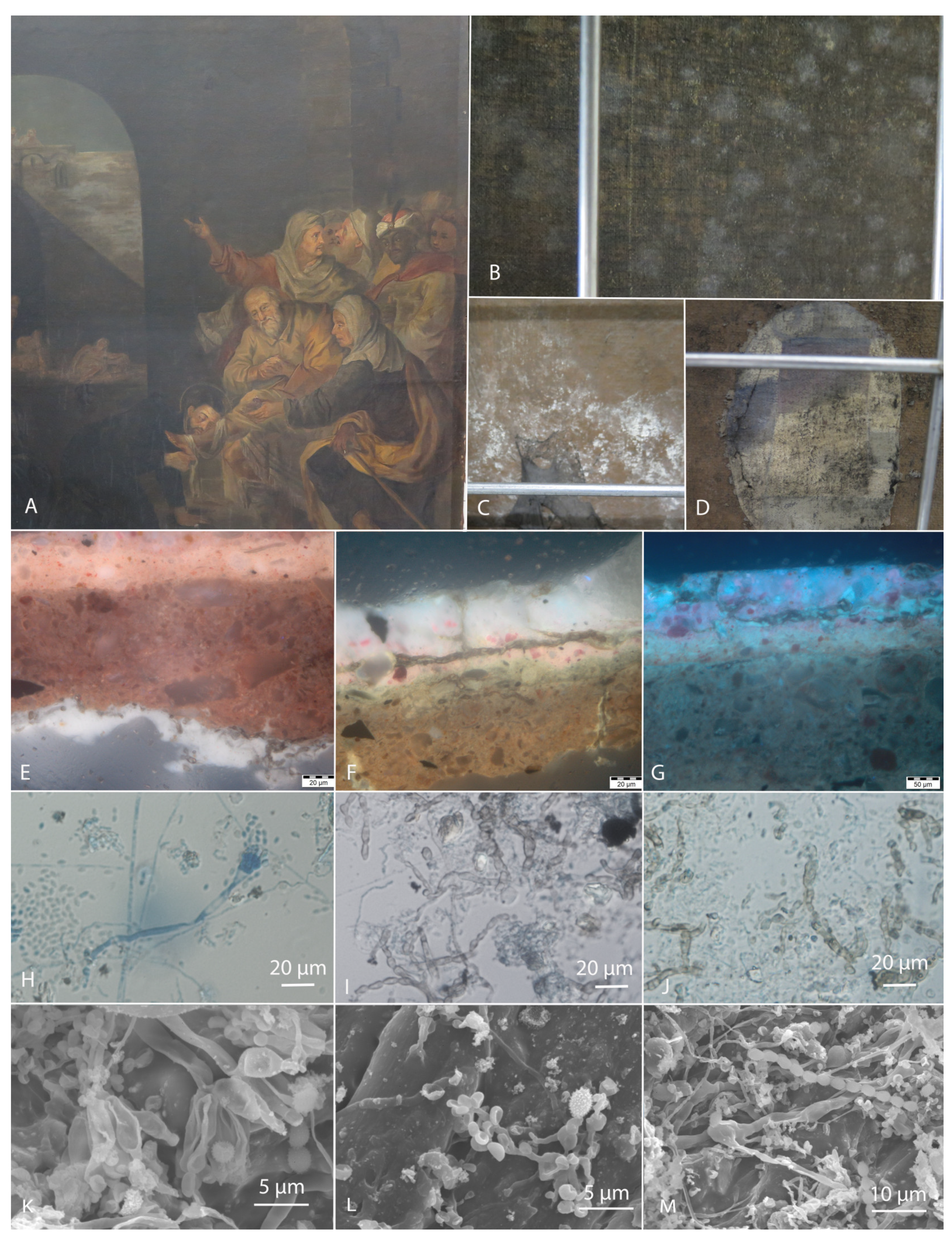
Fungi contaminate both the front (verso) and the back (recto) of canvas paintings (Figure 1). Some studies suggest that front side communities of oil paintings are inactive, while those observed on the back side are considered to be involved in biodeterioration [19]. The infestation on the back side is initiated by the hydration of the organic textile fibres [20], the weakening of the adjacent layers, and the mechanical penetration of the hyphae into the paint layer, resulting in cracks [7,21], considered as hotspots for fungal growth [22]. On the other hand, infestation on the front side depends on the ground, paint, and varnish layers and their mode of application. After gravitational deposition of transient airborne bacterial and fungal spores on the surface, they form a subaerial community that eventually grows and degrades these materials, especially in the presence of high humidity [19].
Although species of the genera Penicillium, Aspergillus, Alternaria, Cladosporium, and Chaetomium dominate the fungal communities on canvas paintings, their biodeterioration potential is not yet fully known due to the paucity of studies [3,11,14,15]. Moreover, these genera contain numerous species [23,24,25,26,27], and new species are constantly being described [27], making it difficult to identify them correctly.
Canvas and protein degradation are the most frequently studied deterioration activities attributed to fungi contaminating paintings. Representatives of the genera mentioned above [7] are known for their ability to disrupt cellulose fibres by affecting the degree of polymerization, chain length, crystallinity, and orientation, thus increasing their susceptibility to biodegradation by cellulolytic enzymes. Enzymes involved in the degradation of textiles are cellulases (endoglucanases, β-glucosidases) [28,29], hemicellulases, lignin-degrading enzymes, and amylases if the canvas is sized with starch or gelatinases if it is sized with animal glues [30]. The degradation of canvas support allows the fungal hyphae to reach the paint layer, which can lead to delamination, cracking, and discoloration [7]. Depending on the type of paint (oil, tempera, synthetic), different binders are used, such as linseed oil, egg yolk, egg white, casein, vegetable oils and gums, wax, and natural or synthetic resins [31]. Casein and egg tempera are the most susceptible to microbial attack, followed by emulsion distemper (greasy tempera), while linseed oil (drying oil) is the most resistant [7]. The proteolytic activity of fungi is widespread and particularly dangerous for damaging natural binders and materials used in conservation processes [32]. Varnishes protect the painted surface. In traditional painting, they are usually applied after dissolving natural resins in oil, such as vernice comune (mixture of colophony/oleoresin and linseed oil), or by dissolving terpenoid resins in spirits of wine and turpentine [33]. There are rare reports of biodegradation of natural resins by bacteria and fungi [34,35,36].
Figure 1. Historic oil painting RCS 15 [37] from the church with signs of biodeterioration on both sides. (A) Front side of the painting, (BD) Back (courtesy of Polona Zalar), (EG) Stratigraphy of the painting under UV light (courtesy of Katja Kavkler) showing the isolation layer between the canvas and the ground (E), the ground layer, the paint layer, and the surface of the varnish (F), and the ground layer and the paint layer (G), all with visible penetration of dark septate hyphae. Light (HJ) (courtesy of Polona Zalar) and Scanning electron microscopy (SEM) (KM) images (courtesy of Petra Bešlagić), show conidiogenous structures of Aspergillus on the back (H,K) and massive growth on the front side of the painting (I,J,L,M).
Jof 10 00076 g001
The present study aims to describe the deterioration potential of xerotolerant/xerophilic and halotolerant/halophilic fungi, which are adapted to dry environments and low water activity (aw) of substrates and were recently reported from historic canvas paintings in Slovenia (Figure 1) [22,37]. The most abundant genus was Aspergillus, with numerous obligate xerophilic species not previously observed on contaminated paintings. Xerophilic/xerotolerant Wallemia, Penicillium, and Cladosporium species were also abundant. Machine learning predictive models, based on the collected data on materials and contaminants, indicated that different Aspergillus species could be associated with all of the materials constituting the paintings [22], in line with the diverse enzymatic activities described in this genus [7,14]. The exact biodeterioration potential, especially of the newly discovered obligate xerophilic species, is not yet known. These fungi have been almost completely neglected in most studies on the deterioration of cultural heritage objects, including canvas paintings, due to the general assumption that low humidity environments prevent fungal growth [22,38]. In this study, 55 fungal strains belonging to 46 species from 16 genera, including 12 obligately xerophilic Aspergillus and Wallemia strains, were evaluated for their biodeterioration abilities. Acidification of the substrate was tested in two ways: (i) in a liquid medium allowing pH measurement after incubation, and (ii) on a solid medium containing calcium carbonate (CaCO3), which dissolves when the medium is acidified [32,39,40]. The strains were screened for the following enzymatic activities, which were selected according to the substrates available in paintings: cellulases (endocellulase, β-glucosidase), amylase, and laccase, which cause canvas degradation, gelatinase and caseinase which cause protein degradation, while esterase and lipase-lecithinase cause degradation of drying oil. The production of fungal pigments, which is dependent on the substrate, was recorded on all media tested that did not contain indicator dyes or reagents that cause colour reactions. Several tests were used for the first time to assess painting biodeterioration: the production of enzymes laccase, urease, and DNase, the formation of siderophores, and the utilization of aromatic and aliphatic sole carbon sources. Laccase, an enzyme known for lignin degradation, can also oxidize other materials containing aromatic rings (e.g., as components of pigments and modern synthetic materials) [41]. Urease can act on several materials in paintings, as urea can be used as a dissolving agent for powdered dyes [1,7] or in combination with esterase can assist in the degradation of wax and synthetic resins [42]. A DNase assay was performed to evaluate the ability of fungi to hydrolyze DNA from surface, formerly active microorganisms and use it as a source of carbon and energy for growth [1]. The siderophore production tested allows the recruitment of scarce Fe from the environment [43,44,45]. The ability to degrade aromatic and aliphatic substrates was used to target the potential degradation of synthetic restoration materials containing aromatic rings or aliphatic chains in their structure, such as the hot-sealing adhesive BEVA 371 and the acrylic glue Lascaux 498 HV, respectively. While the enzyme screening of the xerotolerant and moderately xerophilic fungi was carried out according to the established culture media and associated protocols, all culture media were adapted to test the enzyme production of the obligate xerophilic fungi by aw reduction with the addition of glycerol. Based on the results obtained, we conclude that the fungal taxa tested are hazardous to paintings, obligately xerophilic ones, even when stored in depositories with recommended environmental measures.

2. Materials and Methods

2.1. Fungal Strains and Inocula Preparation

Identified fungal strains were selected from the studies of Kavkler et al. (2022) [37] and Zalar et al. (2023) [22]. They were mainly isolated from deteriorated historic paintings exhibited in churches or were deposited for restoration in authorised depository (Supplementary Table S1). Only two strains originate from the air in the painting depository of the Restoration Centre of the Institute for the Protection of Cultural Heritage of Slovenia (RCS). Four strains were isolated as contaminants of mock-up samples treated with restoration materials. The later were identified using DNA sequences of genus-specific molecular markers evident in Supplementary Table S1 as described in Zalar et al. (2023) [22]. Nucleotide sequences were deposited to NCBI (PP033021, PP034128, PP034131, PP034132). All the strains are preserved in the Microbial Culture Collection Ex within Infrastructural Centre Mycosmo (Department of Biology, Biotechnical Faculty, University of Ljubljana, Ljubljana, Slovenia).
Fungal cultures were grown on Malt Extract Agar (MEA) prepared according to Galloway and Burgess (1952) (0.02% malt extract, 0.02% glucose, 0.001% pepton, 0.02% (m/V) agar; pH = 5–5.5) [46] or on Dichloran Glycerol Agar [47] (DG18; Biolife Italiana, Milano, Italy) prepared according to the producer’s instructions, for 7–14 days. Conidia/spore suspensions of sporulating fungi were prepared with concentration of 107 spores/mL, while suspensions of mycelial fragments of nonsporulating fungi were not standardized. The inocula of xerotolerant and moderately xerophilic species were prepared in 0.9% sodium chloride (NaCl), while those of obligate xerophiles were prepared in 10% NaCl.

2.2. Growth at Different Water Activities and Temperatures

All selected strains (see Section 2.1) were tested for growth on media without and with added solutes (glycerol, NaCl) to determine their xero-(halo)tolerance/xero-(halo)philic nature. Xerotolerance was tested on DG18 culture medium base (Biolife Italiana, Milano, Italy) [47] without (DG0; aw = 0.99) and with the addition of different amounts of glycerol: 18% (DG18; aw = 0.942), 30% (DG30; aw = 0.882), and 40% (w/v) (DG40; aw = 0.827). Halotolerance test was performed on MEA (described in 2.1) supplemented with different concentrations of NaCl: 10% (w/v) (MEA10; aw = 0.930), 20% (MEA20; aw = 0.852), and 25% (MEA25; aw = 0.802). Colony diameters were measured for both tests after 28 days of incubation at 24 °C. Water activity of the media was measured using AquaLab Series 3TE (Decagon Devices, Pullman, WA, USA).
Strains were tested for growth at 15 °C (recommended temperature in depositories), 24 °C (museum temperature), and 37 °C (temperature used to test potential hazards to human [48]) on MEA for xerotolerant and moderately xerophilic fungi, or on DG18 for obligately xerophilic species. Colony diameters were measured after 14 days using a ruler.

2.3. Biodegradation Assays

2.3.1. Excretion of Enzymes

Enzyme screening assays were selected based on the commonly used materials in canvas paintings and in their conservation–restoration. The selection according to a specific painting layer is presented in Figure 2. The composition of paint layers according to the painting technique (egg tempera, oil paints, other paints, or synthetic paints) is indicated with orange rectangles, e.g., greasy tempera contains pigments, drying oil, and egg. Each component can be degraded by specific enzymes, e.g., pigments by laccases, drying oil by esterases and lipases, and egg by lecithinases. The potential hazard of one enzymatic activity may affect multiple layers of a painting, as, e.g., in the case of esterase activity.
The following enzyme activities were tested on agar media in 9 cm Petri dishes: amylase, β-glucosidase, endocellulase, protease (caseinase as general activity, gelatinase as specific activity), esterase, lecithinase, lipase, and laccase. Media were 3-point inoculated with 5 µL of conidial suspension and incubated at 24 °C for 7–28 days. The deterioration profile of xerotolerant and moderately xerophilic taxa was tested on established culture media, as described in detail below, while for the testing of obligately xerophilic fungi, the water activity (aw) of all test media was adjusted to 0.94 by adding 18% (w/v) glycerol. Agar media for lecithinase activity was the only one prepared without additional solutes, but it was incubated for a longer period (28 days) until sufficiently dried to allow the growth of obligately xerophilic species.
The overview of performed enzyme tests, the main substrate, and positive results is presented in Table 1, while the exact composition and preparation of the test media are described below.
Amylolytic activity was tested for degradation of starch used as a canvas consolidant [1,7]. The medium used was Czapek agar, prepared from starch solution (5% soluble starch in dH2O) (Sigma-Aldrich, Steinheim, Germany), 50 mL of Czapek solution A (4% NaNO3, 1% KCl, 0.02% MgSO4 × 7H2O in dH2O), 50 mL of Czapek solution C (2% K2HPO4 in dH2O), 1 mL of zinc solution (1% ZnSO4 × 7H2O in dH2O), 1 mL of copper solution (0.5% CuSO4 × 5H2O in dH2O), 1.2% agar, and dH2O up to 1 L. After 14 days of incubation, plates were flooded with iodine solution for 30 s, and the excess iodine was poured off; the clear zone around colonies in otherwise black/blue-stained medium indicated starch degradation [49].
Endocellulase and β-glucosidase activities were tested as indicators for canvas degradation [1,7]. To test β-glucosidase activity, aesculin (6,7-dihydroxycoumarin 6-glucoside) (Merck, Darmstadt, Germany) was added to Czapek agar at the concentration 0.3%; after 14 days of incubation, a black zone around colonies indicated a positive reaction. Endocellulase activity was tested by adding 4% carboxymethyl cellulose (CMC; Sigma-Aldrich, Steinheim, Germany) to Czapek agar; after 21 days of incubation, media were stained with 0.3% Congo red (Merck, Darmstadt, Germany) and rinsed after 30 min with 1 M NaCl solution; a yellow clear zone around colonies indicated a positive reaction [49].
Caseinase activity was tested as the general proteolytic activity [1,7] on agar medium containing 5% skim milk (Merck, Darmstadt, Germany) [50]. The medium was prepared from two separately prepared and autoclaved solutions: (1) 0.2% yeast extract, 4% agar in dH2O; (2) 10% skim milk in dH2O. After autoclaving, 500 mL of solution (1) was aseptically and homogeneously added to the same amount of solution (2); a clear zone in an otherwise opaque medium indicated proteolytic activity which was read after 14 days.
Degradation of gelatine, a specific protein often used as a sizing agent to alter the absorbent qualities of canvas [1,7], was tested on Czapek agar medium described above with addition of 1% sucrose and 12% gelatine (Sigma-Aldrich, Steinheim, Germany) in 1 L of dH2O. After 14 days of incubation, a positive reaction resulted in liquefied medium around colony [49].
Esterase, lecithinase, and lipase were tested, because they can be involved in the drying oil degradation [1,7]. Esterolytic activity was determined on an agar medium containing 1% Tween-80 (polyoxyethylene-sorbitan-monooleate) (Sigma-Aldrich Steinheim, Germany), 1% peptone, 0.5% NaCl, 0.01% CaCl2 × 2H2O, 1.5% agar, and 0.0025% pH indicator Bromocresol purple (Sigma-Aldrich, Steinheim, Germany) with final pH of 5.4 [51]; the increase in pH due to cleavage of ester bonds resulted in a colour change of the pH indicator, switching from yellow to purple/blue, and in precipitation of calcium salts around the colonies; test was read after 14 days. Lecithinase activity (esterolytic) was determined on egg yolk agar that containing 1.5% pancreatic digest of casein (Sigma-Aldrich, Steinheim, Germany), 1% vitamin K1, 0.5% NaCl, 0.5% papaic digest of soybean meal, 0.5% yeast extract, 0.04% L-cystine, 0.5% hemin, 10% egg yolk emulsion, 2% agar in dH2O, pH 7.0 [52]; a positive reaction was evident when a white opaque zone formed around the colonies; the reaction was read after 28 days. Lipase activity (esterolytic) was assayed on medium containing 0.5% mycological peptone, 0.3% yeast extract, and 1% agar. After autoclaving and cooling to 60 °C, filter sterilized tributyrin (Sigma-Aldrich, Steinheim, Germany) was added to the final concentration of 0.1% [53]; clearing of the opaque medium around the colonies indicated positive reaction, read after 28 days.
Ureolytic activity was tested, because urea can be used as dissolving agent for powdered dyes [1,7]. Urease in combination with esterase supports the degradation of wax and synthetic resins [42]. The testing medium was prepared in two steps: Medium containing 0.1% peptone, 0.1% glucose, 0.5% NaCl, 0.2% KH2PO4, 0.0012% phenol red (Sigma-Aldrich, Steinheim, Germany), 2% agar in dH2O, pH 6.8 to 7.0, was autoclaved. After cooling, filter-sterilized urea solution (Merck, Darmstadt, Germany) was added to the final concentration of 2% [54]. Medium was aseptically poured into sterile tubes and cooled down in a slanted position. After 14 days, ureolytic strains increased pH of the medium due to urea degradation, visible as change in pH indicator colour from yellow to purple.
Laccase (ligninolytic) activity was tested for lignin degradation either in textile (jute, hemp or linen) or mainly in wooden support or non-specific oxidative action on resins, varnishes, and pigments [1,7]. Laccase (ligninolytic) activity was tested on potato dextrose agar (PDA) (Biolife Italiana, Milano, Italy) [55] containing either 0.0015% (m/v) ABTS (2-2′-azino-bis-[3-ethyl benzthiazoline-6-sulfonic acid]) (Sigma-Aldrich, Steinheim, Germany) or 0.0005% (m/v) guaiacol (Sigma-Aldrich, Steinheim, Germany) as separate substrates; purple and orange/brown colour around colonies, respectively, indicated laccase activity.
The ability to hydrolyse DNA was tested to assess the ability of fungi to utilize DNA as the residues of microbes deposited in the dust or present on the surface (see Section 2.3) [1,7]. DNase agar [56] (Becton and Dickinson, Franklin Lakes, NJ, USA) was prepared according to the manufacturer’s instructions; after 7 days of incubation, plates were flooded with 1 M HCl; a change in the medium from an opaque white colour to a transparent, indicated a positive reaction.

2.3.2. Substrate Acidification

Acidification due to the growth of fungi can cause depigmentation of canvas paintings and alter chemical composition of the materials [11,14,29]. To determine acidification properties of the selected isolates, we used two tests: (i) Modified protocol of Borrego et al. (2010) [57]: Minimal liquid medium, containing 0.3% NaNO3, 0.1% K2HPO4, 0.05% MgSO4 × 7H2O, 0.05% KCl, 0.001% FeSO4 × 7H2O, and 0.1% glucose, was prepared in distilled H2O. Ten mL of the medium was aliquoted into 20 mL test tubes and autoclaved. Prior autoclaving-adjusted pH 7 dropped to pH 6 after autoclaving. A loop full of mycelium in case of nonsporulating fungi or 105 of spores was inoculated into the test medium. After 7 days of incubation, the resulting pH was measured using pH indicator strips (Macherey-Nagel, Düren, Germany and Supelco, Bellefonte, PA, USA). Acidification properties were also evaluated by the ability of fungal strains to dissolve calcium carbonate (CaCO3), on agar medium prepared according to [58], containing 0.5% CaCO3, 1% glucose, 1.5% agar in distilled H2O, pH 8.0. The excreted acids may alter CaCO3 ground supporting the canvas. After 14 days of incubation at 24 °C, a clear zone around colonies in otherwise opaque medium indicated acid production, which caused calcite dissolution.

2.3.3. Excretion of Soluble Pigments

Microbially produced pigments excreted in paintings pose an aesthetic problem [49]. Production of soluble pigments was recorded on all solid culture media used in this study that did not contain dyes or colour indicators that could possibly interfere with pigment production. These were MEA, MEA + 10, 20, 25% NaCl, DG18, 30, 40, caseinase, gelatinase tests described in Section 2.3. The production of pigments was recorded after 28 days of incubation.

2.4. Assimilation of Hydrocarbons

To evaluate the ability of the strains to utilize aliphatic and aromatic substrates, such as synthetic polymers used in conservation–restoration actions for consolidation, lining, or for painting protection, their growth was tested on mineral oil (aliphatic) and hexadecane (aromatic). The selected strains were inoculated into a liquid medium containing yeast nitrogen base (YNB; Becton and Dickinson, Franklin Lakes, NJ, USA) (pH 7.0) and 0.5% (w/v) NH4(SO4)2 with 20% (w/v) filter-sterilized mineral oil/hexadecane (Sigma-Aldrich, Steinheim, Germany). The assay included two controls, a negative (non-inoculated YNB) and a positive control (inoculated YNB with 2% (w/v) glucose). To evaluate the utilization of mineral oil/hexadecane of obligately xerophilic fungi, water activity (aw) was decreased to 0.94 by adding 10% (w/v) NaCl into the medium. The utilization of mineral oil/hexadecane as the sole carbon source was evaluated after 28 days of incubation at 24 °C [59].

2.5. Siderophore Formation

To investigate the possibility of Fe3+ sequestration from organic pigments, siderophore production was evaluated. The ability of fungal strains to produce siderophores was tested on chrome azurol S (CAS) agar [45]. Medium was mixed from separately prepared and autoclaved solution 1 and solution 2 in ratio 1:9. Solution 1 contained 0.061% (m/v) CAS (Acros Organics, Geel, Belgium), 10% (v/v) iron (III) solution [83 µL HCl (37%, 12 M) and 27 mg FeCl3 × 6H2O in 100 mL MiliQ water], and 0.073% CTAB in 100 mL of dH2O. Solution 2 was prepared from 0.034% (m/v) PIPES, 3.78% NaOH, 0.022% (m/v) malt extract, 0.001% (m/v) peptone, 0.022% (m/v) glucose in 900 mL of dH2O. The positive reaction was a colour reaction ranging from blue to orange, purple, or magenta. The plates were observed after 14 days of incubation at 24 °C.

2.6. Evaluation and Categorization of Fungal Activities

To allow the comparison among the tested strains, fungi were categorized into groups, or factors were determined according to below described criteria.
Based on the ability to grow on media with different water activities obtained by the addition of glycerol, fungi were categorized into six groups. Group »0« represents non-xerotolerant fungi (growth only in medium without added glycerol). Groups 1–2 indicated xerotolerant fungi, where »1« defines weakly xerotolerant (growth on agar media without and with 18% (w/v) glycerol), »2« moderately xerotolerant, and »3« moderately xerophilic (growth on all tested agar media). Groups 4 and 5 indicate obligately or extremely xerophilic fungi, where »4« means that fungi required 18% (w/v) glycerol for growth, and »5« defined fungi that grow only in the presence of 30 and 40% (w/v) glycerol.
Based on their ability to grow on agar media with different concentrations of NaCl, fungi were categorized into six groups: »0« non-halotolerant (no growth on media supplemented with 10% NaCl); »1« weakly halotolerant (growth without and with 10% NaCl); »2« moderately halotolerant (growth without and with maximally 15% NaCl); and »3« moderately halophilic (growth at 10, 20, and 25% NaCl). Groups 4 and 5 indicate obligately or extremely halophilic fungi, where »4« represents fungi growing only in the presence of 10 or 15% NaCl, and »5« is fungi growing at minimally 15% NaCl.
According to the growth at different temperatures, fungi were categorized into four groups: »1« psychrophile (growth only at 15 °C); »2« psychrotolerant (growth at 15 and 24 °C); »3« psychrotolerant mesophile (growth at 15, 24, 37 °C); and »4« mesophile (growth at 24 and 37 °C).
To allow the comparison of enzymatic activities between the tested strains, results were evaluated, when possible, as enzyme coefficients according to the following adapted equation [60]: F = diameter of the coloration zone/colony diameter. Enzymatic activity factors (relative values of enzymatic activity) were determined based on the range of calculated enzymatic activity coefficients. Coefficients equal to or greater than 1 were divided into five equal activity classes, where »1« represented the lowest and »5« the highest activity. The most extreme values were excluded when setting the criteria. Lecithinase activity and urease activity were scored as positive/growth (1) or negative reaction/no growth (0).
According to the measured pH after incubation, fungi were categorized into three groups: »0« when final pH remained 6.0–6.1; »1« when final pH was 5.0–5.9, and »2« when final pH was reduced to 4.0–4.9. Ability to solubilize calcium carbonate (CaCO3) was scored with three categories; »0« negative reaction (no solubilization of CaCO3); »1« weak solubilization; and »2« strong solubilization. Production of soluble pigments was scored as positive »1« (pigmentation of the medium) or negative »0« (no pigmentation).
Assimilation of hydrocarbons was scored as positive, where »1« represents weak growth, »2« very good growth, and »0« as negative or no growth. Siderophore formation was scored the same as enzymatic activities, with final factors from 0 to 5.

2.7. Statistical Analyses

To investigate the possible differences between the enzymatic profiles of xerotolerant and moderately xerophilic, and xerophilic species, a table of the presence and absence of specific enzymatic activities of the tested fungal strains as well as their ability to change (reduce) pH and pigmentation was prepared visually as described below. The final figures were prepared by assembling the individual panels in Inkscape [61].
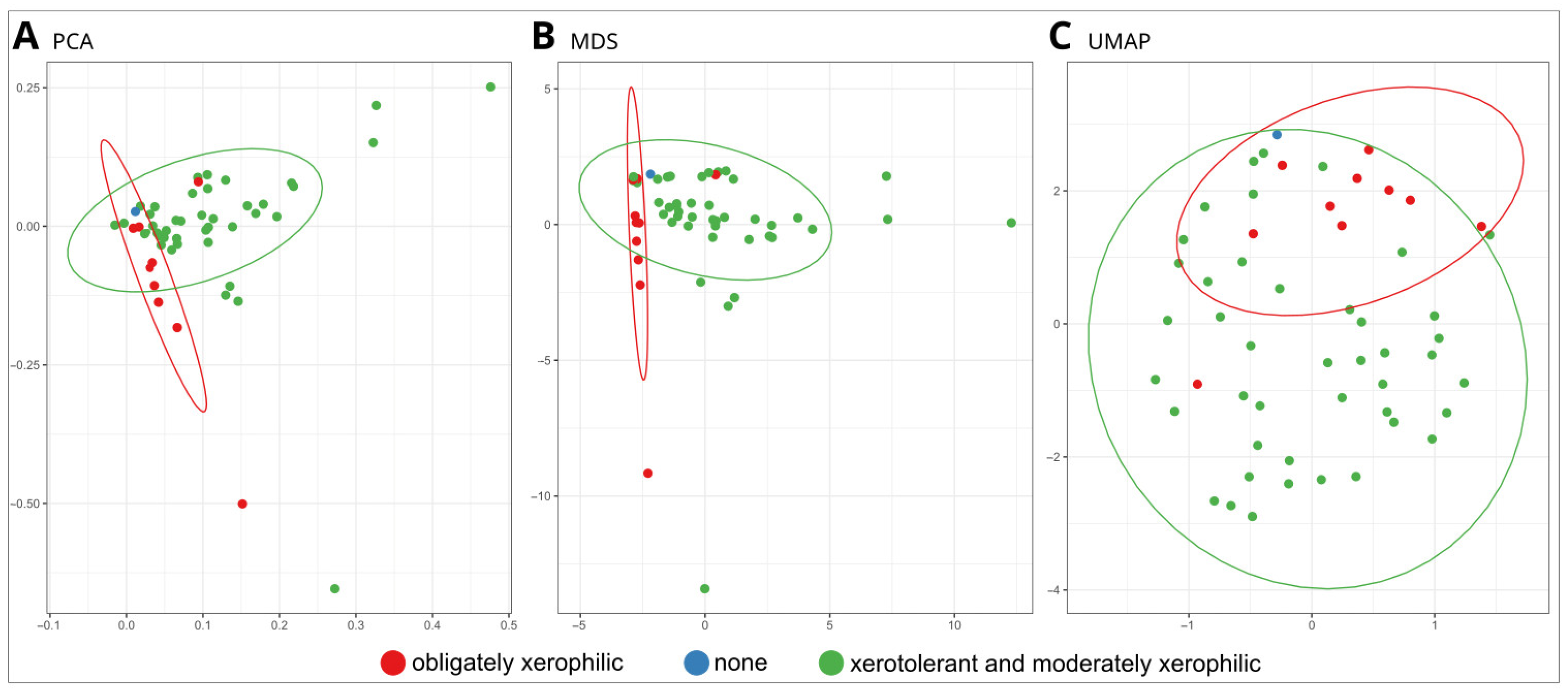
Fungal isolates were clustered with Principal Component Analysis (PCA), Multidimensional Scaling (MDS), and UMAP, Uniform Manifold Approximation and Projection (UMAP). Analyses were performed in the environment R [62] using the function ‘prcomp()’ (PCA) and the function ‘cmdscale()’, both from the package ‘stats’ (MDS), and the function ‘umap()’ from the package ‘umap’ [63]. A table of species and their enzymatic activities was used as input. The ellipses surrounding groups of strains were added with the ‘stat_ellipse()’ function of ‘ggplot2’ [64].
The presence and absence of specific enzymatic activities in tested strains as well as selected strain characteristics (pH-change, xerophily/halophily, pigmentation) were visualised with UpSet plots in the environment R [62] using the package ‘ggplot2’ [64].

3. Results

3.1. Physiological Characterisation: Growth at Different Water Activities, Temperatures, and Production of Acids and Pigments

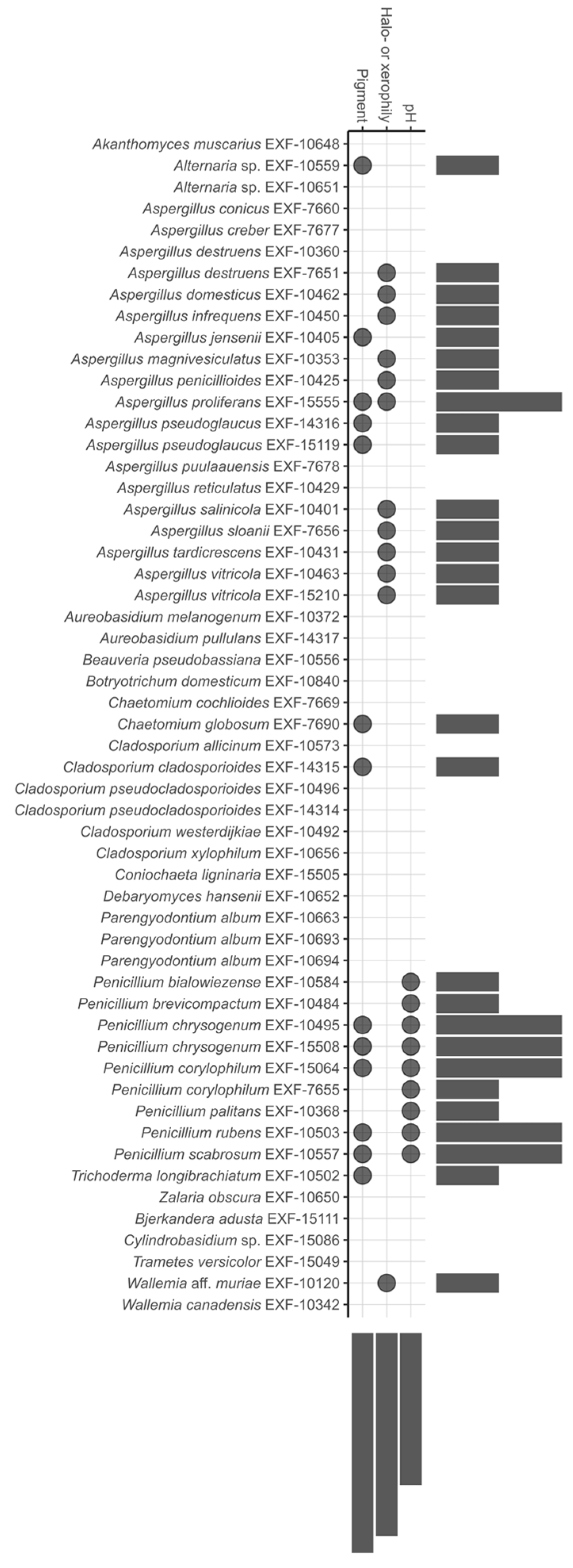
Figure 3 provides an overview of the three characteristics studied: xerophily, significant acidification of the liquid medium (from pH 6 to pH 5.1), and soluble pigment production, each of which is represented by a dot. More detailed results and additional data on growth at different temperatures are summarized in Supplementary Table S2.
More than one third of all tested strains, mostly species of the genus Aspergillus and Wallemia aff. muriae, showed an obligate need for low water activity of the medium, regardless of the solute and could thus be designated as obligate or extreme xerophiles or halophiles (17.5%). If no preference was evident for NaCl in comparison to glycerol, they were categorized as extreme xerophiles. The only species preferring NaCl over glycerol was A. proliferans. Differences were observed between A. destruens strains, with one being obligately xerophilic and the other only tolerant.
All strains except for Penicillium were able to maintain pH 6. Most Penicillium strains substantially lowered the pH of the medium: P. bialowiezense, P. brevicompactum, P. corylophilum and P. rubens for 0.9–1 pH unit, and P. chrysogenum, P. palitans and P. scabrosum for 1.1–2 pH units (Supplementary Table S2). Although, the CaCO3 solubilization test (implemented in Figure 4) showed that an even smaller pH drop resulted in a positive result, evident in many aspergilli and some other fungi (Supplementary Table S3).
Fifteen strains, mostly penicillia, were able to excrete different pigments observed on at least one of the culture media viewed.
Almost all tested strains were able to grow at temperatures recommended for storage and exposition of canvas paintings (Supplementary Table S2). In total, 3 strains grew only at 15 °C and were identified as psychrophiles, 31 strains were psychrotolerant, and 19 strains were psychrotolerant mesophiles and grew at all tested incubation temperatures (15, 24, and 37 °C).

3.2. Biodeteriorative Potential of Fungi: Enzymatic Activities, Ability to Assimilate Hydrocarbons, to Solubilize CaCO3, and Synthesize Siderophores

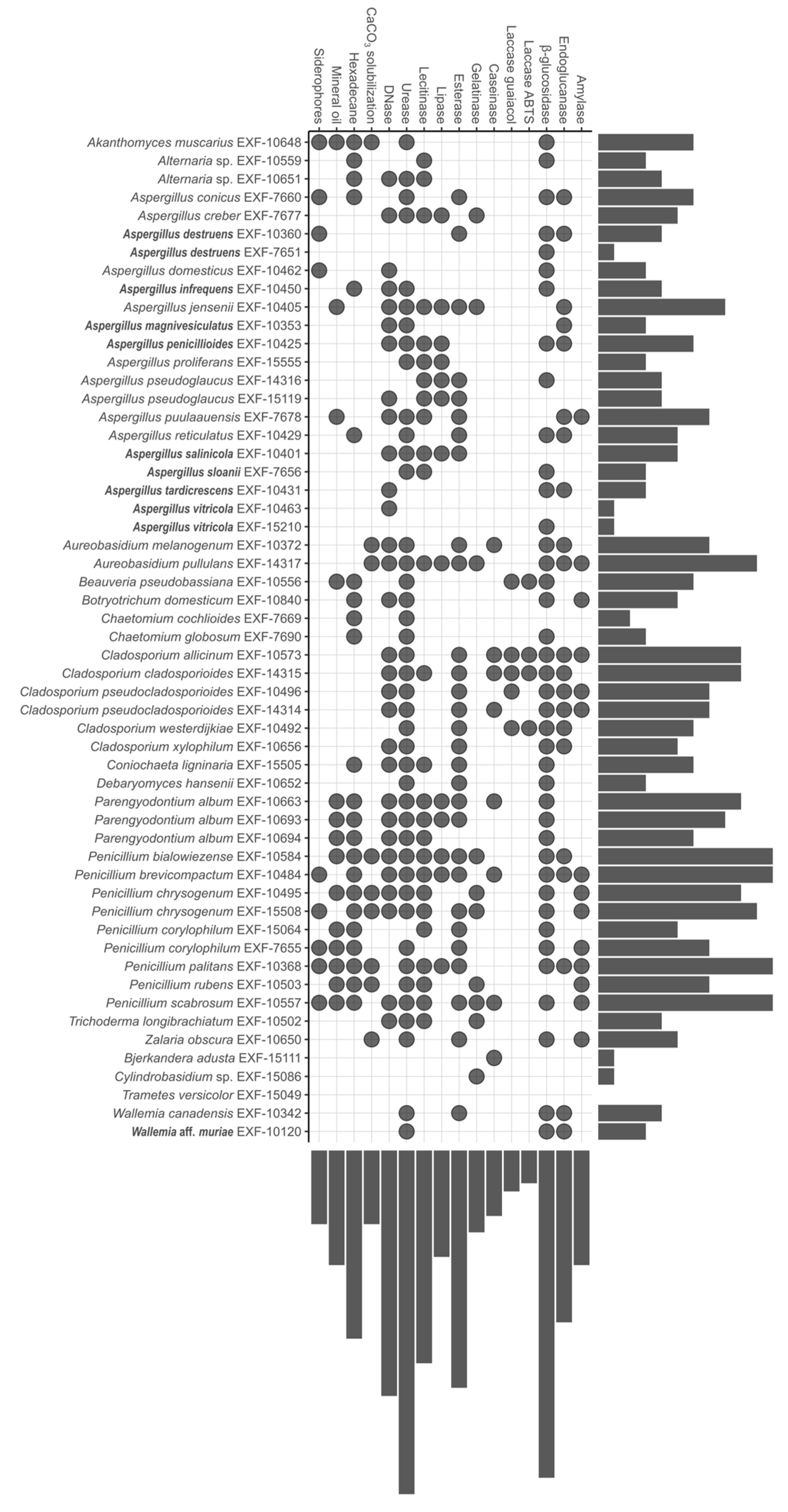
An overview of the strains that displayed the strongest enzymatic activities or strong ability to assimilate hydrocarbons, solubilize CaCO3, and sequester iron by producing siderophores (factor 2 or higher) is shown in Figure 4. Detailed results are summarized in Supplementary Table S3, and the majority of the tests are presented in Supplementary Figure S1.
Strains that displayed the largest number of strong activities, 11 strong activities out of 16, belong to different xerotolerant or moderately xerophilic Penicillium species: P. bialowiezense, P. brevicompactum, P. palitans and P. scabrosum, and Penicillium chrysogenum (EXF-15508), while Aureobasidium pullulans displayed 10 strong activities. Three strains showed a single strong activity: A. destruens (EXF-7651) β-glucosidase, A. vitricola (EXF-10463 DNase), Bjerkandera adusta caseinase, and Cylindrobasidium sp. gelatinase, while Trametes versicolor demonstrated no strong activities.

3.2.1. Enzymatic Activity

The majority of xerotolerant and moderately xerophilic strains showed a strong urease activity (80.0%), and less strains demonstrated β-glucosidase (68.9%), esterase (62.2%), DNase, and lecithinase (both 51.1%) activities. Endoglucanase, amylase, lipase, gelatinase, caseinase, and laccase were displayed as weak reactions. Penicillium brevicompactum displayed almost all strong enzymatic activities and represents the most active and enzymatically diverse species tested, followed by Aureobasidium pullulans, Cladosporium allicinum, and C. cladosporioides. The least enzymatically active xerotolerant or moderately xerophilic fungi were Bjerkandera adusta, Chaetomium cochlioides, and Cylindrobasidium sp. With a single activity, while drought sensitive Trametes versicolor did not display any of the tested activities.
Among obligately xerophilic isolates, more than two thirds showed β-glucosidase activity (75%), and more than half expressed DNase (58.3%) and urease (50.0%). Aspergillus penicillioides displayed five and A. salinicola six strong enzymatic activities. As mentioned above, only one of two tested A. destruens (EXF-10360) and A. vitricola (EXF-10463) strains showed a single strong enzymatic activity, β-glucosidase and DNA-se, respectively.
In comparison, more than a half of the obligately xerophilic strains showed strong β-glucosidase, DNase, and urease activities, while more than half of the xerotolerant and moderately xerophilic strains displayed strong esterase and lecithinase activities. The frequency of the strong enzymatic activities was at least 16.7% among obligately xerophilic fungi, excluding gelatinase, caseinase, laccase, and amylase activities that were not recorded. Only weak amylase activity was present among obligately xerophilic strains. In contrast, among xerotolerant and moderately xerophilic fungi, all of the strong enzymatic activities were detected in at least 20% of the strains, excluding laccase activity that was less frequent. In conclusion, this group of fungi displayed more diverse and stronger enzymatic activities.

3.2.2. Assimilation of Hydrocarbons, Solubilization of CaCO3, and Production of Siderophores

In general, 42.1% of all tested strains were able to assimilate hexadecane, while other strong activities were less frequent. Xerotolerant and moderately xerophilic strains were able to assimilate hexadecane (51.1%) in an even higher proportion, while other activities were less common. Two strains strongly displayed all four activities, Akanthomyces muscarius and P. palitans, while moderately xerophilic P. bialowiezense, both strains of P. chrysogenum, P. corylophilum (EXF-7655), P. rubens, and P. scabrosum displayed at least three strong activities of four tested. Almost 40% of xerotolerant and moderately xerophilic strains did not show any of these activities.
A strong ability to synthesize siderophores and an ability to assimilate hexadecane were detected as well among obligately xerophilic strains, however in less than 20%, while none of them were able to assimilate mineral oil and to solubilize CaCO3. Four strains out of twelve showed only one strong activity. A. destruens (EXF-10360) and A. domesticus both produced siderophores, A. infrequens grew abundantly on hexadecane, and A. conicus, A. domesticus, and A. destruens produced siderophores. Other obligately xerophilic strains did not display any of these activities.
The comparison of xerotolerant and moderately xerophilic fungi to obligately xerophilic fungi showed that, in general, the former were able to solubilize CaCO3 to assimilate hydrocarbons, particularly hexadecane (51.1%), and synthesize siderophores. In contrast, obligately xerophilic fungi only assimilated hexadecane and synthesized siderophores (16%).

3.2.3. Statistical Analyses

The phenotypic traits of tested fungal strains (the presence of different enzymatic activities, the presence of pigmentation, and change in pH) were distinctly different between obligately xerophilic and xerotolerant and moderately xerophilic strains. Dimensionality-reduction methods clustered obligately xerophilic strains into a relatively tight cluster that was only partially overlapping with a larger cluster of xerotolerant and moderately xerophilic strains in both PCA and MDS analyses and to a somewhat lesser extent using UMAP (Figure 5).

4. Discussion

Canvas paintings are objects susceptible to biodeterioration, because they are rich in organic and inorganic materials that provide food and support for fungi under favourable environmental conditions [1]. Fungi are ubiquitous, and their propagules are constantly present in the air and dust, so inoculum is constantly available [66], but environmental conditions control their growth, particularly RH. Studies of biodeteriorated paintings usually focus on biodiversity [3,67], to a lesser extent on the detection of viable taxa, e.g., the presence of active mycelium, and even less on the biodeterioration potential in terms of enzyme and acid excretion [11,14,32]. The aim of this study was to investigate the biodeterioration potential of xerophilic species likely to be active in low humidity environments.

4.1. Xerophily Is the Crucial Factor That Allows the Biodeterioration of Paintings Indoors

Although definitions of xerophily can vary from publication to publication, Pitt (2008) defined xerophile as a fungus capable of growing under at least one set of conditions with a water activity of 0.85 or less [68]. In our study, this aw was only achieved on DG40 (aw = 0.827), MEA + 20% NaCl (aw = 0.852), and MEA + 25% NaCl (aw = 0.802), which means that all fungi classified as groups 3–5 on the glycerol and NaCl media can be considered as xerophiles. For practical use, we distinguish obligate xerophiles from moderate xerophiles, with the former requiring a reduction in aw for growth.
RH and T are recognised as risk factors in museums/storage facilities and are therefore maintained at 40–60% RH with 5–10% variation over 24 h and in the T range of 16–25 °C [69]. Nevertheless, fungal growth may occur due to local RH increases in the indoor microclimate, e.g., near walls or in poorly ventilated areas [2,70,71], allowing the growth of xerotolerant and xerophilic taxa. However, xerophilic fungi with a wide aw range can grow under the recommended environmental regime and cause biodeterioration. This phenomenon is partly due to their ability to accumulate and retain extraordinary amounts of glycerol for osmotic adjustment [72]. In recent years, more studies have pointed to xerophilic fungal communities that are not constrained by the above mentioned environmental parameters [38] and to their as yet unassessed biodegradation potential [22].
Among the limited number of fungal genera with xerophilic species, the genus Aspergillus (Ascomycota, Eurotiales) is one of the most common on paintings and contains several groups of xerophilic species restricted to the subgenus Aspergillus. While it was generally known that metabolic activity and cell division of the most extreme xerophilic aspergilli cease between aw 0.700 and 0.640 (about 70–64% RH), A. penicillioides, the most xerophilic species of the section Restricti [23], germinated even at aw 0.640 when culture media were supplemented with glycerol. Furthermore, extrapolations made for this species indicated a theoretical water activity minimum at aw 0.570, 57% RH [72], which is already in the range of recommended RH for museums/depositories. Half of the species accommodated to the section Restricti have been recently described as new species [27], also as painting contaminants [22], detected in high CFU numbers. They differ slightly in xerophily, but the majority of species cannot grow without a minimum of 5% NaCl, with an optimum around 10% and a maximum at 20% NaCl [27]. The exact experiments on the limiting aw values for their germination and growth have not yet been studied, but it is quite possible that they are also among the most extreme. Tolerance to low aw is also known in Penicillium, e.g., the most extreme species P. chrysogenum was observed to germinate at aw 0.78 [68]. Among Cladosporium isolates, the C. sphaerospermum species complex, in particular C. sphaerospermum and C. halotolerans, are the most extreme, growing at aw ≥ 0.82, while species of other species complexes have an aw minimum of 0.85 [73]. Basidiomycetes in general are less known from low aw environments, the only exception being Wallemia, which germinates at aw 0.75 [68]. In terms of environmental parameters, particularly RH, Aspergillus and Penicillium are among the most extreme and are likely to be active on paintings.

4.2. Testing the Biodeterioration Potential of Obligate Xerophiles by Adapting Culture Media

Biodeterioration tests performed on painting contaminants in previous studies [22] were carried out on culture media with aw close to 1, which had to be reduced to allow testing of obligately xerophilic species unable to grow without additional solutes. We used the same amounts of glycerol as in dichloran glycerol agar (DG18), suitable for the enumeration and isolation of moderately xerophilic fungi [68], to obtain aw close to 0.942, which is much higher than the recommended indoor RH conditions but allows for relatively rapid growth, in up to 28 days. However, this aw was not suitable for testing most extreme obligate xerophiles, such as A. halophilicus, which would require even lower aw. Although glycerol can be used as the C source with many species, we obtained similar positive reactions in the tests performed in terms of changed/stained/cleared areas in at least some of the strains tested. While testing the use of cyclic or aliphatic hydrocarbons as C sources, solutes were required that could not be assimilated, so we used NaCl at a concentration with a similar aw.

4.3. Biodeterioration of Canvas and Proteins

According to previously published data on biodeteriorated paintings, the raw canvas on the recto side of the painting, which is potentially exposed to higher humidity behind paintings facing the wall [7], and/or the presence of proteins in the painting [22], are the critical factors for moulding in paintings. While the strong and diverse activities associated with cellulose degradation (endoglucanase, β-glucosidase, laccase) were abundant in xerotolerant Cladosporium species, obligately xerophilic Aspergillus and xerophilic Penicillium often expressed β-glucosidase but rarely endoglucanase. Surprisingly, the basidiomycetous taxa tested did not show strong cellulolytic activities. Fungi with laccase activity may also be involved in other biodegradation phenomena due to their non-specific activity, e.g., they may contribute to varnish and pigment degradation [74].
The genera Aspergillus and Penicillium, which are generally considered to be good producers of proteases potentially dangerous for proteinaceous paint layers adhesives [32], did not express or expressed strong gelatinase activity. Some Cladosporium strains were positive for caseinase. Fungal proteolytic activity is particularly dangerous for damaging natural binders and materials used in conservation procedures [32]. The results of different studies [15] were not always congruent in detected activities, which may indicate either strain specificity or that less strong activity was expressed in our study and not marked in the UpSet plot.
Linseed oil, the binder used in oil paintings, is a natural drying oil composed of a mixture of triglycerides derived primarily from unsaturated oleic, linoleic, and linolenic acids [10]. Esterases hydrolyse ester bonds in soluble and insoluble lipids in the binder of paintings and are the main contributors to the biodeterioration of paint layers [19,34]. Among the newly tested obligate xerophiles, the A. salinicola strain and one of the two A. destruens strains were positive for esterase activity, while all other obligate xerophiles were negative. Lipolytic and lecithinase activities were mainly attributed to Aspergillus and Penicillium strains. Aspergillus proliferans, A. pseudoglaucus, P. bialowiezense, P. brevicompactum, and P. palitans were indeed strong producers, with many species also expressing esterase activity.

4.4. Biodeterioration of Varnish and Wax

Traditionally, natural resins such as terpenoid resins, mastic, sandarac, copal, dammar, elemi, or rosin resins are used as varnishes for canvas paintings and are considered less acceptable for biodeterioration [34,35,36]. Various mixtures can also be used, such as terpenoid resin-based oil varnishes, which consist of a drying oil (linseed oil), a diterpenoid resin (colophony), and a triterpenoid resin (mastic) [34]. Due to their complex structure and composition, the degradation of varnishes requires combined enzymatic activities, such as esterase, urease, and possibly oxidising enzymes such as laccase and other lignin oxidases. The ability to grow on hydrocarbons (cyclic, aliphatic) may also contribute to varnish degradation. Several xerophilic fungi from our study had esterase and urease activity and the ability to use hydrocarbons as the sole carbon source: P. chrysogenum, P. corylophilum, P. palitans, P. scabrosum, A. pullulans, and Parengyodontium album. In fact, P. chrysogenum has already been reported to have these activities [75,76], and indeed, it can also degrade the sandarac (Thuja articulata) coating and induced changes in Manila Copal, both of which have been studied in mock-up experiments [35]. There have also been reports on the ability of P. corylophilum to degrade hydrocarbons [77], the urease activity and ability of A. pullulans to degrade hydrocarbons [78,79], and the urease activity of P. album [80]. None of the obligate xerophilic species possessed these abilities; although, there is a report on A. destruens degrading pollutant hydrocarbons under saline conditions [81].
Wax (beeswax), traditionally used in conservation–restoration to consolidate layers of paint, to line canvas, or as an additive to some varnishes, is a complex mixture of hydrocarbons, esters, and free fatty acids [82]. Fungi with high esterase activity and a strong ability to degrade hydrocarbons may also affect wax. In our study, these were numerous xerophilic Penicillium species (P. bialowiezense, P. brevicompactum, P. chrysogenum, P. corylophilum, P. palitans, and P. scabrosum), A. conicus, A. reticulatus, A. jensenii, A. puulaauensis, as well as xerotolerant Coniochaeta ligninaria, and Parengyodontium album. A painting with a protective layer of beeswax was deteriorated by an unidentified Penicillium species [83] and a wax seal by Aureobasidium pullulans and Aspergillus, Penicillium, and Chaetomium species [84,85]. Few Penicillium and Cladosporium species have also been isolated from wax drops on ancient manuscripts [86]. Recently, the growth of the xerophilic species A. destruens, A. jensenii, and the obligately xerophilic Wallemia species on canvas has been associated with wax [22], which could imply the involvement of different mechanisms of its degradation or co-metabolism, since only A. jensenii expressed all three activities listed, whereas A. destruens expressed only esterase and Wallemia only urease. However, as obligate xerophiles as single species do not possess the enzymatic activities mentioned, they are not considered to be the main degraders of these compounds.

4.5. Biodeterioration of Synthetic Polymers

Synthetic polymers, mainly polyesters, polyacrylics, and alkyd polymers, are the most stable materials used in cultural heritage art. However, in recent years it has become increasingly apparent that polymers can be susceptible to biodeterioration and incompatible with other materials [87,88,89,90]. Synthetic polymers can be used in a variety of ways: as canvas, as consolidants for paints, and as varnishes. Although there are some reports of fungal growth on these materials, the exact mechanisms of degradation are not yet known. Growth of certain species of the genera Aspergillus, Aureobasidium, Cladosporium, Gliocladium, and Penicillium has been observed on alkyd resins used as binders for pigments [8,91]. Members of all of these genera, as well as members of the genera Aureobasidium, Cenococcum, Eladia, Epicoccum, Glyphium, Penicillium, Phoma, and Talaromyces, have been identified on acrylic resins used for the restoration of various works of art [5,8,91,92,93,94,95]. The enzymes involved in degradation are esterases/lipases, proteases, and ureases with the ability to cleave ester bonds [96], together with the ability to degrade hydrocarbons, particularly polyurethane [97,98]. As fungi of the genus Cladosporium have been reported to grow well on alkyd and acrylic resins, a high level of esterase and urease activity, as well as the ability to degrade hydrocarbons, has been suggested [91,92,99]. This was confirmed in our study as all Cladosporium species showed strong esterase and urease activity and the ability to assimilate hexadecane. Similarly, Chaetomium globosum, previously reported to be able to grow on alkyd and acrylic resins [8,100], also showed these activities. Aureobasidium pullulans, which can grow on alkyd resin and polyurethane [91,95,101], showed strong esterase and urease activity in our study, in agreement with previous studies [101]. In conclusion, most xerophilic species of Aspergillus and Penicillium and xerotolerant species of Aureobasidium and Cladosporium in in our study showed esterase and urease activities, as well as the ability to assimilate hydrocarbons. Indeed, many species of Aspergillus, Cladosporium, and Penicillium are known degraders of plastics and have shown potential for use in bioremediation as good degraders of polycyclic aromatic hydrocarbons (PAHs) [77,81,102,103,104,105,106]. Penicillium citrinum is also commonly found on polyurethane and acrylic synthetic resins [95]. The xerophilic species A. conicus and possibly A. infrequens are also potential degraders of synthetic polymers.

4.6. Novel Tests for Biodeterioration Studies: Degradation of Environmental Debris and Ability to Sequester Fe Ions

Some oligotrophic fungi can live on the surfaces of paintings on dust deposits and various organic environmental debris, such as the remains of bacteria, spiders and their webs, and small arthropods, which can also act as microbial vectors [107]. DNA from dead organisms (bacteria, fungi, insects) is also a source of nucleotides, carbon, nitrogen, and phosphorus, made possible by the production of extracellular DNases. DNase activity is most often associated with fungal pathogenicity to animals and only in a few cases to saprobic taxa (reviewed in [108]). In our study, extracellular DNase activity was exhibited by all tested species of the genera Aureobasidium, Cladosporium, Parengyodontium, and most Penicillium and Aspergillus species, including the majority of extremely xerophilic species.
Various pigments can be sources of certain essential metals, such as iron, which can be biosequestered by synthesised microbial siderophores—ligands that chelate iron. This ability has been reported mainly in the context of microbial pathogenicity and is considered as an important virulence factor [45]. It was rarely detected in our study. Among the xerotolerant taxa, it was detected in the entomopathogenic Akanthomyces muscarius and Beauveria pseudobassiana, which could be in line with their pathogenicity, while the presence of siderophores in A. conicus, A. destruens, and in A. domesticus cannot be commented yet and requires further investigation.

5. Conclusions

Moderately xerophilic fungi of the genera Penicillium, Aspergillus, and Cladosporium show a high degree of metabolic versatility, possessing enzymes that allow the biodegradation of canvas and various paint layers, as well as environmental DNA. The greatest risk is posed by various Penicillium species such as P. chrysogenum, P. scabrosum, P. palitans, P. brevicompactum, and P. bialowiezense, which also excrete acids and pigments. Obligately xerophilic species, tested on media supplemented with glycerol to lower their aw, showed less diverse enzymatic activities. Nevertheless, they have the ability to grow under low RH conditions, where they can degrade the canvas and potentially affect the varnish and synthetic polymers, especially in combination with other taxa expressing lipase-esterase activities. Most of these species expressed DNase activity, indicating the importance of dust deposition for their establishment on painting surfaces. Aspergillus domesticus and A. destruens may affect paint layers and related synthetic materials. Interestingly, A. vitricola and A. destruens, which are the most common obligate xerophilic species on easel paintings [22], were found to degrade only a limited number of painting compounds, showing not only β-glucosidase and DNase activity but also excreting acids, so their biodeterioration potential should not be neglected and should be further investigated.
With only one exception, all of the isolates selected from the deteriorated paintings were xerotolerant or xerophilic and could grow at ambient temperatures (15–25 °C). Thirty-nine per cent of the strains also grew at 37 °C, indicating the need for precautionary measures for conservators–restorers when dealing with contaminated paintings.
Xerotolerant and moderately xerophilic fungi possess a wide range of enzymes, including urease, β-glucosidase, esterase, lecithinase, and DNase, which can potentially degrade all painting components under conditions of high water availability. The xerophilic strains that pose the greatest threat to paintings are P. scabrosum, P. palitans, P. brevicompactum, P. bialowiezense, and P. chrysogenum.
Obligate xerophiles had a narrower range of enzymatic activity, with β-glucosidase, DNase, urease, and endoglucanase being the most common enzymes.
Xerophilic fungi pose a threat to canvas paintings at low RH, especially to a canvas or partially degraded canvas, due to endoglucanase and β-glucosidase activity. Almost all strains tested can degrade environmental DNA through DNase activity. The greatest threat to paintings is posed by Aspergillus penicillioides, A. salinicola, A. infrequens, Wallemia aff. Muriae, A. tardicrescens, and A. magnivesiculatus.
Given the growth of obligate xerophilic fungi and their minimal growth requirements in terms of environmental conditions, this study provides additional evidence to the studies of Bastholm et al. (2022) [38] and Paiva de Carvalho et al. (2018) [69] that new measures should be considered in museums and painting repositories.

Supplementary Materials

The following supporting information can be downloaded at: https://www.mdpi.com/article/10.3390/jof10010076/s1, Table S1: Cultures used in this study, their source and identification. Table S2: Adaptation to low aw (xero-tolerance/-phily, halo-tolerance/-phily), growth at different temperatures, production of acids, and pigments of tested fungal species. Table S3: Enzymatic (amylase, endocellulase, β-glucosidase, caseinase, gelatinase, esterase, lipase, lecithinase, laccase, urease, and DNase), and other activities (assimilation of hydrocarbons, dissolution of CaCO3, and siderophore formation) of tested fungal species. Figure S1: Biodegradation assays with negative (upper row) and positive (lower row) enzymatic activities.

Author Contributions

Conceptualization, P.Z. and A.K.; methodology, P.Z.; software, C.G.; validation, A.K. and P.Z.; formal analysis, A.K., N.G., C.G., K.K. and P.Z.; investigation, A.K., N.G., C.G., and P.Z.; resources, P.Z. and N.G.-C.; data curation, A.K.; writing—original draft preparation, A.K., K.K. and P.Z.; writing—review and editing, A.K., P.Z., and N.G-C.; visualization, A.K., C.G., K.K. and P.Z.; supervision, P.Z; project administration, P.Z.; funding acquisition, P.Z. and N.G.-C. All authors have read and agreed to the published version of the manuscript.

Funding

This research was funded by Slovenian Research and Innovation Agency (ARIS) as the Young Researcher Grant to A. Kujović, research programs ARIS P1-0198 (Molecular Biology of Microorganisms) and ARIS P4-0432 (Marine and microbial Biotechnology); research project ARIS J7-1815 (Restoration of moldy canvas paintings: improvement or deterioration?), and MRIC UL, I0-0022 (IC Mycosmo).

Institutional Review Board Statement

Not applicable.

Informed Consent Statement

Not applicable.

Data Availability Statement

Data are contained within the article and supplementary materials.

Acknowledgments

We thank Mojca Matul and Barbara Kastelic Bokal for their excellent technical assistance and Petra Bešlagić for scanning electron microscopy images.

Conflicts of Interest

The authors declare no conflicts of interest. The funders had no role in the design of the study; in the collection, analyses, or interpretation of data; in the writing of the manuscript; or in the decision to publish the results.

References

  1. Ciferri, O. Microbial Degradation of Paintings. Appl. Environ. Microbiol. 1999, 65, 879–885. [Google Scholar] [CrossRef] [PubMed]
  2. Sterflinger, K. Fungi: Their Role in Deterioration of Cultural Heritage. Fungal Biol. Rev. 2010, 24, 47–55. [Google Scholar] [CrossRef]
  3. López-Miras, M.D.M.; Martín-Sánchez, I.; Yebra-Rodríguez, Á.; Romero-Noguera, J.; Bolívar-Galiano, F.; Ettenauer, J.; Sterflinger, K.; Piñar, G. Contribution of the Microbial Communities Detected on an Oil Painting on Canvas to Its Biodeterioration. PLoS ONE 2013, 8, e80198. [Google Scholar] [CrossRef]
  4. Cappitelli, F.; Sorlini, C. Microorganisms Attack Synthetic Polymers in Items Representing Our Cultural Heritage. Appl. Environ. Microbiol. 2008, 74, 564–569. [Google Scholar] [CrossRef] [PubMed]
  5. Farmakalidis, H.V.; Douvas, A.M.; Karatasios, I.; Sotiropoulou, S.; Boyatzis, S.; Argitis, P.; Chryssoulakis, Y.; Kilikoglou, V. Accelerated Thermal Ageing of Acrylic Copolymers, Cyclohexanone-Based and Urea-Aldehyde Resins Used in Paintings Conservation. Mediterr. Archaeol. Archaeom. 2016, 16, 213–228. [Google Scholar] [CrossRef]
  6. Cappitelli, F.; Villa, F.; Sanmartín, P. Interactions of Microorganisms and Synthetic Polymers in Cultural Heritage Conservation. Int. Biodeterior. Biodegrad. 2021, 163, 105282. [Google Scholar] [CrossRef]
  7. Poyatos, F.; Morales, F.; Nicholson, A.W.; Giordano, A. Physiology of Biodeterioration on Canvas Paintings. J. Cell. Physiol. 2018, 233, 2741–2751. [Google Scholar] [CrossRef]
  8. Doménech-Carbó, M.T.; Bitossi, G.; Cruz-Cañizares, J.D.; Bolívar-Galiano, F.; López-Miras, M.M.; Romero-Noguera, J.; Martín-Sánchez, I.; Gimeno-Adelantado, J.V.; Doménech-Carbó, A. Study on the Biodeterioration of Alkyd Ah Resin Used As a Binding Medium for Modern Paintings By Pyrolysis-Gas Chromatography-Mass Spectrometry and Ftir Spectroscopy. Arche 2008, 3, 191–196. [Google Scholar]
  9. Leonardi, R. Nuclear Physics and Painting: Sub-Topic of the Wide and Fascinating Field of Science and Art. Nucl. Phys. A 2005, 752, 659–674. [Google Scholar] [CrossRef]
  10. López-Miras, M.; Piñar, G.; Romero-Noguera, J.; Bolívar-Galiano, F.C.; Ettenauer, J.; Sterflinger, K.; Martín-Sánchez, I. Microbial Communities Adhering to the Obverse and Reverse Sides of an Oil Painting on Canvas: Identification and Evaluation of Their Biodegradative Potential. Aerobiologia 2013, 29, 301–314. [Google Scholar] [CrossRef]
  11. Văcar, C.L.; Mircea, C.; Pârvu, M.; Podar, D. Diversity and Metabolic Activity of Fungi Causing Biodeterioration of Canvas Paintings. J. Fungi 2022, 8, 589. [Google Scholar] [CrossRef]
  12. Ortiz-Miranda, A.S.; Doménech-Carbó, A.; Doménech-Carbó, M.T.; Osete-Cortina, L.; Bolívar-Galiano, F.; Martín-Sánchez, I. Analyzing Chemical Changes in Verdigris Pictorial Specimens upon Bacteria and Fungi Biodeterioration Using Voltammetry of Microparticles. Herit. Sci. 2017, 5, 8. [Google Scholar] [CrossRef]
  13. Zhgun, A.; Avdanina, D.; Shumikhin, K.; Simonenko, N.; Lyubavskaya, E.; Volkov, I.; Ivanov, V. Detection of Potential Biodeterioration Risks for Tempera Painting in 16th Century Exhibits from State Tretyakov Gallery. PLoS ONE 2020, 15, e0230591. [Google Scholar] [CrossRef] [PubMed]
  14. Savković, Ž.; Stupar, M.; Unković, N.; Ivanović, Ž.; Blagojević, J.; Vukojević, J.; Ljaljević Grbić, M. In Vitro Biodegradation Potential of Airborne Aspergilli and Penicillia. Sci. Nat. 2019, 106, 8. [Google Scholar] [CrossRef] [PubMed]
  15. Trovão, J.; Portugal, A. Current Knowledge on the Fungal Degradation Abilities Profiled through Biodeteriorative Plate Essays. Appl. Sci. 2021, 11, 4196. [Google Scholar] [CrossRef]
  16. Romero, S.M.; Giudicessi, S.L.; Vitale, R.G. Is the Fungus Aspergillus a Threat to Cultural Heritage? J. Cult. Herit. 2021, 51, 107–124. [Google Scholar] [CrossRef]
  17. Pinna, D.; Salvadori, O. Processes of Biodeterioration: General Mechanisms. In Plant Biology for Cultural Heritage: Biodeterioration and Conservation; Caneva, G., Nugari, M.P., Salvadori, O., Eds.; Getty Publications: Los Angeles, CA, USA, 2008; ISBN 9780892369393. [Google Scholar]
  18. Sazanova, K.V.; Shchiparev, S.M.; Vlasov, D.Y. Formation of Organic Acids by Fungi Isolated from the Surface of Stone Monuments. Microbiology 2014, 83, 516–522. [Google Scholar] [CrossRef]
  19. Kumar, S.; Priyanka; Kumar, U. Microbial Community Present on the Reverse Side of a Deteriorated Canvas BT. In Microbial Biotechnology Approaches to Monuments of Cultural Heritage; Yadav, A.N., Rastegari, A.A., Gupta, V.K., Yadav, N., Eds.; Springer: Singapore, 2020; pp. 1–12. ISBN 978-981-15-3401-0. [Google Scholar]
  20. Berovič, M. Biodeterioration Studies on Pastels and Oil-Based Paintings. In Art, biology, and Conservation: Biodeterioration of Works of Art; Metropolitan Museum of Art: New York, NY, USA, 2003; pp. 50–59. ISBN 9781588391070. [Google Scholar]
  21. Poyatos Jiménez, F. Procesos de Microbiodeterioro En Pinturas Sobre Lienzo Del Museo de Bellas Artes de Granada Examen Visual y Gráfico. Ph.D. Thesis, University of Granada, Granada, Spain, 2007. [Google Scholar]
  22. Zalar, P.; Graf Hriberšek, D.; Gostinčar, C.; Breskvar, M.; Džeroski, S.; Matul, M.; Novak Babič, M.; Čremožnik Zupančič, J.; Kujović, A.; Gunde-Cimerman, N.; et al. Xerophilic Fungi Contaminating Historically Valuable Easel Paintings from Slovenia. Front. Microbiol. 2023, 14, e1258670. [Google Scholar] [CrossRef]
  23. Houbraken, J.; Kocsubé, S.; Visagie, C.M.; Yilmaz, N.; Wang, X.C.; Meijer, M.; Kraak, B.; Hubka, V.; Bensch, K.; Samson, R.A.; et al. Classification of Aspergillus, Penicillium, Talaromyces and Related Genera (Eurotiales): An Overview of Families, Genera, Subgenera, Sections, Series and Species. Stud. Mycol. 2020, 95, 5–169. [Google Scholar] [CrossRef]
  24. Lawrence, D.P.; Rotondo, F.; Gannibal, P.B. Biodiversity and Taxonomy of the Pleomorphic Genus Alternaria. Mycol. Prog. 2016, 15, 3. [Google Scholar] [CrossRef]
  25. Bensch, K.; Groenewald, J.Z.; Braun, U.; Dijksterhuis, J.; de Jesús Yáñez-Morales, M.; Crous, P.W. Common but Different: The Expanding Realm of Cladosporium. Stud. Mycol. 2015, 82, 23–74. [Google Scholar] [CrossRef]
  26. Wang, X.W.; Houbraken, J.; Groenewald, J.Z.; Meijer, M.; Andersen, B.; Nielsen, K.F.; Crous, P.W.; Samson, R.A. Diversity and Taxonomy of Chaetomium and Chaetomium-like Fungi from Indoor Environments. Stud. Mycol. 2016, 84, 145–224. [Google Scholar] [CrossRef]
  27. Sklenář, F.; Jurjević; Zalar, P.; Frisvad, J.C.; Visagie, C.M.; Kolařík, M.; Houbraken, J.; Chen, A.J.; Yilmaz, N.; Seifert, K.A.; et al. Phylogeny of Xerophilic Aspergilli (Subgenus Aspergillus) and Taxonomic Revision of Section Restricti. Stud. Mycol. 2017, 88, 161–236. [Google Scholar] [CrossRef] [PubMed]
  28. Mazzoli, R.; Giuffrida, M.G.; Pessione, E. Back to the Past: “Find the Guilty Bug—Microorganisms Involved in the Biodeterioration of Archeological and Historical Artifacts”. Appl. Microbiol. Biotechnol. 2018, 102, 6393–6407. [Google Scholar] [CrossRef] [PubMed]
  29. Sterflinger, K.; Piñar, G. Microbial Deterioration of Cultural Heritage and Works of Art—Tilting at Windmills? Appl. Microbiol. Biotechnol. 2013, 97, 9637–9646. [Google Scholar] [CrossRef] [PubMed]
  30. Caneva, G.; Maggi, O.; Nugari, M.P.; Pietrini, A.M.; Piervittori, R.; Ricci, S.; Roccardi, A. The Biological Aerosol as a Factor of Biodeterioration BT. In Cultural Heritage and Aerobiology: Methods and Measurement Techniques for Biodeterioration Monitoring; Mandrioli, P., Caneva, G., Sabbioni, C., Eds.; Springer: Dordrecht, The Netherlands, 2003; pp. 3–29. ISBN 978-94-017-0185-3. [Google Scholar]
  31. Stoner Joyce Hill, R.R. (Ed.) Conservation of Easel Paintings, 2nd ed.; Routledge: London, UK; Taylor and Francis: London, UK, 2021; ISBN 9780429680977. [Google Scholar]
  32. Unković, N.; Dimkić, I.; Stupar, M.; Stanković, S.; Vukojević, J.; Grbić, M.L. Biodegradative Potential of Fungal Isolates from Sacral Ambient: In Vitro Study as Risk Assessment Implication for the Conservation of Wall Paintings. PLoS ONE 2018, 13, e0190922. [Google Scholar] [CrossRef]
  33. Sabin, A.H. The Industrial and Artistic Technology of Paint Varnish; Wiley: New York, NY, USA, 1927. [Google Scholar]
  34. Doménech-Carbó, M.T.; Osete-Cortina, L.; De La Cruz Cañizares, J.; Bolívar-Galiano, F.; Romero-Noguera, J.; Fernández-Vivas, M.A.; Martín-Sánchez, I. Study of the Microbiodegradation of Terpenoid Resin-Based Varnishes from Easel Painting Using Pyrolysis-Gas Chromatography-Mass Spectrometry and Gas Chromatography-Mass Spectrometry. Anal. Bioanal. Chem. 2006, 385, 1265–1280. [Google Scholar] [CrossRef]
  35. Romero-Noguera, J.; Martín-Sánchez, I.; Doménech-Carbó, M.T.; Osete-Cortina, L.; López-Miras, M.M.; Bolívar-Galiano, F. Analytical Characterisation of the Biodeterioration of Diterpenoid Labdanic Varnishes Used in Pictorial Techniques: Sandarac and Manila Copal. Int. Biodeterior. Biodegrad. 2014, 90, 99–105. [Google Scholar] [CrossRef]
  36. Romero-Noguera, J.; Bailón-Moreno, R.; Bolívar-Galiano, F. Varnishes with Biocidal Activity: A New Approach to Protecting Artworks. Appl. Sci. 2020, 10, 7319. [Google Scholar] [CrossRef]
  37. Kavkler, K.; Humar, M.; Kržišnik, D.; Turk, M.; Tavzes, Č.; Gostinčar, C.; Džeroski, S.; Popov, S.; Penko, A.; Gunde-Cimerman, N.; et al. A Multidisciplinary Study of Biodeteriorated Celje Ceiling, a Tempera Painting on Canvas. Int. Biodeterior. Biodegrad. 2022, 170, 105389. [Google Scholar] [CrossRef]
  38. Bastholm, C.J.; Madsen, A.M.; Andersen, B.; Frisvad, J.C.; Richter, J. The Mysterious Mould Outbreak—A Comprehensive Fungal Colonisation in a Climate-Controlled Museum Repository Challenges the Environmental Guidelines for Heritage Collections. J. Cult. Herit. 2022, 55, 78–87. [Google Scholar] [CrossRef]
  39. Ortega-Morales, B.O.; Narváez-Zapata, J.; Reyes-Estebanez, M.; Quintana, P.; De la Rosa-García, S.D.C.; Bullen, H.; Gómez-Cornelio, S.; Chan-Bacab, M.J. Bioweathering Potential of Cultivable Fungi Associated with Semi-Arid Surface Microhabitats of Mayan Buildings. Front. Microbiol. 2016, 7, 1–12. [Google Scholar] [CrossRef] [PubMed]
  40. Pangallo, D.; Chovanová, K.; Šimonovičová, A.; Ferianc, P. Investigation of Microbial Community Isolated from Indoor Artworks and Air Environment: Identification, Biodegradative Abilities, and DNA Typing. Can. J. Microbiol. 2009, 55, 277–287. [Google Scholar] [CrossRef] [PubMed]
  41. Yadav, A.; Yadav, P.; Kumar Singh, A.; Kumar, V.; Chintaman Sonawane, V.; Markandeya; Naresh Bharagava, R.; Raj, A. Decolourisation of Textile Dye by Laccase: Process Evaluation and Assessment of Its Degradation Bioproducts. Bioresour. Technol. 2021, 340, 125591. [Google Scholar] [CrossRef] [PubMed]
  42. Gaytán, I.; Burelo, M.; Loza-Tavera, H. Current Status on the Biodegradability of Acrylic Polymers: Microorganisms, Enzymes and Metabolic Pathways Involved. Appl. Microbiol. Biotechnol. 2021, 105, 991–1006. [Google Scholar] [CrossRef]
  43. Charlang, G.; Ng, B.; Horowitz, N.H.; Horowitz, R.M. Cellular and Extracellular Siderophores of Aspergillus Nidulans and Penicillium Chrysogenum. Mol. Cell. Biol. 1981, 1, 94–100. [Google Scholar] [CrossRef]
  44. Rapti, S.; Boyatzis, S.C.; Rivers, S.; Pournou, A. Siderophores and Their Applications in Wood, Textile, and Paper Conservation BT. In Microorganisms in the Deterioration and Preservation of Cultural Heritage; Joseph, E., Ed.; Springer International Publishing: Cham, Switzerland, 2021; pp. 301–339. ISBN 978-3-030-69411-1. [Google Scholar]
  45. Milagres, A.M.; Machuca, A.; Napoleão, D. Detection of Siderophore Production from Several Fungi and Bacteria by a Modification of Chrome Azurol S (CAS) Agar Plate Assay. J. Microbiol. Methods 1999, 37, 1–6. [Google Scholar] [CrossRef]
  46. Galloway, L.D.; Burgess, R. Applied Mycology and Bacteriology, 3rd ed.; Leonard Hill: London, UK, 1952. [Google Scholar]
  47. Hocking, A.D.; Pitt, J.I. Dichloran-Glycerol Medium for Enumeration of Xerophilic Fungi from Low-Moisture Foods. Appl. Environ. Microbiol. 1980, 39, 488–492. [Google Scholar] [CrossRef]
  48. Pinheiro, A.C.; Sequeira, S.O.; Macedo, M.F. Fungi in Archives, Libraries, and Museums: A Review on Paper Conservation and Human Health. Crit. Rev. Microbiol. 2019, 45, 686–700. [Google Scholar] [CrossRef]
  49. Bridge, R.R.M.P.; Bridge, P.D. Biochemical Techniques for Filamentous Fungi; Wallingford CAB International: Wallingford, UK, 1994. [Google Scholar]
  50. Brizzio, S.; Turchetti, B.; de García, V.; Libkind, D.; Buzzini, P.; van Broock, M. Extracellular Enzymatic Activities of Basidiomycetous Yeasts Isolated from Glacial and Subglacial Waters of Northwest Patagonia (Argentina). Can. J. Microbiol. 2007, 53, 519–525. [Google Scholar] [CrossRef]
  51. Lelliott, R.A.; Stead, D.E. Methods for the Diagnosis of Bacterial Diseases of Plants; Wiley–Blackwell: Oxford, UK, 1987; ISBN 978-0632012336. [Google Scholar]
  52. Cappuccino, J.G.; Sherman, N. Microbiology: A Laboratory Manual; Pearson Education Boston: Boston, MA, USA, 2014; ISBN 9780321864864. [Google Scholar]
  53. Lima, N.; Teixeira, J.A.; Mota, M. Deep Agar-Diffusion Test for Preliminary Screening of Lipolytic Activity of Fungi. J. Microbiol. Methods 1991, 14, 193–200. [Google Scholar] [CrossRef]
  54. Christensen, W.B. Urea Decomposition as a Means of Differentiating Proteus and Paracolon Cultures from Each Other and from Salmonella and Shigella Types. J. Bacteriol. 1946, 52, 461–466. [Google Scholar] [CrossRef] [PubMed]
  55. Senthivelan, T.; Kanagaraj, J.; Panda, R.C.; Narayani, T. Screening and Production of a Potential Extracellular Fungal Laccase from Penicillium Chrysogenum: Media Optimization by Response Surface Methodology (RSM)and Central Composite Rotatable Design (CCRD). Biotechnol. Reports 2019, 23, e00344. [Google Scholar] [CrossRef] [PubMed]
  56. Jeffries, C.D.; Holtman, D.F.; Guse, D.G. Rapid Method for Determining the Activity of Microorganisms on Nucleic Acids. J. Bacteriol. 1957, 73, 590–591. [Google Scholar] [CrossRef]
  57. Borrego, S.; Guiamet, P.; Gómez de Saravia, S.; Batistini, P.; Garcia, M.; Lavin, P.; Perdomo, I. The Quality of Air at Archives and the Biodeterioration of Photographs. Int. Biodeterior. Biodegrad. 2010, 64, 139–145. [Google Scholar] [CrossRef]
  58. Albertano, P.; Urzı, C. Structural Interactions among Epilithic Cyanobacteria and Heterotrophic Microorganisms in Roman Hypogea. Microbiol. Ecol. 1999, 38, 244–252. [Google Scholar] [CrossRef]
  59. Satow, M.M.; Attili-Angelis, D.; de Hoog, G.S.; Angelis, D.F.; Vicente, V.A. Selective Factors Involved in Oil Flotation Isolation of Black Yeasts from the Environment. Stud. Mycol. 2008, 61, 157–163. [Google Scholar] [CrossRef]
  60. Sharma, R.R.; Singh, D.; Singh, R. Biological Control of Postharvest Diseases of Fruits and Vegetables by Microbial Antagonists: A Review. Biol. Control 2009, 50, 205–221. [Google Scholar] [CrossRef]
  61. Inkscape. The Inkscape Project; Inkscape: Boston, MA, USA, 2023. [Google Scholar]
  62. R Core Team. R: A Language and Environment for Statistical Computing; R Foundation for Statistical Computing: Vienna, Austria, 2022. [Google Scholar]
  63. McInnes, L.; Healy, J. UMAP: Uniform Manifold Approximation and Projection for Dimension Reduction. arXiv 2023, arXiv:1802.03426. [Google Scholar]
  64. Wickham, H. Ggplot2: Elegant Graphics for Data Analysis, 2nd ed.; Springer: New York, NY, USA, 2016; ISBN 9783319242774. [Google Scholar]
  65. Lex, A.; Gehlenborg, N.; Strobelt, H.; Vuillemot, R.; Pfister, H. UpSet: Visualization of Intersecting Sets. IEEE Trans. Vis. Comput. Graph. 2014, 20, 1983–1992. [Google Scholar] [CrossRef]
  66. Visagie, C.M.; Houbraken, J.; Frisvad, J.C.; Hong, S.B.; Klaassen, C.H.W.; Perrone, G.; Seifert, K.A.; Varga, J.; Yaguchi, T.; Samson, R.A. Identification and Nomenclature of the Genus Penicillium. Stud. Mycol. 2014, 78, 343–371. [Google Scholar] [CrossRef]
  67. Caselli, E.; Pancaldi, S.; Baldisserotto, C.; Petrucci, F.; Impallaria, A.; Volpe, L.; D’Accolti, M.; Soffritti, I.; Coccagna, M.; Sassu, G.; et al. Characterization of Biodegradation in a 17th Century Easel Painting and Potential for a Biological Approach. PLoS ONE 2018, 13, e0207630. [Google Scholar]
  68. Pitt, J.I.; Hocking, A.D. The Ecology of Fungal Food Spoilage. In Fungi and Food Spoilage; Springer: Boston, MA, USA, 2009. [Google Scholar]
  69. Paiva de Carvalho, H.; Mesquita, N.; Trovão, J.; Fernández Rodríguez, S.; Pinheiro, A.C.; Gomes, V.; Alcoforado, A.; Gil, F.; Portugal, A. Fungal Contamination of Paintings and Wooden Sculptures inside the Storage Room of a Museum: Are Current Norms and Reference Values Adequate? J. Cult. Herit. 2018, 34, 268–276. [Google Scholar] [CrossRef]
  70. Caneva, G. La Biología en la Restauración: Arte y Restauración; NEREA: San Sebastian, Spain, 2000. [Google Scholar]
  71. Camuffo, D. Microclimate for Cultural Heritage, 1st ed.; Elsevier: Amsterdam, The Netherlands, 1998; ISBN 9780080536538. [Google Scholar]
  72. Stevenson, A.; Hamill, P.G.; O’Kane, C.J.; Kminek, G.; Rummel, J.D.; Voytek, M.A.; Dijksterhuis, J.; Hallsworth, J.E. Aspergillus Penicillioides Differentiation and Cell Division at 0.585 Water Activity. Environ. Microbiol. 2017, 19, 687–697. [Google Scholar] [CrossRef]
  73. Segers, F.J.J.; Meijer, M.; Houbraken, J.; Samson, R.A.; Wösten, H.A.B.; Dijksterhuis, J. Xerotolerant Cladosporium Sphaerospermum Are Predominant on Indoor Surfaces Compared to Other Cladosporium Species. PLoS ONE 2015, 10, e0145415. [Google Scholar] [CrossRef]
  74. Janusz, G.; Pawlik, A.; Świderska-Burek, U.; Polak, J.; Sulej, J.; Jarosz-Wilkołazka, A.; Paszczyński, A. Laccase Properties, Physiological Functions, and Evolution. Int. J. Mol. Sci. 2020, 21, 966. [Google Scholar] [CrossRef]
  75. Fang, C.; Kumari, D.; Zhu, X.; Achal, V. Role of Fungal-Mediated Mineralization in Biocementation of Sand and Its Improved Compressive Strength. Int. Biodeterior. Biodegrad. 2018, 133, 216–220. [Google Scholar] [CrossRef]
  76. Yang, S.; Zhang, J.; Liu, Y.; Feng, W. Biodegradation of Hydrocarbons by Purpureocillium Lilacinum and Penicillium Chrysogenum from Heavy Oil Sludge and Their Potential for Bioremediation of Contaminated Soils. Int. Biodeterior. Biodegrad. 2023, 178, 105566. [Google Scholar] [CrossRef]
  77. Dhar, K.; Dutta, S.; Anwar, M.N. Biodegradation of Petroleum Hydrocarbon by Indigenous Fungi Isolated from Ship Breaking Yards of Bangladesh. Int. Res. J. Biol. Sci. 2014, 3, 22–30. [Google Scholar]
  78. Federici, F. Extracellular Enzymatic Activities in Aureobasidium pullulans. Mycologia 1982, 74, 738–743. [Google Scholar] [CrossRef]
  79. Schoeman, M.W.; Dickinson, D.J. Aureobasidium Pullulans Can Utilize Simple Aromatic Compounds as a Sole Source of Carbon in Liquid Culture. Lett. Appl. Microbiol. 1996, 22, 129–131. [Google Scholar] [CrossRef]
  80. Kanegae, H.; Tomino, N.; Nakamura, Y.; Minakawa, T.; Yaguchi, T.; Izawa, T.; Sano, A.; Itano, E.N.; Ueda, K. Parengyodontium Album Isolated from Cutaneous Lesions of a Pacific White-Sided Dolphin (Lagenorhynchus Obliquidens) During Treatment for Paracoccidioidomycosis Ceti. Mycopathologia 2020, 185, 1021–1031. [Google Scholar] [CrossRef] [PubMed]
  81. González-Abradelo, D.; Pérez-Llano, Y.; Peidro-Guzmán, H.; Sánchez-Carbente, M.D.R.; Folch-Mallol, J.L.; Aranda, E.; Vaidyanathan, V.K.; Cabana, H.; Gunde-Cimerman, N.; Batista-García, R.A. First Demonstration That Ascomycetous Halophilic Fungi (Aspergillus Sydowii and Aspergillus Destruens) Are Useful in Xenobiotic Mycoremediation under High Salinity Conditions. Bioresour. Technol. 2019, 279, 287–296. [Google Scholar] [CrossRef]
  82. Horie, C.V. Materials for Conservation: Organic Consolidants, Adhesives, and Coatings; Butterworths London: London, UK, 1987; Volume 11, p. 281. ISBN 0408015314. [Google Scholar]
  83. Vieto, S.; Escudero-Leyva, E.; Avendaño, R.; Rechnitzer, N.; Barrantes-Madrigal, M.D.; Conejo-Barboza, G.; Herrera-Sancho, O.A.; Chaverri, P.; Chavarría, M. Biodeterioration and Cellulolytic Activity by Fungi Isolated from a Nineteenth-Century Painting at the National Theatre of Costa Rica. Fungal Biol. 2021, 126, 101–112. [Google Scholar] [CrossRef]
  84. Šoltys, K.; Planý, M.; Biocca, P.; Vianello, V.; Bučková, M.; Puškárová, A.; Sclocchi, M.C.; Colaizzi, P.; Bicchieri, M.; Pangallo, D.; et al. Lead Soaps Formation and Biodiversity in a XVIII Century Wax Seal Coloured with Minium. Environ. Microbiol. 2020, 22, 1517–1534. [Google Scholar] [CrossRef]
  85. Szulc, J.; Jablonskaja, I.; Jabłońska, E.; Ruman, T.; Karbowska-Berent, J.; Gutarowska, B. Metabolomics and Metagenomics Characteristic of Historic Beeswax Seals. Int. Biodeterior. Biodegrad. 2020, 152, 105012. [Google Scholar] [CrossRef]
  86. Pavlović, J.; Sclocchi, M.C.; Planý, M.; Ruggiero, D.; Puškárová, A.; Bučková, M.; Šoltys, K.; Colaizzi, P.; Riccardi, M.L.; Pangallo, D.; et al. The Microbiome of Candle Beeswax Drops on Ancient Manuscripts. Int. Biodeterior. Biodegrad. 2022, 174, 105482. [Google Scholar] [CrossRef]
  87. Esposito Corcione, C.; De Simone, N.; Santarelli, M.L.; Frigione, M. Protective Properties and Durability Characteristics of Experimental and Commercial Organic Coatings for the Preservation of Porous Stone. Prog. Org. Coatings 2017, 103, 193–203. [Google Scholar] [CrossRef]
  88. Sabatini, L.; Sisti, M.; Campana, R. Evaluation of Fungal Community Involved in the Bioderioration Process of Wooden Artworks and Canvases in Montefeltro Area (Marche, Italy). Microbiol. Res. 2018, 207, 203–210. [Google Scholar] [CrossRef]
  89. Zhang, H.; Liu, Q.; Liu, T.; Zhang, B. The Preservation Damage of Hydrophobic Polymer Coating Materials in Conservation of Stone Relics. Prog. Org. Coatings 2013, 76, 1127–1134. [Google Scholar] [CrossRef]
  90. Zander, Z.; Newton, D.; Scaglione, H.; Reiber, A.; Agarwal, P. Microbial Susceptibility of Various Polymers and Evaluation of Thermoplastic Elastomers with Antimicrobial Additives. Polym. Eng. Sci. 2021, 61, 3029–3036. [Google Scholar] [CrossRef]
  91. Cappitelli, F.; Vicini, S.; Piaggio, P.; Abbruscato, P.; Princi, E.; Casadevall, A.; Nosanchuk, J.D.; Zanardini, E. Investigation of Fungal Deterioration of Synthetic Paint Binders Using Vibrational Spectroscopic Techniques. Macromol. Biosci. 2005, 5, 49–57. [Google Scholar] [CrossRef] [PubMed]
  92. Cappitelli, F.; Nosanchuk, J.D.; Casadevall, A.; Toniolo, L.; Brusetti, L.; Florio, S.; Principi, P.; Borin, S.; Sorlini, C. Synthetic Consolidants Attacked by Melanin-Producing Fungi: Case Study of the Biodeterioration of Milan (Italy) Cathedral Marble Treated with Acrylics. Appl. Environ. Microbiol. 2007, 73, 271–277. [Google Scholar] [CrossRef] [PubMed]
  93. Pinna, D.; Salvadori, O. Biological Growth on Italian Monuments Restored with Organic or Carbonatic Compounds. In Of Microbes and Art: The Role of Microbial Communities in the Degradation and Protection of Cultural Heritage; Ciferri, O., Mastromei, G., Tiano, P., Eds.; Plenum Publishers: New York, NY, USA, 1999; pp. 149–154. [Google Scholar]
  94. Kigawa, R. Evaluation of Mould Resistance of Various Synthetic Resins Used in Conservation of Historic Sites. Sci. Conserv. Hozon Kagaku 2005, 44, 149–156. [Google Scholar]
  95. Campana, R.; Sabatini, L.; Giorgi, L.; Pettinari, G.; Valentini, L.; Gobbi, P. A Multidisciplinary Approach in Examining the Susceptibility to Microbial Attack of Polyacrylic and Polyurethane Resins Used in Art Restoration. Int. J. Mol. Sci. 2022, 23, 11725. [Google Scholar] [PubMed]
  96. Scott, G. Polymer and the Environment; RSC Paperbacks: Cambridge, MA, USA, 1999. [Google Scholar]
  97. Lucas, N.; Bienaime, C.; Belloy, C.; Queneudec, M.; Silvestre, F.; Nava-Saucedo, J.E. Polymer Biodegradation: Mechanisms and Estimation Techniques—A Review. Chemosphere 2008, 73, 429–442. [Google Scholar] [CrossRef]
  98. do Canto, V.P.; Thompson, C.E.; Netz, P.A. Polyurethanases: Three-Dimensional Structures and Molecular Dynamics Simulations of Enzymes That Degrade Polyurethane. J. Mol. Graph. Model. 2019, 89, 82–95. [Google Scholar] [CrossRef] [PubMed]
  99. Cappitelli, F.; Principi, P.; Pedrazzani, R.; Toniolo, L.; Sorlini, C. Bacterial and Fungal Deterioration of the Milan Cathedral Marble Treated with Protective Synthetic Resins. Sci. Total Environ. 2007, 385, 172–181. [Google Scholar] [CrossRef]
  100. Abdel-Kareem, O. Microbiological Testing to Assess the Susceptibility of Museum Textiles Conserved with Polymers to Fungal Deterioration. In Polymer Science: Research Advances, Practical Applications and Educational Aspects; Formatex Research Center S.L.: Badajoz, Spain, 2016. [Google Scholar]
  101. Crabbe, J.R.; Campbell, J.R.; Thompson, L.; Walz, S.L.; Schultz, W.W. Biodegradation of a Colloidal Ester-Based Polyurethane by Soil Fungi. Int. Biodeterior. Biodegrad. 1994, 33, 103–113. [Google Scholar] [CrossRef]
  102. Srikanth, M.; Sandeep, T.S.R.S.; Sucharitha, K.; Godi, S. Biodegradation of Plastic Polymers by Fungi: A Brief Review. Bioresour. Bioprocess. 2022, 9, 42. [Google Scholar] [CrossRef]
  103. Potin, O.; Veignie, E.; Rafin, C. Biodegradation of Polycyclic Aromatic Hydrocarbons (PAHs) by Cladosporium Sphaerospermum Isolated from an Aged PAH Contaminated Soil. FEMS Microbiol. Ecol. 2004, 51, 71–78. [Google Scholar] [CrossRef] [PubMed]
  104. Birolli, W.G.D.A.; Santos, D.; Alvarenga, N.; Garcia, A.C.F.S.; Romão, L.P.C.; Porto, A.L.M. Biodegradation of Anthracene and Several PAHs by the Marine-Derived Fungus Cladosporium Sp. CBMAI 1237. Mar. Pollut. Bull. 2018, 129, 525–533. [Google Scholar] [CrossRef] [PubMed]
  105. Peidro-Guzmán, H.; Pérez-Llano, Y.; González-Abradelo, D.; Fernández-López, M.G.; Dávila-Ramos, S.; Aranda, E.; Hernández, D.R.O.; García, A.O.; Lira-Ruan, V.; Pliego, O.R.; et al. Transcriptomic Analysis of Polyaromatic Hydrocarbon Degradation by the Halophilic Fungus Aspergillus Sydowii at Hypersaline Conditions. Environ. Microbiol. 2021, 23, 3435–3459. [Google Scholar] [CrossRef] [PubMed]
  106. Leitão, A.L. Potential of Penicillium Species in the Bioremediation Field. Int. J. Environ. Res. Public Health 2009, 6, 1393–1417. [Google Scholar] [CrossRef]
  107. Trovão, J.; Mesquita, N.; Paiva, D.S.; Paiva de Carvalho, H.; Avelar, L.; Portugal, A. Can Arthropods Act as Vectors of Fungal Dispersion in Heritage Collections? A Case Study on the Archive of the University of Coimbra, Portugal. Int. Biodeterior. Biodegrad. 2013, 79, 49–55. [Google Scholar] [CrossRef]
  108. Park, H.-J.; Wang, W.; Curlango-Rivera, G.; Xiong, Z.; Lin, Z.; Huskey, D.A.; Hawes, M.C.; VanEtten, H.D.; Turgeon, B.G. A DNase from a Fungal Phytopathogen Is a Virulence Factor Likely Deployed as Counter Defense against Host-Secreted Extracellular DNA. mBio 2019, 10, 02805-18. [Google Scholar] [CrossRef]
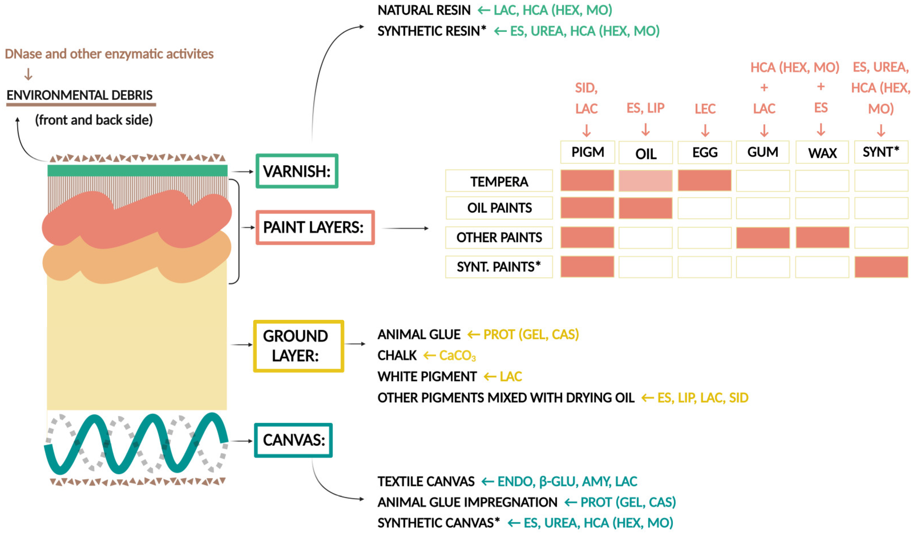
Figure 2. Graphical visualization of the selected enzyme tests according to the materials used in painting layers. AMY: amylase; ENDO: endocellulase; β-GLU: β-glucosidase; PROT: proteinase activity (CAS: caseinase; GEL: gelatinase); ES: esterase, LIP: lipase; LEC: lecithinase; LAC: laccase, CaCO3: carbonate dissolution; UREA: urease; HCA: hydrocarbon assimilation (HEX: hexadecane, MO: mineral oil); SID: siderophore formation; PIGM: pigments; DRY: drying oil; EGG: egg; GUM: gum; WAX: wax; SYNT: synthetic materials; * contemporary techniques using synthetic materials. (Created with BioRender.com).
Jof 10 00076 g002
Figure 3. The UpSet plot [65] listing fungi showing selected features under in-vitro conditions that, if performed also on canvas paintings, could have a damaging effect, like acidifying abilities (change of pH from 6 to 5.1), growth at extreme aw conditions (obligate halo- or xerophily), and soluble pigment production. Dots represent positive character expressions of a specified taxon and horizontally aligned bars the number of expressed characters (0–2). Vertically arranged bars specify the number of taxa expressing a certain character in relative terms. The figure was generated by ‘ggplot2’ [64].
Jof 10 00076 g003
Figure 4. The UpSet plot [65] shows strong activities (factor 2 or higher) of the tested fungal strains, presented in an alphabetical order of ascomycetes, continuing by alphabetical order of basidiomycetes, from top to bottom, at the left side of the plot. Beside enzyme activities (amylase, endoglucanase, β-glucosidase, laccase, caseinase, gelatinase, esterase, lipase, lecithinase, urease, and DNase), the ability to solubilize CaCO3, growth on hexadecane and mineral oil as sole carbon sources, and siderophore production are shown. Obligately xerophilic taxa are written in bold. Dots represent positive character expressions of a specified taxon and horizontally aligned bars the number of expressed characters (0–11). Vertically arranged bars specify the number of taxa expressing a certain character in relative terms. The figure was generated by ‘ggplot2’ [64].
Jof 10 00076 g004
Figure 5. Comparison of dimensionality reduction results based on the presence/absence of enzymatic activities, pigmentation, and pH reduction. (A) principal components analysis (PCA); (B) multidimensional scaling (MDS); and (C) Uniform Manifold Approximation and Projection (UMAP). Each individual strain is labelled with a dot, and groups are marked using colour: obligate xerophiles in red, xerotolerant and moderately xerophilic in green, non-xerotolerant in blue.
Jof 10 00076 g005
Table 1. Performed enzyme tests with the main substrates, their producers, incubation conditions, and positive reactions of enzymatic assays.
Incubation Conditions
Enzymatic
Activity
Substrate
(Producer)
Temperature
[°C]
Time [days]Positive ReactionReference
amylasesoluble starch
(Sigma-Aldrich)
2414a clear zone around colonies in black/blue stained medium after flooding with iodine solution[49]
β-glucosidaseaesculin (6,7-dihydroxycoumarin 6-glucoside) (Merck)2414a black zone around colonies in light-yellow medium[49]
endocellulasecarboxymethyl cellulose
(Sigma-Aldrich)
2421a yellow clear zone around colonies in red stained medium after flooding with Congo red and rinsing with NaCl solution[49]
gelatinasegelatine
(Sigma-Aldrich)
2414liquefied medium around the colonies in yellow transparent medium[49]
caseinaseskim milk
(Merck)
2414a clear zone around colonies in opaque medium[50]
esteraseTween-80
(Sigma-Aldrich)
2414a purple/blue zone around colonies in yellow medium due to colour change in the indicator; precipitation of calcium salts[51]
lecithinaseegg yolk2428a white opaque zone around the colonies in yellow medium[52]
lipasetributyrin
(Sigma-Aldrich)
2428a clear zone around the colonies in opaque medium[53]
ureaseurea
(Merck)
2414change in colour of the medium from yellow to purple[54]
laccaseABTS
(Sigma-Aldrich)
2414a purple halo around the colonies in light yellow transparent medium[55]
laccaseguaiacol
(Sigma-Aldrich)
2414an orange/brown halo around the colonies in light yellow transparent medium[55]
DNaseDNase agar
(Beckton and Dickinson)
247a transparent halo around colonies in opaque white medium after flooding with 1 M HCl[56]
Disclaimer/Publisher’s Note: The statements, opinions and data contained in all publications are solely those of the individual author(s) and contributor(s) and not of MDPI and/or the editor(s). MDPI and/or the editor(s) disclaim responsibility for any injury to people or property resulting from any ideas, methods, instructions or products referred to in the content.