Article

Mutations in Exons 8 and 11 of c-kit Gene in Canine Subcutaneous Mast Cell Tumors and Their Association with Cell Proliferation

1
College of Veterinary Medicine, Michigan State University, East Lansing, MI 48824, USA
2
Department of Veterinary Medical Sciences, University of Bologna, 40126 Bologna, Italy
3
Veterinary Diagnostic Laboratory, Michigan State University, Lansing, MI 48910, USA
*
Author to whom correspondence should be addressed.
Vet. Sci. 2022, 9(9), 493; https://doi.org/10.3390/vetsci9090493
Submission received: 8 August 2022 / Revised: 1 September 2022 / Accepted: 7 September 2022 / Published: 10 September 2022
(This article belongs to the Special Issue Current Advances in Veterinary Oncology)

Abstract

:

Simple Summary

Mast cell tumors (MCTs) are one of the most common skin tumors in dogs with variable clinical behavior ranging from benign lesions to those causing widespread metastasis. Prognostic factors have been intensively studied in cutaneous MCTs but are less commonly investigated in subcutaneous MCTs as the majority are benign. Activating mutations in exons 8 and 11 of c-kit, a gene that regulates proliferation and differentiation of mast cells, occur commonly in canine cutaneous MCTs and are strong predictors of prognosis. c-kit mutations have rarely been reported in subcutaneous MCTs. The goal of this study was to identify the prevalence of c-kit mutations in exons 8 and 11 in 216 canine subcutaneous MCTs and to investigate their association with other prognostic factors, including mitotic count, histologic grade, KIT pattern and proliferation markers. We detected c-kit mutations in exons 8 and 11 in 23 (10.6%) and 12 (5.56%) subcutaneous MCTs, respectively. c-kit mutations in exon 11 were associated with histologic high grade and a high mitotic count, suggesting that these parameters can predict the biological behavior of subcutaneous MCTs in a similar manner as in their cutaneous counterparts.

Abstract

The prognostic significance of internal tandem duplication (ITD) mutations in exons 8 and 11 of c-kit has been well-described for canine cutaneous mast cell tumors (MCTs), but c-kit mutations have rarely been reported in subcutaneous MCTs. The objective of this study was to identify the prevalence of ITD mutations in exons 8 and 11 of c-kit in canine subcutaneous MCTs and to investigate its association with histologic grade, KIT pattern, and proliferation markers. ITD mutations in exons 8 and 11 of c-kit, mitotic count, Ki67 index, AgNOR number, Ki67xAgNOR score, KIT pattern, and histologic grade (two-tier system) were retrospectively recorded for 216 dogs with subcutaneous MCTs. ITD mutations in exons 8 and 11 of c-kit were detected in 23 (10.6%) and 12 (5.56%) subcutaneous MCTs, respectively. Exon 11 mutations were significantly associated with Kiupel high grade (p < 0.001) and increased mitotic count (p < 0.001) compared to subcutaneous MCTs with no mutations in exons 8 or 11 (p = 0.002) or subcutaneous MCTs with a mutation in exon 8 (p = 0.001). There was no significant association of either c-kit mutation with KIT patterns or proliferation activity. This study identified a higher prevalence of ITD mutations in exons 8 and 11 of c-kit in subcutaneous MCTs than previously reported. Like their cutaneous counterpart, subcutaneous MCTs with exon 11 mutations were more likely to be histologically high grade and have a higher mitotic count, whereas such associations were not observed in subcutaneous MCTs with exon 8 mutations.

1. Introduction

Mast cell tumors (MCTs) are one of the most common skin tumors in dogs with variable biological behavior, ranging from benign localized masses, for which surgical excision is curative, to aggressive tumors with increased recurrence, metastasis, and mortality [1,2]. Based on their location within the skin, MCTs have been divided into cutaneous and subcutaneous tumors. This anatomical division is important, as more than 90% of subcutaneous MCTs are considered benign and can be controlled with complete surgical excision [3,4,5,6]. However, this still leaves almost 10% of dogs diagnosed with subcutaneous MCTs succumbing to MCT-related disease [6]. Based on the published literature, approximately 8–9% of subcutaneous MCTs will recur, and 4–6% will metastasize [5,6]. Lastly, 11% of dogs with subcutaneous MCTs will develop another MCT [6].
Histologic grading has been the primary tool to prognosticate canine MCTs [1,2,7,8,9]. While the Patnaik grading system applied the histologic grading to both cutaneous and subcutaneous MCTs, the more recent two-tier system has been shown to be prognostically significant for cutaneous MCTs only [1,7,8,9]. However, some criteria that are being evaluated as part of the two-tier grading system, such as a mitotic count (>4 in 10 HPFs) and multinucleated giant cells (at least one cell in 10 HPFs), have been shown to be prognostically significant in subcutaneous MCTs, albeit with different established cut-off values [6,10]. Studies evaluating the prognostic significance of the two-tier system for canine subcutaneous MCTs are lacking. In addition to histologic grading, proliferation markers, such as argyrophilic nucleolar organizing regions (AgNORs) and Ki67, have been successfully utilized to prognosticate subcutaneous MCTs [5,10]. A Ki67 labeling index of 21 and a combined AgNORxKi67 score above 54 have been associated with more aggressive behavior in cutaneous MCTs [1,11].
The tyrosine kinase receptor KIT plays an important role in mast cell proliferation, differentiation, migration, and survival [12,13,14,15]. As such, tyrosine kinase inhibitors are a popular targeted treatment modality in MCT treatment [12,16,17,18]. Aberrant expression of the KIT protein as determined by immunohistochemistry (IHC) has been associated with a negative prognosis [10,11,15]. Activating mutations of predominantly internal tandem duplication (ITD) and, less often, small insertions/deletions of the c-kit gene have been identified in exons 8, 9, 11, 12, and 17, with the highest frequency in the first three [9,13,14,15,19,20,21,22,23,24,25]. ITD mutations in exon 11 of c-kit have been reported to occur in approximately 20–45% of canine cutaneous MCTs and have been associated with aberrant KIT protein localization, higher grade, and higher proliferation activity as evidenced by a higher Ki67 index and AgNORxKi67 score [9,12,13,15,21,25,26]. Dogs with cutaneous MCTs with ITD mutations in exon 11 of c-kit have an increased incidence of recurrent disease, decreased survival times, and a high risk for MCT-related mortality [2,15,27]. This is in stark contrast to ITD mutations in exon 8 of c-kit, which have been detected in up to 33% of canine cutaneous MCTs but have not been associated with poor prognosis [1,13,27]. Mast cell tumors with exon 8 c-kit mutations were associated with longer overall survival times in dogs with cutaneous MCTs than those without exon 8 or 11 mutations [1]. MCTs with exon 8 mutations of c-kit had a lower histologic grade and proliferation activity and, less often, an aberrant KIT localization [27].
Interestingly, c-kit mutations have rarely been reported in canine subcutaneous MCTs [22,24]. An ITD mutation in exon 8 has recently been reported in a single subcutaneous MCT [22], while previous studies did not detect such mutations [9,10].

2. Materials and Methods

2.1. Case Selection

The study population was comprised of subcutaneous MCTs submitted as surgical biopsies to the Michigan State University (MSU) Veterinary Diagnostic Laboratory (VDL) between April 2011 and December 2019. Inclusion criteria required the submitted MCT to be located in the subcutis with no involvement of the overlying dermis or deeper fascial planes. All subcutaneous MCTs included in this study were submitted for a prognostic MCT panel that included histologic grading, KIT expression pattern and Ki67 index both determined by immunohistochemistry, and AgNOR count, as well as the calculated combined AgNORxKi67 score.

2.2. Signalment

The breed and age of the dog at histologic diagnosis were recorded for each biopsy specimen.

2.3. Histologic Grading

The histologic grade was recorded even though it has not been validated for subcutaneous MCTs. Histologic grading was performed on HE-stained sections of all 216 MCTs in accordance with the 2-tier grading system by Kiupel et al. for cutaneous MCTs [7]. A high histologic grade was assigned to subcutaneous MCT that met at least one of the following published criteria: at least 7 mitotic figures in 10 high-power fields (hpfs, equal to 2.37 mm2), or karyomegaly (i.e., nuclear diameters of at least 10% of neoplastic cells vary by at least two-fold), or at least 3 bizarre nuclei in 10 hpfs, or at least 3 multinucleated (3 or more nuclei) cells in 10 hpfs [7]. MCTs that did not meet these criteria were diagnosed as histologically low grade.

2.4. KIT Expression Patterns

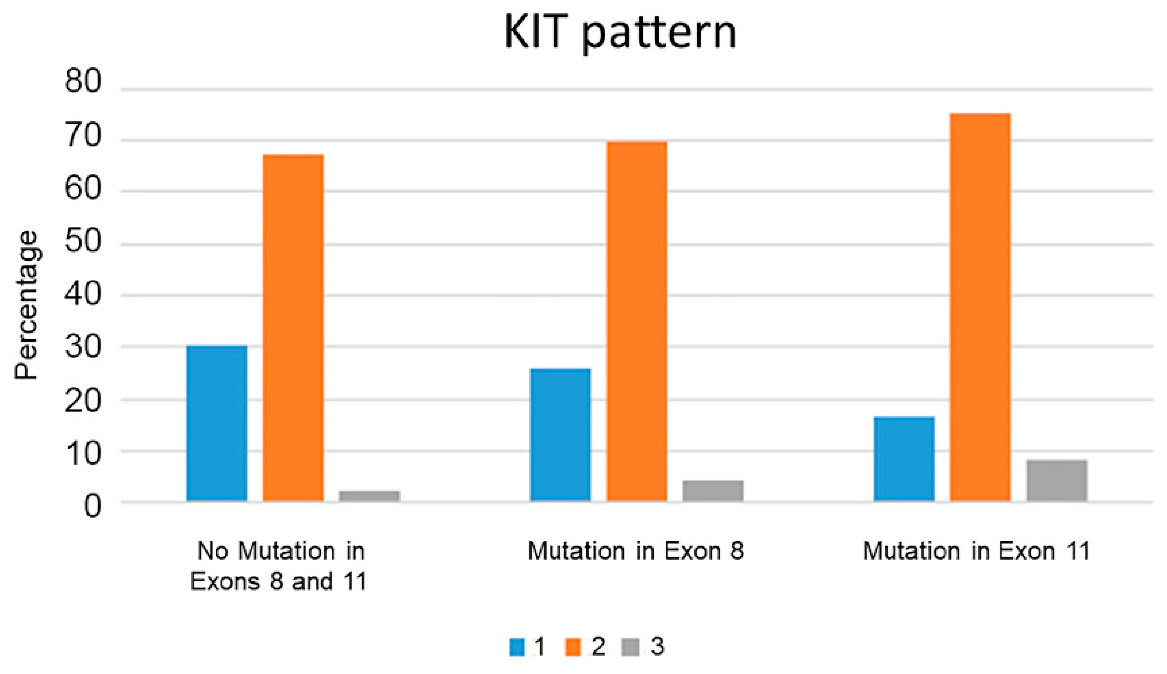
Cellular localization of the KIT protein was determined by IHC labeling as previously described [11]. Cellular localization patterns are classified as follows: 1. KIT pattern I: predominantly membranous labeling; 2. KIT pattern II: focal to stippled cytoplasmic labeling with decreased membrane-associated labeling; and 3. KIT pattern III: diffuse cytoplasmic labeling [1,11].

2.5. Ki67 Index, AgNOR Count, and Combined AgNOR x Ki67 Score

IHC for Ki67 was determined by IHC labeling for the Ki67 as previously described [11], and Ki67-positive cells were quantified as the average number of positively labeled neoplastic nuclei per area of a 1 cm2 optical grid reticle at a magnification of 40× (5 grid areas counted) in the highest labeling area [1,11]. Histochemical staining for AgNORs was performed as previously described [1,11]. AgNORs were counted in 100 randomly selected neoplastic mast cells throughout the tumor at 1000× magnification. Individual AgNORs were resolved by focusing up and down while counting within individual nuclei. Average AgNOR counts/cells were then determined on the basis of averaging the counts within these 100 random neoplastic cells [1,11]. Quantification of tumor proliferation (combined AgNOR x Ki67 score) was performed by multiplying the Ki67 index with the AgNOR count.

2.6. Screening for Mutations in Exons 8 and 11 of c-kit

ITD mutations in exons 8 and 11 of c-kit were determined by polymerase chain reaction (PCR) using previously described primer pairs [19]. For exon 8, primers were located in introns 7 to 8 and amplified the previously reported 12-bp duplication mutation in canine MCTs [13,27]. For exon 11, primers flanked exon 11 and the 5′ end of intron 11, which amplifies the previously described region of the ITD mutation in canine MCTs [14,15]. Amplifications for both primer sets were performed using the Type-it Mutation Detection PCR Kit (Qiagen) as previously described [15,27]. PCR products were visualized on the QIAxcel Capillary Electrophoresis System (Qiagen) [19].

2.7. Statistical Analyses

Categorical variables were summarized as frequency (percentage), and numerical variables were summarized as median (range). For each case, the histologic grade (2-tier Kiupel system), mitotic count, Ki67 index, AgNOR numbers, combined AgNOR x Ki67 score, KIT pattern (1, 2, 3), and c-kit mutation status for exons 8 and 11 were determined. The distribution of the aforementioned tumor markers and signalment were compared in subcutaneous MCTs with or without c-kit mutations. Specifically, MCTs with exon 8 and exon 11 mutations were compared to each other and to subcutaneous MCTs without exon 8 or 11 mutations.
Mann–Whitney U test was used to determine differences in continuous variables (i.e., age, mitotic count, Ki67 index, AgNOR numbers, and AgNOR x Ki67 score), whereas categorical variables (i.e., histologic grade, breed, and KIT pattern) were assessed by means of chi-squared/Fisher’s exact test. Significance was set at p < 0.05.

3. Results

Two hundred and sixteen samples diagnosed as subcutaneous MCTs were included in this study. Of these, breeds included 67 mixed-breed dogs, 37 Labrador Retrievers, 12 Golden Retrievers, 7 Boxers, 7 Pugs, 6 Beagles, 6 Shih Tzu dogs, 5 American Pit Bull Terriers, 3 Boston Terriers, 3 Chihuahuas, 3 German Shorthaired Pointers, 3 Maltese dogs, 3 Siberian Huskies, 3 Vizslas, 2 American Bulldogs, 2 Cocker Spaniels, 2 Dachshunds, 2 Doberman Pinschers, 2 English Setters, 2 French Bulldogs, 2 Great Pyrenees, 2 Havanese, 2 Miniature Schnauzers, 2 Rhodesian Ridgebacks, 2 Staffordshire Terriers, 1 Australian Shepherd, 1 Belgian Malinois, 1 Bernese Mountain Dog, 1 Blue Heeler, 1 Bullmastiff, 1 Coton de Tulear, 1 Fox Terrier, 1 Jack Russell Terrier, 1 Toy Poodle, 1 Portuguese Water Dog, 1 Schnauzer, 1 Shetland Sheepdog, 1 Tibetan Terrier, 1 Weimaraner, 1 Yorkshire Terrier, and 14 unspecified others.
ITD mutations of c-kit were detected in exon 11 in 12/216 (5.6%) subcutaneous MCTs and in exon 8 in 23/216 (10.6%) cases. No mutations in exons 8 or 11 were detected in 181/216 (83.8%) subcutaneous MCTs. No tumors in this study had a concurrent mutation in exons 8 and 11.
Labrador Retrievers had a high prevalence of subcutaneous MCTs with c-kit mutations, with 8 of 37 (22%) MCTs having exon 8 mutations and 4 of 37 (11%) MCTs having exon 11 mutations. A distinction between black Labradors and yellow Labradors was not available from this database. No other breed predilections were identified with our sample size.
The median age of dogs with subcutaneous MCTs with c-kit mutations in exon 8 was 5 years, which was significantly lower than the median age of dogs with no mutations in exons 8 or 11 (8 years, p = 0.001). Dogs with subcutaneous MCTs with c-kit mutations in exon 11 also had a significantly higher median age (9 years, p = 0.017).
There were 173 (80.1%) low-grade and 43 (19.9%) high-grade tumors. The proportion of high-grade subcutaneous MCTs was significantly higher in tumors with c-kit mutations in exon 11 (8/12; 66.7%), compared to those with exon 8 mutations (3/23; 13%; p = 0.002) or to MCTs with no c-kit mutations in exons 8 and 11 (32/181; 17.6%; p < 0.001; Figure 1).
The median mitotic count was 1 for both low-grade and high-grade subcutaneous MCTs tumors (range, 0–49 for high-grade and 0–6 for low-grade). Of the high-grade subcutaneous MCTs, 29/43 (67.4%) had a mitotic count of 7 or higher, while none of the low-grade tumors had a count of 7 or higher. Exon 11 c-kit mutations were also associated with an increased mitotic count (median mitotic count, 2, range 1–10; Figure 2) compared to mutations in exon 8 (median mitotic count, 1, range 0–11; p = 0.001) or dogs with no mutations in exons 8 or 11 (median mitotic count, 1, range 0–49; p < 0.001). There were no statistically significant differences in histologic grades between subcutaneous MCTs with a mutation in exon 8 and no mutations in exons 8 or 11.
There were no statistically significant differences in proliferation markers (Ki67 index, AgNOR count, AgNOR x Ki67 index) or in KIT protein localization patterns in subcutaneous MCTs with mutations in either exon 8 or exon 11 when compared to each other or to subcutaneous MCTs without exon 8 or 11 mutations (Figure 3). Of the high-grade subcutaneous MCTs, 23/43 (53.5%) had a Ki67 index of 23 or higher and a combined AgNOR x Ki67 index of 54 or higher. Only 19/173 (10.9%) low-grade subcutaneous MCTs had a Ki67 index of 23 or higher.

4. Discussion

This is the first large-scale study investigating the c-kit mutation status in canine subcutaneous MCTs. In contrast to previous studies, we identified ITD c-kit mutations in exon 11 and exon 8 in 5.6% and 10.6% of examined cases, respectively. Compared to the study population, Labrador Retrievers had a higher prevalence at 11% and 22%, respectively. As Labrador Retrievers have been reported to have a higher risk for developing MCTs and low-grade MCTs in particular, larger-scale studies are necessary to determine whether MCTs in this breed have a higher prevalence of c-kit mutations than MCTs in other breeds [28,29]. The high prevalence of subcutaneous MCTs with a mutation in exon 8 is especially unusual, as approximately 4% of cutaneous MCTs carry this mutation [13]. Furthermore, the prevalence of subcutaneous MCTs with either mutation was less than half the prevalence that has been reported for cutaneous MCTs [1,13,15,19,27]. Interestingly, the ratio of subcutaneous MCTs with a mutation in exon 11 compared to subcutaneous MCTs with a mutation in exon 8 (ratio 1:2) was inversely related to what has been reported for cutaneous MCTs (ratio 2:4:1) [1,27]. The relatively lower number of subcutaneous MCTs with a mutation in exon 11 correlates with the less aggressive biological behavior of these tumors, as ITD mutations in exon 11 of c-kit have been associated with a poor prognosis for dogs with cutaneous MCTs [2,6,9,10,15,27].
While the current study lacked clinical outcome data, both a high mitotic count as well as a high histologic grade based on the two-tier grading system were significantly associated with exon 11 mutations, while for subcutaneous MCTs with no mutations in exons 8 and 11 or with a mutation in exon 8, no such association was detected. As an increased mitotic count has been demonstrated to predict higher local recurrence and metastasis in subcutaneous MCTs [5,6,10], we surmise that the detection of an ITD c-kit mutation in exon 11 also indicates a more aggressive biological behavior in subcutaneous MCTs, similar to their cutaneous counterpart.
Both a high mitotic count and an exon 11 mutation were significantly associated with a higher grade. A prospective study should confirm the prognostic significance of the two-tier grading system and the detection of ITD mutations in exon 11 of c-kit in subcutaneous MCTs. A total of 8/43 (18.60%) subcutaneous MCTs classified as high-grade had an exon 11 ITD mutation. The prevalence of exon 11 mutations tends to be much higher in high-grade cutaneous MCTs [9,21,26], and a recent study reported such a mutation in 62/75 (82%) cutaneous MCTs [27]. The much lower prevalence of aggressive subcutaneous MCTs compared to high-grade cutaneous MCTs and the lower prevalence of such high-grade subcutaneous MCTs to carry an exon 11 mutation may be responsible for previous studies not being able to identify exon 11 mutations in subcutaneous MCTs [10]. While 32/43 (74.4%) subcutaneous MCTs classified as high-grade had no mutations in exons 8 and 11, a mitotic count above 7 in 67.4% of tumors in this group still supports an aggressive behavior [6,10]. These data may also suggest that exon 11 mutations of c-kit are less often the driver of aggressive behavior in canine subcutaneous MCTs compared to cutaneous MCTs. Interestingly, only 53.5% of these high-grade subcutaneous MCTs had a Ki67 index above 23 or an AgNORxKi67 index above 54. The larger number of cases with a high mitotic count than those with a Ki67 index above the threshold established for cutaneous MCTs may reflect karyologic abnormalities rather than an increased proliferation activity. Regardless, a prospective study will be necessary to fully establish the prognostic significance of histologic grading for subcutaneous MCTs.
Exon 11 c-kit mutations play a crucial role in the oncogenesis of cutaneous MCTs, especially in mast cell proliferation, and we expected that the various proliferation markers investigated in this study would be significantly higher in subcutaneous MCTs with a mutation in exon 11 compared to subcutaneous MCTs with no mutation in exons 8 and 11, similar to what has been reported for cutaneous MCTs [1,11,27]. Furthermore, the aberrant expression of KIT is more commonly observed in cutaneous MCTs with an exon 11 mutation [6,15,19,27,30]. Neither finding could be confirmed for the subcutaneous MCTs with an exon 11 mutation in this study. As we detected only 12 subcutaneous MCTs with such a mutation, the low number may have negatively impacted statistical analysis.
Similar to recently published data for cutaneous MCTs, a mutation in exon 8 of c-kit was not associated with any parameter investigated in this study that predicts a poor prognosis [27]. Based on these previously published data and the data presented here, it seems unlikely that a c-kit mutation in exon 8 causes a gain-of-function of KIT that would thereby stimulate increased tumor proliferation.
Although the overall number of mutations detected in exons 8 and 11 of c-kit in subcutaneous MCTs was low, this retrospective study suggests a prognostic significance for detecting mutations. Furthermore, the histologic grade using the two-tier system may also be helpful in identifying subcutaneous mast cell tumors with a more aggressive biological behavior. Future prospective studies investigating the role of c-kit mutations and histologic grade in subcutaneous MCTs for predicting clinical disease progression and risk for metastatic disease and MCT-associated mortality are needed to confirm these hypotheses.

5. Conclusions

Mutations in exon 8 and 11 of c-kit have been reported in up to 45% of canine cutaneous MCTs [9,13,14,15,19,20,21,22,24,25,27], but only a single case of an exon 8 mutation has been reported in a subcutaneous MCT [22]. This study of 216 dogs identified a higher than anticipated prevalence of approximately 11% and 6% of ITD mutations in exons 8 and 11 of c-kit in subcutaneous MCTs, respectively. Similar to cutaneous MCTs, our study demonstrated that c-kit mutations exon 11 in subcutaneous MCTs were significantly associated with a histologic high grade (p < 0.001) and an increased mitotic count (p < 0.001) compared to subcutaneous MCTs with no c-kit mutations in exons 8 or 11 (p = 0.002) or subcutaneous MCTs with a c-kit mutation in exon 8 (p = 0.001). The larger number of exon 8 then exon 11 c-kit mutations in subcutaneous MCTs represents an inverse relationship compared to cutaneous MCTs that correlates with the less aggressive biological behavior of subcutaneous MCTs. There was no significant association of either c-kit mutation with KIT patterns or proliferation activity. These results provide future directions for prospective studies to confirm c-kit mutations in exon 11 and a histologic high grade as prognostic factors for subcutaneous MCTs.

Author Contributions

Conceptualization, M.K.; Acquisitions, analysis, or interpretation, P.C., S.S. and M.K.; Drafted manuscript, P.C. and M.K.; Substantively revised and edited the manuscript, L.M., S.S. and M.K.; Provided final approval and agrees to be accountable for all aspects of work ensuring integrity and accuracy, P.C., L.M., S.S. and M.K. All authors have read and agreed to the published version of the manuscript.

Funding

This research received no external funding.

Institutional Review Board Statement

Not applicable.

Informed Consent Statement

Not applicable.

Data Availability Statement

The data presented in this study are available on request from the corresponding author.

Acknowledgments

The authors would like to thank the MSU VDL Histology and Virology Laboratories for performing the histology, immunohistochemistry, and PCR testing. We thank the referring veterinarians for providing these cases.

Conflicts of Interest

The authors declare no conflict of interest.

References

  1. Kiupel, M.; Camus, M. Diagnosis and Prognosis of Canine Cutaneous Mast Cell Tumors. Vet. Clin. N. Am. Small Anim. Pract. 2019, 49, 819–836. [Google Scholar] [CrossRef] [PubMed]
  2. Tamlin, V.S.; Bottema, C.D.K.; Peaston, A.E. Comparative aspects of mast cell neoplasia in animals and the role of KIT in prognosis and treatment. Vet. Med. Sci. 2019, 6, 3–18. [Google Scholar] [CrossRef] [PubMed]
  3. Gill, V.; Leibman, N.; Monette, S.; Craft, D.M.; Bergman, P.J. Prognostic Indicators and Clinical Outcome in Dogs with Subcutaneous Mast Cell Tumors Treated with Surgery Alone: 43 Cases. J. Am. Anim. Hosp. Assoc. 2020, 56, 215–225. [Google Scholar] [CrossRef] [PubMed]
  4. Horta, R.S.; LaValle, G.E.; Monteiro, L.N.; Souza, M.C.C.; Cassali, G.D.; Araújo, R.B. Assessment of Canine Mast Cell Tumor Mortality Risk Based on Clinical, Histologic, Immunohistochemical, and Molecular Features. Vet. Pathol. 2018, 55, 212–223. [Google Scholar] [CrossRef] [PubMed]
  5. Newman, S.; Mrkonjich, L.; Walker, K.; Rohrbach, B. Canine Subcutaneous Mast Cell Tumour: Diagnosis and Prognosis. J. Comp. Pathol. 2007, 136, 231–239. [Google Scholar] [CrossRef]
  6. Thompson, J.J.; Pearl, D.L.; Yager, J.A.; Best, S.J.; Coomber, B.L.; Foster, R.A. Canine Subcutaneous Mast Cell Tumor: Characterization and Prognostic indices. Vet. Pathol. 2010, 48, 156–168. [Google Scholar] [CrossRef]
  7. Kiupel, M.; Webster, J.D.; Bailey, K.L.; Best, S.; DeLay, J.; Detrisac, C.J.; Fitzgerald, S.D.; Gamble, D.; Ginn, P.E.; Goldschmidt, M.H.; et al. Proposal of a 2-Tier Histologic Grading System for Canine Cutaneous Mast Cell Tumors to More Accurately Predict Biological Behavior. Vet. Pathol. 2010, 48, 147–155. [Google Scholar] [CrossRef]
  8. Patnaik, A.K.; Ehler, W.J.; MacEwen, E.G. Canine Cutaneous Mast Cell Tumor: Morphologic Grading and Survival Time in 83 Dogs. Vet. Pathol. 1984, 21, 469–474. [Google Scholar] [CrossRef]
  9. Takeuchi, Y.; Fujino, Y.; Watanabe, M.; Takahashi, M.; Nakagawa, T.; Takeuchi, A.; Bonkobara, M.; Kobayashi, T.; Ohno, K.; Uchida, K.; et al. Validation of the prognostic value of histopathological grading or c-kit mutation in canine cutaneous mast cell tumours: A retrospective cohort study. Vet. J. 2013, 196, 492–498. [Google Scholar] [CrossRef]
  10. Thompson, J.J.; Yager, J.A.; Best, S.J.; Pearl, D.L.; Coomber, B.L.; Torres, R.N.; Kiupel, M.; Foster, R.A. Canine Subcutaneous Mast Cell Tumors: Cellular Proliferation and KIT Expression as Prognostic Indices. Vet. Pathol. 2010, 48, 169–181. [Google Scholar] [CrossRef]
  11. Webster, J.D.; Yuzbasiyan-Gurkan, V.; Miller, R.A.; Kaneene, J.B.; Kiupel, M. Cellular Proliferation in Canine Cutaneous Mast Cell Tumors: Associations with c-KIT and Its Role in Prognostication. Vet. Pathol. 2007, 44, 298–308. [Google Scholar] [CrossRef] [PubMed]
  12. Gil da Costa, R.M. C-kit as a prognostic and therapeutic marker in canine cutaneous mast cell tumours: From laboratory to clinic. Vet. J. 2015, 205, 5–10. [Google Scholar] [CrossRef] [PubMed]
  13. Letard, S.; Yang, Y.; Hanssens, K.; Palmérini, F.; Leventhal, P.S.; Guéry, S.; Moussy, A.; Kinet, J.-P.; Hermine, O.; Dubreuil, P. Gain-of-Function Mutations in the Extracellular Domain of KIT Are Common in Canine Mast Cell Tumors. Mol. Cancer Res. 2008, 6, 1137–1145. [Google Scholar] [CrossRef]
  14. London, C.A.; Galli, S.J.; Yuuki, T.; Hu, Z.-Q.; Helfand, S.C.; Geissler, E.N. Spontaneous canine mast cell tumors express tandem duplications in the proto-oncogene c-kit. Exp. Hematol. 1999, 27, 689–697. [Google Scholar] [CrossRef]
  15. Webster, J.D.; Yuzbasiyan-Gurkan, V.; Kaneene, J.B.; Miller, R.; Resau, J.H.; Kiupel, M. The Role of c-KIT in Tumorigenesis: Evaluation in Canine Cutaneous Mast Cell Tumors. Neoplasia 2006, 8, 104–111. [Google Scholar] [CrossRef]
  16. Hadzijusufovic, E.; Peter, B.; Gleixner, K.V.; Schuch, K.; Pickl, W.F.; Thaiwong, T.; Yuzbasiyan-Gurkan, V.; Mirkina, I.; Willmann, M.; Valent, P. H1-receptor antagonists terfenadine and loratadine inhibit spontaneous growth of neoplastic mast cells. Exp. Hematol. 2010, 38, 896–907. [Google Scholar] [CrossRef]
  17. Isotani, M.; Ishida, N.; Tominaga, M.; Tamura, K.; Yagihara, H.; Ochi, S.; Kato, R.; Kobayashi, T.; Fujita, M.; Fujino, Y.; et al. Effect of Tyrosine Kinase Inhibition by Imatinib Mesylate on Mast Cell Tumors in Dogs. J. Vet. Intern. Med. 2008, 22, 985–988. [Google Scholar] [CrossRef] [PubMed]
  18. London, C.A.; Malpas, P.B.; Wood-Follis, S.L.; Boucher, J.F.; Rusk, A.W.; Rosenberg, M.P.; Henry, C.J.; Mitchener, K.L.; Klein, M.K.; Hintermeister, J.G.; et al. Multi-center, Placebo-controlled, Double-blind, Randomized Study of Oral Toceranib Phosphate (SU11654), a Receptor Tyrosine Kinase Inhibitor, for the Treatment of Dogs with Recurrent (Either Local or Distant) Mast Cell Tumor Following Surgical Excision. Clin. Cancer Res. 2009, 15, 3856–3865. [Google Scholar] [CrossRef] [PubMed]
  19. Downing, S.; Chien, M.B.; Kass, P.H.; Moore, P.F.; London, C.A. Prevalence and importance of internal tandem duplications in exons 11 and 12 of c- kit in mast cell tumors of dogs. Am. J. Vet. Res. 2002, 63, 1718–1723. [Google Scholar] [CrossRef]
  20. Cameron, L.R.J.; Grahn, R.A.; Chien, M.B.; Lyons, L.A.; London, C.A. Detection of c-kit Mutations in Canine Mast Cell Tumors using Fluorescent Polyacrylamide Gel Electrophoresis. J. Vet. Diagn. Investig. 2004, 16, 95–100. [Google Scholar] [CrossRef] [Green Version]
  21. Tamlin, V.; Kessell, A.; Mccoy, R.; Dobson, E.; Smith, T.; Hebart, M.; Brown, L.; Mitrovic, D.; Peaston, A. Prevalence of exon 11 internal tandem duplications in the C-KIT proto-oncogene in Australian canine mast cell tumours. Aust. Vet. J. 2017, 95, 386–391. [Google Scholar] [CrossRef] [PubMed]
  22. Vozdova, M.; Kubickova, S.; Fictum, P.; Cernohorska, H.; Fröhlich, J.; Rubes, J. Mutation and methylation status of KIT and TP53 in canine cutaneous and subcutaneous mast cell tumours. Vet. Comp. Oncol. 2019, 18, 438–444. [Google Scholar] [CrossRef] [PubMed]
  23. Vozdova, M.; Kubickova, S.; Fictum, P.; Fröhlich, J.; Jelinek, F.; Rubes, J. Prevalence and prognostic value of c-kit and TP53 mutations in canine mast cell tumours. Vet. J. 2019, 247, 71–74. [Google Scholar] [CrossRef] [PubMed]
  24. Vozdova, M.; Kubickova, S.; Pal, K.; Fröhlich, J.; Fictum, P.; Rubes, J. Recurrent gene mutations detected in canine mast cell tumours by next generation sequencing. Vet. Comp. Oncol. 2020, 18, 509–518. [Google Scholar] [CrossRef] [PubMed]
  25. Zemke, D.; Yamini, B.; Yuzbasiyan-Gurkan, V. Mutations in the juxtamembrane domain of c-KIT are associated with higher grade mast cell tumors in dogs. Vet. Pathol. 2002, 39, 529–535. [Google Scholar] [CrossRef]
  26. Mochizuki, H.; Thomas, R.; Moroff, S.; Breen, M. Genomic profiling of canine mast cell tumors identifies DNA copy number aberrations associated with KIT mutations and high histological grade. Chromosom. Res. 2017, 25, 129–143. [Google Scholar] [CrossRef]
  27. Brocks, B.A.W.; Bertram, C.A.; Bartel, A.; Kirpensteijn, J.; Collins-Webb, A.; Catlin, C.; Thaiwong, T.; Kiupel, M. Internal Tandem Duplication of Exon 8 of c-kit Is Associated With Longer Total Survival in Canine Cutaneous Mast Cell Tumors. Vet. Pathol. 2020, 58, 315–324. [Google Scholar] [CrossRef]
  28. Martins, A.; Faria, F.; Mesquita, J.; Rtner, F.; Amorim, I. Analysis of risk factors for canine mast cell tumors based on the Kiupel and Patnaik grading system among dogs with skin tumors. Open Vet. J. 2021, 11, 619. [Google Scholar] [CrossRef]
  29. Pierini, A.; Lubas, G.; Gori, E.; Binanti, D.; Millanta, F.; Marchetti, V. Epidemiology of Breed-Related Mast Cell Tumour Occurrence and Prognostic Significance of Clinical Features in a Defined Population of Dogs in West-Central Italy. Vet. Sci. 2019, 6, 53. [Google Scholar] [CrossRef]
  30. Thompson, J.J.; Morrison, J.A.; Pearl, D.L.; Boston, S.E.; Wood, G.; Foster, R.A.; Coomber, B.L. Receptor Tyrosine Kinase Expression Profiles in Canine Cutaneous and Subcutaneous Mast Cell Tumors. Vet. Pathol. 2015, 53, 545–558. [Google Scholar] [CrossRef] [Green Version]
Figure 1. Histogram of percentage of low- (blue) and high-grade (orange) subcutaneous mast cell tumors associated with each of the three groups of different mutation status. A high grade was significantly associated with an internal tandem duplication mutation in exon 11 (p < 0.001), but not with a mutation in exon 8 or with no mutations in exons 8 and 11.
Vetsci 09 00493 g001
Figure 2. Boxplot of mitotic count per 10 high-power fields in subcutaneous mast cell tumors with different mutation status. Internal tandem duplication mutations in exon 11 (gray) were significantly associated with a higher mitotic count compared to exon 8 (orange) mutations (p = 0.001) and no (blue) mutations (p < 0.001), but there was no difference between the latter two groups.
Vetsci 09 00493 g002
Figure 3. Histogram of percentage of KIT patterns (1 (perimembranous; blue), 2 (focal or stippled cytoplasmic; orange), and 3 (diffuse cytoplasmic, gray)) in subcutaneous mast cell tumors with different mutation status. While there was an overall increase in aberrant labeling (patterns 2 and 3) in mast cell tumors with an internal tandem duplication mutation in exon 11, there was no statistically significant difference.
Vetsci 09 00493 g003
Publisher’s Note: MDPI stays neutral with regard to jurisdictional claims in published maps and institutional affiliations.