Article

Perfluorooctanoic Acid (PFOA) Induces Redox Status Disruption in Swine Granulosa Cells

by
Giuseppina Basini
*,
Simona Bussolati
,
Veronica Torcianti
and
Francesca Grasselli
Dipartimento di Scienze Medico-Veterinarie, Università di Parma, Via del Taglio 10, 43126 Parma, Italy
*
Author to whom correspondence should be addressed.
Vet. Sci. 2022, 9(6), 254; https://doi.org/10.3390/vetsci9060254
Submission received: 28 April 2022 / Revised: 23 May 2022 / Accepted: 24 May 2022 / Published: 26 May 2022

Abstract

:
Perfluorooctanoic acid (PFOA) is employed in the production and processing of several plastic materials, mainly during the production of waterproof fabrics or nonstick cookware. PFOA is identified as a substance of very high concern, as it is classified as a persistent, bioaccumulative, and toxic (PBT) substance because of its persistence in the environment and its potential accumulation in organisms. Thus, safe levels of exposure cannot be established, and PFOA emissions should be minimized. PFOA has recently been linked to several health concerns in humans. In particular, a disruptive effect on redox status homeostasis has been documented, with a potential impairment of normal reproductive function that requires adequate oxidative balance. Therefore, the aim of the present study was to evaluate the effects of PFOA (2, 20, and 200 ng/mL) on ovarian granulosa cells, a model of reproductive cells. The obtained results reveal that PFOA stimulated cell viability (p < 0.05). Regarding the effects on free radical production, O2, NO, and H2O2 were significantly inhibited (p < 0.05), while the nonenzymatic antioxidant power was not significantly modified. Collectively, the present results deserve attention since free radical molecules play a crucial role in ovarian follicle development leading to a successful ovulation.

1. Introduction

In recent years, perfluoroalkyl substances (PFAS) have gained increasing attention as an emerging category of pollutants. This family of chemical compounds were produced in the late 1940s through electrochemical fluorination or telomerization. Since their chemical structure renders them stable from a chemical and thermal point of view, and difficult to biodegrade in the environment, they have wide industrial applications; they are used as surfactants and emulsifiers, in the production of other fluorinated chemicals, stain removers, friction reducers, firefighting foams, and waterproofing and insulating agents [1]. Potential routes of human exposure to PFAS include the ingestion of dust present in the environment, inhalation, skin absorption, and the consumption of contaminated drinking water or food [2]. Direct exposure occurs when PFAS are present in food or in house dust, while indirect exposure happens when PFAS are formed through the transformation of fluorotelomeric-based materials, including polyfluoroalkyl phosphate esters (PAP), fluorotelomer acrylate (FTAc), fluorotelomeric iodides (FTI), and fluorotelomeric alcohols (FTOH) [3]. PFAS are particularly resistant to hydrolysis, photolysis, or microbial degradation due to their chemical structure, and they can easily accumulate in organisms. Thus, these compounds are ubiquitous, and their presence was documented in biota and human serum [4]. Perfluorooctanoic acid (PFOA), a well-studied and ubiquitous PFAS, is an eight-carbon fluorocarbon and carboxylic acid group detectable in wild animal serum [5]. Once absorbed by humans, PFOA binds to blood proteins, mainly albumin, and it is not easily metabolized, leading to a half-life of 2.3–8.5 years [6]. Among the various uses of PFAS, PFOA is widely employed for the production of fluoropolymers such as polytetrafluoroethylene (better known as Teflon), of which the nonstick properties and chemical inertness are widely known, and polyvinyldenefluoride (PVDF). A further application of PFOA is represented by Gore-Tex®, a resistant waterproof material characterized by breathability and biocompatibility that is used in the production of technical sports clothing and medical-health items, such as vascular prostheses, heart valves, and sutures, and in cosmetic surgery. Experiments performed on animals showed that PFOA mainly accumulates in the liver, kidneys, and serum [7], causing negative health effects, including hepatotoxicity, genotoxicity, immunotoxicity, and neurotoxicity, which raise increasing concerns due to its long half-life. In 2016, it was also classified by the International Agency for Research on Cancer (IARC) as “possibly carcinogenic to humans”, belonging to class 2B. Furthermore, several studies on animal exposure to PFOA have suggested possible interference with endocrine signaling [8]. In particular, a study conducted by Di Nisio et al. showed that PFOA disrupts the male hypothalamus–pituitary–gonadal (HPG) axis [9]. The present work investigates the possible consequences of PFOA on female gonads by means of our well-validated model of endocrine reproductive cells [10,11,12]. In particular, we hypothesized a disruption in the fine balance between free radicals and antioxidant mechanisms induced by PFOA. To this goal, granulosa cells were collected from follicles. The pig was chosen as a valuable animal model for translational medicine, as it is similar to humans from anatomical, genetic, and physiological points of view [13]. The effect of PFOA was studied on granulosa cell viability and on oxidative-stress-related parameters, since redox status is crucial for correct follicular function leading to ovulation [14].

2. Materials and Methods

All reagents were purchased from Sigma Chemical Co., Ltd. (St. Louis, MO, USA), while the plastics were from Sarstedt AG & Co. (Numbrecht, Germany). If purchased elsewhere, it is highlighted in the text.

2.1. Isolation and Culture of Granulosa Cells

To obtain the granulosa cells, swine ovaries were collected at a local slaughterhouse and placed in a refrigerated container with phosphate-buffered saline (PBS; 4 °C) with the addition of penicillin (100 Ul/mL), streptomycin (100 Ul/mL), and amphotericin B (2.5 µg/mL), stored in a freezer bag, and transported to the laboratory within 1 h. In order to improve cleaning in the laboratory, the ovaries were immersed for 1 min in 70% ethanol and subjected to further washing with PBS [15]; during the selection, ovaries with cystic or hemorrhagic follicles were discarded. Granulosa cells were aspirated from preovulatory follicle with a diameter greater than 5 mm with a 26-gauge needle [16,17]. Cells were then subjected to centrifugation at 450× g for 10 min, and the cell pellet was treated with ammonium chloride 0.17M at 37 °C for 1 min to eliminate any red blood cells in the precipitate. Treatment with ammonium chloride could be repeated if a high number of erythrocytes are present. The cell number was counted by viable trypan blue dye (0.4% w/v). Cells were then plated and cultured in a validated serum-free system composed by DMEM/Ham’s F12 medium supplemented with penicillin (100 µg/mL), amphotericin B (2.5 µg/mL), streptomycin (100 µg/mL), sodium selenite (5 ng/mL) and transferrin (5 µg/mL) [10,18,19], indicated hereafter as CM. This serum-free medium is a culture system designed to keep granulosa cells differentiated and prevent luteinization. At the time of seeding in 96-well plates, cells were treated with PFOA (2, 20, and 200 ng/mL) on the basis of tested concentrations in previous works [20,21]. Cells were then incubated at 37 °C under humidified conditions (5% CO2) for 48 h.

2.1.1. Granulosa Cell Viability

The vitality of granulosa cells was evaluated using a bioluminescent assay (ATP-lite; Packard Bioscience, Groningen, The Netherlands). ATP is a marker of cell viability because it is present in all metabolically active cells, while its concentration declines very rapidly in the presence of necrosis or apoptosis; the test is based on light emission due to the reaction among ATP, luciferase, and luciferin. The emitted light is proportional to the ATP concentration. The test was validated by plating different viable cell numbers (from 2.5 × 10³ to 4 × 10⁶/100 µL) The curve was repeated three times. The relationship between cell number and luminescence was linear (r = 0.95). Cells were seeded in 96-well plates at a density of 2 × 10⁵ cells/100 µL CM, and treated with PFOA as detailed above. After 48 h of incubation with the treatments, by adding the kit reagents, it was possible to measure luminescence, proportional to the number of viable cells, using the luminometer Victor Nivo (Perkin Elmer, Groningen, The Netherlands) [12].

2.1.2. Granulosa Cell Redox Status

Granulosa Cell Superoxide (O2) Production

O2 production was evaluated by WST-1 (4-[3-(4-iodophenyl)-2-(4-nitrophenyl)-2H-5-tetrazolium]-1,3-benzene disulfonate) test (Roche, Mannheim, Germany). The assay was based on the cleavage of the water-soluble tetrazolium salt, WST-1, to water-soluble formazan as already reported [20]. For the test, cells were seeded in 96-well plates at a concentration of 104 cells/100 µL CM and incubated with PFOA as above indicated. During the last 4 h of incubation, 20 µL of WST-1 reagent was added to cells, and the absorbance of the developed color was determined using the Victor Nivo at a wavelength of 450 nm with a reference length of 620 nm. The coefficients of variation were less than 3%.

Granulosa Cell Hydrogen Peroxide (H2O2) Production

For the test, cells were seeded in 96-well plates at a density of 2 × 10⁵ viable cells/200 μL CM and treated with PFOA as mentioned above. After centrifugation for 10 min at 400× g, the supernatant was discarded, and cells were lysed by adding cold Triton 0.5% + PMSF in PBS (200 μL/well) and incubating on ice for 30 min. H2O2 production was measured through a sensitive one-step analysis using the Amplex Red reagent; the kit used is the Amplex Red Hydrogen Peroxide Assay Kit (Molecular Probes, PoortGebouw, The Netherlands); the reagent reacts with H2O2 in the sample to produce the fluorescent oxidation product resorufin. Briefly, 5 µL of cell lysates were dispensed in each well of a 96-well plate and mixed with 45 µL of reaction buffer (0.05 M sodium phosphate, pH 7.4). Thereafter, 50 µL of Amplex Red reagent (100 µM)/HRP (0.2 U/mL) working solution were added to each microplate well, incubated at room temperature for 30 min, protected from light, and read against a standard curve of H2O2 ranging from 0.39 to 50 µM. Absorbance was determined with Victor Nivo using a 540 nm filter [22].

Granulosa Cell Nitric Oxide (NO) Production

In total, 2 × 10⁵ viable cells/200 μL CM were seeded in 96-well plates and treated with PFOA as previously detailed. Plates were then subjected to centrifugation for 10 min at 400× g; the test allowed for evaluating NO levels by measuring the nitrites in the supernatant of the samples using a microplate method based on the formation of an azo dye after the reaction with the Greiss reagent, which was prepared fresh daily by mixing equal volumes of 1% sulfanilamide, 5% phosphoric acid (stock solution 1) and 0.1% N-[naphtyl] ethylenediaminedihydrochloride (stock solution 2). After incubation with a Greiss reagent at room temperature, the colorimetric reaction was determined with the Victor Reader using a 540 nm against 620 nm filter. Furthermore, a standard curve between 25 and 0.39 μM was set up by diluting the 10 mM sodium nitrite in CM and on which the absorbances of the samples were interpolated [23].

Non-Enzymatic Scavenging Activity

The ferric reducing ability of plasma (FRAP) test is a direct measure of the total reducing power of a solution; is a colorimetric method that is based on the reduction, by antioxidant chemical agents present in the sample, in ferric-tripiridyltriazine (Fe3+ TPTZ) into a ferrous form (Fe2+ TPTZ). Fe2+ is measured spectrophotometrically by determining its colored complex with 2,4,6-tris(2-pyridyl)-s-triazine (Fe2+ TPTZ). The TPTZ reagent was prepared before use by mixing 25 mL of acetate buffer, 2.5 mL of 2,4,6-Tris(2-pyridyl)-s-triazine (TPTZ) 10 mM in HCl 40 mM, and FeCl3−6H2O solution. For the test, cells were seeded in 96-well plates at a density of 2 × 10⁵ cells/200 μL CM and treated with PFOA. Lastly, plates were subjected to centrifugation for 10 min at 400× g; supernatants were discarded, and cells were lysed by adding in an ice bath for 30 min cold Triton 0.5% + PMSF in PBS (200 μL/well). For the test, 40 μL of cell lysates added to Fe3+ TPTZ reagent are loaded in the wells. After 30 min incubation at 37 °C, absorbance of Fe2+ TPTZ was determined by Victor Reader at 595 nm. To quantify the absorbance, a standard curve must be prepared starting from a stock solution (1 mM) consisting of 0.0278 g of FeSO4 in 100 mL of distilled H2O, from which the other dilutions are prepared: 750 μM-500 μM-250 μM-100 μM [24].

2.2. Statistical Analysis

The experiments were repeated five times on the granulosa cells, each time starting from ovaries of about 40 gilts. Each experimental treatment was set up each time using mixed cells collected from all 40 gilts. Therefore, no animal effect was present in our experimental model. Each time, six replicates were made in cell cultures for controls and PFOA treatments. Data are presented as mean ± SEM. Statistical differences were calculated by One Way ANOVA considering treatment as the main factor using Statgraphics software (STC Inc., Rockville, MD, USA); in the presence of a significant difference (p < 0.05), the means were subjected to the Scheffè F test for comparisons multiples.

3. Results

3.1. Effect of PFOA on Swine Granulosa Cells Viability

All the examined PFOA concentrations significantly increased (p < 0.05) cell metabolic activity, evaluated as ATP production (Figure 1).

3.2. Effect of PFOA on Swine Granulosa Cells Redox Status

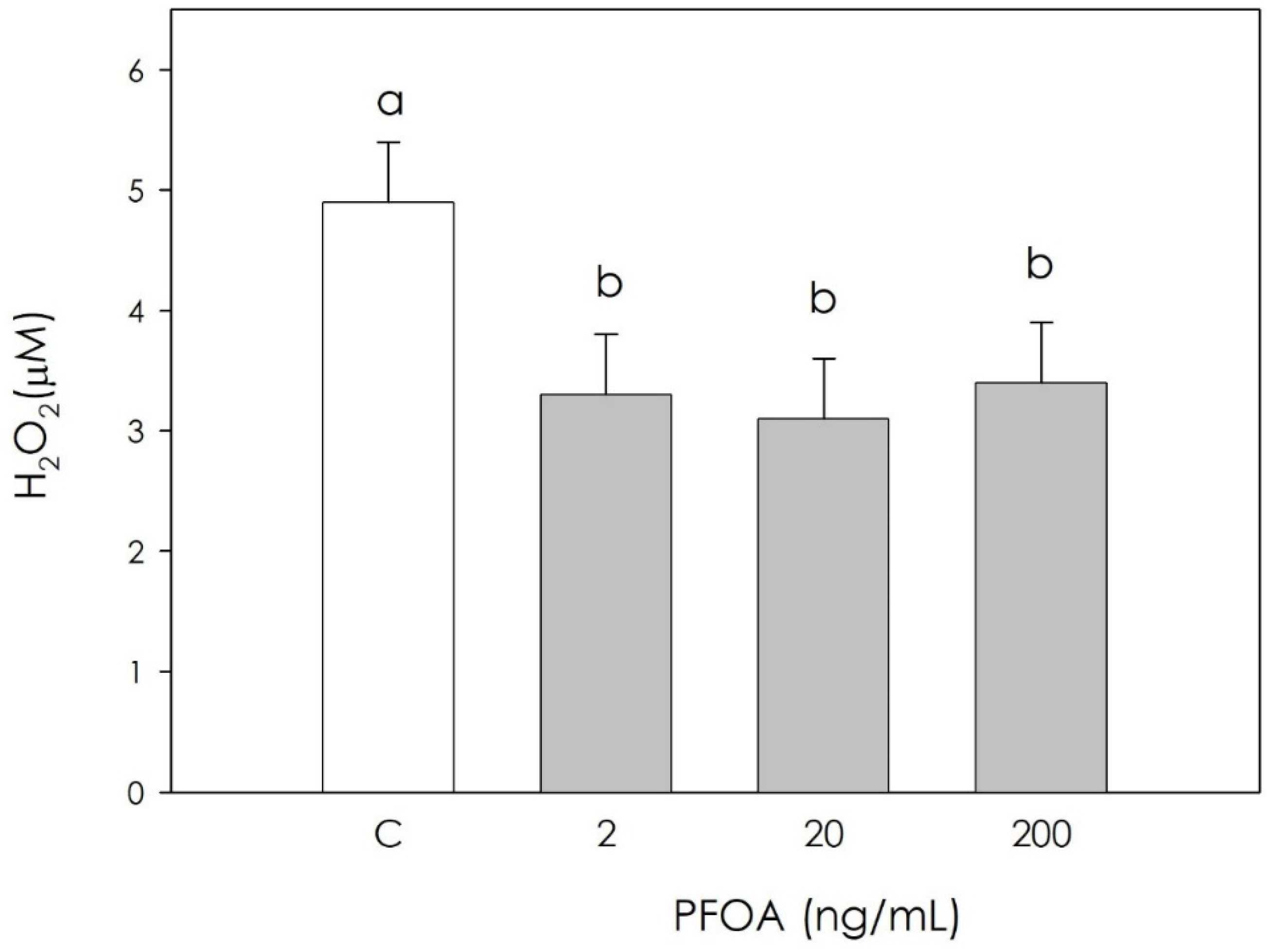
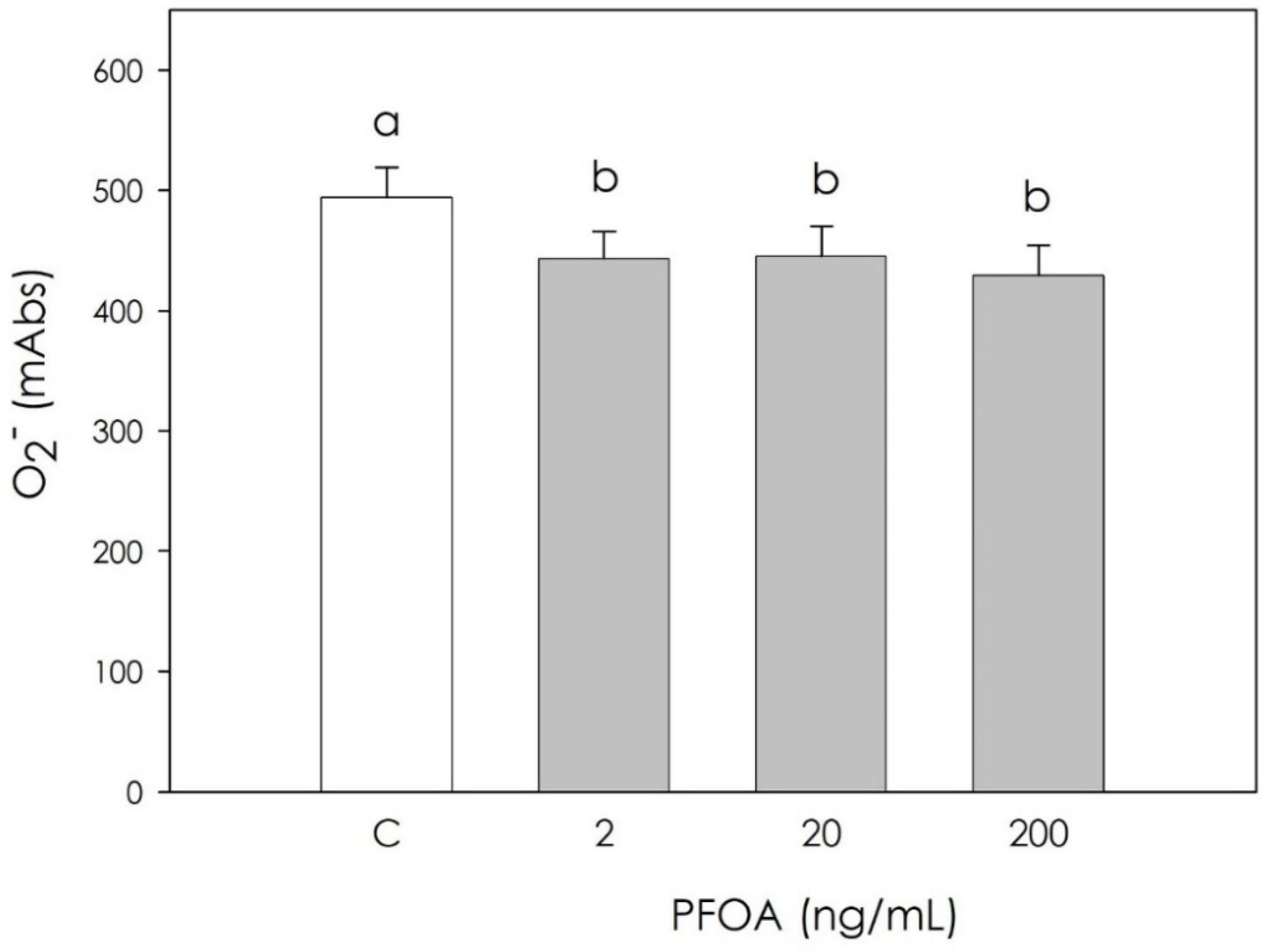
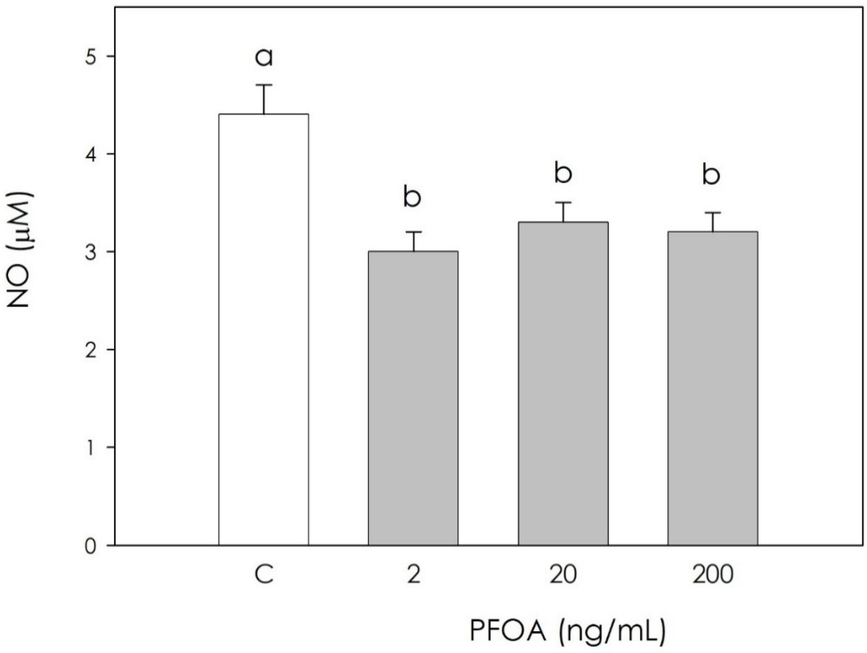
In the production of free radicals, H2O2, O2, and NO were significantly inhibited (p < 0.05) by PFOA at all examined concentrations (Figure 2, Figure 3 and Figure 4).
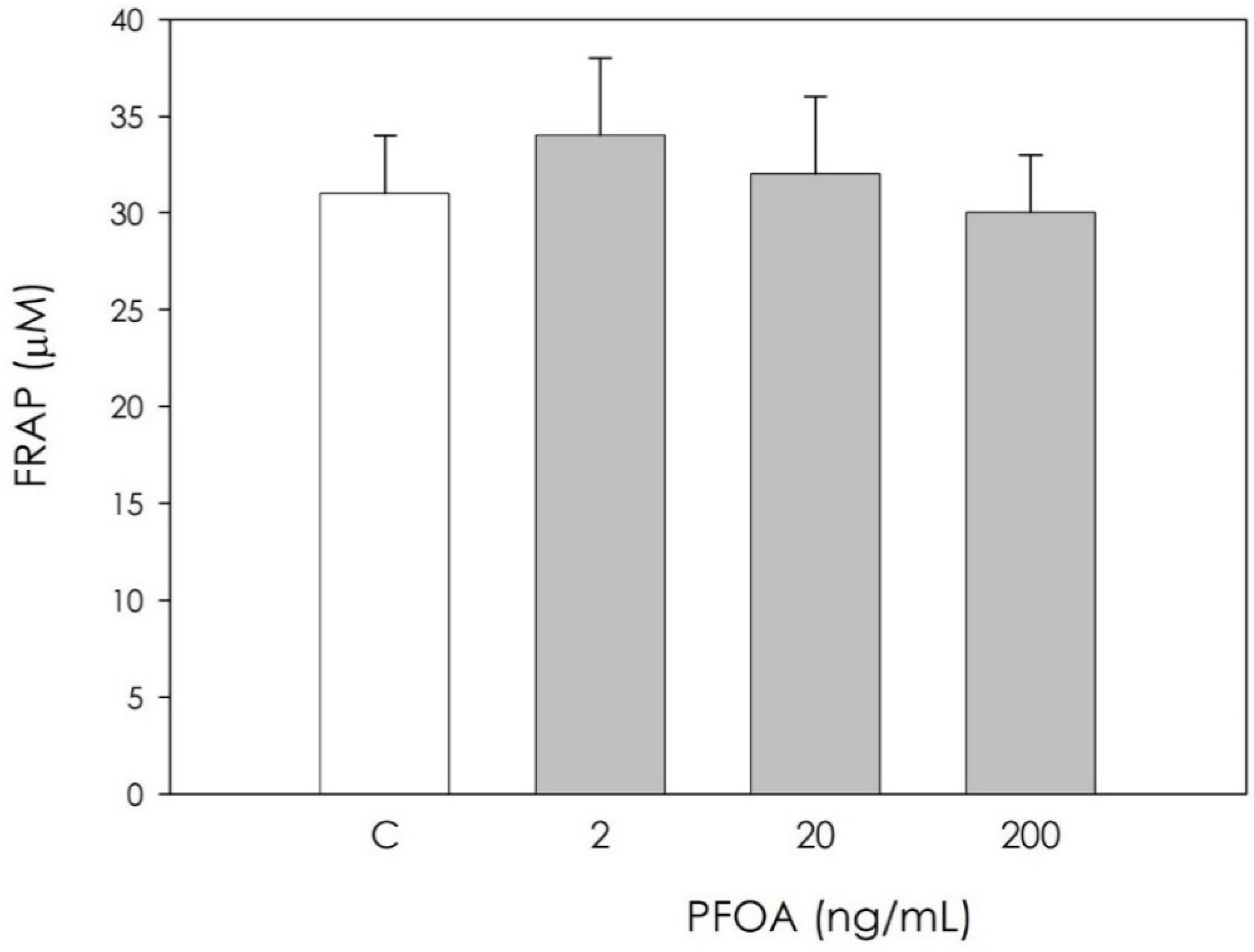
Scavenging activity represented by nonenzymatic antioxidant power was unaffected by PFOA (Figure 5).

4. Discussion

Perfluoroalkyl substances (PFAS) represents a broad group of artificial chemicals whose potential impact on human and animal health is gaining increasing attention and concern. Their peculiar chemical and physical properties have led to a massive use in many consumer and industrial products, with a resulting accumulation in both the environment and in organisms, mainly due to their strong resistance to degradation [4]. Particular concerns have been raised against perfluorooctanoic acid (PFOA), a “forever pollutant”. Due to long persistence, reproduction could be one of the main targets of its disruptive action [25]. Reproduction in mammals is in fact dependent on the hypothalamic–pituitary–gonadal axis, and can be modified by endogenous and exogenous signals. Within the ovary, coordinated interactions among theca cells, granulosa cells, and the oocyte are essential in follicular development, oocyte maturation, and ovulation. On this basis, this research was undertaken to explore the potential effect of PFOA on swine granulosa cells, a model of endocrine reproductive cells [11,12,19]. First, we investigated the PFOA effect on cell metabolic activity, evaluated as ATP production, and we found a significant increase. Krawczyk et al. [26] reported the effects of a mixture of endocrine disrupting chemicals (EDCs) including PFOA in human granulosa cells, documenting both increased mitochondrial activity and ATP content, which in turn is an index of cell viability. On the other hand, research on different cell types showed the inhibition of metabolic activity after exposure to PFOA: in mouse liver cells, the main PFOA target, Sun et al. [27] highlighted a decrease in cell viability, as assessed by ATP production after treatment with PFOA 200 μM for 28 days. Souders et al. [28] studied human neuron cells, observing that 24 and 48 h exposure to PFOA (400 µM PFOA) inhibits ATP production. In addition, exposure to PFOA 250 µM resulted in a decrease in ATP synthase activity. Since granulosa cell redox status is directly involved in the regulation of ovarian physiology follicular function regulation [14], we measured several oxidative stress markers in order to verify a potential impairment of follicular function by PFOA. Our results indicate that PFOA treatment significantly inhibited the production of superoxide anion, NO and hydrogen peroxide, while nonenzymatic antioxidant power was unaffected. Studies conducted on the modulation of oxidative stress induced by PFOA in the ovary are limited. Divergence between the few available results in the literature and our current observations may depend on differences in the experimental model, and various times of exposure to the substance and different concentrations of PFOA. Specific studies on the interaction of PFOA with ovarian cells are limited. Among these, a study conducted in 2019 by Lopez-Arellano et al. [29] on mouse oocytes exposed to PFOA (50, 100, and 150 μM) for 24 h documented that ROS levels increased significantly and concentration-dependently. These results agree with those obtained in an in vivo study [30] that reported that PFOA treatment inhibited the activities of superoxide dismutase and catalase, and increased the generation of hydrogen peroxide and malondialdehyde in the ovaries of pregnant mice. A recent study on cumulus oophore cells (oocyte and granulosa cells) documented that exposure to 20 and 40 μM PFOA for 44 h significantly stimulated ROS production [31]; the observed increase in ROS was attributed to antioxidant system damage, as already reported in a study by Xu et al. [32], possibly leading to the lipid peroxidation of mitochondrial membranes. This would result in ion losses, which can affect the proton gradient and oxidative phosphorylation required to produce ATP, thus contributing to the further generation of ROS [33].

5. Conclusions

This research demonstrated that PFOA disrupts the metabolic activity and redox status of swine ovarian cells. In conclusion, collected data from the present research added to the limited knowledge on PFOA interaction with the reproductive system, and may provide a stimulus to continue the evaluation of the potentially critical effects of this substance. Further studies are needed to better explore the effect of PFOA in granulosa cells, in particular to verify its potential effects on steroidogenic activity.

Author Contributions

Conceptualization, supervision, writing—original draft preparation, writing—review and editing, G.B.; data curation, S.B. and V.T.; writing—review and editing, F.G. All authors have read and agreed to the published version of the manuscript.

Funding

This research was supported by the FIL program of University of Parma.

Institutional Review Board Statement

The study was conducted using discarded samples collected at a slaughterhouse.

Informed Consent Statement

Not applicable.

Data Availability Statement

Data are available upon request.

Conflicts of Interest

The authors declare no conflict of interest.

References

  1. Evich, M.G.; Davis, M.J.B.; McCord, J.P.; Acrey, B.; Awkerman, J.A.; Knappe, D.R.U.; Lindstrom, A.B.; Speth, T.F.; Tebes-Stevens, C.; Strynar, M.J.; et al. Per-and polyfluoroalkyl substances in the environment. Science 2022, 375, eabg9065. [Google Scholar] [CrossRef]
  2. DeLuca, N.M.; Minucci, J.M.; Mullikin, A.; Slover, R.; Cohen Hubal, E.A. Human exposure pathways to poly- and perfluoroalkyl substances (PFAS) from indoor media: A systematic review. Environ. Int. 2022, 162, 107149. [Google Scholar] [CrossRef]
  3. Eichler, C.M.A.; Bi, C.; Wang, C.; Little, J.C. A modular mechanistic framework for estimating exposure to SVOCs: Next steps for modeling emission and partitioning of plasticizers and PFAS. J. Expo. Sci. Environ. Epidemiol. 2022, 32, 356–365. [Google Scholar] [CrossRef]
  4. Milinovic, J.; Lacorte, S.; Vidal, M.; Rigol, A. Sorption behaviour of perfluoroalkyl substances in soils. Scien. Total Environ. 2015, 511, 63–71. [Google Scholar] [CrossRef]
  5. Beale, D.J.; Nilsson, S.; Bose, U.; Bourne, N.; Stockwell, S.; Broadbent, J.A.; Gonzalez-Astudillo, V.; Braun, C.; Baddiley, B.; Limpus, D.; et al. Bioaccumulation and impact of maternal PFAS offloading on egg biochemistry from wild-caught freshwater turtles (Emydura macquarii macquarii). Sci. Total Environ. 2022, 817, 153019. [Google Scholar] [CrossRef]
  6. Fu, J.; Gao, Y.; Cui, L.; Wang, T.; Liang, Y.; Qu, G.; Yuan, B.; Wang, Y.; Zhang, A.; Jiang, G. Occurrence, temporal trends, and half-lives of perfluoroalkyl acids (PFAAs) in occupational workers in China. Sci. Rep. 2016, 6, 38039. [Google Scholar] [CrossRef] [Green Version]
  7. Vanden Heuvel, J.P.; Kuslikis, B.I.; Van Rafelghem, M.J.; Peterson, R.E. Tissue distribution, metabolism, and elimination of perfluorooctanoic acid in male and female rats. J. Biochem. Toxicol. 1991, 6, 83–92. [Google Scholar] [CrossRef]
  8. Fenton, S.E.; Reiner, J.L.; Nakayama, S.F.; Delinsky, A.D.; Stanko, J.P.; Hines, E.P.; White, S.S.; Lindstrom, A.B.; Strynar, M.J.; Petropoulou, S.E. Analysis of PFOA in dosed CD-1 mice. Part 2. Disposition of PFOA in tissues and fluids from pregnant and lactating mice and their pups. Reprod. Toxicol. 2009, 27, 365–372. [Google Scholar] [CrossRef] [Green Version]
  9. Di Nisio, A.; Sabovic, I.; Valente, U.; Tescari, S.; Rocca, M.S.; Guidolin, D.; Dall’Acqua, S.; Acquasaliente, L.; Pozzi, N.; Plebani, M.; et al. Endocrine Disruption of Androgenic Activity by Perfluoroalkyl Substances: Clinical and Experimental Evidence. J. Clin. Endocrinol. Metab. 2019, 104, 1259–1271. [Google Scholar] [CrossRef] [Green Version]
  10. Basini, G.; Baioni, L.; Bussolati, S.; Grasselli, F.; Daquino, C.; Spatafora, C.; Tringali, C. Antiangiogenic properties of an unusual benzo[k,l]xanthene lignan derived from CAPE (caffeic acid phenethyl ester). Investig. New Drugs 2012, 30, 186–190. [Google Scholar] [CrossRef]
  11. Basini, G.; Bussolati, S.; Ciccimarra, R.; Grasselli, F. Melatonin potentially acts directly on swine ovary by modulating granulosa cell function and angiogenesis. Reprod. Fertil. Dev. 2017, 29, 2305–2312. [Google Scholar] [CrossRef]
  12. Basini, G.; Bussolati, S.; Andriani, L.; Grolli, S.; Ramoni, R.; Bertini, S.; Iemmi, T.; Menozzi, A.; Berni, P.; Grasselli, F. Nanoplastics impair in vitro swine granulosa cell functions. Domest. Anim. Endocrinol. 2021, 76, 106611. [Google Scholar] [CrossRef]
  13. Tumbleson, M.E.; Schook, L.B. Advances in Swine in Biomedical Research; Plenum Press: New York, NY, USA, 1996. [Google Scholar]
  14. Timóteo-Ferreira, F.; Abreu, D.; Mendes, S.; Matos, L.; Rodrigues, A.R.; Almeida, H.; Silva, E. Redox imbalance in age-related ovarian dysfunction and perspectives for its prevention. Ageing Res. Rev. 2021, 68, 101345. [Google Scholar] [CrossRef]
  15. Basini, G.; Tamanini, C. Interrelationship between nitric oxide and prostaglandins in bovine granulosa cells. Prostaglandins Other Lipid. Mediat. 2001, 66, 179–202. [Google Scholar] [CrossRef]
  16. Foxcroft, G.R.; Hunter, M.G. Basic physiology of follicular maturation in the pig. J. Reprod. Fertil. Suppl. 1985, 33, 1–19. [Google Scholar]
  17. Basini, G.; Falasconi, I.; Bussolati, S.; Grolli, S.; Di Lecce, R.; Grasselli, F. Swine granulosa cells show tipical endothelial cell characteristics. Reprod. Sci. 2016, 23, 630–637. [Google Scholar] [CrossRef]
  18. Basini, G.; Baioni, L.; Bussolati, S.; Grolli, S.; Kramer, L.H.; Wagner, G.F.; Grasselli, F. Expression and localization of stanniocalcin 1 in swine ovary. Gen. Comp. Endocrinol. 2010, 166, 404–408. [Google Scholar] [CrossRef]
  19. Basini, G.; Bianchi, F.; Bussolati, S.; Baioni, L.; Ramoni, R.; Grolli, S.; Conti, V.; Bianchi, F.; Grasselli, F. Atrazine disrupts steroidogenesis, VEGF and NO production in swine granulosa cells. Ecotoxicol. Environ. Saf. 2012, 85, 59–63. [Google Scholar] [CrossRef]
  20. Gogola, J.; Hoffmann, M.; Ptak, A. Persistent endocrine-disrupting chemicals found in human follicular fluid stimulate the proliferation of granulosa tumor spheroids via GPR30 and IGF1R but not via the classic estrogen receptors. Chemosphere 2019, 217, 100–110. [Google Scholar] [CrossRef]
  21. Gogola, J.; Hoffmann, M.; Ptak, A. Persistent endocrine-disrupting chemicals found in human follicular fluid stimulate IGF1 secretion by adult ovarian granulosa cell tumor spheroids and thereby increase proliferation of non-cancer ovarian granulosa cells. Toxicol. Vitr. Int. J. Publ. Assoc. BIBRA 2020, 65, 104769. [Google Scholar] [CrossRef]
  22. Basini, G.; Simona, B.; Santini, S.E.; Grasselli, F. Reactive oxygen species and anti-oxidant defences in swine follicular fluids. Reprod. Fertil. Dev. 2008, 20, 269–274. [Google Scholar] [CrossRef] [PubMed]
  23. Dong, Y.L.; Yallampalli, C. Interaction between nitric oxide and prostaglandin E2 pathways in pregnant rat uteri. Am. J. Physiol. Endocrinol. Metab. 1996, 270, E471–E476. [Google Scholar] [CrossRef]
  24. Basini, G.; Santini, S.E.; Bussolati, S.; Grasselli, F. The phytoestrogen quercetin impairs steroidogenesis and angiogenesis in swine granulosa cells in vitro. J. Biomed. Biotechnol. 2009, 2009, 419891. [Google Scholar]
  25. Chambers, W.S.; Hopkins, J.G.; Richards, S.M. A Review of Per-and Polyfluorinated Alkyl Substance Impairment of Reproduction. Front. Toxicol. 2021, 3, 732436. [Google Scholar] [CrossRef] [PubMed]
  26. Krawczyk, K.; Marynowicz, W.; Gogola-Mruk, J.; Jakubowska, K.; Tworzydło, W.; Opydo-Chanek, M.; Ptak, A. A mixture of persistent organic pollutants detected in human follicular fluid increases progesterone secretion and mitochondrial activity in human granulosa HGrC1 cells. Reprod. Toxicol. 2021, 104, 114–124. [Google Scholar] [CrossRef]
  27. Sun, S.; Guo, H.; Wang, J.; Dai, J. Hepatotoxicity of perfluorooctanoic acid and two emerging alternatives based on a 3D spheroid model. Environ. Pollut. 2019, 246, 955–962. [Google Scholar] [CrossRef]
  28. Souders, C.L.; Sanchez, C.L.; Malphurs, W.; Aristizabal-Henao, J.J.; Bowden, J.A.; Martyniuk, C.J. Metabolic profiling in human SH-SY5Y neuronal cells exposed to perfluorooctanoic acid (PFOA). Neurotoxicology 2021, 85, 160–172. [Google Scholar] [CrossRef]
  29. López-Arellano, P.; López-Arellano, K.; Luna, J.; Flores, D.; Jiménez-Salazar, J.; Gavia, G.; Teteltitla, M.; Rodríguez, J.J.; Domínguez, A.; Casas, E.; et al. Perfluorooctanoic acid disrupts gap junction intercellular communication and induces reactive oxygen species formation and apoptosis in mouse ovaries. Environ. Toxicol. 2019, 34, 92–98. [Google Scholar] [CrossRef] [Green Version]
  30. Chen, Y.; Zhou, L.; Xu, J.; Zhang, L.; Li, M.; Xie, X.; Xie, Y.; Luo, D.; Zhang, D.; Yu, X.; et al. Maternal exposure to perfluorooctanoic acid inhibits luteal function via oxidative stress and apoptosis in pregnant mice. Reprod. Toxicol. 2017, 69, 159–166. [Google Scholar] [CrossRef]
  31. Teteltitla, M.; Ducolomb, Y.; Souza, V.; Dominguez, A.; Rodriguez-Mercado, J.; Flores, D.; Bonilla, E.; Casas, E.; Altamirano, M.; Lopez, A.; et al. Effects of perfluorooctanoic acid in oxidative stress generation, DNA damage in cumulus cells, and its impact on in vitro maturation of porcine oocytes. Environ. Toxicol. 2022, 37, 1394–1403. [Google Scholar]
  32. Xu, M.; Cui, Z.; Zhao, L.; Hu, S.; Zong, W.; Liu, R. Characterizing the binding interactions of PFOA and PFOS with catalase at the molecular level. Chemosphere 2018, 203, 360–367. [Google Scholar] [CrossRef] [PubMed]
  33. Suh, K.S.; Choi, E.M.; Kim, Y.J.; Hong, S.M.; Park, S.Y.; Rhee, S.Y.; Oh, S.; Kim, S.W.; Pak, Y.K.; Choe, W.; et al. Perfluorooctanoic acid induces oxidative damage and mitochondrial dysfunction in pancreatic β-cells. Mol. Med. Rep. 2017, 15, 3871–3878. [Google Scholar] [CrossRef] [PubMed] [Green Version]
Figure 1. Effect of 48 h treatment with or without (C) perfluorooctanoic acid (PFOA) (2, 20 and 200 ng/mL) on cell metabolic activity quantified using ATP production in swine granulosa cell culture media. Data expressed as counts per second (CPS) represent the mean ± SEM of six replicates/treatment repeated in five different experiments. Different letters indicate a significant difference (p < 0.05).
Vetsci 09 00254 g001
Figure 2. Effect of 48 h treatment with or without (C) perfluorooctanoic acid (PFOA) (2, 20 and 200 ng/mL) on hydrogen peroxide (H2O2) production in swine granulosa cell lysates. Data expressed as µM represent the mean ± SEM of six replicates/treatment repeated in five different experiments. Different letters indicate a significant difference (p < 0.05).
Vetsci 09 00254 g002
Figure 3. Effect of 48 h treatment with or without (C) perfluorooctanoic acid (PFOA) (2, 20, and 200 ng/mL) on superoxide anion (O2) generation in swine granulosa cell culture media. Data expressed as milliabsorbance units (milliAbs), represent the mean ± SEM of six replicates/treatment repeated in five different experiments. Different letters indicate a significant difference (p < 0.05).
Vetsci 09 00254 g003
Figure 4. Effect of 48 h treatment with or without (C) perfluorooctanoic acid (PFOA) (2, 20 and 200 ng/mL) on NO production quantified using Griess reagent in swine granulosa cell culture media. Data expressed as µM represent the mean ± SEM of six replicates/treatment repeated in five different experiments. Different letters indicate a significant difference (p < 0.05).
Vetsci 09 00254 g004
Figure 5. Effect of 48 h treatment with or without (C) perfluorooctanoic acid (PFOA) (2, 20 and 200 ng/mL) on nonenzymatic scavenger activity quantified using FRAP method in swine granulosa cell culture media. Data, expressed as µM, represent the mean ± SEM of six replicates/treatment repeated in five different experiments.
Vetsci 09 00254 g005
Publisher’s Note: MDPI stays neutral with regard to jurisdictional claims in published maps and institutional affiliations.