Article

Development of a Multiplex RT-PCR Assay for Simultaneous Detection of Four Potential Zoonotic Swine RNA Viruses

1
Heilongjiang Provincial Key Laboratory of Animal and Comparative Medicine, State Key Laboratory of Veterinary Biotechnology, Harbin Veterinary Research Institute, Chinese Academy of Agricultural Sciences, Harbin 150069, China
2
Chongqing Academy of Animal Science, Chongqing 408599, China
3
College of Veterinary Medicine, Southwest University, Chongqing 400715, China
*
Authors to whom correspondence should be addressed.
These authors contributed equally to this work.
Vet. Sci. 2022, 9(4), 176; https://doi.org/10.3390/vetsci9040176
Submission received: 3 March 2022 / Revised: 26 March 2022 / Accepted: 4 April 2022 / Published: 7 April 2022
(This article belongs to the Special Issue Diagnostic PCR on Animal Diseases: From Extraction to Amplification)

Abstract

:
Swine viruses like porcine sapovirus (SaV), porcine encephalomyocarditis virus (EMCV), porcine rotavirus A (RVA) and porcine astroviruses (AstV) are potentially zoonotic viruses or suspected of potential zoonosis. These viruses have been detected in pigs with or without clinical signs and often occur as coinfections. Despite the potential public health risks, no assay for detecting them all at once has been developed. Hence, in this study, a multiplex RT-PCR (mRT-PCR) assay was developed for the simultaneous detection of SaV, EMCV, RVA and AstV from swine fecal samples. The PCR parameters were optimized using specific primers for each target virus. The assay’s sensitivity, specificity, reproducibility, and application to field samples have been evaluated. Using a pool of plasmids containing the respective viral target fragments as a template, the developed mRT-PCR successfully detected 2.5 × 10³ copies of each target virus. The assay’s specificity was tested using six other swine viruses as a template and did not show any cross-reactivity. A total of 280 field samples were tested with the developed mRT-PCR assay. Positive rates for SaV, EMCV, RVA, and AstV were found to be 24.6% (69/280), 5% (14/280), 4.3% (12/280), and 17.5% (49/280), respectively. Compared to performing separate assays for each virus, this mRT-PCR assay is a simple, rapid, and cost-effective method for detecting mixed or single infections of SaV, EMCV, RVA, and AstV.

1. Introduction

Swine viruses cause a major economic loss in the swine industry [1,2,3,4,5]. Thus far, more than sixteen viruses have been detected from porcine fecal specimens [6,7,8,9]. From these swine viruses, due to the asymptomatic nature of infections, unless there is a confounding factor or a mixed infection of another pathogenic virus, infections from porcine sapovirus (SaV), encephalomyocarditis virus (EMCV), porcine rotavirus A (RVA) and porcine astrovirus (AstV) might not be noticed [10,11,12,13]. Though the economic impact of SaV, EMCV, RVA and AstV viruses on the swine industry might not be significant enough to attract the attention of pig producers, their potential to become zoonotic diseases warrants proper attention [12]. More importantly, even asymptomatic pigs can shed the virus into the environment or directly transmit it to people at risk [9,10,11,12]. Likewise, SaV, EMCV, RVA, and AstV were found to be genetically closely related to human viruses and hence have a zoonotic potential and/or are suspected of zoonosis [5,11,12,14,15,16,17,18].
To develop vaccines against the prevalent viral strains, early and accurate virus detection is needed and hence, early virus detection is an integral part of disease control and prevention programs [19]. However, coinfection complicates early and accurate virus detection. In the presence of coinfection, it is often difficult to know the underlining cause of disease [20,21]. This is because, in the presence of coinfection, viruses that do not usually cause apparent clinical signs during a single infection could contribute to the severity of an existing infection, further complicating disease diagnosis [11,22]. Moreover, compared to other swine viruses, these potential zoonotic viruses, due to their high coinfection rate, higher virus shedding, fecal–oral transmission, asymptomatic nature, and potential to transmit to humans, demands special attention for early detection and accurate identification.
Virus isolation, electron microscopy, serological tests, and nucleic acid detection methods are the most commonly used methods for detecting viruses [21,23,24]. Recently nucleic acid-based and serological detection methods are being used widely for diagnosing swine viral infections [23,24,25]. Nucleic acid-based detection methods are preferred over serological methods for determining current infection status [26]. The existing nucleic acid-based diagnostic assays for detecting potential zoonotic swine viruses depend on a single PCR. These single PCR-based detection methods consume more time and resources to detect each virus at a one-time point, whereas using multiplex PCR assays, nucleic acid detection methods have another added advantage for the simultaneous detection of viruses [24,25]. Multiplex reverse transcriptase PCR (mRT-PCR) based virus diagnostic assays provide both the affordability and accuracy of virus detection, enabling faster and wider field-based applications [23,26]. In this study, in an attempt to solve challenges associated with detecting potential zoonotic swine viruses, a multiplex PCR assay that could simultaneously detect SaV, EMCV, RVA and AstV from porcine fecal samples has been developed.

2. Materials and Methods

2.1. Nucleic Acid Extraction and Reverse Transcription

From May to September 2021, 280 fecal samples from pigs of different age groups were collected from the Heilongjiang province in China. Prior to storage, 10% (w/v) stool suspensions were mixed with PBS (phosphate-buffered saline, 7.4 PH), centrifuged at 400× g for 20 min at 4 °C, and collected supernatants. Total RNA was extracted from fecal supernatants using the Tiangen virus RNA extraction kit (Tiangen Biotech, Beijing, China), according to manufacturer instructions.
Complementary DNA (cDNA) was synthesized in a 20 µL reverse transcription (RT) reaction mixture containing 2 µL of Golden MLV buffer, 1 µL Golden MLV enzyme, 1 µL Random hexamer primers and 1 µL dNTP, 0.5 µL Rnase inhibitor and 1 µg of total RNA and RNase free double distilled water (ddH2O) and then incubated at 37 °C for 15 min and at 85 °C for 5 s (TakaRa, Dalian, China).

2.2. Primer Design and Construction of SaV, EMCV, RVA and AstV Plasmids

Nucleotide sequences were downloaded from NCBI and aligned prior to primer design to identify conserved regions for each selected virus. The primers (Table 1) used to amplify the fragments of SaV, EMCV, RVA, and AstV were designed by primer3 [27], and then primer properties such as primer heterodimer, self-dimer and hairpin were checked by OligoAnalyzer (https://www.idtdna.com/calc/analyzer, accessed on 20 June 2020). Each amplicon was purified and cloned into the pMD18-T vector (TaKaRa, Dalian, China). The plasmids were transformed into competent Escherichia coli DH5α cells. The plasmids were extracted using a mini plasmid extraction kit (Tiangen, Beijing, China) from a bacterial solution cultured at 37 ℃ for 14–16 h and quantified by a NanoDrop spectrophotometer (Thermo Fisher, Waltham, MA, USA). The plasmid constructs were then confirmed by PCR and sequencing (CometBio, Jilin, China). For mRT-PCR optimization, the plasmids were used as templates.

2.3. Single RT-PCR and mRT-PCR Reaction Optimization

Prior to performing mRT-PCR, the designed primers were used to perform single RT-PCR (sRT-PCR) for SaV, EMCV, RVA, and AstV. A 40 µL PCR reaction was prepared using 5 µL of cDNA, 5% DMSO, rTaq enzyme (0.5–1 unit), 0.25 µM of each primer, 1.5 mM MgCl2, 0.2 mM dNTP, 4X rTaq buffer and autoclaved ddH2O was added to make 40 µL volume. In the negative control reaction, ddH2O was used as a template. In a Bio-Rad PCR Thermo Cycler (Bio-Rad, Hercules, CA, USA), the prepared reaction was amplified as follows: one cycle at 94 °C for 5 min; 35 cycles of denaturation at 98 °C for 10 s; gradient annealing (48 °C to 58 °C) for 30 s; 72 °C for 45 s extension; and a final extension step of 10 min at 72 °C. Gel electrophoresis with a 1.5% agarose gel in 1X TAE buffer was used to examine PCR results.
The mRT-PCR reactions were optimized by varying a single parameter while keeping other parameters constant. Parameter variables including annealing temperature (48 to 58 °C), number of PCR cycle (25 to 40), concentrations of primer (0.05 μM to 0.4 μM), MgCl2 (1.0 to 4.0 mM), dNTP (0.3–0.9 mM), and TakaRa rTaq DNA Polymerase (2 to 6 U) were optimized. The PCR products were visualized by electrophoresis through 1.5% agarose gel in 1× TAE buffer. Following mRT-PCR condition optimization, the amplicons were sequenced for confirmation (CometBio, Jilin, China).

2.4. Assay Sensitivity, Specificity, and Reproducibility

The sensitivity of sRT-PCR and mRT-PCR was compared using a 10-fold serially diluted standard plasmids of known DNA copy number. The DNA copy number of each standard plasmid was calculated using the formula described in [24]. To serve as a template for the sRT-PCR and mRT-PCR, 106 copies/μL of each cloned virus fragment were mixed into a single tube and diluted 10-fold serially at a concentration gradient of 100 to 106 copies/μL.
Specificity of the developed mRT-PCR was tested by using cDNA of porcine epidemic diarrhea virus (PEDV), Seneca Valley virus (SVV), transmissible gastroenteritis virus (TGEV), porcine respiratory and reproductive syndrome virus (PRRSV), porcine delta coronavirus (PDCoV), porcine sapelovirus (PSV) and porcine enterovirus (PEV) mixed with cDNA of each of the viruses included in the mRT-PCR as templates. The amplicons were purified and sequenced to confirm the specificity of the assay.
To evaluate the intra-assay reproducibility of the developed assay, mRT-PCR reactions were performed in triplicate on each optimized parameter. Furthermore, inter-assay reproducibility was tested by running four different PCR reactions under optimized conditions with freshly prepared templates.

2.5. Detecting Target Viruses from Field Samples Using mRT-PCR

The developed mRT-PCR assay was used to test a pool of 280 porcine fecal field samples for SaV, EMCV, RVA, and AstV, and 130 of these samples were also tested using single PCR Primers for each virus.

2.6. Phylogenetic Analysis of the Detected Viruses

Some of the porcine fecal field samples that were mRT-PCR positive for SaV, EMCV, RVA and AstV were reamplified using sRT-PCR. The amplified and purified PCR products were sequenced. MEGA-X program was used to align the nucleotide sequences. The phylogenetic trees were constructed with MEGA-X software using the neighbor-joining method [28], Kimura-2 distances, and a bootstrap sampling technique with 1000 replicates [29].

3. Results

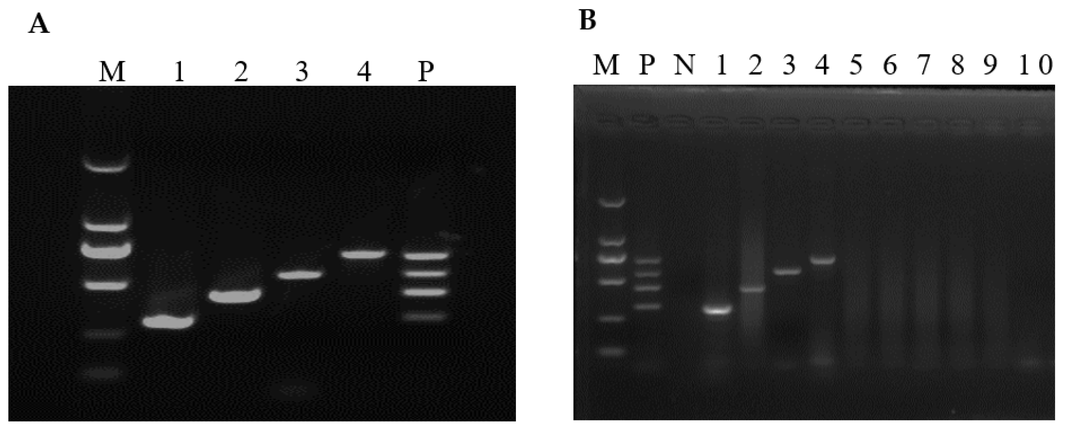
3.1. Optimized Conditions of the mRT-PCR

Before mRT-PCR reaction optimization, sRT-PCR standardization and optimum annealing temperature determination were done using gradient PCR for each virus at a temperature of 48 to 58 °C. Primers of SaV, EMCV, RVA and AstV produced an amplicon size of 305 bp, 438 bp, 570 bp and 702 bp, respectively. The annealing temperature was optimized at 50–52 °C. After optimizing the annealing temperature, each PCR reaction condition was optimized one at a time by keeping other conditions constant. After repetitive experiments, an optimum concentration of dNTP, MgCl2 and Taq polymerase was determined at 0.6 mM, 0.35 mM and 6 units, respectively. Primer concentration were optimized at 0.15, 0.113, 0.15 and 0.1 µM for SaV, EMCV, RVA and AstV, respectively. All reactions were performed at 40 µL reaction volume and 35 cycles. Each of the amplicons was visualized by electrophoresing 10 µL aliquots through 1.5% agarose gels in 1× TAE (40 mM Tris-acetate [pH 8.0], 1 mM EDTA). Using the optimized mRT-PCR conditions, all four viral fragments were amplified successfully (Figure 1A).

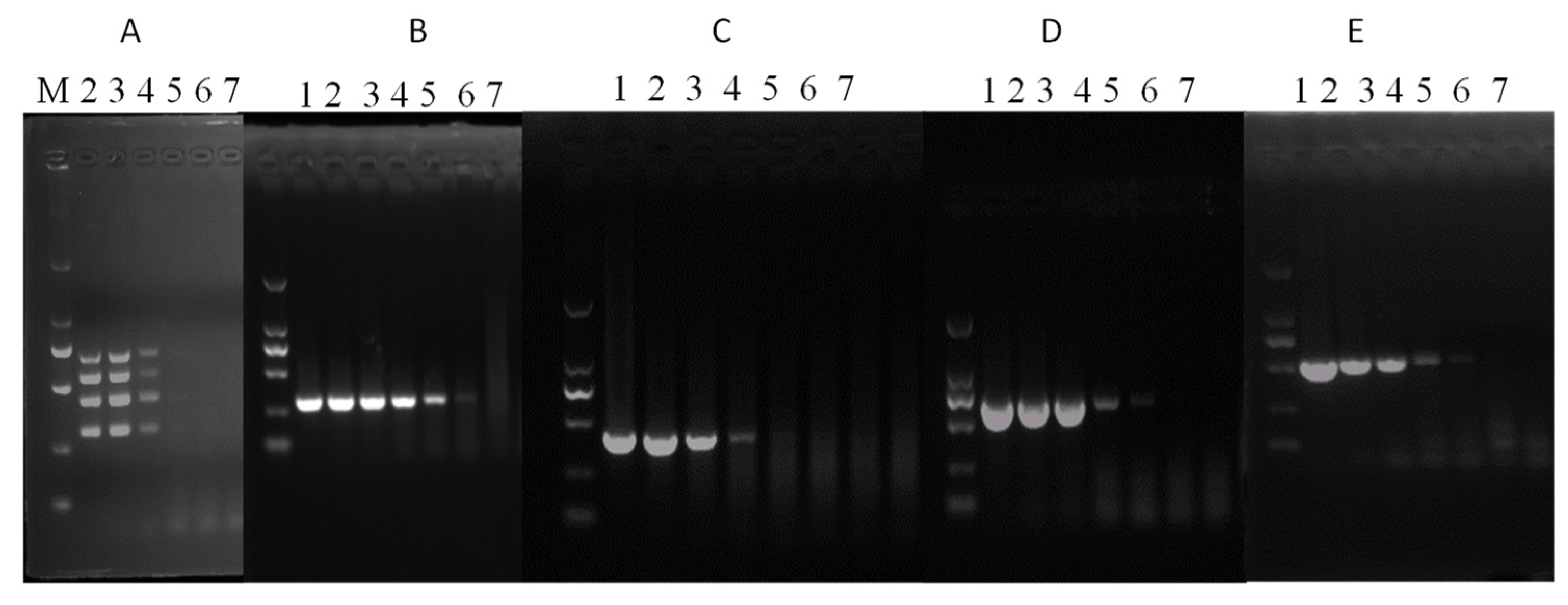
3.2. Assay Sensitivity

The sensitivity of sRT-PCR and mRT-PCR was tested using ten-fold serial dilutions of the SaV, EMCV, RVA and AstV plasmid constructs. It was found that the developed mRT-PCR assay could simultaneously detect up to 2.5 × 10³ copies of each template, while single PCR could detect 2.5 × 101, 2.5 × 10³, 2.5 × 10², and 2.5 × 10² copies of SaV, EMCV, RVA and AstV, respectively (Figure 2).

3.3. Assay Specificity

The specificity of each primer pair was determined using sRT-PCR and mRT-PCR (Figure 1B). Both sRT-PCR and mRT-PCR were found specific for the target viral agent because no amplicons were produced with other viral agents, including PEDV, SVV, TGEV, PRRSV, PDCoV, and PSV (Figure 1B, lanes 5–10). All positive amplicons were sequenced to check the presence of potential false-positive results. Basic Local Alignment Search Tool (BLAST; http://www.ncbi.nlm.nih.gov, accessed on 10 December 2020) was used to search homologous sequences and analyses of the sequences obtained corresponding to 305 bp for SaV, 438 bp for EMCV, 570 bp for RVA, and 702 bp AstV, respectively, were found to be identical to each virus. This result showed that the developed mRT-PCR assay is specific.

3.4. Assay Reproducibility

Four mRT-PCR reactions were performed at different times to assess inter-assay reproducibility using the same reaction conditions. A freshly extracted plasmid was used as a template for each PCR reaction. Each of the four mRT-PCR reactions showed a similar result (data not shown), indicating the reproducibility of the assay. Besides inter-assay reproducibility, intra-assay reproducibility was checked by carrying out triplicate mRT-PCR reactions (data not shown). The developed mRT-PCR assay’s intra-assay and inter-assay reproducibility tests showed that it could be used to detect potentially zoonotic swine viruses.

3.5. Detection of Field Samples

A total of 280 porcine fecal samples were tested for SaV, EMCV, RVA and AstV using the developed mRT-PCR assay. One hundred of the 280 tested samples were found to be positive for at least one virus. A positive rate of 24.6%, 5%, 4.3% and 17.5% was observed for SaV, EMCV, RVA and AstV (Table 2). Though only RVA showed a statistically significant difference in prevalence based on age (p < 0.05, X2 = 8), as swine age increases, the remaining three viruses showed a slight decrease in prevalence (Table 2). A statistically significant difference (p < 0.05, X2 = 5.3) in prevalence was found between diarrheic and non-diarrheic pigs. In contrast, the prevalence of the other three viruses did not show any statistically significant difference between diarrheic and non-diarrheic pigs.
With varying degrees of double and triple infection, a mixed infection rate of 6.4% (18/280) was detected. Out of the 100 samples found positive for at least one virus, a coinfection rate of 5% (SaV, RVA and AstV), 4% (SaV, EMCV and AstV), 1% (EMCV, RVA and AstV), 8% (SaV and Astv), 3% (SaV and EMCV), 2% (RVA and AstV) and 1% (RVA and SaV) was observed (Figure 3).
For checking the reliability of the mRT-PCR assay, 130 samples containing 40 samples from diarrheic pigs were selected and further tested using sRT-PCR for SaV, EMCV, RVA and AstV. Except for SaV (Table 3), samples positive for each virus using sRT-PCR test also showed similar positive results when tested by mRT-PCR. Some of the mRT-PCR positive samples were also checked by sequencing. The concordance of sequence results were then checked by subjecting them to NCBI nucleotide blast and no false-positive results were found.
SaV infection is the most common of the four viruses tested, while RVA infection is the least common. Test results of mRT-PCR showed 98.5% agreement with the test results of sRT-PCR. This result indicated that, like sRT-PCR, the developed mRT-PCR is sensitive enough to detect field samples.

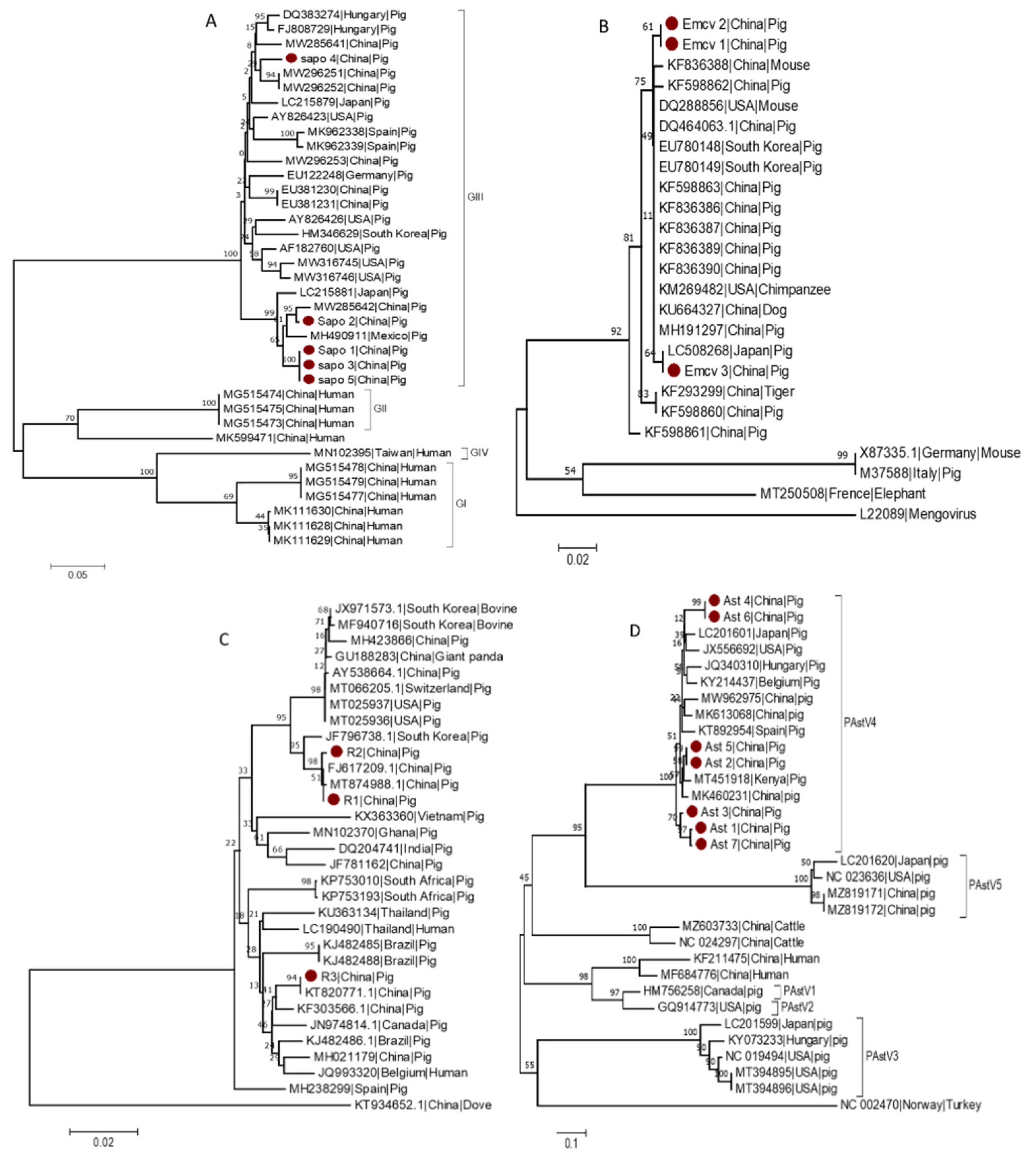
3.6. Phylogenetic Analysis

Phylogenetic trees were constructed using the neighbor-joining method. When constructing the phylogenetic trees, geographic location, host type, and virus genotype were all considered.
Within the Caliciviridae family, based on their complete VP1 nucleotide sequences, sapoviruses could be classified into 15 genogroups, eight of which have been detected in swine (GIII, GV–GXI) [30,31], but only the GI, GII, GIV, and GV genogroups are known to infect humans [30,32,33]. To investigate the genetic diversity of several of the sequences identified in this study, a phylogenetic tree for SaV was constructed based on the partial sequence of the ORF1 region using 5 identified strains (Sapo 1 to Sap 5) and 32 reference strains. The SaV strains identified in this study named (Sapo 1, Sapo 2, Sapo 3 and Sapo 5) clustered within the GIII genogroup and were found to be closely related to other GIII SaV strains detected in China (MW285642) and Mexico (MH490911). Whereas Sapo 4, which is also classified as a GIII genogroup member, formed a separate clade (Figure 4A).
EMCV is a Picornaviridae virus that infects many animals, including pigs, mice, primates, and humans [34]. The EMCV strains isolated from various animals, for example, pigs and rats, demonstrated a high degree of homology [35]. Thus, to investigate the genetic variability of the detected EMCV strains, a phylogenetic tree for EMCV was constructed based on the partial sequence of the 3D region using three identified strains (EMCV 1 to EMCV 3) and 22 reference strains from GenBank. According to the partial sequence of the 3D region, EMCV 3 detected in this study clustered with other EMCV strains from Japan (LC508268), whereas EMCV 1 and EMCV 2 clustered separately and more closely related to strains from China (Figure 4B).
A phylogenetic tree for RVA was constructed based on the partial sequence of the VP6 region using three identified strains (R1 to R3) and 30 reference strains from GenBank. The RVA variants identified in this study clustered into two distinct clades, with R1 and R2 belonging to one clade and R3 to another clade. However, all RVA variants identified in this study are more closely related to RVA previously described from China (MT874988, FJ617209 and KT82077), implying that they may have originated from the common ancestor. R1 and R2 clustered with MT874988 and FJ617209, previously described from China, whereas R3 clustered with KT820771, another Chinese RVA strain (Figure 4C).
Based on the partial sequence of the ORF1ab region, a phylogenetic tree for AstV was constructed using three identified strains (Ast 1 to Ast 7) and 25 reference strains from GenBank. All AstV variants detected during this study clustered under AstV4. Furthermore, Ast 1, 2, 3, 5 and Ast 7 clustered closely with AstV from China (MK460231) and Kenya (MT451918), whereas Ast 4 and Ast 6 clustered independently and close to strain from the USA (JX556692) (Figure 4D).

4. Discussion

Despite the presence of a plethora of different types of diagnostic assays, nucleic acid-based assays offer an added advantage of high specificity and sensitivity with the possibility of a lower detection limit [36]. Recently, mRT-PCR is being used as one of the most important diagnostic methods for rapidly and simultaneously detecting viral pathogens. Additionally, hence, different mRT-PCR assays for simultaneously detecting nine [37], six [25,38], five [39], four [23,24,26,40], three [41] and two [42,43] swine enteric viruses have been developed. Due to the asymptomatic nature of SaV, EMCV, RVA, and AstV infections, these viruses have received insufficient attention, and no single diagnostic assay capable of simultaneously detecting these viruses from porcine fecal samples has been developed.
A mRT-PCR assay capable of simultaneously detecting SaV, EMCV, RVA, and AstV RNA from porcine fecal samples was developed in this study. Our assay and other previously developed assays [23,26] for other viruses suggest that sRT-PCR may be more specific than mRT-PCR, but the improvement in turnaround time and cost-effectiveness would compensate for this minor reduction in sensitivity. Yet, compared to sRT-PCR, mRT-PCR is more economical and rapid. The sensitivity of the developed mRT-PCR assay using plasmids containing the specific viral target fragments was 2.5 × 10³ copies for each template. Similarly, Zhao et al. [23] reported a similar mRT-PCR assay sensitivity of 2.17 × 10³, 2.1 × 10³, 1.74 × 10⁴ and 1.26 × 10⁴ for porcine epidemic diarrhea virus, transmissible gastroenteritis virus, RVA, and porcine circovirus 2, respectively. Thus, the currently developed mRT-PCR assay has similar sensitivity with Hu et al. [40] and Liu et al. [26], a higher sensitivity compared to Day et al. [44] and Cagirgan and Yazici [45], and lower sensitivity compared to Liu et al. [39]. Further, 130 fecal samples were tested for checking the difference in sensitivity of sRT-PCR and mRT-PCR assays. Test results of mRT-PCR showed an overall concordance rate of 98.5% (128/130) (Table 3) to the test results of sRT-PCR. Besides sensitivity, test results of specificity and reproducibility of this assay indicate that similar to the sRT-PCR, the developed mRT-PCR could be employed to detect SaV, EMCV, RVA, and AstV from porcine fecal samples. Similarly, previous reports [25,37] and others suggested that mRT-PCR could be a highly sensitive and specific assay that could be used for the rapid detection of swine viruses from fecal specimens.
To further confirm the validity of the developed mRT-PCR assay, 280 porcine fecal samples were tested and the positive rates of SaV, EMCV, RVA and AstV was found to be 24.6% (69/280), 5% (14/280), 4.3% (12/280), and 17.5% (49/280), respectively. This shows that, compared to the other two viruses, SaV has the highest positive rate and RVA the lowest positive rate. Similarly, SaV, EMCV, RVA, and AstV have been detected in diarrheic and non-diarrheic swine feces [5,11,12,13,15], highlighting the importance of detecting these viruses in swine farms. In swine, coinfection of enteric viruses is common. In this study, coinfection rate of 5% (SaV, RVA and AstV), 4% (SaV, EMCV and AstV),1% (EMCV, RVA and AstV), 8% (SaV and Astv), 3% (SaV and EMCV), 2% (RVA and AstV) and 1% (RVA and SaV) has been observed. Similar to the current study, coinfections of pigs with different enteric viruses have been reported from China [25,46], the United States [47] and Belgium [48].
A phylogenetic analysis was also made to explore further the epidemiologic characteristics of the detected viruses. The phylogenetic analysis revealed that the SaV detected in this study belongs to the GIII genogroup. Similar to this study, previous research [49,50] indicated that from the eight SaV genogroups detected in swine [30,31], the GIII genogroup is the most prevalent in China. Phylogenetic analysis of the partial sequence of the 3D region of EMCV, similar to previous studies from China [51,52], revealed that the EMCV variants detected in this study clustered into two groups. Yuan, Song, Zhang, Zhang and Sun [52] assigned the EMCV-30 strain (DQ288856) from the USA and the Korean strains K3 (EU780148) and K11 (EU780149) to the EMCV group one lineage while strains PV2 (X87335) and D variant (M37588) to group two lineage. Based on Yuan, Song, Zhang, Zhang and Sun [52], three of the EMCV strains identified in this study may belong to group one lineage, regardless of their clustering pattern. Based on the VP7 gene sequence, Group A rotaviruses are classified into 36 G genotypes, of which 12 G-genotypes (G1–G6, G8–G12, and G26) have been identified in porcine [18,53]. All three RVA strains detected in this study were found closely grouped with strains NJ2012 (MT874988.1) and TA-3-1 (KT820771.1), both of which are G9 genotypes, implying that the RVA strains detected in this study could be G9 genotype. According to phylogenetic analysis, the AstV variants detected in this study belong to AstV4. Similarly, previous reports from the United States [54], Thailand [55] and Slovakia [8] indicated that AstV4 is the most prevalent type of AstV. In contrast, a higher prevalence of AstV2 from China [56] and AstV5 from China’s Hunan province [57] have also been observed, highlighting the importance of further AstV epidemiology studies. These findings lend credence to the notion that coinfection of these viruses is common in swine. Through recombination and mutations, the coinfection of viruses accelerates the evolution of coinfected viruses [58,59], allowing more virulent virus strains to emerge. As a result, the developed mRT-PCR assay could play a critical role in controlling and preventing SaV, EMCV, RVA, and AstV through early and accurate detection.

5. Conclusions

The assay sensitivity, specificity, and repeatability results demonstrated that the developed mRT-PCR could be employed to detect SaV, EMCV, RVA, and AstV simultaneously and rapidly in swine fecal specimens. Potential zoonotic swine viruses such as SaV, EMCV, RVA, and AstV are prevalent in Heilongjiang province, China. These viruses frequently coinfect pigs, and detecting them quickly and cost-effectively may necessitate specialized diagnostic techniques such as mRT-PCR. The current mRT-PCR may assist in lowering the cost and time associated with sample processing and testing during large-scale field sample screening for SaV, EMCV, RVA, and AstV. As a result, this assay may help control and prevent potential zoonotic swine viruses by detecting them early and accurately.

Author Contributions

Conceptualization, Y.W. and H.C.; methodology, G.M.W., H.Z. and Y.M.I.; formal analysis, G.M.W.; investigation, G.M.W., H.Z., Y.X., Y.M.I., W.Z., L.Z., Y.P. and W.W.; writing—original draft preparation, G.M.W.; writing—review and editing, G.M.W., L.F. and Y.W.; supervision, Y.W. and L.F.; project administration, Y.W. and H.C.; fund acquisition, Y.W. All authors have read and agreed to the published version of the manuscript.

Funding

This research was supported by the National Key Research & Development Program of China (2021YFF0703100) and Heilongjiang province (GZ20210010).

Institutional Review Board Statement

Not applicable.

Informed Consent Statement

Informed consent was obtained from farm owners for each of the animals included in this study.

Data Availability Statement

The data presented in this study can be found in the manuscript.

Conflicts of Interest

The authors declare no conflict of interest.

References

  1. Zhou, P.; Fan, H.; Lan, T.; Yang, X.-L.; Shi, W.-F.; Zhang, W.; Zhu, Y.; Zhang, Y.-W.; Xie, Q.-M.; Mani, S. Fatal swine acute diarrhoea syndrome caused by an HKU2-related coronavirus of bat origin. Nature 2018, 556, 255–258. [Google Scholar] [CrossRef] [PubMed]
  2. Ge, F.-F.; Yang, D.-Q.; Ju, H.-B.; Wang, J.; Liu, J.; Liu, P.-H.; Zhou, J.-P. Epidemiological survey of porcine epidemic diarrhea virus in swine farms in Shanghai, China. Arch. Virol. 2013, 158, 2227–2231. [Google Scholar] [CrossRef] [PubMed]
  3. Mandelik, R.; Sarvas, M.; Jackova, A.; Salamunova, S.; Novotny, J.; Vilcek, S. First outbreak with chimeric swine enteric coronavirus (SeCoV) on pig farms in Slovakia–lessons to learn. Acta Vet. Hung. 2018, 66, 488–492. [Google Scholar] [CrossRef] [PubMed]
  4. Bank-Wolf, B.R.; König, M.; Thiel, H.-J. Zoonotic aspects of infections with noroviruses and sapoviruses. Vet. Microbiol. 2010, 140, 204–212. [Google Scholar] [CrossRef] [Green Version]
  5. Midgley, S.E.; Bányai, K.; Buesa, J.; Halaihel, N.; Hjulsager, C.K.; Jakab, F.; Kaplon, J.; Larsen, L.E.; Monini, M.; Poljšak-Prijatelj, M. Diversity and zoonotic potential of rotaviruses in swine and cattle across Europe. Vet. Microbiol. 2012, 156, 238–245. [Google Scholar] [CrossRef] [PubMed]
  6. Saif, L.J. Comparative pathogenesis of enteric viral infections of swine. In Mechanisms in the Pathogenesis of Enteric Diseases 2; Paul, P.S., Francis, D.H., Eds.; Springer: Boston, MA, USA, 1999; Volume 473, pp. 47–59. [Google Scholar] [CrossRef]
  7. Zhou, W.; Ullman, K.; Chowdry, V.; Reining, M.; Benyeda, Z.; Baule, C.; Juremalm, M.; Wallgren, P.; Schwarz, L.; Zhou, E. Molecular investigations on the prevalence and viral load of enteric viruses in pigs from five European countries. Vet. Microbiol. 2016, 182, 75–81. [Google Scholar] [CrossRef] [PubMed]
  8. Salamunova, S.; Jackova, A.; Mandelik, R.; Novotny, J.; Vlasakova, M.; Vilcek, S. Molecular detection of enteric viruses and the genetic characterization of porcine astroviruses and sapoviruses in domestic pigs from Slovakian farms. BMC Vet. Res. 2018, 14, 313. [Google Scholar] [CrossRef] [PubMed]
  9. Pan, Y.; Tian, X.; Qin, P.; Wang, B.; Zhao, P.; Yang, Y.-L.; Wang, L.; Wang, D.; Song, Y.; Zhang, X. Discovery of a novel swine enteric alphacoronavirus (SeACoV) in southern China. Vet. Microbiol. 2017, 211, 15–21. [Google Scholar] [CrossRef] [PubMed]
  10. Theuns, S.; Conceicao-Neto, N.; Zeller, M.; Heylen, E.; Roukaerts, I.D.; Desmarets, L.M.; Van Ranst, M.; Nauwynck, H.J.; Matthijnssens, J. Characterization of a genetically heterogeneous porcine rotavirus C, and other viruses present in the fecal virome of a non-diarrheic Belgian piglet. Infect. Genet. Evol. 2016, 43, 135–145. [Google Scholar] [CrossRef] [PubMed]
  11. Chao, D.Y.; Wei, J.Y.; Chang, W.F.; Wang, J.; Wang, L.C. Detection of multiple genotypes of calicivirus infection in asymptomatic swine in Taiwan. Zoonoses Public Health 2012, 59, 434–444. [Google Scholar] [CrossRef] [PubMed]
  12. Machnowska, P.; Ellerbroek, L.; Johne, R. Detection and characterization of potentially zoonotic viruses in faeces of pigs at slaughter in Germany. Vet. Microbiol. 2014, 168, 60–68. [Google Scholar] [CrossRef]
  13. Monini, M.; Di Bartolo, I.; Ianiro, G.; Angeloni, G.; Magistrali, C.F.; Ostanello, F.; Ruggeri, F.M. Detection and molecular characterization of zoonotic viruses in swine fecal samples in Italian pig herds. Arch. Virol. 2015, 160, 2547–2556. [Google Scholar] [CrossRef]
  14. Dufkova, L.; Scigalkova, I.; Moutelikova, R.; Malenovska, H.; Prodelalova, J. Genetic diversity of porcine sapoviruses, kobuviruses, and astroviruses in asymptomatic pigs: An emerging new sapovirus GIII genotype. Arch. Virol. 2013, 158, 549–558. [Google Scholar] [CrossRef] [PubMed]
  15. Mendenhall, I.H.; Smith, G.J.; Vijaykrishna, D. Ecological drivers of virus evolution: Astrovirus as a case study. J. Virol. 2015, 89, 6978–6981. [Google Scholar] [CrossRef] [PubMed] [Green Version]
  16. Czechowicz, J.; Huaman, J.L.; Forshey, B.M.; Morrison, A.C.; Castillo, R.; Huaman, A.; Caceda, R.; Eza, D.; Rocha, C.; Blair, P.J. Prevalence and risk factors for encephalomyocarditis virus infection in Peru. Vector-Borne Zoonotic Dis. 2011, 11, 367–374. [Google Scholar] [CrossRef] [PubMed]
  17. Martella, V.; Lorusso, E.; Banyai, K.; Decaro, N.; Corrente, M.; Elia, G.; Cavalli, A.; Radogna, A.; Costantini, V.; Saif, L. Identification of a porcine calicivirus related genetically to human sapoviruses. J. Clin. Microbiol. 2008, 46, 1907–1913. [Google Scholar] [CrossRef] [PubMed] [Green Version]
  18. Vlasova, A.N.; Amimo, J.O.; Saif, L.J. Porcine Rotaviruses: Epidemiology, Immune Responses and Control Strategies. Viruses 2017, 9, 48. [Google Scholar] [CrossRef] [PubMed]
  19. Lipkin, W.I.; Firth, C. Viral surveillance and discovery. Curr. Opin. Virol. 2013, 3, 199–204. [Google Scholar] [CrossRef] [Green Version]
  20. Akdag, A.I.; Gupta, S.; Khan, N.; Upadhayay, A.; Ray, P. Epidemiology and clinical features of rotavirus, adenovirus, and astrovirus infections and coinfections in children with acute gastroenteritis prior to rotavirus vaccine introduction in Meerut, North India. J. Med. Virol. 2020, 92, 1102–1109. [Google Scholar] [CrossRef] [PubMed]
  21. Zhang, Q.; Hu, R.; Tang, X.; Wu, C.; He, Q.; Zhao, Z.; Chen, H.; Wu, B. Occurrence and investigation of enteric viral infections in pigs with diarrhea in China. Arch. Virol. 2013, 158, 1631–1636. [Google Scholar] [CrossRef] [PubMed]
  22. Valko, A.; Marosi, A.; Csagola, A.; Farkas, R.; Ronai, Z.; Dan, A. Frequency of diarrhoea-associated viruses in swine of various ages in Hungary. Acta Vet. Hung. 2019, 67, 140–150. [Google Scholar] [CrossRef] [PubMed]
  23. Zhao, J.; Shi, B.J.; Huang, X.G.; Peng, M.Y.; Zhang, X.M.; He, D.N.; Pang, R.; Zhou, B.; Chen, P.Y. A multiplex RT-PCR assay for rapid and differential diagnosis of four porcine diarrhea associated viruses in field samples from pig farms in East China from 2010 to 2012. J. Virol. Methods 2013, 194, 107–112. [Google Scholar] [CrossRef]
  24. Yue, F.; Cui, S.; Zhang, C.; Yoon, K.J. A multiplex PCR for rapid and simultaneous detection of porcine circovirus type 2, porcine parvovirus, porcine pseudorabies virus, and porcine reproductive and respiratory syndrome virus in clinical specimens. Virus Genes 2009, 38, 392–397. [Google Scholar] [CrossRef]
  25. Ding, G.; Fu, Y.; Li, B.; Chen, J.; Wang, J.; Yin, B.; Sha, W.; Liu, G. Development of a multiplex RT-PCR for the detection of major diarrhoeal viruses in pig herds in China. Transbound. Emerg. Dis. 2020, 67, 678–685. [Google Scholar] [CrossRef] [PubMed] [Green Version]
  26. Liu, S.; Zhao, Y.; Hu, Q.; Lv, C.; Zhang, C.; Zhao, R.; Hu, F.; Lin, W.; Cui, S. A multiplex RT-PCR for rapid and simultaneous detection of porcine teschovirus, classical swine fever virus, and porcine reproductive and respiratory syndrome virus in clinical specimens. J. Virol. Methods 2011, 172, 88–92. [Google Scholar] [CrossRef] [PubMed]
  27. Untergasser, A.; Cutcutache, I.; Koressaar, T.; Ye, J.; Faircloth, B.C.; Remm, M.; Rozen, S.G. Primer3—New capabilities and interfaces. Nucleic Acids Res. 2012, 40, e115. [Google Scholar] [CrossRef] [PubMed] [Green Version]
  28. Saitou, N.; Nei, M. The neighbor-joining method: A new method for reconstructing phylogenetic trees. Mol. Biol. Evol. 1987, 4, 406–425. [Google Scholar] [CrossRef] [PubMed]
  29. Kumar, S.; Stecher, G.; Li, M.; Knyaz, C.; Tamura, K. MEGA X: Molecular evolutionary genetics analysis across computing platforms. Mol. Biol. Evol. 2018, 35, 1547. [Google Scholar] [CrossRef]
  30. Oka, T.; Lu, Z.; Phan, T.; Delwart, E.L.; Saif, L.J.; Wang, Q. Genetic characterization and classification of human and animal sapoviruses. PLoS ONE 2016, 11, e0156373. [Google Scholar] [CrossRef] [PubMed]
  31. Scheuer, K.A.; Oka, T.; Hoet, A.E.; Gebreyes, W.A.; Molla, B.Z.; Saif, L.J.; Wang, Q. Prevalence of porcine noroviruses, molecular characterization of emerging porcine sapoviruses from finisher swine in the United States, and unified classification scheme for sapoviruses. J. Clin. Microbiol. 2013, 51, 2344–2353. [Google Scholar] [CrossRef] [PubMed] [Green Version]
  32. Yinda, C.K.; Conceição-Neto, N.; Zeller, M.; Heylen, E.; Maes, P.; Ghogomu, S.M.; Van Ranst, M.; Matthijnssens, J. Novel highly divergent sapoviruses detected by metagenomics analysis in straw-colored fruit bats in Cameroon: Divergent bat sapoviruses. Emerg. Microbes Infect. 2017, 6, 1–7. [Google Scholar] [CrossRef] [Green Version]
  33. Wang, L.; Marthaler, D.; Fredrickson, R.; Gauger, P.C.; Zhang, J.; Burrough, E.R.; Petznick, T.; Li, G. Genetically divergent porcine sapovirus identified in pigs, United States. Transbound. Emerg. Dis. 2020, 67, 18–28. [Google Scholar] [CrossRef]
  34. Carocci, M.; Bakkali-Kassimi, L. The encephalomyocarditis virus. Virulence 2012, 3, 351–367. [Google Scholar] [CrossRef] [PubMed] [Green Version]
  35. Liu, H.; Li, Y.; Zhang, G.; Sang, S.; Wang, C.; Chang, H. Complete genome sequences and phylogenetic analysis of encephalomyocarditis virus strains isolated from pigs and rats origin. Infect. Genet. Evol. 2017, 55, 277–280. [Google Scholar] [CrossRef] [PubMed]
  36. Templeton, K.E.; Scheltinga, S.A.; Graffelman, A.W.; Van Schie, J.M.; Crielaard, J.W.; Sillekens, P.; Van Den Broek, P.J.; Goossens, H.; Beersma, M.F.; Claas, E.C. Comparison and evaluation of real-time PCR, real-time nucleic acid sequence-based amplification, conventional PCR, and serology for diagnosis of Mycoplasma pneumoniae. J. Clin. Microbiol. 2003, 41, 4366–4371. [Google Scholar] [CrossRef] [PubMed] [Green Version]
  37. Ogawa, H.; Taira, O.; Hirai, T.; Takeuchi, H.; Nagao, A.; Ishikawa, Y.; Tuchiya, K.; Nunoya, T.; Ueda, S. Multiplex PCR and multiplex RT-PCR for inclusive detection of major swine DNA and RNA viruses in pigs with multiple infections. J. Virol. Methods 2009, 160, 210–214. [Google Scholar] [CrossRef]
  38. Xu, X.G.; Chen, G.D.; Huang, Y.; Ding, L.; Li, Z.C.; Chang, C.D.; Wang, C.Y.; Tong, D.W.; Liu, H.J. Development of multiplex PCR for simultaneous detection of six swine DNA and RNA viruses. J. Virol. Methods 2012, 183, 69–74. [Google Scholar] [CrossRef] [PubMed]
  39. Liu, H.; Shi, K.; Sun, W.; Zhao, J.; Yin, Y.; Si, H.; Qu, S.; Lu, W. Development a multiplex RT-PCR assay for simultaneous detection of African swine fever virus, classical swine fever virus and atypical porcine pestivirus. J. Virol. Methods 2021, 287, 114006. [Google Scholar] [CrossRef] [PubMed]
  40. Hu, L.; Lin, X.Y.; Yang, Z.X.; Yao, X.P.; Li, G.L.; Peng, S.Z.; Wang, Y. A multiplex PCR for simultaneous detection of classical swine fever virus, African swine fever virus, highly pathogenic porcine reproductive and respiratory syndrome virus, porcine reproductive and respiratory syndrome virus and pseudorabies in swines. Pol. J. Vet. Sci. 2015, 18, 715–723. [Google Scholar] [CrossRef] [PubMed] [Green Version]
  41. Nakhaie, M.; Soleimanjahi, H.; Mollaie, H.R.; Arabzadeh, S.M.A. Development of multiplex reverse transcription-polymerase chain reaction for simultaneous detection of influenza A, B and adenoviruses. Iran. J. Pathol. 2018, 13, 54. [Google Scholar] [CrossRef] [PubMed]
  42. Kim, O.; Choi, C.; Kim, B.; Chae, C. Detection and differentiation of porcine epidemic diarrhoea virus and transmissible gastroenteritis virus in clinical samples by multiplex RT-PCR. Vet. Rec. 2000, 146, 637–640. [Google Scholar] [CrossRef] [PubMed]
  43. Huang, C.; Hung, J.J.; Wu, C.Y.; Chien, M.S. Multiplex PCR for rapid detection of pseudorabies virus, porcine parvovirus and porcine circoviruses. Vet. Microbiol. 2004, 101, 209–214. [Google Scholar] [CrossRef] [PubMed]
  44. Day, J.M.; Spackman, E.; Pantin-Jackwood, M. A multiplex RT-PCR test for the differential identification of turkey astrovirus type 1, turkey astrovirus type 2, chicken astrovirus, avian nephritis virus, and avian rotavirus. Avian. Dis. 2007, 51, 681–684. [Google Scholar] [CrossRef]
  45. Cagirgan, A.A.; Yazici, Z. Development of a multiplex RT-PCR assay for the routine detection of seven RNA viruses in Apis mellifera. J. Virol. Methods 2020, 281, 113858. [Google Scholar] [CrossRef]
  46. Zhao, Z.P.; Yang, Z.; Lin, W.D.; Wang, W.Y.; Yang, J.; Jin, W.J.; Qin, A.J. The rate of co-infection for piglet diarrhea viruses in China and the genetic characterization of porcine epidemic diarrhea virus and porcine kobuvirus. Acta Virol. 2016, 60, 55–61. [Google Scholar] [CrossRef] [PubMed] [Green Version]
  47. Chen, Q.; Wang, L.; Zheng, Y.; Zhang, J.; Guo, B.; Yoon, K.-J.; Gauger, P.C.; Harmon, K.M.; Main, R.G.; Li, G. Metagenomic analysis of the RNA fraction of the fecal virome indicates high diversity in pigs infected by porcine endemic diarrhea virus in the United States. Virol. J. 2018, 15, 95. [Google Scholar] [CrossRef] [PubMed] [Green Version]
  48. Theuns, S.; Vanmechelen, B.; Bernaert, Q.; Deboutte, W.; Vandenhole, M.; Beller, L.; Matthijnssens, J.; Maes, P.; Nauwynck, H.J. Nanopore sequencing as a revolutionary diagnostic tool for porcine viral enteric disease complexes identifies porcine kobuvirus as an important enteric virus. Sci. Rep. 2018, 8, 9830. [Google Scholar] [CrossRef] [PubMed] [Green Version]
  49. Li, J.; Shen, Q.; Zhang, W.; Zhao, T.; Li, Y.; Jiang, J.; Yu, X.; Guo, Z.; Cui, L.; Hua, X. Genomic organization and recombination analysis of a porcine sapovirus identified from a piglet with diarrhea in China. Virol. J. 2017, 14, 57. [Google Scholar] [CrossRef] [PubMed] [Green Version]
  50. Liu, Z.K.; Li, J.Y.; Pan, H. Seroprevalence and molecular detection of porcine sapovirus in symptomatic suckling piglets in Guangdong Province, China. Trop. Anim. Health Prod. 2014, 46, 583–587. [Google Scholar] [CrossRef] [PubMed]
  51. Chang, H.; Chen, L.; Zhao, J.; Wang, X.; Yang, X.; Yao, H.; Wang, C. Complete genome sequence of porcine encephalomyocarditis virus from an aardvark in China. Genome Announc. 2014, 2, e00017-14. [Google Scholar] [CrossRef] [PubMed] [Green Version]
  52. Yuan, W.; Song, Q.; Zhang, X.; Zhang, L.; Sun, J. Isolation and molecular analysis of porcine encephalomyocarditis virus strain BD2 from northern China. Infect. Genet. Evol. 2014, 21, 303–307. [Google Scholar] [CrossRef] [PubMed]
  53. Matthijnssens, J.; Ciarlet, M.; McDonald, S.M.; Attoui, H.; Bányai, K.; Brister, J.R.; Buesa, J.; Esona, M.D.; Estes, M.K.; Gentsch, J.R. Uniformity of rotavirus strain nomenclature proposed by the Rotavirus Classification Working Group (RCWG). Arch. Virol. 2011, 156, 1397–1413. [Google Scholar] [CrossRef] [PubMed] [Green Version]
  54. Xiao, C.-T.; Giménez-Lirola, L.G.; Gerber, P.F.; Jiang, Y.-H.; Halbur, P.G.; Opriessnig, T. Identification and characterization of novel porcine astroviruses (PAstVs) with high prevalence and frequent co-infection of individual pigs with multiple PAstV types. J. Gen. Virol. 2013, 94, 570–582. [Google Scholar] [CrossRef] [PubMed]
  55. Kumthip, K.; Khamrin, P.; Saikruang, W.; Kongkaew, A.; Vachirachewin, R.; Ushijima, H.; Maneekarn, N. Detection and genetic characterization of porcine astroviruses in piglets with and without diarrhea in Thailand. Arch. Virol. 2018, 163, 1823–1829. [Google Scholar] [CrossRef] [PubMed]
  56. Su, M.; Qi, S.; Yang, D.; Guo, D.; Yin, B.; Sun, D. Coinfection and Genetic Characterization of Porcine Astrovirus in Diarrheic Piglets in China from 2015 to 2018. Front. Vet. Sci. 2020, 7, 462. [Google Scholar] [CrossRef] [PubMed]
  57. Xiao, C.-T.; Luo, Z.; Lv, S.-L.; Opriessnig, T.; Li, R.-C.; Yu, X.-L. Identification and characterization of multiple porcine astrovirus genotypes in Hunan province, China. Arch. Virol. 2017, 162, 943–952. [Google Scholar] [CrossRef] [Green Version]
  58. Boniotti, M.B.; Papetti, A.; Lavazza, A.; Alborali, G.; Sozzi, E.; Chiapponi, C.; Faccini, S.; Bonilauri, P.; Cordioli, P.; Marthaler, D. Porcine Epidemic Diarrhea Virus and Discovery of a Recombinant Swine Enteric Coronavirus, Italy. Emerg. Infect. Dis. 2016, 22, 83–87. [Google Scholar] [CrossRef] [PubMed]
  59. Akimkin, V.; Beer, M.; Blome, S.; Hanke, D.; Höper, D.; Jenckel, M.; Pohlmann, A. New chimeric porcine coronavirus in swine feces, Germany, 2012. Emerg. Infect. Dis. 2016, 22, 1314. [Google Scholar] [CrossRef] [Green Version]
Figure 1. mRT-PCR optimization and assay specificity. M: 2000 bp marker, 1: SaV, 2: EMCV, 3: RVA and 4: AstV, P: standard plasmid template mix containing SaV, EMCV, RVA and AstV fragments. (A) Optimization of mRT-PCR using plasmids containing SaV, EMCV, RVA and AstV fragments. (B) Assay specificity. N: negative control, 1: SaV, 2: EMCV, 3: RVA, 4: AstV and 5–10: PEDV, SVV, TGEV, PRRSV, PDCoV and PSV, respectively.
Vetsci 09 00176 g001
Figure 2. mRT-PCR assay sensitivity. The sensitivity of mRT-PCR and sRT-PCR assays was checked using 10-fold serially diluted plasmids containing SaV, EMCV, RVA and AstV fragments as a template. M: DL2000 DNA Marker, 1: 2.5 × 10⁶ copies, 2: 2.5 × 10⁵ copies, 3: 2.5 × 10⁴ copies, 4: 2.5 × 10³ copies, 5: 2.5 × 10² copies, 6: 2.5 × 101 copies and 7: 2.5 × 100 copies. (A) mRT-PCR, (B) SaV, (C) EMCV, (D) RVA, and (E) AstV.
Vetsci 09 00176 g002
Figure 3. The number of single and mixed infections of Ast, EMCV, RVA and SaV. The coinfection rate of each virus was calculated using 100 samples that tested positive for at least one virus.
Vetsci 09 00176 g003
Figure 4. Phylogenetic analysis of SaV, EMCV, RVA and AstV. The strains identified in this study (marked with red dots) and reference strains from GenBank were used to construct phylogenetic trees. Scales indicate units of the number of base substitutions per site. (A) Phylogenetic tree of SaV. The tree was built using 224 positions from the ORF1 region of SaV. (B) Phylogenetic tree of EMCV. The tree was constructed using 211 positions from the 3D region of the EMCV. Mengovirus was used as an outgroup. (C) Phylogenetic tree of RVA. The tree was built based on 545 positions of the VP6 region. Rotavirus A from dove (avian) was used as an outgroup. (D) Phylogenetic tree of AstV. The tree was built based on 379 positions of the ORF1ab region. As an outgroup, Turkey (avian) Astrovirus is included.
Vetsci 09 00176 g004
Table 1. List of primers used to detect SaV, EMCV, RVA and AstV by the mRT-PCR.
PrimerSequenceTarget GeneAccession No.Target Region (bp)Amplicon Size (bp)
SaV-FAGCCAGAAGTGTTCGTGATGGORF1MK965898.1 5124–5429 306
SaV-RGGACARGTGRAGYGTGTARGG
EMCV-FCCGTCAAGTCTTCCAACCAG3DMH191297.16288–6725438
EMCV-RGCGGCTTGAACCTTCTCTATC
RVA-FGCAAACGAAGTCTTCGACATGGVP6MH308723.16–575570
RVA-RGGCGTTAATCCACATAGTYCCCA
AstV -FTTGTGGAGCTTGACTGGACCORF1abMK613068.13341–4042702
AstV-RCTGTGAGTCTTGCAGGCAGA
Table 2. Prevalence of AstV, EMCV, RVA and SaV.
Age GroupHealth StatusNumber of Samples (n)% (Number of Positive/n)
SaVEMCVPRVAAstV
PigletDiarrheic3171 (22/31)19.4 (6/31)19.4 (6/31)41.9 (13/31)
Non-Diarrheic417.3 (3/41)2.4 (1/41)2.4 (1/41)17.1 (7/41)
Sub-total7234.7 (25/72)9.7 (7/72)9.7 (7/72)27.8 (20/72)
WeanerDiarrheic1275 (9/12)0.016.7 (2/12)50 (6/12)
Non-Diarrheic3010 (3/30)13.3 (4/30)3.3 (1/30)16.7 (5/30)
Sub-total4228.6 (12/42)9.5 (4/42)7.1 (3/42)26.2 (11/42)
Fattening pigDiarrheic2080 (16/20)15 (3/20)5 (1/20)30 (6/20)
Non-Diarrheic422.4 (1/42)007.1 (3/42)
Sub-total6227.4 (17/62)4.8 (3/62)1.6 (1/62)14.5 (9/62)
SowDiarrheic1978.9 (15/19)05.3 (1/19)26.3 (5/19)
Non-Diarrheic850004.7 (4/85)
Sub-total10414.4 (15/104)0.01 (1/104)8.7 (9/104)
28024.6 (69/280)5 (14/280)4.3 (12/280)17.5 (49/280)
Table 3. Detection of four viruses in 130 field samples by sRT-PCR and mRT-PCR.
AssayNumber of Positive Samples
SaVEMCVRVAAstV
sRT-PCR298213
mRT-PCR27 *8213
* The results were similar except for two samples that were positive for SaV by sRT-PCR, but negative by mRT-PCR.
Publisher’s Note: MDPI stays neutral with regard to jurisdictional claims in published maps and institutional affiliations.

