


Feline and Canine Cutaneous Lymphocytosis: Reactive Process or Indolent Neoplastic Disease?
 , ,
, ,  and
and
Abstract
1. Introduction
2. Materials and Methods
2.1. Animals
2.2. Histopathology
2.3. Immunohistochemistry
2.4. PARR
3. Results
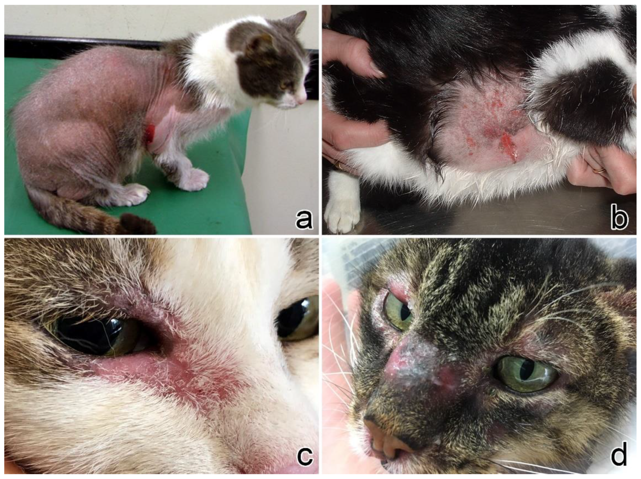
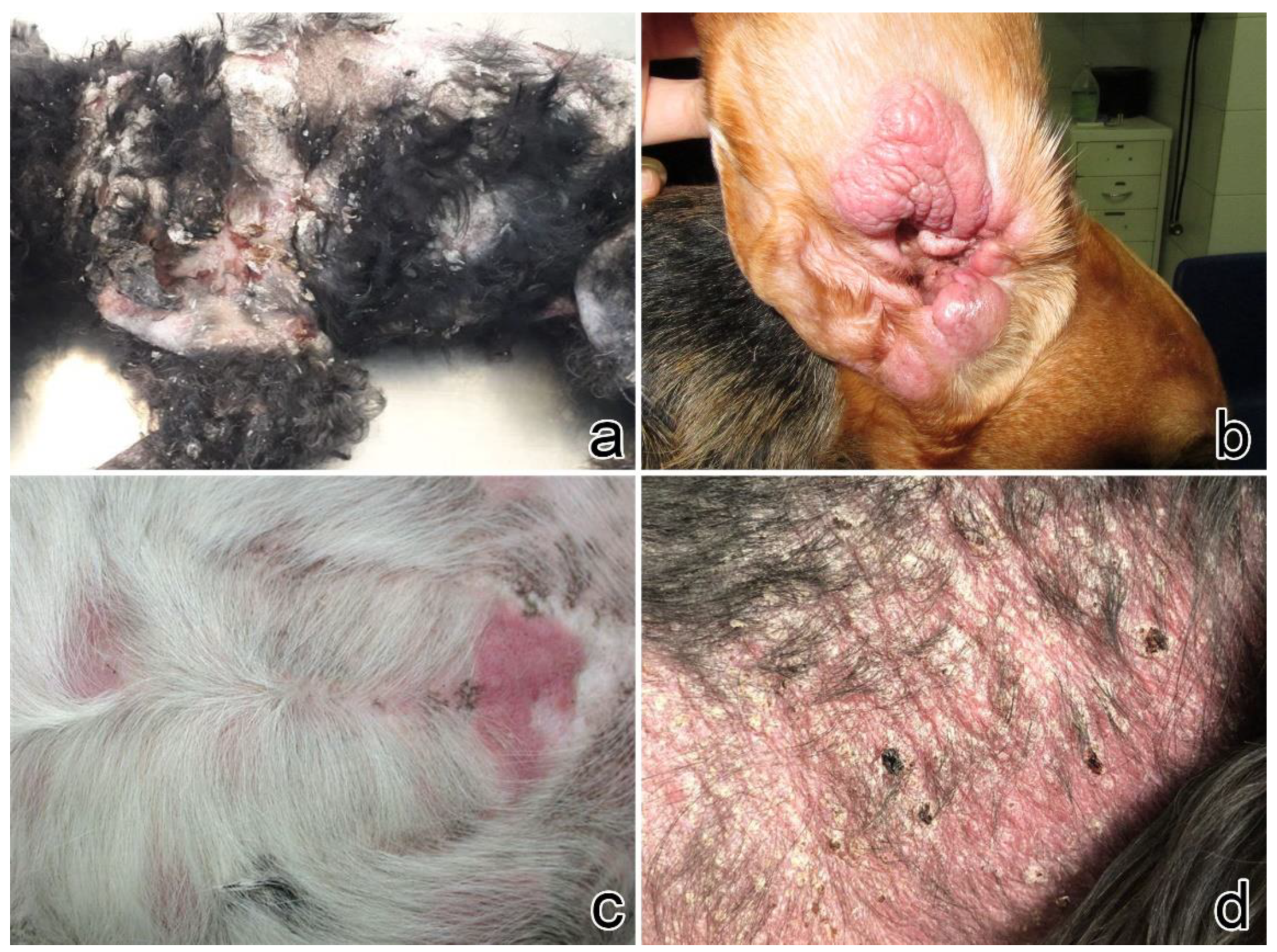
3.1. Clinical Presentation
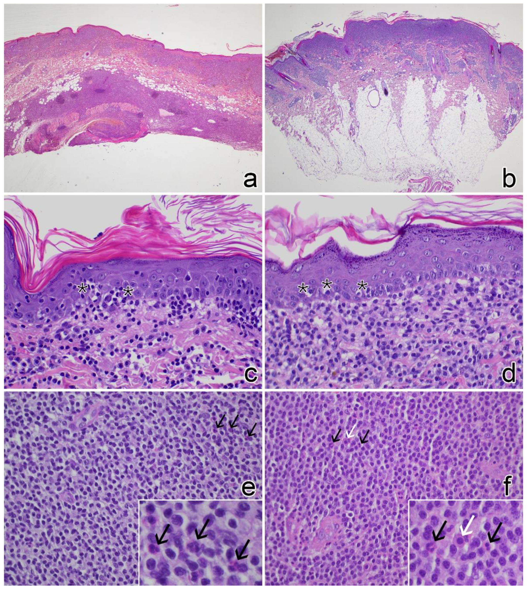
3.2. Histopathology
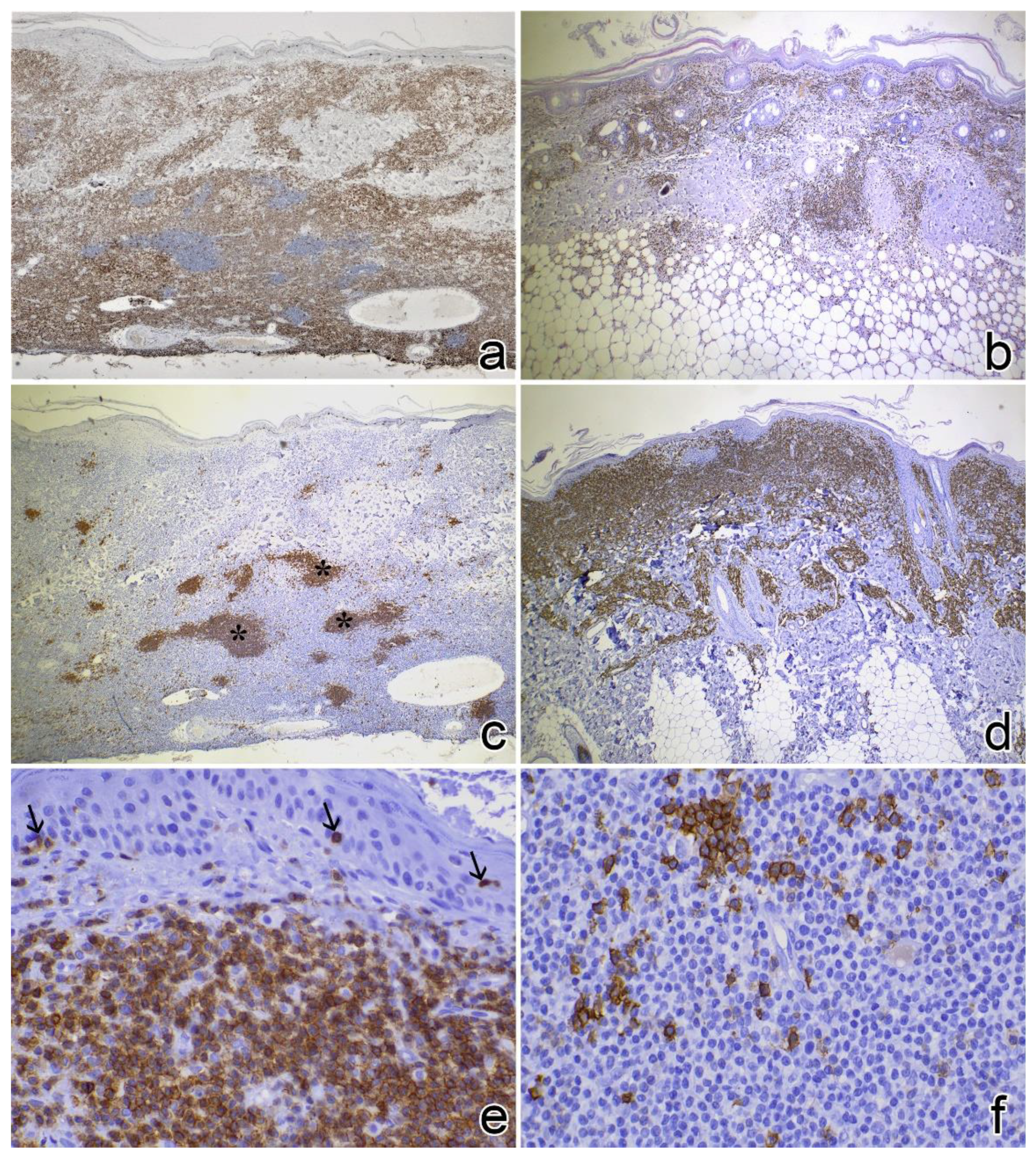
3.3. Immunophenotyping
3.4. Clonality Test
3.5. Staging and Clinical Follow-Up
4. Discussion
5. Conclusions
Supplementary Materials
Author Contributions
Funding
Institutional Review Board Statement
Informed Consent Statement
Data Availability Statement
Acknowledgments
Conflicts of Interest
References
- Ploysangam, T.; Breneman, D.L.; Mutasim, D.F. Cutaneous pseudolymphomas. J. Am. Acad. Dermatol. 1998, 38, 877–898. [Google Scholar] [CrossRef] [Scilit]
- Bergman, R. Pseudolymphoma and cutaneous lymphoma: Facts and controversies. Clin. Dermatol. 2010, 28, 568–574. [Google Scholar] [CrossRef] [Scilit] [PubMed]
- Gilliam, A.C.; Wood, G.S. Cutaneous lymphoid hyperplasias. Semin. Cutan. Med. Surg. 2000, 19, 133–141. [Google Scholar] [CrossRef] [Scilit] [PubMed]
- Gilbert, S.; Affolter, V.K.; Gross, T.L.; Moore, P.F.; Ihrke, P.J. Clinical, morphological and immunohistochemical characterization of cutaneous lymphocytosis in 23 cats. Vet. Dermatol. 2004, 15, 3–12. [Google Scholar] [CrossRef] [Scilit] [PubMed]
- Affolter, V.K.; Gross, T.L.; Moore, P.F. Indolent cutaneous T-cell lymphoma presenting as cutaneous lymphocytosis in dogs. Vet. Dermatol. 2009, 20, 577–585. [Google Scholar] [CrossRef] [Scilit] [PubMed]
- Pariser, M.S.; Gram, D.W. Feline cutaneous lymphocytosis: Case report and summary of the literature. J. Feline Med. Surg. 2014, 16, 758–763. [Google Scholar] [CrossRef] [Scilit]
- Gross, T.L. Skin Diseases of the Dog and Cat: Clinical and Histopathologic Diagnosis; Blackwell Science: Ames, IA, USA, 2005; ISBN 978-0-632-06452-6. [Google Scholar]
- Bagg, A. Immunoglobulin and T-cell receptor gene rearrangements: Minding your B’s and T’s in assessing lineage and clonality in neoplastic lymphoproliferative disorders. J. Mol. Diagn. 2006, 8, 426–429. [Google Scholar] [CrossRef] [Scilit]
- Waugh, E.M.; Gallagher, A.; Haining, H.; Johnston, P.E.J.; Marchesi, F.; Jarrett, R.F.; Morris, J.S. Optimisation and validation of a PCR for antigen receptor rearrangement (PARR) assay to detect clonality in canine lymphoid malignancies. Vet. Immunol. Immunopathol. 2016, 182, 115–124. [Google Scholar] [CrossRef] [Scilit]
- Ehrhart, E.J.; Wong, S.; Richter, K.; Zismann, V.; Grimes, C.; Hendricks, W.; Khanna, C. Polymerase chain reaction for antigen receptor rearrangement: Benchmarking performance of a lymphoid clonality assay in diverse canine sample types. J. Vet. Intern. Med. 2019, 33, 1392–1402. [Google Scholar] [CrossRef] [Scilit]
- Hristov, A.C.; Tejasvi, T.; Wilcox, R.A. Mycosis fungoides and Sézary syndrome: 2019 update on diagnosis, risk-stratification, and management. Am. J. Hematol. 2019, 94, 1027–1041. [Google Scholar] [CrossRef] [Scilit]
- Gilbert, S.; Affolter, V.K.; Schmidt, P.; Kosten, S.; Kramme, P.M.; Gross, T.L.; Ihrke, P.; Moore, P.F. Clonality studies of feline cutaneous lymphocytosis. Vet. Dermatol. 2004, 15, 24. [Google Scholar] [CrossRef] [Scilit]
- Ramos-Vara, J.A.; Kiupel, M.; Baszler, T.; Bliven, L.; Brodersen, B.; Chelack, B.; West, K.; Czub, S.; Del Piero, F.; Dial, S.; et al. Suggested Guidelines for Immunohistochemical Techniques in Veterinary Diagnostic Laboratories. J. Vet. Diagn. Investig. 2008, 20, 393–413. [Google Scholar] [CrossRef] [Scilit]
- Burnett, R.C.; Vernau, W.; Modiano, J.F.; Olver, C.S.; Moore, P.F.; Avery, A.C. Diagnosis of Canine Lymphoid Neoplasia Using Clonal Rearrangements of Antigen Receptor Genes. Vet. Pathol. 2003, 40, 32–41. [Google Scholar] [CrossRef] [PubMed]
- Mochizuki, H.; Nakamura, K.; Sato, H.; Goto-Koshino, Y.; Sato, M.; Takahashi, M.; Fujino, Y.; Ohno, K.; Uchida, K.; Nakayama, H.; et al. Multiplex PCR and Genescan analysis to detect immunoglobulin heavy chain gene rearrangement in feline B-cell neoplasms. Vet. Immunol. Immunopathol. 2011, 143, 38–45. [Google Scholar] [CrossRef] [Scilit] [PubMed]
- Moore, P.F.; Woo, J.C.; Vernau, W.; Kosten, S.; Graham, P.S. Characterization of feline T cell receptor gamma (TCRG) variable region genes for the molecular diagnosis of feline intestinal T cell lymphoma. Vet. Immunol. Immunopathol. 2005, 106, 167–178. [Google Scholar] [CrossRef] [Scilit] [PubMed]
- Keller, S.M.; Vernau, W.; Moore, P.F. Clonality Testing in Veterinary Medicine: A Review With Diagnostic Guidelines. Vet. Pathol. 2016, 53, 711–725. [Google Scholar] [CrossRef] [Scilit] [PubMed]
- Rook, K.A. Canine and Feline Cutaneous Epitheliotropic Lymphoma and Cutaneous Lymphocytosis. Vet. Clin. N. Am. Small Anim. Pract. 2019, 49, 67–81. [Google Scholar] [CrossRef] [Scilit] [PubMed]
- Siewert, J.; Pellin, M.A.; Husbands, B.D.; Curran, K.M.; Scavelli, D.; Sampene, E. Feline cutaneous lymphoma: An evaluation of disease presentation and factors affecting response to treatment. J. Feline Med. Surg. 2021, 1098612X211028837. [Google Scholar] [CrossRef] [Scilit] [PubMed]
- Fontaine, J.; Heimann, M.; Day, M.J. Cutaneous epitheliotropic T-cell lymphoma in the cat: A review of the literature and five new cases. Vet. Dermatol. 2011, 22, 454–461. [Google Scholar] [CrossRef] [Scilit]
- Mitteldorf, C.; Kempf, W. Cutaneous pseudolymphoma—A review on the spectrum and a proposal for a new classification. J. Cutan. Pathol. 2020, 47, 76–97. [Google Scholar] [CrossRef] [Scilit] [PubMed]
- Laprais, A.; Olivry, T. Is CCNU (lomustine) valuable for treatment of cutaneous epitheliotropic lymphoma in dogs? A critically appraised topic. BMC Vet. Res. 2017, 13, 61. [Google Scholar] [CrossRef] [Scilit] [PubMed]

| Case No. | Signalment | Clinical Features of the Lesions | Other Clinical Findings | ||||||||||||
|---|---|---|---|---|---|---|---|---|---|---|---|---|---|---|---|
| Breed | Sex | Age (Years) | Site | Longest Diameter (cm) | Distribution | Duration (Months) | U | Pr | A | Er | Ex | Pl | Additional Features | ||
| 1 | DSH | F | 12 | Head, neck, forelimb (shoulder) | 1.5 | Multifocal | 3 | X | Nodules | None | |||||
| 2 | DSH | M | 15 | Head (upper lip) | 4 | Focal | 6 | X | X | X | None | ||||
| 3 | DSH | M | 14 | Neck, dorsum, perineum, limbs | Diffuse | 24 | X | X | X | X | Papules, crusts | Otitis | |||
| 4 | DSH | F | 14 | Flank | 4 | Focal | 2 weeks | X | X | X | X | None | |||
| 5 | DSH | M | 9 | Head (upper lip, periocular) | 1 | Focal | 18 | X | X | X | X | None | |||
| 6 | DSH | M | 11 | Head (eyelid) | Focal | 12 | X | ||||||||
| 7 | DSH | M | 16 | Thorax-lumbar | 10 | Focal | 24 | X | X | X | X | None | |||
| 8 | DSH | F | 10 | Neck, axillae, dorsum | Multifocal | 7 | X | X | X | X | Anorexia, hyperthermia, melena, weight loss | ||||
| 9 | DSH | F | 14 | Head (periocular), hindlimb (metatarsus), tail | 5 | Multifocal | 6 | X | X | X | X | None | |||
| 10 | DSH | F | 10 | Head (upper lip), flank (bilateral) | 5 | Multifocal | 3 | X | X | X | Hyperpigmentation | None | |||
| 11 | DSH | F | 10 | Flank | Focal | 9 | X | ||||||||
| 12 | DSH | F | 13 | Forelimb (elbow) | 4 | Focal | 2 | X | X | X | None | ||||
| 13 | DSH | F | 10 | Thorax, axillae | 4 | Multifocal | 12 | X | X | X | None | ||||
| 14 | DSH | M | 7 | Head, abdomen (bilateral) | Diffuse | 3 | X | X | X | X | Weight loss, submandibular lymphadenomegaly, FIV-positive | ||||
| 15 | DSH | F | 9 | Abdomen | 1 | Focal | 2 | Nodule | None | ||||||
| 16 | DSH | M | 14 | Hindlimb (3rd digit) | 0.2 | Focal | 12 | X | X | Nodule | None | ||||
| 17 | DSH | F | 7 | Head (perilabial), neck, abdomen, trunk, hindlimb (thighs) | Diffuse | 24 | X | X | X | X | Bronchitis | ||||
| 18 | DSH | F | 13 | Neck, flank, hindlimb (thigh) | 20 | Multifocal | 60 | X | X | X | None | ||||
| 19 | DSH | F | 14 | Head, dorsum, abdomen | 30 | Multifocal | 6 | X | X | X | X | Dryness, crusts | None | ||
| Case No. | Signalment | Clinical Features of the Lesions | Other Clinical Findings | ||||||||||||
|---|---|---|---|---|---|---|---|---|---|---|---|---|---|---|---|
| Breed | Sex | Age (Years) | Site | Longest Diameter (cm) | Distribution | Duration (Months) | U | Pr | A | Er | Ex | Pl | Additional Features | ||
| 1 | Beagle | F | 13 | Abdomen, hindlimb | 2 | Multifocal | 2 weeks | X | None | ||||||
| 2 | Beagle | M | 6 | Head (ear), perianal | 3 | Multifocal | 1 | X | X | X | None | ||||
| 3 | Shar Pei | F | 12 | Thorax | 2 | Multifocal | 1 | X | X | None | |||||
| 4 | Toy poodle | F | 13 | Dorsum, flank, abdomen | 2 | Multifocal | 1 | X | X | X | X | Lymphadenomegaly | |||
| 5 | Mixed breed | M | 8 | Thorax | 7 | Focal | 5 | X | None | ||||||
| 6 | Breton | M | 11 | Neck, flank, thorax | 20 | Multifocal | 2 | X | X | X | Crusts | None | |||
| 7 | Mixed breed | F | 15 | Perianal | 8 | Focal | 1 | X | X | X | Lymphocytic leukemia, mesenchymal gastric tumour | ||||
| 8 | Mixed breed | F | 12 | Trunk, flank | 2 | Multifocal | 10 | X | X | X | X | X | Ataxia, hypoglycemic crisis | ||
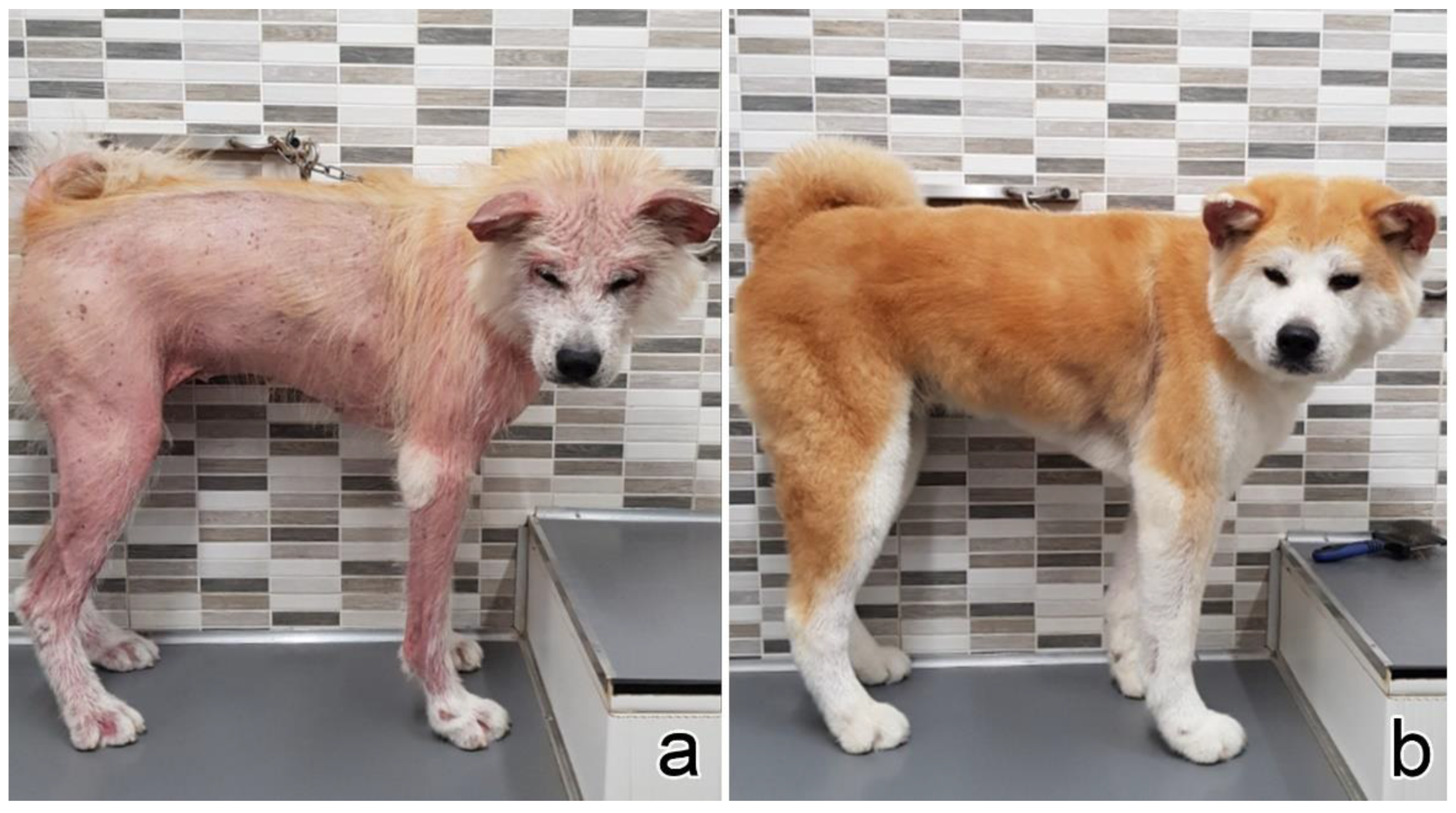
| 9 | Akita Inu | F | 9 | Axilla, forelimb (shoulder), hindlimb | Diffuse | 12 | X | X | X | Anemia, thrombocytopenia | |||||
| 10 | Dachshund | F | 8 | Abdomen | 5 | Multifocal | 2 weeks | X | X | X | None | ||||
| Case No. | PARR | Staging | Therapy | Response to Therapy | Overall Survival (Days) | Cause of Death |
|---|---|---|---|---|---|---|
| 1 | neg | neg (HB, U, Rx) | Corticosteroids | PR | 365 | Lost to follow-up |
| 2 | neg | neg (HB) | None | SD | 1080 | Unrelated (renal failure) |
| 3 | neg | n.a. | None | SD | 720 | Unrelated (renal failure) |
| 4 | neg | n.a. | None | SD | 240 | Unknown |
| 5 | neg | n.a. | Corticosteroids, chlorambucil | PR | 540 | Lost to follow-up |
| 6 | neg | n.a. | None | SD | 90 | Unrelated (renal failure) |
| 7 | neg | n.a. | Corticosteroids, chlorambucil | SD | 570 | Lost to follow-up |
| 8 | neg | n.a. | Corticosteroids | SD | 30 | Unknown |
| 9 | neg | neg (lnC) | Corticosteroids, cyclosporine | PR | 480 | Alive |
| 10 | neg | neg (HB, U) | Corticosteroids, cyclosporine | PR | 1080 | Alive |
| 11 | neg | n.a. | n.a. | n.a. | n.a. | n.a. |
| 12 | neg | n.a. | Corticosteroids | CR | 1080 | Alive |
| 13 | neg | n.a. | Chlorambucil | SD | 81 | Alive |
| 14 | neg | Submandibular lymphoadenomegaly (U, CT) | Corticosteroids | PR | 90 | Unknown |
| 15 | neg | n.a. | Surgery | CR | 365 | Unknown (not investigated visceral mass) |
| 16 | neg | n.a. | Surgery | CR | 570 | Alive |
| 17 | neg | neg (HB, U) | Corticosteroids | PR | 180 | Alive |
| 18 | neg | neg (HB, spC, livC) | Corticosteroids | PR | 1680 | Alive |
| 19 | neg | neg (HB, spC, livC) | Corticosteroids, chlorambucil | SD | 1080 | Alive |
| Case No. | PARR | Staging | Therapy | Response to Therapy | Overall Survival (Days) | Cause of Death |
|---|---|---|---|---|---|---|
| 1 | neg | n.a. | None | CR | 28 | Lost to follow-up |
| 2 | TCR/BCR clonal | neg (lnC) | Corticosteroids, cyclosporine | SD | 365 | Lost to follow-up |
| 3 | BCR clonal | neg (HB, US, spC, livC) | Surgery | CR | 630 | Alive |
| 4 | neg | neg (HB, US, spC, livC) | Lomustine, chlorambucil * | PD | 360 | Related (euthanasia for progressive disease) |
| 5 | neg | n.a. | Surgery | CR | 720 | Alive |
| 6 | BCR clonal | neg (HB, US, spC, livC) | Corticosteroids, chlorambucil | SD | 240 | Alive |
| 7 | BCR clonal | lymphocitic leukemia (HB, U) | Corticosteroids, lomustine | PR | 240 | Alive |
| 8 | neg | neg (HB, US, spC, livC) | Lomustine, chlorambucil * | PR | n.a. | Unknown |
| 9 | TCR clonal | mild anemia and thrombocytopenia (HB) | Corticosteroids, cyclosporine, chlorambucil, lomustine * | PR (after drug withdrawal) | 334 | Alive |
| 10 | BCR clonal | neg (U, lnC) | Surgery | CR | 150 | Alive |
Publisher’s Note: MDPI stays neutral with regard to jurisdictional claims in published maps and institutional affiliations. |
© 2022 by the authors. Licensee MDPI, Basel, Switzerland. This article is an open access article distributed under the terms and conditions of the Creative Commons Attribution (CC BY) license (https://creativecommons.org/licenses/by/4.0/).
Share and Cite
Albanese, F.; Abramo, F.; Marino, M.; Massaro, M.; Marconato, L.; Minoli, L.; Martini, V.; Aresu, L. Feline and Canine Cutaneous Lymphocytosis: Reactive Process or Indolent Neoplastic Disease? Vet. Sci. 2022, 9, 26. https://doi.org/10.3390/vetsci9010026
Albanese F, Abramo F, Marino M, Massaro M, Marconato L, Minoli L, Martini V, Aresu L. Feline and Canine Cutaneous Lymphocytosis: Reactive Process or Indolent Neoplastic Disease? Veterinary Sciences. 2022; 9(1):26. https://doi.org/10.3390/vetsci9010026
Chicago/Turabian StyleAlbanese, Francesco, Francesca Abramo, Michele Marino, Maria Massaro, Laura Marconato, Lucia Minoli, Valeria Martini, and Luca Aresu. 2022. "Feline and Canine Cutaneous Lymphocytosis: Reactive Process or Indolent Neoplastic Disease?" Veterinary Sciences 9, no. 1: 26. https://doi.org/10.3390/vetsci9010026
APA StyleAlbanese, F., Abramo, F., Marino, M., Massaro, M., Marconato, L., Minoli, L., Martini, V., & Aresu, L. (2022). Feline and Canine Cutaneous Lymphocytosis: Reactive Process or Indolent Neoplastic Disease? Veterinary Sciences, 9(1), 26. https://doi.org/10.3390/vetsci9010026

