Skip to Content
Veterinary SciencesVeterinary Sciences
  • Case Report
  • Open Access

22 April 2026

9 Pages

First PCR-Confirmed Case of Feline Hemoplasmosis in Bosnia and Herzegovina with a Long-Term Follow-Up


,
,
and
1
Department of Clinical Sciences of Veterinary Medicine, Veterinary Faculty, University of Sarajevo, 71 000 Sarajevo, Bosnia and Herzegovina
2
Department of Pathobiology and Epidemiology, Veterinary Faculty, University of Sarajevo, 71 000 Sarajevo, Bosnia and Herzegovina
*
Author to whom correspondence should be addressed.
This article belongs to the Section Veterinary Internal Medicine

Simple Summary

Feline patients may frequently develop anemia in association with a variety of underlying concurrent conditions, primarily as a direct consequence of metabolic disorders or infectious agents such as hemoplasmas. Additionally, anemia may arise as a part of a primary myeloproliferative disease, particularly associated with retroviral infections. Recent data from owners and veterinarians indicate that feline hemoplasmosis, despite its worldwide occurrence, is not routinely considered as the differential diagnosis for anemia in cats of Bosnia and Herzegovina. The infection is often underdiagnosed and overlooked, primarily due to the absence of reliable rapid testing methods and lack of hemoplasmosis PCR testing availability in Bosnia and Herzegovina, prior to this study. This is the first reported case of PCR-confirmed hemoplasmosis in Bosnia and Herzegovina. It further provides detailed information on patients’ signalment, clinical and molecular diagnostic approaches, treatment, and long-term follow-up. Despite being immunocompromised due to feline immunodeficiency virus infection, no recurrence of hemoplasmosis or retroviral reactivation was observed over an almost five-year period. This case also emphasizes the importance of considering hemoplasmosis in anemic cats, particularly those that do not respond to the treatment, and demonstrates the value of molecular methods in establishing definitive diagnosis.

Abstract

Hemotropic mycoplasmas are Gram-negative bacteria and are recognized as primary causative agents of feline infectious anemia, with a worldwide distribution and variable pathogenicity. This case report describes an unusual clinical presentation of hemoplasmosis in a seventeen-year-old spayed female cat that presented with severe flea infestation accompanied by marked weakness and lethargy. It was febrile, tachycardic, and tachypnoic, with prominent pallor of the mucous membranes. Laboratory analysis revealed severe, non-regenerative, microcytic and (falsely) hyperchromic anemia, with mild lymphopenia and slightly increased plateletcrit. The cat also tested positive for feline immunodeficiency virus (FIV). Blood smear examination raised suspicion of hemoplasmosis, which was later confirmed and identified by polymerase chain reaction (PCR) as Candidatus Mycoplasma haematominutum. Following the treatment of ectoparasites, the cat was successfully treated with enrofloxacin and erythropoiesis-stimulating agents (ESAs), avoiding blood transfusion, and afterwards made a full clinical recovery. Although immunocompromised due to FIV, the cat lived for an additional five years without further relapses. To the best of the authors’ knowledge, this is the first documented and PCR-confirmed case of feline hemoplasmosis in Bosnia and Herzegovina. It also highlights the need for conducting a larger study in this region to evaluate hemoplasma prevalence in this species.

1. Introduction

Hemotropic mycoplasmas are Gram-negative bacteria, also referred to as hemoplasmas, and are recognized as causative agents of feline infectious anemia. Due to their hemotropic nature, they parasitize the surface of red blood cells [1]. The three most common feline hemoplasmas identified to date are Mycoplasma haemofelis (Mhf), Candidatus Mycoplasma haematominutum (CMhm), and Candidatus Mycoplasma turicense (CMt) [1,2,3]. These organisms have been reported worldwide [1]. The highest prevalence of feline hemoplasmosis in Europe has been reported in Portugal (43.43% and 27.1%) [4,5]. The most commonly reported hemoplasma in European cats was CMhm, with the lowest prevalence noted in Catalonia, Spain (6/135; 4.4%) [6] and the highest in Portugal (33/320; 41.56%) [4]. Among the other European countries following the same trend of CMhm being the most frequently identified hemoplasma are the Czech Republic (CMhm 35.6%) [7], Turkey (CMhm 17.7%) [8], Latvia (CMhm 15%) [9], Northern Serbia (CMhm 12.6%) [10], and Greece (CMhm 10.3%) [11]. Although Mhf, CMhm, and CMt are widespread among wild cats in Europe [12], none of these hemoplasmas were identified in European wild cats in Bosnia and Herzegovina [13]. Phylogenetic analyses revealed that those identified in four individuals instead represent two different nucleotide sequences: type A forms a separate clade genetically distinct from the haematominutum group (Candidatus Mycoplasma haematoparvum and Candidatus Mycoplasma haematominutum) and, in contrast, type B belongs to the haemofelis group (uncultured Mycoplasma sp. from raccoons and feline hemoplasmas, such as Candidatus Mycoplasma turicensis). These hemoplasmas may represent previously undescribed and novel species [13]. Transmission occurs primarily via horizontal routes [1,2], although vertical transmission has also been documented [14]. Arthropods, particularly fleas (Ctenocephalides felis), are implicated as major vectors [15]. Iatrogenic transmission through blood transfusion has also been reported [3]. Older cats are considered as a target group due to their weaker immune response. However, infections in these cats are often subclinical, with CMhm being the most commonly identified hemoplasma. In contrast, younger cats tend to show more overt clinical signs due to their naïve immune system and susceptibility to the more pathogenic Mhf [16]. Male cats are more predisposed to infection due to territorial aggression, with saliva identified as a potential source of transmission [17]. Consequently, outdoor lifestyle and the aforementioned pathways are considered risk factors for hemoplasma infection [1,2,3]. Mhf is the most pathogenic hemoplasma, capable of inducing fatal hemolytic anemia in acutely infected cats, even in the absence of concurrent disease [1]. Chronic infection is typically mild, particularly during the carrier phase, and may occur without clinically evident anemia [1]. Candidatus species are generally less pathogenic [1,18], but can cause clinical disease in association with other infectious agents or retroviral immunodeficiency, such as feline immunodeficiency virus (FIV) [19,20,21], and less frequently Feline Leukemia Virus (FeLV) [2]. CMhm infection concurrent with FeLV has been linked to myeloproliferative disorders [22]. Moreover, CMhm alone has been shown to cause anemia [23]. Clinical manifestations of hemoplasmosis include moderate to severe hemolytic anemia, often accompanied by pallor of the mucous membranes [1]. Hemolysis may occur extravascularly [24] or intravascularly [25]. Despite the hemolytic nature of the anemia, icterus is rarely observed [1,2]. Severe cases may present with tachycardia, tachypnea, anorexia, dehydration, and intermittent pyrexia. Clinical signs, combined with severe anemia, risk factors, and medical history, often place hemoplasmosis high on the differential diagnosis list [1,2]. Definitive diagnosis requires PCR testing, as blood smear examination is considered insensitive due to fluctuations in bacteremia and its inability to reliably differentiate hemoplasmas [1]. This case report describes the first clinically evident hemoplasma infection confirmed by PCR in a cat in Bosnia and Herzegovina, with complete recovery.

2. Case Description

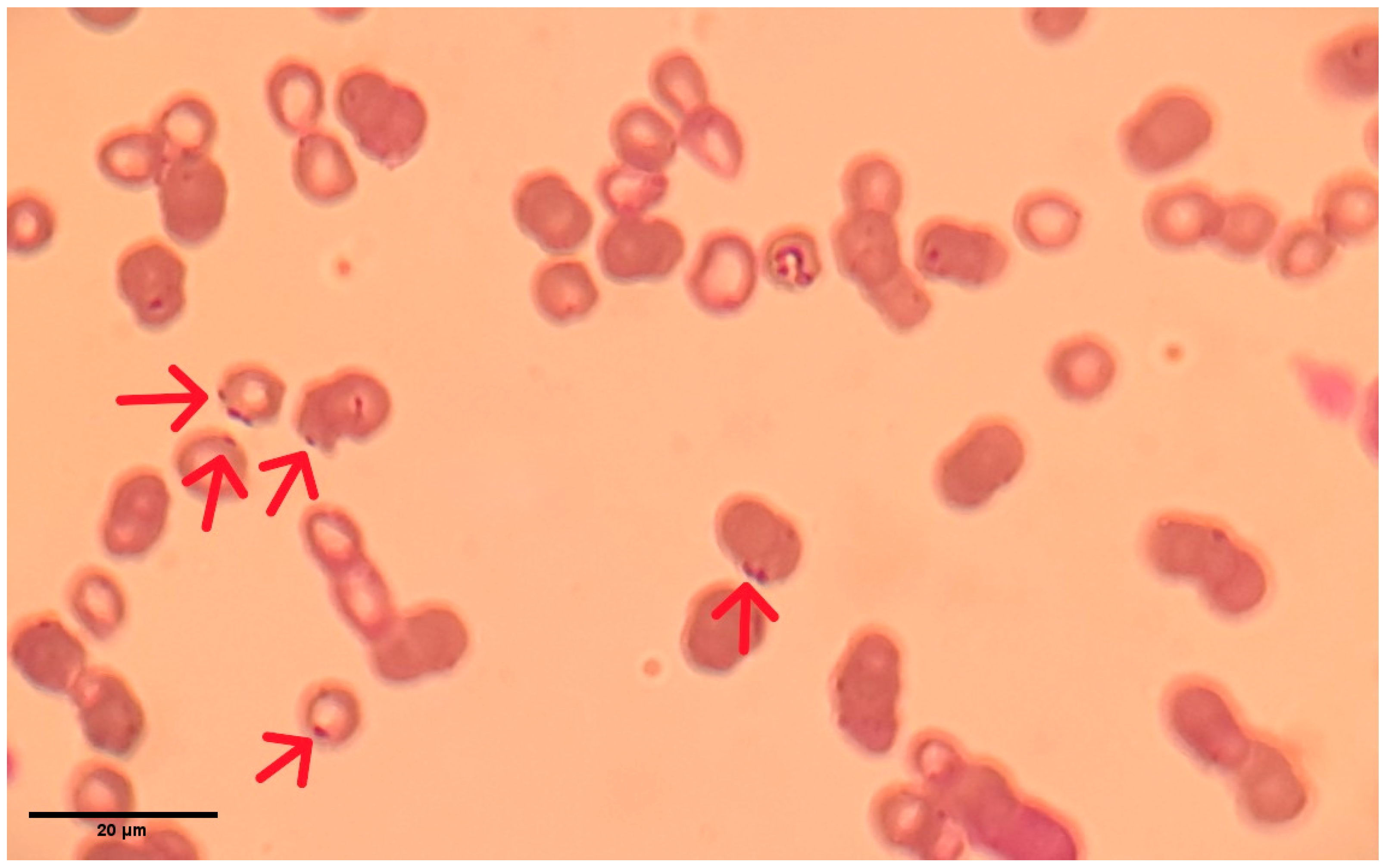
A 17-year-old, domestic shorthaired, spayed female cat was referred to the Clinic for Internal Diseases, Oncology, and Emergency Medicine of Small Animals, Veterinary Faculty, University of Sarajevo, for abdominal ultrasonography due to severe lethargy and anorexia. The interval from onset to first visit was two days, following referral by the primary veterinarian. At presentation, the cat was recumbent, very weak, lethargic, and depressed. Consequently, the owner consented to a complete clinical examination, diagnostics, and treatment. The cat had been kept strictly indoors for the past three years, following 14 years of outdoor access. It was the only cat in the household. On physical examination, severe flea infestation was noted. The mucous membranes were markedly pale, rendering capillary refill time immeasurable. The cat was febrile (39.7 °C), dehydrated (5%), and tachycardic with a weak pulse, and it had a mildly increased respiratory rate. The body condition score was 3/9, with moderate sarcopenia. Thoracic auscultation revealed normal rhythm with accentuated cardiac sounds and vesicular breath sounds. Abdominal palpation was non-painful and revealed no abnormalities. A complete blood count (Idexx ProCyte Dx®, IDEXX Laboratories Inc., Westbrook, ME, USA) demonstrated severe non-regenerative anemia (hematocrit 6%; reference range 30.3–52.3%), appearing falsely hyperchromic due to pronounced microcytosis. Mild lymphopenia was present, and plateletcrit (PCT%) was slightly elevated (Table 1). Serological testing using the SNAP FIV/FeLV Combo Test (IDEXX Laboratories Inc.) revealed that the cat tested positive for feline immunodeficiency virus (FIV) and negative for Feline Leukemia Virus (FeLV). Serum biochemistry (Catalyst One Chemistry Analyzer, IDEXX Laboratories Inc.) was within reference ranges. Abdominal ultrasonography showed no abnormalities. Microscopic examination of a Diff-Quik-stained blood smear revealed reduced erythrocyte number, microcytosis, and numerous dark blue microorganisms adherent to erythrocyte membranes (Figure 1). Based on clinical and laboratory findings, hemoplasmosis was suspected. The blood sample was submitted for PCR analysis on the same day, prior to initiation of treatment. Antimicrobial therapy (enrofloxacin) was started only after sample submission, once confirmation was obtained that the laboratory had received the sample, thereby avoiding any potential interference with PCR results. Results of PCR testing were available within seven days. DNA was extracted from EDTA-anticoagulated blood using the DNeasy Blood and Tissue Kit (QIAGEN GmbH, Hilden, Germany). Universal hemoplasma screening was conducted via SYBR Green real-time PCR [26] using the QuantiNova SYBR Green PCR Master Mix (QIAGEN GmbH, Hilden, Germany) and primers (Eurofins, MWG, Operon, Louisville, KY, USA). Identification of feline hemoplasmas, Mhf, CMhm, and CMt [5], was performed by specific real-time PCR assays, using Luna® Universal Probe qPCR Master Mix (New England Biolabs, Ipswich, MA, USA), primers, and probes specific to Mhf, CMhm, and CMt (Eurofins, MWG, Operon, Louisville, KY, USA). A sample was considered positive for Haemoplasma spp., when the cycle threshold (Ct) value was ≤35, whereas positivity for CMhm was defined at Ct ≤ 40. The active infection was confirmed, and CMhm was identified. Supportive therapy was initiated due to the patient’s weakness and anorexia. Flea infestation was treated with topical moxidectin and imidacloprid (Advocate™, Elanco, Indianapolis, IN, USA). Intravenous fluid therapy with Ringer’s lactate (B. Braun Melsungen AG, Melsungen, Germany) was administered according to protocols for severely anemic patients [19], maintaining hydration and microvascular perfusion, without exacerbating anemia [27]. Darbepoetin alfa (Aranesp®, 10 µg/0.4 mL; Amgen Europe B.V., Netherlands) was administered subcutaneously at 1 µg/kg once weekly (for two weeks) until hematocrit exceeded 20%, which was discontinued on the thirteenth day of treatment, when control hematology analysis was performed. Darbepoetin alfa was accompanied by intramuscular hydroxocobalamin (OHB12 injectable solution 2500 µg/2 mL, Galenika a.d., Belgrade, Serbia) supplementation (250 µg once weekly) for the full course of the treatment. Enrofloxacin (Enroxil Flavor 15 mg, KRKA, Novo Mesto, Slovenia) was given orally at 5 mg/kg q24h for 28 days. Due to financial constraints, blood transfusion was not performed initially, though the owner was advised it would be necessary if clinical deterioration occurred. Marked clinical improvement was observed within the first week of treatment, including increased appetite, responsiveness, and activity. Mucous membranes were no longer severely pale. Hematological improvement (hematocrit increase from 6% to 25.9%) was documented on day 13 of treatment (Table 1) and revealed mild normocytic anemia, without additional abnormalities. The cat remained clinically healthy for five years without recurrence of hemoplasmosis.
Table 1. Hematological analysis before and during enrofloxacin hemoplasmosis treatment.
Figure 1. Haemoplasma spp. observed in a stained peripheral blood smear from a domestic cat. The organisms appear as small, dark blue structures adherent to the surface of erythrocytes (red arrows).

3. Discussion

Feline hemoplasmas identified to date have variable pathogenicity. Mhf is generally considered the most pathogenic, capable of inducing severe, potentially fatal anemia in acutely infected cats, although clinical signs may be absent in chronic infections or immunocompetent individuals [1,2,3]. In contrast, CMhm, despite its high worldwide prevalence, is less pathogenic, with infections often remaining subclinical. Similarly, monoinfections with CMt, the least prevalent feline hemoplasma, are typically subclinical [1,2,3]. Concurrent illnesses or immunosuppressive conditions, such as retroviral infections or hemoplasma coinfections, can exacerbate disease severity and lead to overt clinical signs [21,22]. Our patient was immunocompromised due to FIV infection and coinfected with hemoplasma, which likely contributed to the complex clinical presentation. Severe flea infestation represented an additional risk factor, as arthropod vectors play the main role in the horizontal transmission of feline hemoplasmas and can exacerbate anemia as well [15,25]. The cat’s history of outdoor access for 14 years further increased the likelihood of exposure to infectious agents and aggressive encounters with infected cats, predisposing her to both FIV and hemoplasma infection [15,28]. Hematological findings in this case differed slightly from those reported in previous studies [29,30]. Pathogenic hemoplasma infections typically result in regenerative, hypochromic, macrocytic anemia, which may be fatal [1]. In contrast, our patient exhibited severe (hematocrit 6%) non-regenerative, microcytic, and falsely hyperchromic anemia, indicating consideration of emergent transfusion. Severe cases of non-regenerative anemia have been reported in hemoplasma monoinfections and in retroviral coinfections [2,4,31,32]. In this case, anemia may have been pre-regenerative, secondary to infection, or truly non-regenerative due to FIV-associated myeloproliferative disease. Hematocrit normalized by the end of treatment, although anemia remained non-regenerative during therapy. The apparent hyperchromia was likely artifactual, resulting from microcytosis and analyzer overestimation of hemoglobin concentration [33]. However, microcytosis could be the result of chronic blood loss, due to flea infestation, decreased erythropoiesis observed in certain inflammatory stages, or reduced red blood cell (RBC) lifespan, which is most likely due to excessive activation of the reticuloendothelial system (RES), resulting in premature removal of RBCs from the circulation [32]. Consequently, darbepoetin alfa was chosen because reduced endogenous erythropoietin production was suspected to be associated with coinfection and chronic inflammation, owing to its longer half-life compared with epoetin [32]. No side effects were observed after administration. Leukopenia, including lymphopenia and basopenia, is frequently observed in hemoplasmosis, while monocytosis may also occur [1]. Mild lymphopenia was evident in this case, with no other leukocyte abnormalities. No abnormalities were detected in biochemistry results. Cytology is generally considered insensitive for hemoplasma diagnosis. Mhf is most frequently identified on blood smears, followed by CMhm because of the rapid fluctuation in bacteriemia, while CMt is rarely detected due to low bacterial load [1,2]. In this case, microscopic examination revealed dark blue epierythrocytic microorganisms suggestive of Mhf, consistent with previous reports [1,2]. However, PCR confirmed CMhm infection. This discrepancy may reflect early smear preparation, interpretation bias, or exacerbated clinical signs due to FIV coinfection. Antibiotic therapy for feline hemoplasmosis typically involves doxycycline or fluoroquinolones, with prolonged courses (≥28 days) recommended to maximize clinical resolution and bacteremia clearance. However, not all hemoplasmas respond equally to these antibiotics. Doxycycline is most effective against Mhf and CMt [1,2,3], while fluoroquinolones (pradofloxacin, marbofloxacin) are preferred for CMhm [1]. Enrofloxacin, although effective against Mhf, is less commonly recommended due to rare reports of retinal toxicity and acute blindness [1,34]. Combination therapy may be considered if monotherapy fails [1,2]. In Bosnia and Herzegovina, veterinary doxycycline was unavailable, and human formulations posed dosing challenges. Marbofloxacin and pradofloxacin were not yet accessible; thus, enrofloxacin was administered. No ophthalmological adverse effects were observed during or after treatment. The cat showed clinical improvement, with hematological parameters returning to reference ranges by the end of therapy, suggesting enrofloxacin efficacy in CMhm infection. Although follow-up PCR testing was not performed to confirm bacteriological clearance, the patient achieved sustained long-term clinical remission. The favorable treatment response supports the hypothesis that concurrent FIV infection enhanced hemoplasma pathogenicity, facilitating clinical disease expression. Despite the absence of FIV viral load assessment, regular annual examinations revealed no evidence of disease progression, with the cat remaining clinically stable and laboratory findings in reference range. The patient remained free of hemoplasmosis relapses and FIV flare-ups for five years following treatment.

4. Conclusions

This case report represents the first documented and PCR-confirmed occurrence of hemoplasmas in the feline population of Bosnia and Herzegovina. Despite being considered less pathogenic, it has been reported that Candidatus Mycoplasma haematominutum may induce anemia as a sole agent; therefore, hemoplasmas should be considered as a potential cause of anemia even in the absence of concurrent diseases. The presence of hemoplasmas in cats of this region underscores the need for conducting larger epidemiological studies, as these microorganisms may persist in the carrier state in asymptomatic cats, consequently causing clinically apparent disease primarily with concurrent conditions including retroviral infections in immunocompromised animals or neoplastic diseases. The absence of follow-up PCR testing to confirm complete bacteriological clearance poses a limitation of this study. Although clinical improvement was observed and laboratory findings remained unremarkable thereafter, complete elimination of the pathogen could not be definitively verified. Future studies incorporating serial PCR testing would be valuable to better assess treatment efficacy and long-term infection dynamics. The uncontrolled growth of stray and feral cat population in this region, along with inadequate control of ectoparasite infestation and other infectious diseases, as well as low neutering/spaying rates, creates favorable conditions for vector-borne pathogens to thrive. Given their clinical significance and impact on feline health, hemoplasmas should be routinely incorporated in infectious disease screening protocols, alongside effective quarantine, anti-ectoparasitic and treatment strategies to prevent further transmission.

Author Contributions

Conceptualization, D.P.; Methodology, Z.M. and D.P.; Validation, Z.M., M.R. and D.Č.; Formal Analysis, Z.M.; Investigation, D.P.; Resources, D.P.; Data Curation, Z.M.; Writing—Original Draft Preparation, D.P.; Writing—Review and Editing, Z.M., M.R. and D.Č.; Visualization, D.P.; Supervision, Z.M. and D.Č.; Project Administration, Z.M. and D.Č. All authors have read and agreed to the published version of the manuscript.

Funding

This research received no external funding.

Institutional Review Board Statement

Ethical review and approval were not required for this study because it involved non-experimental animals and was conducted in accordance with internationally recognized standards of veterinary clinical practice.

Data Availability Statement

The raw data supporting the conclusions of this article will be made available by the authors on request.

Acknowledgments

The positive control—DNA of Candidatus Mycoplasma haematominutum—was obtained with the courtesy of Regina Hofmann-Lehmann and Barbara Willi (Vetsuisse Faculty, University of Zurich, Switzerland).

Conflicts of Interest

The authors declare no conflicts of interest.

Abbreviations

The following abbreviations are used in this manuscript:
MhfMycoplasma haemofelis
CMhmCandidatus Mycoplasma haematominutum
CMtCandidatus Mycoplasma turicensis
FIVFeline immunodeficiency virus
FeLVFeline Leukemia Virus
PCRPolymerase chain reaction
DNADesoxyribonucleic acid
EDTAEthylenediaminetetraacetic acid

References

  1. Tasker, S.; Hofmann-Lehmann, R.; Belák, S.; Frymus, T.; Addie, D.D.; Pennisi, M.G.; Boucraut-Baralon, C.; Egberink, H.; Hartmann, K.; Hosie, M.J.; et al. Haemoplasmosis in cats: European guidelines from the ABCD on prevention and management. J. Feline Med. Surg. 2018, 20, 256–261. [Google Scholar] [CrossRef] [Scilit] [PubMed]
  2. Willi, B.; Boretti, F.S.; Baumgartner, C.; Tasker, S.; Wenger, B.; Cattori, V.; Meli, M.L.; Reusch, C.E.; Lutz, H.; Hofmann-Lehmann, R. Prevalence, risk factor analysis, and follow-up of infections caused by three feline hemoplasma species in cats in Switzerland. J. Clin. Microbiol. 2006, 44, 961–969. [Google Scholar] [CrossRef] [Scilit] [PubMed]
  3. Tasker, S. Haemotropic mycoplasmas: What’s their real significance in cats? J. Feline Med. Surg. 2010, 12, 369–381. [Google Scholar] [CrossRef] [Scilit] [PubMed]
  4. Martínez-Díaz, V.L.; Silvestre-Ferreira, A.C.; Vilhena, H.; Pastor, J.; Francino, O.; Altet, L. Prevalence and co-infection of haemotropic mycoplasmas in Portuguese cats by real-time polymerase chain reaction. J. Feline Med. Surg. 2013, 15, 879–885. [Google Scholar] [CrossRef] [Scilit]
  5. Duarte, A.; Marques, V.; Correia, J.H.; Neto, I.; Bráz, B.S.; Rodrigues, C.; Martins, T.; Rosado, R.; Ferreira, J.P.; Santos-Reis, M.; et al. Molecular detection of haemotropic Mycoplasma species in urban and rural cats from Portugal. J. Feline Med. Surg. 2015, 17, 516–522. [Google Scholar] [CrossRef] [Scilit]
  6. Álvarez-Fernández, A.; Maggi, R.; Martín-Valls, G.E.; Baxarias, M.; Breitschwerdt, E.B.; Solano-Gallego, L. Prospective serological and molecular cross-sectional study focusing on Bartonella and other blood-borne organisms in cats from Catalonia (Spain). Parasites Vectors 2022, 15, 6. [Google Scholar] [CrossRef] [Scilit]
  7. Lobová, D.; Konvalinová, J.; Bedáňová, I.; Filipejová, Z.; Molínková, D. Infections of cats with blood mycoplasmas in various contexts. Acta Vet. Brno 2021, 90, 211–219. [Google Scholar] [CrossRef] [Scilit]
  8. Cetinkaya, H.; Haktanir, D.; Arun, S.; Vurusaner, C. Molecular detection and prevalence of feline hemotropic mycoplasmas in Istanbul, Turkey. Acta Parasitol. 2016, 61, 165–171. [Google Scholar] [CrossRef] [Scilit]
  9. Berzina, I.; Capligina, V.; Namina, A.; Visocka, A.; Ranka, R. Haemotropic Mycoplasma species in pet cats in Latvia: A study, phylogenetic analysis and clinical case report. J. Feline Med. Surg. Open Rep. 2021, 7, 20551169211028088. [Google Scholar] [CrossRef] [Scilit]
  10. Sarvani, E.; Tasker, S.; Kovačević Filipović, M.; Andrić, J.F.; Andrić, N.; Aquino, L.; English, S.; Attipa, C.; Leutenegger, C.M.; Helps, C.R.; et al. Prevalence and risk factor analysis for feline haemoplasmas in cats from Northern Serbia, with molecular subtyping of feline immunodeficiency virus. J. Feline Med. Surg. Open Rep. 2018, 4, 2055116918770037. [Google Scholar] [CrossRef] [Scilit]
  11. Maher, I.E.; Tasker, S.; Polizopoulou, Z.; Dasopoulou, A.; Egan, K.; Helps, C.R.; Papasouliotis, K. Polymerase chain reaction survey of feline haemoplasma infections in Greece. J. Feline Med. Surg. 2010, 12, 601–605. [Google Scholar] [CrossRef] [Scilit] [PubMed]
  12. Willi, B.; Filoni, C.; Catão-Dias, J.L.; Cattori, V.; Meli, M.L.; Vargas, A.; Martínez, F.; Roelke, M.E.; Ryser-Degiorgis, M.-P.; Leutenegger, C.M.; et al. Worldwide occurrence of feline hemoplasma infections in wild felid species. J. Clin. Microbiol. 2007, 45, 1159–1166. [Google Scholar] [CrossRef] [Scilit] [PubMed]
  13. Hodžić, A.; Alić, A.; Duscher, G.G. High diversity of blood-associated parasites and bacteria in European wild cats in Bosnia and Herzegovina: A molecular study. Ticks Tick-Borne Dis. 2018, 9, 589–593. [Google Scholar] [CrossRef] [Scilit] [PubMed]
  14. Barker, E.N. Update on Feline Hemoplasmosis. Vet. Clin. N. Am. Small Anim. Pract. 2019, 49, 733–743. [Google Scholar] [CrossRef] [Scilit]
  15. Barrs, V.R.; Beatty, J.A.; Wilson, B.J.; Evans, N.; Gowan, R.; Baral, R.; Lingard, A.; Perkovic, G.; Hawley, J.; Lappin, M.R. Prevalence of Bartonella species, Rickettsia felis, haemoplasmas and the Ehrlichia group in the blood of cats and fleas in eastern Australia. Aust. Vet. J. 2010, 88, 160–165. [Google Scholar] [CrossRef] [Scilit]
  16. Jenkins, K.S.; Dittmer, K.E.; Marshall, J.C.; Tasker, S. Prevalence and risk factor analysis of feline haemoplasma infection in New Zealand domestic cats using a real-time PCR assay. J. Feline Med. Surg. 2013, 15, 1063–1069. [Google Scholar] [CrossRef] [Scilit]
  17. Dean, R.S.; Helps, C.R.; Gruffydd Jones, T.J.; Tasker, S. Use of real-time PCR to detect Mycoplasma haemofelis and ‘Candidatus Mycoplasma haemominutum’ in the saliva and salivary glands of haemoplasma-infected cats. J. Feline Med. Surg. 2008, 10, 413–417. [Google Scholar] [CrossRef] [Scilit]
  18. Sykes, J.E.; Drazenovich, N.L.; Ball, L.M.; Leutenegger, C.M. Use of conventional and real-time polymerase chain reaction to determine the epidemiology of hemoplasma infections in anemic and nonanemic cats. J. Vet. Intern. Med. 2007, 21, 685–693. [Google Scholar] [CrossRef]
  19. Tasker, S.; Caney, S.M.; Day, M.J.; Dean, R.S.; Helps, C.R.; Knowles, T.G.; Lait, P.J.; Pinches, M.D.; Gruffydd-Jones, T.J. Effect of chronic FIV infection, and efficacy of marbofloxacin treatment, on Mycoplasma haemofelis infection. Vet. Microbiol. 2006, 117, 169–179. [Google Scholar] [CrossRef] [Scilit]
  20. Macieira, D.B.; de Menezes Rde, C.; Damico, C.B.; Almosny, N.R.; McLane, H.L.; Daggy, J.K.; Messick, J.B. Prevalence and risk factors for hemoplasmas in domestic cats naturally infected with feline immunodeficiency virus and/or feline leukemia virus in Rio de Janeiro—Brazil. J. Feline Med. Surg. 2008, 10, 120–129. [Google Scholar] [CrossRef] [Scilit]
  21. Gentilini, F.; Novacco, M.; Turba, M.E.; Willi, B.; Bacci, M.L.; Hofmann-Lehmann, R. Use of combined conventional and real-time PCR to determine the epidemiology of feline haemoplasma infections in northern Italy. J. Feline Med. Surg. 2009, 11, 277–285. [Google Scholar] [CrossRef] [Scilit]
  22. George, J.W.; Rideout, B.A.; Griffey, S.M.; Pedersen, N.C. Effect of preexisting FeLV infection or FeLV and feline immunodeficiency virus coinfection on pathogenicity of the small variant of Haemobartonella felis in cats. Am. J. Vet. Res. 2002, 63, 1172–1178. [Google Scholar] [CrossRef] [Scilit] [PubMed]
  23. Reynolds, C.A.; Lappin, M.R. “Candidatus Mycoplasma haemominutum” infections in 21 client-owned cats. J. Am. Anim. Hosp. Assoc. 2007, 43, 249–257. [Google Scholar] [CrossRef] [Scilit] [PubMed]
  24. Maede, Y.; Hata, R. Studies on feline haemobartonellosis. II. The mechanism of anemia produced by infection with Haemobartonella felis. Nihon Juigaku Zasshi 1975, 37, 49–54. [Google Scholar] [CrossRef] [Scilit] [PubMed]
  25. Willi, B.; Boretti, F.S.; Cattori, V.; Tasker, S.; Meli, M.L.; Reusch, C.; Lutz, H.; Hofmann-Lehmann, R. Identification, molecular characterization, and experimental transmission of a new hemoplasma isolate from a cat with hemolytic anemia in Switzerland. J. Clin. Microbiol. 2005, 43, 2581–2585. [Google Scholar] [CrossRef] [Scilit]
  26. Willi, B.; Meli, M.L.; Lüthy, R.; Honegger, H.; Wengi, N.; Hoelzle, L.E.; Reusch, C.E.; Lutz, H.; Hofmann-Lehmann, R. Development and application of a universal Hemoplasma screening assay based on the SYBR green PCR principle. J. Clin. Microbiol. 2009, 47, 4049–4054. [Google Scholar] [CrossRef] [Scilit]
  27. Davis, H.; Jensen, T.; Johnson, A.; Knowles, P.; Meyer, R.; Rucinsky, R.; Shafford, H. 2013 AAHA/AAFP fluid therapy guidelines for dogs and cats. J. Am. Anim. Hosp. Assoc. 2013, 49, 149–159. [Google Scholar] [CrossRef] [Scilit]
  28. Huggins, L.G.; Baydoun, Z.; Mab, R.; Khouri, Y.; Schunack, B.; Traub, R.J.; Colella, V. Transmission of haemotropic mycoplasma in the absence of arthropod vectors within a closed population of dogs on ectoparasiticides. Sci. Rep. 2023, 13, 10143. [Google Scholar] [CrossRef] [Scilit]
  29. Stojanovic, V.; Foley, P. Infectious disease prevalence in a feral cat population on Prince Edward Island, Canada. Can. Vet. J. 2011, 52, 979–982. [Google Scholar]
  30. Santos, A.P.; Conrado Fde, O.; Messick, J.B.; Biondo, A.W.; de Oliveira, S.T.; Guimaraes, A.M.S.; Nascimento, N.C.D.; Pedralli, V.; Lasta, C.S.; González, F.H.D. Hemoplasma prevalence and hematological abnormalities associated with infection in three different cat populations from Southern Brazil. Rev. Bras. Parasitol. Vet. 2014, 23, 428–434. [Google Scholar] [CrossRef] [Scilit]
  31. Korman, R.M.; Hetzel, N.; Knowles, T.G.; Harvey, A.M.; Tasker, S. A retrospective study of 180 anaemic cats: Features, aetiologies and survival data. J. Feline Med. Surg. 2013, 15, 81–90. [Google Scholar] [CrossRef] [Scilit]
  32. Winzelberg Olson, S.; Hohenhaus, A.E. Feline non-regenerative anemia: Diagnostic and treatment recommendations. J. Feline Med. Surg. 2019, 21, 615–631. [Google Scholar] [CrossRef] [Scilit]
  33. Couto, G.C. Hematology. In Small Animal Internal Medicine; JAI-Elsevier Science Inc.: Amsterdam, The Netherlands, 2014; pp. 1201–1219. [Google Scholar]
  34. Dowers, K.L.; Olver, C.; Radecki, S.V.; Lappin, M.R. Use of enrofloxacin for treatment of large-form Haemobartonella felis in experimentally infected cats. J. Am. Vet. Med. Assoc. 2002, 221, 250–253. [Google Scholar] [CrossRef] [Scilit]
Disclaimer/Publisher’s Note: The statements, opinions and data contained in all publications are solely those of the individual author(s) and contributor(s) and not of MDPI and/or the editor(s). MDPI and/or the editor(s) disclaim responsibility for any injury to people or property resulting from any ideas, methods, instructions or products referred to in the content.

Article Metrics

Citations

Article Access Statistics

Multiple requests from the same IP address are counted as one view.