Article

Variants in Nucleotide Sequences; Gene Expression; and Hematological, Immune, and Antioxidant Biomarkers Linked to Pneumonia Risk in Holstein Calves

1
Department of Animal Health and Poultry, Animal and Poultry Production Division, Desert Research Center (DRC), Cairo 11753, Egypt
2
Department of Animal Medicine (Internal Medicine), Faculty of Veterinary Medicine, Arish University, Arish 45511, Egypt
3
Department of Respiratory Center, College of Applied Medical Sciences-Jubal 4030 (CAMSJ), Imam Abdulrahman Bin Faisal University, Al Jubail 38516, Saudi Arabia
4
Department of Development of Animal Wealth, Faculty of Veterinary Medicine, Mansoura University, Mansoura 35516, Egypt
5
Department of Clinical Pathology, Faculty of Veterinary Medicine, Mansoura University, Mansoura 35516, Egypt
6
Department of Biology, College of Science, Princess Nourah bint Abdulrahman University, P.O. Box 84428, Riyadh 11671, Saudi Arabia
7
Department of Chemistry, Faculty of science, Majmaah University, Al Majmaah 11952, Saudi Arabia
*
Authors to whom correspondence should be addressed.
Vet. Sci. 2025, 12(7), 620; https://doi.org/10.3390/vetsci12070620
Submission received: 16 April 2025 / Revised: 11 June 2025 / Accepted: 23 June 2025 / Published: 26 June 2025

Simple Summary

Pneumonia is a serious problem that influences the health and productivity of calves and results in various losses. This work aimed to examine the immunological and antioxidant responses, as well as genetic and molecular differences, in calves that are prone to pneumonia. Out of the 225 calves, 180 Holstein calves had respiratory symptoms, while 45 cases seemed to be healthy. Blood was collected from the diseased and apparently healthy calves for CBC and RNA extraction. The pneumonic calves had significantly different hematological, immunological, and antioxidant blood levels; expression patterns; and SNPs of genes related to immunity and antioxidants when compared to healthy calves. These findings offer a practical strategy for reducing the incidence of pneumonia in calves by selective breeding based on genetic markers.

Abstract

Pneumonia is a major issue that affects calves’ health and performance and causes numerous losses despite treatment. Investigating genetic and molecular differences, as well as immunological and antioxidant responses, in calves at risk for pneumonia was the aim of this study. A total of 225 calves were studied, including 180 Holstein calves with respiratory signs and 45 calves that were apparently healthy. Blood samples were collected for CBC, RNA extractions, and immunological and antioxidant analysis. In contrast to the control group, the pneumonic one showed a considerable (p < 0.05) increase in the expression levels of cytokines and antioxidant genes IL1α, IL-1β, IL-6, IFN-γ, TNF-α, and NOX4. In contrast, the values of IL10, PRDX6, ATG7, and NDUFS6 were in the opposite range. The pneumonic and healthy calves were found to differ in the nucleotide sequences of the genes under analysis. In pneumonic calves, a substantial (p ˂0.05) reduction was detected in RBCs, Hb count, PCV%, and lymphocytes count, and a notable (p ˂ 0.05) increase in WBCs and neutrophil count was correlated with healthy control calves. The findings of the serum profile showed that there was a meaningful (p ˂ 0.05) rise in the serum values of IL-1α, IL-1β, IL-6, TNF-α, IFN-γ, and MDA, with significant reductions in the SOD, GSH, TAC, and IL-10 in the pneumonic compared to the healthy calves. Our results provide valuable information about the nucleotide sequence, gene expression, and serum profile differences of putative indicators for pneumonia in calves. This could be applied in monitoring calves’ pneumonia through the discriminate breeding of naturally resistant animals.

1. Introduction

Respiratory disease syndrome, which is a prevalent ailment among calves in all of the major nations that produce calves, is brought on by the lungs’ bronchioles and alveoli responding inflammatorily to infectious agents, causing pulmonary consolidation [1,2]. It is thought of as a complex disease that involves interactions between the host (physiological and immunological), several agents (bacterial, viral, and parasitic), nutrition, stress, and environmental factors [3,4]. It results in significant financial losses, including decreased growth performance, medical costs, malfunctioning reproductive organs, early culling, and death of sick calves [5]. Conferring to [6], lung resistance mechanisms are debilitated and illness can progress when a minimum level of infectious agents, host susceptibility, and non-specific resistance mechanisms is extended. As the size of the herd grows, the disease also grows. It is more common during the growing season, particularly in animals that are one to three months old [7]. Calves that are raised in cramped quarters and have environmental respiratory issues are also more likely to experience this condition. Crowding in barns with inadequate ventilation and the buildup of urine and manure cause the air in the barn to become heavily contaminated with ammonia, which severely irritates the mucous membrane of the upper respiratory tract and increases the risk of inflammation [8]. The erratic weather and drastic temperature swings between day and night make calves more susceptible to respiratory illnesses in the early spring and autumn [9]. According to [10,11], respiratory diseases cause a morbidity rate of roughly 30–65% per year and a mortality rate of 10–31% in calves. The main symptoms in infected calves include fever, coughing, abnormal breath and tear production, and abnormal lung auscultation sounds [12]. The complicated mechanisms of pneumonia development make prevention difficult even with advances in vaccine technology [13,14]. Numerous metabolite markers found by [15] were in line with earlier research, indicating that metabolomics could be helpful as a means of confirming bovine respiratory disorders (BRDs) in feedlot cattle after the first visual diagnosis.
An assessment of the seriousness of the injury to the body’s tissues could be made by detecting a deviation of some blood standards from their regular ranges [16]. An imbalance among antioxidants and oxidants, like free radicals or reactive oxygen species, frequently results in oxidative stress. However, through defense mechanisms, animal bodies can control excess free radicals [17,18].
Tumor necrosis factor (TNF), IL1, IL-6, IL-8, and other pro- and anti-inflammatory cytokines are released by the body during a natural infection or following vaccination [19,20]. In addition to attracting neutrophils and monocytes to the infection place, these cytokines are essential for promoting the production of acute-phase proteins, which are critical during acute inflammation [19]. Nonetheless, proinflammatory cytokines, including IL-1β, IL-8, and TNF-α, demonstrated 90% (68.3–98.8%), 100% (83.2–100%), and 85% (62.1–96.8%) diagnostic precision, respectively, in feedlot cattle, signifying that their application to BRD diagnosis may be possible [21]. A prior study likewise indicated that a combination of cytokines and APPs at arrival could aid in the development of a guide that can be used to more precisely identify animals that are more prone to having BRD [22].
Modern molecular genetic approaches are employed as a supplement to treat the disease and improve animal health [23]. Researchers have successfully reported genetic markers and primarily single-nucleotide polymorphisms (SNPs) for cattle disease susceptibility or resistance [24,25,26]. It implies that variations exist in the adaptability or resilience of host genomes to the disease [27].
Studies on the serum levels of proinflammatory cytokines and antioxidants and their possible application in the early diagnosis of pneumonia infection in calves are relatively scarce. In addition, no comprehensive investigation of the relationship between SNPs and altered gene expression, increased production of proinflammatory cytokines, and antioxidant response in pneumonic calves has been conducted. We hypothesize that the proinflammatory cytokines and antioxidant biomarkers can serve as diagnostic and monitoring tools for pneumonia in young calves [28]. Consequently, the objective of this work was to investigate the association between calf pneumonia and SNPs, gene expression, the serum profile of immunological and antioxidant indicators variations, and further hazard issues.

2. Material and Methods

2.1. Animals and Study Design

A total of 225 male Holstein calves with an average age of 3.1 ± 0.9 months (1.6–4.5 months) and average body weight of 74.7 ± 37 kg (30–140 kg), from eight different farms in Siwa Oasis, Egypt (lat., 29°06″ 29°24″ N; long., 25°16″ 26°12″ E), situated 330 km southwest of the Mediterranean coastline and 65 km east of the Libyan boundaries, were involved in the present study, from December 2023 to May 2024.
A systematic clinical examination was made [10]. All calves were evaluated for animal posture (normal vs. low head), mental status (alertness vs. unawareness), appetite (normal vs. appetite loss), rectal temperature (38.8 vs. 41.3 °C), pulse (93.8 vs. 145.4 beat/min), respiration rates (29.4 vs. 51.9 beat/min), nostril and eye secretion (presence vs. absence), and pulmonary auscultation (normal vs. abnormal lung sounds as crackles and wheezes). According to the clinical examination, calves were allocated into two groups. The control group (CON; n = 45) consisted of clinically healthy calves without presenting signs of pneumonia, and the pneumonic group (PG; n = 180) consisted of calves that were naturally infected and exhibited a range of clinical signs. The vaccination status of the calves and dam was undetermined. The calves lived in semi-open sheltered pens and were fed on a basal diet prepared for beef cattle [29]. The basal diet comprised 40% wheat straw and 60% concentrate. Free water was offered, along with a diet twofold a day in the morning and evening.

2.2. Blood Sampling

Jugular venipuncture was performed to collect 10 mL of blood from every calf immediately after clinical diagnosis. The blood was alienated into two types of collection tubes: EDTA-containing tubes for whole blood, and plain tubes without anticoagulants for serum separation. Immediately after collection, all samples were placed on crushed ice and promptly carried to the laboratory for supplementary processing. EDTA-treated whole blood was used for complete blood count (CBC) analysis and RNA extraction. Plain blood tubes were left overnight at room temperature, and then they were centrifuged at 3000 rpm for 15 min. The separated sera were fractionated, frozen at –20 °C, and stored for following immunological analysis.

2.3. Hematological Parameters

Hematological parameters, including red blood cell count (RBCs), hemoglobin concentration (Hb), packed cell volume (PCV), mean corpuscular volume (MCV), mean corpuscular hemoglobin (MCH), mean corpuscular hemoglobin concentration (MCHC), total white blood cell count (WBC), neutrophil count, and lymphocyte count, were measured using an automated hematology analyzer (Exigo EOS Veterinary Hematology System, Boule Medical AB, Domnarvsgatan 4a 163 53, Spånga, Sweden).

2.4. Total RNA Extraction, Reverse Transcription, and Quantitative Real-Time PCR

In accordance with the manufacturer’s references, overall RNA was isolated from calf blood with TRIzol reagent (RNeasy Mini Ki, Catalogue no. 74104, Qiagen Inc., Germantown, MD, USA). The NanoDrop® ND-1000 Spectrophotometer was used to quantify and qualify the amount of isolated RNA. Each sample’s cDNA was created in accordance with the manufacturing process (Thermo Fisher, Waltham, WA, USA, Catalog no, EP0441). Using quantitative RT-PCR with SYBR Green PCR Master Mix (2× SensiFastTM SYBR, Bioline, CAT No: Bio-98002), the gene expression patterns for coding segments of genes encoding immunity (IL-1α, IL1B, IL6, TNF-α, IL10, and IFN-γ) and antioxidant (PRDX6, ATG7, NDUFS6, and NOX4) were evaluated. Via real-time PCR with the SYBR Green PCR Master Mix (Quantitect SYBR green PCR kit, Catalogue no. 204141, Qiagen, Hilden, Germany), the relative mRNA level was measured.
Primers were created based on the Bos taurus nucleotides that was issued in PubMed, as indicated in Table 1. As a constitutive control for normalization, the housekeeping gene GAPDH was employed. The reaction mixture, which had a total volume of 25 µL, contained 3 µL of total RNA, 4 µL of 5× Trans Amp buffer, 0.25 µL of reverse transcriptase, 0.5 µL of each primer, 12.5 µL of 2× Quantitect SYBR green PCR master mix, and 8.25 µL of RNase-free water. After putting the finished reaction mixture in a thermal cycler, the following procedure was run: reverse transcription for 30 min at 50 °C; primary denaturation for 10 min at 94 °C; 40 cycles of 94 °C for 15 s; annealing temperatures for 1 min, as indicated in Table 1; and 72 °C for 30 s. A melting curve analysis was conducted at the conclusion of the amplification step to verify the PCR product’s specificity. Using the 2−ΔΔCt technique, the relative expression of each gene per sample was compared to that of the GAPDH gene [30].

2.5. DNA Sequencing and Polymorphism Detection

Removal of primer dimmers, nonspecific bands, and other contaminants was performed prior to DNA sequencing. As described by [31], a PCR purification kit was used in accordance with the manufacturer’s instructions to purify real-time PCR yields with the desired size (target bands) (Jena Bioscience # pp-201×s/Jene, Germany). The Nanodrop (UV-Vis spectrophotometer Q5000/USA, NanoDrop Technologies, Wilmington, DE, USA) was used to quantify the PCR product for producing superior results and guarantee sufficient concentrations and transparency of the PCR yields [32]. PCR results containing the target band were sent for forward and reverse DNA sequencing using an ABI 3730XL DNA sequencer (Applied Biosystem, Foster City, CA, USA) in order to identify SNPs in genes examined in control and pneumonia-affected calves. This was performed using an enzyme-chain terminator technique created by [33]. Chromas 1.45 and blast 2.0 tools were used to analyze the DNA-sequencing data [34]. PCR results of the genes under investigation and reference sequences found in GenBank were compared, and differences were categorized as single-nucleotide polymorphisms (SNPs). Based on DNA sequencing-data alignment, the MEGA6 software tool was used to compare the amino acid sequences of the genes under investigation among enrolled calves [35].

2.6. Inflammatory and Antioxidant Biomarkers

Serum immunological investigations were performed by profitable test kits, following the manufacturers’ typical protocols. The subsequent kits were used to enumerate the serum level: IL 1 α, IL 1 β and IFNᵧ ELISA Kit (Ray Biotech, Inc., CAT No: ELB-IL1a-1 and ELH-IL1b-1 and ELB-IFNg-1, respectively); IL 6, IL10 (BOSTER BIOLOGICAL TECHNOLOGY, CAT No: EK0412 and EK0418, respectively), and TNF-α ELISA Kit (AVIVA SYSTEM BIOLOGY); and malondialdehyde (MDA), antioxidants (super oxide dismutase (SOD), glutathione reduced (GSH), and total antioxidant capacity (TAC) (spectrophotometrically, using commercial kits from Biodiagnostic Company®, Giza, Egypt).

2.7. Statistical Analysis

The SPSS software version 23 was utilized to compare the measured variable means of the PG and CON groups utilizing independent-samples t-test. The PG and CG variables were all displayed as mean ± SD. The correlations between the genetic and immunological factors were determined using Pearson’s simple correlation test. Difference in the frequencies of each gene SNP between pneumonic and healthy calves was statistically assessed using Chi-square test to match the distribution of the recognized SNPs among the two groups, using SPSS version 23, USA. A Linear Discriminant Analysis (LDA) was conducted to determine whether gene-level SNP averages could be used to differentiate between pneumonic and healthy calves. The 21 genes’ average scores served as predictor variables, and the health status (pneumonic vs. healthy) was the grouping variable. A level of p = 0.05 was considered significant for all tests.

3. Results

3.1. Clinical Examination and Hematological Profile

In comparison to clinically healthy calves, pneumonic calves displayed a wide range of the greatest common detectable clinical symptoms of respiratory disease syndrome and a marked increase in body temperature (41.3 ± 0.3 °C), respiratory rate (145.4 ± 4.5 beat/min, and pulse rate (51.9 ± 2.3 breath/min) (Table 2).
Lower (p = 0.001) RBCs, Hb count, PCV, and lymphocytes (p = 0.03) count; and higher WBCs (p = 0.001) and neutrophil (p = 0.004) counts were observed in the PG than in the CON group (Table 3).

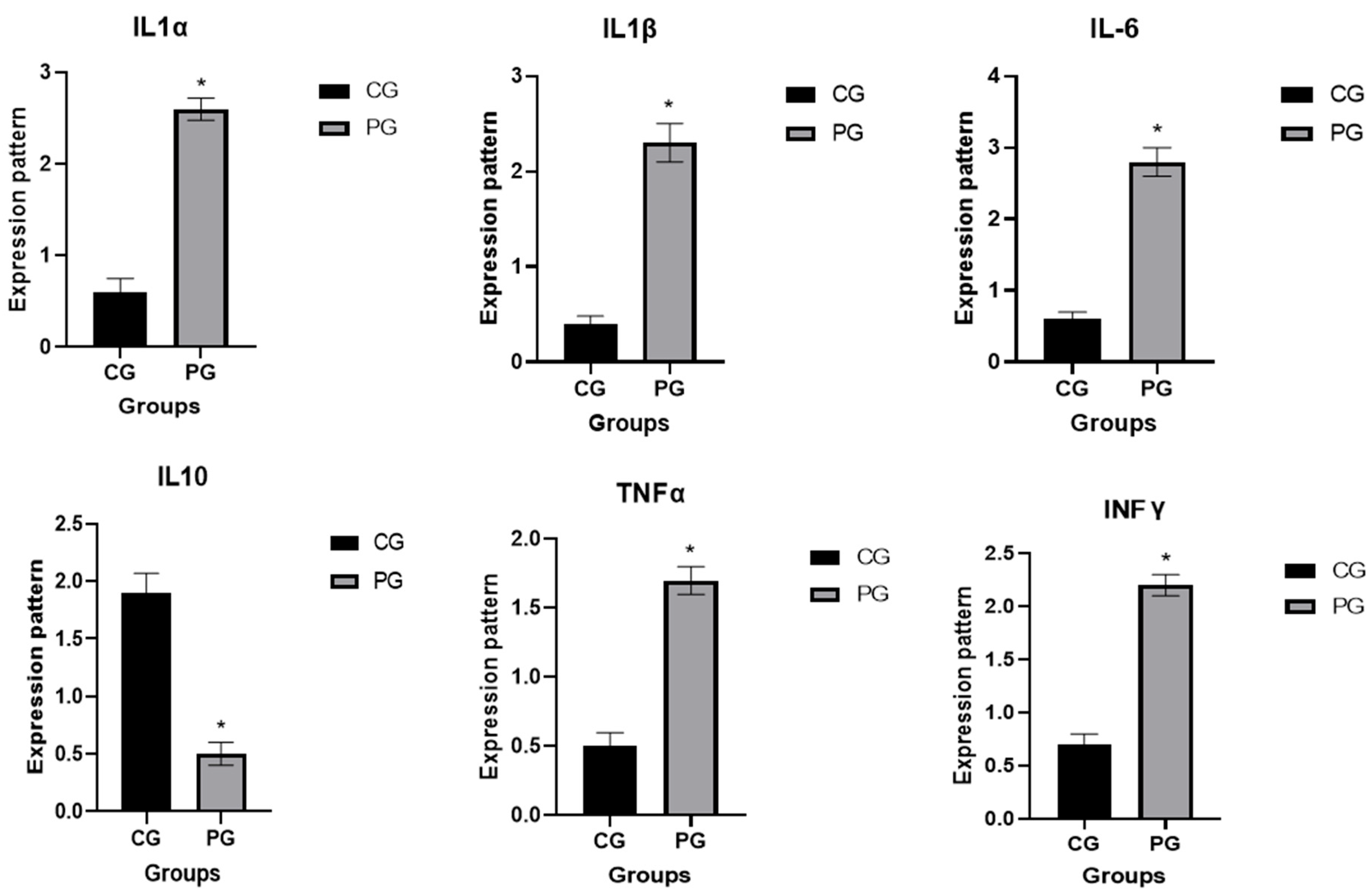
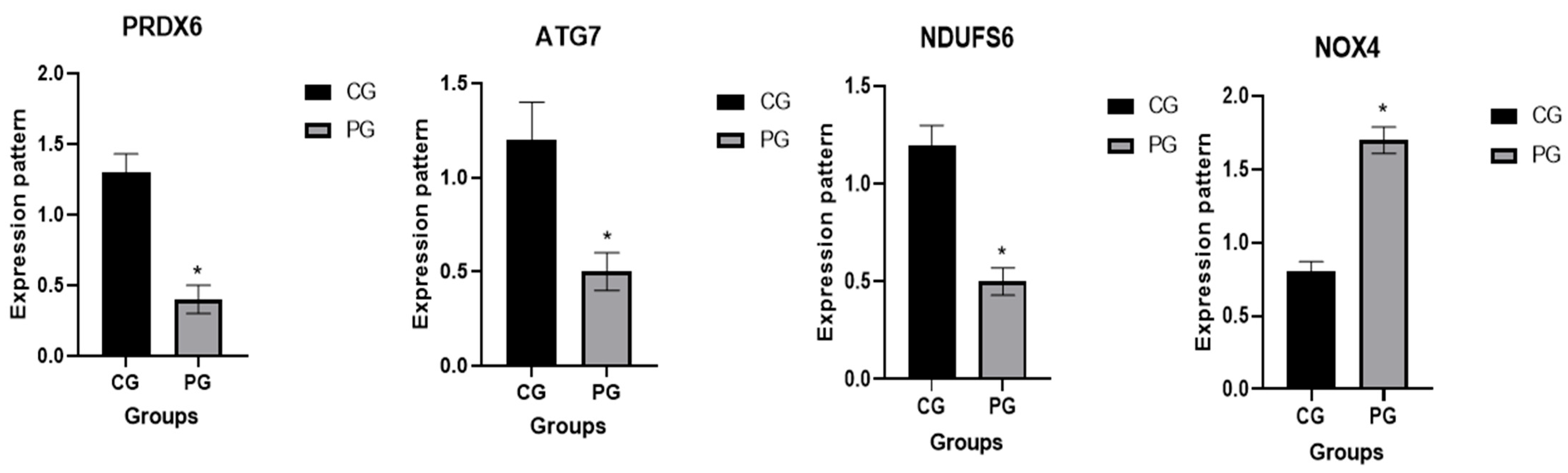
3.2. Patterns for Transcript Levels of Immune Indicators

The transcription patterns for the valued immunological and antioxidant markers are shown in Figure 1 and Figure 2, respectively. The genes IL-1α, IL1B, IL6, TNF-α, IL10, IFN-γ, and NOX4 had significantly greater gene expression levels in pneumonia-affected calves compared to the CON group. Meanwhile, the levels of ATG7, NDUFS6, PRDX6, and IL10 dropped.
Pneumonic and healthy calves were compared for the highest and lowest levels of expression of immunological and antioxidant genes. The utmost potential level of mRNA for pneumonic calves was 2.82 ± 0.21 for IL-6, whereas the lowest extent of each marker was 0.45 ± 0.12 for PRDX6. Among all the genes tested in the CON group, IL-10 had the highest possible value of mRNA (1.96 ± 0.17), whereas IL1B had the least quantity (0.49 ± 0.08).

3.3. Genetic Polymorphisms of Immune and Antioxidant Genes

Using PCR-DNA sequence verdicts, the IL-1α (356-bp), IL-1β (423-bp), IL-6 (384-bp), TNFα (490-bp), IL-10 (306-bp), IFN-γ (321-bp), PRDX6 (434-bp), ATG7 (416-bp), NDUFS6 (405-bp), and NOX4 (396-bp) genes were found to have different SNPs in the amplified DNA bases linked to pneumonia. All of the detected SNPs were authorized based on DNA sequence variances between immunological and antioxidant markers tested in the calves under research, as well as reference gene sequences obtained from GenBank (Figures S1–S10).
The coding DNA sequences of the affected calves differed from those of the healthy calves due to the exonic-region abnormalities that are seen in Table 4 in all of the immunological and antioxidant markers tested. Using DNA sequencing of immune and antioxidant genes, 22 SNPs were found. Eight of them were non-synonymous, and fourteen of them were synonymous. Six of the eight non-synonymous SNPs were associated with healthy calves, while two were associated with pneumonic calves. The incidence of pneumonia, on the other hand, was linked to seven of the fourteen synonymous SNPs revealed.
Two of the recurrent SNPs found by DNA sequencing for the immunological markers of the IL-1α gene (356-bp) were G56C and A182G, which resulted in non-synonymous alterations S19R and K61R, respectively. The 100N amino acid, on the other hand, was identified as the consequence of the synonymous mutation T300C. The IL-1β gene’s 423 bp DNA sequence was examined, and one recurrent SNP—C153T—was found to carry the synonymous mutation 51I. The IL-6 (384 bp) gene was sequenced, and two repeated SNPs were discovered. S74N was a non-synonymous mutation in G221A, while R108K was involved in G323A. DNA sequencing revealed that the IL-10 gene contains four regular SNPs (306 bp); three of them, A105T, G129C, and C168T, corresponded to synonymous alterations of 35S, 43L, and 56C, respectively. The G203C SNP, meanwhile, resulted in the non-synonymous mutation R68K. Located in the 490 bp DNA structure of the TNFα marker, three frequently occurring synonymous SNPs—A120G, C213T, and T468C—were associated with amino acids 40Q, 71D, and 156Y, respectively. The IFN-γ gene’s 321 bp sequencing revealed one frequent SNP, where the G35T variation initiated the non-synonymous mutation G12V.
According to antioxidant markers, DNA sequencing revealed that the PRDX6 gene (434 bp) includes two recurrent SNPs that resulted in synonymous mutations, 39N and 75D, respectively. Three SNPs were trapped within the 416 bp AT7G gene. Two SNPs, T48C and T333C, were synonymous and connected to the amino acids 16S and 111A, respectively. Meanwhile, the amino acid V72I was generated by the non-synonymous SNP G214A. A synonymous SNP was found in the NDUFS6 gene’s nucleotide sequence (405 bp); the G354A SNP produced the 118S result. Sequencing the NOX4 (405 bp) gene revealed two recurrent SNPs. Non-synonymous mutations F67Y were involved in T200A. Conversely, the synonymous mutation 71S was triggered by the G213A SNP.
A significant difference was detected in the frequencies of all examined genes’ SNPs among pneumonic and healthy calves (p < 0.005). Chi-square analysis was carried out for comparison of the distribution of all identified SNPs in all genes between pneumonic and healthy calves. Total Chi-square value showed significant variation among the identified SNPs in all genes between resistant and affected animals (p < 0.05; Table 4).
Table 5 shows the discriminant analysis results for the arrangement of kinds of genes and healthy status. The organization consequences showed that the model properly classified 100% of the cases overall for both the CON group and the PG. These results indicate that the SNP markers included in the model possess a good level of discriminatory power and may be useful as potential genetic indicators for pneumonia susceptibility in calves.

3.4. Antioxidants and Immunological Profile

In comparison to the CON group, pneumonic calves showed a considerable drop in SOD, GSH, and TAC, and a large increase in MDA, a biomarker of oxidative stress (Table 5). A significant rise (p < 0.01) in serum concentrations of IL-1α (155.1 ± 3.9 pg/mL), IL-1β (141.3 ± 5.3 pg/mL), IL-6 (140.5 ± 1.2 pg/mL), TNF-α (117.1 ± 3.0 pg/mL), and IFN-γ (143.8 ± 8.2 pg/mL) was observed in pneumonic calves (PG) related to the CON group, indicating a strong innate immune response. In contrast, the anti-inflammatory cytokine IL-10 was significantly lesser (p < 0.01) in PG (27.5 ± 1.7 pg/mL) than in the CON group (Table 5).

3.5. Association Among Gene Expression Pattern and Serum Profile of Immunological and Antioxidant Biomarkers

Overall, gene expression pattern was strongly correlated with antioxidant or immunological biomarkers (Table 6).

4. Discussion

Due to its frequent occurrence, high rate of morbidity and mortality, slow growth, and veterinary costs, pneumonia is supposed to be a main cause of financial loss for calves of all ages and types [4]. This work aimed to monitor changes in gene expression and nucleotide sequences of immunological markers of pneumonic calves.
In comparison to the CON group, pneumonic calves demonstrated a marked increase in body temperature, respiratory rate, and pulse rate. Our clinical findings were comparable to that observed by [12,36,37,38,39,40].
We investigated the alterations in the immunological and antioxidant status of calves with pneumonia in comparison to healthy calves by assessing the mRNA values of immune and antioxidant biomarkers. The expression of the genes IL-1α, IL1B, IL6, TNF-α, IL10, and IFN-γ was much greater in the affected calves than in the CON group. This is the first report to thoroughly examine the transcription levels of the antioxidant and immunological biomarkers associated with pneumonia in calves. As a result, quantitative variations in the expression of the genes under investigation occur before pneumonia manifests itself. The immune genes’ mRNA levels in goats (SLC11A1, CD-14, CCL2, TLR1, TLR7, TLR8, TLR9, defensin, SP110, SPP1, BP1, A2M, ADORA3, CARD15, IRF3, and SCART1) varied between the healthy and infected goats with pneumonia [41]. Gene expression outlining revealed that Toll-like receptors (TLRs) and complement genes were accompanying infectious pneumonia in sheep [42]. When comparing pneumonia-affected sheep to resistant ewes, the IL-1α, IL1B, IL6, and TNF-α genes were markedly upregulated [43]. The gene that codes for IL10, on the other hand, was downregulated.
Our investigation used a PCR-DNA-sequencing procedure to illustrate the difference in immunological (IL-1α, IL1B, IL6, TNF-α, IL10, and IFN-γ) and antioxidant (PRDX6, ATG7, NDUFS6, and NOX4) genes in calves with pneumonia and healthy calves. The results demonstrate that there are transformations in the SNPs involving the two classes. It is fundamental to stress that, in contrast to the similar datasets obtained from GenBank, the polymorphisms discovered and presented, in this circumstance, offer extra evidence for the evaluated indicators. No research has looked at the relationship between calves’ risk of pneumonia and SNPs in the immune and antioxidant genes. This link is initially shown by the Bos taurus gene sequences used in our study, which were published in PubMed. However, ruminant susceptibility to pneumonia was assessed using the candidate gene technique. For instance, there is evidence linking MHC-DRB1 genotypes to either resistance or susceptibility to Mycoplasma ovipneumoniae in sheep [44]. Furthermore, it has been demonstrated that the consequences of ovine TMEM154 gene polymorphisms during normal exposure influence progressive pneumonia virus susceptibility [45]. There have been reports linking TLR gene variants to pneumococcal disease in humans [46]. Through PCR-DNA sequencing in Baladi goats with and without pneumonia, SNPs linked to pneumonia susceptibility and resistance were discovered in the immunological (SLC11A1, CD-14, CCL2, TLR1, TLR7, TLR8, TLR9, defensin, SP110, SPP1, BP1, A2M, ADORA3, CARD15, IRF3, and SCART1) genes [41].
Mutation is the central cause of selection and adaptability [47]. Exonic area mutations were presented in all examined markers under investigation in this case, leading to different coding DNA sequences in pneumonic as opposed to healthy calves. Eight non-synonymous and fourteen synonymous SNPs were identified through DNA sequencing of investigated genes. Protein sequences are altered by non-synonymous mutations, and natural selection usually targets animals with these changes [47]. Genetic discrepancy caused by non-synonymous SNPs adjusts the fixed amino acid at the mutant site, potentially leading to structural and practical deviations in the mutated protein [48]. It was long thought that there was either no selection on synonymous mutations or very little selection [47]. The examined immune genes must be precisely characterized at the molecular level in order to comprehend the physiological alterations between pneumonic and healthy calves in terms of resistance and susceptibility. According to our research, polymorphisms based on the translated DNA sequence of calves are further valued than intronic regions.
In the context of inflammation, cytokines, including NFKB, IL-1α, TNF-α, IFN-γ, IL-6, and IL1B, act as indirect biomarkers [49]. IL-10 is an anti-inflammatory cytokine that prevents macrophages from producing natural killer cells (IL-1 and TNF) and Th1 lymphocytes from producing IFN- and IL-2 [50,51]. Through its interaction with regulating suppressor T CD8+ cells, IL-10, which is formed by a variety of T cells, has been shown to contain anti-inflammatory characteristics that defend uterine tissues from the highly pathogenic impact of inflammatory cells and mediators [52]. The IL10, TNF-α, IL1B, and IL6 genes were PCR-DNA sequenced to identify SNPs with nucleotide sequence variations connected to diarrhea resistance or vulnerability in lambs [53]. Diarrheal lambs had noticeably greater intensities of TNF-α, IL1B, and IL6 gene expression than resistant lambs. In diarrheal lambs, the IL10 gene was significantly downregulated compared to resistant lambs. The nucleotide sequences of healthy and endometritis-affected cows were found to differ by using PCR-DNA sequencing for the IL10 gene. The IL10 gene was expressed at much lower levels in cows suffering from endometritis [54].
A conserved ionized thiol group enables the peroxiredoxin (PRDX) family of antioxidant oxidoreductase enzymes to induce the reduction of hydrogen peroxide (H2O2). Thiol-specific peroxidases act as sensors for signaling events triggered by hydrogen peroxide and contribute to cellular defense, contrary to oxidative stress, by detoxifying peroxides and sulfur-containing radicals [55]. Complex I, the major enzyme complex in the mitochondrial electron transport chain, is encoded in part by the NADH ubiquinone oxidoreductase subunit S6 (NDUFS6) gene [56]. Through the action of Complex I, NADH donates electrons to the mitochondrial respiratory chain. Mutations in the NDUFS6 gene can lead to mitochondrial Complex I deficiency, which is associated with neurodegenerative diseases in adults, as well as severe illness in neonates [56]. It has formerly been stated that the NDUFS6 gene, set on BTA20 in cattle, has a 900 bp deletion that removes exon 2 entirely [57]. There is a measurable trait locus for SCS in this area of the genome [58].
PCR-DNA sequence revealed nucleotide sequence differences in the PRDX6 and NDUFS6 genes among control and mastitis-affected she-camels [59]. Significantly lower levels of PRDX6 and NDUFS6 gene expression were observed in mastitic camels. Differences in the PRDX6 gene’s nucleotide sequence were observed between calves with diarrhea and healthy calves [60]. Diarrheic calves have significantly higher levels of PRDX6 expression than resistant calves. In buffaloes with endometritis, NDUFS6 was expressed at significantly lower levels [61].
Numerous autophagy-associated genes (ATGs) are implicated in molecular pathways, and autophagy is a complicated and highly controlled process [62]. According to [63], ATG7 is an E1-like ligase that facilitates the elongation of separation membranes and is essential for the development of autophagosomes. According to [64], the expression of important genes linked to antioxidant replies, such as the ATG7 gene, was markedly downregulated by bacterial LPS. The catalytic NOX subunit of NADPH oxidase 4 (NOX4), a member of the NADPH oxidase family, converts electrons from NADPH to oxygen to produce ROS [65]. Numerous pathogeneses, such as cell senescence [66], apoptosis [67], endothelial dysfunction [68], angiogenesis [69], atherosclerosis and vascular ageing [70], cardiac remodeling [71], and neoplasms [72], have been linked to the NOX4 gene. NOX4 gene polymorphism alters the relationships between dietary caloric consumption and peripheral blood mononuclear cell ROS levels [73].
The altered immune expression patterns observed in calves with pneumonia may result from the induction of inflammatory and antioxidant biomarkers triggered by lipopolysaccharides or endotoxins [74]. When LPSs excite macrophages, inflammatory gene expressions treble, mRNA levels increase by approximately one-hundred-fold, and the amount of protein released can rise by up to 10,000 times [75]. Moreover, damaged tissues generate more free radical reactions than healthy tissues [76,77]. In contrast to the resistant genotypes, which considerably grow the expressions of IL-2 and IL-10 to suppress the inflammatory response, the vulnerable genotypes increase the inflammatory response via the large release of inflammatory biomarkers [78]. Calves with pneumonia also produce a range of cellular immunological mechanisms that bind to cell surface receptors to arbitrate and control the inflammatory response and immune activity [79]. Numerous investigations have shown that different cell immunological components have intricate interactions. The formation of various immune globulins, complements, and acute phase reactive proteins, for example, has been discovered to be influenced by the interactions of these proteins, resulting in a complex network structure [80]. We therefore expect that the majority of the pneumonia cases in our analysis were caused by infectious pneumonia. Moreover, our real-time PCR results give a credible suggestion that animals suffering from pneumonia have a robust inflammatory reaction.
The obvious drop in the values of RBCs, hemoglobin, and PCV (%) in pneumonic calves is accordant with conclusions of other works [20,37,81]. Pneumonic calves showed a significant reduction in lymphocytes but a considerable rise in WBCs and neutrophils when compared to the CON group. These findings were compatible with those of [20,37,81,82]. Increases in WBC counts, particularly neutrophils, are frequently observed in a variety of disorders as a result of inflammatory courses, like those in respiratory disorders. According to [83,84], inflammatory lesions and the attendance of bacterial infection may be the cause of the significant increase in WBCs and neutrophils that happened in pneumonia. According to [82,85], calves with respiratory infections may experience severe lymphopenia as a result of the high-concentration endotoxin’s ability to trigger lymphocyte lysis. However, the significant reduction in lymphocytes may be due to immunological suppression brought on by a variety of stressors or may be explained by the adrenal gland being stimulated by tissue infected by bacterial toxins [86,87].
When oxidants and antioxidants are not balanced, oxidative stress occurs, which damages cells and tissues. Oxidative stress eventually results in mechanical injury and loss of function in cells and tissues when there is an imbalance caused by an increase in oxidants or a decrease in antioxidants. This can have a wide range of detrimental effects on animals, from decreased production to death. Thus, it has been reported that oxidative stress had a significant purpose in the etiology and pathogenesis of a number of animal diseases, particularly those affecting the respiratory system [88]. Calves with pneumonia had significantly higher MDA values and significantly lower SOD, GSH, and TAC activity in their oxidant/antioxidant profiles as opposed to the CON group. Our findings were close to that observed by [40,89,90,91]. By inhibiting the peroxidation procedure and the generation of additional free radical molecules [92], the rise in serum MDA was linked to cellular lipid peroxidation, and the fall in SOD, GSH, and TAC levels was linked to their consumption in cells’ defense against oxidative damage.
According to this investigation, PG showed a significant rise in pro-inflammatory cytokines in contrast to the CON group. Our findings were comparable to that found by [20,36,40] in cattle calves [93], in buffalo calves [94], in camel calves [95,96,97], in sheep [37], and in cattle and in pigs [98]. As per [95], reduced anti-inflammatory cytokine levels in the pneumonic group likely exacerbated the illness by facilitating an aggressive innate immune response. The inflammatory response during infection is made worse by this misalignment of pro- and anti-inflammatory signals [97]. In the early stages of bacterial infection, resident alveolar macrophages are stimulated and release pro-inflammatory cytokines, eventually leading to the formation of a pro-inflammatory amplification loop between lymphocytes and local or recruited neutrophils or macrophages. A repeat recurrence of anti-inflammatory cytokines is generated as the infection is eradicated in order to pinpoint the location of the inflammatory response in the lung microenvironment and, eventually, to suppress this immune response [99].
This current work has several restrictions that should be addressed in upcoming research. First, this study focused on a single breed, so applying this research to different calf breeds would provide more accurate health assessments. Second, only a limited set of immunity and antioxidant-related genes were examined, indicating the need for broader gene profiling in subsequent studies. Third, bacterial species and viruses present in the pneumonic calves were not isolated or identified, and future research should aim to address them. Finally, histological alterations in the infected lungs were not evaluated in this study; thus, analyzing the histopathological changes is essential for a comprehensive understanding of the disease.

5. Conclusions

Our results emphasize the nucleotide sequence variations and the expression profiles of the immunological and antioxidant genes studied as a cause or a mechanism-mediated response for pneumonia resistance or susceptibility in calves. The findings provide strong evidence of noteworthy immunological and oxidative fluctuations associated with pneumonia in calves, particularly concerning their blood antioxidant and immunological profiles. The distinct expression patterns of pneumonia-resistant and susceptible calves may represent biomarkers and references to monitor these animals’ health. In line with genetic markers interrelated to innate immunity to infection, these results present a viable method for lowering calves’ risk of pneumonia through discriminatory breeding.

Supplementary Materials

The following supporting information can be downloaded at https://www.mdpi.com/article/10.3390/vetsci12070620/s1, Figure S1: An example of the alignment of the IL-1α gene (356 bp) between calves with pneumonia (P) and that are healthy (H). Figure S2: An example of the alignment of the IL-1β gene (423 bp) among calves with pneumonia (P) and that are healthy (H). Figure S3: An example of the alignment of the IL-6 gene (384 bp) concerning calves with pneumonia (P) and that are healthy (H). Figure S4: An example of the alignment of the IL-10 gene (306 bp) amongst calves with pneumonia (P) and that are healthy (H). Figure S5: An example of the alignment of the TNFα gene (490-bp) between calves with pneumonia (P) and that are healthy (H). Figure S6: An example of the alignment of the IFN-γ gene (321-bp) between calves with pneumonia (P) and that are healthy (H). Figure S7: An example of the alignment of the PRDX6 gene (434-bp) between calves with pneumonia (P) and that are healthy (H). Figure S8: An example of the alignment of the ATG7 gene (416-bp) between calves with pneumonia (P) and that are healthy (H). Figure S9: An example of the alignment of the NDUFS6 gene (405-bp) between calves with pneumonia (P) and that are healthy (H). Figure S10: An example of the alignment of the NOX4 gene (396-bp) between calves with pneumonia (P) and that are healthy (H).

Author Contributions

A.E. and A.A. proposed the study and gathered the samples, interpreted the results, conducted gene and blood biochemical analysis, and recruited the principal manuscript; A.E.-S., D.E., H.G., H.M.A., K.M.A., M.A.B. and A.A.E. helped formulate the research and develop the article. Each author has revised and accepted the published version of the work. All authors have read and agreed to the published version of the manuscript.

Funding

Princess Nourah bint Abdulrahman University, located in Riyadh, Saudi Arabia, provided funding for this study under the Researchers Supporting Project number (PNURSP2025R454).

Institutional Review Board Statement

The Desert Research Centre (DRC) Ethics Committee in Egypt gave its approval to the procedures for sample collection and animal care in this investigation (code DRC-021-1-23) on 1 January 2020.

Informed Consent Statement

Every farmer has given their informed consent to take part in this study.

Data Availability Statement

The relevant author will grant supporting material for the study’s results upon reasonable request.

Acknowledgments

The authors are thankful to Princess Nourah bint Abdulrahman University, set in Riyadh, Saudi Arabia, for its Academics Reinforcing Project number (PNURSP2025R454). The authors extend their appreciation to the deanship of postgraduate studies and scientific research at Majmaah University for funding this research work through the project number (R-2025-1852).

Conflicts of Interest

The authors declare no conflicts of interest.

References

  1. Kamel, M.S.; Davidson, J.L.; Verma, M.S. Strategies for Bovine Respiratory Disease (BRD) Diagnosis and Prognosis: A Comprehensive Overview. Animals 2024, 14, 627. [Google Scholar] [CrossRef] [PubMed]
  2. Murray, G.M.; More, S.J.; Clegg, T.A.; Earley, B.; O’Neill, R.G.; Johnston, D.; Gilmore, J.; Nosov, M.; McElroy, M.C.; Inzana, T.J. Risk factors associated with exposure to bovine respiratory disease pathogens during the peri-weaning period in dairy bull calves. BMC Vet. Res. 2018, 14, 53. [Google Scholar] [CrossRef]
  3. Buhler, V.M.; Cash, K.R.; Hurley, D.J.; Credille, B.C. Characterization and comparison of cell-mediated immune responses following ex vivo stimulation with viral and bacterial respiratory pathogens in stressed and unstressed beef calves. J. Anim. Sci. 2019, 97, 2739–2749. [Google Scholar] [CrossRef] [PubMed]
  4. Nagy, A.; Abdallah, F.; El Damaty, H.M.; Tariq, A.; Merwad, A.M.; Alhatlani, B.Y.; Elsohaby, I. Genetic characterization of upper respiratory tract virome from nonvaccinated egyptian cow-calf operations. PLoS ONE 2022, 17, e0267036. [Google Scholar] [CrossRef] [PubMed]
  5. Johnson, K.K.; Pendell, D.L. Market impacts of reducing the prevalence of bovine respiratory disease in United States beef cattle feedlots. Front. Vet. Sci. 2017, 4, 189. [Google Scholar] [CrossRef]
  6. Donia, G.R.; El Ebissy, I.A.; Wassif, I.M. Biochemical and immunological studies on the respiratory diseases in sheep in north western coast. Eur. J. Biomed. 2018, 5, 34–41. [Google Scholar]
  7. Donlon, J.D.; Mee, J.F.; McAloon, C.G. Prevalence of respiratory disease in Irish preweaned dairy calves using hierarchical Bayesian latent class analysis. Front. Vet. Sci. 2023, 10, 1149929. [Google Scholar] [CrossRef]
  8. Teixeira, A.; McArt, J.; Bicalho, R. Efficacy of tildipirosin metaphylaxis for the prevention of respiratory disease, otitis and mortality in pre-weaned Holstein calves. Vet. J. 2017, 219, 44–48. [Google Scholar] [CrossRef]
  9. Zeineldin, M.; Lowe, J.; Aldridge, B. Contribution of the mucosal microbiota to bovine respiratory health. Trends Microbiol. 2019, 27, 753–770. [Google Scholar] [CrossRef]
  10. Constable, P.D.; Hinchcliff, K.W.; Done, S.H.; Grünberg, W. Veterinary Medicine: A Textbook of the Diseases of Cattle, Horses, Sheep, Pigs and Goats; Elsevier Health Sciences: Amsterdam, The Netherlands, 2016. [Google Scholar]
  11. Dubrovsky, S.; Van Eenennaam, A.; Karle, B.; Rossitto, P.; Lehenbauer, T.; Aly, S. Epidemiology of bovine respiratory disease (BRD) in preweaned calves on California dairies: The BRD 10K study. J. Dairy Sci. 2019, 102, 7306–7319. [Google Scholar] [CrossRef]
  12. Bozukluhan, K.; Merhan, O.; Kiziltepe, S.; Egritag, H.E.; Akyuz, E.; Gokce, H.I. Determination of haptoglobin, some biochemical and oxidative stress parameters in calves with pneumonia. Fresenius Environ. Bull. 2021, 30, 9485–9489. [Google Scholar]
  13. Fulton, R.W.; Confer, A.W. Laboratory test descriptions for bovine respiratory disease diagnosis and their strengths and weaknesses: Gold standards for diagnosis, do they exist? Can. Vet. J. 2012, 53, 754–761. [Google Scholar]
  14. Wolfger, B.; Timsit, E.; White, B.J.; Orsel, K. A systematic review of bovine respiratory disease diagnosis focused on diagnostic confirmation, early detection, and prediction of unfavorable outcomes in feedlot cattle. Vet. Clin. Food Anim. Pract. 2015, 31, 351–365. [Google Scholar] [CrossRef] [PubMed]
  15. Blakebrough-Hall, C.; Dona, A.; D’occhio, M.; McMeniman, J.; A Gonzále, L. Diagnosi di malattia respiratoria bovina in animali allevati in feedlot mediante metabolomica 1H NMR su sangue. Summa Anim. Reddito 2020, 15, 17. [Google Scholar]
  16. Abebe, W.; Getinet, A.; Mekonnen, H. Study on live weight, carcass weight and dressing percentage of Issa camels in Ethiopia. Rev. Méd. Vétér. 2002, 153, 713–716. [Google Scholar]
  17. Sharma, N.; Singh, N.; Singh, O.; Pandey, V.; Verma, P. Oxidative stress and antioxidant status during transition period in dairy cows. Asian-Australas. J. Anim. Sci. 2011, 24, 479–484. [Google Scholar] [CrossRef]
  18. Surai, P.F.; Earle-Payne, K. Antioxidant defences and redox homeostasis in animals. Antioxidants 2022, 11, 1012. [Google Scholar] [CrossRef]
  19. McGill, J.L.; Sacco, R.E. The immunology of bovine respiratory disease: Recent advancements. Vet. Clin. N. Am. Food Anim. Pract. 2020, 36, 333–348. [Google Scholar] [CrossRef]
  20. Ramadan, M.; Ghanem, M.; El Attar, H.E.; Abdel-Raoof, Y. Evaluation of clinical and hematobiochemical alterations in naturally occurring bovine respiratory disease in feedlot cattle calves in Egypt. Benha Vet. Med. J. 2019, 36, 305–313. [Google Scholar] [CrossRef]
  21. El-Deeb, W.; Fayez, M.; Elsohaby, I.; Salem, M.; Alhaider, A.; Kandeel, M. Investigation of acute-phase proteins and cytokines response in goats with contagious caprine pleuropneumonia with special reference to their diagnostic accuracy. PeerJ 2020, 8, e10394. [Google Scholar] [CrossRef]
  22. Chitko-McKown, C.G.; Bennett, G.L.; Kuehn, L.A.; DeDonder, K.D.; Apley, M.D.; Harhay, G.P.; Clawson, M.L.; Workman, A.M.; White, B.J.; Larson, R.L. Cytokine and haptoglobin profiles from shipping through sickness and recovery in metaphylaxis-or un-treated cattle. Front. Vet. Sci. 2021, 8, 611927. [Google Scholar] [CrossRef] [PubMed]
  23. Berry, D.P.; Bermingham, M.L.; Good, M.; More, S.J. Genetics of animal health and disease in cattle. Ir. Vet. J. 2011, 64, 5. [Google Scholar] [CrossRef]
  24. Pierce, C.F.; Brown, V.R.; Olsen, S.C.; Boggiatto, P.; Pedersen, K.; Miller, R.S.; Speidel, S.E.; Smyser, T.J. Loci associated with antibody response in feral swine (Sus scrofa) infected with Brucella suis. Front. Vet. Sci. 2020, 7, 554674. [Google Scholar] [CrossRef] [PubMed]
  25. Sallam, A.M.; Abou-Souliman, I.; Reyer, H.; Wimmers, K.; Rabee, A.E. New insights into the genetic predisposition of brucellosis and its effect on the gut and vaginal microbiota in goats. Sci. Rep. 2023, 13, 20086. [Google Scholar] [CrossRef] [PubMed]
  26. Zhang, L.; Nie, M.-Y.; Liu, F.; Chen, J.; Wei, L.-J.; Hua, Q. Multiple gene integration to promote erythritol production on glycerol in Yarrowia lipolytica. Biotechnol. Lett. 2021, 43, 1277–1287. [Google Scholar] [CrossRef]
  27. Adams, L.; Schutta, C.J. Natural resistance against brucellosis: A review. Open Vet. Sci. J. 2010, 4, 61–71. [Google Scholar] [CrossRef]
  28. Saco, Y.; Bassols, A. Acute phase proteins in cattle and swine: A review. Vet. Clin. Pathol. 2023, 52, 50–63. [Google Scholar] [CrossRef]
  29. National Academies of Sciences, Engineering, and Medicine; Division on Earth, Life Studies; Committee on Nutrient Requirements of Beef Cattle. Nutrient Requirements of Beef Cattle; National Academies Press: Washington, DC, USA, 2016. [Google Scholar]
  30. Pfaffl, M.W. A new mathematical model for relative quantification in real-time RT–PCR. Nucleic Acids Res. 2001, 29, e45. [Google Scholar] [CrossRef]
  31. Boom, R.; Sol, C.; Salimans, M.; Jansen, C.; Wertheim-van Dillen, P.; Van der Noordaa, J. Rapid and simple method for purification of nucleic acids. J. Clin. Microbiol. 1990, 28, 495–503. [Google Scholar] [CrossRef]
  32. Boesenberg-Smith, K.A.; Pessarakli, M.M.; Wolk, D.M. Assessment of DNA yield and purity: An overlooked detail of PCR troubleshooting. Clin. Microbiol. Newsl. 2012, 34, 1–6. [Google Scholar] [CrossRef]
  33. Sanger, F.; Nicklen, S.; Coulson, A.R. DNA sequencing with chain-terminating inhibitors. Proc. Natl. Acad. Sci. USA 1977, 74, 5463–5467. [Google Scholar] [CrossRef] [PubMed]
  34. Altschul, S.F.; Gish, W.; Miller, W.; Myers, E.W.; Lipman, D.J. Basic local alignment search tool. J. Mol. Biol. 1990, 215, 403–410. [Google Scholar] [CrossRef] [PubMed]
  35. Tamura, K.; Dudley, J.; Nei, M.; Kumar, S. MEGA4: Molecular evolutionary genetics analysis (MEGA) software version 4.0. Mol. Biol. Evol. 2007, 24, 1596–1599. [Google Scholar] [CrossRef]
  36. El-Seedy, F.; Hassan, H.; Nabih, A.; Salem, S.; Khalifa, E.; Menshawy, A.; Abed, A. Respiratory affections in calves in upper and middle Egypt: Bacteriologic, immunologic and epidemiologic studies. Adv. Anim. Vet. Sci 2020, 8, 558–569. [Google Scholar] [CrossRef]
  37. Allam, T.; Said, L.; Elsayed, M.; Saleh, N. Clinical investigation of the pathogenicity of pasteurella multocida isolated from cattle in egypt regarding its effect on hematological, biochemical, and oxidant-antioxidant biomarkers as well as proinflammatory cytokines and acute phase proteins. Adv. Anim. Vet. Sci 2021, 9, 792–801. [Google Scholar] [CrossRef]
  38. Su, M.; She, Y.; Deng, M.; Guo, Y.; Li, Y.; Liu, G.; Sun, B.; Liu, D. Effect of Capsaicin Addition on Antioxidant Capacity, Immune Performance and Upper Respiratory Microbiota in Nursing Calves. Microorganisms 2023, 11, 1903. [Google Scholar] [CrossRef]
  39. El-Sheikh, M.E.-S.; El-Mekawy, M.F.; Eisa, M.I.; Abouzeid, N.Z.; Abdelmonim, M.I.; Bennour, E.M.; Yousef, S.G. Effect of two different commercial vaccines against bovine respiratory disease on cell-mediated immunity in Holstein cattle. Open Vet. J. 2024, 14, 1921–1927. [Google Scholar] [CrossRef]
  40. Saher, A.S.; Raza, A.; Qiu, F.; Mehmood, K.; Hussain, R.; Qayyum, A.; Idris, M.; Almutairi, M.H.; Li, K. Detection of haptoglobin and serum amyloid A as biomarkers in naturally infected Mycoplasma bovis calves. Acta Trop. 2024, 254, 107215. [Google Scholar] [CrossRef]
  41. Ateya, A.; Al-Sharif, M.; Abdo, M.; Fericean, L.; Essa, B. Individual genomic loci and mRNA levels of immune biomarkers associated with pneumonia susceptibility in baladi goats. Vet. Sci. 2023, 10, 185. [Google Scholar] [CrossRef]
  42. Abendaño Carbajo, N.; Esparza Baquer, A.; Bernales Pujana, I.; Reina, R.; De Andrés, D.; Jugo Orrantia, B.M. Gene Expression Profiling Reveals New Pathways and Genes Associated with Visna/Maedi Viral Disease. Animals 2021, 11, 1785. [Google Scholar] [CrossRef]
  43. Sayed, A.E.; Hafez, A.; Ateya, A.; Darwish, A.; Tahoun, A. Single nucleotide polymorphisms, gene expression and evaluation of immunological, antioxidant, and pathological parameters associated with bacterial pneumonia in Barki sheep. Ir. Vet. J. 2025, 78, 11. [Google Scholar] [CrossRef] [PubMed]
  44. Wang, K.; Liu, X.; Li, Q.; Wan, K.; Gao, R.; Han, G.; Li, C.; Xu, M.; Jia, B.; Shen, X. MHC-DRB1 exon 2 polymorphism and its association with mycoplasma ovipneumonia resistance or susceptibility genotypes in sheep. J. Genet. 2020, 99, 22. [Google Scholar] [CrossRef] [PubMed]
  45. Leymaster, K.; Chitko-McKown, C.; Clawson, M.; Harhay, G.; Heaton, M. Effects of TMEM154 haplotypes 1 and 3 on susceptibility to ovine progressive pneumonia virus following natural exposure in sheep. J. Anim. Sci. 2013, 91, 5114–5121. [Google Scholar] [CrossRef]
  46. Salamaikina, S.; Karnaushkina, M.; Korchagin, V.; Litvinova, M.; Mironov, K.; Akimkin, V. TLRs Gene Polymorphisms Associated with Pneumonia before and during COVID-19 Pandemic. Diagnostics 2022, 13, 121. [Google Scholar] [CrossRef]
  47. Barton, N.H. Mutation and the evolution of recombination. Philos. Trans. R. Soc. B Biol. Sci. 2010, 365, 1281–1294. [Google Scholar] [CrossRef] [PubMed]
  48. Dakal, T.C.; Kala, D.; Dhiman, G.; Yadav, V.; Krokhotin, A.; Dokholyan, N.V. Predicting the functional consequences of non-synonymous single nucleotide polymorphisms in IL8 gene. Sci. Rep. 2017, 7, 6525. [Google Scholar] [CrossRef]
  49. Salim, T.; Sershen, C.L.; May, E.E. Investigating the role of TNF-α and IFN-γ activation on the dynamics of iNOS gene expression in LPS stimulated macrophages. PLoS ONE 2016, 11, e0153289. [Google Scholar] [CrossRef]
  50. Martinez-Espinosa, I.; Serrato, J.A.; Ortiz-Quintero, B. Role of IL-10-producing natural killer cells in the regulatory mechanisms of inflammation during systemic infection. Biomolecules 2021, 12, 4. [Google Scholar] [CrossRef]
  51. Wang, Z.; Guan, D.; Huo, J.; Biswas, S.K.; Huang, Y.; Yang, Y.; Xu, S.; Lam, K.-P. IL-10 enhances human natural killer cell effector functions via metabolic reprogramming regulated by mTORC1 signaling. Front. Immunol. 2021, 12, 619195. [Google Scholar] [CrossRef]
  52. Iyer, S.S.; Cheng, G. Role of interleukin 10 transcriptional regulation in inflammation and autoimmune disease. Crit. Rev.™ Immunol. 2012, 32, 23–63. [Google Scholar] [CrossRef]
  53. Darwish, A.; Ebissy, E.; Hafez, A.; Ateya, A.; El-Sayed, A. Nucleotide sequence variants, gene expression and serum profile of immune and antioxidant markers associated with bacterial diarrhea susceptibility in Barki lambs. BMC Vet. Res. 2024, 20, 462. [Google Scholar] [CrossRef] [PubMed]
  54. Al-Sharif, M.; Abdo, M.; Shabrawy, O.E.; El-Naga, E.M.A.; Fericean, L.; Banatean-Dunea, I.; Ateya, A. Investigating polymorphisms and expression profile of immune, antioxidant, and erythritol-related genes for limiting postparturient endometritis in Holstein cattle. Vet. Sci. 2023, 10, 370. [Google Scholar] [CrossRef]
  55. Wadley, A.J.; Aldred, S.; Coles, S.J. An unexplored role for Peroxiredoxin in exercise-induced redox signalling? Redox Biol. 2016, 8, 51–58. [Google Scholar] [CrossRef]
  56. Kirby, D.M.; Salemi, R.; Sugiana, C.; Ohtake, A.; Parry, L.; Bell, K.M.; Kirk, E.P.; Boneh, A.; Taylor, R.W.; Dahl, H.-H.M. NDUFS6 mutations are a novel cause of lethal neonatal mitochondrial complex I deficiency. J. Clin. Investig. 2004, 114, 837–845. [Google Scholar] [CrossRef] [PubMed]
  57. Boussaha, M.; Esquerré, D.; Barbieri, J.; Djari, A.; Pinton, A.; Letaief, R.; Salin, G.; Escudié, F.; Roulet, A.; Fritz, S. Genome-wide study of structural variants in bovine Holstein, Montbéliarde and Normande dairy breeds. PLoS ONE 2015, 10, e0135931. [Google Scholar] [CrossRef]
  58. Durán Aguilar, M.; Román Ponce, S.; Ruiz López, F.; González Padilla, E.; Vásquez Peláez, C.; Bagnato, A.; Strillacci, M.G. Genome-wide association study for milk somatic cell score in holstein cattle using copy number variation as markers. J. Anim. Breed. Genet. 2017, 134, 49–59. [Google Scholar] [CrossRef] [PubMed]
  59. Ateya, A.; Safhi, F.A.; El-Emam, H.; Marawan, M.A.; Fayed, H.; Kadah, A.; Mamdouh, M.; Hizam, M.M.; Al-Ghadi, M.Q.; Abdo, M. Combining Nucleotide Sequence Variants and Transcript Levels of Immune and Antioxidant Markers for Selection and Improvement of Mastitis Resistance in Dromedary Camels. Agriculture 2023, 13, 1909. [Google Scholar] [CrossRef]
  60. Ateya, A.; Al-Sharif, M.; Faraj, S.H.; Abdo, M.; Fericean, L.; Banatean-Dunea, I.; Hammad, S.J.; Mamdouh, M.; Fayed, H.; Marawan, M.A. Exploring genetic polymorphisms and transcript levels of antioxidant and metabolic markers for prediction and monitoring diarrhea in Holstein dairy calves. Acta Agric. Scand. Sect. A—Anim. Sci. 2024, 73, 86–96. [Google Scholar] [CrossRef]
  61. El-Sayed, A.; Faraj, S.H.; Marghani, B.H.; Safhi, F.A.; Abdo, M.; Fericean, L.; Banatean-Dunea, I.; Alexandru, C.-C.; Alhimaidi, A.R.; Ammari, A.A. The Transcript Levels and the Serum Profile of Biomarkers Associated with Clinical Endometritis Susceptibility in Buffalo Cows. Vet. Sci. 2024, 11, 340. [Google Scholar] [CrossRef]
  62. Khan, M.Z.; Huang, B.; Kou, X.; Chen, Y.; Liang, H.; Ullah, Q.; Khan, I.M.; Khan, A.; Chai, W.; Wang, C. Enhancing bovine immune, antioxidant and anti-inflammatory responses with vitamins, rumen-protected amino acids, and trace minerals to prevent periparturient mastitis. Front. Immunol. 2024, 14, 1290044. [Google Scholar] [CrossRef]
  63. Bernabucci, U.; Ronchi, B.; Lacetera, N.; Nardone, A. Influence of body condition score on relationships between metabolic status and oxidative stress in periparturient dairy cows. J. Dairy Sci. 2005, 88, 2017–2026. [Google Scholar] [CrossRef] [PubMed]
  64. Dai, H.; Coleman, D.; Hu, L.; Martinez-Cortés, I.; Wang, M.; Parys, C.; Shen, X.; Loor, J. Methionine and arginine supplementation alter inflammatory and oxidative stress responses during lipopolysaccharide challenge in bovine mammary epithelial cells in vitro. J. Dairy Sci. 2020, 103, 676–689. [Google Scholar] [CrossRef]
  65. He, W.; Wang, Q.; Gu, L.; Zhong, L.; Liu, D. NOX4 rs11018628 polymorphism associates with a decreased risk and better short-term recovery of ischemic stroke. Exp. Ther. Med. 2018, 16, 5258–5264. [Google Scholar] [CrossRef]
  66. Kofler, P.A.; Pircher, H.; von Grafenstein, S.; Diener, T.; Höll, M.; Liedl, K.R.; Siems, K.; Jansen-Dürr, P. Characterisation of Nox4 inhibitors from edible plants. Planta Medica 2013, 79, 244–252. [Google Scholar] [CrossRef]
  67. Li, J.-Z.; Yu, S.-Y.; Wu, J.-H.; Shao, Q.-R.; Dong, X.-M. Paeoniflorin protects myocardial cell from doxorubicin-induced apoptosis through inhibition of NADPH oxidase. Can. J. Physiol. Pharmacol. 2012, 90, 1569–1575. [Google Scholar] [CrossRef] [PubMed]
  68. Stasi, M.A.; Scioli, M.G.; Arcuri, G.; Mattera, G.G.; Lombardo, K.; Marcellini, M.; Riccioni, T.; De Falco, S.; Pisano, C.; Spagnoli, L.G. Propionyl-l-carnitine improves postischemic blood flow recovery and arteriogenetic revascularization and reduces endothelial NADPH-oxidase 4–mediated superoxide production. Arterioscler. Thromb. Vasc. Biol. 2010, 30, 426–435. [Google Scholar] [CrossRef]
  69. Santos, C.X.; Hafstad, A.D.; Beretta, M.; Zhang, M.; Molenaar, C.; Kopec, J.; Fotinou, D.; Murray, T.V.; Cobb, A.M.; Martin, D. Targeted redox inhibition of protein phosphatase 1 by Nox4 regulates eIF 2α-mediated stress signaling. EMBO J. 2016, 35, 319–334. [Google Scholar] [CrossRef] [PubMed]
  70. de Dios, S.T.; Sobey, C.G.; Drummond, G.R. Oxidative stress and endothelial dysfunction. In Endothelial Dysfunction and Inflammation; Springer: Basel, Switzerland, 2010; pp. 37–64. [Google Scholar]
  71. Endemann, D.H.; Schiffrin, E.L. Endothelial dysfunction. J. Am. Soc. Nephrol. 2004, 15, 1983–1992. [Google Scholar] [CrossRef]
  72. Shimokawa, H.; Morikawa, K. Hydrogen peroxide is an endothelium-derived hyperpolarizing factor in animals and humans. J. Mol. Cell. Cardiol. 2005, 39, 725–732. [Google Scholar] [CrossRef]
  73. Liu, Q.; Li, H.; Wang, N.; Chen, H.; Jin, Q.; Zhang, R.; Wang, J.; Chen, Y. Polymorphism of rs1836882 in NOX4 gene modifies associations between dietary caloric intake and ROS levels in peripheral blood mononuclear cells. PLoS ONE 2013, 8, e85660. [Google Scholar] [CrossRef]
  74. Pham, T.X.; Park, Y.-K.; Bae, M.; Lee, J.-Y. The potential role of an endotoxin tolerance-like mechanism for the anti-inflammatory effect of Spirulina platensis organic extract in macrophages. J. Med. Food 2017, 20, 201–210. [Google Scholar] [CrossRef] [PubMed]
  75. Bannerman, D. Pathogen-dependent induction of cytokines and other soluble inflammatory mediators during intramammary infection of dairy cows. J. Anim. Sci. 2009, 87, 10–25. [Google Scholar] [CrossRef] [PubMed]
  76. Wei, W.; Feng, W.; Xin, G.; Tingting, N.; Zhanghe, Z.; Haimin, C.; Xiaojun, Y. Enhanced effect of κ-carrageenan on TNBS-induced inflammation in mice. Int. Immunopharmacol. 2016, 39, 218–228. [Google Scholar] [CrossRef] [PubMed]
  77. Aslan, O.; Goksu, A.G.; Apaydin, Ν. The evaluation of oxidative stress in lambs with Pestivirus infection. J. Hell. Vet. Med. Soc. 2017, 68, 299–306. [Google Scholar] [CrossRef]
  78. Blattman, A.; Kinghorn, B.; Gray, G.; Hulme, D.; JBeh, K.; Woolaston, R. A search for associations between major histocompatibility complex restriction fragment length polymorphism bands and resistance to Haemonchus contortus infection in sheep. Anim. Genet. 1993, 24, 277–282. [Google Scholar] [CrossRef]
  79. Zhang, W.; Li, J.; McManus, D.P. Concepts in immunology and diagnosis of hydatid disease. Clin. Microbiol. Rev. 2003, 16, 18–36. [Google Scholar] [CrossRef]
  80. Moroz, A.; Czopowicz, M.; Sobczak-Filipiak, M.; Dolka, I.; Rzewuska, M.; Kizerwetter-Świda, M.; Chrobak-Chmiel, D.; Mickiewicz, M.; Witkowski, L.; Szaluś-Jordanow, O. The prevalence of histopathological features of pneumonia in goats with symptomatic caprine arthritis-encephalitis. Pathogens 2022, 11, 629. [Google Scholar] [CrossRef]
  81. Ismael, M.; El-Sayed, M.S.; Metwally, A.M.; Ibrahim, Z.K.; El-Saman, A.E.-R.M. Clinical and Haematobiochemical Evaluation of Pneumonia in Calves with Special Reference to Oxidant/Antioxidant Indices. Alex. J. Vet. Sci. 2017, 54, 40. [Google Scholar] [CrossRef]
  82. Kumar, P.; Jain, V.; Kumar, T.; Kumar, V.; Rana, Y. Clinical and haematobiochemical studies on respiratory disease in buffaloes. Int. J. Livest. Res. 2018, 8, 178–184. [Google Scholar] [CrossRef]
  83. Sayed, A.; Ali, A.; Mottelib, A.; El-Rahman, A. Bronchopneumonia in buffalo-calves in Assiut Governorate. I-Studies on bacterial causes, clinical, haematological and biochemical changes associated with the disease. Assiut Vet. Med. J. 2002, 46, 138–155. [Google Scholar]
  84. Smith, B.P. Large Animal Internal Medicine, 5th ed.; Elsevier Health Sciences: Amsterdam, The Netherlands, 2014. [Google Scholar]
  85. Akgul, O.; Kozat, S.; Ozkan, C.; Kaya, A.; Akgul, Y. Evaluation of acute phase protein levels and some cytokine levels in pneumonic calves. Med. Weter. 2019, 75, 343–365. [Google Scholar]
  86. El-Naser, E.; Khamis, G. Some hematological and blood serum biochemical indices associated with respiratory affections by camels. Assiut Vet. Med. J. 2009, 55, 154–162. [Google Scholar]
  87. Aytekin, İ.; Mamak, N.; Ulucan, A.; Kalinbacak, A. Clinical, haematological, biochemical and pathological findings in lambs with peste des petits ruminants. Kafkas Üniversitesi Vet. Fakültesi Derg. 2011, 17, 349–355. [Google Scholar]
  88. Aktas, M.S.; Kandemir, F.M.; Kirbas, A.; Hanedan, B.; Aydin, M.A. Evaluation of oxidative stress in sheep infected with Psoroptes ovis using total antioxidant capacity, total oxidant status, and malondialdehyde level. J. Vet. Res. 2017, 61, 197–201. [Google Scholar] [CrossRef] [PubMed]
  89. Yurdakul, İ.; Aydogdu, U. The effect of enteritis, pneumonia and omphalitis on oxidative/antioxidant balance in the calves. Turk. J. Agric.-Food Sci. Technol. 2019, 7, 539–542. [Google Scholar] [CrossRef]
  90. Özbek, M.; Özkan, C. Oxidative stress in calves with enzootic pneumonia. Turk. J. Vet. Anim. Sci. 2020, 44, 1299–1305. [Google Scholar] [CrossRef]
  91. Salci, H.; Ozdemir Salci, E.S.; Ozakin, C.; Seyrek Intas, K. A brief study on hematological, sero-biochemical and microbiological results of umbilical lesions in calves. Inter. J. Vet. Sci. 2017, 6, 86–89. [Google Scholar]
  92. Chirase, N.K.; Greene, L.W.; Purdy, C.W.; Loan, R.W.; Auvermann, B.W.; Parker, D.B.; Walborg, E.F.; Stevenson, D.E.; Xu, Y.; Klaunig, J.E. Effect of transport stress on respiratory disease, serum antioxidant status, and serum concentrations of lipid peroxidation biomarkers in beef cattle. Am. J. Vet. Res. 2004, 65, 860–864. [Google Scholar] [CrossRef] [PubMed]
  93. Abdulla, O.; Kilany, O.; Dessouki, A.; Mohmoud, N. Clinico-Pathological Studies in Some Respiratory Diseases in Newly Born Buffalo Calves. Suez Canal Vet. Med. J. SCVMJ 2015, 20, 15–26. [Google Scholar] [CrossRef]
  94. El-Deeb, W. Acute phase response and oxidative stress parameters in pneumonic camel calves (Camelus dromedarius). Bulg. J. Vet. Med. 2015, 18, 258–269. [Google Scholar] [CrossRef]
  95. Fernández, S.; Galapero, J.; Rey, J.; Pérez, C.; Ramos, A. Cytokines study in lungs infected with Pasteurellaceae and Mycoplasma spp. from fattening lambs. J. Med. Microbiol. Immunol. Res 2018, 2, 1–7. [Google Scholar]
  96. Rizk, M.A.; El-Sayed, S.A.E.-S.; Salman, D.; Marghani, B.H.; Gadalla, H.E.; Sayed-Ahmed, M.Z. Immunomodulatory effect of vitamin C on proinflammatory cytokines production in Ossimi Lambs (Ovis aries) with Pneumonic Pasteurellosis. Animals 2021, 11, 3374. [Google Scholar] [CrossRef] [PubMed]
  97. Arbaga, A.; Hassan, H.; Anis, A.; Osthman, N. Hematological changes and serum minerals concentrations in pneumonic sheep. Benha Vet. Med. J. 2022, 42, 143–146. [Google Scholar] [CrossRef]
  98. Van Reeth, K.; Nauwynck, H. Proinflammatory cytokines and viral respiratory disease in pigs. Vet. Res. 2000, 31, 187–213. [Google Scholar] [CrossRef]
  99. Moore, T.A.; Standiford, T.J. The role of cytokines in bacterial pneumonia: An inflammatory balancing act. Proc. Assoc. Am. Physicians 1998, 110, 297–305. [Google Scholar]
Figure 1. Normal (CG) and pneumonia-affected caves (PG) have different immune gene transcript levels. When p is less than 0.05, significance is showed by the sign *: p < 0.05.
Vetsci 12 00620 g001
Figure 2. Normal (CG) and pneumonia-affected caves (PG) have different antioxidant gene transcript levels. When p is less than 0.05, significance is directed by the mark *: p < 0.05.
Vetsci 12 00620 g002
Table 1. Forward and reverse oligonucleotide primers for real-time PCR targeting the immunological and antioxidant genes under investigation.
Examined IndicatorPrimerProduct Size (bp)Annealing Temperature (°C)GenBank Isolate
IL-1αF5′-GTCCCTGACCTCTTTGAAGACCT-3
R5′-ACGTTACTCTGGAAGCTGTAATG-3′
35660NM_174092.1
IL-1βF5′-GAGAATGAGCTGTTATTTGAG-3
R5′-GCTCATGCAGAACACCACTTC-3′
42358NM_174093.1
IL-6F5′-AGCGCCTTCACTCCATTCGCT-3′
R5′-CTCATACTCGTTCTGGAGGTAG-3′
38458NM_173923.2
IL10F5′-TACCTGGGTTGCCAAGCCTTGTC-3′
R5′-CTTTTGCATCTTCGTTGTCAT-3′
30658NM_174088.1
TNFαF5′-TCCTTCCTCCTGGTTGCAGGAGC-3′
R5′-CTCCTCCCTGGTAGATGGGTTC-3′
49060NM_173966.3
IFN-γF5′-TATACAAGCTATTTCTTAGCT-3′
R5′-AGAGCTGCCATTCAAGAACTTC-3′
32158NM_174086.1
PRDX6F5′-ACGGAGCTCGGCAGAGCAGCA-3′
R5′-ATGGTTGGAAGGACCATCACGC-3′
43460NM_174643.1
ATG7F5′-GGTGTCAGACACATCACGTTCGT-3′
R5′-CTGGGCTTCTTCAGGCCGTGTC-3′
41660NM_001142967.1
NDUFS6F5′-ACGCGCGGCCTGCCCGGGGGC-3′
R5′-TCCTGTCCTGGAAGCTCTGTGCG-3′
40558XM_005221658.4
NOX4F5′-CACCTCTGCCTGCTTATCTGGC-3′
R5′-TCGGTATCTTGCTGCATTCAGT-3′
39660NM_001304775.1
GAPDHF5′-CCTGCCCGTTCGACAGATAG-3′
R5′-ATGGCGACGATGTCCACTTT-3′
15358NM_001034034.2
IL1α, interleukin-1 alpha; IL1β, interleukin-1beta; IL6, interleukin 6; IL10, interleukin 10; TNFα, tumor necrosis factor alpha; INFγ, interferon gamma; PRDX6, peroxiredoxin 6; AT7G, autophagy-related gene 7; NDUFS6, NADH/ubiquinone oxidoreductase subunit S6; NOX4, NADPH oxidase 4; GAPDH, Glyceraldehyde-3-Phosphate Dehydrogenase.
Table 2. Clinical examination of healthy and pneumonic calves.
ParametersHealthy CalvesBRD Calvesp-Values
Temperature (°C)38.8 ± 0.0541.3 ± 0.3 *0.005
Pulse rate (beat/min)93.8 ± 0.4145.4 ± 4.5 *0.002
Respiration rate (breath/min)29.4 ± 0.351.9 ± 2.3 *0.003
Significant changes among the two groups shown as *: p < 0.05.
Table 3. Hematological parameters of apparently health and pneumonic calves.
ParametersHealthy CalvesPneumonic Calvesp-Values
RBCs (×106/µL)6.3 ± 0.64.2 ± 0.5 *0.001
Hb (g/dL)11.9 ± 1.58.1 ± 1.01 *0.001
PCV (%)32.1 ± 0.522 ± 0.3 *0.001
MCV (fl)34.4 ± 1.232.9 ± 0.50.1
MCH (pg)22.1 ± 1.919.1 ± 0.880.09
MCHC (%)36.1 ± 1.433.4 ± 0.80.07
WBC (×103/µL)11.22 ± 1.418.11 ± 3.32 *0.001
Neutrophils (×103/µL)4.02 ± 0.088.99 ± 0.29 *0.004
Lymphocyte (×103/µL)4.1 ± 0.082.11 ± 0.55 *0.03
WBC, total leukocytes count; RBCs, erythrocytes count; Hb, hemoglobin; PCV, packed cell volume; MCV, mean corpuscular volume; MCH, mean corpuscular hemoglobin; MCHC, mean corpuscular hemoglobin concentration. Significant differences between the two groups are indicated by *: p < 0.05.
Table 4. Pneumonic and healthy calves’ immunological and antioxidant biomarker distributions with a single base differential and possible genetic change.
GenesSNPsHealthy
n = 45
Pneumonia
n = 180
Total
n = 225
kind of Inherited ChangeAmino Acid Order and SortChi Squar
Value X2
p-Value
IL-1αG56C21/45-/18021/225Non-synonymous19 S to R92.6<0.001
A182G34/45-/18034/225Non-synonymous61 K to R149.2<0.001
T300C-/4567/18067/225Synonymous100 N23.8<0.001
IL-1βC153T19/45-/18018/225Synonymous51 I37.5<0.001
IL-6G221A-/45131/180131/225Non-synonymous74 S to N78.3<0.001
G323A28/45-/18028/225Non-synonymous108 R to K127.9<0.001
IL-10A105T39/45-/18039/225Synonymous35 S188.7<0.001
G129C-/4551/18051/225Synonymous43 L16.4<0.001
C168T-/4576/18076/225Synonymous56 C28.6<0.001
G203A37/45-/18037/225Non-synonymous68 R to K177.1<0.001
TNFαA120G26/45-/18026/225Synonymous40 Q117.5<0.001
C213T-/4539/18039/225Synonymous71 D11.7<0.001
T468C18/45-/18018/225Synonymous156 Y78.2<0.001
IFN-γG35T-/4558/18058/225Non-synonymous12 G to V19.5<0.001
PRDX6T117C-/4582/18082/225Synonymous39 N20.4<0.001
T225C-/45126/180126/225Synonymous75 D71.5<0.001
AT7GT48C38/45-/18038/225Synonymous16 S182.8<0.001
G214A31/45-/18031/225Non-synonymous72 V to I143.8<0.001
C333T19/45-/18019/225Synonymous111 A83<0.001
NDUFS6G354A-/45103/180103/180Synonymous118 S47.9<0.001
NOX4T200A28/45-/18028/225Non-synonymous67 F to Y127.9<0.001
G213A21/45-/18021/225Synonymous71 S92.6<0.001
IL1α, interleukin-1 alpha; IL1β, interleukin-1beta; IL6, interleukin 6; IL10, interleukin 10; TNFα, tumor necrosis factor alpha; INFγ, interferon gamma; PRDX6, peroxiredoxin 6; AT7G, autophagy-related gene 7; NDUFS6, NADH/ubiquinone oxidoreductase subunit S6; NOX4, NADPH oxidase 4; A = alanine; C = cysteine; D = aspartic acid; F = phenylalanine; G = glycine; I = isoleucine; K = lysine; L = leucine; N = asparagine; Q = glutamine; R = arginine; S = serine; V = valine; and Y = tyrosine.
Table 5. Antioxidants and immunological parameters of control and pneumonic calves.
ParametersHealthy CalvesBRD Calvesp-Values
SOD (U/mL)55 ± 0.540 ± 0.5 *0.001
GSH (mg/dl)37.6 ± 0.826.3 ± 0.8 *0.001
TAC (mM/L)49 ± 0.535 ± 0.5 *0.001
MDA (nmol/mL)5.8 ± 0.0510 ± 0.1 *0.002
IL1α (pg/mL)32.6 ± 2.7155.1 ± 3.9 *0.002
IL1β (pg/mL)25.4 ± 1.6141.3 ± 5.3 *0.001
IL 6 (pg/mL)26 ± 1.2140.5 ± 1.2 *0.001
IL 10 (pg/mL)119.4 ± 2.527.5 ± 1.7 *0.002
TNFα (pg/mL)29.3 ± 1.9117.1 ± 3 *0.001
IFNγ (Pg/mL)46.7 ± 0.9 143.8 ± 8.2 *0.007
SOD, superoxide dismutase; GSH, glutathione; TAC, total antioxidant capacity; MDA, malondialdehyde; IL1α, interleukin-1 alpha; IL1β, interleukin-1beta; IL6, interleukin 6; IL10, interleukin 10; TNFα, tumor necrosis factor alpha; INFγ, interferon gamma. *: p < 0.05
Table 6. Correlation between gene expression pattern and serum profile of immunological biomarkers.
Gene ExpressionSerum Profile
PRDX6SOD (r = 0.877 and p = 0.024)
GSH (r = 0.924 and p = 0.008)
TAC (r = 0.961 and p = 0.0.002)
NDUFS6SOD (r = 0.917 and p = 0.01)
GSH (r = 0.925 and p = 0.008)
TAC (r = 0.904 and p = 0.0.01)
MDA (r = −0.887 and p = 0.01)
NOX4SOD (r = −0.975 and p = 0.001)
GSH (r = −0.973 and p = 0.001)
TAC (r = −0.9634 and p = 0.003)
MDA (r = 0.951 and p = 0.003)
ATG7GSH (r = 0.853 and p = 0.013)
TAC (r = 0.849 and p = 0.03)
IL1α INFγ (r = −0.997 and p = 0.04)
IL10TNFα (r = −0.998 and p = 0.04)
INFγTNFα (r = 1 and p = 0.005)
PRDX6, peroxiredoxin 6; NDUFS6, NADH/ubiquinone oxidoreductase subunit S6; NOX4, NADPH oxidase 4; AT7G, autophagy-related gene 7; IL1α, interleukin-1 alpha; IL10, interleukin 10; INFγ, interferon gamma.
Disclaimer/Publisher’s Note: The statements, opinions and data contained in all publications are solely those of the individual author(s) and contributor(s) and not of MDPI and/or the editor(s). MDPI and/or the editor(s) disclaim responsibility for any injury to people or property resulting from any ideas, methods, instructions or products referred to in the content.

