Article

Molecular Epidemic Characteristics and Genetic Evolution of Porcine Circovirus Type 2 in Henan, China

1
College of Veterinary Medicine, Henan University of Animal Husbandry and Economy, Zhengzhou 450046, China
2
Institute of Animal Product Quality and Safety Technology, Henan University of Animal Husbandry and Economy, Zhengzhou 450046, China
3
The School of Food Technology and Nature Science, Massey University, Palmerston North 4410, New Zealand
4
College of Marine Sciences, South China Agriculture University, Guangzhou 510642, China
*
Authors to whom correspondence should be addressed.
Vet. Sci. 2025, 12(4), 343; https://doi.org/10.3390/vetsci12040343
Submission received: 12 March 2025 / Revised: 31 March 2025 / Accepted: 2 April 2025 / Published: 7 April 2025

Simple Summary

This study provides an updated overview of the porcine circovirus 2 (PCV2) situation in China following the African swine fever outbreak. The molecular detection rate at population level was 71.17%. The molecular detection rates in 2020, 2021, 2022 and 2023 were 81.16%, 72.41%, 62.50% and 53.49%, respectively. Phylogenetic clustering patterns based on the origin year of the samples were observed, suggesting that nucleotide mutations occur over time. The PCV2d genotype is the dominant genotype in China. The substitution rate for the PCV2 ORF2 sequence in this study was estimated at 1.098 × 10⁻³ substitutions/site/year (ssy), aligning with the high substitution rate expected for PCV2 strains. These findings provide valuable insights into the emergence and evolution of PCV2, and genetic diversity of PCV2, enhancing our understanding of its evolutionary dynamics.

Abstract

The widespread distribution and genetic diversity of porcine circovirus type 2 (PCV2) seriously threatens the swine industry worldwide. This study investigates the molecular epidemiology of PCV2 in Henan Province (2020–2023) through PCR screening (385 samples) and whole-genome sequencing (34 strains). The overall detection rate was 71.17% (274/385), with annual rates of 81.16% (112/138) in 2020, 72.41% (84/116) in 2021, 62.50% (55/88) in 2022, and 53.49% (23/43) in 2023, indicating a declining trend. Phylogenetic analysis revealed the dominance of the PCV2d genotype, comprising 82.4% (28/34) of sequenced strains. Evolutionary analysis identified strong negative selection pressure on ORF2, with an elevated substitution rate of 1.098 × 10⁻³ ssy. These findings provide critical insights into the predominance and adaptive evolution of PCV2d, and significantly improve our understanding of its genetic diversity and evolutionary dynamics.

1. Introduction

Porcine circovirus-associated diseases (PCVADs) are one of major threats to the swine industry worldwide, causing huge economic losses each year [1,2,3,4,5,6,7]. PCVADs are characterized by immunosuppression and diverse clinical manifestations, including postweaning multisystemic wasting syndrome (PMWS), porcine respiratory disease complex (PRDC), porcine dermatitis and nephropathy syndrome (PDNS), and reproductive disorders in sows [8,9]. These diseases pose serious challenges to the sustainable development of the swine industry worldwide. Key risk factors driving PCV2 transmission include high-density farming practices, suboptimal biosecurity protocols, frequent cross-regional pig movements, and co-infections with pathogens such as PRRSV and Mycoplasma hyopneumoniae [10,11,12]. Additionally, incomplete vaccination coverage and viral mutations in immunodominant epitopes further exacerbate viral persistence and spread [11].
The etiologic agent PCV2 is a member of family Circoviridae, genus Circovirus [13]. To date, nine PCV2 genotypes (PCV2a–PCV2i) have been identified [6]. Epidemiological investigations indicate that prior to 2008, circulating PCV2 strains were predominantly restricted to three genotypes (PCV2a–PCV2c). Among these, PCV2c was initially isolated from asymptomatic porcine sera in Denmark, and later in Brazil, China, and the Netherlands [14,15,16]. PCV2d, identified in 2010, has dominated globally since 2012 [17]. Recent studies further characterize novel genotypes (PCV2e–PCV2i), underscoring ongoing viral diversification [6,18,19]. The genome of PCV2 is predicted to contain 11 open reading frames (ORFs), with ORF1 and ORF2 being the primary functional ORFs [20]. The ORF1 encodes replication-related proteins [21], while the ORF2 encodes the capsid protein (Cap), which facilitates the viral entry and induces the production of neutralizing antibodies [22,23,24]. ORF2 is highly variable and is commonly used in phylogenetic analyses of PCV2 [2,25,26].
PCVADs result in 4–20% mortality in affected herds and reduce average daily weight gain by 15–30% in pigs. Furthermore, in breeding herds, PCV2 infection increases stillbirth rates (≤40%) and reduces litter sizes, with annual losses of GBP 5–10 per sow in the EU [27]. Since its first report in China, PCV2 has become an important pathogen affecting the country’s swine industry [28]. Notably, as the world’s largest pork producer, China incurs annual losses of CNY 12–15 billion (USD 1.7–2.1 billion) due to PCV2-related morbidity. Several studies have investigated the epidemiological and genetic characteristics of PCV2 in different regions of China [3,26,29,30,31,32]. Similar to the global frequency situation, PCV2a, PCV2b, and PCV2d are also the predominate genotypes of PCV2 in China [3,15,33]. Moreover, PCV2 has undergone rapid evolution, with a genotype shift from PCV2b to PCV2d [1,34]. Previous studies have investigated the genetic evolution and genetic characteristics of PCV2 in Henan Province [26,31]. During 2020 and 2021, high pig prices led to the transportation of a large number of piglets to Henan Province. Nevertheless, due to the ongoing epidemic of African swine fever (ASF), most small-scale pig farms were shut down in 2022 and 2023, significantly reducing the density of pig herds and farms. These changes undoubtedly affected the prevalence of PCV2. Notably, while ASF caused acute mortality and immediate production collapse [35], PCV2 imposed chronic economic burdens through subclinical infections and secondary diseases [27]. However, recent data on PCV2 epidemiology remain limited. Given the continuing economic impact of PCV2 on the global swine industry, updating the genetic characteristics of current PCV2 strains is crucial for optimizing PCVAD management and control. In this study, the epidemiological characteristics of PCV2 in Henan Province during 2020 and 2023 were investigated. Additionally, the complete genome sequencing and analysis of 34 PCV2 strains were performed to enhance our understanding of its genetic evolution and diversity.

2. Materials and Methods

2.1. Sample Collection and Processing

A total of 385 tissue samples were collected from pig farms across 18 cities in Henan Province between February 2020 and May 2023 (clinical case backgrounds summarized in Supplementary Table S1). During necropsy, tissue samples including spleen and inguinal lymph nodes were systematically collected from pigs that had died from illness with different clinical symptoms. A subset of samples submitted to the laboratory for analysis also included lung tissue and blood. Samples in this study were from Xinyang (n = 16), Zhenghzou (n = 11), Shangqiu (n = 22), Zhoukou (n = 29), Xuchang (n = 19), Luoyang (n = 18), Kaifeng (n = 27), Sanmenxia (n = 18), Pingdingshan (n = 21), Luohe (n = 15), Jiaozuo (n = 24), Nanyang (n = 26), Zhumadian (n = 32), Xinxiang (n = 36), Hebi (n = 8), Jiyuan (n = 9), Puyang (n = 16), and Anyang (n = 38). Samples were processed following standard operating procedures. The tissue samples from the same pig were diluted with three volumes of phosphate-buffered saline (PBS, pH 7.2), freeze-thawed three times, and homogenized with TissueLyser II (QIAGEN, Germany). Subsequently, the samples were centrifuged at 8000× g for 10 min at 4 °C. The supernatants were transferred to a 1.5 mL tube and stored at −80 °C until use. Information on the clinical samples is presented in Figure 1.

2.2. DNA Extraction and PCR Detection of PCV2

Viral genomic DNA was extracted from the supernatant with the Magnetic Universal Genomic DNA Kit (TianGen, Beijing, China) following the manufacturer’s instructions. All extracted genomic DNA (310–650 ng/μL) met the minimum threshold for PCR detection and was stored at −80 °C until use. The viral DNA extracted from the tissue samples was used to detect PCV2. To detect PCV2 in clinical samples, a PCR test was performed with the primers (Table 1) as described previously [36]. In brief, the 20 μL reaction mixture consisted of 10 μL of 2× Taq PCR Mix (TianGen, Beijing, China), 0.5 μL (25 μM) of each primer, 1 μL of DNA template, and 8 μL of ddH2O. Amplification was performed with an initial denaturation at 94 °C for 10 min, followed by 40 cycles of 94 °C for 30 s, 58 °C for 45 s, 72 °C for 1 min, and a final extension at 72 °C for 10 min. Nucleic acid electrophoresis of PCR products was analyzed by nucleic acid electrophoresis on a 1.5% agarose gel stained with StarStain Red Plus Nucleic Acid Dye (GenStar, Beijing, China) and visualized using ChemiDoc XRS+ Imager (Bio-Rad, Hercules, CA, USA). Samples were considered PCV2-positive if a 486 bp amplicon was detected on the agarose gels using the molecular marker DL2000 DNA ladder (100–2000 bp range; veterinary diagnostic laboratory, Henan University of Animal Husbandry and Economy).

2.3. Amplification and Sequencing of Complete Genome of PCV2 Strains

To characterize the PCV2 strains circulating in Henan Province between 2020 and 2023, 1 PCV2-positive sample was randomly selected from each batch of 3–6 samples. A total of 34 complete PCV2 genomes were amplified from the positive samples by PCR with the primers listed in Table 1, as previously described [37]. PCR amplification was performed as follows: initial denaturation at 94 °C for 5 min, followed by 30 cycles of 94 °C for 30 s, 58 °C for 30 s, and 72 °C for 60 s, with a final extension at 72 °C for 10 min. The amplified PCR products were purified using the TIANgel Purification Kit (TianGen, Beijing, China) and subsequently cloned into the vector pMD-18 using the original TA Cloning kit (TaKaRa, Dalian, China). The recombinant plasmids were introduced into E. coli DH5α cells via electroporation (2.5 kV, 200 Ω, 25 μF) and cultured on LB agar containing 100 μg/mL ampicillin. To avoid misleading results caused by PCR artefacts, three random clones were sequenced for each of the PCV2 strains. In total, the 102 clones containing PCV2 genomic DNAs were sequenced by Sangon Biotech (Shanghai, China). The full-length PCV2 genomes were assembled using the CExpress software (v6.2, NovelBio Inc., Winnipeg, Canada) with a redundant base coverage strategy (minimum 10 × depth per position). The PCV2 sequences obtained in this study have been deposited in GenBank.

2.4. Sequence Comparison and Phylogenetic Analysis

The complete genomes of PCV2 representative strains, available PCV2 commercial vaccine strains, and all PCV2 strains obtained from Henan Province were downloaded from NCBI database (https://www.ncbi.nlm.nih.gov/) (accessed on 28 August 2023) for genetic analysis. The GenBank accession numbers of PCV2 reference strains are listed in Supplementary Table S2. Multiple sequence alignments of the PCV2 stains obtained in this study, along with reference strains, were performed using the Clustal W function of the MegAlign V7.1.0 program (Lasergene, DNAStar, Madison, WI, USA). The deduced amino acids of ORF2 were analyzed in a similar manner. Furthermore, phylogenetic reconstruction of the complete PCV2 nucleic acid sequence and ORF2 deduced amino acid sequences was conducted in MEGA 6.0 using the neighbor-joining method under a p-distance model, respectively. The robustness of the topology was evaluated by 1000 bootstrap replications.

2.5. Rates of Substitution

The substitution rates of the PCV2 ORF2 nucleotide sequences were estimated using the Bayesian Markov chain Monte Carlo (MCMC) algorithm implemented in BEAST v1.10.4 to [38]. Three independent MCMC computations were performed on 200 PCV2 ORF2 sequences under the relaxed molecular clock model, the Hasegawa–Kishono–Yano (HKY) substitution model, and the γ-site heterogeneity model. To account for dynamic population changes over time, the Bayesian Skygrid tree was selected, configured with 52 parameters and a single transition point at the end [39]. MCMC chains were run for 2 × 10⁸ iterations, with posterior probability distributions sampled every 1000 steps. Convergence was assessed using Tracer v1.7.2 via visual inspection of trace plots and ensuring an effective sample size exceeded 200 after a 10% burn-in. Substitution rates from three independent runs were obtained using LogCombiner v1.10.4 (part of the BEAST v1.10.4 package), with 95% confidence intervals calculated. Subsequently, the MCMC runs were independently repeated on subsets of the PCV2a, PCV2b, and PCV2d cap gene nucleotide sequence data using the same methodology.

3. Results

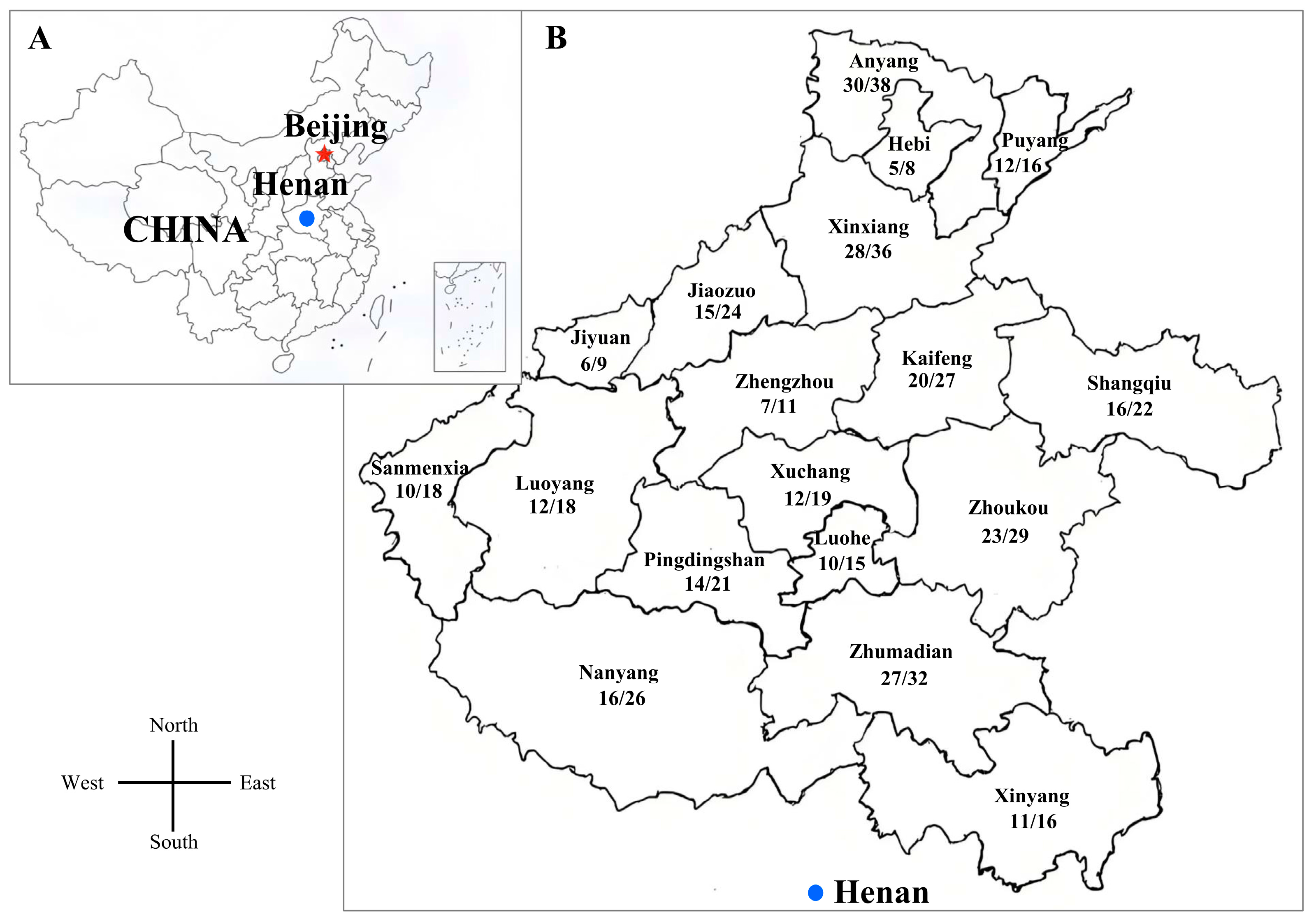
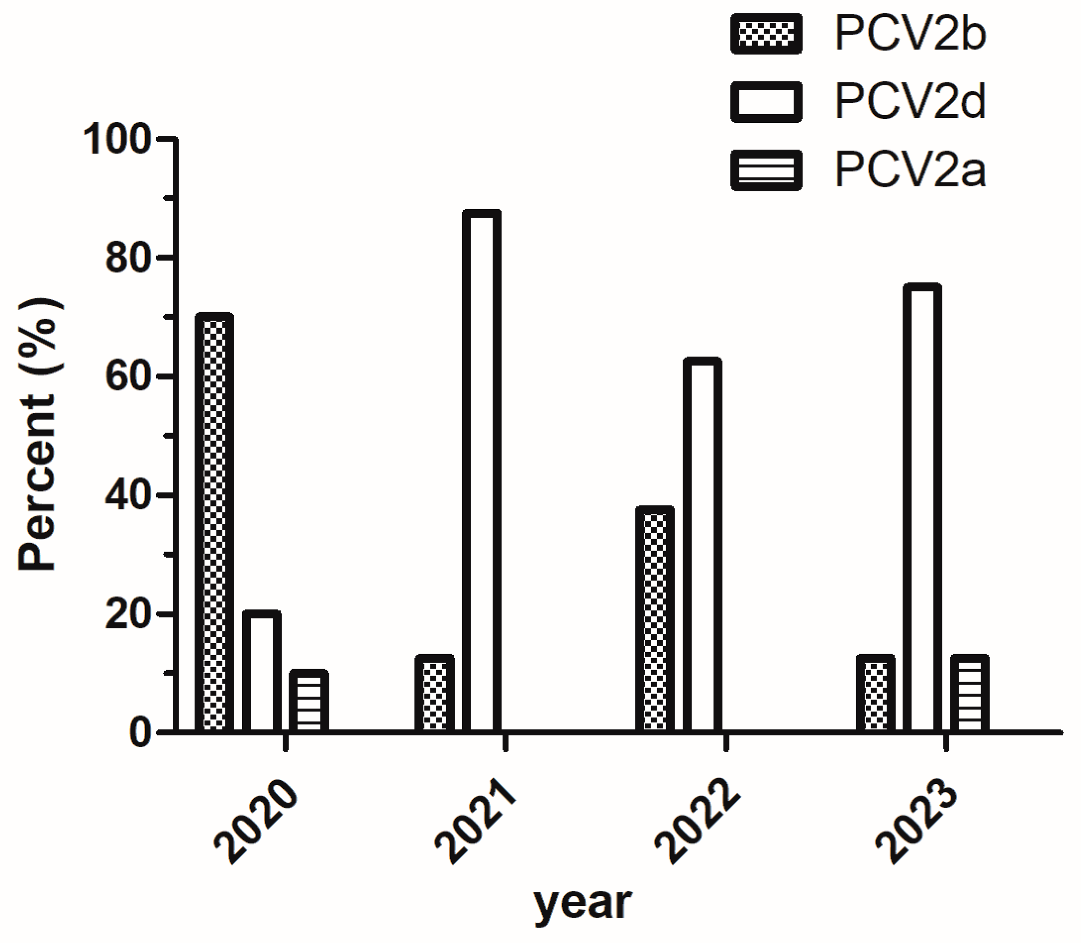
3.1. The Frequency of PCV2 in Different Regions During 2020 and 2023

From 2020 and 2023, a total of 385 clinical samples were collected, with 274 testing positive for PCV2, yielding an overall positivity rate of 71.17% (274/385) (Table 2). Among the 18 regions in Henan Province, the top three detection rates of PCV2 were 84.38% in Zhumadian, 79.31% in Zhoukou, and 78.95% in Anyang. The lowest detection rate of PCV2 was 55.56% in Sanmenxia (Figure 1). A temporal analysis of PCV2 detection rates, conducted using SPSS v28.0 with sampling duration-adjusted weights, revealed a significant decreasing trend over time: 81.16% (112/138) in 2020, 72.41% (84/116) in 2021, 62.50% (55/88) in 2022, and 53.49% (23/43) in 2023 (χ2 = 9.8, df = 3, p = 0.021). Post hoc comparisons with Bonferroni correction confirmed a marked decline between 2020 and 2023 (p = 0.003). However, key limitations should be noted. The small sample size (n = 43) may have reduced statistical power, and potential residual bias may exist due to the truncated 2023 sampling period (five months). Additionally, weighting adjustments for partial-year data introduce inherent assumptions that require careful interpretation. Despite these constraints, the observed decline suggests a potential epidemiological shift in PCV2 prevalence, warranting further validation through standardized longitudinal surveillance programs.

3.2. Genome Sequence Analysis of PCV2

To characterize the genetic diversity of recently prevalent PCV2 strains in Henan Province, 34 PCV2-positive samples were randomly selected from multiple regions for full-genome amplification, sequencing, and analysis. All 34 PCV2 strains had a genome length of 1767 nt. The following sequences were deposited in GenBank: OQ749408-OQ749433, OR114683-OR114690 (Table 3). The ORF1 gene and ORF2 genes were 945 nt and 702 nt in length, respectively. Pairwise sequence comparisons revealed nucleotide homologies ranging from 94.10% to 100.00% for the complete genome, 96.60% to 100.00% for ORF1, and 89.00% to 100.00% for ORF2. At the amino acid level, homology was found to be 98.10% to 100.00% for Rep and 87.20% to 100.00% for Cap.

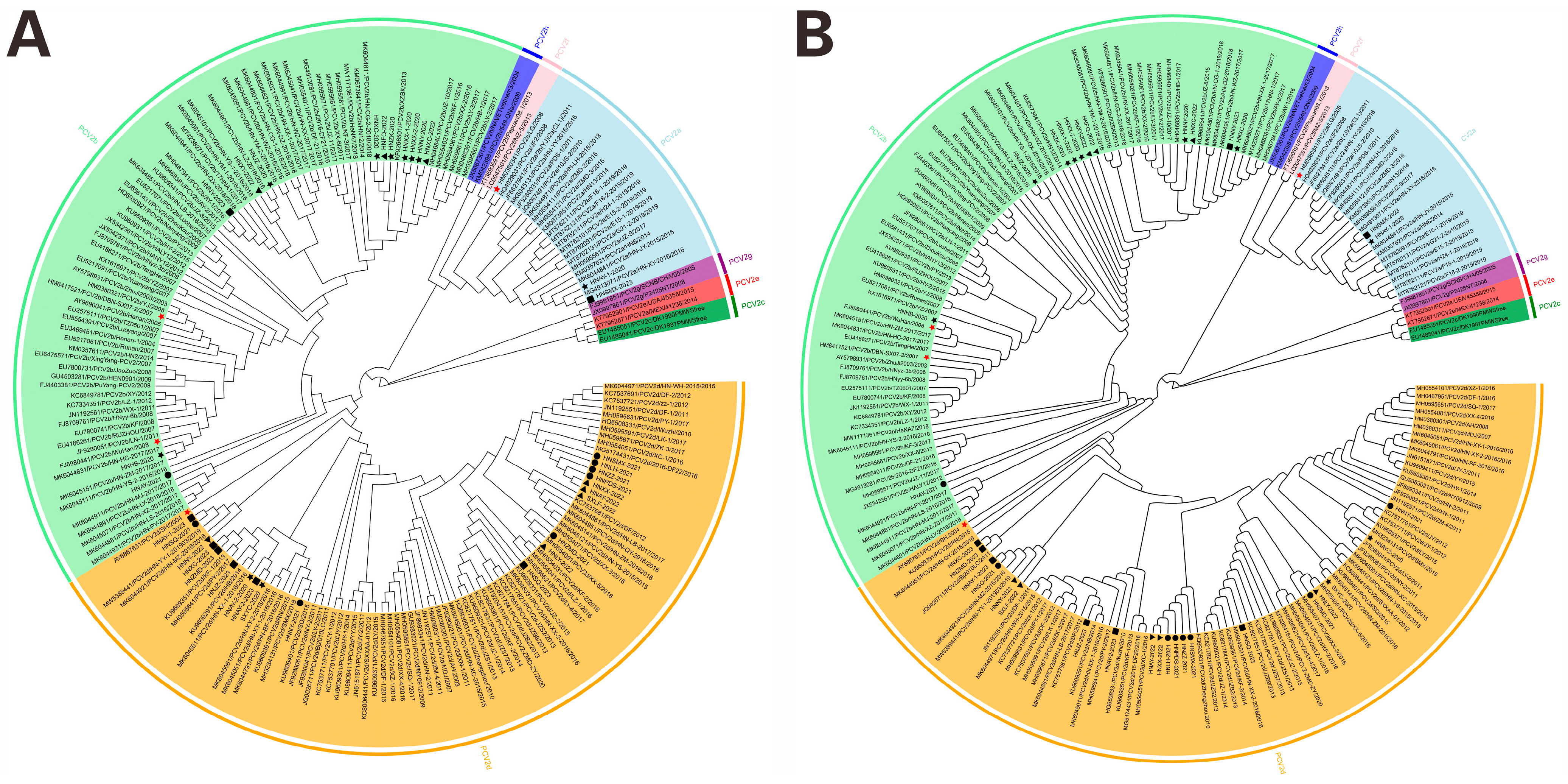
3.3. Phylogenetic Analysis of PCV2 Strains

Phylogenetic analysis of PCV2 strains based on complete PCV2 genome sequences and ORF2 amino acid sequences from the 34 strains showed that three sub-genotypes (PCV2a, PCV2b, and PCV2d) were identified in the current study (Figure 2), 58.82% (20/34) strains belonged to the sub-genotype PCV2d; and 35.30% (12/34) strains belonged to the sub-genotype PCV2b. Only two strains (5.88%, 2/34), HNAY-1-2020 and HNSMX-2023, were clustered with PCV2a strains. The percentages of PCV2d strains in 2021 (87.50%), 2022 (62.50%) and 2023 (75.00%) were higher than that in 2020 (20.00%) (Figure 3). Noticeably, 90.00% (18/20) of PCV2d strains were collected during 2021–2023, whereas 41.67% (5/12) PCV2b strains were collected during the same period.

3.4. Mutation Analysis of Cap Proteins

To explore the characteristics of the amino acid sequences of Cap of the PCV2 strains in this study, the amino acid sequence alignment was performed between the 34 PCV2 strains and the 28 reference PCV2 strains, including PCV2 commercial vaccine strains, LG (HM038034.1, PCV2a), WuHan (FJ5980441.1, PCV2b), and DBN-SX07 (HM641752.1, PCV2b) (Supplementary Figure S1). The results showed that the lengths of Cap amino acid sequences of 34 PCV2 strains were 234 amino acids. Genotype-specific substitutions were identified: PCV2b exhibited V57I, I89R/L, P134T, and D210E (absent in PCV2a/d), while PCV2d showed S68N, I89L, T/P134N, and S169G/R (absent in PCV2a/b). In addition, in this study, no amino acid deletions or insertions were observed among the 34 PCV2 strains. Thirty-one critical amino acid substitution sites were found in the Cap proteins, predominantly clustering in immunodominant regions (Table 4).

3.5. Rates of Substitution

Bayesian MCMC analysis of the PCV2 ORF2 nucleotide sequences revealed evolutionary rates, which are summarized in Table 5. Convergence diagnostics confirmed stable posterior distributions across three independent runs (10% burn-in), with effective sample size (ESS) values exceeding 200 for all parameters. Genotype-specific analyses demonstrated distinct substitution rates: PCV2a (4.658 × 10⁻⁴ ssy), PCV2b (6.476 × 10⁻⁴ ssy), and PCV2d (2.127 × 10⁻³ ssy). When all three genotypes were analyzed collectively, the substitution rate for the PCV2 ORF2 nucleotide sequences was estimated to be 1.098 × 10⁻³ ssy.

4. Discussion

PCV2 has been endemic in Chinese swine populations for decades, with PCVADs posing significant economic and health challenges to the swine industry [1,34,40]. The rapid evolution of PCV2 is driven by international breeding pig transportation, natural viral evolution, and immune pressure from vaccination [36]. Two major genotype transitions have been documented in China: PCV2a dominated prior to 2003, PCV2b emerged as the primary genotype between 2003 and 2010, and PCV2d has become predominant since 2010 [11,36,41,42]. Notably, the co-circulation of multiple genotypes within individual farms highlights the genetic plasticity of PCV2 and complicates disease management [1,30,43,44].
In this study, PCV2 was detected in 71.17% (274/385) of tissue samples collected from Henan Province (2020–2023), a rate comparable to historical regional data (62.4–72.9%, 2015–2019) [30,31], but higher than that reported in Shanghai (57.78%) [2], Yunnan (60.93%) [45], Shandong (36.98%) [11], and Hebei Province (39.96%) [32]. Taken together, these aforementioned studies indicated a high PCV2 detection rate in China. The consistently high detection rates suggest widespread PCV2 circulation in China. However, our findings indicate a gradual decline in detection rates from 2020 to 2023, likely due to structural transformations within the swine industry. Henan Province dominated China’s swine production, with tens of millions of pigs raised in thousands of pig farms of various scale every year. Owing to the high pig price (from 2020 to 2021), millions of piglets were transferred to Henan Province, which may be the reason of high frequency of PCV2 from 2020 to 2021 in Henan Province. Nevertheless, post-ASF industry restructuring led to the closure of small-scale farms with poor biosecurity, reducing swine density and inter-regional transport—key drivers of PCV2 transmission—which may explain the declining detection rates from 2022 onward.
To date, phylogenetic analyses of PCV2 genomic sequences currently delineate nine genotypes (PCV2a–i), with China exhibiting sustained co-circulation of multiple genotypes [2,34,46]. Notably, evolutionary surveillance since 2020 has identified PCV2d as the dominant circulating genotype in Chinese swine populations, replacing previously prevalent strains [2]. In this study, phylogenetic trees based on whole genome sequences and ORF2 encoded amino acid sequences classified the 34 PCV2 strains PCV2a (5.88%, 2/34), PCV2b (35.30%, 12/34), and PCV2d (58.82%, 20/34), reinforcing PCV2d’s predominance in Henan Province. This finding aligns with previous studies, including a 2018–2020 central China survey (PCV2d: 47.06%) [26] and a 2020–2021 Henan-specific analysis (PCV2d: 40.00%) [47], confirming the ongoing shift toward PCV2d dominance in China’s swine populations.
The capsid (Cap) protein, the sole structural protein of PCV2, plays a crucial role in viral entry, replication, and host immune responses [46,48]. Specially, residues 1–41 are involved in the nuclear localization of the virus [49], while residues 47–63 are essential for the recognition of PCV2 epitopes [50]. Notably, PCV2 exhibits higher substitution rates (10−3–10−4 substitutions/site/year) than typical DNA viruses [51,52], as evidenced by genotype-specific rates in this study: PCV2a (4.658 × 10⁻⁴ ssy), PCV2b (6.476 × 10⁻⁴ ssy), and PCV2d (2.127 × 10⁻³ ssy). The accelerated evolution of PCV2 likely contributes to amino acid substitutions in Cap, potentially altering viral characteristics and immune escape potential [23,33].
Analysis of ORF2 sequences from the 34 PCV2 strains revealed substantial variability (nucleotide: 88.90–100.00%; amino acid: 88.60–100.00%). In addition, three variable regions (residues 53–91, 121–136, 190–206) (Table 4) overlapped immunodominant domains (residues 65–87, 113–147, and 193–207) in the Cap protein [53]. Notably, residue 59 (Alanine in PCV2a), a neutralizing epitope, exhibited mutations that may facilitate antigenic drift and variant emergence [54]. While previous studies have postulated a correlation between Cap protein variability and PCV2 pathogenicity [54], virulence determinants are likely multigenic. Therefore, comprehensive structural analyses of genotype-specific antigenic conformations are needed to elucidate pathogenesis mechanisms and guide the development of high-efficacy PCV2 vaccines.
This study has several limitations: first, its small sample size (n = 34) and geographic biases; second, temporal sampling discontinuities hindering evolutionary trend resolution. Despite these constraints, our findings provide insights into PCV2 evolution and epidemiology. Future studies should address these limitations by incorporating larger cohorts and longitudinal sampling strategies.

5. Conclusions

This study delineates critical epidemiological transitions in PCV2 within one of China’s major swine-producing regions (Henan Province), where declining detection rates and genotype shifts (PCV2d dominance) mirror global trends, providing actionable insights for refining vaccine strategies and containment protocols in intensive pig production systems worldwide. Observed antigenic divergence in PCV2d Cap epitopes highlights potential gaps in PCV2a-based vaccine efficacy, urgently requiring PCV2d-targeted or multivalent vaccine development. Despite post-African swine fever (ASF) biosecurity improvements, persistent co-circulation of multiple genotypes highlights the necessity to integrate real-time genomic surveillance with enhanced containment measures, including optimized farm biosecurity protocols and regionally tailored vaccination programs. Collectively, these measures are critical to mitigate economic losses and preempt PCVAD resurgence in China’s swine industry.

Supplementary Materials

The following supporting information can be downloaded at https://www.mdpi.com/article/10.3390/vetsci12040343/s1: Supplementary Table S1: Background of clinical cases of porcine circovirus-associated diseases from pig farms distributed in Henan Province, China; Supplementary Table S2: Accession numbers of PCV2 strains isolated in Henan Province and other reference PCV2 strains used in this study; Supplementary Figure S1: Amino acid sequence alignment of the PCV2 Cap protein.

Author Contributions

Conceptualization, C.B. and Y.S.; methodology, H.Z., H.L. (Huifang Lv) and L.Z.; software, Y.H. and H.L. (Huawei Li); validation, Z.P., K.Z. and H.L. (Huifang Lv); formal analysis, Z.P.; investigation, Z.P. and H.L. (Huifang Lv); resources, Z.P.; data curation, L.Z. and H.L. (Huifang Lv); writing—original draft preparation, Z.P., H.L. (Huifang Lv) and H.Q.; writing—review and editing, Z.P., H.L. (Huifang Lv), L.Z., H.L. (Huawei Li), Y.S. and C.B.; visualization, H.L. (Huifang Lv); supervision, H.Q.; project administration, Z.P.; funding acquisition, C.B. All authors have read and agreed to the published version of the manuscript.

Funding

This work was supported by the grants from Key Discipline of Veterinary Medicine of Henan Province (serial number: 312), Key Discipline of Preventive Veterinary Medicine of Henan University of Animal Husbandry and Economy (XJK202202), Scientific and Technological Project of Henan Province (242102110002; 232102111048) and Henan Province key research and development project (241111113400).

Institutional Review Board Statement

This study was approved by Animal Ethical Committee of Henan University of Animal Husbandry and Economy, code: HNUAHEER2425D228 and date: 2020-02-06.

Informed Consent Statement

Informed consent was obtained from all subjects involved in the study.

Data Availability Statement

All data generated or analyzed during this study are included in this published article [and its Supplementary Information Files].

Acknowledgments

We are very grateful to Yongsheng Wang (Henan Dahenong Animal Husbandry Co. Ltd.) for kindly providing tissue samples and epidemic information.

Conflicts of Interest

The authors declare no conflicts of interest.

References

  1. Yang, Y.; Xu, T.; Wen, J.; Yang, L.; Lai, S.; Sun, X.; Xu, Z.; Zhu, L. Prevalence and phylogenetic analysis of porcine circovirus type 2 (PCV2) and type 3 (PCV3) in the Southwest of China during 2020–2022. Front. Vet. Sci. 2022, 9, 1042792. [Google Scholar] [CrossRef]
  2. Kang, L.; Wahaab, A.; Shi, K.; Mustafa, B.E.; Zhang, Y.; Zhang, J.J.; Li, Z.J.; QIU, Y.F. Molecular Epidemic Characteristics and Genetic Evolution of Porcine Circovirus Type 2 (PCV2) in Swine Herds of Shanghai, China. Viruses 2022, 14, 289. [Google Scholar] [CrossRef]
  3. Li, N.; Liu, J.; Qi, J.; Hao, F.; Xu, L.; Guo, K. Genetic Diversity and Prevalence of Porcine Circovirus Type 2 in China During 2000–2019. Front. Vet. Sci. 2021, 8, 788172. [Google Scholar] [CrossRef]
  4. Tegegne, D.; Tsegaye, G.; Aman, S.; Faustini, G.; Franzo, G. Molecular Epidemiology and Genetic Characterization of PCV2 Circulating in Wild Boars in Southwestern Ethiopia. J. Trop. Med. 2022, 2022, 5185247. [Google Scholar] [CrossRef]
  5. Liu, Y.; Gong, Q.L.; Nie, L.B.; Wang, Q.; Ge, G.Y.; Li, D.L.; Ma, B.Y.; Sheng, C.Y.; Su, N.; Zong, Y.; et al. Prevalence of porcine circovirus 2 throughout China in 2015-2019: A systematic review and meta-analysis. Microb. Pathog. 2020, 149, 104490. [Google Scholar] [CrossRef] [PubMed]
  6. Wang, Y.; Noll, L.; Lu, N.; Porter, E.; Stoy, C.; Zheng, W.; Liu, X.; Peddireddi, L.; Niederwerder, M.; Bai, J. Genetic diversity and prevalence of porcine circovirus type 3 (PCV3) and type 2 (PCV2) in the Midwest of the USA during 2016-2018. Transbound. Emerg. Dis. 2020, 67, 1284–1294. [Google Scholar] [CrossRef]
  7. de Sousa Moreira, A.; Santos-Silva, S.; Mega, J.; Palmeira, J.D.; Torres, R.T.; Mesquita, J.R. Epidemiology of Porcine Circovirus Type 2 Circulating in Wild Boars of Portugal during the 2018-2020 Hunting Seasons Suggests the Emergence of Genotype 2d. Animals 2022, 12, 451. [Google Scholar] [CrossRef]
  8. Ouyang, T.; Zhang, X.W.; Liu, X.H.; Ren, L.Z. Co-Infection of Swine with Porcine Circovirus Type 2 and Other Swine Viruses. Viruses 2019, 11, 185. [Google Scholar] [CrossRef] [PubMed]
  9. Harding, J.C. The clinical expression and emergence of porcine circovirus 2. Vet. Microbiol. 2004, 98, 131–135. [Google Scholar] [CrossRef]
  10. Rose, N.; Opriessnig, T.; Grasland, B.; Jestin, A. Epidemiology and transmission of porcine circovirus type 2 (PCV2). Virus Res. 2012, 164, 78–89. [Google Scholar] [CrossRef]
  11. Ma, Z.; Liu, M.; Liu, Z.; Meng, F.; Wang, H.; Cao, L.; Li, Y.; Jiao, Q.; Han, Z.; Liu, S. Epidemiological investigation of porcine circovirus type 2 and its coinfection rate in Shandong province in China from 2015 to 2018. BMC Vet. Res. 2021, 17, 17. [Google Scholar] [CrossRef]
  12. Chae, C. Porcine respiratory disease complex: Interaction of vaccination and porcine circovirus type 2, porcine reproductive and respiratory syndrome virus, and Mycoplasma hyopneumoniae. Vet. J. 2016, 212, 1–6. [Google Scholar] [CrossRef]
  13. Tischer, I.; Mields, W.; Wolff, D.; Vagt, M.; Griem, W. Studies on epidemiology and pathogenicity of porcine circovirus. Arch. Virol. 1986, 91, 271–276. [Google Scholar] [CrossRef]
  14. Franzo, G.; Cortey, M.; de Castro, A.M.; Piovezan, U.; Szabo, M.P.; Drigo, M.; Segalés, J.; Richtzenhain, L.J. Genetic characterisation of Porcine circovirus type 2 (PCV2) strains from feral pigs in the Brazilian Pantanal: An opportunity to reconstruct the history of PCV2 evolution. Vet. Microbiol. 2015, 178, 158–162. [Google Scholar] [CrossRef] [PubMed]
  15. Xiao, C.T.; Halbur, P.G.; Opriessnig, T. Global molecular genetic analysis of porcine circovirus type 2 (PCV2) sequences confirms the presence of four main PCV2 genotypes and reveals a rapid increase of PCV2d. J. Gen. Virol. 2015, 96, 1830–1841. [Google Scholar] [CrossRef] [PubMed]
  16. Zhai, S.L.; He, D.S.; Qi, W.B.; Chen, S.N.; Deng, S.F.; Hu, J.; Li, X.P.; Li, L.; Chen, R.A.; Luo, M.L.; et al. Complete genome characterization and phylogenetic analysis of three distinct buffalo-origin PCV2 isolates from China. Infect. Genet. Evol. 2014, 28, 278–282. [Google Scholar] [CrossRef]
  17. Guo, L.J.; Lu, Y.H.; Wei, Y.W.; Huang, L.P.; Liu, C.M. Porcine circovirus type 2 (PCV2): Genetic variation and newly emerging genotypes in China. Virol. J. 2010, 7, 273. [Google Scholar] [CrossRef]
  18. Jantafong, T.; Boonsoongnern, A.; Poolperm, P.; Urairong, K.; Lekcharoensuk, C.; Lekcharoensuk, P. Genetic characterization of porcine circovirus type 2 in piglets from PMWS-affected and -negative farms in Thailand. Virol. J. 2011, 8, 88. [Google Scholar] [CrossRef]
  19. Bao, F.; Mi, S.; Luo, Q.; Guo, H.; Tu, C.; Zhu, G.; Gong, W. Retrospective study of porcine circovirus type 2 infection reveals a novel genotype PCV2f. Transbound. Emerg. Dis. 2018, 65, 432–440. [Google Scholar] [CrossRef]
  20. Yang, X.; Chen, F.; Cao, Y.; Pang, D.; Ouyang, H.; Ren, L. Complete genome sequence of porcine circovirus 2b strain CC1. J. Virol. 2012, 86, 9536. [Google Scholar] [CrossRef]
  21. Mankertz, A.; Mankertz, J.; Wolf, K.; Buhk, H.J. Identification of a protein essential for replication of porcine circovirus. J. Gen. Virol. 1998, 79, 381–384. [Google Scholar] [CrossRef]
  22. Finsterbusch, T.; Mankertz, A. Porcine circoviruses--small but powerful. Virus Res. 2009, 143, 177–183. [Google Scholar] [CrossRef] [PubMed]
  23. Nawagitgul, P.; Morozov, I.; Bolin, S.R.; Harms, P.A.; Sorden, S.D.; Paul, P.S. Open reading frame 2 of porcine circovirus type 2 encodes a major capsid protein. J. Gen. Virol. 2000, 81, 2281–2287. [Google Scholar] [CrossRef]
  24. Shi, R.; Hou, L.; Liu, J. Host immune response to infection with porcine circoviruses. Anim. Dis. 2021, 1, 23. [Google Scholar] [CrossRef]
  25. Olvera, A.; Cortey, M.; Segalés, J. Molecular evolution of porcine circovirus type 2 genomes: Phylogeny and clonality. Virology 2007, 357, 175–185. [Google Scholar] [CrossRef]
  26. Xu, T.; Zhang, Y.H.; Tian, R.B.; Hou, C.Y.; Li, X.S.; Zheng, L.L.; Wang, L.Q.; Chen, H.Y. Prevalence and genetic analysis of porcine circovirus type 2 (PCV2) and type 3 (PCV3) between 2018 and 2020 in central China. Infect. Genet. Evol. 2021, 94, 105016. [Google Scholar] [CrossRef] [PubMed]
  27. Alarcon, P.; Rushton, J.; Wieland, B. Cost of post-weaning multi-systemic wasting syndrome and porcine circovirus type-2 subclinical infection in England—An economic disease model. Prev. Vet. Med. 2013, 110, 88–102. [Google Scholar] [CrossRef]
  28. Lang, H.W.; Zhang, G.C.; Wu, F.Q.; Zhang, C.G. Detection of serum antibody against postweaning multisystemic wasting syndrome in pigs. Chin. J. Vet. Sci. Technol. 2000, 30, 3–5. [Google Scholar]
  29. Wang, S.Y.; Sun, Y.F.; Wang, Q.; Yu, L.X.; Zhu, S.Q.; Liu, X.M.; Yao, Y.; Wang, J.; Shan, T.L.; Zheng, H.; et al. An epidemiological investigation of porcine circovirus type 2 and porcine circovirus type 3 infections in Tianjin, North China. PeerJ 2020, 8, e9735. [Google Scholar] [CrossRef]
  30. Zheng, G.; Lu, Q.; Wang, F.; Xing, G.; Feng, H.; Jin, Q.; Guo, Z.; Teng, M.; Hao, H.; Li, D.; et al. Phylogenetic analysis of porcine circovirus type 2 (PCV2) between 2015 and 2018 in Henan Province, China. BMC Vet. Res. 2020, 16, 6. [Google Scholar] [CrossRef]
  31. Jia, Y.; Zhu, Q.; Xu, T.; Chen, X.; Li, H.; Ma, M.; Zhang, Y.; He, Z.; Chen, H. Detection and genetic characteristics of porcine circovirus type 2 and 3 in Henan province of China. Mol. Cell Probes 2022, 61, 101790. [Google Scholar] [CrossRef]
  32. Han, L.; Yuan, G.F.; Chen, S.J.; Dai, F.; Hou, L.S.; Fan, J.H.; Zuo, Y.Z. Porcine circovirus type 2 (PCV2) infection in Hebei Province from 2016 to 2019: A retrospective study. Arch. Virol. 2021, 166, 2159–2171. [Google Scholar] [CrossRef] [PubMed]
  33. Franzo, G.; Cortey, M.; Segalés, J.; Hughes, J.; Drigo, M. Phylodynamic analysis of porcine circovirus type 2 reveals global waves of emerging genotypes and the circulation of recombinant forms. Mol. Phylogenetics Evol. 2016, 100, 269–280. [Google Scholar] [CrossRef]
  34. Xiao, Q.; Wen, L.; Yin, L.; Zhang, F.; Li, T.; Banma, Z.; He, K.; Suolang, S.; Nan, W.; Wu, J.; et al. Prevalence and genetic diversity of porcine circovirus type 2 in northern Guangdong Province during 2016–2021. Front. Vet. Sci. 2022, 9, 932612. [Google Scholar] [CrossRef]
  35. Xu, G.; Sarkar, A.; Qian, L.; Shuxia, Z.; Rahman, M.A.; Yongfeng, T. The impact of the epidemic experience on the recovery of production of pig farmers after the outbreak-Evidence from the impact of African swine fever (ASF) in Chinese pig farming. Prev. Vet. Med. 2022, 199, 105568. [Google Scholar] [CrossRef] [PubMed]
  36. Xu, P.L.; Zhao, Y.; Zheng, H.H.; Tian, R.B.; Han, H.Y.; Chen, H.Y.; Zheng, L.L. Analysis of genetic variation of porcine circovirus type 2 within pig populations in central China. Arch. Virol. 2019, 164, 1445–1451. [Google Scholar] [CrossRef]
  37. Hu, Y.; Zhan, Y.; Wang, D.; Xie, X.; Liu, T.; Liu, W.; Wang, N.; Deng, Z.; Lei, H.; Yang, Y.; et al. Evidence of natural co-infection with PCV2b subtypes in vivo. Arch. Virol. 2017, 162, 2015–2020. [Google Scholar] [CrossRef]
  38. Drummond, A.J.; Suchard, M.A.; Xie, D.; Rambaut, A. Bayesian phylogenetics with BEAUti and the BEAST 1.7. Mol. Biol. Evol. 2012, 29, 1969–1973. [Google Scholar] [CrossRef]
  39. Hill, V.; Baele, G. Bayesian Estimation of Past Population Dynamics in BEAST 1.10 Using the Skygrid Coalescent Model. Mol. Biol. Evol. 2019, 36, 2620–2628. [Google Scholar] [CrossRef]
  40. Suh, J.; Oh, T.; Park, K.; Yang, S.; Cho, H.; Chae, C. A Comparison of Virulence of Three Porcine Circovirus Type 2 (PCV2) Genotypes (a, b, and d) in Pigs Singularly Inoculated with PCV2 and Dually Inoculated with PCV2 and Porcine Reproductive and Respiratory Syndrome Virus. Pathogens 2021, 10, 891. [Google Scholar] [CrossRef]
  41. Constans, M.; Ssemadaali, M.; Kolyvushko, O.; Ramamoorthy, S. Antigenic Determinants of Possible Vaccine Escape by Porcine Circovirus Subtype 2b Viruses. Bioinform. Biol. Insights 2015, 9, 1–12. [Google Scholar] [CrossRef] [PubMed]
  42. Hou, Z.; Wang, H.; Feng, Y.; Song, M.; Li, Q.; Li, J. Genetic variation and phylogenetic analysis of Porcine circovirus type 2 in China from 2016 to 2018. Acta Virol. 2019, 63, 459–468. [Google Scholar] [CrossRef]
  43. Yue, W.; Li, Y.; Zhang, X.; He, J.; Ma, H. Prevalence of Porcine Circoviruses in Slaughterhouses in Central Shanxi Province, China. Front. Vet. Sci. 2022, 9, 820914. [Google Scholar] [CrossRef]
  44. Jang, G.; Yoo, H.; Kim, Y.; Yang, K.; Lee, C. Genetic and phylogenetic analysis of porcine circovirus type 2 on Jeju Island, South Korea, 2019-2020: Evidence of a novel intergenotypic recombinant. Arch. Virol. 2021, 166, 1093–1102. [Google Scholar] [CrossRef] [PubMed]
  45. Lv, N.; Zhu, L.; Li, W.; Li, Z.; Qian, Q.; Zhang, T.; Liu, L.; Hong, J.; Zheng, X.; Wang, Y.; et al. Molecular epidemiology and genetic variation analyses of porcine circovirus type 2 isolated from Yunnan Province in China from 2016–2019. TBMC Vet. Res. 2020, 16, 96. [Google Scholar] [CrossRef]
  46. Huang, Y.; Chen, X.; Long, Y.; Yang, L.; Song, W.; Liu, J.; Li, Q.; Liang, G.; Yu, D.; Huang, C.; et al. Epidemiological Analysis From 2018 to 2020 in China and Prevention Strategy of Porcine Circovirus Type 2. Front. Vet. Sci. 2021, 8, 753297. [Google Scholar] [CrossRef]
  47. Xu, T.; Hou, C.Y.; Zhang, Y.H.; Li, H.X.; Chen, X.M.; Pan, J.J.; Chen, H.Y. Simultaneous detection and genetic characterization of porcine circovirus 2 and 4 in Henan province of China. Gene 2022, 808, 145991. [Google Scholar] [CrossRef]
  48. Zhan, Y.; Yu, W.; Cai, X.; Lei, X.; Lei, H.; Wang, A.; Sun, Y.; Wang, N.; Deng, Z.; Yang, Y. The Carboxyl Terminus of the Porcine Circovirus Type 2 Capsid Protein Is Critical to Virus-Like Particle Assembly, Cell Entry, and Propagation. J. Virol. 2020, 94, e00042-00020. [Google Scholar] [CrossRef]
  49. Hou, Q.; Hou, S.; Chen, Q.; Jia, H.; Xin, T.; Jiang, Y.; Guo, X.; Zhu, H. Nuclear localization signal regulates porcine circovirus type 2 capsid protein nuclear export through phosphorylation. Virus Res. 2018, 246, 12–22. [Google Scholar] [CrossRef]
  50. Lekcharoensuk, P.; Morozov, I.; Paul, P.S.; Thangthumniyom, N.; Wajjawalku, W.; Meng, X.J. Epitope mapping of the major capsid protein of type 2 porcine circovirus (PCV2) by using chimeric PCV1 and PCV2. J. Virol. 2004, 78, 8135–8145. [Google Scholar] [CrossRef]
  51. Tan, C.Y.; Thanawongnuwech, R.; Arshad, S.S.; Hassan, L.; Fong, M.W.C.; Ooi, P.T. Genotype Shift of Malaysian Porcine Circovirus 2 (PCV2) from PCV2b to PCV2d within a Decade. Animals 2022, 12, 1849. [Google Scholar] [CrossRef] [PubMed]
  52. Duffy, S.; Shackelton, L.A.; Holmes, E.C. Rates of evolutionary change in viruses: Patterns and determinants. Nat. Rev. Genet. 2008, 9, 267–276. [Google Scholar] [CrossRef] [PubMed]
  53. Mahé, D.; Blanchard, P.; Truong, C.; Arnauld, C.; Le Cann, P.; Cariolet, R.; Madec, F.; Albina, E.; Jestin, A. Differential recognition of ORF2 protein from type 1 and type 2 porcine circoviruses and identification of immunorelevant epitopes. J. Gen. Virol. 2000, 81, 1815–1824. [Google Scholar] [CrossRef]
  54. Larochelle, R.; Magar, R.; D’Allaire, S. Genetic characterization and phylogenetic analysis of porcine circovirus type 2 (PCV2) strains from cases presenting various clinical conditions. Virus Res. 2002, 90, 101–112. [Google Scholar] [CrossRef] [PubMed]
Figure 1. The geographical locations in Henan Province of the samples collected in this study. (A) Geographic location of Henan Province in China. (B) The geographical locations in Henan Province of China where 385 samples were collected. The numbers indicate the detection rate of PCV2 in different cities. Red star: Beijing (China’s capital); Blue dot: Henan Province.
Vetsci 12 00343 g001
Figure 2. Phylogenetic trees based on the PCV2 strains obtained in this study and reference strains. (A) Phylogenetic tree of the complete genome sequences. (B) Phylogenetic tree of deduced amino acid sequences of ORF2. The phylogenetic reconstruction was performed in MEGA6.0 software using the neighbor-joining algorithm with 1000 bootstrap replicates. Red star represented PCV2 commercial vaccine strains. Black stars, black circles, black triangles and black squares represented PCV2 strains obtained in this study in 2020, 2021, 2022 and 2023, respectively.
Vetsci 12 00343 g002
Figure 3. The proportion of PCV2 subtypes in different years in this study. A total of 385 clinical samples were collected from pig farms in 18 cities in Henan Province during February 2020 and May 2023, with 274 testing positive for PCV2. The percentages of PCV2d strains in 2021 (87.50%), 2022 (62.50%) and 2023 (75.00%) were higher than that in 2020 (20.00%).
Vetsci 12 00343 g003
Table 1. Primers used in this study.
Primer NameSequence (5′−3′)LengthAnnealing TemperaturePurpose
PCV2-FCTGTTTTCGAACGCAGTGCC486 bp56 °CDetection of PCV2 [36]
PCV2-RGCATCTTCAACACCCGCCT
PCV2-C-FCGGGGTACCACTGAGTCTTTTTTATCACTTCG1767–1768 bp58 °CPCV2 whole-genome amplification [37]
PCV2-C-RCCCAAGCTTAAGACTCAGTAATTTATTTCATATGG
Table 2. The frequency of PCV2 in different regions in Henan Province of China during 2020 and 2023.
RegionNo. of Tested SamplesNo. of PCV2 Positive SamplesFrequency
Anyang383078.95%
Puyang161275.00%
Hebi8562.50%
Xinxiang362877.78%
Jiaozuo241562.50%
Jiyuan9666.67%
Zhenzhou11763.64%
Kaifeng272074.07%
Shangqiu221672.73%
Zhoukou292379.31%
Xuchang191263.16%
Luohe151066.67%
Zhumadian322784.38%
Pingdingshan211466.67%
Nanyang261661.54%
Luoyang181266.67%
Sanmenxia181055.56%
Xinyang161168.75%
Total38527471.17%
Table 3. Detailed information of PCV2 strains sequenced in this study.
StrainCollection YearIsolation OriginGenotypeAccession No.Length (nt)
HNZK-20202020Zhoukou, HenanPCV2bOQ7494081767
HNXX-1-20202020Xinxiang, HenanPCV2bOQ7494091767
HNNY-20202020Nanyang, HenanPCV2bOQ7494101767
HNJZ-20202020Jiaozuo, HenanPCV2bOQ7494111767
HNXC-20202020Xuchang, HenanPCV2bOQ7494121767
HNAY-1-20202020Anyang, HenanPCV2aOQ7494131767
HNXX-2-20202020Xinxiang, HenanPCV2bOQ7494141767
HNHB-20202020Hebi, HenanPCV2bOQ7494151767
HNAY-2-20202020Anyang, HenanPCV2dOQ7494161767
HNJY-20202020Jiyuan, HenanPCV2dOQ7494171767
HNAY-20212021Anyang, HenanPCV2bOQ7494181767
HNZZ-20212021Zhengzhou, HenanPCV2dOQ7494191767
HNPDS-20212021Pingdingshan, HenanPCV2dOQ7494201767
HNSQ-20212021Shangqiu, HenanPCV2dOQ7494211767
HNLH-20212021Luohe, HenanPCV2dOQ7494221767
HNNY-20212021Nanyang, HenanPCV2dOQ7494231767
HNZMD-20212021Zhumadian, HenanPCV2dOQ7494241767
HNSMX-20212021Sanmenxia, HenanPCV2dOQ7494251767
HNXX-20222022Xinxiang, HenanPCV2dOQ7494261767
HNAY-20222022Anyang, HenanPCV2dOQ7494271767
HNXY-20222022Xinyang, HenanPCV2bOQ7494281767
HNXC-20222022Xuchang, HenanPCV2bOQ7494291767
HNLY-20222022Luoyang, HenanPCV2dOQ7494301767
HNNY-20222022Nanyang, HenanPCV2dOQ7494311767
HNFQ-20222022Xinxiang, HenanPCV2bOQ7494321767
HNLY-20222022Luoyang, HenanPCV2dOQ7494331767
HNJZ-20232023Jiaozuo, HenanPCV2dOR1146831767
HNZMD-20232023Zhumadian, HenanPCV2dOR1146841767
HNXC-20232023Xuchang, HenanPCV2dOR1146851767
HNSQ-20232023Shangqiu, HenanPCV2dOR1146861767
HNNY-20232023Nanyang, HenanPCV2bOR1146871767
HNAY-1-20232023Anyang, HenanPCV2dOR1146881767
HNSMX-20232023Sanmenxia, HenanPCV2aOR1146891767
HNAY-2-20232023Anyang, HenanPCV2dOR1146901767
Table 4. Critical amino acid substitutions in the Cap proteins of PCV2 strains identified in this study.
PositionPCV2aPCV2bPCV2dPositionPCV2aPCV2bPCV2d
8FF(1/12/Y(11/12)F(16/20)/Y(4/20)121SST
47ATT131MTT
53FF(11/12)/I(1/12)I133VAA
57VIV134PTN
59AK(11/12)/R(1/12)K136QLL
63SK(1/12)/R(11/12)R151PTT
68SAN169SS(11/12)/C(1/12)R(3/20)/G(17/20)
73LMM185MLL
77LII190SA(1/12)/T(11/12)T
78DNN191RGG
80VLL206T/KII
86TSS210DED
88KPP215VVI
89IR(11/12)/L(1/12)L232NNN
90SST234KK(1/12)/(11/12)K
91IVV
Table 5. Substitution rates of PCV2 ORF2 nucleotide sequences.
PCV2 GenotypeMean Rate of Substitution (Substitution/site/year)95% Highest Posterior DensityEffective Sample Size
PCV2a4.658 × 10−48.643 × 10−4245,643
PCV2b6.476 × 10−41.063 × 10−3254,638
PCV2d2.127 × 10−32.853 × 10−367,627
Combined analysis for all genotypes1.098 × 10−31.381 × 10−381,829
Disclaimer/Publisher’s Note: The statements, opinions and data contained in all publications are solely those of the individual author(s) and contributor(s) and not of MDPI and/or the editor(s). MDPI and/or the editor(s) disclaim responsibility for any injury to people or property resulting from any ideas, methods, instructions or products referred to in the content.

