Article

Rhodotorula Yeast Culture Improved the Antioxidant Capacity, Lipid Metabolism, and Immunity of Sheep Livers

1
College of Animal Science, Inner Mongolia Agricultural University, Hohhot 010018, China
2
Hetao College, Bayannur 015000, China
3
Inner Mongolia Herbivorous Livestock Feed Engineering Technology Research Center, Hohhot 010018, China
*
Authors to whom correspondence should be addressed.
Vet. Sci. 2025, 12(4), 314; https://doi.org/10.3390/vetsci12040314
Submission received: 15 February 2025 / Revised: 18 March 2025 / Accepted: 28 March 2025 / Published: 30 March 2025

Simple Summary

The use of feed additives to ensure sheep health has attracted increasing attention under China’s current policy of completely prohibiting the addition of antibiotics to feed. Rhodotorula is a kind of fungus that can produce carotenoids. Previous findings showed that R. mucilaginosa offers great potential as a source of antioxidants. Yeast culture, a type of yeast additive, exhibits beneficial effects for lipid metabolism and immunity. Therefore, we hypothesized that R. mucilaginosa yeast cultures have the potential to replace feed antibiotics. To test this hypothesis, we explored the effects of R. mucilaginosa yeast cultures as an additive on the antioxidant capacity, lipid metabolism, and immunity in sheep liver samples. Adding 20 g/day R. mucilaginosa yeast culture significantly increased the activities of antioxidant enzymes, decreased blood lipid levels, improved fatty acid composition, and decreased the levels of pro-inflammatory factors in sheep livers. These results indicate that R. mucilaginosa yeast culture contributed to maintaining a healthy sheep herd, providing a scientific basis for using R. mucilaginosa yeast culture as a feed additive for sheep.

Abstract

There is currently no research on the application evaluation of R. mucilaginosa yeast culture (RYC) in animal production. Therefore, this study investigated the effects of RYC on the antioxidant capacity, lipid metabolism, and immunity of sheep livers. Twenty-four 3-month-old Duhan male sheep (36 ± 4 kg) were divided into four groups. The control group received a basal diet, the L group received a basal diet + 10 g/sheep/day RYC, the M group received a basal diet + 20 g/sheep/day RYC, and the H group received a basal diet + 40 g/sheep/day RYC. The trial lasted for 75 days. The results showed that the content of glutathione peroxidase in the livers of sheep in group M was significantly increased by 26.6%, and the content of malondialdehyde was significantly decreased by 38% (p < 0.05). Additionally, the serum levels of total cholesterol, triglycerides, high-density lipoprotein cholesterol, and low-density lipoprotein cholesterol significantly decreased; the liver content of C16:0 decreased; and the levels of C18:2n6C and C20:1 increased (p < 0.05). Furthermore, the contents of cytokines TNF-α and IFN-γ in sheep livers from the M group were also significantly decreased by 20% and 24.8%, respectively (p < 0.05). These findings suggest that supplementation with 20 g/sheep/day RYC can enhance antioxidant capacity, improve lipid metabolism, and reduce inflammation in sheep livers, which is advantageous for farming healthy sheep.

1. Introduction

Sheep play an important role in the Chinese husbandry industry as an important source of meat. However, many common adverse factors exist in current sheep-feeding processes, such as a high proportion of concentrate [1], moldy feed [2], and an unsuitable growth environment [3], that negatively impact the health and production performance of sheep. Previously, antibiotics were commonly added to feed to counteract these negative effects or improve production efficiency. However, unregulated use has led to antibiotic residues in animal products and the emergence of drug-resistant strains in livestock and poultry. These resistant strains can colonize consumers, thereby diminishing the efficacy of antibiotics in treating human diseases [4,5]. Considering the health of consumers, China issued an announcement in 2020 calling for a total ban on the addition of antibiotics during feed production [6]. Therefore, safe and effective feed additives are urgently required for the sheep industry.
Yeast culture (YC) is a unique feed additive produced by fermenting live yeast under fixed culture conditions and then drying the entire medium. Its main components are yeast cell walls and various metabolites [7]. Studies on monogastric animals have shown that YC can affect immunity and intestinal microbial ecology [8,9,10] and increase serum antioxidant enzyme activity [11]. Studies on ruminants have shown that YC can significantly affect the antioxidant level, immune factor content, and fatty acid content in the back muscles of sheep [12,13]. Moreover, YC also impacts rumen microbial diversity, which subsequently affects lipid metabolism-related metabolites within the rumen or modulates the overall rumen environment [14,15]. When used in conjunction with glycerol, YC can improve the energy status of dairy cows and the expression of gluconeogenic enzymes in the liver [16]. Meanwhile, Feng et al.’s research indicates that YC also significantly affects the lipid metabolism pathways in fish liver [17].
Rhodotorula species, such as R. mucilaginosa and R. minuta, are widely distributed fungi, which can produce carotenoids, digestive enzymes, β-glucan, vitamins, and other active metabolites [18,19,20]. The results of a recent mouse study showed that R. mucilaginosa significantly improved antioxidant capacity and reduced cyclophosphamide-induced immunosuppression and immune organ atrophy [21]. Hu et al. [19] found that R. mucilaginosa improved growth performance, enhanced antioxidant capacity, strengthened gastrointestinal digestion, and maintained the intestinal microbial balance in piglets. Sun et al. [22] showed that an R. mucilaginosa solid-state fermentation product improved the laying performance and intestinal flora of hens, speculating that the improved performance might have been related to its metabolites, such as β-glucan. Chen et al. [23] found that the hydrolysate of R. mucilaginosa enhanced the antioxidant capacity of juvenile Nile tilapia. In general, previous research has shown great potential of R. mucilaginosa as an antioxidant agent.
The liver is central for various metabolic activities in the body, maintaining homeostasis and playing important roles in oxidative stress, lipid metabolism, and immunity [24,25,26]. Current studies have shown that YC has certain effects on sheep antioxidants, lipid metabolism, and immunity. R. mucilaginosa has the characteristics of producing carotenoids. Therefore, we speculate that the yeast culture made of R. mucilaginosa may have the effect of YC and also show the effect of carotenoids. However, there is no study on the effect of R. mucilaginosa yeast culture (RYC) on sheep. Therefore, this study attempts to explore the effects of dietary supplementation of RYC on antioxidants, lipid metabolism, and the immunity of sheep livers and provide a tentative exploration for whether RYC can be used as an alternative antibiotic feed additive.

2. Materials and Methods

The animal study protocol was approved by the Animal Welfare and Ethics Committee of Inner Mongolia Agricultural University (approval number NND2022110). RYC was provided by the Chinese Academy of Agricultural Sciences Beijing Institute of Animal Husbandry and Veterinary Medicine, using soybean meal as the solid-state fermentation substrate, and inoculated with the liquid fermentation broth of R. mucilaginosa.

2.1. Experimental Design

Twenty-four 3-month-old male Duhan sheep (36 ± 4 kg) were randomly assigned to four groups, with six sheep in each group. These groups included the control (CON) group (fed a basal diet), L group (fed basal diet + 10 g/sheep/day RYC), M group (fed basal diet + 20 g/sheep/day RYC), and H group (fed basal diet + 40 g/sheep/day RYC). The entire experiment lasted 90 days (15-day acclimatization phase + 75-day trial period). Throughout the study, all sheep were fed twice daily at 8:00 and 18:00 and were provided with ad libitum access to water. During the trial period, each sheep was fed a specific amount of RYC before 8:00 AM daily, followed by uniform feeding of the basal diet. On the last day of the trial period, blood was drawn from all sheep in the morning before they were fed, and serum samples were subsequently obtained. Fasting and water-deprivation for a whole night. On the following day, five sheep were randomly selected from each group for the Islamic method of slaughter [27] and sampling.

2.2. Diet Composition

The basic diet used in this experiment was prepared by Fuchuan Feed Co., Ltd. (Inner Mongolia, China) of Inner Mongolia according to the “NY/T 816-2021 Nutritional Requirements for Mutton Sheep in China”. The ratio of concentrate to roughage was 5:5. The nutritional components of the basal diet used in this study are presented in Table 1.

2.3. Sampling

Sheep blood was collected from the jugular vein using a disposable vacuum blood-collection vessel. After standing at room temperature for 40 min, each blood sample was centrifuged at 3000 rpm for 10 min, after which the serum was collected and stored at −20 °C. After the sheep were slaughtered, the surface blood stains were washed with cold normal saline. The central portion of the liver from each sheep was then excised using surgical scissors and placed into a cryogenic storage tube (Corning, NY, USA). Following this, the samples were immediately frozen in liquid nitrogen and subsequently transferred to a −80 °C freezer for storage.

2.4. Liver-Antioxidant Capacity

Total superoxide dismutase (T-SOD), catalase (CAT), glutathione peroxidase (GPx), total antioxidative capacity (T-AOC), malondialdehyde (MDA), and protein levels were measured using appropriate kits from Nanjing Jiancheng Bioengineering Institute (Nanjing, China), per the manufacturer’s instructions. The liver homogenate for the experiment was prepared according to the instructions.

2.5. Serum Lipid Metabolism

Serum glucose (GLU), total cholesterol (TC), triglycerides (TG), high-density lipoprotein cholesterol (HDL-C), and low-density lipoprotein cholesterol (LDL-C) concentrations were determined using appropriate kits from Nanjing Jiancheng Bioengineering Institute (Nanjing, China), per the manufacturer’s instructions. Free fatty acid (FFA) contents were determined using an ELISA kit from Baoman Biological Technology Co., Ltd. (Shanghai, China).

2.6. Liver Fatty Acid Content

Livers (0.6 g) were weighed, ground into powder, added to 0.7 mL of 10 mol/L KOH and 5.3 mL of methanol, mixed well, placed in a 55 °C water bath for 1.5 h, with occasional oscillatory mixing during the period, and cooled to room temperature; then, 0.58 mL of 12 mol/L H2SO4 was added; it was mixed again, mixed in a 55 °C water bath for 1.5 h, with occasional oscillatory mixing during the period, and cooled to room temperature; 3 mL n-hexane was added, it was vortexed for 5 min and centrifuged at 1500× g for 5 min. Then, 1 mL supernatant filtered through a 0.22 μm filter membrane was collected by loading a glass bottle. Fatty acid contents were determined using a 6890N gas chromatograph (Agilent Technologies Inc., Santa Clara, CA, USA), according to a previous method described by Zhao [28].

2.7. Liver Immune Cytokines

We used appropriate ELISA kits (Baoman Biological Technology Co., Ltd., Shanghai, China) to measure interleukin 1β (IL-1β), interleukin 6 (IL-6), interleukin 10 (IL-10), tumor necrosis factor-α (TNF-α), and interferon-γ (IFN-γ) levels in the homogenates. The liver homogenate for the experiment was prepared according to the instructions. Protein quantification was performed as described in Section 2.4.

2.8. Liver RNA Extraction and Quantitative Real-Time PCR

Liver samples (0.05 g) were added to 1 mL of RNAiso Plus reagent (Takara, Dalian, China), and total RNA was extracted per the manufacturer’s instructions. An Implen P330 device (Implen, Munich, Germany) was used to determine the RNA concentrations. Complementary DNA (cDNA) was generated using an ABI Veriti 96 instrument (Applied Biosystems, Waltham, MA, USA) and a reverse transcription kit (AGbio Co., Ltd., Changsha, China). The cDNA was temporarily stored at −20 °C. A qPCR kit (AGbio Co., Ltd., Changsha, China) and LightCycler 480II Instrument (Roche, Basel, Switzerland) were used to detect the mRNA-expression levels of genes related to antioxidant, lipid-metabolism, and immune activities. β-actin expression was detected as an internal reference gene. The relative expression results were calculated using the 2−ΔΔCt method. Primer design was first performed by checking the relevant mRNA sequences using the Primer-BLAST function of the National Center for Biotechnology. The primers were synthesized by Beijing Liuhe BGI Co., Ltd. (Beijing, China). The specific primer sequences are shown in Table 2.

2.9. Data Analysis

All data normality and homoscedasticity were verified through Shapiro–Wilk’s and Levene’s test, respectively, and p > 0.05 was considered as normally distributed data [29]. Subsequently, a one-way ANOVA was conducted across the four groups [30]. The model used for the one-way ANOVA can be expressed as
Y ij = μ + τ i + £ ij
where Y ij represents the j-th observed value of the i-th treatment group; μ is the overall mean; τ i is the effect of the i-th treatment group; and £ ij is the random error term.
Following the one-way ANOVA, a Duncan’s multiple range test was employed for post hoc comparisons to identify specific differences among the treatment groups. The Duncan test adjusts for multiple comparisons by comparing the means of all groups in a stepwise manner, thereby controlling the family-wise error rate.
All statistical analyses were performed using IBM SPSS Statistics software (Version 21.0; Armonk, NY, USA). p < 0.05 was defined as significant difference, and 0.05 ≤ p < 0.1 was defined as a trend of difference. The table data are expressed as the mean and standard error of mean (SEM) [31,32], and the picture data are expressed as the mean ± standard deviation (SD). Drawings were made with GraphPad Prism 9.5 (GraphPad, Boston, MA, USA).

3. Results

3.1. Liver Antioxidant Capacity

Compared with the CON group, the liver T-SOD activity in the H group was significantly increased (p < 0.05); the GPx activity in the L, M, and H groups was significantly increased (p < 0.05), but there was no significant difference among the three experimental groups. Compared with CON group, the content of MDA in the L, M, and H groups was significantly decreased (p < 0.05), but there was no significant difference among the three experimental groups (Table 3).

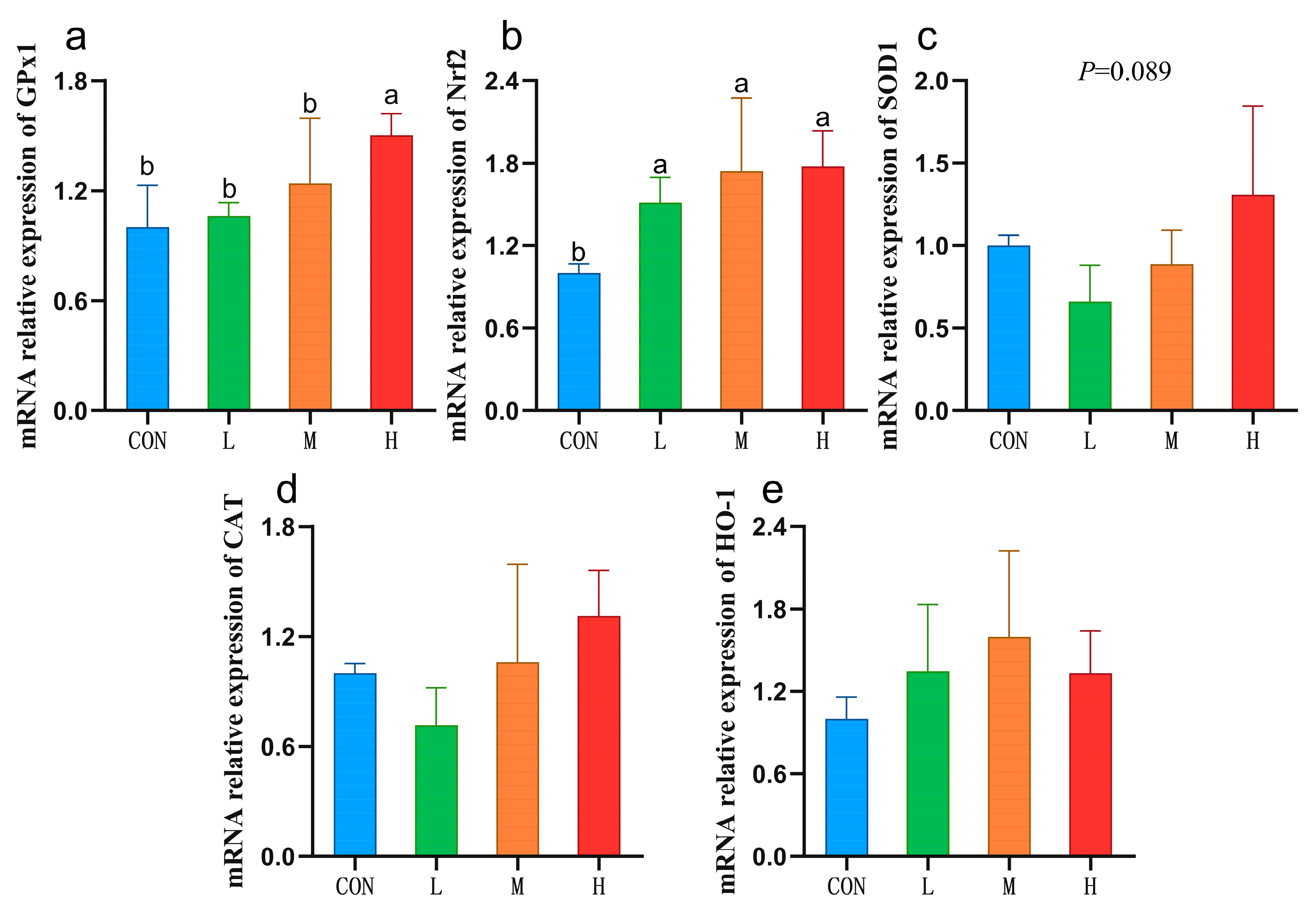
3.2. Expression of Liver Antioxidant-Related Genes

Compared with the CON group, the mRNA expression of GPx1 in the H group was significantly increased (p < 0.05), and a trend towards increased SOD1 mRNA expression was observed (p = 0.089). Compared with CON group, the mRNA expression of nuclear factor erythroid 2-related factor 2(Nrf2) in the L, M, and H groups was significantly increased (p < 0.05), and no significant differences were observed among the three experimental groups (Figure 1).

3.3. Serum Lipid Metabolism

Compared with the CON group, the TC content in the L and M groups was significantly decreased (p < 0.05); however, no significant difference occurred between the L and M groups. Compared with the CON group, the TG content in the M and H groups was significantly decreased (p < 0.05); however, no significant difference occurred between the M and H groups. Compared with the CON group, HDL-C and LDL-C in the L, M, and H groups were significantly decreased (p < 0.05), and no significant differences were observed among the three experimental groups (Table 4).

3.4. Liver Fatty Acid Profile

Compared with the CON group, we found that the C16:0 contents in livers from the M group significantly decreased (p < 0.05), C18:2n6C and C20:1 contents were significantly increased (p < 0.05), the C22:2n6 contents of the H group were significantly increased (p < 0.05), and the C18:3n6 contents in the H group tended to increase (p = 0.05). The ΣPUFAs in the M group was significantly increase (p < 0.05; Table 5).

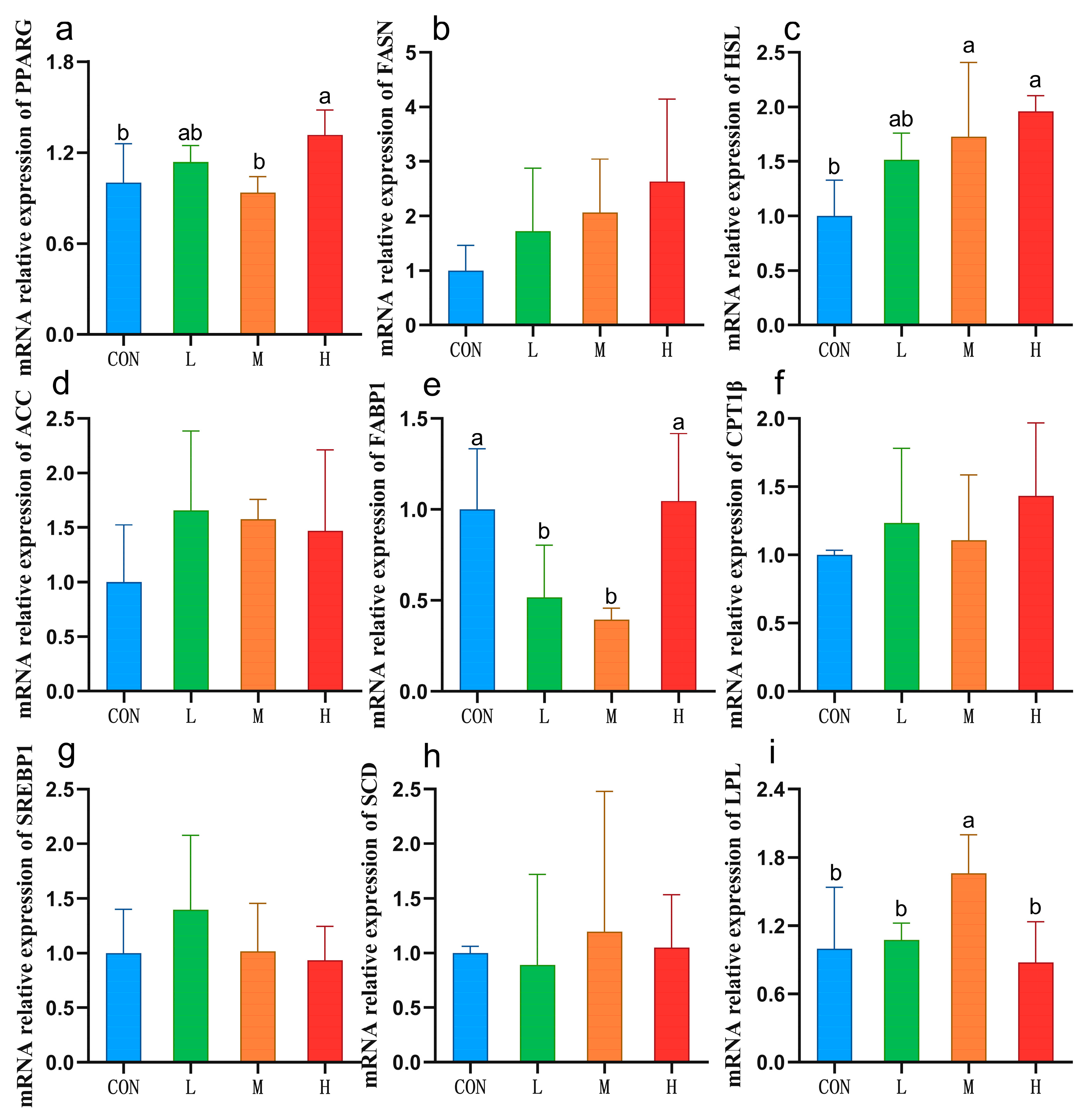
3.5. Expression of Liver Lipid Metabolism-Related Genes

Compared with the CON group, the mRNA expression of PPARG in the livers from the H group was significantly (p < 0.05). In addition, hormone-sensitive lipase (HSL) mRNA expression of the M and H groups was significantly increased (p < 0.05), fatty acid binding protein 1 (FABP1) mRNA expression in the L and M groups was significantly decreased (p < 0.05), and the mRNA expression of lipoprotein lipase (LPL) in the M group was significantly increased (p < 0.05; Figure 2).

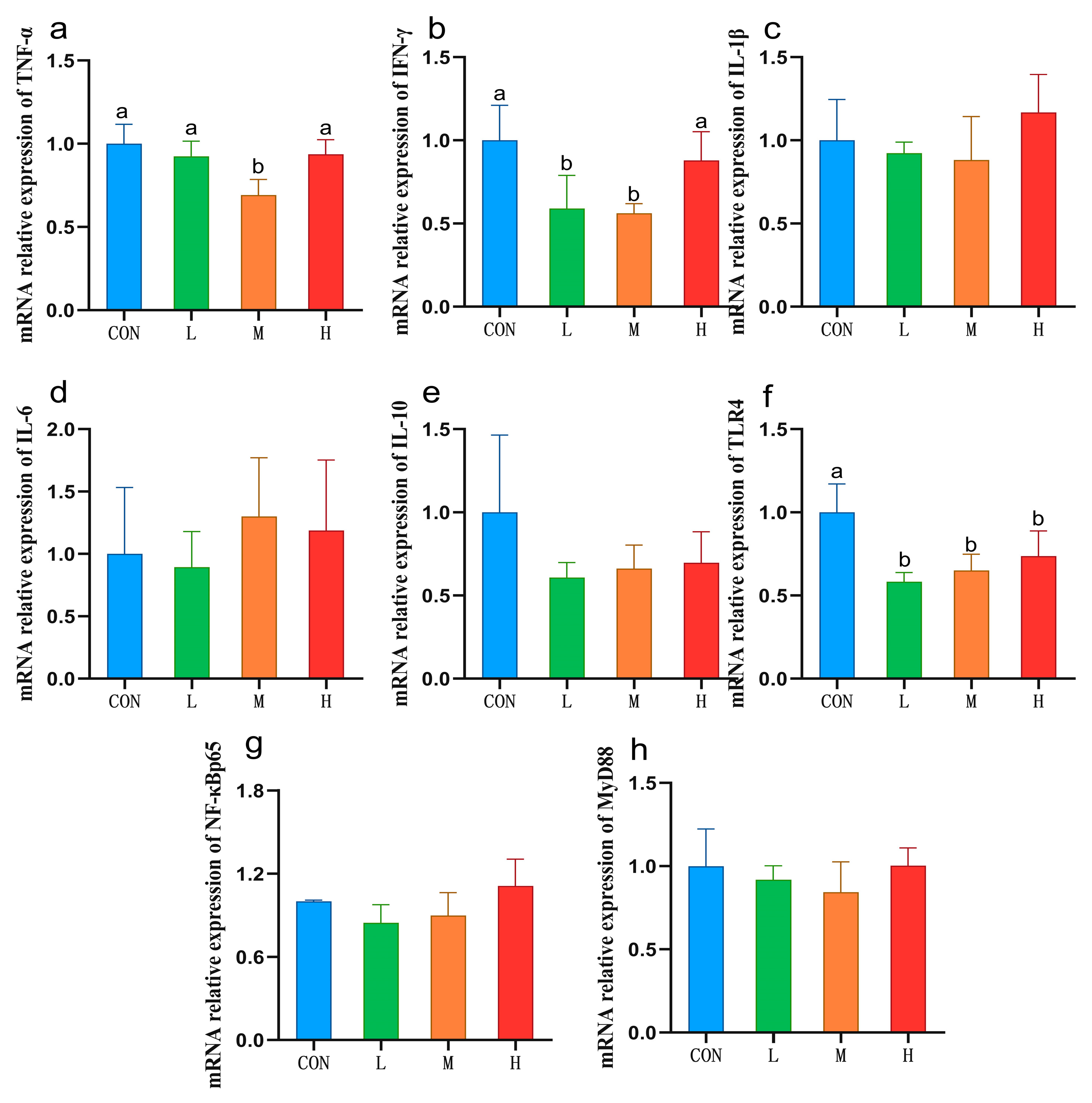
3.6. Expression of Liver Immune Cytokines and Related Genes

Compared with the CON group, the contents of TNF-α and IFN-γ in the liver of sheep in the M group decreased significantly (p < 0.05; Table 6). Compared with the CON group, the mRNA expression of TNF-α in the liver of the M group was significantly decreased (p < 0.05), the mRNA expression of IFN-γ in the L and M groups was significantly decreased (p < 0.05), and there was no significant difference between the two groups. Compared with the CON group, the mRNA expression of TLR4 in the L, M, and H groups all significantly decreased (p < 0.05), and there was no significant difference among the three experimental groups (Figure 3).

4. Discussion

Livestock are exposed to various stressors during growth that cause oxidative stress and damage their health. MDA is generally considered the marker of oxidative stress, and the SOD, GPx, and CAT enzymes play important antioxidant roles [33,34]. In this study, all three doses of RYC significantly increased GPx activity and decreased MDA contents in sheep livers, and a daily intake of 40 g RYC significantly increased T-SOD activity. This is consistent with the results of Chen et al. [12]. These findings indicate that RYC can stimulate the antioxidant enzyme system in sheep livers to adapt to ROS attacks and reduce liver oxidative damage. Specifically, RYC can protect cells by increasing GPx activity to remove H2O2 and increasing T-SOD activity to reduce cell membrane lipid peroxidation [12]. Carotenoids are the main metabolites of Rhodotorula, with conjugated double bonds, which can eliminate free radicals in the order of electron transfer, hydrogen extraction, and addition [35], which may be the reason why RYC exerts antioxidant capacity.
Nrf2 is a key regulator of metabolic redox reactions that directly regulate GPx expression [36]. Here, we observed that all three doses of RYC tested significantly increased Nrf2 and GPx1 mRNA expression (consistent with the trends in their enzyme activities), indicating that RYC increased GPx activity by upregulating the Nrf2-GPx1 signaling pathway. We also found that SOD1 mRNA expression tended to be elevated in sheep given 40 g/day RYC, whereas the SOD enzyme activity increased significantly. There may be two reasons for this statistical inconsistency in gene- and protein-expression levels. One is related to the nature of SOD. The activity of T-SOD measured in this experiment is the sum of SOD1 and SOD2 isoenzymes. RYC may increase the expression of both isoenzyme genes and eventually lead to an increase in total enzyme activity. The second is the cascade amplification of protein translation, which makes SOD significantly increase at the protein level.
The health status of domestic animals can be reflected by specific blood indicators. In ruminants, the source of GLU mainly depends on endogenous synthesis, particularly the gluconeogenesis pathway [37]. Therefore, GLU levels often reflect the energy level and health status of animals, influenced by their diet and foraging behaviors for a period of time. In this study, we found no differences in GLU levels between the groups, suggesting that the energy intake of sheep in each group was consistent. This finding is also consistent with Ge et al. [16]; they found that YC (S. cerevisiae) supplementation did not affect the blood GLU content of dairy cows before and after calving. This indicates that, akin to S. cerevisiae YC, RYC as a feed additive does not influence the energy status of sheep. In this experiment, the energy intake of sheep in each group was consistent. TC is mainly synthesized in the liver of animals and depends on different lipoproteins for transport in the body to form LDL-C and HDL-C [38]. Goldstein et al. [39] demonstrated that the TC concentration in the blood correlated positively with low-density lipoprotein levels. The results of that study’s serum TC contents were markedly lower in the L and M groups than in the CON group. Additionally, the LDL-C contents in the L, M, and H groups decreased by 40.5%, 32.4%, and 29.7%, respectively, and the HDL-C contents decreased by 14.9%, 19%, and 16.5% in the same groups. These findings indicate that RYC can decrease TC, LDL-C, and HDL-C levels in sheep, with a greater reduction in LDL-C than in HDL-C, which is beneficial to sheep health. This effect may be related to the presence of β-glucan in RYC [40].
TGs are esterified by one molecule of glycerol and three molecules of fatty acids, which can be hydrolyzed into FFAs to generate energy for the body [41]. However, elevated FFA blood levels can lead to ectopic lipid deposition, which adversely affects animal health [42]. FFA levels reliably reflect the nutritional status and current response levels of sheep to environmental stresses [37]. Carpinelli et al. [43] found that YC reduced the FFA content in the blood of perinatal cows. Malekkhahi et al. [44] found that YC reduced the TG content in the blood of Baluchi lambs fed a high level of YC. Consistently, in this study, we found that adding 20 g or 40 g RYC to the feed per day significantly reduced the serum TG contents of sheep and tended to reduce the serum FFA contents. It indicated that RYC could improve lipid metabolism and reduce disease risk in sheep, with 20 g RYC/day being the most effective.
Fatty acids in the animal body mainly originate from an exogenous daily diet and endogenous de novo synthesis [45,46]. The main product of de novo synthesis is C16:0, and longer fatty acids need to be synthesized on this basis [47]. However, mammals lack desaturation enzymes involved in the synthesis of C18:2n6 and C18:3n3. These two FAs must be obtained from food. A previous report showed that C16:0 can induce inflammation and cell death in animals [48]. Therefore, the significant reduction of C16:0 in group M observed in this study may suggest a diminished risk of inflammation in sheep. This reduction is likely due to the promotion of the conversion to C18:0, which in turn results in an increasing trend in the content of C18:0.
C20:1 can either be obtained from food or synthesized from C18:1 in the body [49]. As all sheep in this study were fed the same basal diet, the observed changes in the C20:1 level were most likely caused by changes in the sheep’s metabolic processes. In this study, the C20:1 level was significantly higher in the M group than in the CON group, whereas C18:1C levels tended to decrease, indicating that the daily addition of 20 g RYC promoted the transformation of C18:1 to C20:1 in sheep livers. This finding differed slightly from those of Li et al. [50]. In their study, YC significantly increased both C18:1C and C20:1 in sheep muscles. This difference may have been caused by different yeasts used in both studies, different principal components in the YC, different sheep breeds, and different tissue specificities.
C18:2n6C enhances the metabolic adaptability and antitumor immunity of CD8 T cells [51] and can be converted into C18:3n6. C22:2n6 has been found to have anti-inflammatory, anti-tumor, and antioxidant functions [52]. In this study, the C18:2n6C and ∑PUFAs contents were significantly higher in the M group than in the CON group, the C18:3n6 content tended to increase in the H group, and the C22:2n6 content was significantly higher, indicating that the daily addition of 20 g RYC to the feed increases the ability of the liver to absorb C18:2n6C without affecting C18:3n6 formation. To improve the ability of the liver to transform C18:3n6 and uptake C22:2n6, 40 g/day of RYC supplementation is required. Similar to our results, Liu et al. [13] also found that high doses of YC could increase the content of C18:2n6C in sheep longissimus dorsi muscles, indicating that some functional components shared by YC may play a role. Moreover, HRE et al. have shown that carotenoids can also affect polyunsaturated fatty acids (PUFAs) in animal livers [53]. Therefore, it cannot be denied that the effect of RYC on liver fatty acid profiles may be the result of the synergistic effect of various functional substances, but it is not known which components are specific, which is also the problem we will continue to study in the next step. In summary, RYC improved the ability of the liver to take up and synthesize PUFAs, improved the fatty acid composition of the sheep liver, and reduced the risk of inflammation. The effects of adding 20 g/sheep and 40 g/sheep per day were different.
To further explore the mechanism whereby RYC affects lipid metabolism in sheep livers, we conducted a relative quantitative analysis of some key genes. LPL and HSL participate in fat degradation and play roles in catalyzing TG degradation. Previous data have shown that a decrease in LPL activity is the main cause of hypertriglyceridemia [54,55]. The results of this study revealed that LPL and HSL mRNA expression were significantly higher in the M group than in the CON group, indicating that the daily addition of 20 g RYC may increase LPL and HSL mRNA expression in sheep, increase LPL and HSL enzyme activities, catalyze TG degradation, and reduce serum TG contents. Sterol regulatory element binding protein 1 (SREBP1) and its target genes, acetyl CoA carboxylase (ACC) and fatty acid synthase (FASN), are related to fatty acid synthesis [56]. No significant changes were observed in the above three genes or in fatty acids shorter than C16:0 in this study, indicating that RYC did not affect the de novo fatty acid synthesis pathway.
The FABP1 transporter can specifically bind to long-chain fatty acids in the cell membrane and participate in fatty acid transport [57]. In this study, we found that FABP1 expression in the L and M groups was significantly lower than that in the CON group. Previous findings showed that FABP1 expression correlated positively with the TG and TC contents [58]. Habashy et al. [59] suggested that FABP1 downregulation led to lower plasma TG concentrations in experimental chickens. These findings indicate that RYC reduced the serum TG and TC contents in sheep by downregulating hepatic FABP1 mRNA expression, which may also explain the increased hepatic ∑PUFAs contents in the M group. That is, decreased FABP1 mRNA expression impeded FABP1 binding to long-chain fatty acids, resulting in more PUFAs remaining in hepatocytes. Peroxisome proliferator-activated receptor γ (PPARγ) is a regulatory factor involved in lipogenesis and lipid metabolism and has been found to have anti-inflammatory and liver protection effects [60]. In this study, we found that daily supplementation of 40 g RYC increased the expression of PPARG in the liver, which was beneficial to the health of sheep.
Inflammatory factors play an important role in immune response. Our findings show that including 20 g/sheep/day RYC in the feed led to significantly decrease IFN-γ content and mRNA expression in sheep livers when compared with the corresponding levels in the CON group. Previous data have shown that Nrf2 activation can inhibit IFN-γ production [61]. As mentioned above, we found that Nrf2 mRNA expression increased in groups with added RYC, which might indicate that RYC can inhibit IFN-γ production by activating Nrf2 mRNA expression. In this study, including 20 g/sheep/day RYC in the sheep feed significantly reduced their hepatic TNF-α contents. Kim et al. [10] reported similar results for chickens. Meanwhile, Li et al. [62] obtained the opposite results on Simmental cattle; they found that YC increased the content of bovine serum IL-1β, IL-6, and IFN-γ. This discrepancy may stem from the utilization of distinct strains, which could engender variations in the active constituents within the YC. Moreover, disparities in experimental animals, feeding conditions, and the sites of index detection might also contribute to these divergent outcomes. We also found that the expression of TLR4 and TNF-α mRNA in the M group was significantly down-regulated compared with CON, while MyD88 and NF-κBp65 were not significantly different. Both TLR4/NF-κB and TLR4/MAPK signaling pathways can activate the expression of TNF-α [63,64]. Therefore, we speculated that RYC may reduce the content and mRNA expression of TNF-α in sheep livers by down-regulating the TLR4/MAPK signaling pathway, rather than the NF-κB signaling pathway.
In this study, 20 g/sheep/day and 40 g/sheep/day RYC both had a positive regulatory effect on sheep. In terms of antioxidant capacity, all three doses of RYC had significant effects on GPx and MDA, and 40 g/sheep/day RYC additionally enhanced SOD activity in sheep livers. In terms of lipid metabolism, medium-dose RYC showed a better effect and regulation effect on more fatty acids. In terms of immunity, only medium-dose RYC showed an enhanced effect. Considering the overall effect and actual feeding cost, we believe that 20 g/sheep/day RYC is the optimal dose choice at present.

5. Conclusions

The results of this study showed that RYC improved the antioxidant capacity in sheep livers by increasing the mRNA-expression levels of Nrf2 and related antioxidant enzymes. RYC reduced the blood lipid levels in sheep and improved the fatty acid composition of the liver by regulating HSL, LPL, and FABP1 mRNA expression. Moreover, RYC reduced the hepatic levels of TNF-α and IFN-γ in sheep. Taking into account the overall efficacy and associated costs, a daily dosage of 20 g/sheep of RYC emerges as the optimal choice. All in all, administering 20 g of RYC daily to sheep can significantly enhance the antioxidant capacity of sheep livers, improve lipid metabolism, and boost immunity. RYC holds great potential as a substitute for feed antibiotics and carries significant implications for the realization of antibiotic-free livestock production. In the future, we will further optimize the dosage or consider incorporating RYC into the total mixed ration to achieve the desired effects for industrial application.

Author Contributions

Conceptualization, Y.L., X.L. (Xinyu Lu), A.G. and Y.W.; methodology, X.L. (Xinyu Lu), H.M. and Y.L.; validation, X.L. (Xinyu Lu), H.M. and M.C.; formal analysis, X.L. (Xinyu Lu); investigation, X.L. (Xinyu Lu), H.M., M.C., J.D., X.S., Y.Z., K.W., G.Y., G.Z. and X.L. (Xiaorui Li); resources, X.L. (Xinyu Lu), H.M., M.C., J.D. and X.S.; data curation, X.L. (Xinyu Lu); writing—original draft preparation, X.L. (Xinyu Lu); writing—review and editing, A.G., Y.W. and Y.L.; visualization, X.L. (Xinyu Lu); supervision, A.G. and Y.W.; project administration, A.G.; funding acquisition, A.G. All authors have read and agreed to the published version of the manuscript.

Funding

This research was funded by the Natural Science Foundation of the Inner Mongolia Autonomous Region, grant number 2023LHMS03064, and the High-level Achievement Cultivation Project of Animal Science College at the Inner Mongolia Agricultural University, grant number BZX202212.

Institutional Review Board Statement

The animal study protocol was approved by the Animal Welfare and Ethics Committee of Inner Mongolia Agricultural University (NND2022110).

Informed Consent Statement

Not applicable.

Data Availability Statement

The original contributions presented in this study are included in the article material.

Acknowledgments

The authors express their gratitude for the support provided by Inner Mongolia Fuchuan Farming Polytron Technologies, Inc. (Bayannur, China) for the care of the animals. The authors express gratitude for the technical support provided by the Key Laboratory of Animal Nutrition and Feed Science (College of Animal Science, Inner Mongolia Agricultural University) and Hai Lahan, Chunxu Yang, Penghui Wu, Shufang Li, Lemei Shu, Hu Wang, and Su Runa for their assistance during the animal experiments.

Conflicts of Interest

The authors declare no conflicts of interest.

References

  1. Meng, M.; Li, X.; Huo, R.; Chang, G.; Shen, X. Effects of dietary disodium fumarate supplementation on muscle quality, chemical composition, oxidative stress and lipid metabolism of Hu sheep induced by high concentrate diet. Meat Sci. 2023, 201, 109176. [Google Scholar] [CrossRef] [PubMed]
  2. Lin, L.X.; Cao, Q.Q.; Zhang, C.D.; Xu, T.-T.; Yue, K.; Li, Q.; Liu, F.; Wang, X.; Dong, H.-J.; Huang, S.-C.; et al. Aflatoxin B1 causes oxidative stress and apoptosis in sheep testes associated with disrupting rumen microbiota. Ecotoxicol. Environ. Saf. 2022, 232, 113225. [Google Scholar] [CrossRef]
  3. Van Wettere, W.H.E.J.; Kind, K.L.; Gatford, K.L.; Swinbourne, A.M.; Leu, S.T.; Hayman, P.T.; Kelly, J.M.; Weaver, A.C.; Kleemann, D.O.; Walker, S.K. Review of the impact of heat stress on reproductive performance of sheep. J. Anim. Sci. Biotechnol. 2021, 12, 26. [Google Scholar] [CrossRef] [PubMed]
  4. Chen, J.; Ying, G.G.; Deng, W.J. Antibiotic Residues in Food: Extraction, Analysis, and Human Health Concerns. J. Agric. Food Chem. 2019, 67, 7569–7586. [Google Scholar] [CrossRef]
  5. Bacanlı, M.G. The two faces of antibiotics: An overview of the effects of antibiotic residues in foodstuffs. Arch. Toxicol. 2024, 98, 1717–1725. [Google Scholar] [CrossRef] [PubMed]
  6. Ministry of Agriculture and Rural Affairs of the People’s Republic of China. Available online: http://www.xmsyj.moa.gov.cn/zcjd/201907/t20190710_6320678.htm (accessed on 5 November 2024).
  7. Shurson, G.C. Yeast and yeast derivatives in feed additives and ingredients: Sources, characteristics, animal responses, and quantification methods. Anim. Feed Sci. Technol. 2018, 235, 60–76. [Google Scholar] [CrossRef]
  8. He, W.; Gao, Y.; Guo, Z.; Yang, Z.; Wang, X.; Liu, H.; Sun, H.; Shi, B. Effects of fermented wheat bran and yeast culture on growth performance, immunity, and intestinal microflora in growing-finishing pigs. J. Anim. Sci. 2021, 99, skab308. [Google Scholar] [CrossRef]
  9. Liu, Y.; Jia, X.; Chang, J.; Pan, X.; Jiang, X.; Che, L.; Lin, Y.; Zhuo, Y.; Feng, B.; Fang, Z.; et al. Yeast culture supplementation of sow diets regulates the immune performance of their weaned piglets under lipopolysaccharide stress. J. Anim. Sci. 2023, 101, skad226. [Google Scholar] [CrossRef]
  10. Kim, E.; Kyoung, H.; Hyung Koh, N.; Lee, H.; Lee, S.; Kim, Y.; Park, K.I.; Heo, J.M.; Song, M. Supplementation of live yeast culture modulates intestinal health, immune responses, and microbiota diversity in broiler chickens. J. Anim. Sci. 2022, 100, skac122. [Google Scholar] [CrossRef]
  11. Lin, Y.; Yu, C.; Ma, Z.; Che, L.; Feng, B.; Fang, Z.; Xu, S.; Zhuo, Y.; Li, J.; Zhang, J.; et al. Effects of Yeast Culture Supplementation in Wheat-Rice-Based Diet on Growth Performance, Meat Quality, and Gut Microbiota of Growing-Finishing Pigs. Animals 2022, 12, 2177. [Google Scholar] [CrossRef]
  12. Chen, H.; Liu, S.; Li, S.; Li, D.; Li, X.; Xu, Z.; Liu, D. Effects of yeast culture on growth performance, immune function, antioxidant capacity and hormonal profile in Mongolian ram lambs. Front. Vet. Sci. 2024, 11, 1424073. [Google Scholar] [CrossRef]
  13. Liu, Y.Z.; Lang, M.; Zhen, Y.G.; Chen, X.; Sun, Z.; Zhao, W.; Zhang, X.; Wang, T.; Qin, G. Effects of yeast culture supplementation and the ratio of non-structural carbohydrate to fat on growth performance, carcass traits and the fatty acid profile of the longissimus dorsi muscle in lambs. J. Anim. Physiol. Anim. Nutr. 2019, 103, 1274–1282. [Google Scholar] [CrossRef]
  14. Wang, H.; Su, M.; Wang, C.; Li, D.; Li, Q.; Liu, Z.; Qi, X.; Wu, Y.; Zhao, Y.; Li, T.; et al. Yeast culture repairs rumen epithelial injury by regulating microbial communities and metabolites in sheep. Front. Microbiol. 2023, 14, 1305772. [Google Scholar] [CrossRef] [PubMed]
  15. Halfen, J.; Carpinelli, N.; Del Pino, F.A.B.; Chapman, J.; Sharman, E.; Anderson, J.; Osorio, J. Effects of yeast culture supplementation on lactation performance and rumen fermentation profile and microbial abundance in mid-lactation Holstein dairy cows. J. Dairy Sci. 2021, 104, 11580–11592. [Google Scholar] [CrossRef]
  16. Ye, G.; Liu, J.; Liu, Y.; Chen, X.; Liao, S.F.; Huang, D.; Huang, K. Feeding glycerol-enriched yeast culture improves lactation performance, energy status, and hepatic gluconeogenic enzyme expression of dairy cows during the transition period. J. Anim. Sci. 2016, 94, 2441–2450. [Google Scholar] [CrossRef]
  17. Feng, Z.; Zhong, Y.; He, G.; Sun, H.; Chen, Y.; Zhou, W.; Lin, S. Yeast culture improved the growth performance, liver function, intestinal barrier and microbiota of juvenile largemouth bass (Micropterus salmoides) fed high-starch diet. Fish Shellfish Immunol. 2022, 120, 706–715. [Google Scholar] [CrossRef]
  18. Mussagy, C.U.; Ribeiro, H.F.; Santos-Ebinuma, V.C.; Schuur, B.; Pereira, J.F.B. Rhodotorula sp.-based biorefinery: A source of valuable biomolecules. Appl. Microbiol. Biotechnol. 2022, 106, 7431–7447. [Google Scholar] [CrossRef]
  19. Hu, P.; Mao, J.; Zeng, Y.; Sun, Z.; Deng, H.; Chen, C.; Sun, W.; Tang, Z. Isolation, Identification, and Function of Rhodotorula mucilaginosa TZR2014 and Its Effects on the Growth and Health of Weaned Piglets. Front. Microbiol. 2022, 13, 922136. [Google Scholar] [CrossRef]
  20. Coutinho, J.O.P.A.; Peixoto, T.S.; de Menezes, G.C.A.; Carvalho, C.R.; Ogaki, M.B.; Gomes, E.C.Q.; Rosa, C.A.; Rosa, L.H.; Arantes, R.M.E.; Nicoli, J.R.; et al. In Vitro and In Vivo Evaluation of the Probiotic Potential of Antarctic Yeasts. Probiotics Antimicrob. Proteins. 2021, 13, 1338–1354. [Google Scholar] [CrossRef]
  21. Kang, K.; Deng, X.; Xie, W.; Chen, J.; Lin, H.; Chen, Z. Rhodotorula mucilaginosa ZTHY2 Attenuates Cyclophosphamide-Induced Immunosuppression in Mice. Animals 2023, 13, 3376. [Google Scholar] [CrossRef]
  22. Sun, J.; Li, M.; Tang, Z.; Zhang, X.; Chen, J.; Sun, Z. Effects of Rhodotorula mucilaginosa fermentation product on the laying performance, egg quality, jejunal mucosal morphology and intestinal microbiota of hens. J. Appl. Microbiol. 2020, 128, 54–64. [Google Scholar] [CrossRef] [PubMed]
  23. Chen, X.Q.; Zhao, W.; Xie, S.W.; Xie, J.-J.; Zhang, Z.-H.; Tian, L.-X.; Liu, Y.-J.; Niu, J. Effects of dietary hydrolyzed yeast (Rhodotorula mucilaginosa) on growth performance, immune response, antioxidant capacity and histomorphology of juvenile Nile tilapia (Oreochromis niloticus). Fish Shellfish Immunol. 2019, 90, 30–39. [Google Scholar] [CrossRef] [PubMed]
  24. Chen, L.; Liu, Y.; Zhang, Y.; Zhang, Y.; Wang, W.; Han, H.; Yang, C.; Dong, X. Superoxide dismutase ameliorates oxidative stress and regulates liver transcriptomics to provide therapeutic benefits in hepatic inflammation. PeerJ 2023, 11, e15829. [Google Scholar] [CrossRef]
  25. Li, T.; Jin, M.; Fei, X.; Yuan, Z.; Wang, Y.; Quan, K.; Wang, T.; Yang, J.; He, M.; Wei, C. Transcriptome Comparison Reveals the Difference in Liver Fat Metabolism between Different Sheep Breeds. Animals 2022, 12, 1650. [Google Scholar] [CrossRef]
  26. Kubes, P.; Jenne, C. Immune Responses in the Liver. Annu. Rev. Immunol. 2018, 36, 247–277. [Google Scholar] [CrossRef]
  27. Bao, G.; Liu, X.; Wang, J.; Hu, J.; Shi, B.; Li, S.; Luo, Y. Effects of Slaughter Age on Myosin Heavy Chain Isoforms, Muscle Fibers, Fatty Acids, and Meat Quality in Longissimus Thoracis Muscle of Tibetan Sheep. Front. Vet. Sci. 2021, 8, 689589. [Google Scholar] [CrossRef]
  28. Zhao, C. Study on the Regulatory Network of Flavor Precursors in Ujimqin Mutton. Master’s Thesis, Inner Mongolia Agricultural University, Hohhot, China, 2022. [Google Scholar] [CrossRef]
  29. Ferlizza, E.; Fasoli, S.; Cavallini, D.; Bolcato, M.; Andreani, G.; Isani, G. Preliminary Study on Urine Chemistry and Protein Profile in Cows and Heifers. Pak. Vet. J. 2020, 40, 413–418. [Google Scholar] [CrossRef]
  30. Felini, R.; Cavallini, D.; Buonaiuto, G.; Bordin, T. Assessing the impact of thermoregulatory mineral supplementation on thermal comfort in lactating Holstein cows. Vet. Anim. Sci. 2024, 24, 100363. [Google Scholar] [CrossRef] [PubMed]
  31. Wilachai, K.; Paengkoum, P.; Taethaisong, N.; Thitisak, P.; Poonsuk, K.; Loor, J.J.; Paengkoum, S. Effect of Isolation Ruminal Yeast from Ruminants on In Vitro Ruminal Fermentation. Vet. Sci. 2025, 12, 155. [Google Scholar] [CrossRef]
  32. Li, Y.; Zhen, S.; Sun, F.; Cao, L.; Wang, L. Effects of γ-Aminobutyric Acid on Growth Performance, Immunity, Antioxidant Capacity, and Intestinal Microbiota of Growing Minks. Vet. Sci. 2024, 11, 398. [Google Scholar] [CrossRef]
  33. Alagbe, E.O.; Schulze, H.; Adeola, O. Growth performance, nutrient digestibility, intestinal morphology, cecal mucosal cytokines and serum antioxidant responses of broiler chickens to dietary enzymatically treated yeast and coccidia challenge. J. Anim. Sci. Biotechnol. 2023, 14, 57. [Google Scholar] [CrossRef] [PubMed]
  34. Roy, Z.; Bansal, R.; Siddiqui, L.; Chaudhary, N. Understanding the Role of Free Radicals and Antioxidant Enzymes in Human Diseases. Curr. Pharm. Biotechnol. 2023, 24, 1265–1276. [Google Scholar] [CrossRef]
  35. Milani, A.; Basirnejad, M.; Shahbazi, S.; Bolhassani, A. Carotenoids: Biochemistry, pharmacology and treatment. Br. J. Pharmacol. 2017, 174, 1290–1324. [Google Scholar] [CrossRef]
  36. Murakami, S.; Kusano, Y.; Okazaki, K.; Akaike, T.; Motohashi, H. NRF2 signalling in cytoprotection and metabolism. Br. J. Pharmacol. 2023. [Preprint]. [Google Scholar] [CrossRef] [PubMed]
  37. Liang, X.; Yamazaki, K.; Kamruzzaman, M.; Bi, X.; Panthee, A.; Sano, H. Effects of Chinese herbal medicine on plasma glucose, protein and energy metabolism in sheep. J. Anim. Sci. Biotechnol. 2013, 4, 51. [Google Scholar] [CrossRef]
  38. Kale, D.; Fatangare, A.; Phapale, P.; Sickmann, A. Blood-Derived Lipid and Metabolite Biomarkers in Cardiovascular Research from Clinical Studies: A Recent Update. Cells 2023, 12, 2796. [Google Scholar] [CrossRef]
  39. Goldstein, J.L.; Brown, M.S. A century of cholesterol and coronaries: From plaques to genes to statins. Cell 2015, 161, 161–172. [Google Scholar] [CrossRef] [PubMed]
  40. Joyce, S.A.; Kamil, A.; Fleige, L.; Gahan, C.G.M. The Cholesterol-Lowering Effect of Oats and Oat Beta Glucan: Modes of Action and Potential Role of Bile Acids and the Microbiome. Front. Nutr. 2019, 6, 171. [Google Scholar] [CrossRef]
  41. Yu, F.; Zong, B.; Ji, L.; Sun, P.; Jia, D.; Wang, R. Free Fatty Acids and Free Fatty Acid Receptors: Role in Regulating Arterial Function. Int. J. Mol. Sci. 2024, 25, 7853. [Google Scholar] [CrossRef]
  42. Henderson, G.C. Plasma Free Fatty Acid Concentration as a Modifiable Risk Factor for Metabolic Disease. Nutrients 2021, 13, 2590. [Google Scholar] [CrossRef]
  43. Carpinelli, N.A.; Halfen, J.; Trevisi, E.; Chapman, J.; Sharman, E.; Anderson, J.; Osorio, J. Effects of peripartal yeast culture supplementation on lactation performance, blood biomarkers, rumen fermentation, and rumen bacteria species in dairy cows. J. Dairy Sci. 2021, 104, 10727–10743. [Google Scholar] [CrossRef] [PubMed]
  44. Malekkhahi, M.; Tahmasbi, A.M.; Naserian, A.A.; Danesh Mesgaran, M.; Kleen, J.L.; Parand, A.A. Effects of essential oils, yeast culture and malate on rumen fermentation, blood metabolites, growth performance and nutrient digestibility of Baluchi lambs fed high-concentrate diets. J. Anim. Physiol. Anim. Nutr. 2015, 99, 221–229. [Google Scholar] [CrossRef]
  45. Bai, H.; Zhang, H.; Wang, C.; Lambo, M.T.; Li, Y.; Zhang, Y. Effects of altering the ratio of C16:0 and cis-9 C18:1 in rumen bypass fat on growth performance, lipid metabolism, intestinal barrier, cecal microbiota, and inflammation in fattening bulls. J. Anim. Sci. Biotechnol. 2024, 15, 94. [Google Scholar] [CrossRef] [PubMed]
  46. Duan, H.; Song, W.; Zhao, J.; Yan, W. Polyunsaturated Fatty Acids (PUFAs): Sources, Digestion, Absorption, Application and Their Potential Adjunctive Effects on Visual Fatigue. Nutrients 2023, 15, 2633. [Google Scholar] [CrossRef]
  47. Wood, J.D.; Enser, M.; Fisher, A.V.; Nute, G.R.; Sheard, P.R.; Richardson, R.I.; Whittington, F.M.; Hughes, S.I. Fat deposition, fatty acid composition and meat quality: A review. Meat Sci. 2008, 78, 343–358. [Google Scholar] [CrossRef]
  48. Shih, L.M.; Tang, H.Y.; Lynn, K.S.; Huang, C.Y.; Ho, H.Y.; Cheng, M.L. Stable Isotope-Labeled Lipidomics to Unravel the Heterogeneous Development Lipotoxicity. Molecules 2018, 23, 2862. [Google Scholar] [CrossRef]
  49. Yang, Z.H.; Emma-Okon, B.; Remaley, A.T. Dietary marine-derived long-chain monounsaturated fatty acids and cardiovascular disease risk: A mini review. Lipids Health Dis. 2016, 15, 201. [Google Scholar] [CrossRef]
  50. Li, X.; Wang, Y.; Xu, J.; Yang, Q.; Sha, Y.; Jiao, T.; Zhao, S. Effects of yeast cultures on meat quality, flavor composition and rumen microbiota in lambs. Curr. Res. Food Sci. 2024, 9, 100845. [Google Scholar] [CrossRef]
  51. Nava Lauson, C.B.; Tiberti, S.; Corsetto, P.A.; Conte, F.; Tyagi, P.; Machwirth, M.; Ebert, S.; Loffreda, A.; Scheller, L.; Sheta, D.; et al. Linoleic acid potentiates CD8+ T cell metabolic fitness and antitumor immunity. Cell Metab. 2023, 35, 633–650.e9. [Google Scholar] [CrossRef]
  52. Tang, Y.X.; Huang, W.; Wang, Y.H.; Chen, H.; Lu, X.-Y.; Tian, Y.; Ji, X.-J.; Liu, H.-H. Engineering Yarrowia lipolytica for sustainable Cis-13, 16-docosadienoic acid production. Bioresour. Technol. 2024, 406, 130978. [Google Scholar] [CrossRef]
  53. Henriquez-Rodriguez, E.; Pena, R.N.; Seradj, A.R.; Fraile, L.; Christou, P.; Tor, M.; Estany, J. Carotenoid intake and SCD genotype exert complementary effects over fat content and fatty acid composition in Duroc pigs. J. Anim. Sci. 2017, 95, 2547–2557. [Google Scholar] [CrossRef]
  54. Pajed, L.; Taschler, U.; Tilp, A.; Hofer, P.; Kotzbeck, P.; Kolleritsch, S.; Radner, F.P.W.; Pototschnig, I.; Wagner, C.; Schratter, M.; et al. Advanced lipodystrophy reverses fatty liver in mice lacking adipocyte hormone-sensitive lipase. Commun. Biol. 2021, 4, 323. [Google Scholar] [CrossRef]
  55. Yuan, C.; Xu, Y.; Lu, G.; Hu, Y.; Mao, W.; Ke, L.; Tong, Z.; Xia, Y.; Ma, S.; Dong, X.; et al. AAV-mediated hepatic LPL expression ameliorates severe hypertriglyceridemia and acute pancreatitis in Gpihbp1 deficient mice and rats. Mol. Ther. 2024, 32, 59–73. [Google Scholar] [CrossRef] [PubMed]
  56. Fang, K.; Wu, F.; Chen, G.; Dong, H.; Li, J.; Zhao, Y.; Xu, L.; Zou, X.; Lu, F. Diosgenin ameliorates palmitic acid-induced lipid accumulation via AMPK/ACC/CPT-1A and SREBP-1c/FAS signaling pathways in LO2 cells. BMC Complement. Altern. Med. 2019, 19, 255. [Google Scholar] [CrossRef]
  57. Wu, Y.; Tang, J.; Wen, Z.; Zhang, B.; Cao, J.; Zhao, L.; Guo, Z.; Xie, M.; Zhou, Z.; Hou, S. Dietary methionine deficiency stunts growth and increases fat deposition via suppression of fatty acids transportation and hepatic catabolism in Pekin ducks. J. Anim. Sci. Biotechnol. 2022, 13, 61. [Google Scholar] [CrossRef]
  58. Xiong, X. Effect of FABP1 on Intramuscular Fat Deposition in Xiangsu Hybrid Pigs. Master’s Thesis, Guizhou University, Guiyang, China, 2023. [Google Scholar] [CrossRef]
  59. Habashy, W.S.; Milfort, M.C.; Fuller, A.L.; Attia, Y.A.; Rekaya, R.; Aggrey, S.E. Effect of heat stress on protein utilization and nutrient transporters in meat-type chickens. Int. J. Biometeorol. 2017, 61, 2111–2118. [Google Scholar] [CrossRef] [PubMed]
  60. Zhao, D.; Xue, C.; Yang, Y.; Li, J.; Wang, X.; Chen, Y.; Zhang, S.; Chen, Y.; Duan, Y.; Yang, X.; et al. Lack of Nogo-B expression ameliorates PPARγ deficiency-aggravated liver fibrosis by regulating TLR4-NF-κB-TNF-α axis and macrophage polarization. Biomed. Pharmacother. 2022, 153, 113444. [Google Scholar] [CrossRef] [PubMed]
  61. Rockwell, C.E.; Zhang, M.; Fields, P.E.; Klaassen, C.D. Th2 skewing by activation of Nrf2 in CD4(+) T cells. J. Immunol. 2012, 188, 1630–1637. [Google Scholar] [CrossRef]
  62. Li, X.; An, N.; Chen, H.; Liu, D. Effects of yeast culture on growth performance, antioxidant capacity, immune function, and intestinal microbiota structure in Simmental beef cattle. Front Vet. Sci. 2025, 11, 1533081. [Google Scholar] [CrossRef]
  63. Chen, S.N.; Tan, Y.; Xiao, X.C.; Li, Q.; Wu, Q.; Peng, Y.-Y.; Ren, J.; Dong, M.-L. Deletion of TLR4 attenuates lipopolysaccharide-induced acute liver injury by inhibiting inflammation and apoptosis. Acta Pharmacol. Sin. 2021, 42, 1610–1619. [Google Scholar] [CrossRef]
  64. Li, Z.; Qi, X.; Zhang, X.; Yu, L.; Gao, L.; Kong, W.; Chen, W.; Dong, W.; Luo, L.; Lu, D.; et al. TRDMT1 exhibited protective effects against LPS-induced inflammation in rats through TLR4-NF-κB/MAPK-TNF-α pathway. Anim. Model Exp. Med. 2022, 5, 172–182. [Google Scholar] [CrossRef] [PubMed]
Figure 1. The mRNA expression of antioxidant-related genes in sheep livers. Data are presented as mean ± SD (n = 5). Values with different letters indicate statistical significance at p < 0.05. CON = fed a basal diet; L = fed a basal diet + 10 g/sheep/day RYC; M = fed a basal diet + 20 g/sheep/day RYC; H = fed a basal diet + 40 g/sheep/day RYC. GPx1: glutathione peroxidase 1; Nrf2: nuclear factor erythroid 2-related factor 2; SOD1: superoxide dismutase 1; CAT: catalase; HO-1: heme oxygenase-1.
Vetsci 12 00314 g001
Figure 2. The mRNA expression of lipid metabolism-related genes in sheep livers. Data are presented as mean ± SD (n = 5). Values with different letters indicate statistical significance at p < 0.05. CON = fed basal diet; L = fed basal diet + 10 g/sheep/day RYC; M = fed basal diet + 20 g/sheep/day RYC; H = fed basal diet + 40 g/sheep/day RYC. PPARG: peroxisome proliferators-activated receptor γ; FASN: fatty acid synthase; HSL: hormone-sensitive lipase; ACC: acetyl CoA carboxylase; FABP1: fatty acid binding protein 1; CPTT1β: carnitine palmitoyltransferase 1β; SREBP1: sterol regulatory element binding protein 1; SCD: stearoyl-CoA Desaturase; LPL: lipoprotein lipase.
Vetsci 12 00314 g002
Figure 3. The mRNA expression of immunity-related genes in sheep livers. Data are presented as mean ± SD (n = 5). Values with different letters indicate statistical significance at p < 0.05. CON = fed basal diet; L = fed basal diet + 10 g/sheep/day RYC; M = fed basal diet + 20 g/sheep/day RYC; H = fed basal diet + 40 g/sheep/day RYC. TNF-α: tumor necrosis factor-α; IFN-γ: interferon-γ; IL-1β: interleukin 1β; IL-6: interleukin 6; IL-10: interleukin 10; TLR4: toll-like receptor 4; NF-κBp65: nuclear factor kappa-B p65; MyD88: myeloid differentiation primary response protein 88.
Vetsci 12 00314 g003
Table 1. Basal diet composition and nutrient levels (dry matter level).
ItemsContent (%)
Ingredients
Sheep grass9.21
Cornstalks32.9
Concentrate supplements31.57
Whole plant corn silage26.32
Total100
Nutrient levels
Metabolic energy 1 (MJ/kg)8.09
Crude protein (%)11.95
Ether extract (%)2.13
Neutral detergent fibers (%)54.86
Acidic detergent fibers (%)21.6
Calcium (%)0.69
Phosphorus (%)0.25
1 Metabolic energy was a calculated value, and the remaining nutrients were measured values.
Table 2. Primer sequences.
GenesSequence(5′-3′)Product Length
(bp)
Accession Number
CATF:CCGCGCAGAAACCTGATGT
R:AAGTAGCCAAAAGCCCCTGC
196XM_060400054.1
SOD1F:GGAGACCTGGGCAATGTGAA
R:CCTCCAGCGTTTCCAGTCTT
182NM_001145185.2
GPx1F:CGGGACTACACCCAGATGAAT
R:GTTCTTGGCGTTTTCCTGATGC
108XM_004018462.5
Nrf2F:TGTGGAGGAGTTCAACGAGC
R:CGCCGCCATCTTGTTCTTG
103XM_042246639.2
HO-1F:AGGGACCAGACCTTCACAGG
R:GCATAAAGCCCCACAGCAAC
166XM_027967703.3
PPARGF:CTTGTGAAGGATGCAAGGGTT
R:CATGCGCCCAAACCTGATG
176NM_001100921.1
FASNF:AGTGGTCATTCAGGTGCGTG
R:ATGACGTAGCTCTTGTGGGC
114XM_027974304.3
HSLF:TCGCCTTTGAAATGCCTCTGACC
R:GCTCCTTGCTGTTCTGTCCTTCC
138NM_001128154.1
LPLF:CCCGGCTTTGATATTGGGAAG
R:CTTTGCCAAGTTTCAGCCAGA
171NM_001009394.1
ACCF:GTGGTGTGAGATCCTGTGCT
R:TTAACGAGTCGCAGTTCGGT
93NM_001009256.1
CPT1βF:AGCAAACCTTAGCTGTGCCA
R:GCGAATCAGGCGTTTCTTCC
168NM_001009259.1
SREBP1F:GACTGCACGTTCGAAGACAT
R:CTCATCGTGGAAGGAGGTGG
164XM_027974786.2
SCDF:ATGGCGTTCCAGAATGACG
R:AAAAGCCACGTCGGGAATTG
103NM_001009254.1
ACOX1F:CTTGCTGAATCAGGGCACCA
R:TCGAAGATGAGTTCCGTGGC
115XM_060395846.1
IL-1βF:TCCTCCGATGAGCTTCTGTG
R:GGAGAGCCTTCAGCACACAT
112NM_001009465.2
IL-6F:ATCGCAGGTCTAATAACCACTCCAG
R:GCAGGAAATTCTCAAGGCTTCTCAG
124NM_001009392.1
IL-10F:GGGTGTCTACAAAGCCATGAGTGAG
R:AGGTTTATGTCGGGGAGTCTAGTCG
143XM_060395938.1
TNF-αF:ACCTGGACTATGCCGAGTCT
R:GAAGGGGATGAGGAGGGTCT
127NM_001024860.1
IFN-γF:AAGTTCTTGAACGGCAGCTCTGAG
R:TGAGGTTAGATTTTGGCGACAGGTC
142NM_001009803.1
TLR4F:TGGGTGCGGAATGAACTGGTAAAG
R:CTGGATGATATTGGCGGCGATGG
114NM_001135930.1
MyD88F:ATGGTGGTGGTTGTCTCTGAC
R:GGAACTCTTTCTTCATTGGCTTGT
139NM_001166183.1
NF-κBp65F:TCTGGCCCCTATGTGGAGAT
R:CCCGTGTAGCCATTGATCTTG
155XM_027959295.2
β-actinF:CCCTGGAGAAGAGCTACGAG
R:GGTAGTTTCGTGAATGCCGC
131NM_001009784.3
Table 3. Effects of RYC on antioxidant capacity of sheep livers.
ItemsCONLMHSEMp-Value
T-SOD (U/mgprot)196.97 b205.09 b196.26 b223.31 a3.7450.007
CAT (U/mgprot)36.4741.7940.2737.941.0860.361
GPx (U/mgprot)10.45 b14.62 a13.23 a13.74 a0.5730.024
T-AOC (μmol/gprot)72.4871.5074.2673.791.3110.911
MDA (nmol/mgprot)1.26 a0.74 b0.78 b0.88 b0.0620.022
Different letter superscripts in the same row indicate significant differences (n = 5). CON = fed basal diet; L = fed basal diet + 10 g/sheep/day RYC; M = fed basal diet + 20 g/sheep/day RYC; H = fed basal diet + 40 g/sheep/day RYC. T-SOD: total superoxide dismutase; CAT: catalase; GPx: glutathione peroxidase; T-AOC: total antioxidative capacity; MDA: malondialdehyde.
Table 4. Effects of RYC on serum lipid metabolism in sheep.
ItemsCONLMHSEMp-Value
GLU (mmol/L)3.413.203.343.340.0760.841
TC (mmol/L)3.06 a2.46 b2.47 b2.66 ab0.0950.040
TG (mmol/L)0.34 a0.31 ab0.22 c0.25 bc0.0110.014
HDL-C (mmol/L)1.21 a1.03 b0.98 b1.01 b0.0260.001
LDL-C (mmol/L)0.37 a0.22 b0.25 b0.26 b0.0170.013
FFA (mmol/L)0.570.560.560.530.0050.056
Different letter superscripts in the same row indicate significant differences (n = 6). CON = fed basal diet; L = fed basal diet + 10 g/sheep/day RYC; M = fed basal diet + 20 g/sheep/day RYC; H = fed basal diet + 40 g/sheep/day RYC. GLU: glucose; TC: total cholesterol; TG: triglycerides; HDL-C: high-density lipoprotein cholesterol; LDL-C: low-density lipoprotein cholesterol; FFA: free fatty acid.
Table 5. Effects of RYC on the fatty acid profile of sheep livers (of total fatty acids,%).
ItemsCONLMHSEMp-Value
C8:00.040.040.040.040.0010.870
C10:00.090.090.090.090.0020.752
C12:00.230.240.230.250.0120.963
C14:01.311.341.091.390.0730.523
C14:10.460.440.430.410.0140.680
C15:00.340.340.320.330.0060.889
C16:023.43 a23.28 a21.60 b23.82 a0.3100.038
C16:11.431.441.221.610.0640.209
C17:00.760.810.810.800.0120.441
C17:10.560.650.590.610.0140.157
C18:024.5325.6126.5723.940.4050.089
C18:1C27.8225.5223.2826.560.6230.052
C18:2n6C12.49 b13.02 b16.02 a13.27 b0.4080.002
C18:3n30.260.270.280.300.0080.336
C18:3n60.350.360.340.410.0100.050
C20:10.26 b0.27 b0.32 a0.25 b0.0090.018
C20:3n34.114.504.734.340.1280.409
C20:3n60.400.430.440.410.0200.888
C22:2n60.21 b0.23 b0.24 ab0.26 a0.0060.013
C22:6n30.911.111.081.050.0500.545
ΣSFAs50.7251.7550.7650.670.2640.435
ΣMUFAs30.5228.3125.8429.440.6630.061
ΣPUFAs18.73 b19.92 b23.14 a20.04 b0.5210.008
Different letter superscripts in the same row indicate significant differences (n = 5). CON = fed basal diet; L = fed basal diet + 10 g/sheep/day RYC; M = fed basal diet + 20 g/sheep/day RYC; H = fed basal diet + 40 g/sheep/day RYC. ΣSFAs = C8:0 + C10:0 + C12:0 + C14:0 + C15:0 + C16:0 + C17:0 + C18:0; ΣMUFAs = C14:1 + C16:1 + C17:1 + C18:1C + C20:1; ΣPUFAs = C18:2n6C + C18:3n3 + C18:3n6 + C20:3n3 + C20:3n6 + C22:2n6 + C22:6n3.
Table 6. Effects of RYC on immune cytokines in sheep livers.
ItemsCONLMHSEMp-Value
IL-1β (pg/mg)6.916.016.486.740.2300.568
IL-6 (pg/mg)17.6115.2115.7016.550.4870.343
IL-10 (pg/mg)2.022.061.832.060.0520.377
TNF-α (pg/mg)11.01 a9.68 ab8.81 b10.25 ab0.2940.041
IFN-γ (pg/mg)60.68 a53.13 ab45.65 b56.42 a1.7770.009
Different letter superscripts in the same row indicate significant differences (n = 5). CON = fed basal diet; L = fed basal diet + 10 g/sheep/day RYC; M = fed basal diet + 20 g/sheep/day RYC; H = fed basal diet + 40 g/sheep/day RYC. IL-1β: interleukin 1β; IL-6: interleukin 6; IL-10: interleukin 10; TNF-α: tumor necrosis factor-α; IFN-γ: interferon-γ.
Disclaimer/Publisher’s Note: The statements, opinions and data contained in all publications are solely those of the individual author(s) and contributor(s) and not of MDPI and/or the editor(s). MDPI and/or the editor(s) disclaim responsibility for any injury to people or property resulting from any ideas, methods, instructions or products referred to in the content.

