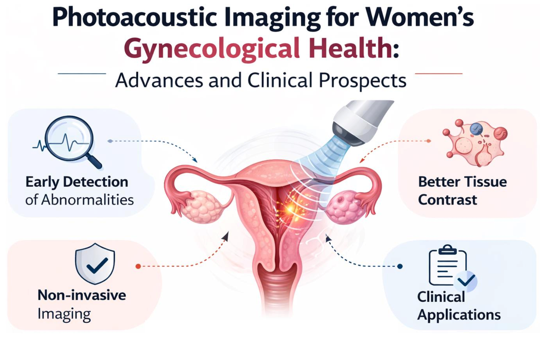
Photoacoustic Imaging for Women’s Gynecological Health: Advances and Clinical Prospects



Abstract
1. Introduction
2. Literature Search Methodology
3. Basic Principle and Fundamentals of Photoacoustic Imaging
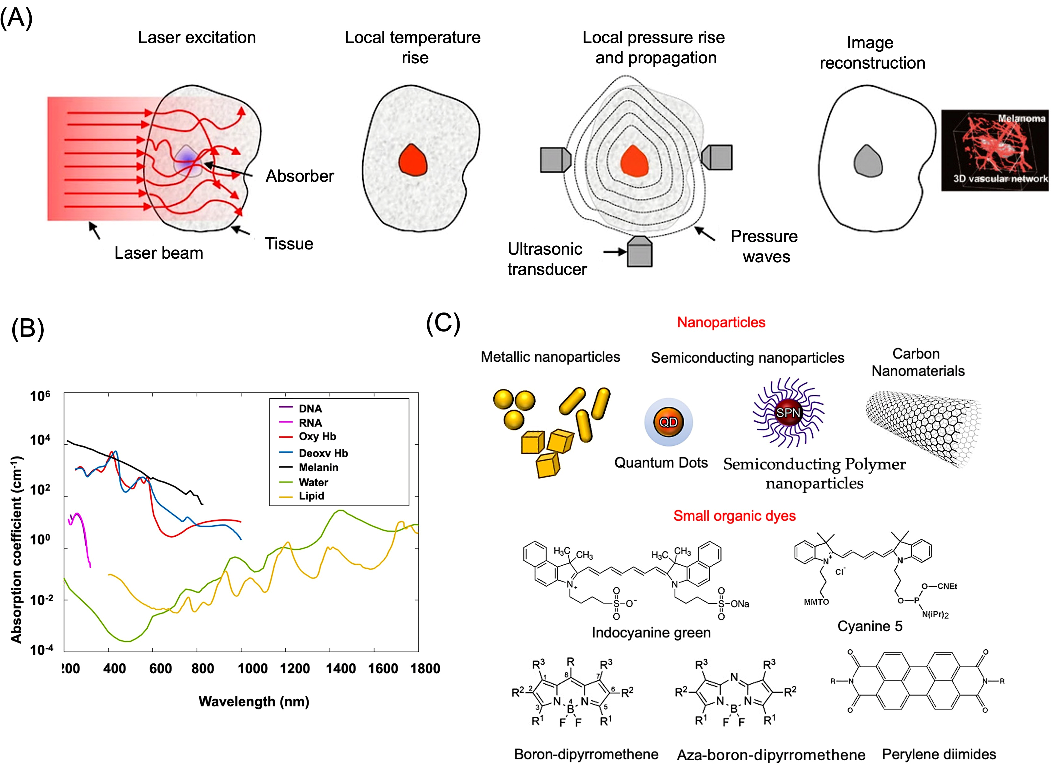
3.1. Basic Principle of Photoacoustic Signal Generation
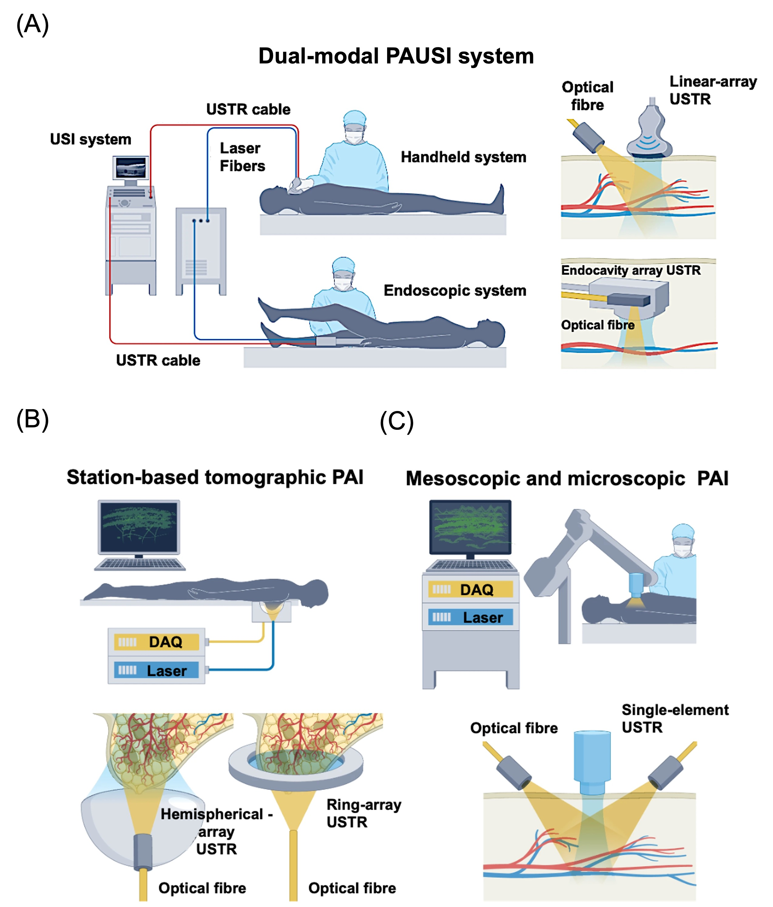
3.2. Imaging Configurations
3.2.1. Photoacoustic Microscopy (PAM)
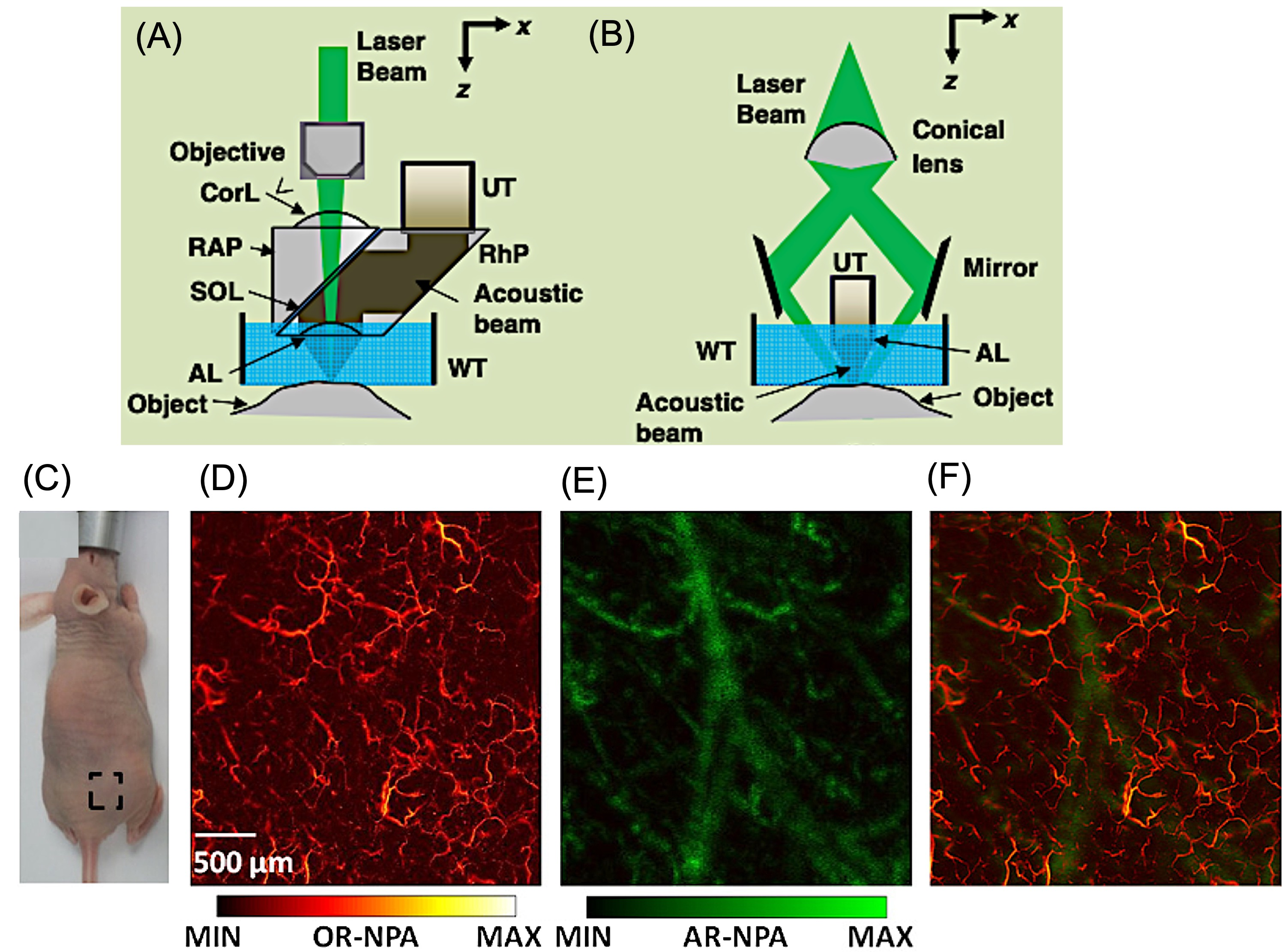
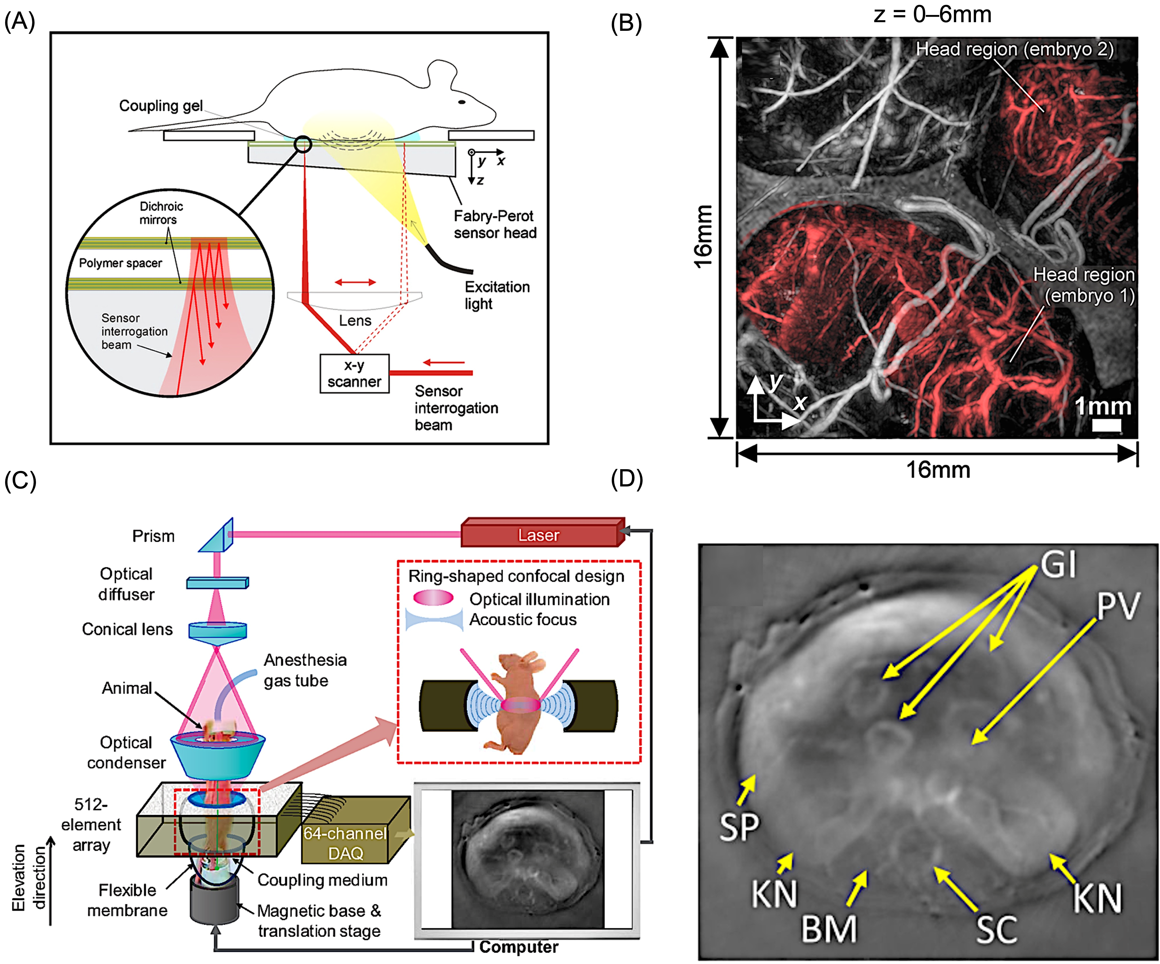
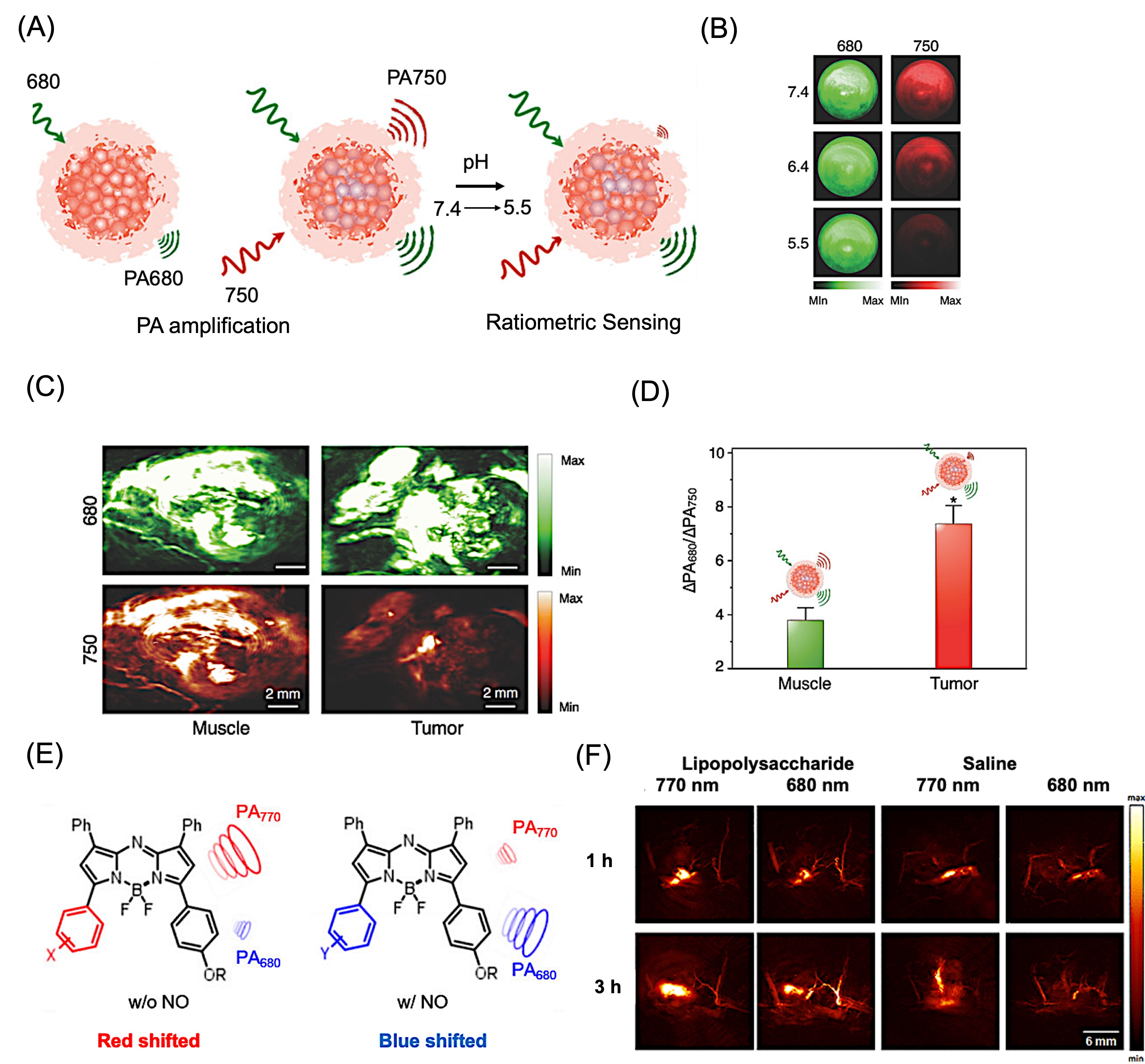
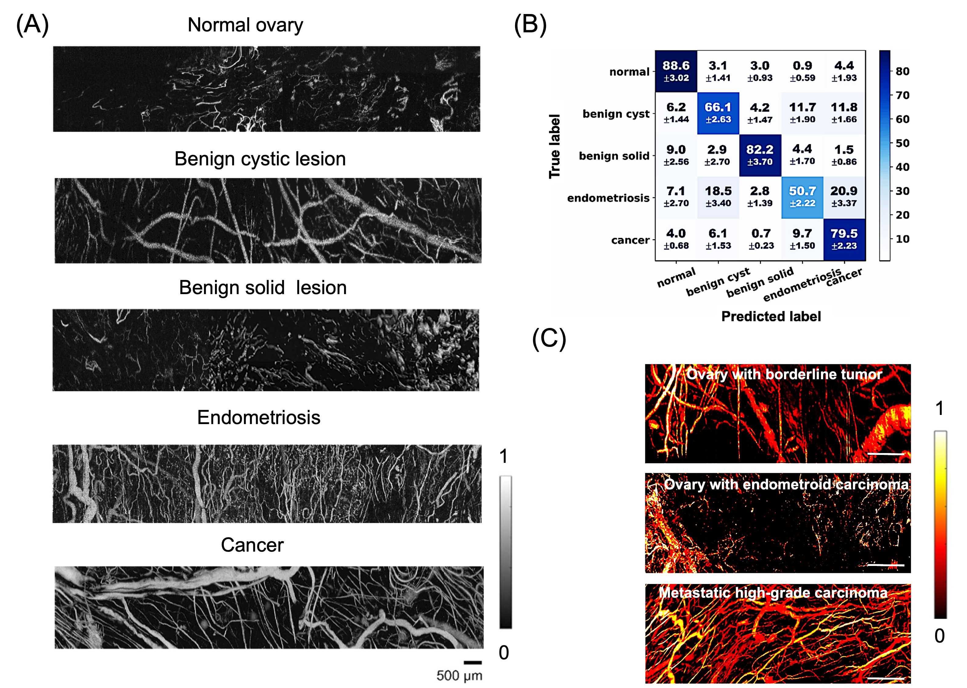
3.2.2. Photoacoustic Computed Tomography (PACT)
3.2.3. Endoscopic and Probe-Based PAI
4. Functional and Molecular Imaging Capability of PAI
4.1. Endogenous Contrast Mechanisms

4.2. Exogenous Contrast Agents for Molecular Photoacoustic Imaging

| Category | Representative Agent | Size | Absorption (nm) | Key Advantages | Limitations (Conventional Issues) | Emerging Direction/Improvement | Application | Refs. |
|---|---|---|---|---|---|---|---|---|
| First-generation (clinical dyes) | Indocyanine Green (ICG) | ~2 nm | ~800 | FDA-approved, strong NIR absorption | Poor stability, aggregation, rapid clearance, non-specific | Encapsulation, conjugation, activatable probes | In vivo imaging | [100,101] |
| Methylene Blue | ~2 nm | 650–700 | Clinically used, easy availability | Low photostability, limited penetration | Modified derivatives, nanoparticle loading | Lymph node imaging | [102,103] | |
| Evans Blue | <2 nm | ~610 | Protein binding, vascular imaging | Non-specific distribution, toxicity concerns | Targeted conjugates | Brain imaging | [104,105] | |
| Second-generation (nanomaterials) | Gold nanorods (AuNRs) | 20–50 nm | 700–900 | Tunable absorption, strong PA signal | Potential toxicity, poor biodegradability | Surface modification, biodegradable coatings | Tumor imaging, PTT | [106,107] |
| Gold nanorods (AuNRs) | 41–45 | 760 | Tunable absorption, strong PA/Raman signal | High sensitivity 17 fM for PAI | Ovarian Cancer | [108] | ||
| Gold nanostars | ~35 nm | 700–900 | High photothermal efficiency | Long-term accumulation | Hybrid/biodegradable systems | Tumor imaging | [109] | |
| Prussian Blue NPs | 20–170 nm | ~700 | Biocompatible, strong absorption | Limited targeting specificity | Functionalization, exosome coating | Brain tumor imaging | [110,111] | |
| Polydopamine NPs | ~200 nm | Broad absorption from UV to NIR | Biocompatible, strong absorption | Signals are not strong as metallic nanoparticles | Biodegradable with wide absorption | Endometriosis treatment | [112] | |
| Hybrid systems | Exosome–Prussian Blue | ~70 nm | 700–750 | Improved targeting, biocompatibility | Complex synthesis | Biomimetic delivery systems | Tumor imaging | [113] |
| BSA-Cerium oxide-ICG | ~28 nm | ~790 | Enhanced stability of ICG | Still partially non-specific | Targeted protein-based systems | Endometriosis | [114] | |
| Silica shell coated AuNRs | 52.75 m | 780 | Enhanced stability and photothermal conversion efficiency | Still partially non-specific | Targeted Nanoformulation via EPR | Endometriosis PTT | [16] | |
| Iron oxide- NIR-830 dye | 22 nm | 820 | Targeted imaging probe | Time consuming | Dual imaging modality with high- resolution and specificity | Ovarian cancer | [115] | |
| Copper sulfide nanodisk/nanoprism | 6 and 26 | 1145 and 1098 nm | Separation between tumor and healthy tissues | Shape-dependent PA signal intensity | Ovarian cancer | [116] | ||
| Emerging small-molecule & activatable probes | BODIPY derivatives | <2 nm | 700–1000 | Tunable structure, high specificity | Limited clinical validation | Enzyme-responsive, ratiometric probes | Molecular imaging/PTT/PDT | [117,118,119] |
| Aza-Bodipy | <2 nm | 750–1050 | Tunable structure | Limited clinical validation | Photostability and thermal conversion | Tumor imaging/PDT/PTT | [120] | |
| NIR dyes (NIRb14, cyanines) | <2 nm | ~800 | Strong NIR absorption, easy modification | Photobleaching, off-target signal | Activatable probes (Metalloprotease, hypoxia) | Tumor imaging, PTT | [121] | |
| Perylene diimide (PDI) | ~50–70 nm | 400–800 | High photostability | Limited water solubility | Functionalized derivatives | Brain imaging/tumor imaging/PTT | [122,123] |
4.2.1. Nanomaterial-Based Contrast Agents

4.2.2. Carbon Nanotube-Based Contrast Agents
4.3. Organic Dyes as Exogenous Contrast Agents
4.3.1. Organic Dye-Based Contrast Agents in the NIR-I Window (650–800 nm)
4.3.2. Organic Dye-Based Contrast Agents with Red-Shifted NIR Absorption (800–1000 nm)
4.3.3. Organic Dyes as Contrast Agents with High NIR Absorption (>1000 nm)
4.4. Comparative Summary of Preclinical and Clinical Studies Using PAI
5. Quantitative and Dynamic Photoacoustic Imaging Techniques
6. Advantages of PAI Compared with Other Imaging Modalities
7. Application of PAI for Gynecological Disorders
7.1. PAI for Uterine and Endometrial Microvessel Disorders

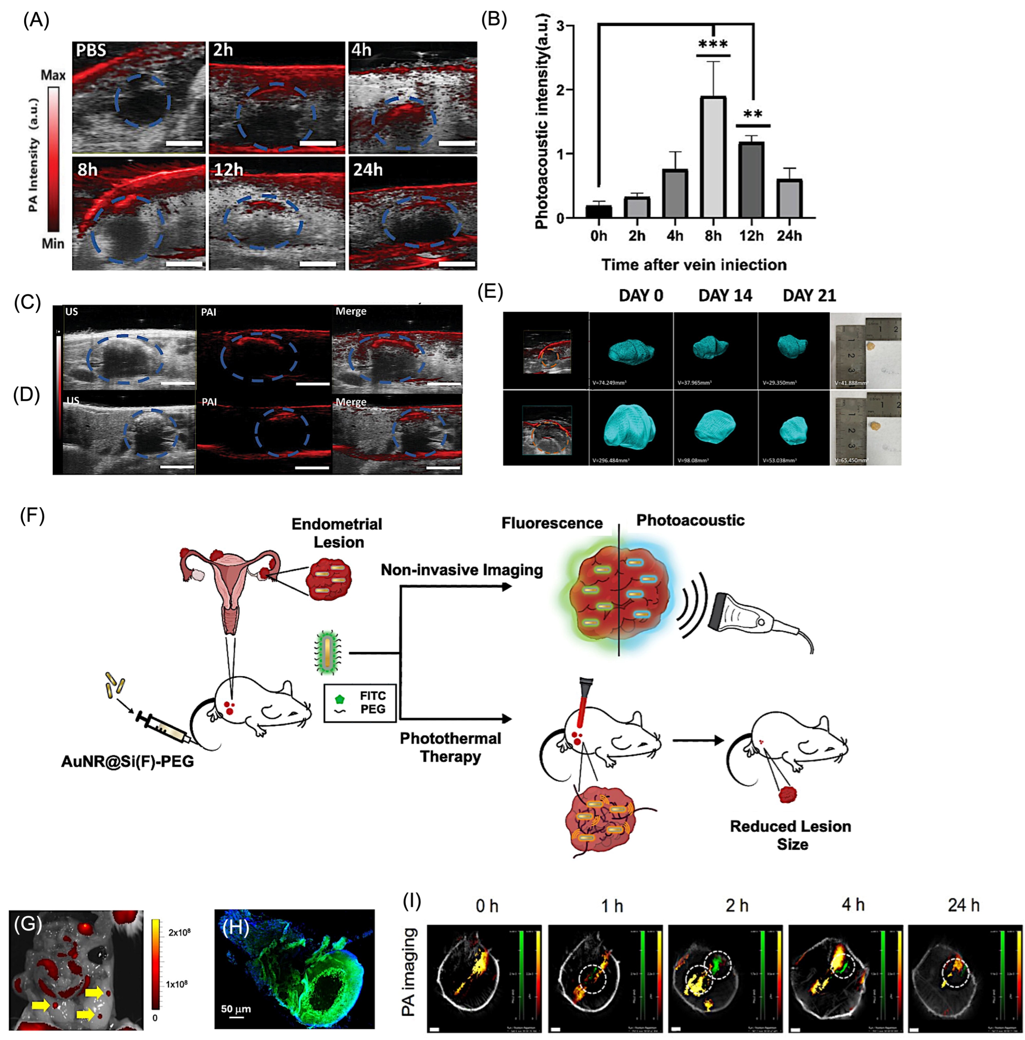
7.2. PAI for Endometriosis
7.2.1. PAI for Non-Invasive Detection of Endometriosis

7.2.2. Endometriosis Treatment Validation Using PAI

7.3. Cervical Tissue Remodeling and Pregnancy-Related Conditions
7.4. Ovarian Pathologies
8. Clinical Translation and Integration

9. Challenges, Design Principles, and Future Directions for Clinical Translation of Photoacoustic Imaging in Women’s Health
10. AI-Driven Photoacoustic Imaging
| Type | Year | AI Model Used | Key Contribution | Ref. |
|---|---|---|---|---|
| Image Analysis / Classification | 2012 | Pattern recognition algorithm (feature-based classification) | Classification of malignant vs benign ovarian tissue using co-registered PA + ultrasound imaging; demonstrated distinct absorption patterns and improved diagnostic differentiation | [220] |
| PAT Reconstruction | 2018 | CNN (U-Net-based) | First DL framework for sparse-data PAT reconstruction | [221] |
| PAT Reconstruction | 2020 | Res-U-Net | End-to-end inverse problem solving for PAT | [222] |
| PAT Reconstruction | 2020 | Hybrid CNN (Y-Net) | Combines raw + beamformed data for improved reconstruction | [223] |
| PAT Reconstruction | 2020 | CNN | Real-time reconstruction for microvascular imaging | [224] |
| PAM Reconstruction | 2021 | FD U-Net | Reconstruction from highly undersampled PAM data | [225] |
| Clinical Imaging Analysis | 2021 | Classical ML models (k-NN, SVM, Random Forest, Neural Network, Logistic Regression) | AI-based classification of rectosigmoid endometriosis using ultrasound-derived features demonstrated comparable performance to traditional statistical models in clinical dataset (n = 333) | [226] |
| Multimodal Image Analysis/Classification | 2022 | SVM | Combined PA/US features for classification of ovarian lesions; demonstrated improved diagnostic accuracy using multimodal feature extraction | [227] |
| Image Processing | 2024 | Noise2Noise CNN | Unsupervised denoising for PA images | [228] |
| Image Processing | 2024 | YOLOv8 + Pix2Pix | Artifact removal + segmentation in PA images | [229] |
| OC-PAM image analysis AI | 2026 | U-Net-type architecture | 3D organoid segmentation | [230] |
11. Conclusions
Funding
Institutional Review Board Statement
Informed Consent Statement
Data Availability Statement
Acknowledgments
Conflicts of Interest
References
- He, X.; Su, J.; Wang, K.; Liang, Y.; Wang, L. Global, Regional, and National Prevalence and Trends of Gynecological Diseases among Women of Childbearing Age from 1990 to 2021: An Analysis of the Global Burden of Disease Study 2021. PLoS ONE 2025, 20, e0329336. [Google Scholar] [CrossRef] [Scilit]
- Gao, Y.; Wang, X.; Wang, Q.; Jiang, L.; Wu, C.; Guo, Y.; Cui, N.; Tang, H.; Tang, L. Rising Global Burden of Common Gynecological Diseases in Women of Childbearing Age from 1990 to 2021: An Update from the Global Burden of Disease Study 2021. Reprod. Health 2025, 22, 57. [Google Scholar] [CrossRef] [Scilit]
- Hill, C.J.; Fakhreldin, M.; Maclean, A.; Dobson, L.; Nancarrow, L.; Bradfield, A.; Choi, F.; Daley, D.; Tempest, N.; Hapangama, D.K. Endometriosis and the Fallopian Tubes: Theories of Origin and Clinical Implications. J. Clin. Med. 2020, 9, 1905. [Google Scholar] [CrossRef] [Scilit]
- Mikhaleva, L.M.; Radzinsky, V.E.; Orazov, M.R.; Khovanskaya, T.N.; Sorokina, A.V.; Mikhalev, S.A.; Volkova, S.V.; Shustova, V.B.; Sinelnikov, M.Y. Current Knowledge on Endometriosis Etiology: A Systematic Review of Literature. Int. J. Womens Health 2021, 13, 525–537. [Google Scholar] [CrossRef] [Scilit] [PubMed]
- Pavaleanu, I.; Ana Balan, T.; Nicolae Poparlan, T.; Maria Haliciu, A.; Andrei Butureanu, T.; Maria Apetrei, A.; Socolov, R.; Ioana Pruteanu, A.; Anca Balan, R. Endometriosis-Associated Ovarian Carcinoma. In Obstetrics and Gynecology; Wu, W., Ju, R., Eds.; IntechOpen: London, UK, 2024; Volume 7. [Google Scholar]
- Jayaprakasan, K.; Ojha, K. Diagnosis of Congenital Uterine Abnormalities: Practical Considerations. J. Clin. Med. 2022, 11, 1251. [Google Scholar] [CrossRef] [Scilit] [PubMed]
- Dykes, T.M.; Siegel, C.; Dodson, W. Imaging of Congenital Uterine Anomalies: Review and Self-Assessment Module. Am. J. Roentgenol. 2007, 189, S1–S10. [Google Scholar] [CrossRef] [Scilit]
- Sahu, S.A.; Shrivastava, D. A Comprehensive Review of Screening Methods for Ovarian Masses: Towards Earlier Detection. Cureus 2023, 15, e48534. [Google Scholar] [CrossRef] [Scilit]
- Matsas, A.; Stefanoudakis, D.; Troupis, T.; Kontzoglou, K.; Eleftheriades, M.; Christopoulos, P.; Panoskaltsis, T.; Stamoula, E.; Iliopoulos, D.C. Tumor Markers and Their Diagnostic Significance in Ovarian Cancer. Life 2023, 13, 1689. [Google Scholar] [CrossRef] [Scilit] [PubMed]
- Tsikouras, P.; Oikonomou, E.; Bothou, A.; Chaitidou, P.; Kyriakou, D.; Nikolettos, K.; Andreou, S.; Gaitatzi, F.; Nalbanti, T.; Peitsidis, P.; et al. The Impact of Endometriosis on Pregnancy. J. Pers. Med. 2024, 14, 126. [Google Scholar] [CrossRef] [Scilit]
- Allaire, C.; Bedaiwy, M.A.; Yong, P.J. Diagnosis and Management of Endometriosis. Can. Med. Assoc. J. 2023, 195, E363–E371. [Google Scholar] [CrossRef] [Scilit]
- Napolitano, R.; Speltri, G.; Martini, P.; Porto, F.; Marvelli, L.; Niorettini, A.; Uccelli, L.; Urso, L.; Filippi, L.; Uslu, H.; et al. Molecular Imaging Advances in Endometriosis: The Promise of Radiopharmaceuticals. Molecules 2025, 31, 93. [Google Scholar] [CrossRef] [Scilit]
- Scutelnicu, L.-A.; Luca, M.; Maftei, R. A Comparative Study on Endometriosis Automatic Diagnosis Using Magnetic Resonance Imaging and Ultrasound. In Human Centred Intelligent Systems; Zimmermann, A., Schmidt, R., Jain, L.C., Howlett, R.J., Eds.; Smart Innovation, Systems and Technologies; Springer Nature: Singapore, 2025; Volume 414, pp. 125–134. [Google Scholar]
- Sebastiano, J.; Rodriguez, C.; Samuels, Z.V.; Pepin, K.; Zeglis, B.M. Molecular Imaging in Gynecology: Beyond Cancer. J. Nucl. Med. 2024, 65, 998–1003. [Google Scholar] [CrossRef] [Scilit]
- Gaia-Oltean, A.I.; Boitor, D.; Pop, L.-A.; Galea, G.; Telecan, T.; Micu, R. Non-Invasive Methods for Early Diagnosis of Endometriosis—A Comprehensive Narrative Literature Review. Healthcare 2025, 13, 3276. [Google Scholar] [CrossRef] [Scilit]
- Kumar, P.P.P.; Chung, S.; Hadrick, K.; Hill, M.L.; Lee, M.; Kim, T.H.; Jeong, J.; Kim, T. Noninvasive Detection and Thermal Ablation Therapy of Endometriosis Using Silica-Coated Gold Nanorods. Adv. NanoBiomed Res. 2025, 5, 2500101. [Google Scholar] [CrossRef] [Scilit]
- Liu, H.; Wang, M.; Ji, F.; Jiang, Y.; Yang, M. Mini Review of Photoacoustic Clinical Imaging: A Noninvasive Tool for Disease Diagnosis and Treatment Evaluation. J. Biomed. Opt. 2024, 29, S11522. [Google Scholar] [CrossRef] [Scilit] [PubMed]
- Wang, L.V.; Hu, S. Photoacoustic Tomography: In Vivo Imaging from Organelles to Organs. Science 2012, 335, 1458–1462. [Google Scholar] [CrossRef] [Scilit] [PubMed]
- Peng, Y.; Zhang, M.; Yan, J.; Wang, R.; Xin, Y.; Zheng, X.; Zhu, L.; Fei, W.; Zhao, M. Emerging Bioengineering Breakthroughs in Precision Diagnosis and Therapy for Endometriosis and Adenomyosis. J. Mater. Chem. B 2025, 13, 742–762. [Google Scholar] [CrossRef] [Scilit] [PubMed]
- Herrara, V.; Tarab-Ravski, D.; Chauhan, S.C.; Narang, N.; Mirazul Islam, M.; Peer, D.; Prasad, R.; Yallapu, M.M. Nanotechnology Strategies for Endometrium Health: Are We on the Right Track? Bioact. Mater. 2025, 54, 423–449. [Google Scholar] [CrossRef] [Scilit]
- Xu, M.; Wang, L.V. Photoacoustic Imaging in Biomedicine. Rev. Sci. Instrum. 2006, 77, 041101. [Google Scholar] [CrossRef] [Scilit]
- Attia, A.B.E.; Balasundaram, G.; Moothanchery, M.; Dinish, U.S.; Bi, R.; Ntziachristos, V.; Olivo, M. A Review of Clinical Photoacoustic Imaging: Current and Future Trends. Photoacoustics 2019, 16, 100144. [Google Scholar] [CrossRef] [Scilit]
- Wang, L.V.; Yao, J. A Practical Guide to Photoacoustic Tomography in the Life Sciences. Nat. Methods 2016, 13, 627–638. [Google Scholar] [CrossRef] [Scilit]
- Yao, J.; Wang, L.V. Sensitivity of Photoacoustic Microscopy. Photoacoustics 2014, 2, 87–101. [Google Scholar] [CrossRef] [Scilit]
- Upputuri, P.K.; Pramanik, M. Recent Advances in Photoacoustic Contrast Agents for in Vivo Imaging. WIREs Nanomed. Nanobiotechnol. 2020, 12, e1618. [Google Scholar] [CrossRef] [Scilit] [PubMed]
- Han, S.; Lee, D.; Kim, S.; Kim, H.-H.; Jeong, S.; Kim, J. Contrast Agents for Photoacoustic Imaging: A Review Focusing on the Wavelength Range. Biosensors 2022, 12, 594. [Google Scholar] [CrossRef] [Scilit]
- Wang, L.; Zeng, W.; Long, K.; Chen, H.; Lan, R.; Liu, L.; Siok, W.T.; Wang, N. Advances in Photoacoustic Imaging Reconstruction and Quantitative Analysis for Biomedical Applications. Vis. Comput. Ind. Biomed. Art 2026, 9, 3. [Google Scholar] [CrossRef] [Scilit]
- Yang, J.J.; Cho, S.-W.; Yao, J. Eavesdrop at Clinical Depths: Deep Photoacoustic Imaging with Internal Light Illumination. Adv. Devices Instrum. 2025, 6, 0100. [Google Scholar] [CrossRef] [Scilit]
- Park, J.; Choi, S.; Knieling, F.; Clingman, B.; Bohndiek, S.; Wang, L.V.; Kim, C. Clinical Translation of Photoacoustic Imaging. Nat. Rev. Bioeng. 2024, 3, 193–212. [Google Scholar] [CrossRef] [Scilit]
- Merdasa, A.; Bunke, J.; Naumovska, M.; Albinsson, J.; Erlöv, T.; Cinthio, M.; Reistad, N.; Sheikh, R.; Malmsjö, M. Photoacoustic Imaging of the Spatial Distribution of Oxygen Saturation in an Ischemia-Reperfusion Model in Humans. Biomed. Opt. Express 2021, 12, 2484. [Google Scholar] [CrossRef] [Scilit] [PubMed]
- Chen, J.; Xia, W. Photoacoustic Imaging: A Unique Imaging Examination for the Assessment of Diabetic Vascular Disease. Front. Clin. Diabetes Healthc. 2025, 6, 1651868. [Google Scholar] [CrossRef] [Scilit] [PubMed]
- Mankovskii, G.; Hysi, E. Photoacoustic Imaging of Metabolic Activities across Biological Length Scales. Physiology 2025, 40, 224–232. [Google Scholar] [CrossRef] [Scilit]
- Dadkhah, A.; Jiao, S. Integrating Photoacoustic Microscopy with Other Imaging Technologies for Multimodal Imaging. Exp. Biol. Med. 2021, 246, 771–777. [Google Scholar] [CrossRef] [Scilit] [PubMed]
- Defining the Clinical Niche for Photoacoustic Imaging. Nat. Rev. Bioeng. 2025, 3, 181. [CrossRef] [Scilit]
- Lin, L.; Wang, L.V. The Emerging Role of Photoacoustic Imaging in Clinical Oncology. Nat. Rev. Clin. Oncol. 2022, 19, 365–384. [Google Scholar] [CrossRef] [Scilit]
- Qiu, T.; Lan, Y.; Gao, W.; Zhou, M.; Liu, S.; Huang, W.; Zeng, S.; Pathak, J.L.; Yang, B.; Zhang, J. Photoacoustic Imaging as a Highly Efficient and Precise Imaging Strategy for the Evaluation of Brain Diseases. Quant. Imaging Med. Surg. 2021, 11, 2169–2186. [Google Scholar] [CrossRef] [Scilit]
- Huang, C.; Zheng, E.; Zheng, W.; Zhang, H.; Cheng, Y.; Zhang, X.; Shijo, V.; Bing, R.W.; Komornicki, I.; Harris, L.M.; et al. Enhanced Clinical Photoacoustic Vascular Imaging through a Skin Localization Network and Adaptive Weighting. Photoacoustics 2025, 42, 100690. [Google Scholar] [CrossRef] [Scilit]
- Huang, M.; Yu, H.; Gao, R.; Liu, Y.; Zhou, X.; Fu, L.; Zhou, J.; Li, L. Photoacoustic Imaging in Inflammatory Orthopedic Diseases: Progress toward Precise Diagnostics and Predictive Regulation. Adv. Sci. 2025, 12, 2412745. [Google Scholar] [CrossRef] [Scilit] [PubMed]
- Zhou, H.-C.; Chen, N.; Zhao, H.; Yin, T.; Zhang, J.; Zheng, W.; Song, L.; Liu, C.; Zheng, R. Optical-Resolution Photoacoustic Microscopy for Monitoring Vascular Normalization during Anti-Angiogenic Therapy. Photoacoustics 2019, 15, 100143. [Google Scholar] [CrossRef] [Scilit]
- Mantri, Y.; Tsujimoto, J.; Donovan, B.; Fernandes, C.C.; Garimella, P.S.; Penny, W.F.; Anderson, C.A.; Jokerst, J.V. Photoacoustic Monitoring of Angiogenesis Predicts Response to Therapy in Healing Wounds. Wound Repair Regen. 2022, 30, 258–267. [Google Scholar] [CrossRef] [Scilit]
- Hu, S.; Wang, L.V. Photoacoustic Imaging and Characterization of the Microvasculature. J. Biomed. Opt. 2010, 15, 011101. [Google Scholar] [CrossRef] [Scilit]
- Sun, M.; Li, C.; Chen, N.; Zhao, H.; Ma, L.; Liu, C.; Shen, Y.; Lin, R.; Gong, X. Full Three-Dimensional Segmentation and Quantification of Tumor Vessels for Photoacoustic Images. Photoacoustics 2020, 20, 100212. [Google Scholar] [CrossRef] [Scilit] [PubMed]
- Wang, T.; Chen, J.; Nie, L.; Li, H. Photoacoustic Microscopy for Multiscale Biological System Visualization and Clinical Translation. Adv. Sci. 2026, 13, e21173. [Google Scholar] [CrossRef] [Scilit]
- Xia, J.; Yao, J.; Wang, L.H.V. Photoacoustic Tomography: Principles and Advances (Invited Review). Prog. Electromagn. Res. 2014, 147, 1–22. [Google Scholar] [CrossRef] [Scilit]
- Hu, S.; Maslov, K.; Wang, L.V. Second-Generation Optical-Resolution Photoacoustic Microscopy with Improved Sensitivity and Speed. Opt. Lett. 2011, 36, 1134. [Google Scholar] [CrossRef] [Scilit] [PubMed]
- Maslov, K.; Zhang, H.F.; Hu, S.; Wang, L.V. Optical-Resolution Photoacoustic Microscopy for in Vivo Imaging of Single Capillaries. Opt. Lett. 2008, 33, 929. [Google Scholar] [CrossRef] [Scilit] [PubMed]
- Maslov, K.; Stoica, G.; Wang, L.V. In Vivo Dark-Field Reflection-Mode Photoacoustic Microscopy. Opt. Lett. 2005, 30, 625–627. [Google Scholar] [CrossRef] [Scilit]
- Xing, W.; Wang, L.; Maslov, K.; Wang, L.V. Integrated Optical- and Acoustic-Resolution Photoacoustic Microscopy Based on an Optical Fiber Bundle. Opt. Lett. 2013, 38, 52. [Google Scholar] [CrossRef] [Scilit]
- Zhu, X.; Menozzi, L.; Cho, S.-W.; Yao, J. High Speed Innovations in Photoacoustic Microscopy. npj Imaging 2024, 2, 46. [Google Scholar] [CrossRef] [Scilit]
- Galanzha, E.I.; Menyaev, Y.A.; Yadem, A.C.; Sarimollaoglu, M.; Juratli, M.A.; Nedosekin, D.A.; Foster, S.R.; Jamshidi-Parsian, A.; Siegel, E.R.; Makhoul, I.; et al. In Vivo Liquid Biopsy Using Cytophone Platform for Photoacoustic Detection of Circulating Tumor Cells in Patients with Melanoma. Sci. Transl. Med. 2019, 11, eaat5857. [Google Scholar] [CrossRef] [Scilit] [PubMed]
- Jeon, S.; Kim, J.; Lee, D.; Baik, J.W.; Kim, C. Review on Practical Photoacoustic Microscopy. Photoacoustics 2019, 15, 100141. [Google Scholar] [CrossRef] [Scilit]
- Cai, X.; Liu, X.; Liao, L.; Bandla, A.; Ling, J.M.; Liu, Y.; Thakor, N.; Bazan, G.C.; Liu, B. Encapsulated Conjugated Oligomer Nanoparticles for Real-Time Photoacoustic Sentinel Lymph Node Imaging and Targeted Photothermal Therapy. Small 2016, 12, 4873–4880. [Google Scholar] [CrossRef] [Scilit]
- Lee, C.; Kim, J.; Zhang, Y.; Jeon, M.; Liu, C.; Song, L.; Lovell, J.F.; Kim, C. Dual-Color Photoacoustic Lymph Node Imaging Using Nanoformulated Naphthalocyanines. Biomaterials 2015, 73, 142–148. [Google Scholar] [CrossRef] [Scilit] [PubMed]
- Laufer, J.; Norris, F.; Cleary, J.; Zhang, E.; Treeby, B.; Cox, B.; Johnson, P.; Scambler, P.; Lythgoe, M.; Beard, P. In Vivo Photoacoustic Imaging of Mouse Embryos. J. Biomed. Opt. 2012, 17, 061220. [Google Scholar] [CrossRef] [Scilit] [PubMed]
- Xia, J.; Chatni, M.R.; Maslov, K.I.; Guo, Z.; Wang, K.; Anastasio, M.A.; Wang, L.V. Whole-Body Ring-Shaped Confocal Photoacoustic Computed Tomography of Small Animals in Vivo. J. Biomed. Opt. 2012, 17, 050506. [Google Scholar] [CrossRef] [Scilit] [PubMed]
- Xia, J.; Chen, W.; Maslov, K.I.; Anastasio, M.A.; Wang, L.V. Retrospective Respiration-Gated Whole-Body Photoacoustic Computed Tomography of Mice. J. Biomed. Opt. 2014, 19, 016003. [Google Scholar] [CrossRef] [Scilit]
- Kruger, R.A.; Kiser, W.L.; Reinecke, D.R.; Kruger, G.A.; Miller, K.D. Thermoacoustic Molecular Imaging of Small Animals. Mol. Imaging 2003, 2, 113–123. [Google Scholar] [CrossRef] [Scilit]
- Brecht, H.-P.; Su, R.; Fronheiser, M.; Ermilov, S.A.; Conjusteau, A.; Oraevsky, A.A. Whole-Body Three-Dimensional Optoacoustic Tomography System for Small Animals. J. Biomed. Opt. 2009, 14, 064007. [Google Scholar] [CrossRef] [Scilit]
- Zhang, K.; Ge, N.; Shen, L.; Yang, F.; Qiu, J.; Guo, J.; Wang, K.; Wang, S.; Yang, F.; Sheng, S.; et al. Recent Research Progress of Photoacoustic Endoscopy in the Digestive System. Endosc. Ultrasound 2025, 14, 99–105. [Google Scholar] [CrossRef] [Scilit]
- Guo, H.; Li, Y.; Qi, W.; Xi, L. Photoacoustic Endoscopy: A Progress Review. J. Biophotonics 2020, 13, e202000217. [Google Scholar] [CrossRef] [Scilit]
- Liang, X.; Zhao, Y.; Li, L.; Sun, H.; Qin, W.; Li, T.; Guo, H.; Qi, W.; Xi, L. Multi-Scenario Photoacoustic Endoscopy for in Vivo Functional Imaging. Photoacoustics 2025, 45, 100750. [Google Scholar] [CrossRef] [Scilit]
- Kim, J.; Heo, D.; Cho, S.; Ha, M.; Park, J.; Ahn, J.; Kim, M.; Kim, D.; Jung, D.H.; Kim, H.H.; et al. Enhanced Dual-Mode Imaging: Superior Photoacoustic and Ultrasound Endoscopy in Live Pigs Using a Transparent Ultrasound Transducer. Sci. Adv. 2024, 10, eadq9960. [Google Scholar] [CrossRef] [Scilit]
- Zhu, Y.; Ni, L.; Hu, G.; Johnson, L.A.; Eaton, K.A.; Wang, X.; Higgins, P.D.R.; Xu, G. Prototype Endoscopic Photoacoustic-Ultrasound Balloon Catheter for Characterizing Intestinal Obstruction. Biomed. Opt. Express 2022, 13, 3355. [Google Scholar] [CrossRef] [Scilit]
- Wen, X.; Lei, P.; Huang, S.; Chen, X.; Yuan, Y.; Ke, D.; Liu, R.; Liang, J.; Wang, E.; Wei, B.; et al. High-Fluence Relay-Based Disposable Photoacoustic-Ultrasonic Endoscopy for in Vivo Anatomical Imaging of Gastrointestinal Tract. Photon. Res. 2023, 11, 55. [Google Scholar] [CrossRef] [Scilit]
- Park, E.; Kim, D.; Ha, M.; Kim, D.; Kim, C. A Comprehensive Review of High-Performance Photoacoustic Microscopy Systems. Photoacoustics 2025, 44, 100739. [Google Scholar] [CrossRef] [Scilit] [PubMed]
- Choi, S.; Kim, J.; Jeon, H.; Kim, C.; Park, E.-Y. Advancements in Photoacoustic Detection Techniques for Biomedical Imaging. npj Acoust. 2025, 1, 1. [Google Scholar] [CrossRef] [Scilit]
- Tsang, V.T.C.; Li, X.; Wong, T.T.W. A Review of Endogenous and Exogenous Contrast Agents Used in Photoacoustic Tomography with Different Sensing Configurations. Sensors 2020, 20, 5595. [Google Scholar] [CrossRef] [Scilit]
- Wu, C.; Zuo, H.; Cui, M.; Deng, H.; Chen, Y.; Wang, X.; Wang, B.; Ma, C. Blood Oxygenation Quantification in Multispectral Photoacoustic Tomography Using a Convex Cone Approach. IEEE Trans. Med. Imaging 2025, 44, 2842–2853. [Google Scholar] [CrossRef] [Scilit]
- Lin, C.-L.; Wu, M.-H.; Ho, Y.-H.; Lin, F.-Y.; Lu, Y.-H.; Hsueh, Y.-Y.; Chen, C.-C. Multispectral Imaging-Based System for Detecting Tissue Oxygen Saturation With Wound Segmentation for Monitoring Wound Healing. IEEE J. Transl. Eng. Health Med. 2024, 12, 468–479. [Google Scholar] [CrossRef] [Scilit] [PubMed]
- Deán-Ben, X.L.; Gottschalk, S.; Mc Larney, B.; Shoham, S.; Razansky, D. Advanced Optoacoustic Methods for Multiscale Imaging of in Vivo Dynamics. Chem. Soc. Rev. 2017, 46, 2158–2198. [Google Scholar] [CrossRef] [Scilit] [PubMed]
- Matsumoto, Y.; Asao, Y.; Yoshikawa, A.; Sekiguchi, H.; Takada, M.; Furu, M.; Saito, S.; Kataoka, M.; Abe, H.; Yagi, T.; et al. Label-Free Photoacoustic Imaging of Human Palmar Vessels: A Structural Morphological Analysis. Sci. Rep. 2018, 8, 786. [Google Scholar] [CrossRef] [Scilit]
- Haedicke, K.; Agemy, L.; Omar, M.; Berezhnoi, A.; Roberts, S.; Longo-Machado, C.; Skubal, M.; Nagar, K.; Hsu, H.-T.; Kim, K.; et al. High-Resolution Optoacoustic Imaging of Tissue Responses to Vascular-Targeted Therapies. Nat. Biomed. Eng. 2020, 4, 286–297. [Google Scholar] [CrossRef] [Scilit]
- Shrestha, B.; DeLuna, F.; Anastasio, M.A.; Yong Ye, J.; Brey, E.M. Photoacoustic Imaging in Tissue Engineering and Regenerative Medicine. Tissue Eng. Part B Rev. 2020, 26, 79–102. [Google Scholar] [CrossRef] [Scilit]
- Yang, X.; Chen, Y.-H.; Xia, F.; Sawan, M. Photoacoustic Imaging for Monitoring of Stroke Diseases: A Review. Photoacoustics 2021, 23, 100287. [Google Scholar] [CrossRef] [Scilit] [PubMed]
- Liu, C.; Wang, L. Functional Photoacoustic Microscopy of Hemodynamics: A Review. Biomed. Eng. Lett. 2022, 12, 97–124. [Google Scholar] [CrossRef] [Scilit]
- Zhang, Y.; Olick-Gibson, J.; Khadria, A.; Wang, L.V. Photoacoustic Vector Tomography for Deep Haemodynamic Imaging. Nat. Biomed. Eng 2023, 8, 701–711. [Google Scholar] [CrossRef] [Scilit]
- Yamaleyeva, L.M.; Sun, Y.; Bledsoe, T.; Hoke, A.; Gurley, S.B.; Brosnihan, K.B. Photoacoustic Imaging for in Vivo Quantification of Placental Oxygenation in Mice. FASEB J. 2017, 31, 5520–5529. [Google Scholar] [CrossRef] [Scilit]
- Bodea, S.-V.; Westmeyer, G.G. Photoacoustic Neuroimaging—Perspectives on a Maturing Imaging Technique and Its Applications in Neuroscience. Front. Neurosci. 2021, 15, 655247. [Google Scholar] [CrossRef] [Scilit] [PubMed]
- Nasri, D.; Manwar, R.; Kaushik, A.; Er, E.E.; Avanaki, K. Photoacoustic Imaging for Investigating Tumor Hypoxia: A Strategic Assessment. Theranostics 2023, 13, 3346–3367. [Google Scholar] [CrossRef] [Scilit]
- Yan, H.; Gou, Z.; Wang, H.; Zhu, X.; Liu, J.; Ling, W.; Huang, L.; Luo, Y. Photoacoustic Oxygenation Imaging to Identify Ischemia/Hypoxia Injury and Necrosis of Intestine after Acute Intussusception: A Comparative Study with CDFI/CEUS. Photoacoustics 2025, 43, 100706. [Google Scholar] [CrossRef] [Scilit]
- Huang, X.; Yu, A.M.; Morais, S.M.A.; Park, J.H.; Zopf, D.A.; Hollister, S.J.; Emelianov, S.Y. Ultrasound-Guided Photoacoustic Monitoring of Vascular Ischemia in Implant-Induced Skin Necrosis. Ultrasound Med. Biol. 2025, 51, 2267–2275. [Google Scholar] [CrossRef] [Scilit]
- Menger, M.M.; Körbel, C.; Bauer, D.; Bleimehl, M.; Tobias, A.L.; Braun, B.J.; Herath, S.C.; Rollmann, M.F.; Laschke, M.W.; Menger, M.D.; et al. Photoacoustic Imaging for the Study of Oxygen Saturation and Total Hemoglobin in Bone Healing and Non-Union Formation. Photoacoustics 2022, 28, 100409. [Google Scholar] [CrossRef] [Scilit] [PubMed]
- He, Y.; Shi, J.; Maslov, K.I.; Cao, R.; Wang, L.V. Wave of Single-Impulse-Stimulated Fast Initial Dip in Single Vessels of Mouse Brains Imaged by High-Speed Functional Photoacoustic Microscopy. J. Biomed. Opt. 2020, 25, 066501. [Google Scholar] [CrossRef] [Scilit]
- Lin, L.; Hu, P.; Shi, J.; Appleton, C.M.; Maslov, K.; Li, L.; Zhang, R.; Wang, L.V. Single-Breath-Hold Photoacoustic Computed Tomography of the Breast. Nat. Commun. 2018, 9, 2352. [Google Scholar] [CrossRef] [Scilit]
- Rich, L.J.; Seshadri, M. Photoacoustic Imaging of Vascular Hemodynamics: Validation with Blood Oxygenation Level–Dependent MR Imaging. Radiology 2015, 275, 110–118. [Google Scholar] [CrossRef] [Scilit]
- Gargiulo, S.; Albanese, S.; Mancini, M. State-of-the-Art Preclinical Photoacoustic Imaging in Oncology: Recent Advances in Cancer Theranostics. Contrast Media Mol. Imaging 2019, 2019, 5080267. [Google Scholar] [CrossRef] [Scilit]
- Ying, Y.; Zhang, H.; Lin, L. Photoacoustic Imaging of Human Skin for Accurate Diagnosis and Treatment Guidance. Optics 2024, 5, 133–150. [Google Scholar] [CrossRef] [Scilit]
- Silverman, R.H.; Kong, F.; Chen, Y.C.; Lloyd, H.O.; Kim, H.H.; Cannata, J.M.; Shung, K.K.; Coleman, D.J. High-Resolution Photoacoustic Imaging of Ocular Tissues. Ultrasound Med. Biol. 2010, 36, 733–742. [Google Scholar] [CrossRef] [Scilit] [PubMed]
- John, S.; Hester, S.; Basij, M.; Paul, A.; Xavierselvan, M.; Mehrmohammadi, M.; Mallidi, S. Niche Preclinical and Clinical Applications of Photoacoustic Imaging with Endogenous Contrast. Photoacoustics 2023, 32, 100533. [Google Scholar] [CrossRef] [Scilit] [PubMed]
- Shu, X.; Li, H.; Dong, B.; Sun, C.; Zhang, H.F. Quantifying Melanin Concentration in Retinal Pigment Epithelium Using Broadband Photoacoustic Microscopy. Biomed. Opt. Express 2017, 8, 2851. [Google Scholar] [CrossRef] [Scilit]
- Shu, X.; Liu, W.; Zhang, H.F. Monte Carlo Investigation on Quantifying the Retinal Pigment Epithelium Melanin Concentration by Photoacoustic Ophthalmoscopy. J. Biomed. Opt. 2015, 20, 106005. [Google Scholar] [CrossRef] [Scilit]
- Fakhoury, J.W.; Lara, J.B.; Manwar, R.; Zafar, M.; Xu, Q.; Engel, R.; Tsoukas, M.M.; Daveluy, S.; Mehregan, D.; Avanaki, K. Photoacoustic Imaging for Cutaneous Melanoma Assessment: A Comprehensive Review. J. Biomed. Opt. 2024, 29, S11518. [Google Scholar] [CrossRef] [Scilit]
- Langhout, G.C.; Grootendorst, D.J.; Nieweg, O.E.; Wouters, M.W.J.M.; Van Der Hage, J.A.; Jose, J.; Van Boven, H.; Steenbergen, W.; Manohar, S.; Ruers, T.J.M. Detection of Melanoma Metastases in Resected Human Lymph Nodes by Noninvasive Multispectral Photoacoustic Imaging. Int. J. Biomed. Imaging 2014, 2014, 163652. [Google Scholar] [CrossRef] [Scilit]
- Zhang, H.F.; Maslov, K.; Stoica, G.; Wang, L.V. Functional Photoacoustic Microscopy for High-Resolution and Noninvasive in Vivo Imaging. Nat. Biotechnol. 2006, 24, 848–851. [Google Scholar] [CrossRef] [Scilit]
- Panja, S.; Sharma, M.; Sharma, H.; Kumar, A.; Chandel, V.; Roy, S.; Biswas, D. A Comprehensive Review on Nanoparticle-Based Photo Acoustic: Current Application and Future Prospective. Discov. Nano 2024, 19, 214. [Google Scholar] [CrossRef] [Scilit]
- Jiang, S.; Lin, J.; Huang, P. Nanomaterials for NIR-II Photoacoustic Imaging. Adv. Healthc. Mater. 2023, 12, 2202208. [Google Scholar] [CrossRef] [Scilit]
- Jiang, Z.; Ding, Y.; Lovell, J.F.; Zhang, Y. Design and Application of Organic Contrast Agents for Molecular Imaging in the Second near Infrared (NIR-II) Window. Photoacoustics 2022, 28, 100426. [Google Scholar] [CrossRef] [Scilit] [PubMed]
- Liu, Y.; Teng, L.; Yin, B.; Meng, H.; Yin, X.; Huan, S.; Song, G.; Zhang, X.-B. Chemical Design of Activatable Photoacoustic Probes for Precise Biomedical Applications. Chem. Rev. 2022, 122, 6850–6918. [Google Scholar] [CrossRef] [Scilit]
- Zou, J.; Kim, Y.; Lee, Y.; Hong, S.; Lee, K.; Zhang, F.; Mao, Z.; Kim, J.S. Activatable Small-Molecule NIR Fluorescent/Photoacoustic Probes for Brain Disease Imaging. Coord. Chem. Rev. 2026, 547, 217149. [Google Scholar] [CrossRef] [Scilit]
- Beziere, N.; Lozano, N.; Nunes, A.; Salichs, J.; Queiros, D.; Kostarelos, K.; Ntziachristos, V. Dynamic Imaging of PEGylated Indocyanine Green (ICG) Liposomes within the Tumor Microenvironment Using Multi-Spectral Optoacoustic Tomography (MSOT). Biomaterials 2015, 37, 415–424. [Google Scholar] [CrossRef] [Scilit]
- Bam, R.; Laffey, M.; Nottberg, K.; Lown, P.S.; Hackel, B.J.; Wilson, K.E. Affibody-Indocyanine Green Based Contrast Agent for Photoacoustic and Fluorescence Molecular Imaging of B7–H3 Expression in Breast Cancer. Bioconjugate Chem. 2019, 30, 1677–1689. [Google Scholar] [CrossRef] [Scilit] [PubMed]
- Wang, J.; Lin, C.-Y.; Moore, C.; Jhunjhunwala, A.; Jokerst, J.V. Switchable Photoacoustic Intensity of Methylene Blue via Sodium Dodecyl Sulfate Micellization. Langmuir 2018, 34, 359–365. [Google Scholar] [CrossRef] [Scilit] [PubMed]
- Jeevarathinam, A.S.; Lemaster, J.E.; Chen, F.; Zhao, E.; Jokerst, J.V. Photoacoustic Imaging Quantifies Drug Release from Nanocarriers via Redox Chemistry of Dye-Labeled Cargo. Angew. Chem. Int. Ed. 2020, 59, 4678–4683. [Google Scholar] [CrossRef] [Scilit]
- Pilatou, M.C.; Marani, E.; De Mul, F.F.M.; Steenbergen, W. Photoacoustic Imaging of Brain Perfusion on Albino Rats by Using Evans Blue as Contrast Agent. Arch. Physiol. Biochem. 2003, 111, 389–397. [Google Scholar] [CrossRef] [Scilit]
- Yao, J.; Maslov, K.I.; Hu, S.; Wang, L.V. Evans Blue Dye-Enhanced Capillary-Resolution Photoacoustic Microscopy in Vivo. J. Biomed. Opt. 2009, 14, 054049. [Google Scholar] [CrossRef] [Scilit]
- Kim, K.; Huang, S.-W.; Ashkenazi, S.; O’Donnell, M.; Agarwal, A.; Kotov, N.A.; Denny, M.F.; Kaplan, M.J. Photoacoustic Imaging of Early Inflammatory Response Using Gold Nanorods. Appl. Phys. Lett. 2007, 90, 223901. [Google Scholar] [CrossRef] [Scilit]
- Li, W.; Chen, X. Gold Nanoparticles for Photoacoustic Imaging. Nanomedicine 2015, 10, 299–320. [Google Scholar] [CrossRef] [Scilit] [PubMed]
- Jokerst, J.V.; Cole, A.J.; Van De Sompel, D.; Gambhir, S.S. Gold Nanorods for Ovarian Cancer Detection with Photoacoustic Imaging and Resection Guidance via Raman Imaging in Living Mice. ACS Nano 2012, 6, 10366–10377. [Google Scholar] [CrossRef] [Scilit]
- Kim, M.; VanderLaan, D.; Lee, J.; Choe, A.; Kubelick, K.P.; Kim, J.; Emelianov, S.Y. Hyper-Branched Gold Nanoconstructs for Photoacoustic Imaging in the Near-Infrared Optical Window. Nano Lett. 2023, 23, 9257–9265. [Google Scholar] [CrossRef] [Scilit]
- Dumani, D.S.; Cook, J.R.; Kubelick, K.P.; Luci, J.J.; Emelianov, S.Y. Photomagnetic Prussian Blue Nanocubes: Synthesis, Characterization, and Biomedical Applications. Nanomed. Nanotechnol. Biol. Med. 2020, 24, 102138. [Google Scholar] [CrossRef] [Scilit] [PubMed]
- Fu, G.; Liu, W.; Feng, S.; Yue, X. Prussian Blue Nanoparticles Operate as a New Generation of Photothermal Ablation Agents for Cancer Therapy. Chem. Commun. 2012, 48, 11567. [Google Scholar] [CrossRef] [Scilit]
- Lv, Q.; Zhang, Y.; Yang, R.; Dai, Y.; Lin, Y.; Sun, K.; Xu, H.; Tao, K. Photoacoustic Imaging Endometriosis Lesions with Nanoparticulate Polydopamine as a Contrast Agent. Adv. Healthc. Mater. 2024, 13, 2302175. [Google Scholar] [CrossRef] [Scilit]
- Hill, M.L.; Chung, S.-J.; Woo, H.-J.; Park, C.R.; Hadrick, K.; Nafiujjaman, M.; Kumar, P.P.P.; Mwangi, L.; Parikh, R.; Kim, T. Exosome-Coated Prussian Blue Nanoparticles for Specific Targeting and Treatment of Glioblastoma. ACS Appl. Mater. Interfaces 2024, 16, 20286–20301. [Google Scholar] [CrossRef] [Scilit] [PubMed]
- Rahman, M.S.; Hadrick, K.; Chung, S.-J.; Carley, I.; Yoo, J.-Y.; Nahar, S.; Kim, T.H.; Kim, T.; Jeong, J.-W. Nanoceria as a Non-Steroidal Anti-Inflammatory Drug for Endometriosis Theranostics. J. Control. Release 2025, 378, 1015–1029. [Google Scholar] [CrossRef] [Scilit]
- Xi, L.; Satpathy, M.; Zhao, Q.; Qian, W.; Yang, L.; Jiang, H. HER-2/Neu Targeted Delivery of a Nanoprobe Enables Dual Photoacoustic and Fluorescence Tomography of Ovarian Cancer. Nanomed. Nanotechnol. Biol. Med. 2014, 10, 669–677. [Google Scholar] [CrossRef] [Scilit]
- Wang, J.; Hsu, S.; Gonzalez-Pech, N.; Jhunjhunwala, A.; Chen, F.; Hariri, A.; Grassian, V.; Tao, A.; Jokerst, J.V. Copper Sulfide Nanodisks and Nanoprisms for Photoacoustic Ovarian Tumor Imaging. Part Part Syst. Charact. 2019, 36, 1900171. [Google Scholar] [CrossRef] [Scilit]
- Zhou, Y.; Ji, P.; Sun, Q.; Gao, H.; Liu, Z. BODIPY-Based Small Molecular Probes for Fluorescence and Photoacoustic Dual-Modality Imaging of Superoxide Anion in Vivo. Talanta 2025, 294, 128269. [Google Scholar] [CrossRef] [Scilit]
- Hu, W.; Ma, H.; Hou, B.; Zhao, H.; Ji, Y.; Jiang, R.; Hu, X.; Lu, X.; Zhang, L.; Tang, Y.; et al. Engineering Lysosome-Targeting BODIPY Nanoparticles for Photoacoustic Imaging and Photodynamic Therapy under Near-Infrared Light. ACS Appl. Mater. Interfaces 2016, 8, 12039–12047. [Google Scholar] [CrossRef] [Scilit] [PubMed]
- Kumar, P.P.P.; Saxena, S.; Joshi, R. BODIPY Dyes: A New Frontier in Cellular Imaging and Theragnostic Applications. Colorants 2025, 4, 13. [Google Scholar] [CrossRef] [Scilit]
- An, D.; Jiang, W.; Zhang, Z.; Wang, S.; Wang, W.; Miao, C.; Shao, J.; Dong, X. Aza-BODIPY Dye with D-π-A Architecture for 1064 Nm Laser-Triggered Photothermal Theranostics. Sens. Actuators B Chem. 2025, 442, 138144. [Google Scholar] [CrossRef] [Scilit]
- Liu, S.; Zhou, X.; Zhang, H.; Ou, H.; Lam, J.W.Y.; Liu, Y.; Shi, L.; Ding, D.; Tang, B.Z. Molecular Motion in Aggregates: Manipulating TICT for Boosting Photothermal Theranostics. J. Am. Chem. Soc. 2019, 141, 5359–5368. [Google Scholar] [CrossRef] [Scilit]
- Fan, Q.; Cheng, K.; Yang, Z.; Zhang, R.; Yang, M.; Hu, X.; Ma, X.; Bu, L.; Lu, X.; Xiong, X.; et al. Perylene-Diimide-Based Nanoparticles as Highly Efficient Photoacoustic Agents for Deep Brain Tumor Imaging in Living Mice. Adv. Mater. 2015, 27, 843–847. [Google Scholar] [CrossRef] [Scilit]
- Li, H.; Yue, L.; Li, L.; Liu, G.; Zhang, J.; Luo, X.; Wu, F. Triphenylamine-Perylene Diimide Conjugate-Based Organic Nanoparticles for Photoacoustic Imaging and Cancer Phototherapy. Colloids Surf. B Biointerfaces 2021, 205, 111841. [Google Scholar] [CrossRef] [Scilit] [PubMed]
- Cheheltani, R.; Ezzibdeh, R.M.; Chhour, P.; Pulaparthi, K.; Kim, J.; Jurcova, M.; Hsu, J.C.; Blundell, C.; Litt, H.I.; Ferrari, V.A.; et al. Tunable, Biodegradable Gold Nanoparticles as Contrast Agents for Computed Tomography and Photoacoustic Imaging. Biomaterials 2016, 102, 87–97. [Google Scholar] [CrossRef] [Scilit] [PubMed]
- Kumar, P.P.P.; Lim, D.-K. Photothermal Effect of Gold Nanoparticles as a Nanomedicine for Diagnosis and Therapeutics. Pharmaceutics 2023, 15, 2349. [Google Scholar] [CrossRef] [Scilit]
- Chen, Y.-S.; Zhao, Y.; Yoon, S.J.; Gambhir, S.S.; Emelianov, S. Miniature Gold Nanorods for Photoacoustic Molecular Imaging in the Second Near-Infrared Optical Window. Nat. Nanotechnol. 2019, 14, 465–472. [Google Scholar] [CrossRef] [Scilit]
- Yim, W.; Borum, R.M.; Zhou, J.; Mantri, Y.; Wu, Z.; Zhou, J.; Jin, Z.; Creyer, M.; Jokerst, J.V. Ultrasmall Gold Nanorod-Polydopamine Hybrids for Enhanced Photoacoustic Imaging and Photothermal Therapy in Second near-Infrared Window. Nanotheranostics 2022, 6, 79–90. [Google Scholar] [CrossRef] [Scilit] [PubMed]
- Wang, B.; Joshi, P.; Sapozhnikova, V.; Amirian, J.; Litovsky, S.H.; Smalling, R.; Sokolov, K.; Emelianov, S. Intravascular Photoacoustic Imaging of Macrophages Using Molecularly Targeted Gold Nanoparticles. In Proceedings of the Photons Plus Ultrasound: Imaging and Sensing 2010, San Francisco, CA, USA; Oraevsky, A.A., Wang, L.V., Eds.; SPIE: Bellingham, WA, USA, 2010; p. 75640A. [Google Scholar]
- Sun, I.-C.; Emelianov, S. Gas-Generating Nanoparticles for Contrast-Enhanced Ultrasound Imaging. Nanoscale 2019, 11, 16235–16240. [Google Scholar] [CrossRef] [Scilit]
- Liu, Y.; He, J.; Yang, K.; Yi, C.; Liu, Y.; Nie, L.; Khashab, N.M.; Chen, X.; Nie, Z. Folding Up of Gold Nanoparticle Strings into Plasmonic Vesicles for Enhanced Photoacoustic Imaging. Angew. Chem. Int. Ed. 2015, 54, 15809–15812. [Google Scholar] [CrossRef] [Scilit]
- Kim, T.; Zhang, Q.; Li, J.; Zhang, L.; Jokerst, J.V. A Gold/Silver Hybrid Nanoparticle for Treatment and Photoacoustic Imaging of Bacterial Infection. ACS Nano 2018, 12, 5615–5625. [Google Scholar] [CrossRef] [Scilit]
- Lee, W.J.; Park, E.-Y.; Choi, D.; Lee, D.; Koo, J.; Min, J.G.; Jung, Y.; Hong, S.B.; Kim, K.; Kim, C.; et al. Colloidal Porous AuAg Alloyed Nanoparticles for Enhanced Photoacoustic Imaging. ACS Appl. Mater. Interfaces 2020, 12, 32270–32277. [Google Scholar] [CrossRef] [Scilit]
- Steinbrueck, A.; Karges, J. Metal Complexes and Nanoparticles for Photoacoustic Imaging. ChemBioChem 2023, 24, e202300079. [Google Scholar] [CrossRef] [Scilit]
- Lee, S.; Lee, D.; Kim, C. Photoacoustic Imaging with Carbon Nanomaterials. In Carbon Nanomaterials for Bioimaging, Bioanalysis, and Therapy; Hui, Y.Y., Chang, H., Dong, H., Zhang, X., Eds.; Wiley: Hoboken, NJ, USA, 2019; pp. 139–166. [Google Scholar]
- Zerda, A.D.L.; Liu, Z.; Bodapati, S.; Teed, R.; Vaithilingam, S.; Khuri-Yakub, B.T.; Chen, X.; Dai, H.; Gambhir, S.S. Ultrahigh Sensitivity Carbon Nanotube Agents for Photoacoustic Molecular Imaging in Living Mice. Nano Lett. 2010, 10, 2168–2172. [Google Scholar] [CrossRef] [Scilit]
- Jeong, S.; Yoo, S.W.; Kim, H.J.; Park, J.; Kim, J.W.; Lee, C.; Kim, H. Recent Progress on Molecular Photoacoustic Imaging with Carbon-Based Nanocomposites. Materials 2021, 14, 5643. [Google Scholar] [CrossRef] [Scilit]
- Lee, I.-Y.S.; Hayama, Y.; Suzuki, H.; Osawa, T. Photoacoustic Sensitization and Laser-Induced Cavitation in Polymer Solutions by Carbon Nanotubes. J. Phys. Chem. C 2010, 114, 22392–22397. [Google Scholar] [CrossRef] [Scilit]
- Kim, J.-W.; Galanzha, E.I.; Shashkov, E.V.; Moon, H.-M.; Zharov, V.P. Golden Carbon Nanotubes as Multimodal Photoacoustic and Photothermal High-Contrast Molecular Agents. Nat. Nanotechnol. 2009, 4, 688–694. [Google Scholar] [CrossRef] [Scilit]
- Xiang, L.; Yuan, Y.; Xing, D.; Ou, Z.; Yang, S.; Zhou, F. Photoacoustic Molecular Imaging with Antibody-Functionalized Single-Walled Carbon Nanotubes for Early Diagnosis of Tumor. J. Biomed. Opt. 2009, 14, 021008. [Google Scholar] [CrossRef] [Scilit] [PubMed]
- Xiang, L.; Xing, D.; Yuan, Y.; Huang, L. In Vivo Photoacoustic Molecular Imaging with Targeting Single-Walled Carbon Nanotubes. In Proceedings of the Photonics and Optoelectronics Meetings, Wuhan, China; Luo, Q., Wang, L.V., Tuchin, V.V., Eds.; SPIE: Bellingham, WA, USA, 2008; p. 728010. [Google Scholar]
- Zanganeh, S.; Aguirre, A.; Biswal, N.C.; Pavlik, C.; Smith, M.B.; Alqasemi, U.; Li, H.; Zhu, Q. Hypoxia Targeted Carbon Nanotubes as a Sensitive Contrast Agent for Photoacoustic Imaging of Tumors. In Proceedings of the Photons Plus Ultrasound: Imaging and Sensing 2011, San Francisco, CA, USA; Oraevsky, A.A., Wang, L.V., Eds.; SPIE: Bellingham, WA, USA, 2011; p. 78991S. [Google Scholar]
- Gifani, M.; Eddins, D.J.; Kosuge, H.; Zhang, Y.; Paluri, S.L.A.; Larson, T.; Leeper, N.; Herzenberg, L.A.; Gambhir, S.S.; McConnell, M.V.; et al. Ultraselective Carbon Nanotubes for Photoacoustic Imaging of Inflamed Atherosclerotic Plaques (Adv. Funct. Mater. 37/2021). Adv. Funct. Mater. 2021, 31, 2170271. [Google Scholar] [CrossRef] [Scilit]
- Wang, C.; Bao, C.; Liang, S.; Fu, H.; Wang, K.; Deng, M.; Liao, Q.; Cui, D. RGD-Conjugated Silica-Coated Gold Nanorods on the Surface of Carbon Nanotubes for Targeted Photoacoustic Imaging of Gastric Cancer. Nanoscale Res. Lett. 2014, 9, 264. [Google Scholar] [CrossRef] [Scilit] [PubMed]
- Koo, J.; Jeon, M.; Oh, Y.; Kang, H.W.; Kim, J.; Kim, C.; Oh, J. In Vivo Non-Ionizing Photoacoustic Mapping of Sentinel Lymph Nodes and Bladders with ICG-Enhanced Carbon Nanotubes. Phys. Med. Biol. 2012, 57, 7853–7862. [Google Scholar] [CrossRef] [Scilit]
- Kang, M.S.; Lee, H.; Jeong, S.J.; Eom, T.J.; Kim, J.; Han, D.-W. State of the Art in Carbon Nanomaterials for Photoacoustic Imaging. Biomedicines 2022, 10, 1374. [Google Scholar] [CrossRef] [Scilit]
- Lalwani, G.; Cai, X.; Nie, L.; Wang, L.V.; Sitharaman, B. Graphene-Based Contrast Agents for Photoacoustic and Thermoacoustic Tomography. Photoacoustics 2013, 1, 62–67. [Google Scholar] [CrossRef] [Scilit]
- Ge, J.; Jia, Q.; Liu, W.; Guo, L.; Liu, Q.; Lan, M.; Zhang, H.; Meng, X.; Wang, P. Red-Emissive Carbon Dots for Fluorescent, Photoacoustic, and Thermal Theranostics in Living Mice. Adv. Mater. 2015, 27, 4169–4177. [Google Scholar] [CrossRef] [Scilit] [PubMed]
- Kawasaki, R.; Kondo, K.; Miura, R.; Yamana, K.; Isozaki, H.; Shimada, R.; Kawamura, S.; Hirano, H.; Nishimura, T.; Tarutani, N.; et al. Theranostic Agent Combining Fullerene Nanocrystals and Gold Nanoparticles for Photoacoustic Imaging and Photothermal Therapy. Int. J. Mol. Sci. 2022, 23, 4686. [Google Scholar] [CrossRef] [Scilit]
- Farooq, A.; Sabah, S.; Dhou, S.; Alsawaftah, N.; Husseini, G. Exogenous Contrast Agents in Photoacoustic Imaging: An In Vivo Review for Tumor Imaging. Nanomaterials 2022, 12, 393. [Google Scholar] [CrossRef] [Scilit]
- Kumar, P.P.P. Organic Dyes for Light-Based Biomedical Imaging and Therapy. Colorants 2026, 5, 10. [Google Scholar] [CrossRef] [Scilit]
- Li, C.; Liu, C.; Fan, Y.; Ma, X.; Zhan, Y.; Lu, X.; Sun, Y. Recent Development of Near-Infrared Photoacoustic Probes Based on Small-Molecule Organic Dye. RSC Chem. Biol. 2021, 2, 743–758. [Google Scholar] [CrossRef] [Scilit]
- Song, K.H.; Stein, E.W.; Margenthaler, J.A.; Wang, L.V. Noninvasive Photoacoustic Identification of Sentinel Lymph Nodes Containing Methylene Blue in Vivo in a Rat Model. J. Biomed. Opt. 2008, 13, 054033. [Google Scholar] [CrossRef] [Scilit] [PubMed]
- Song, L.; Kim, C.; Maslov, K.; Shung, K.K.; Wang, L.V. High-Speed Dynamic 3D Photoacoustic Imaging of Sentinel Lymph Node in a Murine Model Using an Ultrasound Array: Dynamic 3D Photoacoustic Imaging of Sentinel Lymph Node. Med. Phys. 2009, 36, 3724–3729. [Google Scholar] [CrossRef] [Scilit] [PubMed]
- Jeon, M.; Song, W.; Huynh, E.; Kim, J.; Kim, J.; Helfield, B.L.; Leung, B.Y.C.; Goertz, D.E.; Zheng, G.; Oh, J.; et al. Methylene Blue Microbubbles as a Model Dual-Modality Contrast Agent for Ultrasound and Activatable Photoacoustic Imaging. J. Biomed. Opt. 2014, 19, 016005. [Google Scholar] [CrossRef] [Scilit]
- Hartono, M.; Baker, A.G.; Else, T.R.; Evtushenko, A.S.; Bohndiek, S.E.; Muñoz-Espín, D.; Fruk, L. Photoacoustic Polydopamine-Indocyanine Green (PDA-ICG) Nanoprobe for Detection of Senescent Cells. Sci. Rep. 2024, 14, 29506. [Google Scholar] [CrossRef] [Scilit]
- Ador, T.; Fournié, M.; Rigollet, S.; Counil, C.; Stupar, V.; Barbier, E.L.; Pichon, C.; Delalande, A. Ultrasound-Assisted Blood–Brain Barrier Opening Monitoring by Photoacoustic and Fluorescence Imaging Using Indocyanine Green. Ultrasound Med. Biol. 2025, 51, 1059–1069. [Google Scholar] [CrossRef] [Scilit]
- Kim, C.; Song, K.H.; Gao, F.; Wang, L.V. Sentinel Lymph Nodes and Lymphatic Vessels: Noninvasive Dual-Modality in Vivo Mapping by Using Indocyanine Green in Rats—Volumetric Spectroscopic Photoacoustic Imaging and Planar Fluorescence Imaging. Radiology 2010, 255, 442–450. [Google Scholar] [CrossRef] [Scilit] [PubMed]
- Miao, Q.; Lyu, Y.; Ding, D.; Pu, K. Semiconducting Oligomer Nanoparticles as an Activatable Photoacoustic Probe with Amplified Brightness for In Vivo Imaging of pH. Adv. Mater. 2016, 28, 3662–3668. [Google Scholar] [CrossRef] [Scilit]
- Reinhardt, C.J.; Zhou, E.Y.; Jorgensen, M.D.; Partipilo, G.; Chan, J. A Ratiometric Acoustogenic Probe for in Vivo Imaging of Endogenous Nitric Oxide. J. Am. Chem. Soc. 2018, 140, 1011–1018. [Google Scholar] [CrossRef] [Scilit]
- Park, E.-Y.; Oh, D.; Park, S.; Kim, W.; Kim, C. New Contrast Agents for Photoacoustic Imaging and Theranostics: Recent 5-Year Overview on Phthalocyanine/Naphthalocyanine-Based Nanoparticles. APL Bioeng. 2021, 5, 031510. [Google Scholar] [CrossRef] [Scilit]
- Zhang, Y.; Jeon, M.; Rich, L.J.; Hong, H.; Geng, J.; Zhang, Y.; Shi, S.; Barnhart, T.E.; Alexandridis, P.; Huizinga, J.D.; et al. Non-Invasive Multimodal Functional Imaging of the Intestine with Frozen Micellar Naphthalocyanines. Nat. Nanotechnol. 2014, 9, 631–638. [Google Scholar] [CrossRef] [Scilit]
- Zhang, Y.; Hong, H.; Sun, B.; Carter, K.; Qin, Y.; Wei, W.; Wang, D.; Jeon, M.; Geng, J.; Nickles, R.J.; et al. Surfactant-Stripped Naphthalocyanines for Multimodal Tumor Theranostics with Upconversion Guidance Cream. Nanoscale 2017, 9, 3391–3398. [Google Scholar] [CrossRef] [Scilit]
- Choi, H.; Choi, W.; Kim, J.; Kong, W.H.; Kim, K.S.; Kim, C.; Hahn, S.K. Multifunctional Nanodroplets Encapsulating Naphthalocyanine and Perfluorohexane for Bimodal Image-Guided Therapy. Biomacromolecules 2019, 20, 3767–3777. [Google Scholar] [CrossRef] [Scilit]
- Jiang, Y.; Upputuri, P.K.; Xie, C.; Lyu, Y.; Zhang, L.; Xiong, Q.; Pramanik, M.; Pu, K. Broadband Absorbing Semiconducting Polymer Nanoparticles for Photoacoustic Imaging in Second Near-Infrared Window. Nano Lett. 2017, 17, 4964–4969. [Google Scholar] [CrossRef] [Scilit]
- Jiang, Y.; Upputuri, P.K.; Xie, C.; Zeng, Z.; Sharma, A.; Zhen, X.; Li, J.; Huang, J.; Pramanik, M.; Pu, K. Metabolizable Semiconducting Polymer Nanoparticles for Second Near-Infrared Photoacoustic Imaging. Adv. Mater. 2019, 31, 1808166. [Google Scholar] [CrossRef] [Scilit] [PubMed]
- Zhang, W.; Sun, X.; Huang, T.; Pan, X.; Sun, P.; Li, J.; Zhang, H.; Lu, X.; Fan, Q.; Huang, W. 1300 Nm Absorption Two-Acceptor Semiconducting Polymer Nanoparticles for NIR-II Photoacoustic Imaging System Guided NIR-II Photothermal Therapy. Chem. Commun. 2019, 55, 9487–9490. [Google Scholar] [CrossRef] [Scilit] [PubMed]
- Guo, B.; Chen, J.; Chen, N.; Middha, E.; Xu, S.; Pan, Y.; Wu, M.; Li, K.; Liu, C.; Liu, B. High-Resolution 3D NIR-II Photoacoustic Imaging of Cerebral and Tumor Vasculatures Using Conjugated Polymer Nanoparticles as Contrast Agent. Adv. Mater. 2019, 31, 1808355. [Google Scholar] [CrossRef] [Scilit]
- Yang, E.; Khaled, A.; Liang, X.; De La Cerda, J.; Schuler, F.W.; Goel, S.; Pagel, M.D. Evaluations of a Cutaneous Wound Healing Model Using Oxygen Enhanced—Dynamic Contrast Enhanced Photoacoustic Imaging (OE-DCE PAI). Mol. Imaging Biol. 2024, 26, 995–1004. [Google Scholar] [CrossRef] [Scilit]
- Chen, Y.-S.; Yoon, S.J.; Frey, W.; Dockery, M.; Emelianov, S. Dynamic Contrast-Enhanced Photoacoustic Imaging Using Photothermal Stimuli-Responsive Composite Nanomodulators. Nat. Commun. 2017, 8, 15782. [Google Scholar] [CrossRef] [Scilit] [PubMed]
- Mallidi, S.; Luke, G.P.; Emelianov, S. Photoacoustic Imaging in Cancer Detection, Diagnosis, and Treatment Guidance. Trends Biotechnol. 2011, 29, 213–221. [Google Scholar] [CrossRef] [Scilit] [PubMed]
- Lahoti, H.S.; Jogdand, S.D. Bioimaging: Evolution, Significance, and Deficit. Cureus 2022, 14, e28923. [Google Scholar] [CrossRef] [Scilit]
- Varani, M.; Auletta, S.; Signore, A.; Galli, F. State of the Art of Natural Killer Cell Imaging: A Systematic Review. Cancers 2019, 11, 967. [Google Scholar] [CrossRef] [Scilit]
- Wu, Y.; Huang, K.; Chen, G.; Lin, L. Advances in Photoacoustic Imaging of Breast Cancer. Sensors 2025, 25, 4812. [Google Scholar] [CrossRef] [Scilit] [PubMed]
- Farajollahi, A.; Baharvand, M. Advancements in Photoacoustic Imaging for Cancer Diagnosis and Treatment. Int. J. Pharm. 2024, 665, 124736. [Google Scholar] [CrossRef] [Scilit]
- Park, B.; Oh, D.; Kim, J.; Kim, C. Functional Photoacoustic Imaging: From Nano- and Micro- to Macro-Scale. Nano Converg. 2023, 10, 29. [Google Scholar] [CrossRef] [Scilit]
- Yao, J.; Wang, L.V. Photoacoustic Brain Imaging: From Microscopic to Macroscopic Scales. Neurophotonics 2014, 1, 011003. [Google Scholar] [CrossRef] [Scilit]
- Xia, J.; Wang, L.V. Small-Animal Whole-Body Photoacoustic Tomography: A Review. IEEE Trans. Biomed. Eng. 2014, 61, 1380–1389. [Google Scholar] [CrossRef] [Scilit] [PubMed]
- Kye, H.; Song, Y.; Ninjbadgar, T.; Kim, C.; Kim, J. Whole-Body Photoacoustic Imaging Techniques for Preclinical Small Animal Studies. Sensors 2022, 22, 5130. [Google Scholar] [CrossRef] [Scilit]
- Wang, Z.; Yang, F.; Zhang, W.; Xiong, K.; Yang, S. Towards in Vivo Photoacoustic Human Imaging: Shining a New Light on Clinical Diagnostics. Fundam. Res. 2024, 4, 1314–1330. [Google Scholar] [CrossRef] [Scilit]
- Yang, C.; Lan, H.; Gao, F.; Gao, F. Review of Deep Learning for Photoacoustic Imaging. Photoacoustics 2021, 21, 100215. [Google Scholar] [CrossRef] [Scilit]
- Deng, H.; Qiao, H.; Dai, Q.; Ma, C. Deep Learning in Photoacoustic Imaging: A Review. J. Biomed. Opt. 2021, 26, 040901. [Google Scholar] [CrossRef] [Scilit]
- Zoń, A.; Bednarek, I. Cisplatin in Ovarian Cancer Treatment—Known Limitations in Therapy Force New Solutions. Int. J. Mol. Sci. 2023, 24, 7585. [Google Scholar] [CrossRef] [Scilit]
- Garg, P.; Krishna, M.; Subbalakshmi, A.R.; Ramisetty, S.; Mohanty, A.; Kulkarni, P.; Horne, D.; Salgia, R.; Singhal, S.S. Emerging Biomarkers and Molecular Targets for Precision Medicine in Cervical Cancer. Biochim. Biophys. Acta (BBA) Rev. Cancer 2024, 1879, 189106. [Google Scholar] [CrossRef] [Scilit]
- Ding, Q.; Guo, A.; Zhang, S.; Gu, C.; Wang, X.; Li, X.; Gu, M.; Kim, J.S. Phototheranostics: An Advanced Approach for Precise Diagnosis and Treatment of Gynecological Inflammation and Tumors. Biomaterials 2025, 316, 123012. [Google Scholar] [CrossRef] [Scilit] [PubMed]
- Christie, E.L.; Bowtell, D.D.L. Acquired Chemotherapy Resistance in Ovarian Cancer. Ann. Oncol. 2017, 28, viii13–viii15. [Google Scholar] [CrossRef] [Scilit] [PubMed]
- Chendian, T.; Guohua, H.; Wang, Z.; Fang, L.; Luo, S.; Liu, X.; Shi, Q. Factors Associated with Thermal Injury of Abdominal Skin in Focused Ultrasound Ablation of Uterine Fibroids. Int. J. Hyperth. 2024, 41, 2295232. [Google Scholar] [CrossRef] [Scilit]
- Liu, X.; Liu, G.; Mao, Y.; Luo, J.; Cao, Y.; Tan, W.; Li, W.; Yu, H.; Jia, X.; Li, H. Engineering Extracellular Vesicles Mimetics for Targeted Chemotherapy of Drug-Resistant Ovary Cancer. Nanomedicine 2024, 19, 25–41. [Google Scholar] [CrossRef] [Scilit]
- Jiang, Y.; Huang, S.; Zhang, L.; Zhou, Y.; Zhang, W.; Wan, T.; Gu, H.; Ouyang, Y.; Zheng, X.; Liu, P.; et al. Targeting the Cdc2-like kinase 2 for overcoming platinum resistance in ovarian cancer. MedComm 2024, 5, e537. [Google Scholar] [CrossRef] [Scilit] [PubMed]
- Spence, T.; De Souza, R.; Dou, Y.; Stapleton, S.; Reilly, R.M.; Allen, C. Integration of Imaging into Clinical Practice to Assess the Delivery and Performance of Macromolecular and Nanotechnology-Based Oncology Therapies. J. Control. Release 2015, 219, 295–312. [Google Scholar] [CrossRef] [Scilit] [PubMed]
- Dong, H.; Tang, R.; Hu, Y.; Zhong, S.; Luo, J. Intrauterine Adhesions Assessment by Photoacoustic Imaging versus High Frequency Ultrasound Imaging in Rats. Biochem. Biophys. Res. Commun. 2024, 741, 151037. [Google Scholar] [CrossRef] [Scilit] [PubMed]
- Xia, Q.; Lv, S.; Xu, H.; Wang, X.; Xie, Z.; Lin, R.; Zhang, J.; Shu, C.; Chen, Z.; Gong, X. Non-Invasive Evaluation of Endometrial Microvessels via in Vivo Intrauterine Photoacoustic Endoscopy. Photoacoustics 2024, 36, 100589. [Google Scholar] [CrossRef] [Scilit]
- Chapron, C.; Marcellin, L.; Borghese, B.; Santulli, P. Rethinking Mechanisms, Diagnosis and Management of Endometriosis. Nat. Rev. Endocrinol. 2019, 15, 666–682. [Google Scholar] [CrossRef] [Scilit]
- Kaspute, G.; Bareikiene, E.; Prentice, U.; Uzieliene, I.; Ramasauskaite, D.; Ivaskiene, T. A Comprehensive Review of Advanced Diagnostic Techniques for Endometriosis: New Approaches to Improving Women’s Well-Being. Medicina 2024, 60, 1866. [Google Scholar] [CrossRef] [Scilit]
- Ding, Y.; Zhang, M.; Lang, J.; Leng, J.; Ren, Q.; Yang, J.; Li, C. In Vivo Study of Endometriosis in Mice by Photoacoustic Microscopy. J. Biophotonics 2015, 8, 94–101. [Google Scholar] [CrossRef] [Scilit]
- Marquardt, R.M.; Nafiujjaman, M.; Kim, T.H.; Chung, S.-J.; Hadrick, K.; Kim, T.; Jeong, J.-W. A Mouse Model of Endometriosis with Nanoparticle Labeling for In Vivo Photoacoustic Imaging. Reprod. Sci. 2022, 29, 2947–2959. [Google Scholar] [CrossRef] [Scilit]
- Yan, Y.; Basij, M.; Garg, A.; Varrey, A.; Alhousseini, A.; Hsu, R.; Hernandez-Andrade, E.; Romero, R.; Hassan, S.S.; Mehrmohammadi, M. Spectroscopic Photoacoustic Imaging of Cervical Tissue Composition in Excised Human Samples. PLoS ONE 2021, 16, e0247385. [Google Scholar] [CrossRef] [Scilit]
- Akins, M.L.; Luby-Phelps, K.; Bank, R.A.; Mahendroo, M. Cervical Softening During Pregnancy: Regulated Changes in Collagen Cross-Linking and Composition of Matricellular Proteins in the Mouse. Biol. Reprod. 2011, 84, 1053–1062. [Google Scholar] [CrossRef] [Scilit]
- Mahendroo, M. Cervical Remodeling in Term and Preterm Birth: Insights from an Animal Model. Reproduction 2012, 143, 429–438. [Google Scholar] [CrossRef] [Scilit]
- Basij, M.; Karpiouk, A.; Winer, I.; Emelianov, S.; Mehrmohammadi, M. Dual-Illumination Ultrasound/ Photoacoustic System for Cervical Cancer Imaging. IEEE Photonics J. 2021, 13, 6900310. [Google Scholar] [CrossRef] [Scilit]
- Yan, Y.; Gomez-Lopez, N.; Basij, M.; Shahvari, A.V.; Vadillo-Ortega, F.; Hernandez-Andrade, E.; Hassan, S.S.; Romero, R.; MehrMohammadi, M. Photoacoustic Imaging of the Uterine Cervix to Assess Collagen and Water Content Changes in Murine Pregnancy. Biomed. Opt. Express 2019, 10, 4643. [Google Scholar] [CrossRef] [Scilit]
- Markel, A.C.; Puri, V.; Domingo, M.J.E.; Miller, K.S.; Bayer, C.L. Spectral Photoacoustic Imaging of Age-Related Reproductive Tract Collagen Changes in a Mouse Model of Prolapse. Photoacoustics 2026, 48, 100810. [Google Scholar] [CrossRef] [Scilit] [PubMed]
- Kamath, S.D.; Ray, S.; Mahato, K.K. Photoacoustic Spectroscopy of Ovarian Normal, Benign, and Malignant Tissues: A Pilot Study. J. Biomed. Opt. 2011, 16, 067001. [Google Scholar] [CrossRef] [Scilit] [PubMed]
- Lin, Y.; Wang, L.; Hagemann, I.S.; Kuroki, L.M.; Sanders, B.E.; Hagemann, A.R.; Siegel, C.; Powell, M.A.; Zhu, Q. Vascular Graph Network for Ovarian Lesion Classification Using Optical-Resolution Photoacoustic Microscopy. Photoacoustics 2026, 47, 100794. [Google Scholar] [CrossRef] [Scilit]
- Leng, X.; Kou, S.; Lin, Y.; Hagemann, A.R.; Hagemann, I.S.; Thaker, P.H.; Kuroki, L.M.; McCourt, C.K.; Mutch, D.G.; Siegel, C.; et al. Quantification of Ovarian Lesion and Fallopian Tube Vasculature Using Optical-Resolution Photoacoustic Microscopy. Sci. Rep. 2022, 12, 15850. [Google Scholar] [CrossRef] [Scilit] [PubMed]
- Nie, H.; Luo, H.; Chen, L.; Zhu, Q. A Coregistered Ultrasound and Photoacoustic Imaging Protocol for the Transvaginal Imaging of Ovarian Lesions. J. Vis. Exp. 2023, 193, 64864. [Google Scholar] [CrossRef] [Scilit]
- Gröhl, J.; Hacker, L.; Cox, B.T.; Dreher, K.K.; Morscher, S.; Rakotondrainibe, A.; Varray, F.; Yip, L.C.M.; Vogt, W.C.; Bohndiek, S.E. The IPASC Data Format: A Consensus Data Format for Photoacoustic Imaging. Photoacoustics 2022, 26, 100339. [Google Scholar] [CrossRef] [Scilit]
- Gröhl, J.; Schellenberg, M.; Dreher, K.; Maier-Hein, L. Deep Learning for Biomedical Photoacoustic Imaging: A Review. arXiv 2020, arXiv:2011.02744. [Google Scholar] [CrossRef] [Scilit]
- Zhao, T.; Desjardins, A.E.; Ourselin, S.; Vercauteren, T.; Xia, W. Minimally Invasive Photoacoustic Imaging: Current Status and Future Perspectives. Photoacoustics 2019, 16, 100146. [Google Scholar] [CrossRef] [Scilit]
- El-Sayed Ellakwa, D.; Ellakwa, T.E. Biocompatible Nanoparticles in Medicine: From Design to Clinical Translation. Discov. Mater. 2025, 6, 28. [Google Scholar] [CrossRef] [Scilit]
- Lee, M.C.; Landers, K.; Chan, J. Activity-Based Photoacoustic Probes for Detection of Disease Biomarkers beyond Oncology. ACS Bio. Med. Chem. Au 2023, 3, 223–232. [Google Scholar] [CrossRef] [Scilit]
- Feng, W.; Cao, X.; Lin, H.; Liang, Y.; Yang, F.; Wu, L. Ratiometric Photoacoustic Imaging Probe for Self-Predicting Nanozyme Therapeutic Effects. Anal. Chem. 2025, 97, 6711–6717. [Google Scholar] [CrossRef] [Scilit]
- Xin, X.; Lin, C.; Wang, X.; Liu, F.; Dong, L.; Liu, L.; Liu, Y.; Leng, J. Dynamically Customizable 4D Printed Shape Memory Polymer Biomedical Devices: A Review. Mater. Futures 2025, 4, 012402. [Google Scholar] [CrossRef] [Scilit]
- Choudhury, S.; Mahadev Patil, P.; Rehman, T.; Pemmaraju, D.B.; Chatterjee, K. NIR-Responsive 4D-Printed Shape Memory Polymer Composites for Imaging-Guided Deployable Biomedical Constructs. Mater. Today Chem. 2026, 52, 103404. [Google Scholar] [CrossRef] [Scilit]
- Pittala, R.K.; Torres, M.A.; Reddy, N.; Swank, S.; Ecker, M. Four-Dimensional Printing of Shape Memory Polymers for Biomedical Applications: Advances in DLP and SLA Manufacturing. Polymers 2025, 18, 24. [Google Scholar] [CrossRef] [Scilit]
- Wei, X.; Feng, T.; Huang, Q.; Chen, Q.; Zuo, C.; Ma, H. Deep Learning-Powered Biomedical Photoacoustic Imaging. Neurocomputing 2024, 573, 127207. [Google Scholar] [CrossRef] [Scilit]
- Abdalla Mohammed, F.S.; Ahmed Eisa, S.M.; Abdalla Madani, A.M.; Alrowili, N.M.F.; Al Ghaythan, A.M.K.; Mohamed Ali, I.M.; Elamin, L.; Abdirahman Salad, N. Artificial Intelligence in Ultrasound-Based Diagnoses of Gynecological Tumors: A Systematic Review. Cureus 2025, 17, e85884. [Google Scholar] [CrossRef] [Scilit] [PubMed]
- Biswas, S.; Chohan, D.P.; Rodrigues, J.; K, A.; Mathew, S.; Mahato, K.K. Photoacoustic Integrated Multimodal Imaging for Breast Cancer Diagnosis: A Review. View 2025, 6, 20250067. [Google Scholar] [CrossRef] [Scilit]
- Brandão, M.; Mendes, F.; Martins, M.; Cardoso, P.; Macedo, G.; Mascarenhas, T.; Mascarenhas Saraiva, M. Revolutionizing Women’s Health: A Comprehensive Review of Artificial Intelligence Advancements in Gynecology. J. Clin. Med. 2024, 13, 1061. [Google Scholar] [CrossRef] [Scilit]
- Shrestha, P.; Poudyal, B.; Yadollahi, S.; Wright, D.E.; Gregory, A.V.; Warner, J.D.; Korfiatis, P.; Green, I.C.; Rassier, S.L.; Mariani, A.; et al. A Systematic Review on the Use of Artificial Intelligence in Gynecologic Imaging—Background, State of the Art, and Future Directions. Gynecol. Oncol. 2022, 166, 596–605. [Google Scholar] [CrossRef] [Scilit]
- Alqasemi, U.; Kumavor, P.; Aguirre, A.; Zhu, Q. Recognition Algorithm for Assisting Ovarian Cancer Diagnosis from Coregistered Ultrasound and Photoacoustic Images: Ex Vivo Study. J. Biomed. Opt 2012, 17, 126003. [Google Scholar] [CrossRef] [Scilit]
- Antholzer, S.; Haltmeier, M.; Schwab, J. Deep Learning for Photoacoustic Tomography from Sparse Data. Inverse Probl. Sci. Eng. 2019, 27, 987–1005. [Google Scholar] [CrossRef] [Scilit] [PubMed]
- Feng, J.; Deng, J.; Li, Z.; Sun, Z.; Dou, H.; Jia, K. End-to-End Res-Unet Based Reconstruction Algorithm for Photoacoustic Imaging. Biomed. Opt. Express 2020, 11, 5321. [Google Scholar] [CrossRef] [Scilit] [PubMed]
- Lan, H.; Jiang, D.; Yang, C.; Gao, F.; Gao, F. Y-Net: Hybrid Deep Learning Image Reconstruction for Photoacoustic Tomography In Vivo. Photoacoustics 2020, 20, 100197. [Google Scholar] [CrossRef] [Scilit] [PubMed]
- Kim, M.; Jeng, G.-S.; Pelivanov, I.; O’Donnell, M. Deep-Learning Image Reconstruction for Real-Time Photoacoustic System. IEEE Trans. Med. Imaging 2020, 39, 3379–3390. [Google Scholar] [CrossRef] [Scilit]
- DiSpirito, A.; Li, D.; Vu, T.; Chen, M.; Zhang, D.; Luo, J.; Horstmeyer, R.; Yao, J. Reconstructing Undersampled Photoacoustic Microscopy Images Using Deep Learning. IEEE Trans Med. Imaging 2021, 40, 562–570. [Google Scholar] [CrossRef] [Scilit]
- Guerriero, S.; Pascual, M.; Ajossa, S.; Neri, M.; Musa, E.; Graupera, B.; Rodriguez, I.; Alcazar, J.L. Artificial Intelligence (AI) in the Detection of Rectosigmoid Deep Endometriosis. Eur. J. Obstet. Gynecol. Reprod. Biol. 2021, 261, 29–33. [Google Scholar] [CrossRef] [Scilit]
- Balica, A.; Dai, J.; Piiwaa, K.; Qi, X.; Green, A.N.; Phillips, N.; Egan, S.; Hacihaliloglu, I. Augmenting Endometriosis Analysis from Ultrasound Data Using Deep Learning. In Proceedings of the Medical Imaging 2023: Ultrasonic Imaging and Tomography, San Diego, CA, USA; Bottenus, N., Boehm, C., Eds.; SPIE: Bellingham, WA, USA, 2023; p. 25. [Google Scholar]
- Cheng, Y.; Zheng, W.; Bing, R.; Zhang, H.; Huang, C.; Huang, P.; Ying, L.; Xia, J. Unsupervised Denoising of Photoacoustic Images Based on the Noise2Noise Network. Biomed. Opt. Express 2024, 15, 4390. [Google Scholar] [CrossRef] [Scilit] [PubMed]
- He, R.; Chen, Y.; Jiang, Y.; Lei, Y.; Yan, S.; Zhang, J.; Cao, H. Deep Learning Realizes Photoacoustic Imaging Artifact Removal. Appl. Sci. 2024, 14, 5161. [Google Scholar] [CrossRef] [Scilit]
- Deloria, A.J.; Csiszar, A.; Deng, S.; Sabbaghi, M.A.; Branciforti, F.; Bugyi, L.; Rotunno, G.; Haindl, R.; Leitgeb, R.; Salvi, M.; et al. Optical Coherence Photoacoustic Microscopy for 3D Cancer Model Imaging with AI-Assisted Organoid Analysis. Light Sci. Appl. 2026, 15, 106. [Google Scholar] [CrossRef] [Scilit] [PubMed]

| Modality | Imaging Principle | Spatial Resolution | Imaging Depth | Advantages | Limitations | Typical Applications |
|---|---|---|---|---|---|---|
| Optical-Resolution PAM (OR-PAM) | Optical focusing defines lateral resolution | ~1–10 µm | ~1–2 mm | Ultra-high resolution; capillary-level imaging | Limited depth due to optical scattering | Microvasculature, skin, superficial tissues |
| Acoustic-Resolution PAM (AR-PAM) | Acoustic focusing defines resolution | ~30–100 µm | ~3–5 mm | Improved depth vs OR-PAM; good vascular imaging | Lower resolution than OR-PAM | Subsurface vasculature, small animal imaging |
| Photoacoustic Computed Tomography (PACT) | Multi-element detection + reconstruction | ~100–500 µm | ~2–7 cm | Deep tissue imaging; whole-organ imaging | Lower resolution than PAM; reconstruction complexity | Tumor imaging, organ-level imaging, clinical translation |
| Endoscopic/Probe-based PAI (PAE/PAUS) | Integrated optical + acoustic probe | ~50–300 µm | ~1–3 cm (localized) | Minimally invasive; real-time imaging | Limited field of view; probe complexity | Gastrointestinal, gynecological, intraluminal imaging |
| Fabry–Perot/Optical Detection PAI | Optical interferometric ultrasound detection | High (depends on system) | Up to several cm | High sensitivity; wide-field imaging | Slower acquisition (scanning-based) | Preclinical imaging, vascular mapping |
| System/Contrast | Model | Imaging Type | Application | Key Outcome | Study Type | Ref. |
|---|---|---|---|---|---|---|
| OR/AR-PAM | Mouse | PAM | Microvascular imaging | Combined superficial + deep vessel visualization | Preclinical | [48] |
| Fabry–Perot PACT | Mouse (pregnancy) | PACT | Embryo imaging | Visualization of embryos and vasculature | Preclinical | [54] |
| Ring-array PACT | Mouse | PACT | Whole-body imaging | Organ-level vascular imaging | Preclinical | [55] |
| Balloon PAE probe | Animal model | PAE | GI imaging | Differentiation of normal vs diseased tissue | Preclinical | [63] |
| PAUS endoscopy | Rat | PAE | GI imaging | Real-time microvascular imaging | Preclinical | [64] |
| Endogenous Hb | Human palm | PAI | Vascular imaging | Depth-resolved vascular mapping | Clinical | [71] |
| Endogenous Hb | Animal brain | PAI | Functional imaging | Oxygenation mapping during stimulation | Preclinical | [83] |
| Endogenous Hb | Human breast | PACT | Breast imaging | 3D vascular visualization | Clinical | [84] |
| SWNT–ICG | Mouse tumor | PAI | Tumor imaging | ~300-fold sensitivity enhancement | Preclinical | [135] |
| Gold-coated CNTs | Mouse tumor | PAI/PTT | Theranostics | High contrast + photothermal ablation | Preclinical | [138] |
| Imaging Modality | Spatial Resolution | Imaging Depth | Contrast Agents | Primary Functional Parameters | Major Advantages | Main Limitations | Typical Gynecological Application |
|---|---|---|---|---|---|---|---|
| NIR-II Imaging | System-dependent (mm–cm scale) | Several mm to cm | Organic fluorophores, inorganic nanoparticles | Vascular morphology, hemodynamics | High spatial resolution, deep tissue penetration, real-time imaging | Contrast agent risks, high equipment cost | Tumor visualization, intraoperative imaging |
| PET | 4–6 mm | Whole body | 18F-FDG, 18F-NaF | Standardized uptake value, metabolic rate, perfusion | Whole-body imaging, high sensitivity, quantitative metabolic information | Radiation exposure, limited spatial resolution, high cost | Ovarian/cervical cancer staging and metastasis detection |
| MRI | 0.3–1 mm | Whole body | Gadolinium-based agents | Degree of stenosis, hemodynamics | Non-invasive, radiation-free, high soft tissue contrast | Long scan time, contrast-related risks, limited microvascular detail | Uterine fibroids, adenomyosis, ovarian lesion characterization |
| CT | 0.2–1 mm | Whole body | Iodinated contrast agents | Degree of stenosis, vessel wall characteristics | High spatial resolution, rapid 3D imaging | Ionizing radiation, nephrotoxicity risk, limited subtle pathology detection | Pelvic lesion localization, cancer staging |
| US | Tens of µm to several mm | Up to several cm | Typically, none (optional microbubbles) | Vascular morphology, blood flow | Real-time, radiation-free, low cost | Operator-dependent, resolution-depth tradeoff | First-line imaging for uterine, ovarian, and pregnancy assessment |
| PAI | Tens to hundreds of µm | Several mm to cm | Endogenous (Hb, melanin, lipid) and exogenous (dyes, nanoparticles) | Oxygen saturation, hemoglobin concentration, vascular morphology, metabolic rate | Combines optical contrast with ultrasound resolution, functional + structural imaging | Limited penetration, ongoing clinical standardization | Endometriosis detection, vascular remodeling, tumor angiogenesis, placental oxygenation, intraoperative guidance |
| Indication | Model | Modality | Comparator | Key Insight | Readiness | Refs. |
|---|---|---|---|---|---|---|
| Endometriosis | Animal | PACT/PAM | Laparoscopy, MRI | Enhanced lesion detection via vascular + molecular imaging | Preclinical | [16,20,112] |
| Uterine disorders | Animal/ex vivo | PACT | Ultrasound, MRI | Functional imaging of vascular remodeling and oxygenation | Early translational | [54,55] |
| Cervical cancer | Animal/pilot human | PACT | MRI, biopsy | Angiogenesis and hypoxia mapping improves tumor characterization | Early clinical | [83,84] |
| Ovarian lesions | Animal | PACT | MRI, ultrasound | Improved deep-tissue contrast with exogenous agents | Preclinical | [55,56] |
| Pregnancy/placenta | Animal/pilot human | PACT | Doppler ultrasound | Real-time placental oxygenation and perfusion monitoring | Translational | [54,83] |
| System | Clinical Status | Notes |
|---|---|---|
| MSOT Acuity/Acuity Echo (iThera Medical) | Clinical research/human trials | Used in clinical research centers for imaging inflammation, cancer, and vascular disease. |
| Imagio Breast Imaging System (Seno Medical) | FDA-cleared clinical device | Used clinically for breast lesion evaluation alongside ultrasound. |
| RSOM Explorer | Clinical research device | Used in human studies for dermatology and microvascular imaging. |
| Twente Photoacoustic Mammoscope | Clinical research prototype | Used in clinical breast cancer imaging trials. |
| Photoacoustic Endoscopy systems | Preclinical/early translational research | Still mainly experimental but progressing toward human trials. |
| Handheld PAI probes | Clinical research prototypes | Used in several pilot clinical studies but not yet standard devices. |
Disclaimer/Publisher’s Note: The statements, opinions and data contained in all publications are solely those of the individual author(s) and contributor(s) and not of MDPI and/or the editor(s). MDPI and/or the editor(s) disclaim responsibility for any injury to people or property resulting from any ideas, methods, instructions or products referred to in the content. |
© 2026 by the authors. Licensee MDPI, Basel, Switzerland. This article is an open access article distributed under the terms and conditions of the Creative Commons Attribution (CC BY) license.
Share and Cite
Kumar, P.P.P.; Lim, D.-K.; Kim, T. Photoacoustic Imaging for Women’s Gynecological Health: Advances and Clinical Prospects. Bioengineering 2026, 13, 476. https://doi.org/10.3390/bioengineering13040476
Kumar PPP, Lim D-K, Kim T. Photoacoustic Imaging for Women’s Gynecological Health: Advances and Clinical Prospects. Bioengineering. 2026; 13(4):476. https://doi.org/10.3390/bioengineering13040476
Chicago/Turabian StyleKumar, Panangattukara Prabhakaran Praveen, Dong-Kwon Lim, and Taeho Kim. 2026. "Photoacoustic Imaging for Women’s Gynecological Health: Advances and Clinical Prospects" Bioengineering 13, no. 4: 476. https://doi.org/10.3390/bioengineering13040476
APA StyleKumar, P. P. P., Lim, D.-K., & Kim, T. (2026). Photoacoustic Imaging for Women’s Gynecological Health: Advances and Clinical Prospects. Bioengineering, 13(4), 476. https://doi.org/10.3390/bioengineering13040476