Article

Advances in Virtual Cutting Guide and Stereotactic Navigation for Complex Tumor Resections of the Sacrum and Pelvis: Case Series with Short-Term Follow-Up

1
Department of Orthopedic Oncology, Division of Surgery, The University of Texas MD Anderson Cancer Center, 1515 Holcombe Blvd, Houston, TX 77030, USA
2
Department of Breast Imaging, Division of Diagnostic Imaging, The University of Texas MD Anderson Cancer Center, 1515 Holcombe Blvd, Houston, TX 77030, USA
3
Department of Plastic Surgery, Division of Surgery, The University of Texas MD Anderson Cancer Center, 1515 Holcombe Blvd, Houston, TX 77030, USA
4
Department of Neurosurgery, Division of Surgery, The University of Texas MD Anderson Cancer Center, 1515 Holcombe Blvd, Houston, TX 77030, USA
*
Author to whom correspondence should be addressed.
Bioengineering 2023, 10(12), 1342; https://doi.org/10.3390/bioengineering10121342
Submission received: 31 August 2023 / Revised: 15 November 2023 / Accepted: 21 November 2023 / Published: 22 November 2023

Abstract

:
Primary malignancies of the sacrum and pelvis are aggressive in nature, and achieving negative margins is essential for preventing recurrence and improving survival after en bloc resections. However, these are particularly challenging interventions due to the complex anatomy and proximity to vital structures. Using virtual cutting guides to perform navigated osteotomies may be a reliable method for safely obtaining negative margins in complex tumor resections of the sacrum and pelvis. This study details the technique and presents short-term outcomes. Patients who underwent an en bloc tumor resection of the sacrum and/or pelvis using virtual cutting guides with a minimum follow-up of two years were retrospectively analyzed and included in this study. Preoperative computer-assisted design (CAD) was used to design osteotomies in each case. Segmentation, delineating the tumor from normal tissue, was performed by the senior author using preoperative CT scans and MRI. Working with a team of biomedical engineers, virtual surgical planning was performed to create osteotomy lines on the preoperative CT and overlaid onto the intraoperative CT. The pre-planned osteotomy lines were visualized as “virtual cutting guides” providing real-time stereotactic navigation. A precision ultrasound-powered cutting tool was then integrated into the navigation system and used to perform the osteotomies in each case. Six patients (mean age 52.2 ± 17.7 years, 2 males, 4 females) were included in this study. Negative margins were achieved in all patients with no intraoperative complications. Mean follow-up was 38.0 ± 6.5 months (range, 24.8–42.2). Mean operative time was 1229 min (range, 522–2063). Mean length of stay (LOS) was 18.7 ± 14.5 days. There were no cases of 30-day readmissions, 30-day reoperations, or 2-year mortality. One patient was complicated by flap necrosis, which was successfully treated with irrigation and debridement and primary closure. One patient had local tumor recurrence at final follow-up and two patients are currently undergoing treatment for metastatic disease. Using virtual cutting guides to perform navigated osteotomies is a safe technique that can facilitate complex tumor resections of the sacrum and pelvis.

1. Introduction

Malignancies originating in the sacrum and pelvis are exceedingly rare clinical conditions that pose an immense surgical challenge. Irrespective of the specific type of malignancy involved, these tumors are known for their aggressive nature. En bloc resection, a surgical approach entailing the removal of both the entirety of the tumor and a portion of surrounding healthy tissue, often emerges as the sole intervention that has demonstrated a definitive survival benefit [1,2]. It is well documented that when resections are inadequate, the risk of tumor recurrence is alarmingly high, reaching up to 100% in cases of intralesional resections and 70% in cases of marginal resections [3,4]. The complexity of the anatomical structures in the sacrum and pelvis, coupled with their proximity to vital neighboring structures, poses a significant challenge for orthopedic and neurosurgical oncologists when attempting to achieve optimal tumor resection and subsequent reconstruction. Additionally, the risk of iatrogenic intraoperative vascular and visceral injuries hovers between 20% and 33% during attempts to achieve the required margins [5,6].
In this context, the utilization of image-guided navigation emerges as a valuable tool for enhancing precision in the quest to achieve negative surgical margins while simultaneously mitigating potential complications in sacral and pelvic tumor resections [7,8,9,10,11,12]. Over the past two decades, the field of computer-assisted design (CAD) has witnessed a surge in popularity, particularly in the realm of maxillofacial surgery, where its application has been instrumental in refining surgical precision [13,14,15]. Currently, CAD is considered as a valuable tool that enhances various aspects of the surgical process, including preoperative planning, customization and personalization of patient-specific models, simulation and training, implant design and fabrication, communication and collaboration among multidisciplinary departments and intraoperative guidance. Additionally, 3D CAD models can help potentially reducing surgery time, contributing to cost saving and in-turn simulation of outcomes [16]. Notably, the fusion of CAD templates with image-guided navigation has been recognized as a dependable method for executing intricate facial reconstructions within maxillofacial surgery [17]. Similarly, within the subspecialties of spine and orthopedic surgery, the image-guided bone scalpel, which is a precision ultrasound-powered bone-cutting tool, has recently emerged as an adjunct that enables precise tumor resection [18].
Traditionally, CAD is used to create 3D-printed physical templates and cutting guides to assist the surgeon with osteotomies, which are bone cuts necessary to execute the surgery. While useful for maxillofacial surgeries, the senior authors have found certain drawbacks to this technology. These physical cutting guides are often characterized as bulky, unwieldy and challenging to use within the highly intricate pelvic space. Additionally, there is always additional labor, time and expense involved in modeling the custom cutting guides and 3D printing them. These limitation spurred an innovative solution which, to the best of our knowledge, has not been previously described: the combination of CAD-created STL files and the use of the image-guided bone scalpel to create what we have termed, “virtual cutting guides” for pelvic osteotomies.
This series represents our initial experience with utilizing virtual cutting guides by overlaying the CAD templates with image-guided navigation and performing navigated osteotomies with an image-guided bone scalpel. It is imperative to note that such techniques have not been previously described for complex tumor resections within the sacrum and pelvis. This manuscript seeks to present the outcomes derived from six cases in which these innovative techniques were applied, and furthermore, it offers a comprehensive review of the existing literature pertaining to similar methods used in the past.

2. Methods

At a single institution between January 2017 and August 2019, a retrospective analysis was performed of patients who underwent an en bloc tumor resection and reconstruction of the sacrum and/or pelvis using virtual cutting guides and navigated osteotomy. STROBE (STrengthening the Reporting of OBservational studies in Epidemiology) guidelines were implemented [19]. Inclusion criteria were any patients aged 18 or above with a minimum follow-up of 24 months. Exclusion criteria were patients lacking a preoperative CAD, metastatic disease, and revision surgery. Demographic data and outcome measures were obtained retrospectively through electronic medical records. Obtained demographics included age, gender, American Anesthesiologists’ Society (ASA) class, BMI, comorbidities, diagnosis for surgery including histologic grade and Musculoskeletal Tumor Society (MSTS) stage, and follow-up. Obtained outcome measures included surgical margins, operative time, estimated blood loss (EBL), perioperative complications, tumor recurrence, 30-day readmission rates, 30-day reoperation rates, 1-year mortality rates, discharge disposition, and postoperative length of stay (LOS). Postoperative LOS was defined as the number of inpatient nights from surgery (or the last surgery if a staged procedure) to time of discharge to their home or a rehabilitation facility.

2.1. CAD Development

Prior to surgery, a fine-cut (0.5 mm slice thickness) computed tomography (CT) scan of the involved region is performed. CT scans of the unilateral or bilateral fibulas are also obtained if reconstruction with free vascularized fibular grafts (FVFG) is planned. Segmentation, delineating the tumor from normal tissue, was performed by the senior author using preoperative CT scans and MRI. Different imaging modalities are first registered together to aid in the anatomy’s segmentation. Bony tissues as well as calcified tumor were segmented from the CT images, and muscular anatomies as well as cartilaginous tumor were segmented from the MR images. Working with a team of biomedical engineers, virtual surgical planning was performed to create osteotomy lines on the preoperative CT. These segmented anatomies as well as virtual cutting planes were saved as objects in an STL file format. The STL files were then uploaded into the stereotactic navigation system, aligned and overlaid on to the intraoperative CT. These images are transferred into Materialise Mimics Medical Innovation Suite V 25.0 software (Materialise/DePuy Synthes, Leuven, Belgium). CAD development is performed through a multidisciplinary collaboration between the surgeons and engineers. Using this software, tumor and surrounding anatomies are segmented and reviewed by the radiologist. Once the required anatomies are segmented, 3D volumes of these anatomies are rendered, and respective CAD models are generated. Virtual 3D cutting planes are then placed on these CAD models at areas of resection (Figure 1).
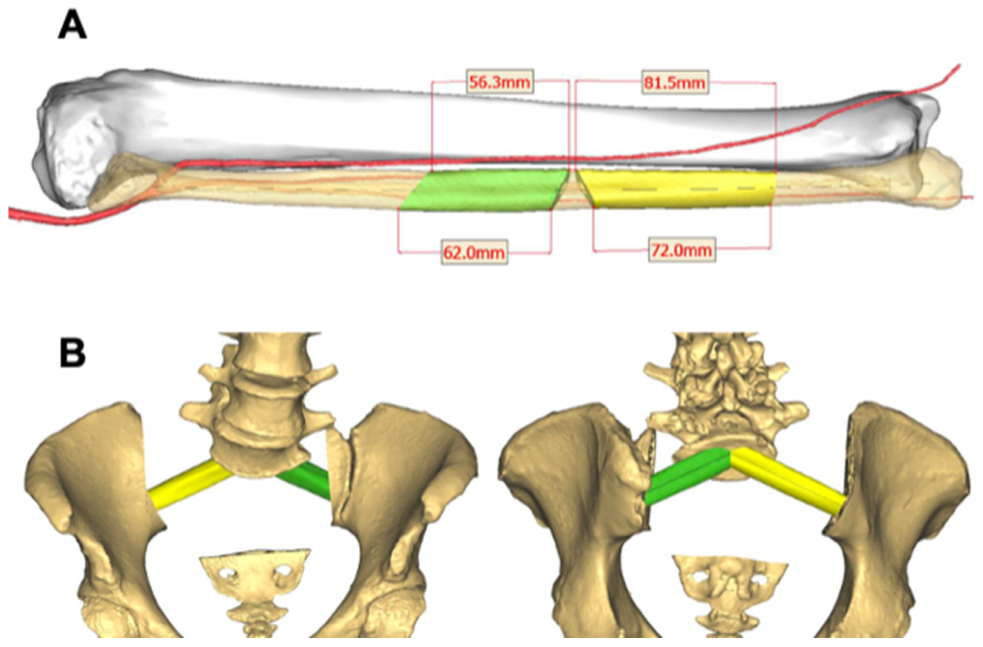
If sacropelvic reconstruction with FVFG is planned, the appropriate resection planes of the fibula are established and incorporated into the post-resection reconstruction model (Figure 2). CAD models of tumor, surrounding anatomies and cutting planes are then exported as STL files. While exporting these as STL files, fiducial markings are placed on every STL file to help with location and realignment.
These STL files along with the preoperative DICOMs used for segmentation are loaded on to the BrainLab Origin Server 3.2 and opened in the iPlan Spine 3.0.6 app. We have noticed that importing these STL files into the software disorients and misaligns objects in the virtual space. Using Advanced Object Planning tab in iPlan and prior placed fiducial markings, STL anatomies and virtual cutting guides are reoriented, aligned and saved. This file is loaded on to the BrainLab Curve (not Kolibri) and opened in Spine and Trauma 3D 2.6.0. Once the intraoperative scan is completed, the preoperative CT and intraoperative CT are aligned using the ImageFusion 4.0 app in the BrainLab Curve. All the anatomies and individual virtual guides can be turned on or off individually as needed by the surgeon.

2.2. Intraoperative CAD Utilization

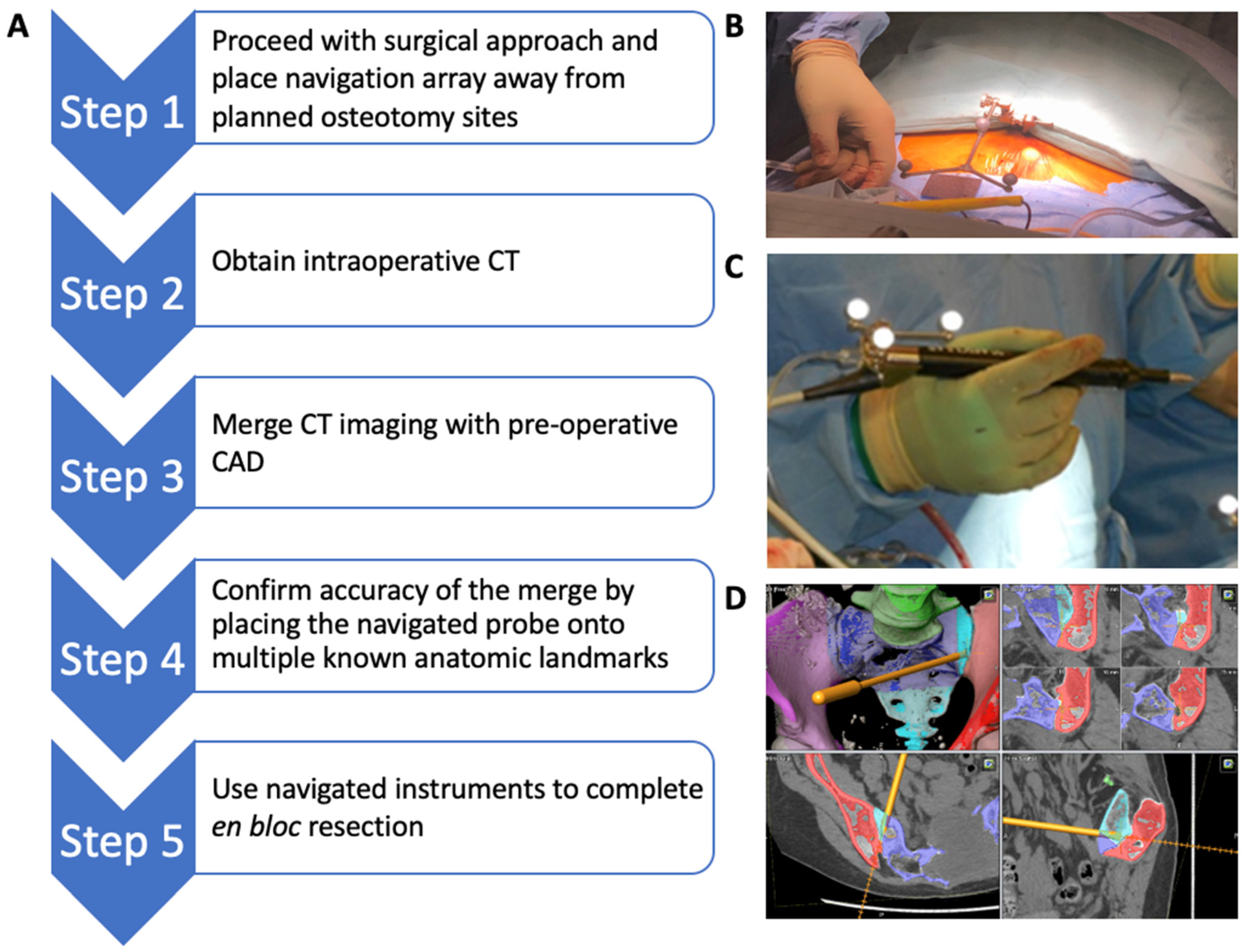
The steps for intraoperative CAD utilization are highlighted in Figure 3. The navigation array is first applied at a location away from the planned osteotomy sites (Figure 3B). Intraoperative CT images are then obtained and imported into the BrainLab Kolibri (BrainLab, Munich, Germany) navigation system. The CAD is overlaid on to the CT images to create a real-time, virtual 3D reconstruction image of the tumor and the planned osteotomy planes. Accuracy of the scan and the CAD overlay is confirmed using the navigated probe on multiple visibly identifiable anatomical landmarks in the coronal, sagittal, and axial planes. A tracking probe clamp is attached to the BoneScalpel (Misonix, Farmingdale, NY, USA) and registered to the navigation platform (Figure 3C). The navigated BoneScalpel is then used to execute the planned osteotomies while ensuring that all bone cuts are made precisely along the osteotomy planes depicted on the virtual guide (Figure 3D).

3. Results

Six patients (mean age 52.2 ± 17.7 years, 2 males, 4 females) underwent CAD-merged image-guided operations during the study period (Table 1). Mean follow-up was 38.0 ± 6.5 months (range, 24.8–42.2). Staged procedures were performed in three patients. Apart from their initial cancer diagnosis, there were few underlying health conditions, with three patients having no comorbidities, two patients having asthma, and one patient having a history of smoking. One patient had Musculoskeletal Tumor Society (MSTS) stage IIB radiation-induced osteosarcoma involving the sacrum and right ilium. One patient had stage IIB primary osteosarcoma involving the sacrum and left ilium. Three patients had stage IIB primary chondrosarcoma involving the pelvis and/or sacrum. One patient had stage III secondary chondrosarcoma with a prior history of multiple hereditary enchondromatosis (MHE). One patient with a primary chondrosarcoma of the acetabulum underwent a pelvis and hip joint reconstruction using a custom endoprosthetic implant. All cases were diagnosed preoperatively with core needle biopsy and margins were confirmed with intraoperative frozen specimens.
Negative margins were achieved in all patients with no intraoperative complications. There were no technical difficulties involving the described technology when carrying out the surgery. Mean operative time was 1229 min (range, 522–2063). Mean EBL was 4425 mL (range, 300–12,150). Mean length of stay (LOS) was 18.7 days (range 7–47). All patients were discharged to a rehabilitation facility. There were no 30-day readmissions, 30-day reoperations, or 2-year mortality at final follow-up. One patient was complicated by flap necrosis, which was successfully treated with debridement and primary closure. One patient had local tumor recurrence at final follow-up. One patient with an initial diagnosis of stage IIB radiation-induced osteosarcoma had local recurrence at 14-month follow-up and is currently undergoing treatment for metastatic disease. One patient with MHE and an initial diagnosis of stage III secondary chondrosarcoma subsequently underwent marginal resections of metastatic disease to the left groin and left shoulder at 24 months and 42 months, respectively, after initial surgery.

4. Discussion

This study describes the surgical technique consisting of using virtual cutting guides to perform navigated osteotomies to safely and accurately perform complex tumor resections of the sacrum and pelvis. Although previous studies report the value of CAD and image-guided navigation, to our knowledge, no prior studies have reported the successful use of merging the two techniques to develop virtual cutting guides, a form of augmented-reality-assisted surgery.
To minimize the incidence of recurrences, an en bloc resection with wide margins is the preferred surgical treatment for primary osteosarcoma and high-grade chondrosarcoma [20,21]. However, performing wide resections in anatomically complex regions such as the lumbar spine, sacrum, and pelvis present challenges for surgeons to obtain adequate margins whilst minimizing morbidity, increasing the risk for intralesional resection and local recurrences [22]. A study by Jeys et al. reported a series of 539 patients undergoing wide resection of primary bone tumors of the sacrum or pelvis using standard surgical techniques without the use of CAD or navigation and found a 29% intralesional resection rate and a 27% local recurrence rate. This was compared to a series of 31 patients who underwent the same procedure using image-guided navigation and reported an 8.7% intralesional resection rate and a 13% local recurrence rate [11]. The authors speculated that although there were significant reductions in intralesional resection and recurrence rates with the use of navigation, using a non-navigated osteotome to perform the osteotomies may reduce the accuracy of the resection planes. Furthermore, without the use of CAD to be overlaid with the navigation imaging, the authors are unable to perform osteotomies precisely through planned resection planes. A recent study by Towner et al. introduced a technique in which an ultrasonic bone scalpel was registered with image-guided navigation to perform precise osteotomies under navigation for spine tumor resections [18]. The authors demonstrated the safety of its use in spine tumor resection and introduced potential advantages including higher precision and decreased operative times. CAD techniques have been recently introduced and are gaining popularity for improving precision in the field of maxillofacial surgery [13,14,15]. A case report by Nguyen et al. reported the first successful application of merging CAD with image-guided navigation for maxillofacial reconstruction of complex facial fractures [17]. Our study combined the use of these novel techniques by developing virtual cutting guides and performing navigated osteotomies to successfully obtain negative margins with no local recurrence in 100% of patients within our series at 12-month follow-up.
In addition to aiding in obtaining negative margins, CAD can effectively be utilized for sacropelvic reconstructions using FVFG as well as for custom endoprosthesis implantation for pelvis and hip reconstruction [23]. Sacropelvic tumor resections often result in large osseous defects and spinopelvic discontinuity and instability. Such resections would benefit from the use of FVFG to reconstruct the sacropelvic junction [23]. However, using the appropriately sized grafts are essential for obtaining union and stability after reconstruction [23,24]. The use of CAD allows the surgeon to preselect the exact length, orientation, and the number of grafts required for appropriate reconstruction, thus maximizing the biomechanical construct and possibly enhancing union rates [23]. Within our case series of six patients, three patients underwent sacropelvic reconstruction with FVFG and the grafts were obtained and appropriately sized using CAD and image-guided navigation (Figure 4).
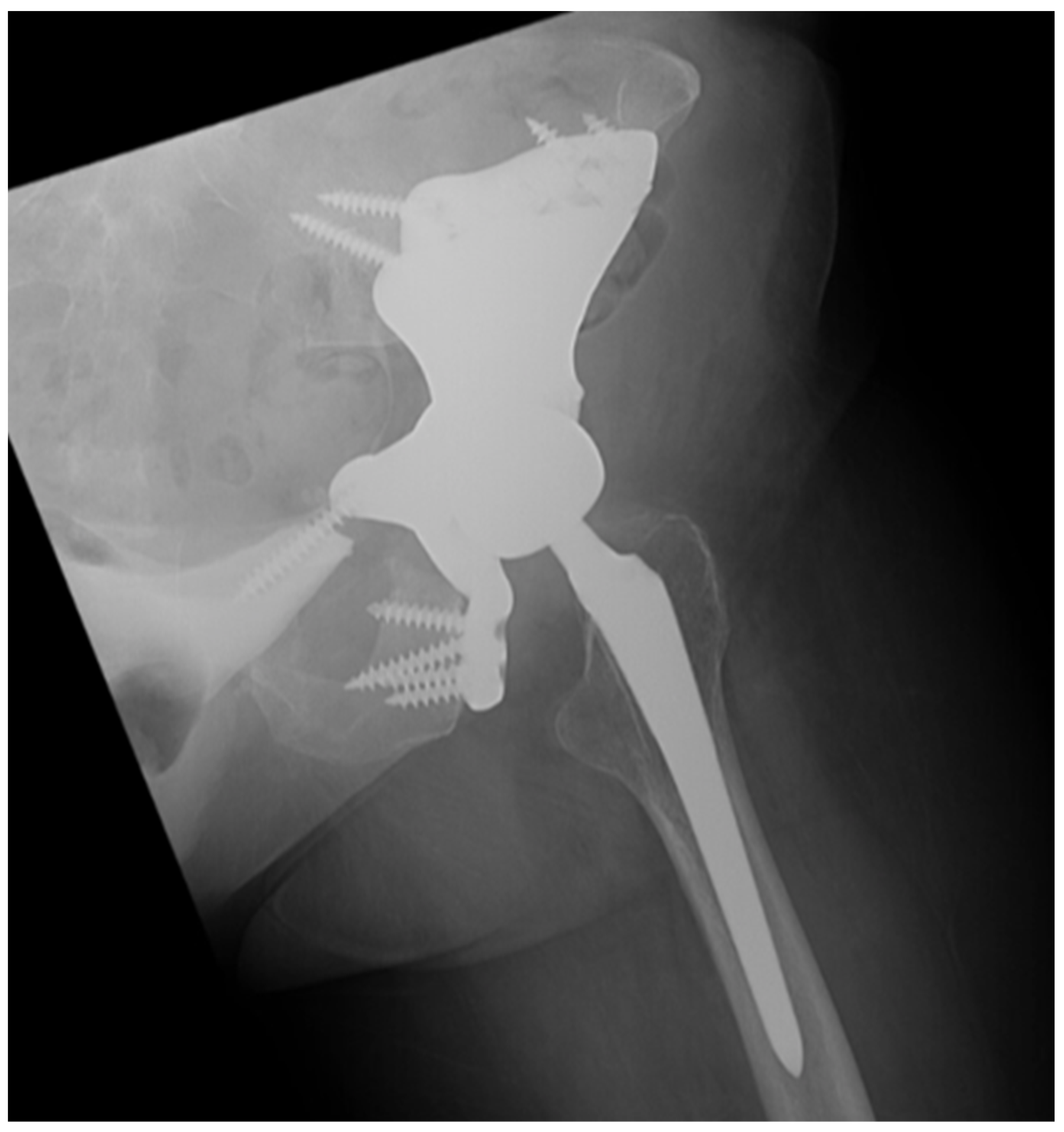
All three patients achieved bony union at 12-month follow-up. With regard to the use of CAD for custom endoprosthesis implantation for pelvis and hip reconstruction, precise osteotomies are necessary to successfully implant a custom endoprosthesis. Recent literature has demonstrated significant advances in 3D technology for this purpose by developing 3D models of the patient anatomy and the custom endoprosthesis as well as 3D cutting guides to be used intraoperatively [25,26]. However, the intraoperative use of 3D models and cutting guides are often challenging due to presence of soft tissues and vital structures surrounding the planned osteotomy sites. Thus, incorporating CAD and virtual gutting guides for executing precise osteotomies is not only useful for en bloc tumor resections but also particularly helpful in preparing the hip and pelvis for a custom endoprosthesis. With our case series, one patient with a primary chondrosarcoma of the acetabulum successfully underwent a pelvis and hip joint reconstruction using a custom endoprosthetic implant without complications (Figure 5).
There are limitations with the use of CAD and image-guided navigation techniques. CAD is developed using CT images acquired preoperatively, which is overlaid with CT images obtained intraoperatively for image-guided navigation. Thus, particularly with fast-growing tumors, there may be slight anatomical differences seen after the image merging process. Therefore, we suggest obtaining the preoperative CT for CAD development as close as possible to the day of surgery. Furthermore, prior studies have described image-to-patient registration errors of up to 2 mm [27,28,29]. Within our study, the accuracy of the scans and the CAD overlays were confirmed using the navigated probe on multiple visibly identifiable anatomical landmarks in the coronal, sagittal, and axial planes, and a less than 1 mm. registration error was confirmed prior to proceeding with the planned osteotomies. Finally, segmentation of tumor can be performed on MRI and merged with intraoperative CT; however, this is often suboptimal due to changes in patient positioning between the two scans.
There are several limitations to this study. Notably, the retrospective nature of the case series with a small sample size is vulnerable to selection bias and is insufficient in terms of statistical power for conducting an analysis. The lack of a control group or comparative analysis complicates the determination of the precise benefits of this method in comparison to traditional approaches. Furthermore, although all reported cases utilized the method of merging CAD with image-guided navigation, the complexity of each case was variable in nature and the aggregate result may not be reflective of all outcomes. This study was also limited by a minimum follow-up period of 24 months; thus, we are unable to comment on survival advantages beyond this time using this operative technique. Future studies should include prospective comparative studies with a larger sample size and longer follow-up that allows for further determination of the efficacy of this surgical technique. Additionally, future work that also applies this technology within other areas of the spine would also contribute to enhancing our comprehension of the effectiveness of this approach.

5. Conclusions

In conclusion, our study demonstrates a novel method of utilizing virtual cutting guides as a safe technique to perform navigated osteotomies that can facilitate complex tumor resections of the sacrum and pelvis. The CAD models, generated from the preoperative images, can be obtained within the institution in timely fashion as well as cost-effectively and could be successfully laid over the intraoperative images to provide guidance during the surgical procedure.

Author Contributions

All authors made substantial contributions to the conception and design of work and provided final approval of the version to be published. Specific additional individual contributions include T.H., interpretation and analysis of data for this work, drafted the introduction, the overall work outline, and made critical final revisions. G.R.M., lead the acquisition of data and drafted the results section. G.L.II, drafted a portion of the discussion section and made critical final revisions. K.T., assisted in the acquisition of data and drafted the results section. R.L.S., senior surgeon with substantial contribution to design of work and made critical final revisions and approval of the version to be published. A.F.M., senior surgeon with substantial contribution to design of work and made critical final revisions and approval of the version to be published. L.D.R., senior surgeon with substantial contribution to design of work and made critical final revisions and approval of the version to be published. J.E.B., senior surgeon with substantial contribution to design of work and made critical final revisions and approval of the version to be published. All authors have read and agreed to the published version of the manuscript.

Funding

This research received no external funding.

Institutional Review Board Statement

The study was conducted in accordance with the Declaration of Helsinki, and approved by the Institutional Review Board of The University of Texas MD Anderson Cancer Center (PRO1077258 on 21 October 2020).

Informed Consent Statement

Not applicable.

Data Availability Statement

The datasets generated and/or analyzed during the current study are available from the corresponding author upon request.

Acknowledgments

Takashi Hirase was supported by a Burroughs Wellcome Fund Physician Scientist Institutional Award to the Texas A&M University Academy of Physician Scientists.

Conflicts of Interest

Takashi Hirase, Grant R McChesney, Garvin Lawrence II, Karthik Tappa, Robert L Satcher, Alexander F Mericli, and Laurence D Rhines declare no potential conflict of interest with respect to research, authorship, and/or publication of this article. Corresponding author, Justin Bird is a paid consultant with GT Medical Technologies and Ico Tec.

References

  1. Bederman, S.S.; Shah, K.N.; Hassan, J.M.; Hoang, B.H.; Kiester, P.D.; Bhatia, N.N. Surgical techniques for spinopelvic reconstruction following total sacrectomy: A systematic review. Eur. Spine J. 2014, 23, 305–319. [Google Scholar] [CrossRef]
  2. Fourney, D.R.; Rhines, L.D.; Hentschel, S.J.; Skibber, J.M.; Wolinsky, J.P.; Weber, K.L.; Suki, D.; Gallia, G.L.; Garonzik, I.; Gokaslan, Z.L. En bloc resection of primary sacral tumors: Classification of surgical approaches and outcome. J. Neurosurg. Spine 2005, 3, 111–122. [Google Scholar] [CrossRef]
  3. Fuchs, B.; Hoekzema, N.; Larson, D.R.; Inwards, C.Y.; Sim, F.H. Osteosarcoma of the pelvis: Outcome analysis of surgical treatment. Clin. Orthop. Relat. Res. 2009, 467, 510–518. [Google Scholar] [CrossRef]
  4. Ozaki, T.; Flege, S.; Kevric, M.; Lindner, N.; Maas, R.; Delling, G.; Schwarz, R.; Von Hochstetter, A.R.; Salzer-Kuntschik, M.; Berdel, W.E.; et al. Osteosarcoma of the pelvis: Experience of the Cooperative Osteosarcoma Study Group. J. Clin. Oncol. 2003, 21, 334–341. [Google Scholar] [CrossRef]
  5. Jackson, R.J.; Gokaslan, Z.L. Spinal-pelvic fixation in patients with lumbosacral neoplasms. J. Neurosurg. 2000, 92, 61–70. [Google Scholar] [CrossRef]
  6. Tomita, K.; Tsuchiya, H. Total sacrectomy and reconstruction for huge sacral tumors. Spine 1990, 15, 1223–1227. [Google Scholar] [CrossRef]
  7. Drazin, D.; Bhamb, N.; Al-Khouja, L.T.; Kappel, A.D.; Kim, T.T.; Johnson, J.P.; Brien, E. Image-guided resection of aggressive sacral tumors. Neurosurg. Focus 2017, 42, E15. [Google Scholar] [CrossRef]
  8. Abraham, J.A.; Kenneally, B.; Amer, K.; Geller, D.S. Can Navigation-assisted Surgery Help Achieve Negative Margins in Resection of Pelvic and Sacral Tumors? Clin. Orthop. Relat. Res. 2018, 476, 499–508. [Google Scholar] [CrossRef]
  9. Ieguchi, M.; Hoshi, M.; Takada, J.; Hidaka, N.; Nakamura, H. Navigation-assisted surgery for bone and soft tissue tumors with bony extension. Clin. Orthop. Relat. Res. 2012, 470, 275–283. [Google Scholar] [CrossRef]
  10. Cho, H.S.; Oh, J.H.; Han, I.; Kim, H.S. The outcomes of navigation-assisted bone tumour surgery: Minimum three-year follow-up. J. Bone Jt. Surg. Br. 2012, 94, 1414–1420. [Google Scholar] [CrossRef]
  11. Jeys, L.; Matharu, G.S.; Nandra, R.S.; Grimer, R.J. Can computer navigation-assisted surgery reduce the risk of an intralesional margin and reduce the rate of local recurrence in patients with a tumour of the pelvis or sacrum? Bone Jt. J. 2013, 95, 1417–1424. [Google Scholar] [CrossRef] [PubMed]
  12. Wong, K.C.; Kumta, S.M. Computer-assisted tumor surgery in malignant bone tumors. Clin. Orthop. Relat. Res. 2013, 471, 750–761. [Google Scholar] [CrossRef] [PubMed]
  13. Seruya, M.; Fisher, M.; Rodriguez, E.D. Computer-assisted versus conventional free fibula flap technique for craniofacial reconstruction: An outcome comparison. Plast. Reconstr. Surg. 2013, 132, 1219–1228. [Google Scholar] [CrossRef]
  14. Klein, M.; Glatzer, C. Individual CAD/CAM fabricated glass-bioceramic implants in reconstructive surgery of the bony orbital floor. Plast. Reconstr. Surg. 2006, 117, 565–570. [Google Scholar] [CrossRef]
  15. Pang, J.H.; Brooke, S.; Kubik, M.W.; Ferris, R.L.; Dhima, M.; Hanasono, M.M.; Wang, E.W.; Solari, M.G. Staged Reconstruction (Delayed-Immediate) of the Maxillectomy Defect Using CAD/CAM Technology. J. Reconstr. Microsurg. 2018, 34, 193–199. [Google Scholar] [CrossRef] [PubMed]
  16. Shirk, J.D.; Reiter, R.; Wallen, E.M. Effect of 3-Dimensional, Virtual Reality Models for Surgical Planning of Robotic Prostatectomy on Trifecta Outcomes: A Randomized Clinical Trial. J. Urol. 2022, 208, 618–625. [Google Scholar] [CrossRef] [PubMed]
  17. Nguyen, A.; Vanderbeek, C.; Herford, A.S.; Thakker, J.S. Use of Virtual Surgical Planning and Virtual Dataset With Intraoperative Navigation to Guide Revision of Complex Facial Fractures: A Case Report. J. Oral. Maxillofac. Surg. 2019, 77, 790.e1–790.e17. [Google Scholar] [CrossRef]
  18. Towner, J.E.; Piper, K.F.; Schoeniger, L.O.; Qureshi, S.H.; Li, Y.M. Use of image-guided bone scalpel for resection of spine tumors: Technical note. AME Case Rep. 2018, 2, 48. [Google Scholar] [CrossRef]
  19. von Elm, E.; Altman, D.G.; Egger, M.; Pocock, S.J.; Gøtzsche, P.C.; Vandenbroucke, J.P. The Strengthening the Reporting of Observational Studies in Epidemiology (STROBE) statement: Guidelines for reporting observational studies. PLoS Med. 2007, 4, e296. [Google Scholar] [CrossRef]
  20. He, F.; Zhang, W.; Shen, Y.; Yu, P.; Bao, Q.; Wen, J.; Hu, C.; Qiu, S. Effects of resection margins on local recurrence of osteosarcoma in extremity and pelvis: Systematic review and meta-analysis. Int. J. Surg. 2016, 36 Pt A, 283–292. [Google Scholar] [CrossRef]
  21. Cloyd, J.M.; Acosta, F.L., Jr.; Polley, M.Y.; Ames, C.P. En bloc resection for primary and metastatic tumors of the spine: A systematic review of the literature. Neurosurgery 2010, 67, 435–445. [Google Scholar] [CrossRef] [PubMed]
  22. Yamazaki, T.; McLoughlin, G.S.; Patel, S.; Rhines, L.D.; Fourney, D.R. Feasibility and safety of en bloc resection for primary spine tumors: A systematic review by the Spine Oncology Study Group. Spine 2009, 34 (Suppl. 22), S31–S38. [Google Scholar] [CrossRef] [PubMed]
  23. Houdek, M.T.; Rose, P.S.; Bakri, K.; Wagner, E.R.; Yaszemski, M.J.; Sim, F.H.; Moran, S.L. Outcomes and Complications of Reconstruction with Use of Free Vascularized Fibular Graft for Spinal and Pelvic Defects Following Resection of a Malignant Tumor. J. Bone Jt. Surg. Am. 2017, 99, e69. [Google Scholar] [CrossRef] [PubMed]
  24. Li, J.; Chen, G.; Lu, Y.; Zhu, H.; Ji, C.; Wang, Z. Factors Influencing Osseous Union Following Surgical Treatment of Bone Tumors with Use of the Capanna Technique. J. Bone Jt. Surg. Am. 2019, 101, 2036–2043. [Google Scholar] [CrossRef] [PubMed]
  25. Ogura, K.; Sakuraba, M.; Miyamoto, S.; Fujiwara, T.; Chuman, H.; Kawai, A. Pelvic ring reconstruction with a double-barreled free vascularized fibula graft after resection of malignant pelvic bone tumor. Arch. Orthop. Trauma. Surg. 2015, 135, 619–625. [Google Scholar] [CrossRef] [PubMed]
  26. Woo, S.H.; Sung, M.J.; Park, K.S.; Yoon, T.R. Three-dimensional-printing Technology in Hip and Pelvic Surgery: Current Landscape. Hip Pelvis 2020, 32, 1–10. [Google Scholar] [CrossRef] [PubMed]
  27. Fang, C.; Cai, H.; Kuong, E.; Chui, E.; Siu, Y.C.; Ji, T.; Drstvenšek, I. Surgical applications of three-dimensional printing in the pelvis and acetabulum: From models and tools to implants. Unfallchirurg 2019, 122, 278–285. [Google Scholar] [CrossRef]
  28. Wong, K.C.; Kumta, S.M. Joint-preserving tumor resection and reconstruction using image-guided computer navigation. Clin. Orthop. Relat. Res. 2013, 471, 762–773. [Google Scholar] [CrossRef]
  29. So, T.Y.; Lam, Y.L.; Mak, K.L. Computer-assisted navigation in bone tumor surgery: Seamless workflow model and evolution of technique. Clin. Orthop. Relat. Res. 2010, 468, 2985–2991. [Google Scholar] [CrossRef]
Figure 1. (A) Tumor involvement within the preoperative 3D CAD. The purple area represents the tumor. (B) Planned resection planes for en bloc resection as represented in red.
Bioengineering 10 01342 g001
Figure 2. (A) Resection planes of the vascularized fibula grafts represented in green and yellow. (B) Virtual sacropelvic reconstruction model with the templated fibula grafts providing a perfect fit within the resected voids.
Bioengineering 10 01342 g002
Figure 3. (A) Outline for intraoperative CAD utilization. (B) Intraoperative placement of navigation array. (C) Attachment of tracking probe clamp to the BoneScalpel. (D) Virtual cutting guide and anterior osteotomies performed using navigated BoneScalpel.
Bioengineering 10 01342 g003
Figure 4. (A) Virtual cutting guide with virtual representations of the navigated BoneScalpel for performing posterior osteotomies around the tumor (red). (B) Intraoperative photo demonstrating navigated BoneScalpel. The use of the navigated bone scalpel with virtual cutting guides significantly decreased the size of the incision and degree of required soft tissue exposure.
Bioengineering 10 01342 g004
Figure 5. Left hip radiograph of a patient with primary chondrosarcoma of the acetabulum that underwent a pelvis and hip joint reconstruction using a custom endoprosthetic implant.
Bioengineering 10 01342 g005
Table 1. Patient Demographics and Outcomes.
Case No.123456
Age (yrs), Sex53, F25, F60, F39, F61, M75, M
ASA Class333333
BMI (kg/m2)17.518.025.931.428.630.4
Follow-Up (months)24.839.740.342.241.539.2
Comorbidities NoneNoneNoneAsthmaFormer smokerAsthma
Diagnosis Radiation-Induced OsteosarcomaPrimary OsteosarcomaPrimary
Chondrosarcoma
MHE, Secondary
Chondrosarcoma
Primary
Chondrosarcoma
Primary
Chondrosarcoma
Histologic GradeHigh gradeHigh gradeHigh gradeDe-differentiatedHigh gradeHigh grade
MSTS StageIIBIIIIIBIIIIIBIIB
LocationSacrum, Rt iliumLS spine, Lt iliumLt acetabulumLt iliumSacrum, Lt iliumSacrum, Lt ilium
Procedures PerformedStage 1: L5-S1 anterior discectomy, anterior sacral osteotomy, fibula flap harvest
Stage 2: sacrectomy, L3-pelvis PSIF, L5-pelvis ASF with vascularized fibula flap, VRAM flap
Stage 1: Rt T12-pelvis PSIF, L3-5 laminectomy, fibula flap harvest;
Stage 2: L4, L5 vertebrectomy, sacrectomy, Lt type I internal hemipelvectomy, Lt L3-pelvis PSIF
Stage 3: L3-pelvis ASF with vascularized fibula flap
Lt type I-II internal hemipelvectomy, custom endoprosthetic pelvic and hip joint reconstructionLt Type I internal hemipelvectomy, vascularized fibula autograft reconstructionStage 1: L3-S3 laminectomy, L5 vertebrectomy, L3-pelvis PSIF; Stage 2: Lt type I internal hemipelvectomy, Lt partial sacrectomy, L5-pelvis ASF with vascularized fibula autograftL5-S1 laminectomy, partial sacrectomy, Lt ilium osteotomy, pedicled Rt gluteus maximus flap
EBL (mL)12,150550014004006800300
OR Time (min)206317395225431994515
MarginsNegativeNegativeNegativeNegativeNegativeNegative
Intraoperative ComplicationsNoneNoneNoneNoneNoneNone
LOS (days)19471612117
Discharge DispositionRehab facilityRehab facilityRehab facilityRehab facilityRehab facilityRehab facility
30-Day ReadmissionNoNoNoNoNoNo
30-Day ReoperationNoNoNoNoNoNo
2-Year MortalityNoNoNoNoNoNo
Postoperative ComplicationsNoneFlap necrosisNoneNoneNoneNone
Local RecurrenceYesNoNoNoNoNo
Metastatic DiseaseYesNoNoYes, left groin, left shoulder s/p resectionNoNo
ASA, American Society of Anesthesiologists; BMI, body mass index; MSTS, Musculoskeletal Tumor Society; EBL, estimated blood loss; OR, operating room; LOS, length of stay; F, female; M, male; LS, lumbosacral; Lt, left; Rt, right; PSIF, posterior spinal instrumented fusion; ASF, anterior spinal fusion; VRAM, vertical rectus abdominis musculocutaneous; MHE, multiple hereditary enchondromatosis.
Disclaimer/Publisher’s Note: The statements, opinions and data contained in all publications are solely those of the individual author(s) and contributor(s) and not of MDPI and/or the editor(s). MDPI and/or the editor(s) disclaim responsibility for any injury to people or property resulting from any ideas, methods, instructions or products referred to in the content.