Article

The Source and Pathophysiologic Significance of Excreted Cadmium

by
Soisungwan Satarug
1,*,
David A. Vesey
1,2,
Werawan Ruangyuttikarn
3,
Muneko Nishijo
4,
Glenda C. Gobe
1,2,5,6 and
Kenneth R. Phelps
7
1
Kidney Disease Research Collaborative, The University of Queensland Faculty of Medicine and Translational Research Institute, Woolloongabba, Brisbane 4102, Australia
2
Department of Nephrology, Princess Alexandra Hospital, Brisbane 4075, Australia
3
Division of Toxicology, Department of Forensic Medicine, Chiang Mai University, Chiang Mai 50200, Thailand
4
Department of Public Health, Kanazawa Medical University, Uchinada, Ishikawa 920-0293, Japan
5
School of Biomedical Sciences, The University of Queensland, Brisbane 4072, Australia
6
NHMRC Centre of Research Excellence for CKD.QLD, UQ Health Sciences, Royal Brisbane and Women’s Hospital, Brisbane 4029, Australia
7
Stratton Veterans’ Affairs Medical Center and Albany Medical College, Albany, NY 12208, USA
*
Author to whom correspondence should be addressed.
Toxics 2019, 7(4), 55; https://doi.org/10.3390/toxics7040055
Submission received: 10 August 2019 / Revised: 10 October 2019 / Accepted: 16 October 2019 / Published: 18 October 2019
(This article belongs to the Special Issue Toxic Metals, Chronic Diseases and Related Cancers)

Abstract

:
In theory, the identification of the source of excreted cadmium (Cd) might elucidate the pathogenesis of Cd-induced chronic kidney disease (CKD). With that possibility in mind, we studied Thai subjects with low, moderate, and high Cd exposure. We measured urine concentrations of Cd, ([Cd]u); N-acetyl-β-d-glucosaminidase, a marker of cellular damage ([NAG]u); and β2-microglobulin, an indicator of reabsorptive dysfunction ([β2MG]u). To relate excretion rates of these substances to existing nephron mass, we normalized the rates to creatinine clearance, an approximation of the glomerular filtration rate (GFR) (ECd/Ccr, ENAG/Ccr, and Eβ2MG/Ccr). To link the loss of intact nephrons to Cd-induced tubular injury, we examined linear and quadratic regressions of estimated GFR (eGFR) on ECd/Ccr, eGFR on ENAG/Ccr, and ENAG/Ccr on ECd/Ccr. Estimated GFR varied inversely with both ratios, and ENAG/Ccr varied directly with ECd/Ccr. Linear and quadratic regressions of Eβ2MG/Ccr on ECd/Ccr and ENAG/Ccr were significant in moderate and high Cd-exposure groups. The association of ENAG/Ccr with ECd/Ccr implies that both ratios depicted cellular damage per surviving nephron. Consequently, we infer that excreted Cd emanated from injured tubular cells, and we attribute the reduction of eGFR to the injury. We suggest that ECd/Ccr, ENAG/Ccr, and eGFR were associated with one another because each parameter was determined by the tubular burden of Cd.

1. Introduction

Cadmium (Cd), a divalent metal used for industrial purposes, is an important environmental pollutant in some regions of the world [1,2,3,4,5]. The metal is conveyed to humans in food, air, and tobacco smoke, and subsequently gains access to the circulation through the gut and lungs [5]. Salts of ionized Cd are absorbed in the duodenum; in addition, complexes of Cd with plant metallothioneins (MT) and phytochelatins (PC) may be absorbed in the colon after liberation by bacteria [6]. In the bloodstream, Cd is bound to red blood cells, albumin, glutathione (GSH), sulfur-containing amino acids, MT, and PC [6,7,8,9,10]. In the liver, hepatocytes take up Cd not bound to MT [10], synthesize MT in response to the metal, and store complexes of CdMT. These complexes are subsequently released from hepatocytes and transported to the kidneys [8,11,12]. Cd in plasma is filterable by glomeruli if it is bound to GSH, amino acids, MT, or PC [8,9], but the fraction of circulating Cd that enters the filtrate is unknown. The proximal tubule reabsorbs and retains most or all of the filtered Cd with an array of channels, solute carriers, and mediators of endocytosis [7,10,13,14,15,16]. Basolateral uptake may also add to the cellular content of Cd in the proximal tubule [16,17,18].
It is currently assumed that the magnitude of a gradually acquired burden determines the toxicity of Cd in tubular cells [19]. The emergence of Cd from lysosomes induces robust intracellular synthesis of MT, which greatly mitigates the injury inflicted by free Cd through complexation of the metal [20]. Nevertheless, a fraction of Cd remains unbound to MT and is presumed to promote autophagy, apoptosis, and necrosis as accumulation of Cd progresses [19,21]. Manifestations of renal toxicity include increased excretion of cellular proteins, impaired reabsorption of filtered substances, histologically demonstrable tissue injury, loss of intact nephrons, and reduction of the glomerular filtration rate (GFR) [5,8,21,22,23,24,25,26,27]. GFR may continue to fall for many years after exogenous exposure ceases [22,24], presumably because traffic of CdMT from the liver to kidneys persists.
In human studies of Cd-induced nephropathy, the most commonly assayed marker of tubular cell damage is the lysosomal enzyme N-acetyl-β-d-glucosaminidase (NAG). Because NAG is too large to be filtered by glomeruli, excessive excretion (ENAG) signifies tubular injury [28]. The most commonly measured indicator of impaired reabsorption is β2-microglobulin (β2MG). This small circulating protein is extremely filterable by glomeruli [29]; ordinarily, more than 99% of filtered β2MG is reabsorbed [30], but that percentage falls early in the course of tubular injury [31].
Although the urinary excretion rate of Cd (ECd) is believed to reflect the body burden of the metal [5], the precise source and pathophysiologic significance of excreted Cd have not been clarified. One possibility is that Cd is excreted because it is filtered and not reabsorbed; an alternate possibility is that excretion reflects liberation of Cd into filtrate from injured or dying tubular cells [32]. This distinction is important because the source of urinary Cd is central to the relationship between Cd accumulation and progression of chronic kidney disease (CKD). Herein, we present evidence that excreted Cd emanates from cells that it has injured. The injury leads to the loss of intact nephrons, reduction of GFR, and impaired reabsorption of filtered β2MG.

2. Materials and Methods

2.1. Study Subjects

The Institutional Ethical Committees of Chulalongkorn University, Chiang Mai University and the Mae Sot Hospital approved the study protocol (Approval No. 142/2544, 5 October 2001) [33]. All participants gave informed consent before participation. Subjects were recruited from urban communities in Bangkok in 2001/2002 and from subsistence farming areas in Mae Sot District, Tak Province, Thailand in 2004/2005 [33]. They had lived at their current addresses for at least 30 years. Exclusion criteria were pregnancy, breast-feeding, a history of metal work, and a hospital record or physician’s diagnosis of an advanced chronic disease. Because occupational exposure was an exclusion criterion, we presumed that all participants had acquired Cd from the environment.
Cd exposure was low in Bangkok and moderate or high in Mae Sot [33,34]. Determination of exposure was based on reported levels of Cd in rice grains grown in the Cd-affected areas of the Mae Sot District [35,36]. After exclusion of subjects with incomplete datasets, we studied 172, 310, and 222 persons from the low, moderate, and high exposure areas, respectively.

2.2. Collection of Biological Specimens and Laboratory Analyses

Second morning-void urine samples were collected after an overnight fast. Within three hours after urine sampling, specimens of whole blood were obtained and serum samples were prepared. Aliquots of urine, whole blood, and serum were transported on ice from a mobile clinic to a laboratory and stored at −20 °C or −80 °C for later analysis. The assay for urine and serum creatinine concentrations ([cr]u, [cr]p]) was based on the Jaffe reaction. The urine NAG assay was based on colorimetry (NAG test kit, Shionogi Pharmaceuticals, Sapporo, Japan). The urine β2MG assay was based on the latex immunoagglutination method (LX test, Eiken 2MGII; Eiken and Shionogi Co., Tokyo, Japan). When the urine concentration of β2MG ([β2MG]u) was below the limit of detection (LOD), 0.5 µg/L, the value assigned to [β2MG]u was LOD/(square root of 2).
For the Bangkok group, [Cd]u was determined by inductively-coupled plasma mass spectrometry (ICP/MS, Agilent 7500, Agilent Technologies), because it had the high sensitivity required to measure Cd concentrations below the detectable limit of atomic absorption spectrophotometry. Multi-element standards (EM Science, EM Industries, Inc., Newark, NJ, USA) were used to calibrate Cd analyses. The accuracy and precision of those analyses were evaluated with reference urine (Lyphochek®, Bio-Rad, Sydney, Australia). When [Cd]u was less than the detection limit of 0.05 μg/L, the concentration assigned was the detection limit divided by the square root of 2.
For the Mae Sot groups, [Cd]u was determined by atomic absorption spectrophotometry (Shimadzu Model AA-6300, Kyoto, Japan). Urine standard reference material No. 2670 (National Institute of Standards, Washington, DC, USA) was used for quality assurance and control purposes. None of the urine samples from the Mae Sot groups were found to have [Cd]u below the detection limit.

2.3. Estimated Glomerular Filtration Rate (eGFR)

The glomerular filtration rate was estimated with equations from the Chronic Kidney Disease Epidemiology Collaboration (CKD-EPI) [37,38]. CKD stages 1, 2, 3, 4, and 5 corresponded to eGFR of 90–119, 60–89, 30–59, 15–29, and <15 mL/min/1.73 m2, respectively. For dichotomous comparisons, CKD was defined as eGFR < 60 mL/min/1.73 m2.

2.4. Normalization of Excretion Rates to Creatinine Clearance (Ccr)

Excretion rates of Cd, NAG, and β2MG were normalized to Ccr to yield the ratios ECd/Ccr, ENAG/Ccr, and Eβ2MG/Ccr in units of mass (amount excreted) per volume of filtrate. For x = Cd, NAG, or β2MG, Ex/Ccr was calculated as [x]u[cr]p/[cr]u ([39]; Supplementary Materials).

2.5. Statistical Analysis

Data were analyzed with SPSS 17.0 (SPSS Inc., Chicago, IL, USA). Distributions of excretion rates for Cd, NAG, and β2MG were examined for skewness, and those showing rightward skewing were subjected to base-10 logarithmic transformation before analysis. Departure of a given variable from normal distribution was assessed with the one-sample Kolmogorov–Smirnov test. For continuous variables not conforming to a normal distribution, the Kruskal–Wallis test was used to determine differences among the three localities in ECd/Ccr, ENAG/Ccr, Eβ2MG/Ccr, and other parameters. The Mann–Whitney U-test was used to compare mean differences between two groups. The Chi-Square test was used to determine differences in percentage and prevalence data. p-values ≤ 0.05 for two-tailed tests were assumed to indicate statistical significance.
Polynomial regression was used to fit lines and curves to the scatterplots of five pairs of variables, including eGFR versus ECd/Ccr, eGFR versus ENAG/Ccr, ENAG/Ccr versus ECd/Ccr, Eβ2MG/Ccr versus ECd/Ccr, and ENAG/Ccr versus Eβ2MG/Ccr. A linear model, y = a + bx, was adopted if the relationship was monotonic. A quadratic model (second-order polynomial), y = a + b1x + b2x2, was used if there was a significant change in the direction of the slope (b1 to b2) for prediction of the dependent variable y. In both types of equations, a represented the y-intercept.
The relationships between x and y were assessed with R2 (the coefficient of determination) and with unstandardized and standardized β coefficients. In linear and quadratic models, R2 is the fraction of variation in y that is explained by the variation in x. In linear models, the unstandardized β coefficient is the slope of the linear regression, and the standardized β coefficient indicates the strength of the association between y and x on a uniform scale. To examine quadratic curves relating eGFR to ECd/Ccr and ENAG/Ccr, we performed slope change analyses with a linear regression method.

3. Results

Table 1 presents data concerning age, gender, blood pressure, smoking status, and renal function of subjects with low, moderate, and high environmental exposure to Cd. There were significant differences in age and percentages of women and smokers across the three exposure subsets. Female gender was overrepresented in the moderate exposure group. More than half of subjects in the high exposure group were smokers. Blood pressures were recorded in the low and moderate exposure groups; systolic and mean pressures were significantly higher in the latter.
Mean eGFR fell and the percentages of stages 2 and 3 CKD rose with intensity of exposure. The mean serum creatinine concentration was higher in the high-Cd locality. In general, urine concentrations of Cd, NAG, and β2MG rose with Cd exposure, but [NAG]u and [β2MG]u were higher in the moderate Cd group than in the other two. ECd/Ccr, ENAG/Ccr, and Eβ2MG/Ccr followed the same pattern.
Figure 1A–D present scatterplots of eGFR against log(ECd/Ccr) in each exposure subset and the entire sample. In each subset, significant linear and quadratic relationships were documented, and quadratic R2 values were slightly higher. Quadratic R2 was 0.228 in the low-Cd group, 0.083 in the moderate-Cd group, 0.154 in the high-Cd group, and 0.378 in the entire sample. In the linear model, standardized β was −0.467 in the low-Cd group, −0.259 in the moderate-Cd group, −0.361 in the high-Cd group, and −0.598 in the entire sample.
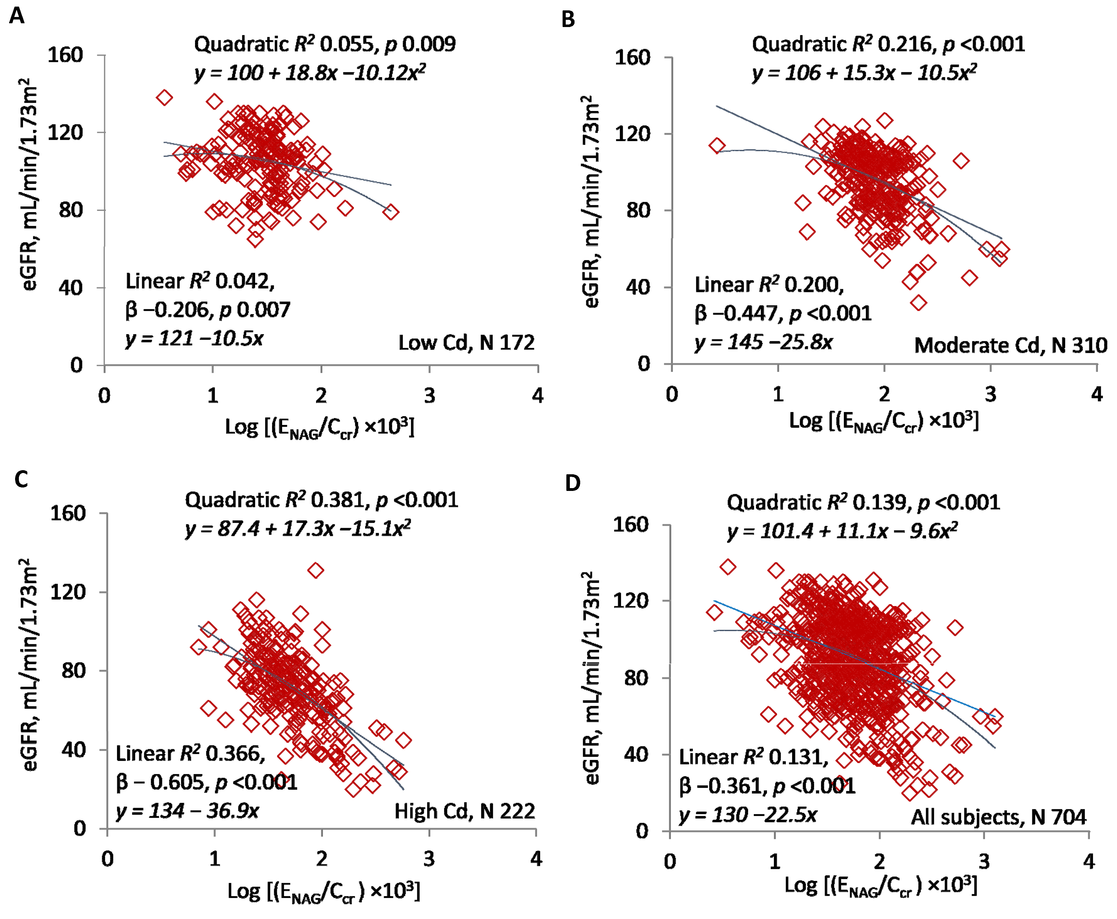
Figure 2A–D present scatterplots of eGFR against log(ENAG/Ccr). As in Figure 1, significant, inverse linear and quadratic relationships were documented in subsets and the entire sample, and quadratic R2 values were slightly higher. Quadratic R2 was 0.055 in the low-Cd group, 0.216 in the moderate-Cd group, 0.381 in the high-Cd group, and 0.139 in the entire sample. In the linear model, standardized β was −0.206 in the low-Cd group, −0.447 in the moderate-Cd group, −0.605 in the high-Cd group, and −0.361 in the entire sample.
The quadratic curves in Figure 1D and Figure 2D indicated that slopes describing rates of GFR reduction varied over the ranges of log[(ECd/Ccr) × 105] and log[(ENAG/Ccr) × 103]. We assumed that log[(ECd/Ccr) × 105] of 3.0 and log[(ENAG/Ccr) × 103] of 1.5 represented the excretion rates of Cd and NAG at which the rates of GFR reduction increased. Table 2 confirms that the slopes changed significantly at these points on the x-axes.
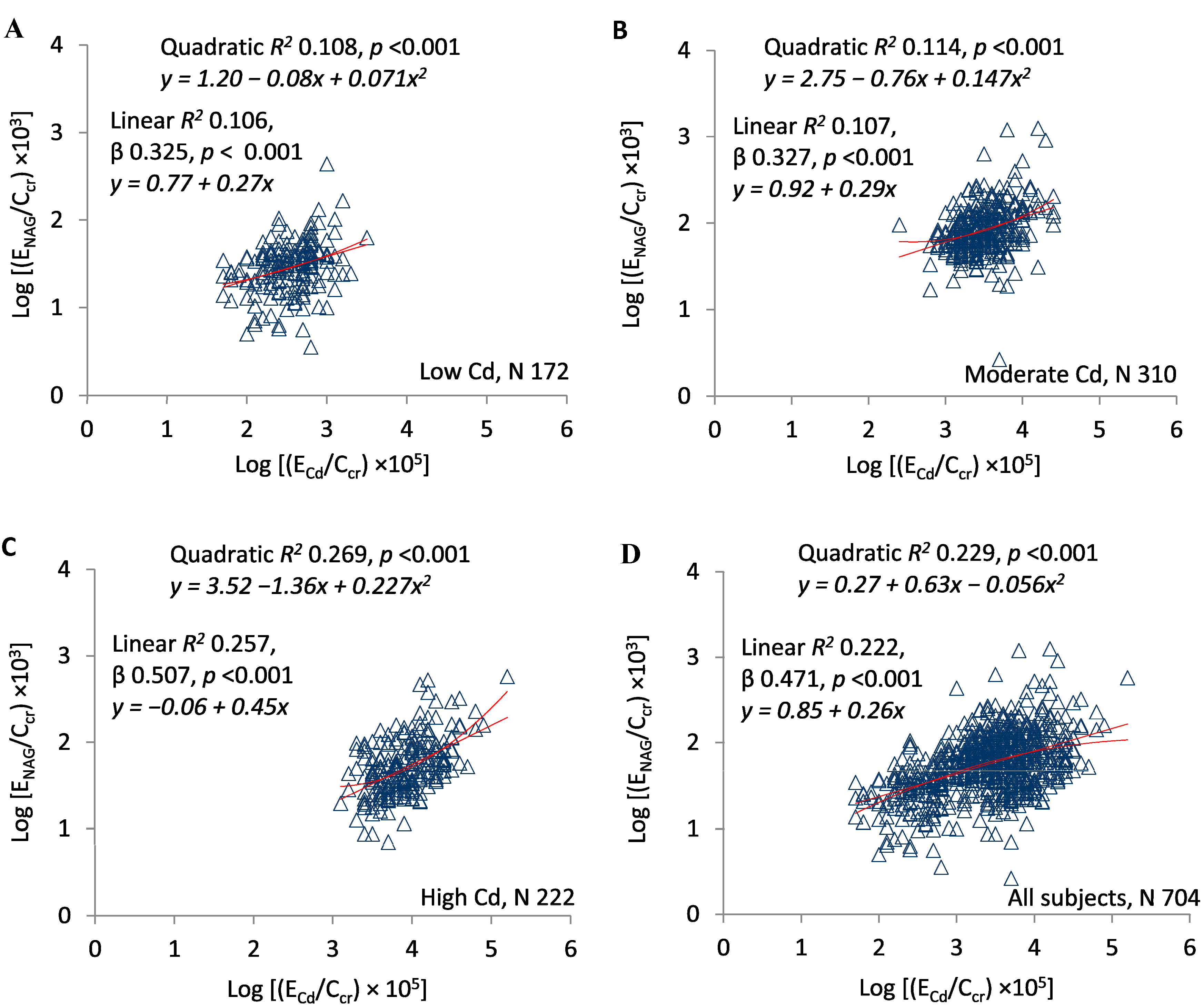
Figure 3A–D present scatterplots of log(ENAG/Ccr) against log(ECd/Ccr) in the exposure subsets and the entire sample. In all subsets, the two ratios varied directly, significant linear and quadratic relationships were documented, and quadratic R2 values were slightly higher. Quadratic R2 was 0.108 in the low-Cd group, 0.114 in the moderate-Cd group, 0.269 in the high-Cd group, and 0.229 in the entire sample. In the linear model, standardized β was 0.325 in the low-Cd group, 0.327 in the moderate-Cd group, 0.507 in the high-Cd group, and 0.471 in the entire sample.
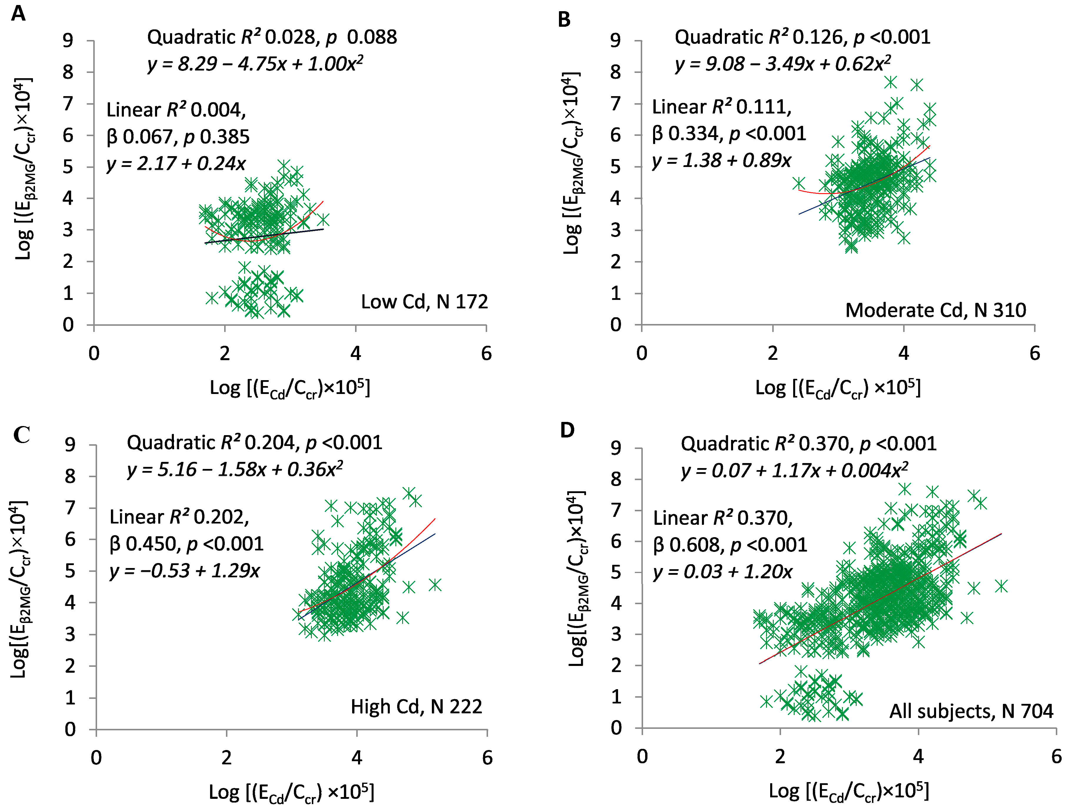
Figure 4A–D present scatterplots of log(Eβ2MG/Ccr) against log(ECd/Ccr) in the exposure subsets and the entire sample. Figure 4A demonstrates the absence of a relationship at the lowest Cd exposure (quadratic R2 = 0.028, p = 0.088). At moderate and high exposure, the two ratios were directly related, significant linear and quadratic relationships were documented, and quadratic R2 values were slightly higher (Figure 4B,C). Quadratic R2 was 0.126 in the moderate exposure group, 0.204 in the high exposure group, and 0.370 in the entire sample. Quadratic and linear relationships in the entire sample were virtually identical (Figure 4D). In the linear model, standardized β was 0.067 in the low-Cd group, 0.334 in the moderate-Cd group, 0.450 in the high-Cd group, and 0.608 in the entire sample.
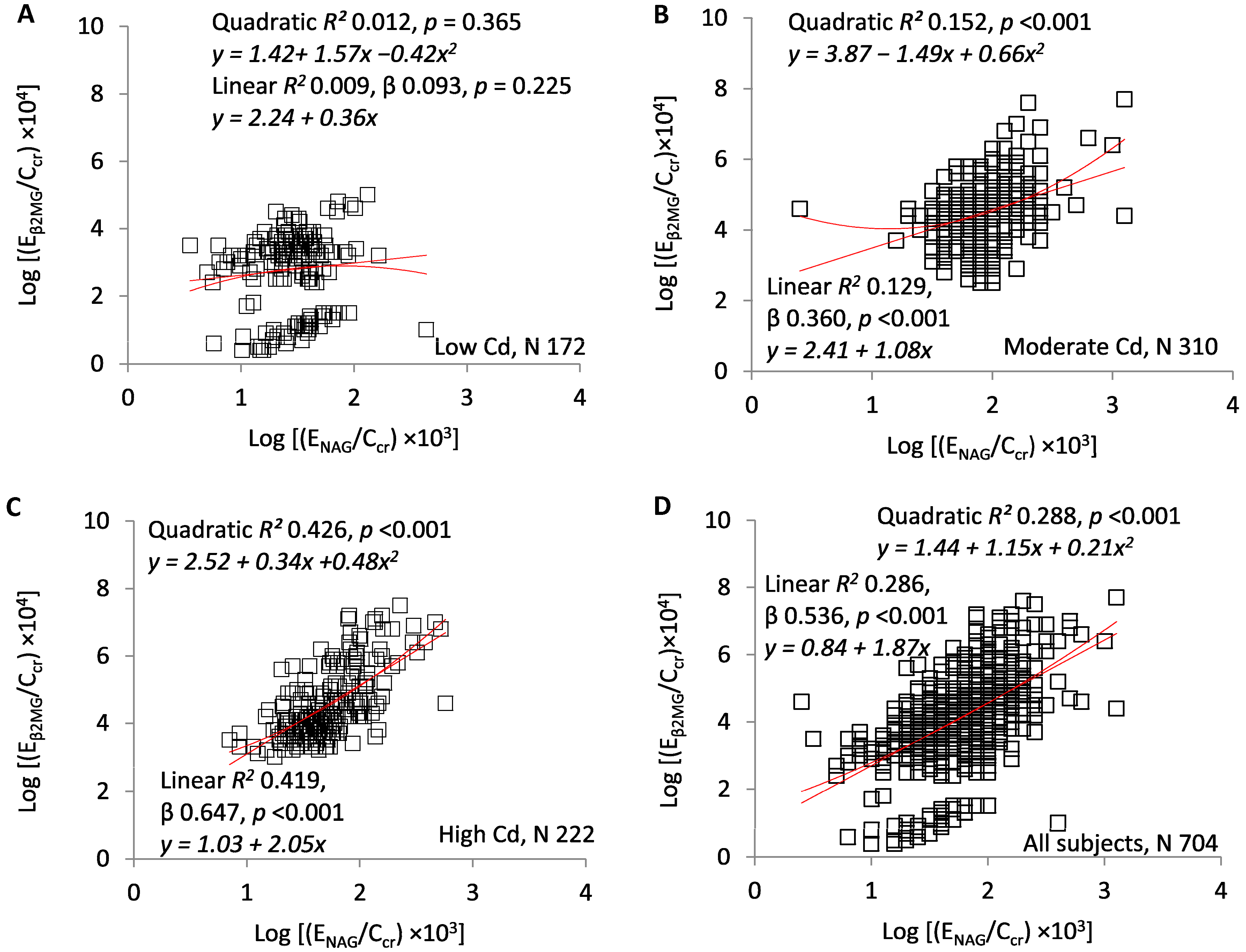
Figure 5A–D present scatterplots of log(Eβ2MG/Ccr) against log(ENAG/Ccr). Figure 5A demonstrates the absence of a relationship at the lowest Cd exposure (linear R2 = 0.009, p = 0.225). At moderate and high exposure, the two ratios were directly related, significant linear and quadratic regressions were documented, and quadratic R2 values were slightly higher (Figure 5B,C). Quadratic R2 was 0.152 in the moderate exposure group, 0.426 in the high exposure group, and 0.288 in the entire sample. In the linear model, standardized β was 0.093 in the low-Cd group, 0.360 in the moderate-Cd group, 0.647 in the high-Cd group, and 0.536 in the entire sample.

4. Discussion

Our goals in the present study were to elucidate the source of urinary Cd and to relate that source to the pathogenesis of Cd nephropathy. Data were obtained from clinically healthy Thai subjects residing in areas with low, moderate, or high exposure to Cd. In Mae Sot District, Tak Province, intensity of exposure was determined from the Cd content of rice grains [35,36]. Exposure in Bangkok was assumed to be low on the basis of food analyses and dietary histories [33].
Subjects in the three subsets were demographically dissimilar (Table 1). Both age and percentage of smokers rose with intensity of exposure. The percentage of women was particularly high in the moderate exposure group, and some women were of childbearing age. Because iron and Cd share a transporter in intestinal epithelium, menstruating women in this group may have absorbed Cd with exceptional avidity and incurred exceptional tubular toxicity secondarily (Table 1). In the high exposure subset, increased age may have conferred additional reasons for deterioration of GFR, and smoking may have provided a second environmental source of Cd. Whether smoking itself could have accelerated the progression of CKD is unresolved [40,41,42]. Neither age, nor smoking per se, nor the source of exogenous Cd obscured the significant relationship between eGFR and ECd/Ccr.
Table 1 shows that with the increasing intensity of exposure, ECd/Ccr rose and eGFR fell in stepwise fashion. In contrast, both ENAG/Ccr and Eβ2MG/Ccr were higher in the moderate than in the high exposure group. Although one might expect a direct relationship between NAG excretion and the number of intact nephrons, the higher ENAG/Ccr in the moderate group implies that the median excretion of NAG per intact nephron was also higher in these subjects. At the same time, the overlap of ENAG/Ccr between the moderate and high exposure groups was substantial (Figure 2 and Figure 5), and consistent relationships among ECd/Ccr, ENAG/Ccr, and eGFR were demonstrable at all intensities of exposure (Figure 1, Figure 2 and Figure 3). We speculate that the number of menstruating women in the moderate exposure subset was sufficient to increase Cd absorption and tubular toxicity in the entire group, but insufficient to disrupt the statistical relationships seen in all groups among ECd/Ccr, ENAG/Ccr, and eGFR.
Analogous statements can be made about β2MG. As impaired reabsorption of this protein is an early sign of proximal tubular injury, it is not surprising that median Eβ2MG/Ccr tracked with median ENAG/Ccr (Table 1). As would be expected, Eβ2MG/Ccr varied directly with ENAG/Ccr in the moderate and high exposure groups, but also varied directly with ECd/Ccr, which increased progressively with the intensity of exposure (Figure 4 and Figure 5).
Although CKD-EPI equations estimate GFR imprecisely [37,38], each exposure group differed significantly from the others with respect to eGFR (Table 1). Estimated GFR was inversely related to ECd/Ccr and ENAG/Ccr in the entire sample and each subset (Figure 1 and Figure 2), and ENAG/Ccr varied directly with ECd/Ccr (Figure 3). For all comparisons in Figure 1, Figure 2 and Figure 3, both linear and quadratic relationships were significant, and with one exception (Figure 1B), R2 rose with the exposure intensity. Standardized β followed the same pattern. Despite the statistical significance of all comparisons, some R2 values indicated that the fractional contribution of ECd/Ccr or ENAG/Ccr to eGFR was <10% (Figure 1B and Figure 2A); simultaneously, however, standardized β indicated robust effects of changes in x on changes in y. Factors other than ECd/Ccr and ENAG/Ccr affected eGFR, but variation in each ratio was associated with substantial variation in eGFR.
Taken together, the graphs in Figure 1, Figure 2 and Figure 3 imply that in each subset, GFR was inversely related to the severity of cellular injury per nephron (ENAG/Ccr), which in turn was associated with the amount of Cd excreted per nephron (ECd/Ccr). In addition, the quadratic relationships in Figure 1 and Figure 2 suggest that small increments in the most advanced injury were accompanied by disproportionate reductions in GFR. Slope analyses of curves in Figure 1D and Figure 2D confirm this inference (Table 2).
Other investigators have described direct relationships of [NAG]u/[cr]u and eGFR to [Cd]u/[cr]u, but we have not found a synthesis of those relationships into a satisfactory pathophysiologic narrative [35,43,44,45,46,47,48]. A cogent interpretation of Figure 1, Figure 2 and Figure 3 must explain how eGFR and ENAG/Ccr—results of cumulative Cd sequestration—were related physiologically to ECd/Ccr, an indicator of Cd excretion at the time of sampling. Cd was excreted for two possible reasons; it was filtered and not reabsorbed, or it was released from tubular cells [32]. Although both processes may have occurred, we do not see how the first, excretion after filtration, could have produced a physiologic connection between ECd/Ccr and ENAG/Ccr (Figure 3). In contrast, if Cd was released from damaged tubules, then Cd and NAG emanated from the same source, and both ECd/Ccr and ENAG/Ccr measured cellular injury. This shared attribute of Cd and NAG explains the statistical association of the ratios.
Additional evidence for the tubular origin of excreted Cd is provided by demonstrated extrusions of MT into tubular lumens [49], documented correlations of ECd with renal tissue content of Cd [50,51,52], and direct relationships between ECd and GFR (number of intact nephrons) [53,54,55]. Experiments in rabbits demonstrated a high tubular maximum for reabsorption of CdMT that would preclude excretion of filtered Cd in the typically intoxicated human [56].
In addition to addressing the likely source of excreted Cd, we must also ask why declining eGFR, the result of continuous loss of intact nephrons over time, was associated in the present study with parameters of current cellular injury, ECd/Ccr and ENAG/Ccr. To address this paradox, we propose that eGFR, ECd/Ccr, and ENAG/Ccr were simultaneous consequences of the tubular content of Cd. As the content rose, cellular injury per nephron and the rate of nephron loss increased proportionately; at any moment in a subject’s exposure history, the three variables were quantitatively associated because they were traceable to the same burden of sequestered Cd.
β2MG, a small protein made by nucleated cells, is almost completely filtered by glomeruli [29]. Ordinarily, the proximal tubule reabsorbs and degrades over 99% of filtered β2MG [30]. Because tubulopathies increase Eβ2MG [31], excessive Eβ2MG is conventionally interpreted as evidence of reabsorptive dysfunction [35,57,58]. This interpretation is understandable, but we suspect that it is an oversimplification. One reason is that endogenous production of β2MG may be increased by chronic inflammatory conditions, solid tumors, lymphatic malignancies, and multiple myeloma [59]. If tubular degradation (TDβ2MG) remains constant as production rises, Eβ2MG also rises even though TDβ2MG has not fallen (SM). Moreover, if both β2MG production and TDβ2MG per volume of filtrate (TDβ2MG/Ccr) remain constant as GFR falls, TDβ2MG also falls, and Eβ2MG rises (SM). Although these inferences are unproven, it seems likely that a combination of reabsorptive dysfunction and reduced GFR caused associations of Eβ2MG/Ccr with ECd/Ccr and ENAG/Ccr (Figure 4 and Figure 5).
An additional observation requires explanation. In the low exposure group, a cluster of subjects exhibited exceptionally low Eβ2MG/Ccr. At a fixed rate of β2MG filtration (equal to endogenous production) and a fixed value of TDβ2MG/Ccr, the rate of β2MG reabsorption increases with the number of intact nephrons, and Eβ2MG decreases simultaneously. In the isolated cluster, mean eGFR was 105.3 mL/min/1.73 m2; in the remainder of subjects in the study, it was 89.3 mL/min/1.73 m2. We suspect that extremely low Eβ2MG/Ccr in the cluster was the result of high Ccr, high TDβ2MG, and secondarily reduced Eβ2MG.
In the present study, we have continued the recently introduced practice of normalizing excretion of Cd, NAG, and β2MG to creatinine clearance instead of creatinine excretion [26]. Because the resulting ratios express ECd, ENAG, and Eβ2MG as functions of intact nephron mass, they nullify sources of imprecision that accompany normalization to Ecr or [cr]u. At any GFR, Ecr is primarily a function of muscle mass [60]; consequently, at a given ECd (for example), [Cd]u/[cr]u may vary by a multiple over the range of human body size. Moreover, multiple groups have reported direct rather than inverse relationships between GFR and [Cd]u/[cr]u after Cd exposure [5,53,54,55]. If the nephron number determines ECd at a given cellular burden of the metal, then [Cd]u/[cr]u may exaggerate the burden at normal GFR and underestimate it at reduced GFR. Normalization of ECd to Ccr—that is, calculation of [Cd]u[cr]p/[cr]u—eliminates the confounding effects of both muscle mass and nephron number on [Cd]u/[cr]u. In addition, because the required measurements are made in aliquots of urine and serum, the calculation quantifies amounts of Cd (or other substances) excreted per volume of filtrate while eliminating the need for timed urine collections and direct determinations of GFR. We plan to address optimal expression of excretion rates relevant to Cd nephropathy in a separate publication.
In summary, we draw the following conclusions from the significant regressions described herein. ENAG/Ccr varied directly with ECd/Ccr because sequestered Cd induced the release of NAG and Cd from tubular cells into filtrate. Estimated GFR varied inversely with both ratios because all three parameters reflected the extent of tubular Cd accumulation. ENAG/Ccr and ECd/Ccr quantified ongoing cellular injury, and eGFR quantified the loss of intact nephrons. We suspect that the significant regressions of Eβ2MG/Ccr on ECd/Ccr and ENAG/Ccr resulted from effects of Cd on both tubular reabsorption and nephron number.

Supplementary Materials

The following are available online at https://www.mdpi.com/2305-6304/7/4/55/s1, The equation for normalization to creatinine clearance.

Author Contributions

S.S., D.A.V., W.R., M.N., and G.C.G. formulated the study designs and protocols. S.S. and W.R. obtained ethical institutional clearances for research on human subjects and supervised the collection of biologic specimens in Thailand. S.S. organized and analyzed the data, created the tables and figures, and revised the manuscript for important intellectual content. K.R.P. proposed normalization of excretion rates to creatinine clearance, provided logical data interpretation, and was the primary author of the manuscript.

Funding

This research received no external funding.

Acknowledgments

This work was partially supported by the Commission for High Education, Thailand Ministry of Education, and the National Science and Technology Development Agency (NSTDA). Additionally, it was supported with resources of the Stratton Veterans’ Affairs Medical Center, Albany, NY, USA, and was made possible by facilities at that institution. Opinions expressed in this paper are those of the authors and do not represent the official position of the United States Department of Veterans’ Affairs.

Conflicts of Interest

The authors have no potential conflicts of interest to declare.

Abbreviations

AbbreviationMeaning
CdCadmium
GFRGlomerular filtration rate, units of volume/time
eGFREstimated glomerular filtration rate, units of mL/min/1.73 m2
CKD-EPIChronic kidney disease epidemiology collaboration
MTMetallothionein
CdMTCadmium–metallothionein complex
PCPhytochelatin
CdPCCadmium–phytochelatin complex
GSHGlutathione
NAGN-acetyl-β-d-glucosaminidase
β2MGBeta2-microglobulin
CcrCreatinine clearance, units of volume/time
VuUrine flow rate, units of volume/time
Ex/CcrExcretion rate of x per volume of filtrate, units of mass/volume, where x = Cd, NAG, or β2MG
TDβ2MGRate of tubular degradation of β2MG, units of mass/time
TDβ2MG/CcrAmount of β2MG degraded per volume of filtrate, units of mass/volume

References

  1. ATSDR (Agency for Toxic Substances and Disease Registry). Toxicological Profile for Cadmium; Department of Health and Humans Services, Public Health Service, Centers for Disease Control and Prevention: Atlanta, GA, USA, 2012. [Google Scholar]
  2. WHO. IPCS (International Programme on Chemical Safety) Environmental Health Criteria 134: Cadmium; WHO: Geneva, Switzerland, 1992. [Google Scholar]
  3. Satarug, S.; Vesey, D.A.; Gobe, G.C. Current health risk assessment practice for dietary cadmium: Data from different countries. Food Chem. Toxicol. 2017, 106, 430–445. [Google Scholar] [CrossRef] [PubMed]
  4. Satarug, S.; Vesey, D.A.; Gobe, G.C. Health risk assessment of dietary cadmium intake: Do current guidelines indicate how much is safe? Environ. Health Perspect. 2017, 125, 284–288. [Google Scholar] [CrossRef] [PubMed]
  5. Satarug, S. Dietary cadmium intake and its effects on kidneys. Toxics 2018, 6, 15. [Google Scholar] [CrossRef] [PubMed]
  6. Langelueddecke, C.; Lee, W.-K.; Thevenod, F. Differential transcytosis and toxicity of the hNGAL receptor ligands cadmium-metallothionein and cadmium-phytochelatin in colon-like Caco-2 cells: Implications for cadmium toxicity. Toxicol. Lett. 2014, 226, 228–235. [Google Scholar] [CrossRef] [PubMed]
  7. Dorian, C.; Gattone, V.H., II; Klaassen, C.D. Discrepancy between the nephrotoxic potencies of cadmium-metallothionein and cadmium chloride and the renal concentration of cadmium in the proximal convoluted tubules. Toxicol. Appl. Pharmacol. 1995, 130, 161–168. [Google Scholar] [CrossRef]
  8. Sabolic, I.; Breljak, D.; Skarica, M.; Herak-Kramberger, C.M. Role of metallothionein in cadmium traffic and toxicity in kidneys and other mammalian organs. Biometals 2010, 23, 897–926. [Google Scholar] [CrossRef]
  9. Fujita, Y.; ElBelbasi, H.I.; Min, K.-S.; Onosaka, S.; Okada, Y.; Matsumoto, Y.; Mutoh, N.; Tanaka, K. Fate of cadmium bound to phytochelatin in rats. Res. Commun. Chem. Pathol. Pharmacol. 1993, 82, 357–365. [Google Scholar]
  10. Sudo, J.-I.; Hayashi, T.; Soyama, M.; Fukata, M.; Kakuino, K. Kinetics of Cd2+ in plasma, liver and kidneys after single intravenous injection of Cd-metallothionein-II. Eur. J. Pharmacol. 1994, 270, 229–235. [Google Scholar] [CrossRef]
  11. Chan, H.M.; Zhu, L.F.; Zhong, R.; Grant, D.; Goyer, R.A.; Cherian, M.G. Nephrotoxicity in rats following liver transplantation from cadmium-exposed rats. Toxicol. Appl. Pharmacol. 1993, 123, 89–96. [Google Scholar] [CrossRef]
  12. Dudley, R.E.; Gammal, L.M.; Klaassen, C.D. Cadmium-induced hepatic and renal injury in chronically exposed rats: Likely role of hepatic cadmium-metallothionein in nephrotoxicity. Toxicol. Appl. Pharmacol. 1985, 77, 414–426. [Google Scholar] [CrossRef]
  13. Dorian, C.; Gattone, V.H., II; Klaassen, C.D. Renal cadmium deposition and injury as a result of accumulation of cadmium-metallothionein (CdMT) by the proximal convoluted tubules—A light microscopic autoradiography study with 109CdMT. Toxicol. Appl. Pharmacol. 1992, 114, 173–181. [Google Scholar] [CrossRef]
  14. Barbier, O.; Jacquillet, G.; Tauc, M.; Poujeol, P.; Cougnon, M. Acute study of interaction among cadmium, calcium, and zinc transport along rat nephron in vivo. Am. J. Physiol. 2004, 287, F1067–F1075. [Google Scholar] [CrossRef] [PubMed]
  15. Felley-Bosco, E.; Diezi, J. Fate of cadmium in rat renal tubules: A micropuncture study. Toxicol. Appl. Pharmacol. 1989, 98, 243–251. [Google Scholar] [CrossRef]
  16. Thevenod, F.; Fels, J.; Lee, W.-K.; Zarbock, R. Channels, transporters and receptors for cadmium and cadmium complexes in eukaryotic cells: Myths and facts. Biometals 2019, 32, 469–489. [Google Scholar] [CrossRef] [PubMed]
  17. Soodvilai, S.; Nantavishit, J.; Muanprasat, C.; Chatsudthipong, V. Renal organic cation transporters mediated cadmium-induced nephrotoxicity. Toxicol. Lett. 2011, 204, 38–42. [Google Scholar] [CrossRef] [PubMed]
  18. Zalups, R.K. Evidence for basolateral uptake of cadmium in kidneys of rats. Toxicol. Appl. Pharmacol. 2000, 164, 15–23. [Google Scholar] [CrossRef]
  19. Prozialeck, W.C.; Edwards, J.R. Mechanisms of cadmium-induced proximal tubule injury: New insights with implications for biomonitoring and therapeutic interventions. J. Pharmacol. Exp. Ther. 2012, 343, 2–12. [Google Scholar] [CrossRef]
  20. Liu, Y.; Liu, J.; Habeebu, S.S.; Klaassen, C.D. Metallothionein protects against the nephrotoxicity produced by chronic CdMT exposure. Toxicol. Sci. 1999, 50, 221–227. [Google Scholar] [CrossRef]
  21. Goyer, R.A.; Miller, C.R.; Zhu, S.-Y.; Victery, W. Non-metallothionein-bound cadmium in the pathogenesis of cadmium nephrotoxicity in the rat. Toxicol. Appl. Pharmacol. 1989, 101, 232–244. [Google Scholar] [CrossRef]
  22. Jarup, L.; Persson, B.; Elinder, C.G. Decreased glomerular filtration rate in solderers exposed to cadmium. Occup. Environ. Med. 1995, 52, 818–822. [Google Scholar] [CrossRef]
  23. Jarup, L.; Hellstrom, L.; Alfven, T.; Carlsson, M.D.; Grubb, A.; Persson, B.; Pettersson, C.; Spang, G.; Schutz, A.; Elinder, C.-G. Low level exposure to cadmium and early kidney damage: The OSCAR study. Occup. Environ. Med. 2000, 57, 668–672. [Google Scholar] [CrossRef] [PubMed]
  24. Roels, H.A.; Lauwerys, R.R.; Buchet, J.P.; Bernard, A.M.; Vos, A.; Oversteyns, M. Health significance of cadmium induced renal dysfunction: A five year follow up. Occup. Environ. Med. 1989, 46, 755–764. [Google Scholar] [CrossRef] [PubMed]
  25. Akesson, A.; Lundh, T.; Vahter, M.; Bjellerup, P.; Lidfeldt, J.; Nerbrand, C.; Samsioe, G.; Strömberg, U.; Skerfving, S. Tubular and glomerular kidney effects in Swedish women with low environmental cadmium exposure. Environ. Health Perspect. 2005, 113, 1627–1631. [Google Scholar] [CrossRef] [PubMed]
  26. Satarug, S.; Boonprasert, K.; Gobe, G.C.; Ruenweerayut, R.; Johnson, D.W.; Na-Bangchang, K.; Vesey, D.A. Chronic exposure to cadmium is associated with a marked reduction in glomerular filtration rate. Clin. Kidney J. 2018, 12, 468–475. [Google Scholar] [CrossRef]
  27. Swaddiwudhipong, W.; Limpatanachote, P.; Mahassakpan, P.; Krintratun, S.; Punta, B.; Funkhiew, T. Progress in cadmium-related health effects in persons with high environmental exposure in northwestern Thailand: A five-year follow-up. Environ. Res. 2012, 112, 194–198. [Google Scholar] [CrossRef]
  28. Price, R.G. Measurement of N-acetyl-beta-glucosaminidase and its isoenzymes in urine: Methods and clinical applications. Eur. J. Clin. Chem. Clin. Biochem. 1992, 30, 693–705. [Google Scholar]
  29. Portman, R.J.; Kissane, J.M.; Robson, A.M. Use of β2 microglobulin to diagnose tubulo-interstitial renal lesions in children. Kidney Int. 1986, 30, 91–98. [Google Scholar] [CrossRef]
  30. Argyropoulos, C.P.; Chen, S.S.; Ng, Y.-H.; Roumelioti, M.-E.; Shaffi, K.; Singh, P.P.; Tzamaloukas, A.H. Rediscovering beta-2 microglobulin as a biomarker across the spectrum of kidney diseases. Front. Med. 2017, 4, 73. [Google Scholar] [CrossRef]
  31. Peterson, P.A.; Evrin, P.-E.; Berggard, I. Differentiation of glomerular, tubular, and normal proteinuria: Determination of urinary excretion of β2-microglobulin, albumin, and total protein. J. Clin. Investig. 1969, 48, 1189–1198. [Google Scholar] [CrossRef]
  32. Chaumont, A.; Voisin, C.; Deumer, G.; Haufroid, V.; Annesi-Maesano, I.; Roels, H.; Thijs, L.; Staessen, J.; Bernard, A. Associations of urinary cadmium with age and urinary proteins: Further evidence of physiological variations unrelated to metal accumulation and toxicity. Environ. Health Perspect. 2013, 121, 1047–1053. [Google Scholar] [CrossRef]
  33. Satarug, S.; Swaddiwudhipong, W.; Ruangyuttikarn, W.; Nishijo, M.; Ruiz, P. Modeling cadmium exposures in low- and high-exposure areas in Thailand. Environ. Health Perspect. 2013, 121, 531–536. [Google Scholar] [CrossRef] [PubMed]
  34. Swaddiwudhipong, W.; Nguntra, P.; Kaewnate, Y.; Mahasakpan, P.; Limpatanachote, P.; Aunjai, T.; Jeekeeree, W.; Punta, B.; Funkhiew, T.; Phopueng, I. Human health effects from cadmium exposure: Comparison between persons living in cadmium-contaminated and non-contaminated areas in northwestern Thailand. Southeast Asian J. Trop. Med. Public Health 2015, 46, 133–142. [Google Scholar]
  35. Honda, R.; Swaddiwudhipong, W.; Nishijo, M.; Mahasakpan, P.; Teeyakasem, W.; Ruangyuttikarn, W.; Satarug, S.; Padungtod, C.; Nakagawa, H. Cadmium induced renal dysfunction among residents of rice farming area downstream from a zinc-mineralized belt in Thailand. Toxicol. Lett. 2010, 198, 26–32. [Google Scholar] [CrossRef] [PubMed]
  36. Simmons, R.W.; Pongsakul, P.; Saiyasitpanich, D.; Klinphoklap, S. Elevated levels of cadmium and zinc in paddy soils and elevated levels of cadmium in rice grain downstream of a zinc mineralized area in Thailand: Implications for public health. Environ. Geochem. Health 2005, 27, 501–511. [Google Scholar] [CrossRef] [PubMed]
  37. Levey, A.S.; Stevens, L.A.; Schmid, C.H.; Zhang, Y.; Castro, A.F., III; Feldman, H.I.; Kusek, J.W.; Eggers, P.; Van Lente, F.; Greene, T.; et al. A new equation to estimate glomerular filtration rate. Ann. Intern. Med. 2009, 150, 604–612. [Google Scholar] [CrossRef] [PubMed]
  38. Levey, A.S.; Becker, C.; Inker, L.A. Glomerular filtration rate and albuminuria for detection and staging of acute and chronic kidney disease in adults: A systematic review. JAMA 2015, 313, 837–846. [Google Scholar] [CrossRef]
  39. Phelps, K.R.; Stote, K.S.; Mason, D. Tubular calcium reabsorption and other aspects of calcium homeostasis in primary and secondary hyperparathyroidism. Clin. Nephrol. 2014, 82, 83–91. [Google Scholar] [CrossRef]
  40. Ricardo, A.C.; Anderson, C.A.; Yang, W.; Zhang, X.; Fischer, M.J.U.; Dember, L.M.; Fink, J.C.; Frydrych, A.; Jensvold, N.; Lustigova, E.; et al. Healthy lifestyle and risk of kidney disease progression, atherosclerotic events, and death in CKD: Findings from the chronic renal insufficiency cohort (CRIC) study. Am. J. Kidney Dis. 2015, 65, 412–424. [Google Scholar] [CrossRef]
  41. Staplin, N.; Haynes, R.; Herrington, W.G.; Reith, C.; Cass, A.; Fellstrom, B.; Jiang, L.; Kasiske, B.L.; Krane, V.; Levin, A.; et al. Smoking and adverse outcomes in patients with CKD: The study of heart and renal protection (SHARP). Am. J. Kidney Dis. 2016, 68, 371–380. [Google Scholar] [CrossRef]
  42. Wang, J.; Wang, B.; Liang, M.; Wang, G.; Li, J.; Zhang, Y.; Huo, Y.; Cui, Y.; Xu, X.; Qin, X. Independent and combined effect of bilirubin and smoking on the progression of chronic kidney disease. Clin. Epidemiol. 2018, 10, 121–132. [Google Scholar] [CrossRef]
  43. Zhang, Y.R.; Wang, P.; Liang, X.X.; Tan, C.S.; Tan, J.B.; Wang, J.; Huang, Q.; Huang, R.; Li, Z.X.; Chen, W.C.; et al. Associations between urinary excretion of cadmium and renal biomarkers in non-smoking females: A cross-sectional study in rural areas of South China. Int. J. Environ. Res. Public Health 2015, 12, 11988–12001. [Google Scholar] [CrossRef]
  44. Bernard, A.; Thielemans, N.; Roels, H.; Lauwerys, R. Association between NAG-B and cadmium in urine with no evidence of a threshold. Occup. Environ. Med. 1995, 52, 177–180. [Google Scholar] [CrossRef] [PubMed]
  45. Liu, C.X.; Li, Y.B.; Zhju, C.S.; Dong, Z.M.; Zhang, K.; Zhao, Y.B.; Xu, Y.L. Benchmark dose for cadmium exposure and elevated N-acetyl-β-d-glucosaminidase: A meta-analysis. Environ. Sci. Pollut. Res. 2016, 23, 20528–20538. [Google Scholar] [CrossRef]
  46. Kawada, T.; Koyama, H.; Suzuki, S. Cadmium, NAG activity, and β2-microglobulin in the urine of cadmium pigment workers. Occup. Environ. Med. 1989, 46, 52–55. [Google Scholar] [CrossRef] [PubMed]
  47. Kawada, T.; Shinmyo, R.R.; Suzuki, S. Urinary cadmium and N-acetyl-β-d-gluosaminidase excretion of inhabitants living in a cadmium-polluted area. Int. Arch. Occup. Environ. Health 1992, 63, 541–546. [Google Scholar] [CrossRef]
  48. Koyama, H.; Satoh, H.; Suzuki, S.; Tohyama, C. Increased urinary cadmium excretion and its relationship to urinary N-acetyl-β-d-gluosaminidase activity in smokers. Arch. Toxicol. 1992, 66, 598–601. [Google Scholar] [CrossRef]
  49. Sabolic, I.; Skarica, M.; Ljubojevic, M.; Breljak, D.; Herak-Kramberger, C.M.; Crljen, V.; Ljubesic, N. Expression and immunolocalization of metallothioneins MT1, MT2 and MT3 in rat nephron. J. Trace Elem. Med. Biol. 2018, 46, 62–75. [Google Scholar] [CrossRef] [PubMed] [Green Version]
  50. Satarug, S.; Baker, J.R.; Reilly, P.E.B.; Moore, M.R.; Williams, D.J. Cadmium levels in the lung, liver, kidney cortex, and urine samples from Australians without occupational exposure to metals. Arch. Environ. Health 2002, 57, 69–77. [Google Scholar] [CrossRef]
  51. Akerstrom, M.; Barregard, L.; Lundh, T.; Sallsten, G. The relationship between cadmium in kidney and cadmium in urine and blood in an environmentally exposed population. Toxicol. Appl. Pharmacol. 2013, 268, 286–293. [Google Scholar] [CrossRef]
  52. Roels, H.A.; Lauwerys, R.; Dardenne, A.N. The critical level of cadmium in human renal cortex: A reevaluation. Toxicol. Lett. 1983, 15, 357–360. [Google Scholar] [CrossRef]
  53. Buser, M.C.; Ingber, S.Z.; Raines, N.; Fowler, D.A.; Scinicariello, F. Urinary and blood cadmium and lead and kidney function: NHANES 2007-2012. Int. J. Hyg. Environ. Health 2016, 219, 261–267. [Google Scholar] [CrossRef] [PubMed]
  54. Weaver, V.M.; Kim, N.-S.; Jaar, B.G.; Schwartz, B.S.; Parsons, P.J.; Steuerwald, A.J.; Todd, A.C.; Simon, D.; Lee, B.-K. Associations of low-level urine cadmium with kidney function in lead workers. Occup. Environ. Med. 2011, 68, 250–256. [Google Scholar] [CrossRef] [PubMed]
  55. Jin, R.; Zhu, X.; Shrubsole, J.M.; Yu, C.; Xia, Z.; Dai, Q. Associations of renal function with urinary excretion of metals: Evidence from NHANES 2003-2012. Environ. Int. 2018, 121, 1355–1362. [Google Scholar] [CrossRef] [PubMed]
  56. Nomiyama, K.; Foulkes, E.C. Reabsorption of filtered cadmium-metallothionein in the rabbit kidney. Proc. Soc. Exp. Biol. Med. 1977, 156, 97–99. [Google Scholar] [CrossRef]
  57. Roels, H.A.; Bernard, A.M.; Buchet, J.P.; Lauwerys, R.R.; Hotter, G.; Ramis, I.U.; Mutti, A.; Franchini, I.; Bundschuh, I.; Stolte, H.; et al. Markers of early renal changes induced by industrial pollutants. III Application to workers exposed to cadmium. Occup. Environ. Med. 1993, 50, 37–48. [Google Scholar] [CrossRef]
  58. Kim, Y.-D.; Yim, D.-H.; Eom, S.-Y.; Moon, S.-I.; Park, C.-H.; Kim, G.-B.; Uy, S.-D.; Choi, B.-S.; Park, J.-D.; Kim, H. Temporal changes in urinary levels of cadmium, N-acetyl-β-d-gluosaminidase and β2-miocroglobuilin in individuals in a cadmium-contaminated area. Environ. Toxicol. Pharmacol. 2015, 39, 35–41. [Google Scholar] [CrossRef]
  59. Forman, D.T. Beta-2 microglobulin—An immunogenetic marker of inflammatory and malignant origin. Ann. Clin. Lab. Sci. 1982, 12, 447–451. [Google Scholar]
  60. Heymsfield, S.B.; Arteaga, C.; McManus, C.; Smith, J.; Moffitt, S. Measurement of muscle mass in humans: Validity of the 24-hour urinary creatinine method. Am. J. Clin. Nutr. 1983, 37, 478–494. [Google Scholar] [CrossRef]
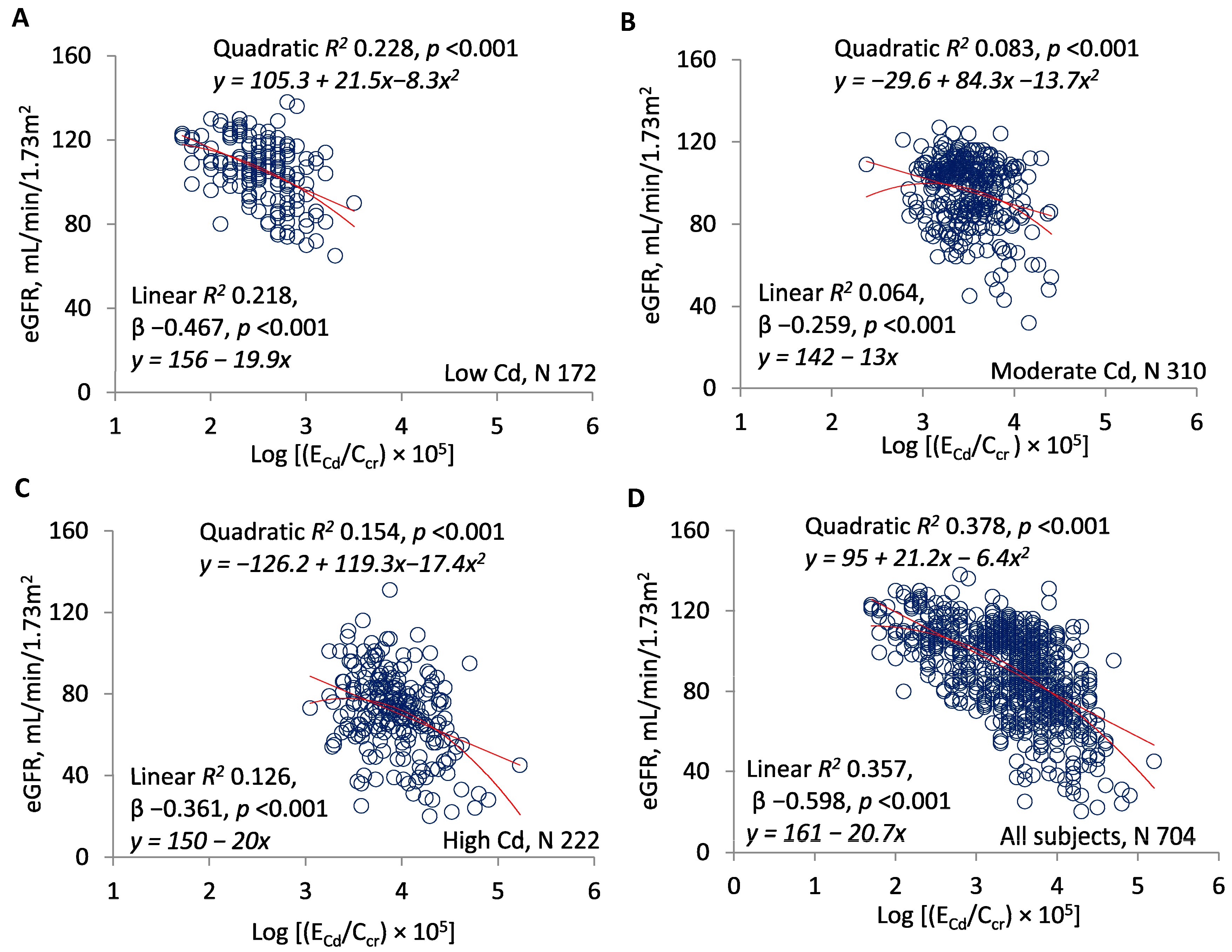
Figure 1. ECd/Ccr as a predictor of the estimated glomerular filtration rate (eGFR). Scatterplots compare eGFR to log[(ECd/Ccr) × 105] in subjects grouped by locality (AC) and in all subjects (D). Quadratic and linear coefficients of determination (R2) are provided together with corresponding equations, standardized β coefficients, and p-values.
Toxics 07 00055 g001
Figure 2. ENAG/Ccr as a predictor of eGFR. Scatterplots compare eGFR to log[(ENAG/Ccr) × 103] in subjects grouped by locality (AC) and in all subjects (D). Quadratic and linear coefficients of determination (R2) are provided together with corresponding equations, standardized β coefficients, and p-values.
Toxics 07 00055 g002
Figure 3. ECd/Ccr as a predictor of ENAG/Ccr. Scatterplots compare log[(ENAG/Ccr) × 103] to log[(ECd/Ccr) × 105] in subjects grouped by locality (AC) and in all subjects (D). Quadratic and linear coefficients of determination (R2) are provided together with corresponding equations, standardized β coefficients, and p-values.
Toxics 07 00055 g003
Figure 4. ECd/Ccr as a predictor of Eβ2MG/Ccr. Scatterplots compare log[(Eβ2MG/Ccr) × 104] to log[(ECd/Ccr) × 105] in subjects grouped by locality (AC) and in all subjects (D). Quadratic and linear coefficients of determination (R2) are provided together with corresponding equations, standardized β coefficients, and p-values.
Toxics 07 00055 g004
Figure 5. ENAG/Ccr as a predictor of Eβ2MG/Ccr. Scatterplots compare log[(ENAG/Ccr) × 103] to log[(Eβ2MG/Ccr) × 104] in subjects grouped by locality (AC) and in all subjects (D). Quadratic and linear coefficients of determination (R2) are provided together with corresponding equations, standardized β coefficients, and p-values.
Toxics 07 00055 g005
Table 1. Study subjects drawn from three localities.
DescriptorsAll SubjectsLocality
Low CdModerate CdHigh Cd
Number of subjects704172310222
Women (%)60.747.772.953.6 *
Smoking (%)43.623.840.663.1 *
Age (years)48.34 ± 11.1138.72 ± 10.2947.24 ± 4.7257.34 ± 11.13
SBP (mmHg)122.7 ± 13.5120.1 ± 10.4124.2 ± 14.7 
DBP (mmHg)78.9 ± 9.578.1 ± 7.679.4 ± 10.4
MBP (mmHg)93.5 ± 9.992.1 ± 7.994.3 ± 3.3 ¶¶
eGFR (mL/min/1.73 m2)90.32 ± 21.84105.46 ± 15.0595.72 ± 16.1371.05 ± 19.65
CKD prevalence (%) 9.503.525.2
Kidney disease stage (%)
Stage 154.884.166.516.2 *
Stage 2 36.615.931.059.9 *
Stage 3 7.702.620.7 **
Stage 41.0003.2
Serum creatinine (mg/dL)0.80 (0.70, 1.0)0.8 (0.7, 0.9)0.8 (0.7, 0.9)1.0 (0.9, 1.2)
Urine creatinine (mg/dL)97 (53, 156)52 (32, 106)110 (62, 171)115 (69, 158)
Urine Cd (μg/L)3.7 (1.1, 8.3)0.2 (0.1, 0.6)3.9 (2.4, 7.2)8.3 (4.7, 13.9)
Urine NAG (units/L)6.4 (2.4, 16.6)1.8 (1.3, 2.9)11.9 (7.1, 19)5.3 (2.7, 9.2)
Urine β2MG (μg/L)154 (33, 778)9.5 (0.3, 42)400 (134, 1118)171 (64, 1368)
ECd/Ccr × 100, µg/L3.1 (1.1, 7.0)0.3 (0.3, 0.6)3.0 (1.7, 5.0)8.2 (4.8, 15)
ENAG/Ccr × 100, units/L5.6 (3.4, 9.4)3.2 (2.0, 4.1)8.0 (5.8, 12)4.9 (3.1. 8.3)
Eβ2MG/Ccr × 100, µg/L137 (29, 604)17 (0.4, 36)368 (92, 808)162 (64, 176)
SBP = systolic blood pressure; DBP = diastolic blood pressure; MBP = Mean arterial pressure; CKD = chronic kidney disease; eGFR = estimated glomerular filtration rate; NAG = N-acetyl-β-d-glucosaminidase. MBP = DBP + (pulse pressure)/3, where pulse pressure = SBP − DBP. Data for age and eGFR are arithmetic mean values ± standard deviation (SD). Data for blood pressure are geometric mean values ± SD. Data for all other continuous variables are the median (25th, 75th percentile) values. * Significant % differences among three groups (p < 0.05, Pearson Chi-Square test). ** Significant % differences between two groups (p < 0.001, Pearson Chi-Square test). Significant mean differences among three groups (p < 0.001, Kruskal–Wallis test). ¶ Significant difference from the low exposure group (p = 0.003, Mann–Whitney U-test). ¶¶ Significant difference from the low exposure group (p = 0.014, Mann–Whitney U-test).
Table 2. Slope analysis for comparing rates of eGFR reduction.
Excretion Rates of Cd or NAGNumber of SubjectseGFR vs. log[(ECd/Ccr) × 105] or log[(ENAG/Ccr) × 103]
β CoefficientsR2p Value
Slope (Unstandardized β) ± SEStandardized β
Log[(ECd/Ccr) × 105]
<3168−17.62 ± 3.20 −0.3920.154<0.001
≥3536−27.71 ± 2.06 −0.5030.253<0.001
All subjects 704−20.86 ± 1.06−0.5980.357<0.001
Log[(ENAG/Ccr) × 103]
<1.5153−15.91 ± 7.62−0.1680.0280.038
≥1.5551−28.35 ± 3.35−0.3390.115<0.001
All subjects704−22.49 ± 2.19−0.3610.131<0.001
The standardized β coefficient indicates the strength of the association of eGFR with log[(ECd/Ccr) × 105] or log[(ENAG/Ccr) × 103]. R2 values are coefficients of determination that indicate the fraction of eGFR variation explained by ECd/Ccr or ENAG/Ccr. p ≤ 0.05 identifies statistically significant eGFR reduction rates or associations of eGFR with urinary Cd or NAG excretion.