1. Introduction
The therapeutic value of
Hibiscus sabdariffa Linnaeus (HSL) is extensively documented. This is owed to its rich concentration of bioactive phytochemicals, including polyphenols, anthocyanins, flavonoids, ascorbic acid, amino acids, and minerals, among others [
1]. The administration of HSL infusions at a concentration of 2–3% has consistently demonstrated high efficacy in restoring the cellular redox balance under pathological conditions characterized by oxidative stress (OS), such as metabolic syndrome and essential hypertension in healthy organisms [
2]. The exogenous antioxidants collaborate with endogenous enzymatic systems to neutralize reactive oxygen species (ROS) and support physiological homeostasis [
3]. However, the conventional assumption that supplementation with a high dose of antioxidants in healthy subjects has arisen.
Prolonged and excessive ingestion of antioxidant-rich diets in healthy states can oversaturate counter-regulatory networks of OS, forcing the cellular environment into a paradoxical condition known as reductive stress (RS) and leading to the loss of redox homeostasis [
4]. RS is defined by an overactivation of enzymatic/non-enzymatic antioxidant systems, an increase in total antioxidant capacity (TAC), and the accumulation of reducing equivalents such as nicotinamide adenine dinucleotide/nicotinamide adenine dinucleotide phosphate (NADH/NADPH), reduced glutathione/glutathione disulfide (GSH/GSSG), and oxidized/reduced thiols [
5]. This results in a critical depletion of physiological ROS that triggers systemic maladaptive cascades. It damages the role of ROS as second messengers regulating cellular proliferation, mechanical vascular contractility, syncytial communication, spermatogenesis, and ovulation among other cellular pathways [
6].
Our research group has systematically explored the multi-organic consequences of RS using a model of chronic consumption of a 6% HSL infusion in healthy Wistar rats. We demonstrated that the excessive intake of this infusion causes systemic alterations, characterized by an upregulated inflammatory profile in plasma, alongside an unexpected elevation in systolic blood pressure (SBP) [
7]. At the vascular level, we reported altered aortic vascular reactivity, which is associated with a decrease in glutathione-S-transferase activity and an increase in TAC [
8]. Furthermore, our recent work in renal pathology revealed that prolonged consumption of the 6% infusion induces severe kidney damage [
9]. This renal impairment is driven by an overproduction of hydrogen sulfide, which elevates cellular reducing power, induces mitochondrial dysfunction via decreased oxidative phosphorylation in complexes I and IV, and results in structural remodeling, indicated by a retraction of the glomerular tuft and tubulointerstitial fibrosis [
10].
On the other hand, the myocardium operates as a highly coordinated, functional syncytium in which mechanical contractility and electrical conduction depend on the integrity of the extracellular matrix (ECM), ionic homeostasis, and an uninterrupted supply of ATP. This mechanical syncytium maintains systemic perfusion, making it uniquely sensitive to structural and metabolic remodeling [
11]. While the detrimental effects of RS caused by the ingestion of 6% HSL on plasma biomarkers, vascular smooth muscle reactivity, and renal architecture have been established by our group, its specific impact on the heart remains unknown. The question arises whether the systemic inflammatory and hypertensive states induced by chronic ingestion of 6% HSL could extend to the cardiac ECM, or if the effect of this sustained antioxidant overload may affect coronary compliance and electrical coupling. In normal conditions, the collagen of the ECM provides the necessary structural support to withstand systolic stress; however, chronic pro-inflammatory profiles alter its turnover, promoting fibrosis and ventricular stiffness [
12]. Cardiac electromechanical coupling also requires the precise synchronization of gap junctions, where connexin 43 (Cx43), the predominant pore-forming protein, is of importance. The assembly and opening of Cx43 is modulated by the redox state, with its cysteine residues being highly susceptible to post-translational modifications [
13]. Under normal physiological conditions, basal ROS maintain appropriate Cx43 phosphorylation, gap-junction function, and syncytial communication. Based on previous experimental evidence, it may be hypothesized that excessive accumulation of reducing equivalents could disrupt this balance, causing dephosphorylation or lateralization of Cx43, thus affecting longitudinal conduction velocity and syncytial communication [
14]. Since maintaining the electrical ionic gradients of the cardiac syncytium and ECM homeostasis are highly energy-dependent processes, an energy deficit deprives the tissue of the metabolic substrates necessary to sustain its function [
15]. Consequently, when faced with hemodynamic challenges such as I/R, a preexisting bioenergetic collapse and electrical uncoupling could alter coronary capacitance, leaving the heart vulnerable to lethal arrhythmias and acute failure.
In this context, the development of cardiac arrhythmias is associated with alterations in tissue excitability and conduction velocity, both of which depend on membrane ion channels, electrochemical gradients, cellular architecture, and intercellular electrical coupling [
16]. Gap-junction uncoupling generally slows impulse propagation and disrupts electrical communication between cardiomyocytes. Moderate reductions in intercellular coupling may decrease the electrotonic load imposed on activated cells and increase the safety factor for conduction. However, more severe or heterogeneous uncoupling can markedly slow conduction, promote conduction block, and create a substrate for reentrant arrhythmias [
17]. This is due to charge accumulation in the intracellular space of individual cardiomyocytes [
18]. However, this increase in intracellular charges leads to faster movement of action potentials across cells but slower movement between them due to cell decoupling [
19]. However, other local, genetic, or environmental factors may exist that are integrated within the mechanisms of damage and could lead to structural or hemodynamic functional alterations in the heart. This is further compromised if there is inflammation and/or oxidative or reductive stress, which could have a direct effect on ventricular dysfunction and arrhythmias [
20]. Therefore, the aim of the present study was to determine whether chronic and excessive ingestion of a 6% HSL infusion for two months compromises the structural, mechanical, and hemodynamic performance of the myocardium in hearts of healthy Wistar rats subjected to I/R, and whether these effects can be reversed if treatment is suspended.
2. Materials and Methods
2.1. Preparation of the HSL 6% Infusion
The HSL infusion was prepared by adding 60 g of dried calyces of HSL to 1 L of boiling water. The mixture was maintained at a boil for 10 min, cooled to room temperature, and subsequently filtered. The resulting infusion was stored at 4 °C until administration. A fresh preparation was made weekly to prevent fermentation. It was offered to the rats ad libitum as their drinking fluid.
2.2. Determinations of Polyphenols, Total Flavonoids, Total Anthocyanins, and Vitamin C in the HSL 6% Infusion
Total vitamin C in the 6% HSL infusion was quantified according to the method described by Jagota. Briefly, 100 μL of the infusion was mixed with 200 μL of 0.20 mM Folin–Ciocalteu reagent. The reaction mixture was vigorously agitated, incubated for 10 min, and the absorbance was recorded at 760 nm. The vitamin C concentration was calculated from a calibration curve prepared with ascorbic acid as the reference standard [
21].
The total flavonoid content in the 6% HSL infusion was measured using the Jia method. This spectrophotometric assay estimates flavanols and flavones, including apigenin, chrysin, luteolin, morin, quercetin, kaempferol, myricetin, and galangin, by measuring absorbance at 510 nm. Quantification was performed using a quercetin calibration curve [
22].
Total anthocyanins were determined following the method of Lee, with absorbance measurements obtained at 520 and 700 nm. The results were expressed as cyanidin-3-glucoside equivalents [
23].
2.3. Calculation for the Sample Size
The mean systolic blood pressure (SBP) in healthy rats was reported to be between 116 mmHg and 112.8 mmHg, with a variance of 19, in rats from the National Institute of Cardiology Ignacio Chavez. Based on this, the sample size per group was estimated by μ (SBP of Wistar rats), with 95% confidence and a maximum error (ME) of 3.2 mmHg, according to the following formula: ME: |μ − x| = |116 − 112.8| = 3.2 mmHg, Confidence: |μ − x| = |116 − 112.8| = 3.2, SS: n = ()2 × σ2/(EM2) = (22) × (19)/(3.2)2= (4 × 19)/(10.24) = 76/10.24 = 7.42, where ME = the maximum error, = the number of standard deviations of the mean estimator, σ2= the variance in the SBP of the rats in our institute, and SS = the sample size.
2.4. Experimental Design
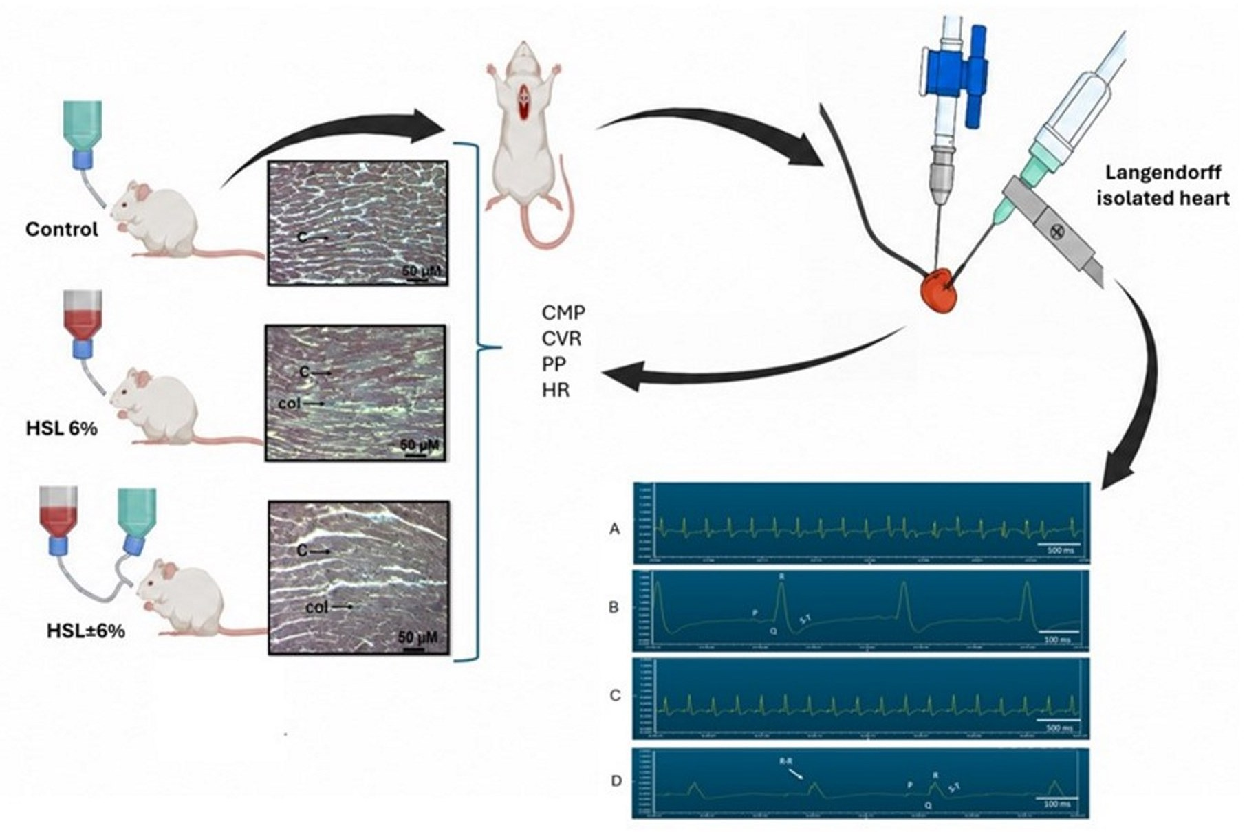
Twenty-four male Wistar rats were allocated to three experimental groups of eight animals each. The control group (C) received tap water ad libitum for two months. The HSL 6% group received a 6% HSL infusion ad libitum for two months. The HSL ± 6% group was given the same infusion for two months, followed by tap water for an additional two months. Throughout the experimental period, the animals were housed under controlled conditions consisting of a 12 h light/12 h dark cycle, an ambient temperature of 18–26 °C, and 40–70% relative humidity. Standard rodent chow was supplied ad libitum and contained 23% crude protein, 4.5% crude fat, 6% crude fiber, 8% ash, and 2.5% minerals (LabDiet 5008; PMI Nutrition International, Richmond, IN, USA). The protocol was approved by the Laboratory Animal Care Committee of the National Institute of Cardiology Ignacio Chávez under approval number INC/CICUAL/009/2023, and all procedures were conducted in accordance with institutional animal care guidelines. At the end of the treatment period, SBP was measured by plethysmography using a Narco Bio-Systems system (Houston, TX, USA) before euthanasia. Signals were recorded and processed with SIEVART software, version 0.1, and five measurements were obtained from each rat. The animals were then anesthetized with sodium pentobarbital at 60 mg/kg body weight. Before euthanasia, heparin was administered at 1000 U/kg body weight using a 1000 U/mL solution.
2.5. Isolated Langendorff Heart Model Under I/R
The heart was rapidly excised following thoracotomy and immediately immersed in ice-cold Krebs–Henseleit solution to induce cardiac arrest and minimize ischemic preconditioning. The ascending aorta was then cannulated without delay, and the organ was connected to a retrograde perfusion apparatus. Mechanical activity was maintained with Krebs–Henseleit buffer containing, in mM, 120 NaCl, 23.4 NaHCO3, 4.8 KCl, 1.2 KH2PO4, 0.86 MgSO4, 1.25 CaCl2, and 11.0 glucose. The solution was adjusted to pH 7.4, maintained at 37 °C, and continuously gassed with 95% O2 and 5% CO2.
A 30 min equilibration period was allowed before the experimental protocol. During this period, the coronary flow was set at 25 mL/min for the first 5 min and subsequently reduced to 14 mL/min for the remaining 25 min. Following equilibration, the perfusion flow was adjusted to 13 mL/min and maintained at this rate throughout the experimental protocol. The heart rate was maintained between 312 and 324 beats/min using square-wave stimulation delivered by a Grass S44F stimulator (Grass Instruments Co., Quincy, MA, USA). The coronary flow was controlled with a peristaltic pump (Masterflex Easy-Load II, model 77200-50; Cole-Parmer Instrument Co., Vernon Hills, IL, USA).
Left intraventricular pressure (LIVP) was measured with a Grass hydropneumatic pressure transducer (Grass Instrument Co., Quincy, MA, USA) connected to a catheter fitted with a latex balloon. The balloon was advanced through the mitral valve into the left ventricular cavity and inflated to establish a diastolic pressure of 5–10 mmHg. Perfusion pressure (PP) was monitored with a second Grass hydropneumatic pressure transducer. Hearts were included in the study only when the initial PP ranged from 55 to 70 mmHg.
All hemodynamic signals were acquired using a computerized recording system (Grass Telefactor, Grass Technologies, Astro-Med, West Warwick, RI, USA) coupled to a Grass model 79D polygraph and Grass PolyView software (Astro-Med, Inc., West Warwick, RI, USA; Grass PolyView version 3.0). Cardiac mechanical performance (CMP) was calculated as the product of heart rate and left intraventricular pressure as follows: CMP = HR × LIVP.
2.6. Heart Homogenization
After completing the experiments on the isolated heart, the organ was removed from the Langendorff system and segmented into three parts, where the superior and apical parts were homogenized under liquid nitrogen after adding KH2PO4 (1 mL) 0.05 mM, pH 7.3, in the presence of 20 µL of antiprotease inhibitors (2 μM leupeptin, 2 μM pepstatin A, 0.1% aprotinin, and 1 mM PMSF).
2.7. Anatomical Changes in the Heart by a Histological Process
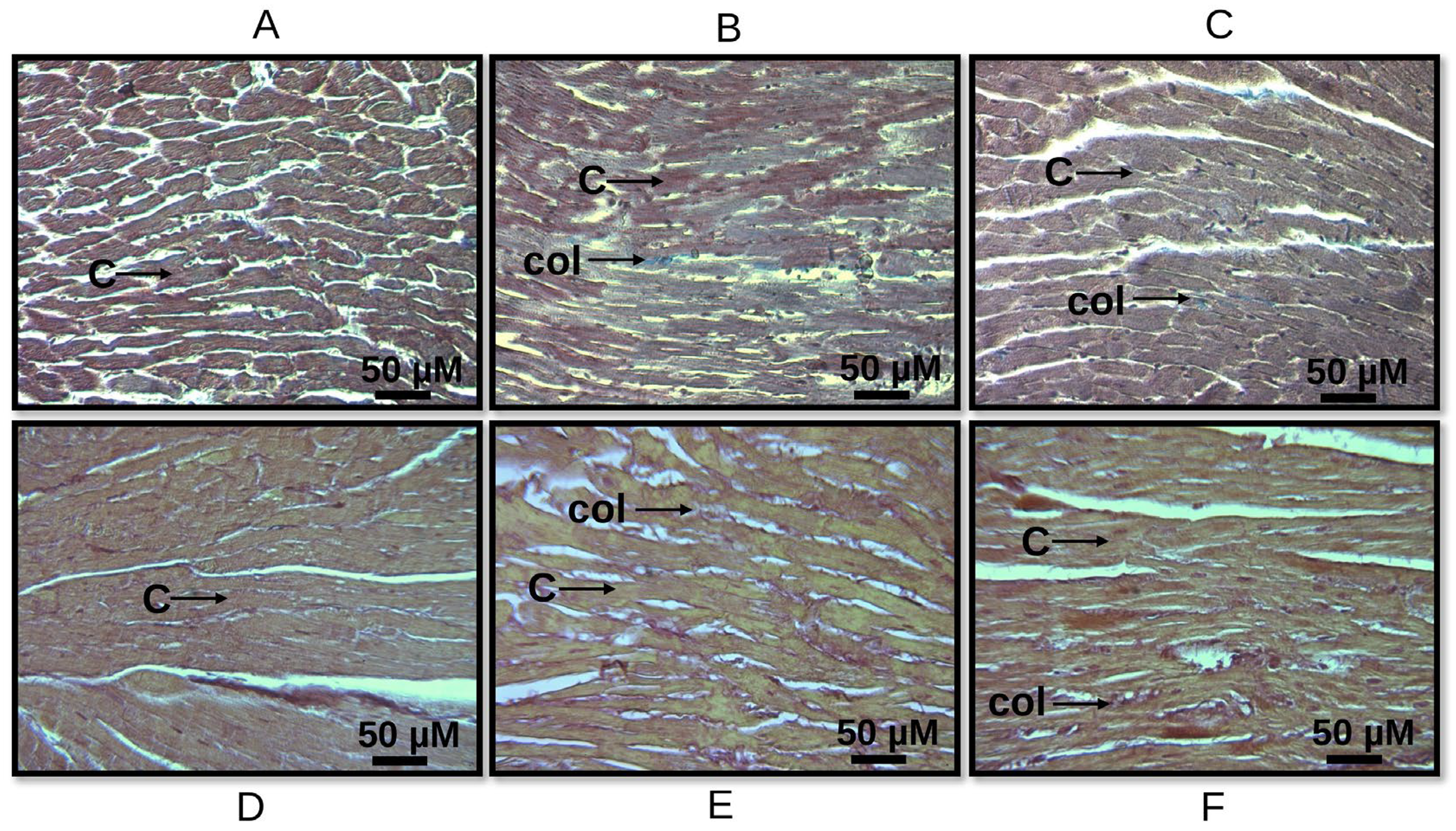
To evaluate the anatomical and structural changes and collagen deposition in cardiac tissue, the central part of each heart was used for histological analysis after completing the experiments in the isolated hearts. The tissue was fixed in a 10% formalin solution for 24 h, gradually dehydrated through increasing concentrations of ethanol, cleared in xylene, embedded in paraffin, and cut into 5 µm thick sections using a microtome (Leica RM212RT, Wetzlar, Germany). The paraffin sections were stained with Masson’s trichrome and Sirius Red.
The histological sections were analyzed at 12.5× magnification using a model 63300 optical microscope (Carl Zeiss, Oberkochen, Germany) equipped with a Tucsen digital camera (Tucsen Photonics Co., Ltd., Fuzhou, China; 18 megapixels) coupled to TSView 7.3.1 software. The histological sections were evaluated by an investigator blinded to the experimental groups, and the evaluation was validated using the Allred scoring system. The microscope illumination was adjusted and kept constant throughout image acquisition. Five fields per heart were analyzed.
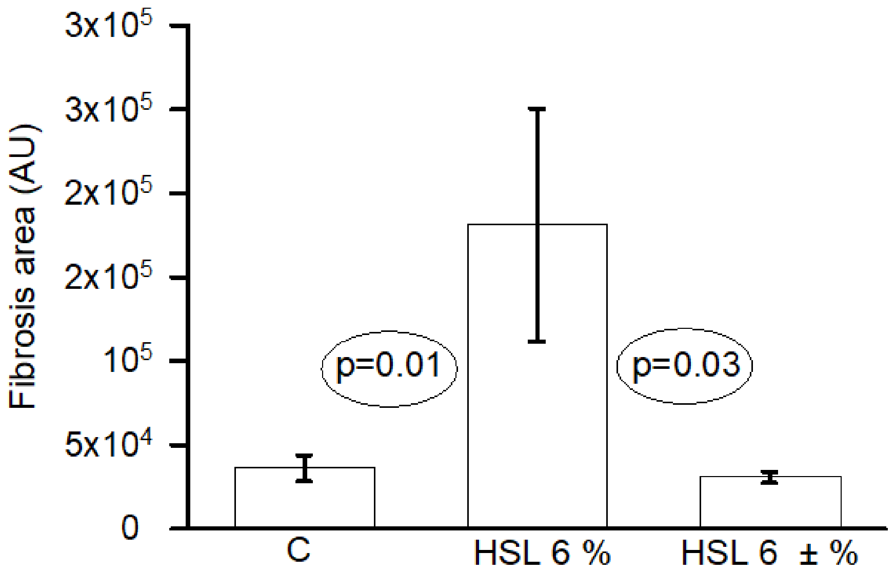
For quantitative collagen analysis, non-overlapping myocardial fields were selected systematically from each section, avoiding tissue folds, tears, staining artifacts, large empty spaces, and areas adjacent to the tissue margins. Collagen deposition was quantified using SigmaScan Pro 5 image-analysis software (Systat Software, Inc. in San Jose, CA, USA). For each image, the total myocardial tissue area and the collagen-positive area were determined after applying a predefined color threshold. The threshold settings were established before group comparisons and maintained unchanged for all images analyzed using the same staining method. The values were expressed in arbitrary units (pixels).
2.8. Evaluation of Total Antioxidant Capacity (TAC)
A total of 100 μg of homogenized heart was added to a mixture that contained C
2H
3O
2 at 300 mM, FeCl
3·6H
2O at 20 mM, 2,4,6-tris-2-pyridyl-s-triazine at 10 mM, and HCl at 40 mM, at pH 3.6 (1.5 mL, at a ratio of 10:1:1
v/
v), and incubated at 37 °C for 15 min. The absorbance was measured at 593 nm [
24].
2.9. Nuclear Factor Erythroid 2 (NrF2)
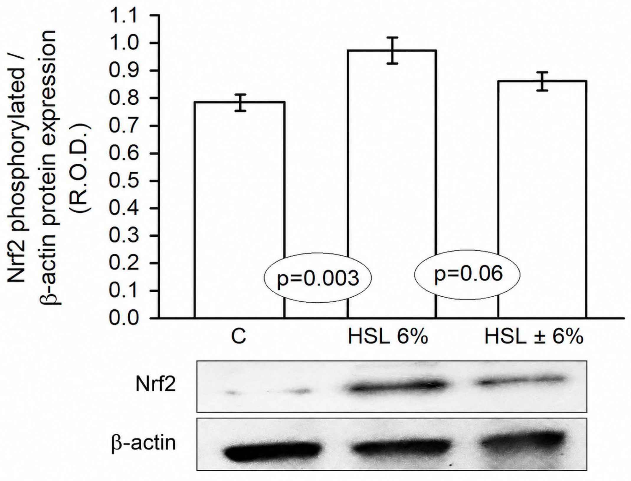
Fifty micrograms of protein from cardiac homogenates were separated by 8% SDS-PAGE and transferred onto PVDF membranes. The membranes were blocked for 1 h at room temperature with 5% skim milk prepared in Tris-buffered saline containing Tween 20 (TBS-T). The membranes were incubated overnight at 4 °C with a monoclonal anti-phospho-Nrf2 (Ser40) antibody diluted to 1:1000 (SAB5701902-100UL; Sigma-Aldrich, St. Louis, MO, USA). After washing with TBS-T, the membranes were incubated with the appropriate secondary antibody diluted to 1:10,000 for 3 h at 4 °C. Protein bands were detected using a Bio-Rad chemiluminescent detection reagent and visualized using a ChemiDoc imaging system (Bio-Rad Laboratories, Inc., Hercules, CA, USA).
β-actin was used as the loading control and was detected using an anti-β-actin antibody diluted to 1:500 (sc-81178; Santa Cruz Biotechnology, Inc., Santa Cruz, CA, USA). After washing with TBS-T, the membranes were incubated with the appropriate secondary antibody diluted to 1:10,000 for 3 h at 4 °C. Band intensity was quantified by densitometry using a GS-800 densitometer (Bio-Rad Laboratories, Inc. in Hercules, CA, USA) and Quantity One 1-D Analysis Software, version 4.6.8 (Bio-Rad Laboratories, Inc., Hercules, CA, USA). Phospho-Nrf2 densitometric values were normalized to the corresponding β-actin signal and expressed as arbitrary units (AU).
2.10. Superoxide Anion Detection
The O
2− in heart homogenates was assessed by monitoring the irreversible oxidation of epinephrine to adrenochrome. Briefly, 50 μg of homogenate protein was mixed with 2 mL of 50 mM glycine buffer, adjusted to pH 10.2, followed by the addition of 50 μL of 60 mM epinephrine. The reaction was carried out at 30 °C, and the increase in absorbance at 480 nm was recorded for 6 min. O
2− was calculated using an extinction coefficient of 4.0 mM
−1 cm
−1 [
25].
2.11. Statistical Analysis
Cardiac mechanical work was analyzed using a two-way repeated measures ANOVA, considering the experimental group as the between-subjects factor, time as the within-subjects factor, and the group × time interaction. Each heart was considered the experimental unit and the repeated measures factor. The stabilization and reperfusion periods were analyzed separately. Measurements during global ischemia were not subjected to inferential analysis because cardiac work was zero. Greenhouse–Geisser correction was applied to the time and group × time effects. Where appropriate, pairwise multiple comparisons were adjusted using the Holm method. Analyses of the adrenochrome densitometry for the Western blot and heart imaging, as well as general characteristics, were performed using one-way ANOVA and Tukey’s post hoc test. Data are presented as mean ± SE. Analyses and graphs were performed using Sigma Plot, version 15 (Systat Software Inc., San Jose, CA, USA). A p-value < 0.05 was considered statistically significant.
4. Discussion
In the present study, we evaluated whether chronic and excessive ingestion of a 6% HSL infusion for two months compromised the structural, mechanical, and hemodynamic performance of the myocardium during I/R in hearts of healthy Wistar rats. Chronic exposure to 6% HSL is a rat experimental model that has been previously characterized by our group as producing systemic and renal alterations associated with antioxidant overload and probably RS [
7,
8,
9,
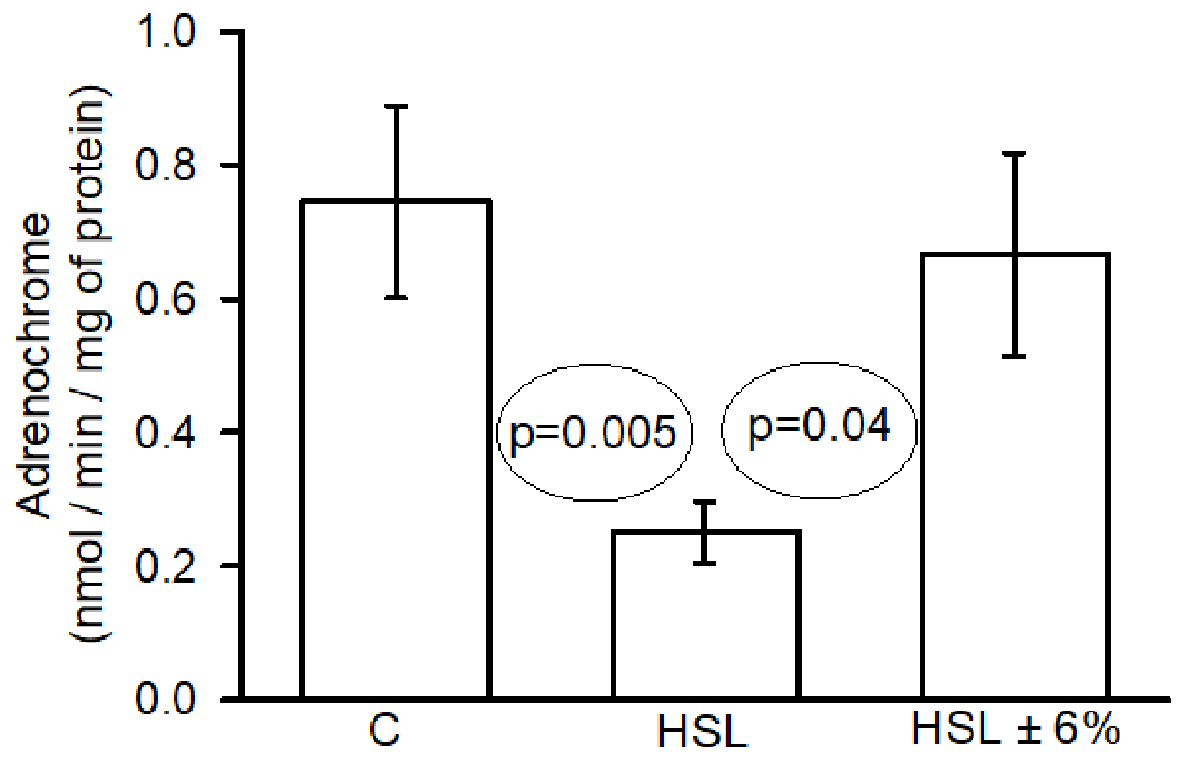
10]. In the present study, the increase in TAC and phospho-Nrf2 expression, together with reduced adrenochrome formation after I/R, indicate an enhanced antioxidant response and a redox environment consistent with increased reduced capacity. In this paper, we also evaluated whether the removal of the infusion could revert the damaging effects of RS associated with the ingestion of the infusion.
The heart is subjected to a period of restricted blood flow (ischemia) followed by the restoration of flow (reperfusion) in experimental models of I/R. While restoring flow is essential to save ischemic tissue, there is a reperfusion paradox that states that it triggers a secondary wave of tissue damage, microvascular dysfunction, and arrhythmias that are caused, in part, by the large amount of ROS generated during this period [
25]. Each of the parameters chosen for evaluation of the damage caused by I/R in this study, including CTM, CVR, PP, and HR, provides a window into the distinct I/R pathological mechanisms [
26]. These parameters do not operate in isolation; they are deeply intertwined components of a dynamic physiological loop [
27].
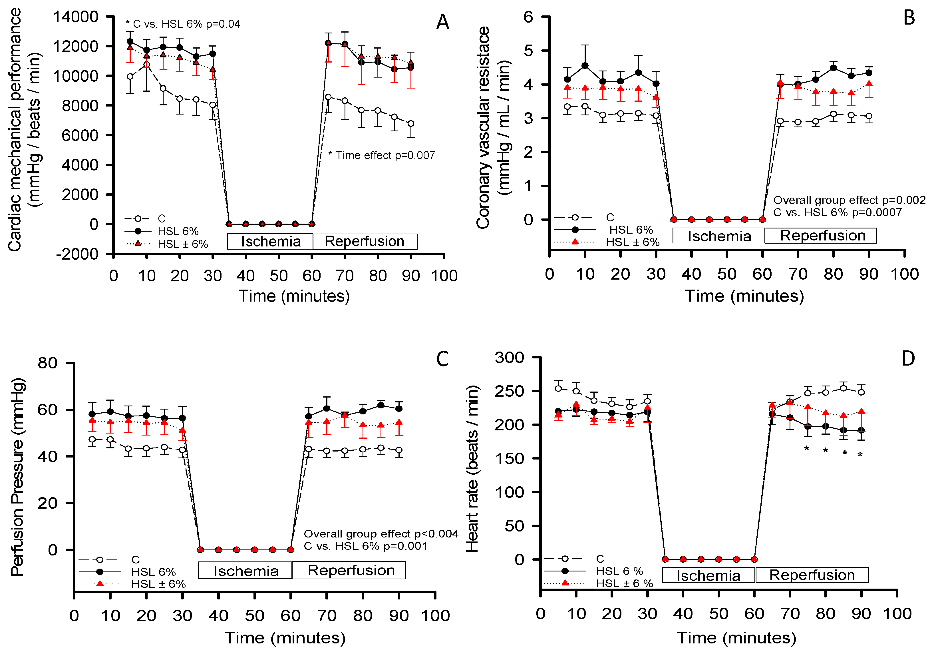
CMP is an indicator of myocardial viability and functional recovery, and it reflects the degree to which the myocardium has suffered irreversible damage versus reversible stunning [
27,
28]. Our results showed that in both the HSL 6% and the HSL ± 6% groups, the CMPs were ~12,000 mmHg/beats/min. This was maintained despite the reperfusion period. In contrast, in the C group, there was a decrease of nearly ~7000. This suggests that the antioxidant overload provided by the HSL infusion could inhibit or neutralize the overproduction of ROS generated during the reperfusion period. Restoring flow and the abrupt return of oxygen that normally triggers ROS overproduction [
8], which saturates the basal antioxidant system, was not observed after ingestion of the infusion. When ROS levels increased at the time of reperfusion, the tissue possibly already had a massive and abnormally high reserve of antioxidants that were accumulated during the two months of treatment [
9]. These accumulated antioxidants may immediately neutralize the ROS generated by reperfusion by reacting with the excess reducing equivalents [
8,
9]. However, in the HSL ± 6% group, the washout period was not long enough, and the heart still showed alterations.
The CVR measures the resistance to flow within the coronary circulation. A rising CVR during reperfusion is a direct hallmark of the “no-reflow” phenomenon and determines the level of protection of the coronary vasculature [
27,
28]. Our results from the C group show that the CVR was constant, which suggests that the intramyocardial coronary arteries were free of external mechanical compression before ischemia occurred; however, after reperfusion, a massive contractile effect occurred, suggesting that the myocardium no longer contracted with force and ceased to compress the blood vessels during systole, eliminating internal mechanical resistance and maintaining low VR [
29]. However, in the 6% HSL group, the results showed a high CRV. This suggests that antioxidant saturation altered basal cell signaling in the myocardial tissue, suggesting chronic structural remodeling of the cardiac muscle, evidenced by histological staining of the heart sections. This structural alteration can cause extrinsic mechanical compression of the coronary microvasculature, physically reducing the diameter of intramural blood vessels. As the caliber of the flow conduction pathways may be decreased, hydrodynamic resistance might be chronically elevated, which is why this group had a higher CVR, even before the recording of the ischemic period. However, once the reperfusion condition is established, the antioxidant molecules provided by the 6% HSL infusion may overexpress the antioxidant system and deplete ROS [
9,
10]. Consequently, the membranes of endothelial cells and smooth vascular muscle are possibly protected from the ROS generated during reperfusion, resulting in the CVR remaining at high levels since the chronic structural compression factor dominates and prevents resistance from decreasing [
30].
In addition, the PP is a direct reflection of the hydrostatic force required to push fluid through the arteries of the heart. Both PP and CVR are directly connected by Ohm’s hydrodynamic law; since the flow is constant, PP behaves exactly like CVR [
30,
31]. Therefore, a low PP in the C group indicates that in this group, vessels are passively and pathologically relaxed, lacking active myogenic regulation due to acute cellular stunning from ischemia–reperfusion. A dramatic spike in PP during reperfusion suggests severe vasoconstriction or microvascular plugging, induced primarily by the ROS burst and cytosolic Ca
2+ overload [
32]. Our results show that a chronic intake of high doses of antioxidants for two months by drinking the 6% HSL solution may have possibly induced an RS state that altered the basal redox signaling pathways necessary for extracellular matrix homeostasis. Furthermore, it generated a structural alteration that might have induced a constant extrinsic mechanical compression on intramural coronary arteries, physically narrowing their internal diameter (vascular lumen) [
10]. With the reduced caliber of the arteries, the system would require significantly more force to push perfusion fluid through them. This structural compression might be the direct reason why the PP was elevated at the beginning of the experiment and remained elevated throughout. In hemodynamic terms, the presence of the perivascular fibrosis exerts extrinsic mechanical compression on the intramural coronary arteries, decreasing the diameter of the vascular lumen and increasing resistance to hydrostatic flow [
33]. The coexistence of collagen accumulation with elevated CMP, CVR, and PP suggests a relationship between myocardial fibrosis and an impaired functioning of the heart. Nevertheless, the current experimental design does not establish a temporal sequence, nor does it prove that altered ROS levels or NrF2 activation directly caused collagen deposition and the subsequent functional abnormalities. Finally, the results from the washout group (HSL ± 6%) showed that removal and reintroduction of tap water partially eliminated the antioxidant overload provided by the HSL infusion, reducing extrinsic mechanical compression in the intramural coronary arteries. As a result, the blood vessels recovered their original internal diameters.
The HR is a variable that dictates both energy supply and demand of the cardiac muscle [
27]. In this sense, the C group exhibited marked bradycardia, electrical instability, and reduced CMP after reperfusion, indicating impaired post-ischemic functional recovery. Alterations in myocardial energetic status, redox regulation, or impulse generation may have contributed to these findings; however, these mechanisms were not directly evaluated in the present study. We were also not able to quantify the incidence of arrhythmia, its duration, or severity. During reperfusion, the low CVR and PP observed in the C group could indicate impaired coronary hemodynamic recovery. Endothelial viability, smooth muscle function, myogenic tone, and vasoplegia were not directly assessed; therefore, these mechanisms remain hypothetical [
34]. The relative preservation of the HR in the HSL-treated groups occurred in parallel with increased reserves of antioxidants and changes in the myocardial redox environment. These findings suggest that modulation of the ROS response may have influenced the chronotropic response during reperfusion. However, the present results do not demonstrate that ROS neutralization directly preserved sinoatrial automaticity or prevented arrhythmogenic mechanisms [
35].
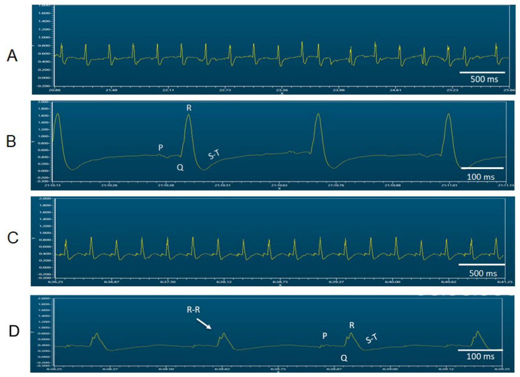
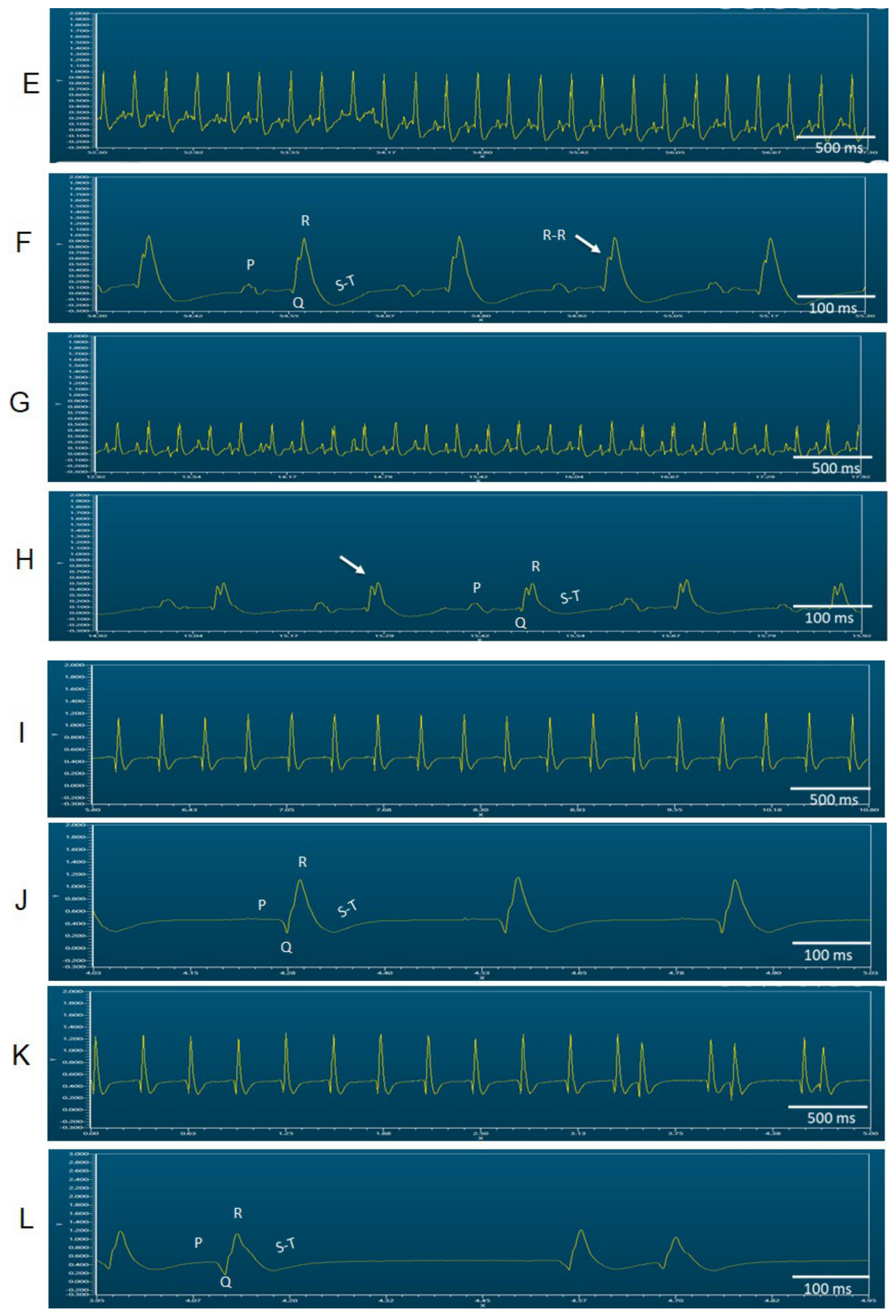
The results obtained in the illustrative representative ECG recordings show electrical abnormalities observed in the experimental groups and could be considered contradictory, since the excess antioxidants and the lack of basal oxidants could act as a shield for protection against ROS; that is, there is normally an increase in the generation of ROS after reperfusion that may cause damage, as observed in the C group. However, in the experimental HSL groups, ROS reacted with the enormous reserve of antioxidants and reducing equivalents accumulated in the tissue instead of causing damage. The combined analysis of representative ECG recordings provides electrophysiological evidence that validates the mechanical (CMP and HR) and hemodynamic (CVR and PP) alterations. For example, in the C group, the adaptation period (0–30 min) caused a widening and distortion of the QRS complex, loss of P-wave definition, and critical alterations in the S-T segment [
36]. The widening and distortion of the ventricular complexes, together with the morphological abnormalities, are compatible with impaired ventricular electrical coordination and possible conduction disturbances. The coexistence of these electrical abnormalities with reduced mechanical performance could suggest altered electromechanical integration during reperfusion [
28,
30]. Nevertheless, conduction velocity, ventricular synchrony, intracellular calcium handling, and excitation–contraction coupling were not directly evaluated and therefore cannot be confirmed from the present data.
In the ECG of the HSL 6% group, excess antioxidants may have acted as a metabolic shield. They neutralized the acute burst of ROS [
9,
10], and consequently, double deflections or notches were observed in the activation complex. The findings in the ECG, histological observations, and the decreased ROS indicate that chronic exposure to the 6% HSL infusion produced a complex myocardial response, in which increased collagen deposition was accompanied by extracellular matrix remodeling and alterations in the ECG. In contrast, in the HSL ± 6% group, the ECG suggests that the PQR and S-T complexes showed a cleaner, sharper tracing, completely free of the notches or double deflections present in the HSL 6% group. This indicates that the two months of prior HSL treatment could indicate induced lasting electrical and metabolic preconditioning. However, upon the onset of I/R, the tissue remained partially protected against ROS damage, preserving automaticity.
Additionally, HSL contains minerals such as aluminum (Al), which are potentially toxic to humans. Al is first eliminated in urine and feces, but part of the Al can be retained in the organism and accumulate. In this sense, a study of volunteers who drank an HSL infusion at 2.5% for 16 days showed a high Al content in their urine that was associated with abnormalities such as diarrhea, gastrointestinal problems, nausea, and dizziness [
37]. This suggests that high concentrations of the HSL infusion could lead to an increased Al body burden and cause cardiovascular problems.
On the other hand, regarding the potential damage caused by excessive antioxidants in human health, it has been described that drug toxicity due to overdoses or drug interactions in healthy subjects and in subjects with comorbidities who chronically consume medications or supplements, whether prescribed by a physician or administered through self-medication, can occur. Although the goal of treatment is to improve physical conditions or alleviate discomfort, the use of some medications, due to drug interactions, can synergize or reduce the kinetic effect of other drugs. Studies in the literature have reported no association between antioxidant use and mortality. However, these studies did not analyze the oxidative or reducing status of the subjects who consumed them [
38,
39]. Therefore, the inclusion of the use of antioxidants by patients in future randomized clinical trials should be considered to allow for the analysis of oxidative or RS as causes of morbidity and mortality worldwide [
40]. Many patients exhibit cardiac remodeling, and medications, along with the individual’s genetic involvement, may modulate Nrf2 [
39]. In this regard, the activation of Nrf2 in the hearts of transgenic mouse models increases endogenous antioxidants and induces RS that is associated with the development of hypertrophic cardiomyopathy [
10]. Over time, this progresses to diastolic dysfunction and heart failure, and the damage is attributed to excessive antioxidant signaling. Therefore, in human patients with heart failure receiving antioxidant supplementation therapy, a thorough assessment of redox homeostasis is suggested and should be performed prior to treatment [
40].




 ,
,