Review

Genetic Polymorphisms as Modifiers of Health Risks from Exposure to Toxic Elements: A Traditional Literature Review
Institute of Clinical Physiology, National Research Council, 56124 Pisa, Italy
*
Authors to whom correspondence should be addressed.
These authors contributed equally to this work.
Toxics 2026, 14(5), 375; https://doi.org/10.3390/toxics14050375
Submission received: 20 March 2026 / Revised: 22 April 2026 / Accepted: 23 April 2026 / Published: 27 April 2026
(This article belongs to the Special Issue Health Effects of Exposure to Environmental Pollutants—2nd Edition)

Abstract

A growing body of epidemiological and toxicological evidence indicates that exposure to toxic elements (TEs), including As, Cd, Cr(VI), Pb, and Hg, is associated with a wide range of adverse health outcomes, including cancer, neurological and cardiovascular diseases. Given their widespread presence and toxicity, understanding the factors underlying inter-individual differences in susceptibility is essential, as not all exposed individuals develop the same health effects. Genetic variability, particularly single-nucleotide polymorphisms (SNPs), is increasingly recognized as a key determinant of individual responses to TE exposure. Variants in genes involved in metal transport, detoxification, and DNA repair, including DMT1, GSTP1, MT2A, hOGG1, and XRCC1, may influence internal dose and biological effects and have been proposed as potential susceptibility markers. However, current evidence remains inconsistent due to small sample sizes, heterogeneous exposure assessments, and limited considerations of ethnic diversity and gene–environment interactions. Future research should prioritize large and well-characterized populations integrating detailed exposure and lifestyle data. This review focuses on genetic susceptibility and gene–environment interactions in TE exposure, with particular emphasis on SNPs as key modulators of individual risk. It summarizes major toxic metals, reviews epidemiological evidence of the associated health risks, and highlights the role of genetic background in modulating TE-induced toxicity.

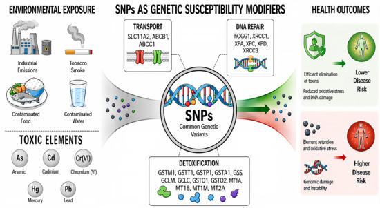
Graphical Abstract

1. Introduction

Potentially toxic elements (PTEs), or toxic elements (TEs), are environmental pollutants of growing scientific concern due to their impact on human health. They are often referred to as “heavy metals,” although this term is imprecise, as it may include metalloids (e.g., arsenic) and elements essential at trace levels [1]. Among TEs, arsenic (As), cadmium (Cd), hexavalent chromium (Cr(VI)), lead (Pb), and mercury (Hg) are the most relevant due to their high toxicity at low concentrations, environmental persistence, and bioaccumulation potential. These elements arise from both natural and anthropogenic sources and are widely distributed across air, water, soil, and food, leading to human exposure primarily through ingestion and inhalation, particularly in industrial and occupational settings [2,3]. Because they are non-biodegradable, toxic elements accumulate in environmental and biological systems, leading to long-term exposure and health risks. Epidemiological and toxicological evidence link As, Cd, Cr(VI), Pb, and Hg exposure to multiple adverse health outcomes affecting the nervous, renal, cardiovascular, immune, endocrine, and reproductive systems [4].
Given their pervasive nature and serious health risks, it is crucial to understand the factors that contribute to differences in susceptibility among individuals [5]. In fact, not all individuals exposed to similar levels of TEs develop the same health effects, reflecting substantial inter-individual variability.
A central mechanism underlying this phenomenon is gene–environment (G × E) interaction, whereby the effects of environmental exposures on disease risk are influenced by an individual’s genetic profile [6]. Among genetic determinants, single-nucleotide polymorphisms (SNPs) are the most common type of variation, present in a population with a frequency ≥ 1% [7]. They can be considered internal contributing factors in the susceptibility of individuals to TE effects. In this regard, SNPs are considered relevant as they belong to essential pathways, such as TE transport and detoxification, oxidative stress and DNA repair [8,9,10].
However, deeper investigations about mechanisms underlying the genetic influence on responses to toxic elements are needed and robust evidence about their potential role as disease predictors are required. Understanding the interplay between genetic factors and environmental exposures can be crucial for identifying individuals at a higher risk of adverse health outcomes, improving risk assessment, and informing preventive or therapeutic strategies. Therefore, this review focuses on genetic susceptibility and gene–environment interactions in the context of TE exposure. In particular, it examines the main toxic elements, summarizes epidemiological evidence of associated health risks, and highlights how genetic variability may modulate individual susceptibility to, or provide partial protection against, TE-related toxicity, based on the current state of the evidence. A bibliographic search was conducted in March 2026. The search was performed using PubMed, with keywords related to the topics mentioned above. To ensure comprehensive coverage, no restrictions were applied regarding publication type or study design. Only articles published in English were included, encompassing original research papers, reviews, and meta-analyses. The reference lists of the selected articles were also screened to identify additional relevant studies.

2. Sources of Toxic Elements and Routes of Exposure

Toxic elements (TEs) are naturally present in the Earth’s crust and are released into the atmosphere, hydrosphere, and lithosphere through processes such as rock weathering, volcanic activity, and geochemical cycling, establishing background levels that vary with local geology. However, over the past century, human activities—including industry, agriculture, and waste management—have significantly increased their mobilization and environmental distribution [11,12].
Arsenic (As) is naturally released through the weathering of As-bearing minerals and geothermal or volcanic activity and can persist in soils and sediments due to its past use in pesticides, feed additives, and wood preservatives. Major anthropogenic sources include mining and smelting, coal combustion, and industrial processes such as electronics and glass production, resulting in emissions to air, water, and waste streams [13]. Cadmium (Cd), naturally present in zinc, lead, and copper ores, is released through weathering and volcanic activity. Anthropogenic inputs are derived mainly from mining, smelting, battery production, wastewater discharge, and the use of Cd-contaminated fertilizers and pesticides, as well as the disposal or incineration of Cd-containing products such as batteries, plastics, pigments, and paints [14].
Hexavalent chromium (Cr(VI)) forms naturally through the oxidation of Cr(III)-bearing minerals, particularly in manganese-oxide-rich or ultramafic soils, but is predominantly associated with anthropogenic activities such as mining, electroplating, leather tanning, and pigment and dye production, as well as improper waste management, leading to the contamination of soils, water, and air [15,16]. Lead (Pb) occurs naturally in rocks and is released via weathering, forest fires, and particulates, but its environmental levels have increased more than 1000-fold in the past three centuries due to mining, smelting, battery production, the use of lead-based paints, fossil fuel combustion (including leaded gasoline), and lead–arsenate pesticides [17]. Mercury (Hg) is naturally emitted through volcanic activity, weathering, and ocean and soil emissions, but about two-thirds of atmospheric Hg is anthropogenic, mainly from coal combustion, mining, cement production, and metal manufacturing, which has significantly altered its natural cycle [18].
Human exposure to TEs occurs mainly through ingestion and inhalation, while dermal absorption is generally less relevant. Dietary intake is a major pathway: As is associated with rice and crops grown in contaminated soils [19]; Cd is found in vegetables, cereals, potatoes, and some fish [20]; Cr(VI) exposure occurs primarily via contaminated drinking water [21]; Pb is present in vegetables and drinking water, particularly from lead service lines [22,23]; and Hg exposure is mainly linked to fish and sea mammals [24].
Inhalation represents another major exposure route, especially in occupational settings. Elevated levels of As, Cd, and Pb have been observed in foundry workers [25], while Hg vapor exposure occurs in dental and industrial environments [24], and Cr(VI) exposure is common in chrome plating, welding, and surface treatment industries [26]. Nevertheless, inhalation exposure is also relevant in indoor environments, including homes, schools, offices, and public transport, where airborne TEs may pose non-carcinogenic and carcinogenic risks, often exceeding outdoor levels and international thresholds [27].
Smoking further increases exposure to TEs, as tobacco plants accumulate As, Cd, Cr, Hg, and Pb [28]. These elements are concentrated in different plant tissues, with Cd and Hg particularly present in leaves, and Cr, As, and Pb accumulating mainly in roots [29,30]. Tobacco smoke therefore exposes both active and passive smokers, with children being especially vulnerable due to higher exposure to contaminated household dust [31].
Dermal exposure is less significant but still possible: arsenite can penetrate the skin [32,33], Cd shows dermal bioaccessibility [34], Cr(VI) can be absorbed from contaminated soils and leather products [35], Pb may contribute to systemic burden in occupational settings [36], and Hg can be absorbed through the skin depending on chemical form and exposure conditions, including cosmetic use [37].

3. Biomonitoring and Health Effects of Toxic Elements

Given their widespread distribution and persistence in the environment, human exposure to TEs is frequent, making the evaluation of their health effects increasingly relevant. In recognition of their potential impact on public health, several TEs—particularly As, Cd, Pb, and Hg—were included among the top ten chemicals of major public health concern by the World Health Organization (WHO) [38]. To assess human exposure to these elements, biomonitoring approaches are widely used, measuring TE concentrations in accessible biological matrices through spectrometric techniques and subsequently evaluating exposure–disease relationships through epidemiological studies. Blood and urine represent the most used matrices for TE biomonitoring, although other matrices, including saliva, hair, nails, teeth, and breast milk, may also provide useful information, each presenting specific advantages and limitations [39]. The choice of matrix often depends on the specific element and the timing or form of exposure. For instance, As exposure is commonly assessed through urinary measurements [40], while Pb levels are typically determined in whole blood [41]. Cr (VI) is preferentially measured in the erythrocytic fraction [42], while Cd can be detected in blood as an indicator of recent exposure or in urine to reflect chronic exposure [43]. Similarly, Hg can be measured in both blood and urine depending on the chemical species involved: urine is more suitable for inorganic Hg forms (including elemental mercury and mercury vapor), whereas blood is the preferred matrix for methylmercury, the organic form mainly derived from fish consumption [44].
A growing body of epidemiological evidence has correlated TE concentrations in the human body with adverse health outcomes affecting multiple physiological systems, particularly cardiovascular, neurological, and oncological diseases, which significantly impact global morbidity, mortality, and quality of life [4]. Chronic exposure to toxic TEs, including As, Cd, Pb, Hg, and Cr(VI), is increasingly recognized as an environmental contributor to major non-communicable diseases. In the cardiovascular system, exposure to several TEs has been associated with an increased risk of cardiovascular diseases, particularly ischemic heart disease, the leading cause of mortality worldwide [45]. Meta-analyses indicate elevated risks associated with Pb, Cd, and As exposure, with more recent pooled analyses confirming increased cardiovascular disease risk for Hg as well [46], while evidence for Cr(VI) remains limited and inconsistent [47]. Neurological disorders represent another major health concern linked to TE exposure. Alzheimer’s disease (AD), the most common cause of dementia accounting for approximately 60–70% of cases, has been associated with chronic exposure to several TEs [48]. Epidemiological studies have reported higher Cd levels in AD patients and evidence linking Cd exposure to cognitive decline [49], while Hg and Pb have also been associated with cognitive impairment and increased dementia risk [50]. In addition, cumulative Pb exposure has been linked to significantly higher incidences of AD [51], while altered As metabolism patterns, elevated blood Cr levels, and Cr(VI)-induced neurotoxicity have also been implicated in neurodegenerative processes [52,53,54]. Importantly, TE exposure is not limited to adult health effects: prenatal or early-life exposure to Pb, Hg, and As has been associated with impaired neurodevelopment, including deficits in cognitive and motor functions as well as behavioral disorders [55].
Beyond cardiovascular and neurological outcomes, several TEs exhibit well-established carcinogenic potential. According to the International Agency for Research on Cancer (IARC), As, Cd, and Cr(VI) are classified as Group 1 human carcinogens, inorganic Pb compounds as Group 2A, and methylmercury as Group 2B. Epidemiological studies consistently associate As exposure with an increased risk of multiple malignancies, including lung, breast, liver, stomach, and hematological cancers [56]. Cd exposure has been linked to pancreatic [57], prostate [58], liver [59], renal [60], breast [47], and thyroid [61] cancers, while Pb exposure is associated with the increased incidence and mortality of several cancers, particularly those of the gastrointestinal and urinary tracts [62]. Hg has also been implicated in thyroid cancer risk [63], and Cr(VI) exposure has been associated with modest but significant increases in overall cancer incidence and mortality, particularly for respiratory and gastrointestinal cancers [64]. Overall, these findings highlight TEs as significant environmental risk factors contributing to the onset and progression of several chronic diseases.

4. Gene–Environment Interaction: The Role of Single-Nucleotide Polymorphisms in Susceptibility to Toxic-Element-Induced Effects

Multifactorial diseases arise from the interplay of genetic and environmental factors. These determinants act both independently and through gene–environment (G×E) interactions, whereby environmental exposures influence gene expression and genetic variations modulate individual susceptibility. In this context, genomic variability, particularly SNPs, contributes to inter-individual differences in response to environmental exposures [65]. SNPs affecting susceptibility to toxic elements can be functionally grouped into key pathways involved in metal transport, detoxification, and DNA repair, reflecting the complex nature of their metabolism and toxicity [8] (Figure 1).

4.1. Transport

SNPs in genes encoding metal transporter proteins play an important role by directly influencing the uptake, distribution, and elimination of TEs.
Divalent Metal Transporter 1 (DMT1), encoded by SLC11A2 located on chromosome 12q13 in humans, is a transmembrane protein that allows the cellular entry of some divalent cations, including Cd, Pb, Co, Mn, Ni, Zn, and Cu, into the cells. DMT1 is also referred to as DCT1 (Divalent Cation Transporter 1), NRAMP2 (Natural Resistance Associated Macrophage Protein 2), and SLC11A2 (Solute Carrier Family 11, member 2). In a study conducted by Kim et al., the relationship between the DMT1 IVS4+44 C/A polymorphism and clinical parameters was investigated in 662 male Korean workers occupationally exposed to Pb. The findings suggested that individuals with the A/A genotype had an increased risk of lead-associated hypertension [66]. Moreover, from another study investigating the role of SLC11A2 (DMT1) polymorphism rs224589 and enrolling 113 workers occupationally exposed to Pb, it emerged that individuals carrying the heterozygous CA genotype (54%) for SLC11A2 showed higher blood Pb concentrations compared with both homozygous CC (wild-type) and AA (mutant) individuals. Furthermore, a significant inverse correlation was observed between blood Pb levels and hemoglobin exclusively in the CA subgroup, indicating a greater vulnerability to lead-related hematological alterations in correlation with this polymorphism [67].
Similarly, genetic variants of ATP-binding cassette transporters critically regulate the intracellular accumulation of TEs by mediating their transport outside from the cells. Polymorphisms in genes encoding for the ABC transporter can influence Hg accumulation and perinatal outcomes. Offspring carrying the ABCC1 rs11075290 C allele have significantly higher cord blood Hg levels and present increased odds of small-for-gestational-age. Similarly, carriers of ABCB1 rs2032582 GG genotype accumulate more mercury in cord blood and show a greater reduction in the mental development index [68].

4.2. Detoxification

Substantial evidence is reported about the contribution in TE toxicity exerted by SNPs of genes involved in detoxification processes, such as glutathione-related genes and metallothioneins, for their role in the binding and metabolism of TEs.

4.2.1. Glutathione-Related Genes

Genes involved in the glutathione pathway are key mediators of detoxification processes, contributing to the conjugation and clearance of toxic compounds, while also counteracting oxidative stress. Genetic variants in these genes, including those encoding glutathione S-transferases (GSTM1, GSTT1, GSTP1) and the modifier (GCLM) and catalytic (GCLC) subunits of glutamate–cysteine ligase, can affect enzymatic function and glutathione biosynthesis, ultimately shaping inter-individual susceptibility to TE-induced toxicity. A study including a Chinese population of 850 subjects exposed to high levels of As in drinking water identified polymorphisms in glutathione-related genes GSTO1 (rs11191979, rs2164624, rs4925), GSTO2 (rs156697, rs2297235), and PNP (rs3790064) as associated with the increased risk of As-induced skin lesions in individuals exposed to high-dose inorganic arsenic, for impairment in the body’s ability to methylate and detoxify arsenic efficiently consequently to the presence of these variants [69].
GSTP1 rs1695 polymorphism (AG + GG genotypes) is associated with an increased urinary percentage of inorganic arsenic (%InAs) and a decreased primary methylation index, indicating the reduced efficiency of these individuals in methylating inorganic As to its less toxic metabolites and increased susceptibility to its toxicity [70]. Altered methylation and reduction steps in As metabolism was confirmed by a recent genome-wide association analysis that reported how genetic variation in the flavin-containing monooxygenase and GSTO gene clusters significantly impacted blood and urinary levels of As metabolites [71].
Moreover, glutathione-related genes can play an important role in individual risk for arsenic-induced carotid atherosclerosis. GSTT1 polymorphism was correlated with high urinary As levels and higher carotid intima-media thickness (IMT), evaluated as a marker of subclinical vascular damage in a cohort of Italian young adults exposed to environmental As [72].
Interestingly, polymorphisms can also be associated with protective effects. A meta-analysis including nine articles and 3324 subjects found that the GSTM1 null genotype (rs4025935) was significantly associated with a lower susceptibility to As poisoning [73].
The presence of the SNPs of this pathway also contributes to the toxic effects observed in populations occupationally exposed to Cd. CAT rs7943316, GSTP1 rs1695, the GSTM1 null genotype, and the GSTT1 null genotype in workers were linked to differences in the phenotypic expression of antioxidant enzymes, suggesting that individuals carrying these genotypes may be more susceptible to oxidative damage from Cd exposure [74].
Genetic polymorphisms in glutathione (GSH)-related genes influence Hg levels. Notably, carriers of the GCLM rs41303970 TT genotype exhibited lower Hg concentrations in both blood and hair compared with C-allele carriers, suggesting more efficient mercury elimination or reduced retention. Conversely, individuals with the GSTM1 null genotype showed higher Hg, indicating greater accumulation of this TE [75].
GSTP1 rs1695 and CAT rs1001179 substantially increase the risk of lung cancer in the context of elevated Cr exposure [76]. Toxic effects due to Hg exposure could be revealed in the presence of the GCLC rs1555903 C allele, as it correlates with a lower estimated glomerular filtration rate in non-exposed individuals and lower beta-2-microglobulin in exposed individuals, both markers of impaired renal function. Similarly, the combined effect of the GSTA1 rs3957356 C and GSS rs3761144 G alleles is associated with higher urinary Hg levels in exposed individuals, suggesting that these variants may also contribute to increased metal retention and potentially greater toxicity. Conversely, the GCLM rs41303970 T allele is associated with protection against toxicity, as this SNP is linked to higher urinary Hg clearance, indicating enhanced elimination of this TE [77].
A cross-sectional study with 236 adults reported that GCLC rs17883901 was linked to enhanced antioxidant response, with higher concentrations of GSH as a function of Pb levels and GCLM rs41303970 exerting a protective effect against Pb accumulation, as carriers of at least one polymorphic allele for this gene had significantly lower blood and plasma Pb levels compared with those with the non-polymorphic genotype [78].
Genotyping of the GSTM1, GSTT1, and GSTP1 genes was performed to investigate their potential association with heavy metal concentrations in 140 children exposed to Pb and Cd near an abandoned mining area in Kabwe, Zambia. The study found that the GSTT1 null genotype was positively correlated with both blood Pb and Cd levels, while the GSTP1 Ile/Val genotype (rs1695) was associated with a higher risk of Pb toxicity. This risk was even greater when these genetic variants co-occurred [79].
Furthermore, maternal polymorphisms in GSH-related genes can influence TE levels, with consequences on perinatal and birth outcomes. Pregnant women with GSTM1-null, GSTT1-null, and GCLM rs41303970 variants exhibit increased Hg accumulation and heightened oxidative stress, which were linked to an increased risk of preterm birth and reduced weight in offspring. Additionally, an increased frequency of children with a lower mental development index occurs when mothers carry the rare G allele of GSTP1 rs1695. Moreover, increasing Hg exposure is associated with a lower psychomotor development index among GCLC rs761142 TT carriers [68].

4.2.2. Metallothioneins

Metallothioneins (MTs) are cysteine-rich proteins that bind and sequester toxic elements, thereby reducing their bioavailability and protecting cells from metal-induced toxicity and oxidative stress [80]. Due to their abundant thiol groups, MTs bind biologically essential metals to help maintain metal homeostasis, as well as bind heavy metals to facilitate their transport and detoxification. Among all MT isoforms—including MT1, MT2, MT3, and MT4—the MT1A and MT2A subtypes are the predominantly expressed isoforms in humans.
A study enrolling 321 women revealed that SNPs of metallothionein 1A and 1B, MT1A rs8044719 and MT1B rs1599823, and 2A, MT2A rs28366003 and MT2A rs10636, are associated with lower urinary Cd, indicating increased tissue retention and susceptibility to this TE [81]. In an analysis of 616 individuals, the MT2A gene polymorphism rs28366003 GG genotype was associated with significantly higher blood Cd and Pb levels compared with other genotypic subgroups [82], suggesting that individuals carrying the GG genotype may be more susceptible to metal toxicity and should take extra precautions to protect their health from the harmful effects of TEs.
MT1A gene polymorphisms rs11640851 and rs8052394 are associated with a negative correlation between creatinine-adjusted urine uric acid concentrations and cumulative blood Pb exposure, indicating that these genotypes may increase susceptibility to Pb-induced renal tubular dysfunction as reflected by uric acid excretion [83].
Regarding susceptibility to Hg exposure, the MT1M rs2270837 AA genotype and MT2A rs10636 CC genotype are associated with lower urinary Hg levels, while the MT1A rs8052394 GA and GG genotypes and MT1M rs9936741 TT genotype are associated with lower hair Hg levels [84]. Influence on Hg levels by SNPs of metallothioneins was subsequently demonstrated by another study with 165 women, in which MT1M rs9936741 was confirmed to be associated with significantly lower hair total Hg levels, suggesting a protective effect against accumulation, while MT1M rs2270836 was linked with higher hair Hg levels, indicating increased susceptibility to retention of this TE [85].
Overall, further investigations are needed on these genetic polymorphisms to better understand their impact on TEs and TE-induced adverse effects.

4.3. DNA Repair: Focus on the Base Excision Repair Pathway

Polymorphisms in DNA repair genes, including hOGG1 and XRCC1, contribute to inter-individual differences in disease susceptibility and cellular responses to DNA damage by altering the efficiency of the base excision repair pathway, which is involved in removing oxidative lesions and maintaining genomic stability.
The hOGG1 Ser326Cys variant is associated with reduced enzymatic activity and impaired removal of oxidative DNA lesions, particularly 8-oxoguanine, leading to increased DNA damage accumulation and genomic instability. This polymorphism is linked to an elevated risk of cancer and other oxidative stress-related disorders, as well as altered sensitivities to chemotherapy and radiotherapy. Similarly, XRCC1 variants such as Arg399Gln and Arg194Trp influence BER efficiency by modifying protein interactions within the repair complex. The Arg399Gln polymorphism is associated with decreased repair capacity and greater cancer risk, particularly under exposure to genotoxic agents, whereas the role of Arg194Trp appears less defined, with some evidence suggesting a potential protective effect. Overall, these variants may modulate individual susceptibility to environmental toxins and ionizing radiation [86].
hOGG1 Ser326Cys (rs1052133) and XRCC1 Arg399Gln (rs25487) polymorphisms can influence susceptibility to TEs. Borghini et al. reported that these polymorphisms in DNA repair genes are associated with increased susceptibility to As exposure in a cohort of 241 Italian young adults [87]. Carriers of the hOGG1 Cys allele and the XRCC1 Gln allele exhibited significantly shorter leukocyte telomere length in the context of higher urinary As levels, indicating enhanced genomic instability. These findings suggest that the combination of elevated As exposure and these DNA repair gene variants exacerbate telomeric DNA damage and may contribute to the development of arsenic-related health effects, highlighting a gene–environment interaction that increases individual vulnerability [87]. Moreover, hOGG1 rs1052133 and rs159153 polymorphisms interact with As exposure and methylation capacity to increase the risk of urothelial carcinoma [88].
hOGG1 rs1052133 and XRCC1 rs25487 are frequently associated with increased DNA damage in workers exposed to Cd, indicating a reduced DNA repair capacity and greater susceptibility to genotoxic effects [9]. hOGG1 rs1052133, along with other polymorphisms in DNA repair genes, XPA rs1800975, and XPC rs2228000 are also associated with the modulation of DNA instability biomarkers in workers occupationally exposed to Pb [89]. Moreover, individuals carrying XRCC1 Arg399Gln (rs25487) and hOGG1 Ser326Cys (rs1052133) polymorphisms exhibited increased DNA damage and oxidative stress when exposed to Hg [90].
Interestingly, the impact of XRCC1 polymorphism on the genotoxicity exerted by the exposure to hexavalent chromium appears controversial. The XRCC1 Arg399Gln (rs25487) homozygous variant genotype (Gln/Gln) is associated with increased chromosomal aberrations and greater susceptibility to chromosomal damage in workers exposed to this TE [91]. This result is in contrast with another study reporting that the XRCC1 Arg399Gln (rs25487) variant is associated with reduced DNA damage in individuals occupationally exposed to Cr(VI), indicating a protective effect of this polymorphism against Cr(VI) toxicity [92]. This discrepancy likely reflects differences in the endpoints measured, population characteristics, or exposure levels. Other DNA repair polymorphisms, correlated with Cr effects, are XPD Lys751Gln (rs13181) and XPC Lys939Gln (rs2228000), which are associated with an increased susceptibility to lung cancer in individuals exposed to this element [93]. Moreover, the interaction between chromate exposure and the XRCC3 rs2295152 T allele has a significant effect on micronuclei frequency, indicating a gene–environment interaction that increases susceptibility to chromate-induced genetic damage [94]. Best characterized SNPs are summarized in Table 1.

5. Conclusions and Future Perspectives: Key Points and Emerging Directions

TEs are bioaccumulative and exert a wide range of adverse effects on the human body, including cancer, neurological, and cardiovascular disorders. Their accumulation is influenced not only by environmental exposure levels but also by biological processes governing absorption, distribution, metabolism, and elimination. These processes vary substantially between individuals and strongly affect internal metal burden and toxicity outcomes. Investigating genetic variants and their interactions with environmental factors therefore provides crucial insights into individual susceptibility to TE-induced health risks.
It is now well established that genetic background represents a central component of the complex network of determinants defining sensitivity to metals. At the same time, additional internal and external factors, such as lifestyle, nutritional status, and co-exposures, further modulate biological responses. The interplay between genetic variability and environmental influences contributes to distinct phenotypes and heterogeneous responses to toxic exposures. Identifying variations in genes involved in TE metabolism remains essential for understanding susceptibility, identifying high-risk populations, and supporting the development of targeted preventive and therapeutic strategies. Among the most extensively studied candidate genes are those involved in transport, detoxification, and DNA repair, including DMT1, GSTP1, MT2A, hOGG1, and XRCC1. Various studies have explored the impact of SNPs within these genes, and some variants have been proposed as potential susceptibility markers for toxic element toxicity. Importantly, the overall risk should also be considered in terms of cumulative exposure across multiple pathways, which collectively influence the internal dose and biological effects of toxic elements.
However, findings remain inconsistent due to the heterogeneity of available studies. Differences in study design, sample size, exposure assessment methods, and population characteristics often hinder the integration of results and contribute to inconsistent findings. In particular, small cohorts and limited statistical power reduce the robustness of associations, while variabilities in exposure type (environmental vs. occupational) and intensity further complicate comparisons across studies. Moreover, the geographic distribution of research is imbalanced, with most evidence generated in specific regions, potentially introducing biases and limiting the generalizability of conclusions to underrepresented populations. In this context, the population-specific distribution of genetic variants warrants careful consideration. Certain alleles may be more prevalent in specific ethnic or geographic groups, and this can influence biological susceptibility to toxic elements and may partly explain inconsistencies across studies. At the same time, this raises important questions regarding the transferability of findings between populations, emphasizing the need for broader representation in genetic and epidemiological research.
Despite these limitations, the investigation of gene–environment interactions holds significant promise for practical applications and important questions regarding the transferability of findings between populations, emphasizing the need for broader representation in genetic and epidemiological research. Genetic variants associated with altered susceptibility could serve as biomarkers for identifying vulnerable individuals or subgroups, thereby improving risk stratification. Their integration into risk assessment frameworks may enhance the accuracy of exposure–response models and support the development of more effective prevention strategies. In the longer term, these insights could contribute to personalized and precision medicine approaches, where interventions are tailored according to individual genetic and environmental risk profiles.
To address current gaps, future research should prioritize multicentric study designs involving larger and more diverse populations exposed to varying levels of toxic elements. The harmonization of methodologies, including exposure assessments and biomarker measurements, is essential to improve comparability and reproducibility. The integration of epidemiological data with computational exposure models, alongside the application of artificial intelligence and machine learning, offers new opportunities to manage complex datasets and uncover subtle interaction patterns. Furthermore, the adoption of multi-omics approaches—encompassing genomics, epigenomics, transcriptomics, and exposomics—will enable a more comprehensive characterization of the exposome, capturing both internal biological responses and external environmental influences. This systems-level perspective is critical for elucidating the mechanisms underlying differential susceptibility.
Recent advances in induced pluripotent stem cell (iPSC) technology provide an additional complementary strategy. By reprogramming accessible somatic cells into iPSCs and differentiating them into tissue-specific models, researchers can investigate gene–environment interactions in a controlled and human-relevant context. These models allow the identification of susceptibility-associated variants, early molecular alterations, and potential therapeutic targets prior to disease onset, thereby supporting earlier and more effective prevention strategies [95,96]. The integration of approaches such as multicentric studies, advanced in vitro platforms, AI-driven data analysis, and multi-omics frameworks will help overcome current limitations and reduce uncertainty in the field. Ultimately, these efforts can improve risk assessment, strengthen prevention strategies, and facilitate the translation of mechanistic insights into public health policies and precision medicine applications.
Overall, this review underscores the role of gene–environment interactions and SNP-based susceptibility in shaping individual responses to toxic element exposure. Progress in this area will depend not only on methodological standardization and broader population representation but also on a stronger emphasis on translating scientific evidence into actionable tools for risk assessment, prevention, and personalized health management.

Author Contributions

Conceptualization, A.B., and F.M.; writing—original draft preparation, M.P. and A.B.; writing—review and editing E.B., S.B., F.G. and F.M. All authors have read and agreed to the published version of the manuscript.

Funding

This research was funded by the Ministry of Health as part of the National Plan for Complementary Investments—“Health, Environments, Biodiversity, and Climate”, project name PNC INSINERGIA, project code (National Research Council) PRR.AP015.165.

Institutional Review Board Statement

Not applicable.

Informed Consent Statement

Not applicable.

Data Availability Statement

No new data were created or analyzed in this study. Data sharing is not applicable to this article.

Acknowledgments

We acknowledge the use of AI assistance, specifically ChatGPT (version 5.0), in the revision of certain parts of this paper to enhance language quality, clarity, and conciseness.

Conflicts of Interest

The authors declare no conflicts of interest.

Abbreviations

The following abbreviations are used in this manuscript:
ADAlzheimer’s disease
AsArsenic
CdCadmium
CrChromium
HgMercury
IARCInternational Agency for Research and Cancer
MTsMetallothioneins
PbLead
PTEsPotentially toxic elements
PVCPolyvinyl Chloride
SNPsSingle-nucleotide polymorphisms
TEsToxic elements
WHOWorld Health Organization

References

  1. Pourret, O.; Hursthouse, A. It’s Time to Replace the Term “Heavy Metals” with “Potentially Toxic Elements” When Reporting Environmental Research. Int. J. Environ. Res. Public Health 2019, 16, 4446. [Google Scholar] [CrossRef] [PubMed]
  2. Kotnala, S.; Tiwari, S.; Nayak, A.; Bhushan, B.; Chandra, S.; Medeiros, C.R.; Coutinho, H.D.M. Impact of heavy metal toxicity on the human health and environment. Sci. Total Environ. 2025, 987, 179785. [Google Scholar] [CrossRef] [PubMed]
  3. Järup, L. Hazards of Heavy Metal Contamination. Br. Med. Bull. 2003, 68, 167–182. [Google Scholar] [CrossRef] [PubMed]
  4. Cheng, Y.F.; Zhao, Y.J.; Chen, C.; Zhang, F. Heavy Metals Toxicity: Mechanism, Health Effects, and Therapeutic Interventions. MedComm 2025, 6, e70241. [Google Scholar] [CrossRef]
  5. Zeise, L.; Bois, F.Y.; Chiu, W.A.; Hattis, D.; Rusyn, I.; Guyton, K.Z. Addressing Human Variability in Next-Generation Human Health Risk Assessments of Environmental Chemicals. Environ. Health Perspect. 2013, 121, 23–31. [Google Scholar] [CrossRef]
  6. Ottman, R. Gene-Environment Interaction: Definitions and Study Designs. Prev. Med. 1996, 25, 764–770. [Google Scholar] [CrossRef]
  7. Azizzadeh-Roodpish, S.; Garzon, M.H.; Mainali, S. Classifying Single Nucleotide Polymorphisms in Humans. Mol. Genet. Genom. 2021, 296, 1161–1173. [Google Scholar] [CrossRef]
  8. Joneidi, Z.; Mortazavi, Y.; Memari, F.; Roointan, A.; Chahardouli, B.; Rostami, S. The Impact of Genetic Variation on Metabolism of Heavy Metals: Genetic Predisposition? Biomed. Pharmacother. 2019, 113, 108642. [Google Scholar] [CrossRef]
  9. Pinto, T.G.; Dias, T.A.; Renno, A.C.M.; Dos Santos, J.N.; Cury, P.R.; Ribeiro, D.A. The Impact of Genetic Polymorphisms for Detecting Genotoxicity in Workers Occupationally Exposed to Metals: A Systematic Review. J. Appl. Toxicol. 2025, 45, 1070–1079. [Google Scholar] [CrossRef]
  10. Kumar, P. Genetic susceptibility to heavy metal toxicity. In Heavy Metal Toxicity and Neurodegeneration; Kumar, P., Gogia, N., Eds.; Elsevier: Amsterdam, The Netherlands, 2025; pp. 179–201. [Google Scholar]
  11. He, Z.L.; Yang, X.E.; Stoffella, P.J. Trace Elements in Agroecosystems and Impacts on the Environment. J. Trace Elem. Med. Biol. 2005, 19, 125–140. [Google Scholar] [CrossRef]
  12. Briffa, J.; Sinagra, E.; Blundell, R. Heavy Metal Pollution in the Environment and Their Toxicological Effects on Humans. Heliyon 2020, 6, e04691. [Google Scholar] [CrossRef]
  13. Garelick, H.; Jones, H.; Dybowska, A.; Valsami-Jones, E. Arsenic Pollution Sources. Rev. Environ. Contam. Toxicol. 2008, 197, 17–60. [Google Scholar] [CrossRef] [PubMed]
  14. Sattar, S.; Yahya, M.; Aslam, S.; Hussain, R.; Mukkarram Shah, S.M.; Rauf, Z.; Zamir, A.; Ullah, R.; Shahzad, A. Environmental Occurrence, Hazards, and Remediation Strategies for the Removal of Cadmium from the Polluted Environment. Results Eng. 2025, 25, 104322. [Google Scholar] [CrossRef]
  15. Ao, M.; Sun, S.; Deng, T.; Zhang, F.; Liu, T.; Tang, Y.; Li, J.; Wang, S.; Qiu, R. Natural Source of Cr(VI) in Soil: The Anoxic Oxidation of Cr(III) by Mn Oxides. J. Hazard. Mater. 2022, 433, 128805. [Google Scholar] [CrossRef]
  16. Ukhurebor, K.E.; Aigbe, U.O.; Onyancha, R.B.; Nwankwo, W.; Osibote, O.A.; Paumo, H.K.; Ama, O.M.; Adetunji, C.O.; Siloko, I.U. Effect of Hexavalent Chromium on the Environment and Removal Techniques: A Review. J. Environ. Manag. 2021, 280, 111809. [Google Scholar] [CrossRef] [PubMed]
  17. Cheema, A.I.; Liu, G.; Yousaf, B.; Abbas, Q.; Zhou, H. A Comprehensive Review of Biogeochemical Distribution and Fractionation of Lead Isotopes for Source Tracing in Distinct Interactive Environmental Compartments. Sci. Total Environ. 2020, 719, 135658. [Google Scholar] [CrossRef] [PubMed]
  18. Kim, M.K.; Zoh, K.D. Fate and Transport of Mercury in Environmental Media and Human Exposure. J. Prev. Med. Public Health 2012, 45, 335–343. [Google Scholar] [CrossRef]
  19. Baker, B.A.; Cassano, V.A.; Murray, C.; ACOEM Task Force on Arsenic Exposure. Arsenic Exposure, Assessment, Toxicity, Diagnosis, and Management: Guidance for Occupational and Environmental Physicians. J. Occup. Environ. Med. 2018, 60, e634–e639. [Google Scholar] [CrossRef]
  20. Zhang, H.; Reynolds, M. Cadmium Exposure in Living Organisms: A Short Review. Sci. Total Environ. 2019, 678, 761–767. [Google Scholar] [CrossRef]
  21. Tumolo, M.; Ancona, V.; De Paola, D.; Losacco, D.; Campanale, C.; Massarelli, C.; Uricchio, V.F. Chromium Pollution in European Water, Sources, Health Risk, and Remediation Strategies: An Overview. Int. J. Environ. Res. Public Health 2020, 17, 5438. [Google Scholar] [CrossRef]
  22. Nag, R.; Cummins, E. Human Health Risk Assessment of Lead (Pb) through the Environmental-Food Pathway. Sci. Total Environ. 2022, 810, 151168. [Google Scholar] [CrossRef] [PubMed]
  23. Lanphear, B.; Navas-Acien, A.; Bellinger, D.C. Lead Poisoning. N. Engl. J. Med. 2024, 391, 1621–1631. [Google Scholar] [CrossRef] [PubMed]
  24. Clarkson, T.W.; Magos, L.; Myers, G.J. The Toxicology of Mercury—Current Exposures and Clinical Manifestations. N. Engl. J. Med. 2003, 349, 1731–1737. [Google Scholar] [CrossRef]
  25. Freire, B.M.; Gonzaga, R.G.; Pedron, T.; Monteiro, L.R.; Lange, C.N.; Pedreira Filho, W.D.R.; Batista, B.L. Occupational Exposure to Potentially Toxic Elements in the Foundry Industry: An Integrated Environmental and Biological Monitoring. Environ. Sci. Pollut. Res. Int. 2021, 28, 34630–34641. [Google Scholar] [CrossRef]
  26. Viegas, S.; Martins, C.; Bocca, B.; Bousoumah, R.; Duca, R.C.; Galea, K.S.; Godderis, L.; Iavicoli, I.; Janasik, B.; Jones, K.; et al. HBM4EU Chromates Study: Determinants of Exposure to Hexavalent Chromium in Plating, Welding and Other Occupational Settings. Int. J. Environ. Res. Public Health 2022, 19, 3683. [Google Scholar] [CrossRef]
  27. Khoshakhlagh, A.H.; Ghobakhloo, S.; Peijnenburg, W.J.G.M.; Gruszecka-Kosowska, A.; Cicchella, D. To Breathe or Not to Breathe: Inhalational Exposure to Heavy Metals and Related Health Risk. Sci. Total Environ. 2024, 932, 172556. [Google Scholar] [CrossRef]
  28. Zumbado, M.; Luzardo, O.P.; Rodríguez-Hernández, Á.; Boada, L.D.; Henríquez-Hernández, L.A. Differential Exposure to 33 Toxic Elements through Cigarette Smoking, Based on the Type of Tobacco and Rolling Paper Used. Environ. Res. 2019, 169, 368–376. [Google Scholar] [CrossRef]
  29. da Silva, C.P.; de Almeida, T.E.; Zittel, R.; de Oliveira Stremel, T.R.; Domingues, C.E.; Kordiak, J.; de Campos, S.X. Translocation of Metal Ions from Soil to Tobacco Roots and Their Concentration in the Plant Parts. Environ. Monit. Assess. 2016, 188, 663. [Google Scholar] [CrossRef]
  30. Uwiringiyimana, E.; Gao, J.; Zhang, D.; Biswash, M.R.; Shi, Y.X. Bioaccumulation and Translocation of Hg and Cr by Tobacco in Sichuan Province, China: Understanding the Influence of Soil pH. Environ. Monit. Assess. 2023, 195, 1193. [Google Scholar] [CrossRef]
  31. Lopez-Galvez, N.; Mahabee-Gittens, E.M.; Quintana, P.J.E.; Merianos, A.L.; Dodder, N.G.; Hoh, E.; Stone, L.; Watanabe, K.; Matt, G.E. Environmental Tobacco Smoke Is a Major Contributor to Lead, Cadmium, and Arsenic in Settled House Dust. Chemosphere 2026, 394, 144820. [Google Scholar] [CrossRef] [PubMed]
  32. Patel, K.S.; Pandey, P.K.; Martín-Ramos, P.; Corns, W.T.; Varol, S.; Bhattacharya, P.; Zhu, Y. A Review on Arsenic in the Environment: Contamination, Mobility, Sources, and Exposure. RSC Adv. 2023, 13, 8803–8821. [Google Scholar] [CrossRef]
  33. Ouypornkochagorn, S.; Feldmann, J. Dermal Uptake of Arsenic through Human Skin Depends Strongly on Its Speciation. Environ. Sci. Technol. 2010, 44, 3972–3978. [Google Scholar] [CrossRef]
  34. Wang, J.; Gao, P.; Li, M.Y.; Ma, J.Y.; Li, J.Y.; Yang, D.L.; Cui, D.L.; Xiang, P. Dermal Bioaccessibility and Cytotoxicity of Heavy Metals in Urban Soils from a Typical Plateau City: Implication for Human Health. Sci. Total Environ. 2022, 835, 155544. [Google Scholar] [CrossRef]
  35. Liu, C.; Liu, C.; Juhasz, A.L.; Dong, W.J.; Ma, L.Q. Human Health Risks of Chromium in Bright-Colored Leather Products: Speciation-Based Insights into Dermal Permeation. Environ. Int. 2025, 200, 109538. [Google Scholar] [CrossRef]
  36. Niemeier, R.T.; Maier, A.; Reichard, J.F. Rapid Review of Dermal Penetration and Absorption of Inorganic Lead Compounds for Occupational Risk Assessment. Ann. Work Expo. Health 2022, 66, 291–311. [Google Scholar] [CrossRef]
  37. Chan, T.Y. Inorganic Mercury Poisoning Associated with Skin-Lightening Cosmetic Products. Clin. Toxicol. 2011, 49, 886–891. [Google Scholar] [CrossRef]
  38. World Health Organization (WHO). 10 Chemicals of Public Health Concern. Available online: https://www.who.int/news-room/photo-story/detail/10-chemicals-of-public-health-concern (accessed on 19 February 2026).
  39. Gil, F.; Hernández, A.F. Toxicological Importance of Human Biomonitoring of Metallic and Metalloid Elements in Different Biological Samples. Food Chem. Toxicol. 2015, 80, 287–297. [Google Scholar] [CrossRef]
  40. Buekers, J.; Baken, K.; Govarts, E.; Martin, L.R.; Vogel, N.; Kolossa-Gehring, M.; Šlejkovec, Z.; Falnoga, I.; Horvat, M.; Lignell, S.; et al. Human Urinary Arsenic Species, Associated Exposure Determinants and Potential Health Risks Assessed in the HBM4EU Aligned Studies. Int. J. Hyg. Environ. Health 2023, 248, 114115. [Google Scholar] [CrossRef] [PubMed]
  41. Barbosa, F., Jr.; Tanus-Santos, J.E.; Gerlach, R.F.; Parsons, P.J. A Critical Review of Biomarkers Used for Monitoring Human Exposure to Lead: Advantages, Limitations, and Future Needs. Environ. Health Perspect. 2005, 113, 1669–1674. [Google Scholar] [CrossRef] [PubMed]
  42. Ndaw, S.; Leso, V.; Bousoumah, R.; Rémy, A.; Bocca, B.; Duca, R.C.; Godderis, L.; Hardy, E.; Janasik, B.; van Nieuwenhuyse, A.; et al. HBM4EU Chromates Study—Usefulness of Measurement of Blood Chromium Levels in the Assessment of Occupational Cr(VI) Exposure. Environ. Res. 2022, 214, 113758. [Google Scholar] [CrossRef] [PubMed]
  43. Adams, S.V.; Newcomb, P.A. Cadmium Blood and Urine Concentrations as Measures of Exposure: NHANES 1999–2010. J. Expo. Sci. Environ. Epidemiol. 2014, 24, 163–170. [Google Scholar] [CrossRef]
  44. So, S.C.A.; Tsoi, M.F.; Cheung, A.J.; Cheung, T.T.; Cheung, B.M.Y. Blood and Urine Inorganic and Organic Mercury Levels in the United States from 1999 to 2016. Am. J. Med. 2021, 134, e20–e30. [Google Scholar] [CrossRef] [PubMed]
  45. Lamas, G.A.; Bhatnagar, A.; Jones, M.R.; Mann, K.K.; Nasir, K.; Tellez-Plaza, M.; Ujueta, F.; Navas-Acien, A.; American Heart Association Council on Epidemiology and Prevention; Council on Cardiovascular and Stroke Nursing; et al. Contaminant Metals as Cardiovascular Risk Factors: A Scientific Statement from the American Heart Association. J. Am. Heart Assoc. 2023, 12, e029852. [Google Scholar] [CrossRef] [PubMed]
  46. Cheema, S.; Hussain, S.I.; Faheem, M.S.B.; Jalal, A.A.; Rifai, M.; Dar, A.; Burhan, M.; Shahid, A.; Ali, M.S.; Anwar, A.; et al. Toxic Heavy Metal Exposure and Heart Health: A Systematic Review and Meta-Analysis of 324,331 Patients. BMC Cardiovasc. Disord. 2025, 25, 792. [Google Scholar] [CrossRef] [PubMed]
  47. Liu, X.; Zhang, D.; Wu, X.; Tu, J.; Gong, C.; Li, Y.; Cui, W.; Chen, J.; Lu, S. Urinary metals as influencing factors of coronary heart disease among a population in Guangzhou, China. Ecotoxicol. Environ. Saf. 2022, 241, 113746. [Google Scholar] [CrossRef]
  48. Bashir, B.; Jarial, S.; Sood, S.; Vishwas, S. Heavy Metals and Alzheimer’s Disease: A Review. In Heavy Metal Toxicity and Neurodegeneration; Kumar, P., Gogia, N., Eds.; Academic Press: Cambridge, MA, USA, 2025; pp. 225–230. [Google Scholar] [CrossRef]
  49. Kooshki, A.; Farmani, R.; Mehrpour, O.; Naghizadeh, A.; Amirabadizadeh, A.; Kavoosi, S.; Vohra, V.; Nakhaee, S. Alzheimer’s Disease and Circulatory Imbalance of Toxic Heavy Metals: A Systematic Review and Meta-Analysis of Clinical Studies. Biol. Trace Elem. Res. 2025, 203, 1871–1885. [Google Scholar] [CrossRef]
  50. Song, Y.; Chen, Y.; Fu, Z.; Wen, Y.; Zhao, W.; Li, J.; Wang, H.; Du, Y.; Deng, Y. Heavy Metal Exposure and Cognitive Impairment: An Umbrella Review of Meta-Analyses. J. Hazard. Mater. 2025, 498, 139881. [Google Scholar] [CrossRef]
  51. Wang, X.; Bakulski, K.M.; Walker, E.; Mukherjee, B.; Dodge, H.; Albin, R.L.; Paulson, H.L.; Park, S.K. Exposure to Lead and Incidence of Alzheimer’s Disease and All-Cause Dementia in the United States. Alzheimers Dement. 2026, 22, e71075. [Google Scholar] [CrossRef]
  52. Yang, Y.W.; Liou, S.H.; Hsueh, Y.M.; Lyu, W.S.; Liu, C.S.; Liu, H.J.; Chung, M.C.; Hung, P.H.; Chung, C.J. Risk of Alzheimer’s Disease with Metal Concentrations in Whole Blood and Urine: A Case-Control Study Using Propensity Score Matching. Toxicol. Appl. Pharmacol. 2018, 356, 8–14. [Google Scholar] [CrossRef]
  53. Strumylaite, L.; Kregzdyte, R.; Kucikiene, O.; Baranauskiene, D.; Simakauskiene, V.; Naginiene, R.; Damuleviciene, G.; Lesauskaite, V.; Zemaitiene, R. Alzheimer’s Disease Association with Metals and Metalloids Concentration in Blood and Urine. Int. J. Environ. Res. Public Health 2022, 19, 7309. [Google Scholar] [CrossRef]
  54. Wise, J.P., Jr.; Young, J.L.; Cai, J.; Cai, L. Current Understanding of Hexavalent Chromium [Cr(VI)] Neurotoxicity and New Perspectives. Environ. Int. 2022, 158, 106877. [Google Scholar] [CrossRef] [PubMed]
  55. da Silva, A.S.; Santos, R.M.S.; De Marco, P.G.; Rezende, V.H.M.; Martins, T.C.; Silva, J.R.; Romano-Silva, M.A.; Miranda, D.M. Neurodevelopmental Outcomes Associated with Early-Life Exposure to Heavy Metals: A Systematic Review. Int. J. Environ. Res. Public Health 2025, 22, 1308. [Google Scholar] [CrossRef]
  56. Rahmani, A.; Khamutian, S.; Doosti-Irani, A.; Shokoohizadeh, M.J.; Shirmohammadi-Khorram, N.; Sahraeei, F.; Khodabakhshi, M.; Ahangaran, N. The Association of Arsenic Exposure with Mortality Due to Cancer, Diabetes, Alzheimer’s and Congenital Anomalies Using Poisson Regression. Sci. Rep. 2023, 13, 15456. [Google Scholar] [CrossRef] [PubMed]
  57. Lee, F.J.; Inchai, H.; Lin, J.T.; Koh, D.; Lin, R.T. Dose-Response Relationship of Cadmium and Pancreatic Cancer Risk: A Meta-Analysis. Occup. Environ. Med. 2025, 82, 407–414. [Google Scholar] [CrossRef]
  58. Firmani, G.; Chiavarini, M.; Dolcini, J.; Quarta, S.; D’Errico, M.M.; Barbadoro, P. The Association Between Cadmium Exposure and Prostate Cancer: An Updated Systematic Review and Meta-Analysis. Int. J. Environ. Res. Public Health 2024, 21, 1532. [Google Scholar] [CrossRef]
  59. Zhang, L.; Wang, Y.; Li, T.; Zhuo, W.; Zhu, Y. Elevated Serum and Hair Levels of Cadmium as a Risk Factor for Liver Carcinoma: A Meta-Analysis. Nutr. Cancer 2023, 75, 1438–1447. [Google Scholar] [CrossRef]
  60. Lee, H.; Jo, Y.; Jung, M.; Lee, J.H.; Kim, T.H.; Lee, J.; Kim, D.J.; Rahmati, M.; Smith, L.; Pizzol, D.; et al. Heavy Metal Exposure and All Health Outcomes: An Umbrella Review of Meta-Analyses. J. Hazard. Mater. 2026, 503, 141141. [Google Scholar] [CrossRef]
  61. Abdelgawwad El-Sehrawy, A.A.M.; Hsu, C.Y.; Saleh, E.A.M.; Moharam, M.M.; Rekha, M.M.; Kundlas, M.; Premkumar, J.; Ray, S.; Hussein, A.R.; Kadhim, M.A. Associations between Cadmium and Lead Exposure and Thyroid Disorders: A Systematic Review and Meta-Analysis. J. Trace Elem. Med. Biol. 2025, 92, 127781. [Google Scholar] [CrossRef]
  62. Vagnoni, G.; Bortolotti, E.; Checchi, S.; Saieva, C.; Berti, G.; Doccioli, C.; Caini, S. Lead (Pb) in Biological Samples in Association with Cancer Risk and Mortality: A Systematic Literature Review. Cancer Epidemiol. 2024, 92, 102630. [Google Scholar] [CrossRef] [PubMed]
  63. Webster, A.M.; Pinion, D.; Pineda, E.; Aboueisha, H.; Hussein, M.H.; Fawzy, M.S.; Toraih, E.A.; Kandil, E. Elucidating the Link between Thyroid Cancer and Mercury Exposure: A Review and Meta-Analysis. Environ. Sci. Pollut. Res. 2024, 31, 12841–12855. [Google Scholar] [CrossRef]
  64. Deng, Y.; Wang, M.; Tian, T.; Lin, S.; Xu, P.; Zhou, L.; Dai, C.; Hao, Q.; Wu, Y.; Zhai, Z.; et al. The Effect of Hexavalent Chromium on the Incidence and Mortality of Human Cancers: A Meta-Analysis Based on Published Epidemiological Cohort Studies. Front. Oncol. 2019, 9, 24. [Google Scholar] [CrossRef]
  65. Virolainen, S.J.; VonHandorf, A.; Viel, K.C.M.F.; Weirauch, M.T.; Kottyan, L.C. Gene-Environment Interactions and Their Impact on Human Health. Genes Immun. 2023, 24, 1–11. [Google Scholar] [CrossRef] [PubMed]
  66. Kim, H.K.; Lee, H.; Kim, H.J. A Polymorphism in DMT1 Is Associated with Lead-Related Hypertensive Status. Mol. Cell. Toxicol. 2013, 9, 415–420. [Google Scholar] [CrossRef]
  67. Mani, M.S.; Dsouza, V.L.; Dsouza, H.S. Evaluation of Divalent Metal Transporter 1 (DMT1) (rs224589) Polymorphism on Blood Lead Levels of Occupationally Exposed Individuals. Toxicol. Lett. 2021, 353, 13–19. [Google Scholar] [CrossRef] [PubMed]
  68. Sadana, A.A.S.; Bakri, S.; Tokonami, S.; Nugraha, E.D.; Amqam, H.; Muniroh, M. Genetic Modulation of Mercury Exposure on Perinatal and Birth Outcomes: A Systematic Review and Meta-Analysis of Gene-Environment Interactions. J. Xenobiot. 2026, 16, 28. [Google Scholar] [CrossRef] [PubMed]
  69. Luo, L.; Li, Y.; Gao, Y.; Zhao, L.; Feng, H.; Wei, W.; Qiu, C.; He, Q.; Zhang, Y.; Fu, S.; et al. Association between Arsenic Metabolism Gene Polymorphisms and Arsenic-Induced Skin Lesions in Individuals Exposed to High-Dose Inorganic Arsenic in Northwest China. Sci. Rep. 2018, 8, 413. [Google Scholar] [CrossRef]
  70. González-Martínez, F.; Sánchez-Rodas, D.; Cáceres, D.D.; Martínez, M.F.; Quiñones, L.A.; Johnson-Restrepo, B. Arsenic Exposure, Profiles of Urinary Arsenic Species, and Polymorphism Effects of Glutathione-S-Transferase and Metallothioneins. Chemosphere 2018, 212, 927–936. [Google Scholar] [CrossRef]
  71. Tamayo, L.I.; Tong, L.; Davydiuk, T.; Vander Griend, D.; Haque, S.E.; Islam, T.; Jasmine, F.; Kibriya, M.G.; Graziano, J.; Chen, L.; et al. Genetic Variation in the FMO and GSTO Gene Clusters Impacts Arsenic Metabolism in Humans. PLoS Genet. 2025, 21, e1011826. [Google Scholar] [CrossRef]
  72. Stea, F.; Faita, F.; Borghini, A.; Faita, F.; Bianchi, F.; Bustaffa, E.; Minichilli, F.; Andreassi, M.G.; Sicari, R. Arsenic and Subclinical Vascular Damage in a Sample of Italian Young Adults: A Cross-Sectional Analysis. Environ. Sci. Pollut. Res. Int. 2016, 23, 20307–20314. [Google Scholar] [CrossRef]
  73. Jin, B.; Wan, S.; Boah, M.; Yang, J.; Ma, W.; Lv, M.; Li, H.; Wang, K. GSTM1 and GSTT1 Null Genotype Polymorphisms and Susceptibility to Arsenic Poisoning: A Meta-Analysis. Biol. Trace Elem. Res. 2021, 199, 2085–2095. [Google Scholar] [CrossRef]
  74. Khan, S.A.; Nurulain, S.M.; Qureshi, R.N.; Zafar, A.; Riaz, Z.; Shoukat, A.; Muneer, Z.; Bibi, N.; Raza, S.; Hussain, S.; et al. Exposure to Heavy Metals, Antioxidant Status, and the Interaction of Single Nucleotide Polymorphisms in the Genes CAT rs7943316, GSTP1 rs1695, as well as GSTM1 and GSTT1 Genes, among Workers in Occupational Settings. Environ. Toxicol. Pharmacol. 2024, 108, 104452. [Google Scholar] [CrossRef] [PubMed]
  75. Barcelos, G.R.; Grotto, D.; de Marco, K.C.; Valentini, J.; Lengert, A.V.; de Oliveira, A.Á.; Garcia, S.C.; Braga, G.Ú.; Schläwicke Engström, K.; Cólus, I.M.; et al. Polymorphisms in Glutathione-Related Genes Modify Mercury Concentrations and Antioxidant Status in Subjects Environmentally Exposed to Methylmercury. Sci. Total Environ. 2013, 463–464, 319–325. [Google Scholar] [CrossRef]
  76. Baszuk, P.; Janasik, B.; Pietrzak, S.; Marciniak, W.; Reszka, E.; Białkowska, K.; Jabłońska, E.; Muszyńska, M.; Lesicka, M.; Derkacz, R.; et al. Lung Cancer Occurrence—Correlation with Serum Chromium Levels and Genotypes. Biol. Trace Elem. Res. 2021, 199, 1228–1236. [Google Scholar] [CrossRef]
  77. Medina Pérez, O.M.; Flórez-Vargas, O.; Rincón Cruz, G.; Rondón González, F.; Rocha Muñoz, L.; Sánchez Rodríguez, L.H. Glutathione-Related Genetic Polymorphisms Are Associated with Mercury Retention and Nephrotoxicity in Gold-Mining Settings of a Colombian Population. Sci. Rep. 2021, 11, 8716. [Google Scholar] [CrossRef]
  78. Gomes, W.R.; Devóz, P.P.; Luiz, B.L.C.; Grotto, D.; Batista, B.L.; Barbosa, F., Jr.; Barcelos, G.R.M. Polymorphisms of Genes Related to Metabolism of Lead (Pb) Are Associated with the Metal Body Burden and with Biomarkers of Oxidative Stress. Mutat. Res. Genet. Toxicol. Environ. Mutagen. 2018, 836, 42–46. [Google Scholar] [CrossRef]
  79. Yohannes, Y.B.; Nakayama, S.M.M.; Yabe, J.; Toyomaki, H.; Kataba, A.; Nakata, H.; Muzandu, K.; Ikenaka, Y.; Choongo, K.; Ishizuka, M. Glutathione S-Transferase Gene Polymorphisms in Association with Susceptibility to Lead Toxicity in Lead- and Cadmium-Exposed Children near an Abandoned Lead-Zinc Mining Area in Kabwe, Zambia. Environ. Sci. Pollut. Res. Int. 2022, 29, 6622–6632. [Google Scholar] [CrossRef]
  80. Yang, R.; Roshani, D.; Gao, B.; Li, P.; Shang, N. Metallothionein: A Comprehensive Review of Its Classification, Structure, Biological Functions, and Applications. Antioxidants 2024, 13, 825. [Google Scholar] [CrossRef]
  81. Adams, S.V.; Barrick, B.; Christopher, E.P.; Shafer, M.M.; Makar, K.W.; Song, X.; Lampe, J.W.; Vilchis, H.; Ulery, A.; Newcomb, P.A. Genetic Variation in Metallothionein and Metal-Regulatory Transcription Factor 1 in Relation to Urinary Cadmium, Copper, and Zinc. Toxicol. Appl. Pharmacol. 2015, 289, 381–388. [Google Scholar] [CrossRef]
  82. Kayaaltı, Z.; Aliyev, V.; Söylemezoğlu, T. The Potential Effect of Metallothionein 2A -5A/G Single Nucleotide Polymorphism on Blood Cadmium, Lead, Zinc and Copper Levels. Toxicol. Appl. Pharmacol. 2011, 256, 1–7. [Google Scholar] [CrossRef] [PubMed]
  83. Yang, C.C.; Chen, H.I.; Chiu, Y.W.; Tsai, C.H.; Chuang, H.Y. Metallothionein 1A Polymorphisms May Influence Urine Uric Acid and N-Acetyl-Beta-D-Glucosaminidase (NAG) Excretion in Chronic Lead-Exposed Workers. Toxicology 2013, 306, 68–73. [Google Scholar] [CrossRef] [PubMed]
  84. Wang, Y.; Goodrich, J.M.; Gillespie, B.; Werner, R.; Basu, N.; Franzblau, A. An Investigation of Modifying Effects of Metallothionein Single-Nucleotide Polymorphisms on the Association between Mercury Exposure and Biomarker Levels. Environ. Health Perspect. 2012, 120, 530–534. [Google Scholar] [CrossRef] [PubMed]
  85. Chan, P.H.Y.; Chan, K.Y.Y.; Schooling, C.M.; Hui, L.L.; Chan, M.H.M.; Li, A.M.; Cheung, R.C.K.; Lam, H.S. Association between Genetic Variations in GSH-Related and MT Genes and Low-Dose Methylmercury Exposure in Children and Women of Childbearing Age: A Pilot Study. Environ. Res. 2020, 187, 109703. [Google Scholar] [CrossRef]
  86. Vodicka, P.; Stetina, R.; Polakova, V.; Tulupova, E.; Naccarati, A.; Vodickova, L.; Kumar, R.; Hanova, M.; Pardini, B.; Slyskova, J.; et al. Association of DNA Repair Polymorphisms with DNA Repair Functional Outcomes in Healthy Human Subjects. Carcinogenesis 2007, 28, 657–664. [Google Scholar] [CrossRef]
  87. Borghini, A.; Faita, F.; Mercuri, A.; Minichilli, F.; Bustaffa, E.; Bianchi, F.; Andreassi, M.G. Arsenic Exposure, Genetic Susceptibility and Leukocyte Telomere Length in an Italian Young Adult Population. Mutagenesis 2016, 31, 539–546. [Google Scholar] [CrossRef]
  88. Huang, C.Y.; Pu, Y.S.; Shiue, H.S.; Chen, W.J.; Lin, Y.C.; Hsueh, Y.M. Polymorphisms of Human 8-Oxoguanine DNA Glycosylase 1 and 8-Hydroxydeoxyguanosine Increase Susceptibility to Arsenic Methylation Capacity-Related Urothelial Carcinoma. Arch. Toxicol. 2016, 90, 1917–1927. [Google Scholar] [CrossRef]
  89. Nunes, E.A.; Silva, H.C.D.; Duarte, N.A.A.; de Lima, L.E.; Maraslis, F.T.; Araújo, M.L.; Pedron, T.; Neves Lange, C.; Freire, B.M.; Matias, A.C.; et al. Impact of DNA Repair Polymorphisms on DNA Instability Biomarkers Induced by Lead (Pb) in Workers Exposed to the Metal. Chemosphere 2023, 334, 138897. [Google Scholar] [CrossRef]
  90. Andreoli, V.; Sprovieri, F. Genetic Aspects of Susceptibility to Mercury Toxicity: An Overview. Int. J. Environ. Res. Public Health 2017, 14, 93. [Google Scholar] [CrossRef] [PubMed]
  91. Halasova, E.; Matakova, T.; Musak, L.; Polakova, V.; Letkova, L.; Dobrota, D.; Vodicka, P. Evaluating Chromosomal Damage in Workers Exposed to Hexavalent Chromium and the Modulating Role of Polymorphisms of DNA Repair Genes. Int. Arch. Occup. Environ. Health 2012, 85, 473–481. [Google Scholar] [CrossRef] [PubMed]
  92. Zhang, X.; Zhang, X.; Zhang, L.; Chen, Q.; Yang, Z.; Yu, J.; Fu, H.; Zhu, Y. XRCC1 Arg399Gln Was Associated with Repair Capacity for DNA Damage Induced by Occupational Chromium Exposure. BMC Res. Notes 2012, 5, 263. [Google Scholar] [CrossRef]
  93. Sarlinova, M.; Majerova, L.; Matakova, T.; Musak, L.; Slovakova, P.; Skereňová, M.; Kavcová, E.; Halašová, E. Polymorphisms of DNA Repair Genes and Lung Cancer in Chromium Exposure. In Lung Cancer and Autoimmune Disorders; Advances in Experimental Medicine and Biology; Springer: Cham, Switzerland, 2015; Volume 833, pp. 1–8. [Google Scholar] [CrossRef]
  94. Long, C.; Liu, J.; Hu, G.; Feng, H.; Zhou, D.; Wang, J.; Zhai, X.; Zhao, Z.; Yu, S.; Wang, T.; et al. Modulation of Homologous Recombination Repair Gene Polymorphisms on Genetic Damage in Chromate Exposed Workers. Environ. Toxicol. Pharmacol. 2019, 66, 126–132. [Google Scholar] [CrossRef]
  95. Fontanelli, L.; Castronovo, A.; Ferri, C.; Vozzi, F.; Recchia, F.A.; Borghini, A. iPSC-Derived Endothelial Cells as Experimental Models for Predictive and Personalized Strategies in Cardiovascular and Cerebrovascular Disease. Int. J. Mol. Sci. 2026, 27, 780. [Google Scholar] [CrossRef] [PubMed]
  96. Chandy, M.; Obal, D.; Wu, J.C. Elucidating Effects of Environmental Exposure Using Human-Induced Pluripotent Stem Cell Disease Modeling. EMBO Mol. Med. 2022, 14, e13260. [Google Scholar] [CrossRef] [PubMed]
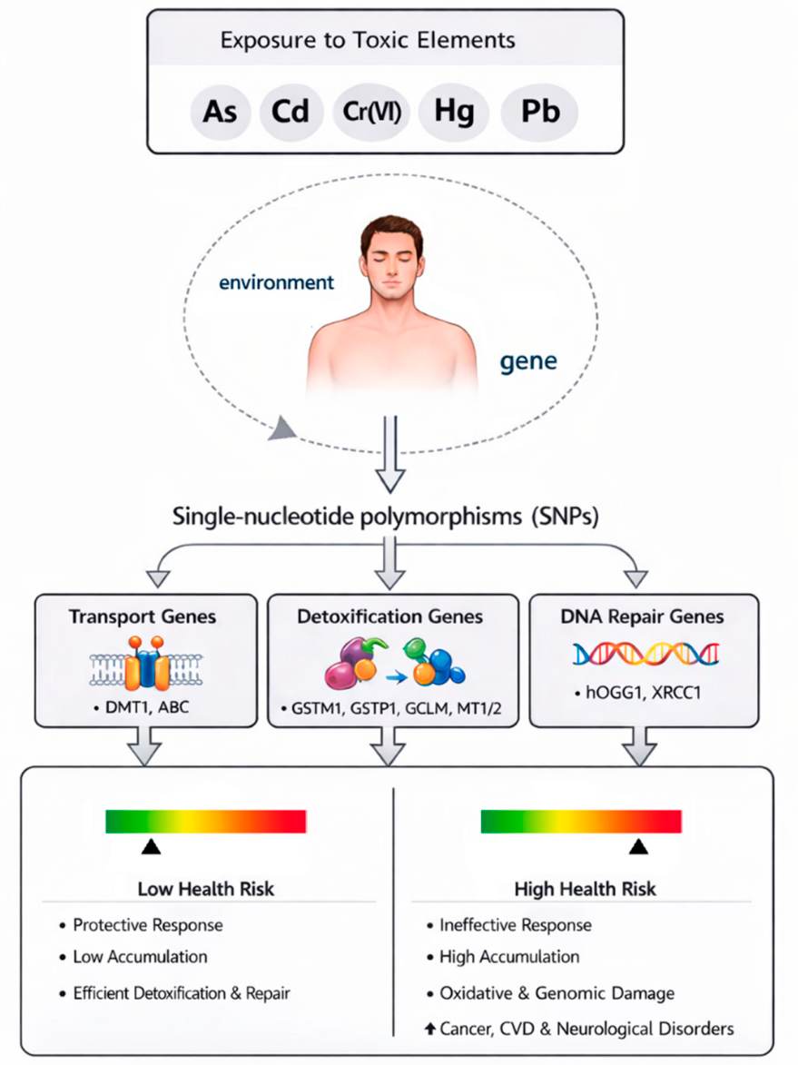
Figure 1. Gene–environment interaction in exposure to toxic elements: single-nucleotide polymorphisms can affect susceptibility to disease risk. The figure shows a schematic overview of gene–environment interactions in exposure to toxic elements, highlighting how single-nucleotide polymorphisms may modulate risk of individual susceptibility to disease. Abbreviations: As: arsenic; Cd: cadmium; Cr(VI): hexavalent chromium; Hg: mercury; Pb: lead; SNPs: single-nucleotide polymorphisms; DNA: deoxyribonucleic acid; CVD: cardiovascular disease. Image partially generated with AI Microsoft Copilot 365.
Toxics 14 00375 g001
Table 1. Best characterized single-nucleotide polymorphisms as promising markers of genetic susceptibility to TE exposure.
Toxic
Element
SNPGeneReferencePathwayEffect
Asrs11191979GSTO1[69]DetoxificationToxics 14 00375 i001Impairment in
body’s ability to methylate and detoxify As efficiently,
↑ risk of skin
lesions
rs2164624
rs4925
rs156697GSTO2
rs2297235
rs3790064PNP
rs1695
AG + GG genotypes
GSTP1[70]Toxics 14 00375 i002↓ Efficiency in methylating inorganic As, ↑ urinary percentage of inorganic AS,
↑ toxicity
+ genotypeGSTT1[72]Toxics 14 00375 i003↑ Urinary As levels and ↑ carotid intima-media thickness
rs4025935
null genotype
GSTM1[73]Toxics 14 00375 i004↓ As poisoning
rs1052133
Ser326Cys
hOGG1[87]DNA repairToxics 14 00375 i005↑ Urinary As levels, ↓ leukocyte telomere length, ↑ genomic instability
rs25487
Arg399Gln
XRCC1
rs1052133hOGG1[88]Toxics 14 00375 i006↑ Risk of urothelial carcinoma
rs159153
Cdrs7943316CAT[74]DetoxificationToxics 14 00375 i007Altered expression of antioxidant enzymes, ↑ oxidative damage
rs1695GSTP1
null genotypeGSTM1
null genotypeGSTT1
null genotypeGSTT1[79]Toxics 14 00375 i008↑ Blood Cd levels
rs8044719MT1A[81]Toxics 14 00375 i009↑ Tissue Cd retention, ↓ urinary Cd
rs1599823MT1B
rs28366003MT2A
rs10636
rs28366003
GG genotype
MT2A[82]Toxics 14 00375 i010↑ Blood Cd levels
rs1052133hOGG1[9]DNA repairToxics 14 00375 i011↓ DNA repair capacity, ↑ genotoxic effects
rs25487XRCC1
Cr(VI)rs1695GSTP1[76]DetoxificationToxics 14 00375 i012↑ Risk of lung cancer
rs1001179CAT
rs25487
Arg399Gln
XRCC1[91]DNA repairToxics 14 00375 i013↑ Chromosomal
aberrations and susceptibility to chromosomal damage
rs25487
Arg399Gln
XRCC1[92]Toxics 14 00375 i014↓ DNA damage
rs13181
Lys751Gln
XPD[93]Toxics 14 00375 i015↑ Susceptibility to lung cancer
rs2228000
Lys939Gln
XPC
rs2295152
T allele
XRCC3[94]Toxics 14 00375 i016↑ Micronuclei frequency and genetic damage
Hgrs11075290
C allele
ABCC1[68]TransportToxics 14 00375 i017↑ Cord blood Hg levels and
↑ odds of
small-for-gestational-age
rs2032582
GG genotype
ABCB1Toxics 14 00375 i018↑ Cord blood Hg levels and
↓ mental
development index
rs41303970
TT genotype
GCLM[75]DetoxificationToxics 14 00375 i019↓ Hg concentrations in blood and hair,
↑ efficient mercury elimination
null genotypeGSTM1Toxics 14 00375 i020↑ Hg accumulation
rs1555903
C allele
GCLC[77]Toxics 14 00375 i021↓ Estimated glomerular filtration rate (eGFR) in non-exposed individuals and ↓ beta-2-microglobulin in exposed individuals, impairment of renal function
rs3957356 C allele
+ rs3761144 G allele
GSTA1
+ GSS
Toxics 14 00375 i022↑ Hg retention
rs41303970 T alleleGCLMToxics 14 00375 i023↑ Urinary Hg clearance, ↑ elimination
null genotypeGSTM1[68]Toxics 14 00375 i024↑ Hg accumulation, ↑ oxidative stress, ↑ risk of preterm birth and ↓ weight in offspring
null genotypeGSTT1
rs41303970GCLM
rs1695
G allele
GSTP1Toxics 14 00375 i025↓ Mental development index in offspring
rs761142
TT genotype
GCLCToxics 14 00375 i026↓ Psychomotor development index
rs2270837
AA genotype
MT1M[84]Toxics 14 00375 i027↓ Urinary Hg levels, ↑ Hg retention
rs10636
CC genotype
MT2A
rs8052394
GA and GG genotypes
MT1AToxics 14 00375 i028↓ Hair Hg levels
rs9936741
TT genotype
MT1M
rs2270836MT1M[85]Toxics 14 00375 i029↑ Hair Hg levels
rs9936741Toxics 14 00375 i030↓ Hair Hg levels
rs25487
Arg399Gln
XRCC1[90]DNA repairToxics 14 00375 i031↑ DNA damage and oxidative stress
rs1052133
Ser326Cys
hOGG1
PbIVS4+44 C/ASLC11A2[66]TransportToxics 14 00375 i032↑ Risk of hypertension
rs224589
CA genotype
SLC11A2[67]Toxics 14 00375 i033↑ Blood Pb concentration and
↑ hematological
alterations
rs17883901GCLC[78]DetoxificationToxics 14 00375 i034↑ Antioxidant response
rs41303970GCLMToxics 14 00375 i035↓ Blood and
plasma Pb levels
null genotypeGSTT1[79]Toxics 14 00375 i036↑ Blood Pb levels
rs1695
Ile/Val genotype
GSTP1Toxics 14 00375 i037↑ Risk of Pb toxicity
rs28366003
GG genotype
MT2A[82]Toxics 14 00375 i038↑ Blood Pb levels
rs11640851MT1A[83]Toxics 14 00375 i039↓ Uric acid elimination, ↑ renal dysfunction
rs8052394
rs1052133hOGG1[89]DNA repairToxics 14 00375 i040↑ DNA instability and genotoxicity biomarkers
rs1800975XPA
rs2228000XPC
Abbreviations: ↓: decrease of; ↑: increase of; As: arsenic; Cd: cadmium; Cr(VI): hexavalent chromium; Hg: mercury; Pb: lead; SNPs: single-nucleotide polymorphisms; DNA: deoxyribonucleic acid. Circles in the “Effect” column are green when SNPs are correlated with a protective action, red with increased toxic effects.
Disclaimer/Publisher’s Note: The statements, opinions and data contained in all publications are solely those of the individual author(s) and contributor(s) and not of MDPI and/or the editor(s). MDPI and/or the editor(s) disclaim responsibility for any injury to people or property resulting from any ideas, methods, instructions or products referred to in the content.