Article

Distinct Effects of PFOS and OBS on Neurotoxicity via PMK-1 Mediated Pathway in Caenorhabditis elegans

Institute of Environmental Systems Biology, College of Environment Science and Engineering, Dalian Maritime University, Linghai Road 1, Dalian 116026, China
*
Author to whom correspondence should be addressed.
Toxics 2025, 13(8), 662; https://doi.org/10.3390/toxics13080662
Submission received: 12 July 2025 / Revised: 2 August 2025 / Accepted: 4 August 2025 / Published: 6 August 2025
(This article belongs to the Special Issue Molecular Mechanisms of PFAS-Induced Toxicity and Carcinogenicity)

Abstract

Sodium p-perfluorous nonenoxybenzenesulfonate (OBS) has been proposed as a substitute for perfluorooctanesulfonic acid (PFOS), yet it has garnered increasing attention due to its environmental persistence and potential toxicity. Despite these concerns, the neurotoxic mechanisms of OBS remain unclear. This study investigates and compares the neurotoxic effects and mechanisms of OBS and PFOS in Caenorhabditis elegans. L4-stage worms were exposed to OBS (0.1–100 μM) or PFOS (100 μM) for 24 h. Neurobehavioral analysis showed that OBS exposure induced concentration-dependent neurobehavioral deficits, with 100 μM OBS significantly reducing pharyngeal pumping rate (29.8%), head swing frequency (23.4%), and body bending frequency (46.6%), surpassing the effects of PFOS. Both compounds decreased the fluorescence intensity of dopaminergic, glutamatergic, and γ-aminobutyric acid neurons and downregulated neurotransmitter-associated genes. They also increased ROS generation and inhibited antioxidant gene expression. Molecular docking revealed that OBS had a stronger binding affinity to p38 MAPK key protein (PMK-1) than PFOS. OBS and PFOS upregulated pmk-1 and skn-1, modulating oxidative stress and neuronal function. pmk-1 mutation differentially affected OBS-induced neurobehavioral changes and gene expression alterations. Our findings indicate that OBS exhibits stronger neurotoxicity than PFOS in Caenorhabditis elegans, mediated through the PMK-1 pathway. These results highlight the need for further investigation into the safety of OBS as a PFOS alternative.

1. Introduction

Pentafluorooctanesulfonate (PFOS), belongs to perfluoroalkyl and polyfluoroalkyl substances (PFASs), which is a globally pervasive environmental pollutant [1]. PFOS is widely utilized in firefighting foams, textile waterproofing treatments, leather processing, and electronic product manufacturing [2]. PFOS exhibits remarkable resistance to biodegradation and a propensity for bioaccumulation, with a half-life of approximately 5 years in the human body [3]. PFOS has been detected in various environmental media and enters human body through contaminated food, water, air, or skin contact [4,5]. It has been shown that PFOS adversely affects multiple organ systems, including the liver, immune system, and endocrine system [6]. Moreover, its neurotoxic effects are increasingly recognized, with studies demonstrating that PFOS can induce oxidative stress and affect dopaminergic neurons, leading to behavioral abnormalities, cognitive dysfunction, and neurodegenerative diseases [7].
Sodium p-perfluorous nonenoxybenzenesulfonate (OBS) is a typical substitute for PFOS, which are primarily used in firefighting foams and oil recovery [8]. Similar with PFOS, OBS exhibits significant hydrophobicity and chemical stability. Despite its reduced bioaccumulative properties, OBS demonstrates high persistence in the environment, accumulating in water bodies, sediments, and organisms [9]. In lakes and sediments surrounding the Daqing Oilfield, OBS detection rates are 91% and 100%, with average concentrations of 409.73 ng/L and 59.87 ng/g, respectively [10]. The average concentration of OBS in the blood of wild crucian carp is 144 μg/L, while in pregnant women’s serum, it is detected at 0.711 ng/mL [11]. OBS exhibits similar acute toxicity to PFOS, causing mortality in fish and tadpoles, with LC50 (96 h) values of 25.5 mg/L and 28.4 mg/L, respectively [10]. OBS has demonstrated developmental toxicity in fish, inducing oxidative stress in the livers of juvenile and adult zebrafish, disrupting circadian rhythms in adult zebrafish [11,12]. OBS is associated with metabolic disorders in mice, increased human obesity, decreased bone density, and cell necrosis and oxidative stress in HepG2 cells [13,14,15,16]. However, potential impacts of OBS on the nervous systems remains unknown.
Caenorhabditis elegans (C. elegans) are transparent C elegans with short lifespan and ease of cultivation, making model organism for studying the neurotoxicity of environmental pollutants [17]. Nervous system of C. elegans is structurally and functionally analogous to mammals, and neuronal damage is reflected in behavioral abnormalities [18,19]. It is reported that many environmental pollutants, including metals, pesticides, and organic compounds, exhibit neurotoxicity in C. elegans, with results often paralleling those in mammalian systems [20]. The transparency of C. elegans and its ease of genetic manipulation, such as with green fluorescent protein (GFP) fusion, enable visualization of cellular morphology and protein expression patterns in vivo [21]. GFP-labelled C. elegans often were utilized to study classical neurotransmitters such as dopamine (DA), glutamate (Glu), gamma-aminobutyric acid (GABA), and acetylcholine (ACh), which linked to neurodegenerative diseases like Alzheimer’s disease (AD) and Parkinson’s disease (PD), elucidating the pathogenesis and progression of these diseases [20].
It is known that p38 MAPK (mitogen-activated protein kinase) is a member of the MAPK family, which plays a crucial role in cellular stress responses [22]. In C. elegans, the homolog of p38 MAPK is PMK-1, which is the key kinase in p38 MAPK pathway [23]. When the nematode is exposed to oxidative stress, PMK-1 phosphorylates the transcription factor SKN-1 (the homolog of Nrf2), promoting its translocation to the nucleus, which is crucial for scavenging reactive oxygen species (ROS) and maintaining cellular homeostasis [24,25]. ROS regulates the expression of antioxidant genes such as gpx-4, gst-4, sod-1, and sod-3 by activating SKN-1 [26,27]. Mutants of PMK-1, such as KU25 [pmk-1 (km25)] strain, are commonly studied variants where the function of the p38 MAPK pathway may be inhibited or altered in the nematode [28]. The pmk-1 mutant exhibits higher sensitivity to various stress conditions, such as heat stress, oxidative stress, and pathogen infection [29,30]. Therefore, the use of pmk-1 mutant can help to further understand the role of PMK-1 in cellular stress responses.
In this study, we systematically investigated the potential neurotoxic effects and underlying mechanisms of exposure to OBS in comparison to PFOS in C. elegans. First, we analyzed the effects of OBS exposure on the neurobehavioral functions of C elegans and the dose–response relationship, and compared it with PFOS to clarify its toxic effects. Then, we explored the interference of PFOS/OBS exposure on oxidative stress and the neurotransmitter system in C elegans. We utilized three GFP-tagged transgenic strains of C. elegans to evaluate neuronal damage. Finally, we elucidated the key role of the p38 MAPK pathway in PFOS/OBS neurotoxicity, by comparing the behavioral, oxidative stress, and neurotransmitter changes in N2 wild-type and pmk-1 mutant C elegans after PFOS/OBS exposure. This study provides an important basis for the scientific assessment of the neurotoxic risks of OBS as a substitute for PFOS.

2. Materials and Methods

2.1. C. elegans Strains and Culture

The following strains of C. elegans were obtained from Caenorhabditis Genetics Center, including N2 (wild-type), BZ555 (dat-1p::GFP, dopaminergic neuron marker), DA1240 (eat-4::GFP), glutamatergic neuron marker), EG1285 (unc-47p::GFP, GABAergic neuron marker), KN259 (sod-3p::GFP, oxidative stress reporter), and KU25 [pmk-1 (km25), p38 MAPK mutant]. Synchronized eggs were obtained by lysing gravid adults with sodium hypochlorite solution (5 M NaOH, NaClO) and then cultured on nematode growth medium (NGM) seeded with Escherichia coli (E. coli) OP50 as a food source. The worms were maintained in a sterile incubator at 20 °C in the dark for 48 h until they reached the L4 larval stage. The NGM culture medium was prepared with 3 g/L NaCl, 2.5 g/L peptone, 17 g/L agar, 1 mM CaCl2, 1 mM MgSO4, 25 mM potassium phosphate buffer (at a pH of 6.0) and 5 μg/mL cholesterol. M9 buffer consisted of 3 g/L K2HPO4, 6 g/L Na2HPO4, 5 g/LNaCl, and 0.5 mM MgSO4. The above various reagents were supplied by Sangon Biotech (Shanghai, China).

2.2. Exposure to PFOS and OBS

PFOS (CAS: 1763-23-1) and OBS (CAS: 70829-87-7) were purchased from Tokyo Chemical Industry Co., Ltd. (Tokyo, Japan) and Shanghai Futian Chemical Technology Co. (Shanghai, China), respectively. The average environmental concentration of OBS is 0.28 µM, with a concentration range of 2.11 × 10⁻⁴ M–6.13 µM [31]. Therefore, a concentration range of 0.1–10 µM OBS was selected to cover environmentally relevant concentrations to higher doses. Meanwhile, PFOS was used as a positive control because exposure to 100 µM PFOS can cause damage to the dopaminergic neurons of C elegans and alter the level of ROS accumulation [32], supporting its use as a positive control for neurotoxicity in our study.
C. elegans were exposed to different concentrations of OBS (0.1, 1, 10, and 100 μM) or PFOS (100 μM, positive control) for 24 h. 0.1% dimethyl sulfoxide (DMSO) was used as vehicle control. In 60 mm solid culture plates, 200 μL of exposure solution at the respective concentrations was added along with synchronized L4-stage C. elegans. The plates were then placed in a biochemical incubator (Lichen, Shaoxing, Zhejiang, China) for 1 day, with three biological replicates per group.

2.3. Neurobehavioral Activity Measurement

After exposure, the C elegans were washed with M9 buffer and transferred to NGM solid plates without food. They were allowed to recover for 1 min before being observed under a stereomicroscope (Mshot, Guangzhou, China). The number of head swings within 30 s was recorded, where one complete swing from left to right and back to left was counted as one event.
Subsequently, the exposed C elegans were transferred to NGM plates supplemented with E. coli OP50, and the number of pharyngeal pumping events within 20 s was recorded. Each upward and downward movement of the pharyngeal pump was counted as one event.
After wash with M9 buffer, the C elegans were placed on NGM solid plates without food for a 1 min recovery period. The number of body bends within 20 s was observed under a stereomicroscope. A body bend was defined as the movement of the nematode in a sinusoidal wave along its longitudinal axis. All experiments included three independent biological replicates, with 15 worms (n = 15) per group in each replicate.

2.4. Neuronal Damage Observation

The GFP-labelled strains BZ555, DA1240, and EG1285 were used to assess neuronal damage. Following exposure, these worms were slowly washed with M9 buffer and collected into 1.5 mL tubes. After allowing them to settle, the supernatant was discarded, and the washing procedure was repeated three times. Subsequently, 5 mM levamisole solution (Aladdin, Shanghai, China) was added to anesthetize the C elegans, and once they were immobilized, they were transferred to glass slides, and imaged under a fluorescence microscope (Nikon, Tokyo, Japan). The average fluorescence intensity of green fluorescent protein expression in the neurons of each strain was measured using ImageJ 2.2.0 software. All experiments included three independent biological replicates, with 20 worms (n = 20) per group in each replicate.

2.5. Reverse Transcription Quantitative PCR (RT-qPCR)

Total RNA was extracted using RNA Isolater reagent (Vazyme Biotech, Nanjing, China). RT-qPCR was performed using SupRealQ Ultra Hunter SYBR qPCR Master Mix (Vazyme Biotech) according to the manufacturer’s protocol. The tba-1 gene was used as a reference, and the relative expression levels were analyzed using the comparative 2−ΔΔCT method. Primer sequences are provided in the Table S1. All experiments included three independent biological replicates, with 15 worms (n = 15) per group in each replicate.

2.6. Molecular Docking

The 2D structure of the small molecule ligand (OBS/PFOS) was retrieved from PubChem, converted to 3D using ChemOffice 20.0 with energy minimization (MMFF94), and saved as a.mol2 file, while the PMK1 target sequence (UniProtKB: Q17446) was modeled via AlphaFold and saved as a.pdb file. Using Molecular Operating Environment 2019 software, the protein was prepared (heteroatom removal, protonation at pH 7.4, AMBER10:EHT minimization) and active pockets were predicted, followed by molecular docking (50 runs, London dG scoring) with binding energy thresholds: ≥−4.25 (weak), <−5.0 (moderate), and <−7.0 kcal/mol (strong). Results were visualized in PyMOL 2.6.0 and Discovery Studio 2019, analyzing interaction patterns (H-bonds, hydrophobic contacts) and pose stability (RMSD clustering).

2.7. Correlation Analysis

The Mantel test is used to analyze the global correlation between gene expression profiles and phenotype feature matrices. It quantitatively evaluates the strength of gene-phenotype association by calculating the Pearson correlation coefficient (with a value range of −1 to 1) between the neuron gene expression matrix and the oxidative stress gene expression matrix. All calculations are implemented through the Vegan package (v2.6.10) in R language (R version 4.4.2).

2.8. Statistical Analysis

Data are presented as means ± SEM. Statistical analysis was performed using one-way ANOVA in GraphPad Prism9.5.0 (GraphPad Software, San Diego, CA, USA). Significant differences were determined by comparing the results of the experimental groups to those of the control group (* p < 0.05, ** p < 0.01 vs. DMSO control; # p < 0.05, ## p < 0.01 vs. PFOS).

3. Results

3.1. Distinct Effects of PFOS/OBS Exposure on Neurobehavioral Activity in C. elegans

We investigated the effects of OBS exposure on neurobehavioral activity, including pharyngeal pumping rate, frequency of head swings and body bends in C. elegans. The results showed that the pharyngeal pumping rates were significantly reduced in 1–100 μM OBS groups compared to the DMSO control group (Figure 1A). OBS exposure decreased frequency of head swings and body bends in a concentration-dependent manner (Figure 1B,C). 100 μM OBS significantly reduced pharyngeal pumping rate (29.8%) and head swing frequency (23.4%), surpassing the effects of PFOS (Figure 1D,E). Both OBS and PFOS led to a significant decrease in the frequency of body bends, with OBS causing a reduction of about 50%, comparable to the decrease seen with PFOS (Figure 1F).

3.2. Distinct Effects of PFOS/OBS Exposure on Specific Neurotransmitter Systems in C. elegans

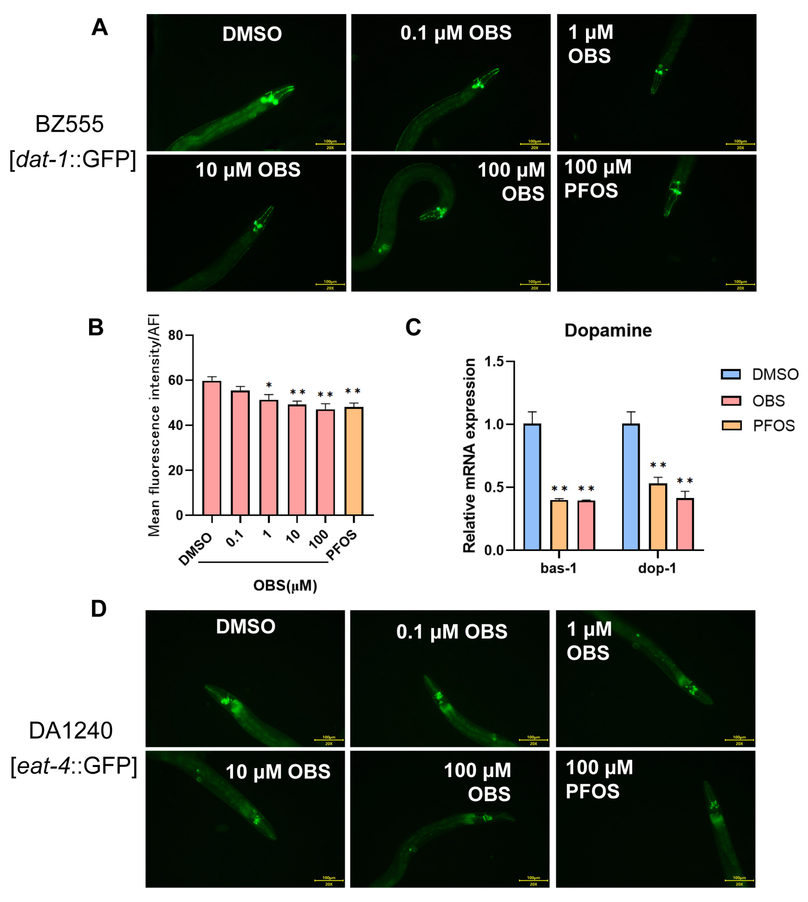
We utilized three transgenic strains (BZ555, DA1240, and EG1285) to specifically target and visualize different types of neurons (dopaminergic, glutamatergic, and GABAergic, respectively) because these neurons play crucial roles in neurobehavioral changes and neurotoxicity. We observed GFP-labelled fluorescence of neurons (Figure 2A,D,G) and measured the relative fluorescence intensity in transgenic worms, by which damage to these neurons can be quantified. The results showed that exposure to OBS (1, 10, 100 μM) significantly reduced the fluorescence intensity of dopaminergic neurons, similar to PFOS (Figure 2B). The genes bas-1 and dop-1 play critical roles in the synthesis and reception of dopamine, ensuring the proper functioning of dopaminergic neurons and the overall dopaminergic neurotransmission in the nervous system. We note that 100 μM OBS significantly caused approximately a 50% decrease in relative mRNA levels of bas-1 and dop-1, similar with the effects of PFOS (Figure 2C).
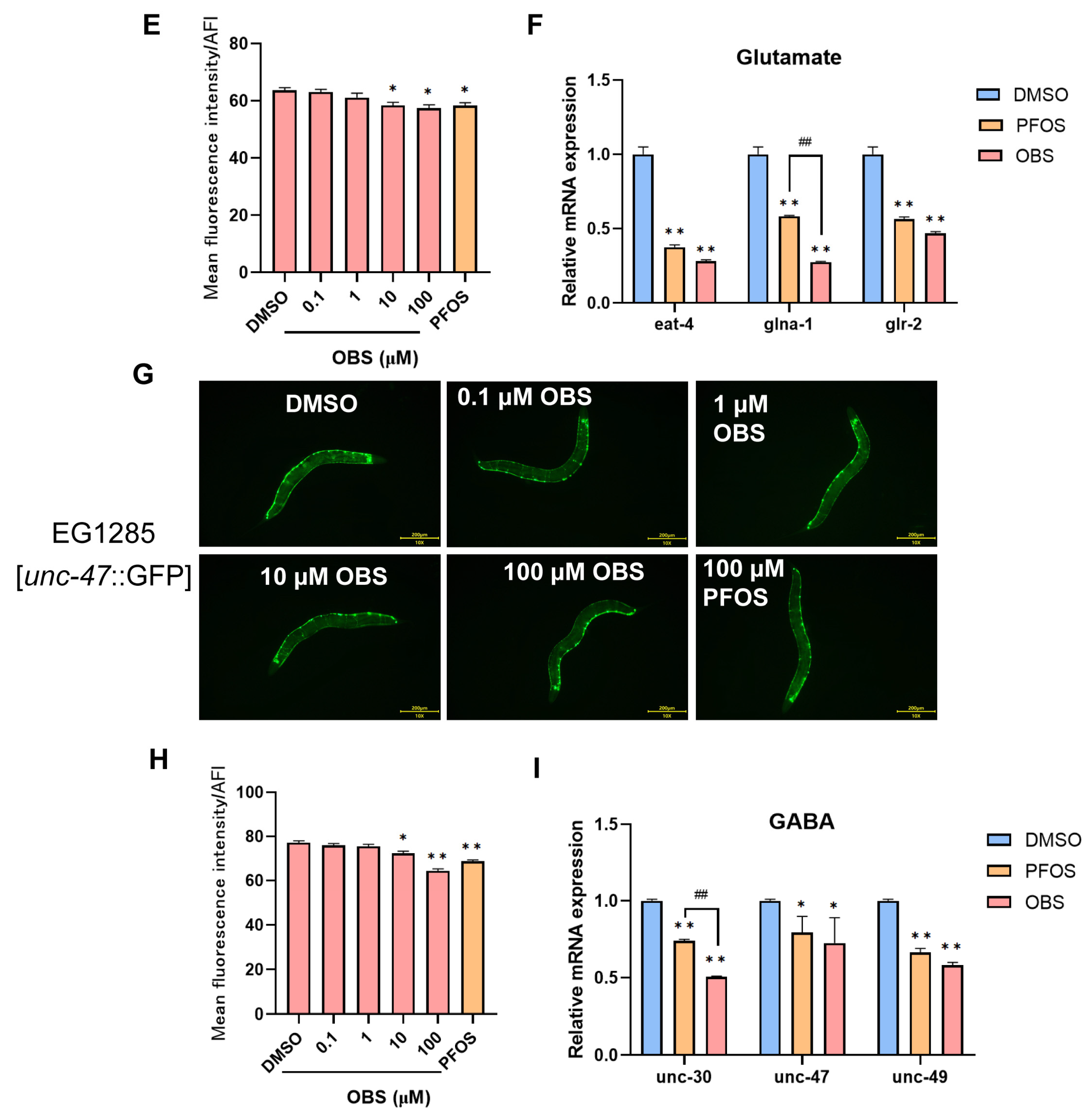
Compared to the control group, exposure to OBS concentrations of 10–100 μM significantly decreased the fluorescence intensities of glutamatergic and GABAergic neurons (Figure 2E,H). The genes eat-4, glna-1, and glr-2 are all related to the function and regulation of glutamatergic neurons in C. elegans. RT-qPCR confirmed the decrease in mRNA levels of eat-4, glna-1 and glr-2 in both PFOS and OBS exposed worms. Notably, glna-1 was significantly down-regulated in OBS group compared to PFOS group (Figure 2F). The genes unc-30, unc-47, and unc-49 play critical roles in the synthesis, transport, and reception of GABA, ensuring the proper functioning of GABAergic neurons and the overall inhibitory neurotransmission in the nervous system. We note that unc-30 was expressed significantly lower in OBS group than PFOS group. Other two genes unc-47 and unc-49 were down-regulated in both OBS and PFOS groups (Figure 2I). These results indicate that OBS has damaging effects on neuronal expression, leading to abnormal levels of neurotransmission-related genes in C. elegans, similar with PFOS. In addition, the impacts of OBS on the expression of certain genes such as glna-1 and unc-30 are more pronounced than that of PFOS.

3.3. Distinct Effects of PFOS/OBS Exposure on Oxidative Stress in C. elegans

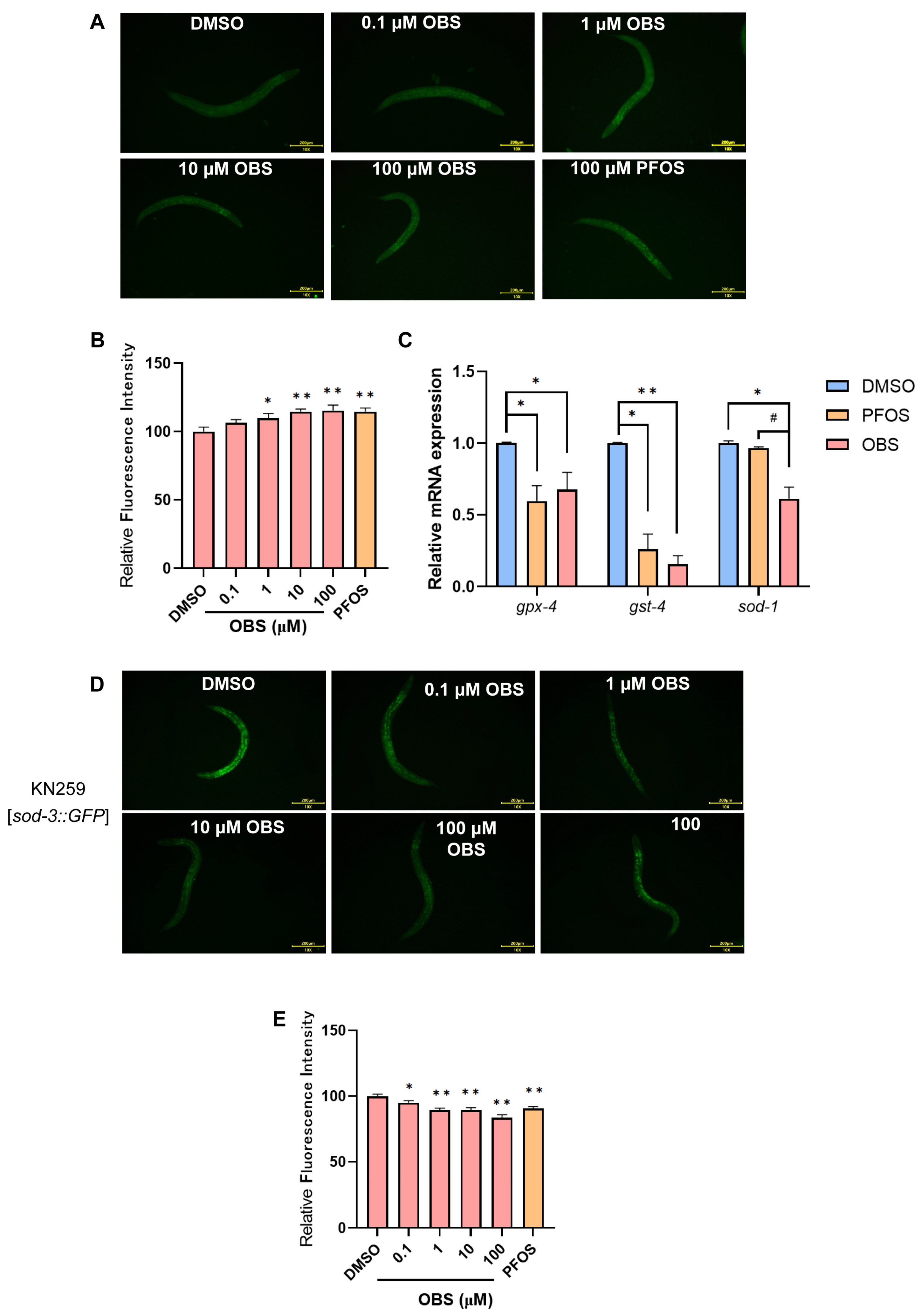
Oxidative stress induces the overproduction of ROS in C. elegans, overwhelming its antioxidant defenses, thereby causing oxidative damage, neurotoxicity, and impaired locomotor behaviors. To investigate the effects of OBS exposure on ROS levels in C elegans, we employed the H2DCFDA fluorescent probe (Nanjing Jiancheng Bioengineering Institute, Nanjing, China) to stain the C elegans. Compared with the DMSO group, the average fluorescence intensity significantly increased in a dose-dependent manner with the elevation of PFOS and OBS exposure concentrations (Figure 3A,B). A total of 100 μM PFOS and OBS remarkably decreased the expression of antioxidants, including gpx-4, gst-4, and sod-1 in C. elegans. Moreover, the reduction in sod-1 expression was more significant in C. elegans exposed to 100 μM OBS than in those exposed to PFOS (Figure 3C).
The KN259 (sod-3p::GFP) strain is a genetically engineered C. elegans strain used as a reporter for oxidative stress, providing a visual and quantitative means to assess the cellular response to oxidative stress. As shown in Figure 3D,E, the fluorescence intensities were significantly reduced in 0.1–100 μM OBS groups and the 100 μM PFOS group compared to the control group.

3.4. Distinct Effects of PFOS/OBS on Oxidative Stress-Neurotransmitter Regulatory Network

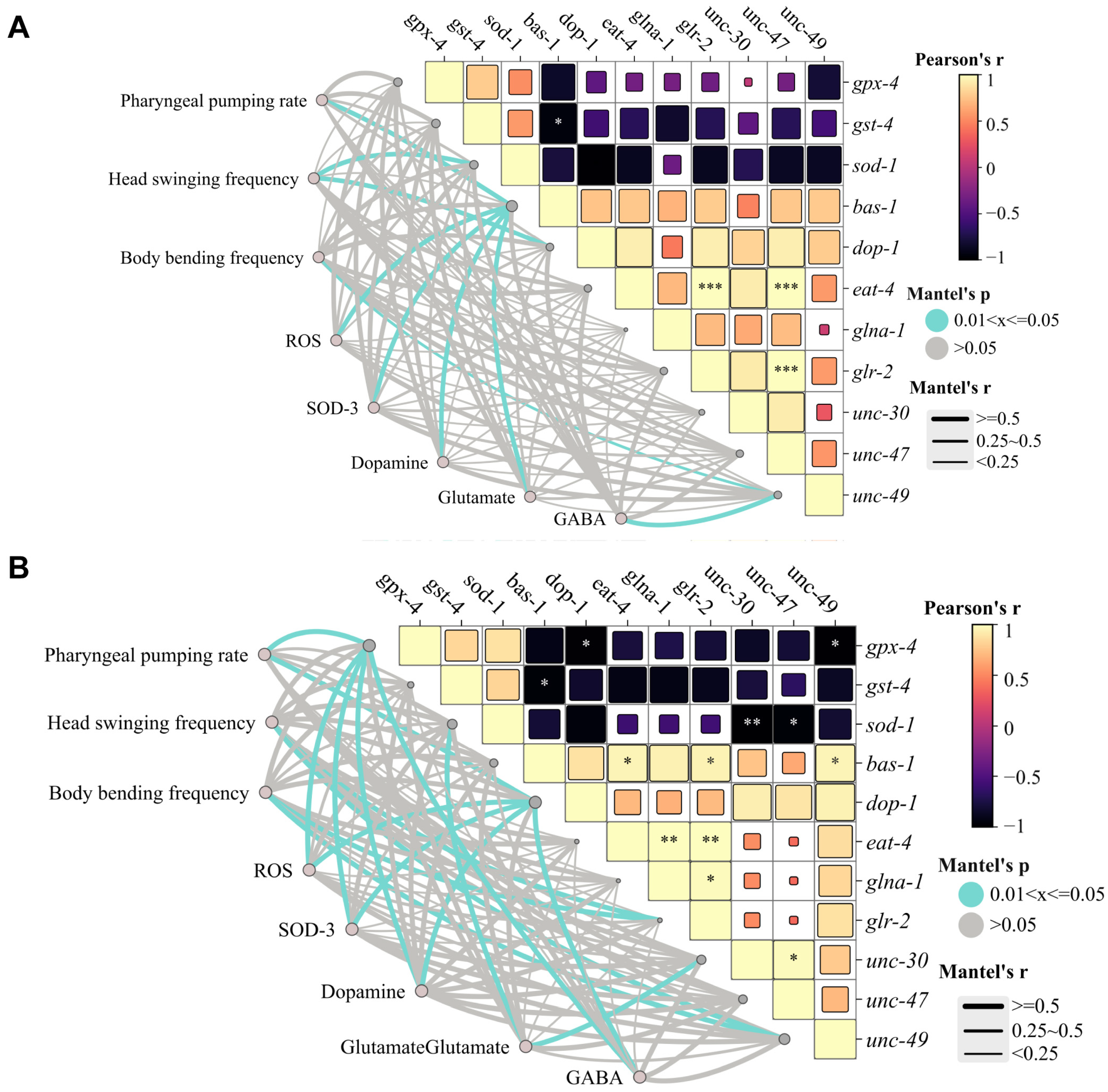
We conducted an in-depth correlation network analysis. In terms of PFOS exposure, pharyngeal pumping rate is significantly positively correlated with the activity of dopaminergic neurons and ROS levels (p ≤ 0.05), suggesting that dysfunction of the dopaminergic system and oxidative stress jointly mediate the inhibition of feeding behavior. Motor behavior indices (frequency of head swinging and body bending) show moderate correlation with glutamate and GABAergic neurons (0.25 ≤ r < 0.5, p ≤ 0.05), confirming that motor coordination is regulated by the dynamic balance of excitatory/inhibitory neurotransmitters. Moreover, the expression of SOD-3 protein and gpx-4 shows a synergistic downregulation trend, pointing to the collapse of the antioxidant defense system as the core mechanism of neurobehavioral damage. The downregulation of bas-1 and dop-1 in the dopaminergic system is consistent with the decreased fluorescence of dopaminergic neurons (0.25 ≤ r < 0.5) and is significantly correlated with the inhibition of pharyngeal pumping rate (p ≤ 0.05), confirming that impaired dopamine synthesis and signaling lead to feeding disorders. In the glutamatergic system, the suppression of glr-2 expression is correlated with the decreased frequency of head swinging and body bending (0.25 ≤ r < 0.5, p ≤ 0.05). The inhibition of antioxidant genes (sod-1, gpx-4, gst-4) forms a positive feedback loop with ROS accumulation (p ≤ 0.05), further exacerbating the damage to dopaminergic and glutamatergic neurons (Figure 4A).
Compared with PFOS, OBS exhibits more significant targeting of neurotransmitter systems. The integrated analysis indicates that OBS induces neurotoxicity through the following pathways: inhibiting the SOD-3 and glutathione system (gpx-4, sod-1), leading to ROS accumulation and oxidative stress; specifically targeting neuronal dopaminergic (dop-1), GABAergic (unc-30, unc-49), and glutamatergic systems (glr-2), resulting in neurotransmitter disorders and ultimately manifesting as abnormal neurobehavioral functions, with differences in toxic mechanisms compared to PFOS (Figure 4B).

3.5. Binding Potential of PFOS and OBS with PMK-1 Protein

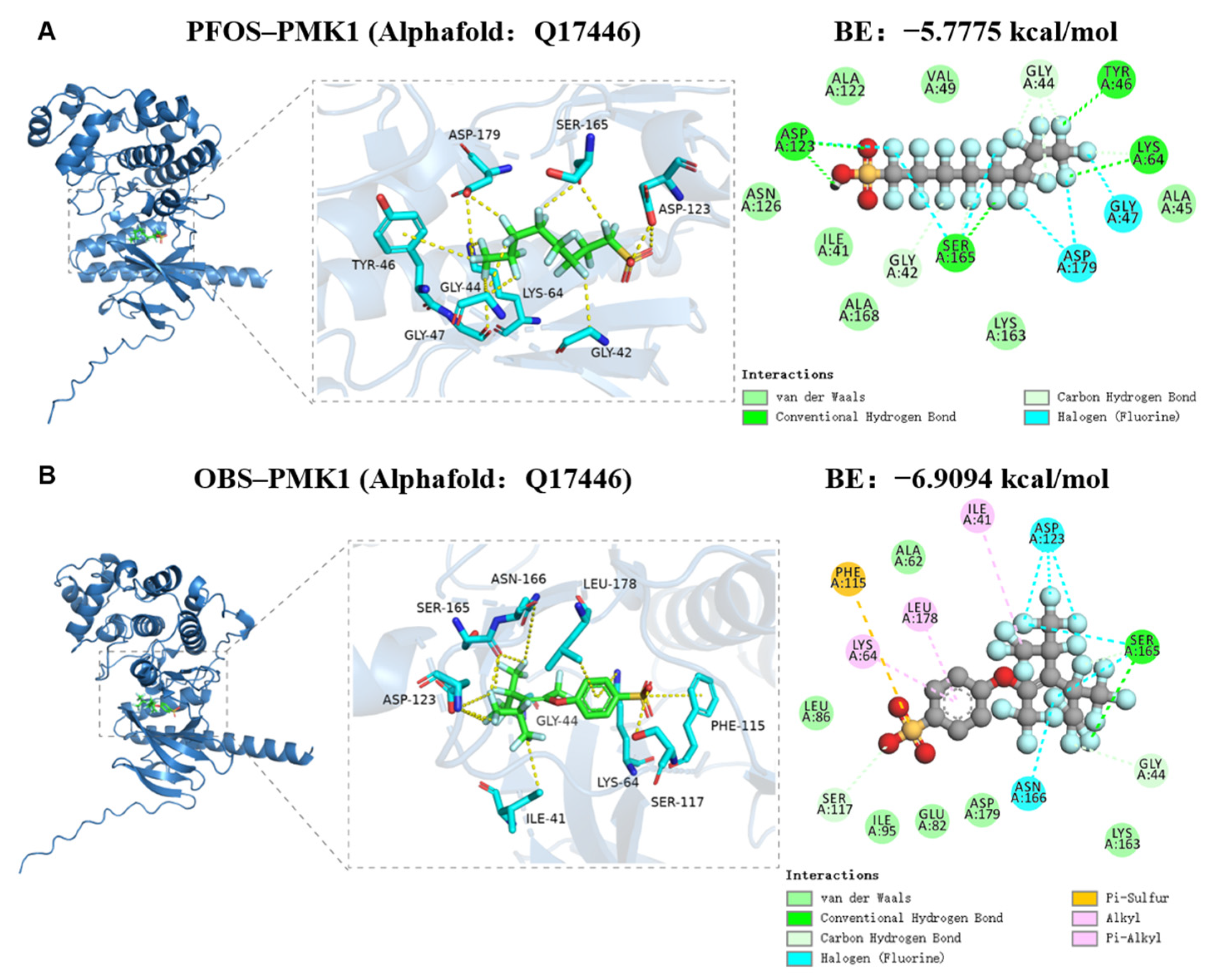
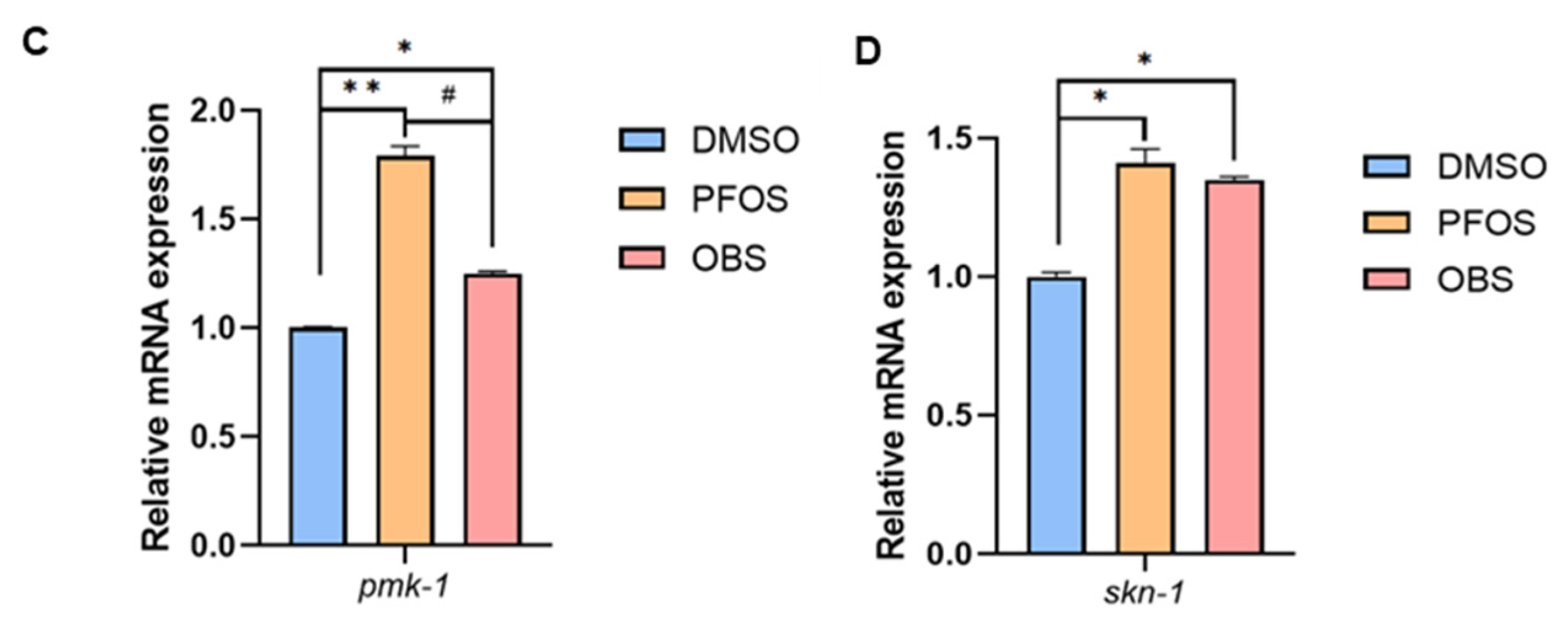
We employed molecular docking to further analyze the interaction between PFOS/OBS and the oxidative stress-related protein PMK-1. PFOS forms hydrogen bonds with residues Asp123, Ser165, Tyr46, and Lys64 on the PMK-1 receptor, and hydrophobic interactions with residues Gly44 and Gly42. Additionally, residues Asp179, Gly47, and Ser165 form halogen bonds with PFOS (Figure 5A). On the PMK-1 receptor, Ser165 interacts via hydrogen bonding with OBS, while residues Ile41, Leu178, and Lys64 interact hydrophobically with OBS. Furthermore, Phe115 on the protein interacts with OBS through a Pi-Sulfur interaction, and residues Ser117 and Gly44 form hydrophobic interactions with this compound. Residues Asn166, Asp123, and Ser165 also form halogen bonds with OBS (Figure 5B). OBS exhibits a higher binding affinity (−6.9094 kcal/mol) compared to PFOS (−5.7775 kcal/mol). Further, we found that PFOS/OBS significantly resulted in the increase in pmk-1 and skn-1 expression in N2 worms (Figure 5C,D). These findings suggest that both OBS and PFOS might modulate the antioxidant defense mechanisms in C elegans by influencing the p38 MAPK signaling pathway.

3.6. Distinct Effects of PFOS/OBS Exposure on Neurotoxicity in pmk-1 Mutant

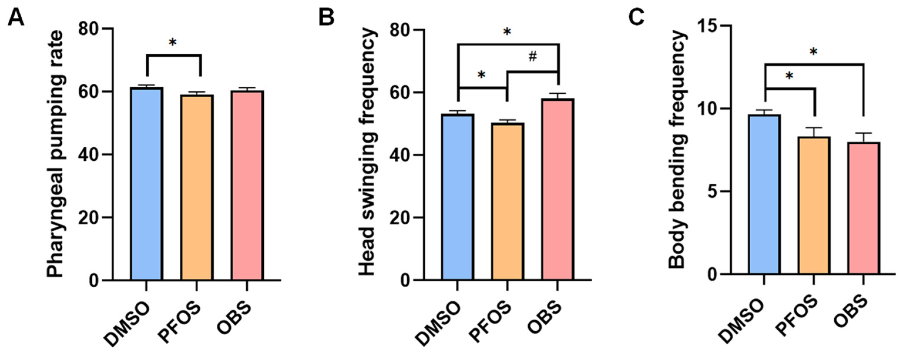
KU25 [pmk-1 (km25)] is pmk-1 mutant of C. elegans with loss of function on PMK-1 protein, thereby often used for interfering the activity of the p38 MAPK signaling pathway. We compared the effects of 100 μM PFOS/OBS exposure on neurobehavioral activity, neurotransmission and oxidative stress in KU25 worms. The results showed that OBS had no significant decrease in the pharyngeal pumping rate compared to the control, but PFOS still reduced pharyngeal pumping rate (Figure 6A). PFOS significantly decreased frequency of head thrashes, whereas OBS significantly increased frequency of head thrashes in KU25 worms (Figure 6B). PFOS/OBS significantly induced the reduction in body bending frequency in KU25 worms (Figure 6C). These results showed that pmk-1 mutation affected the alterations of pharyngeal pumping rate and frequency of head thrashes induced by OBS, but not PFOS.
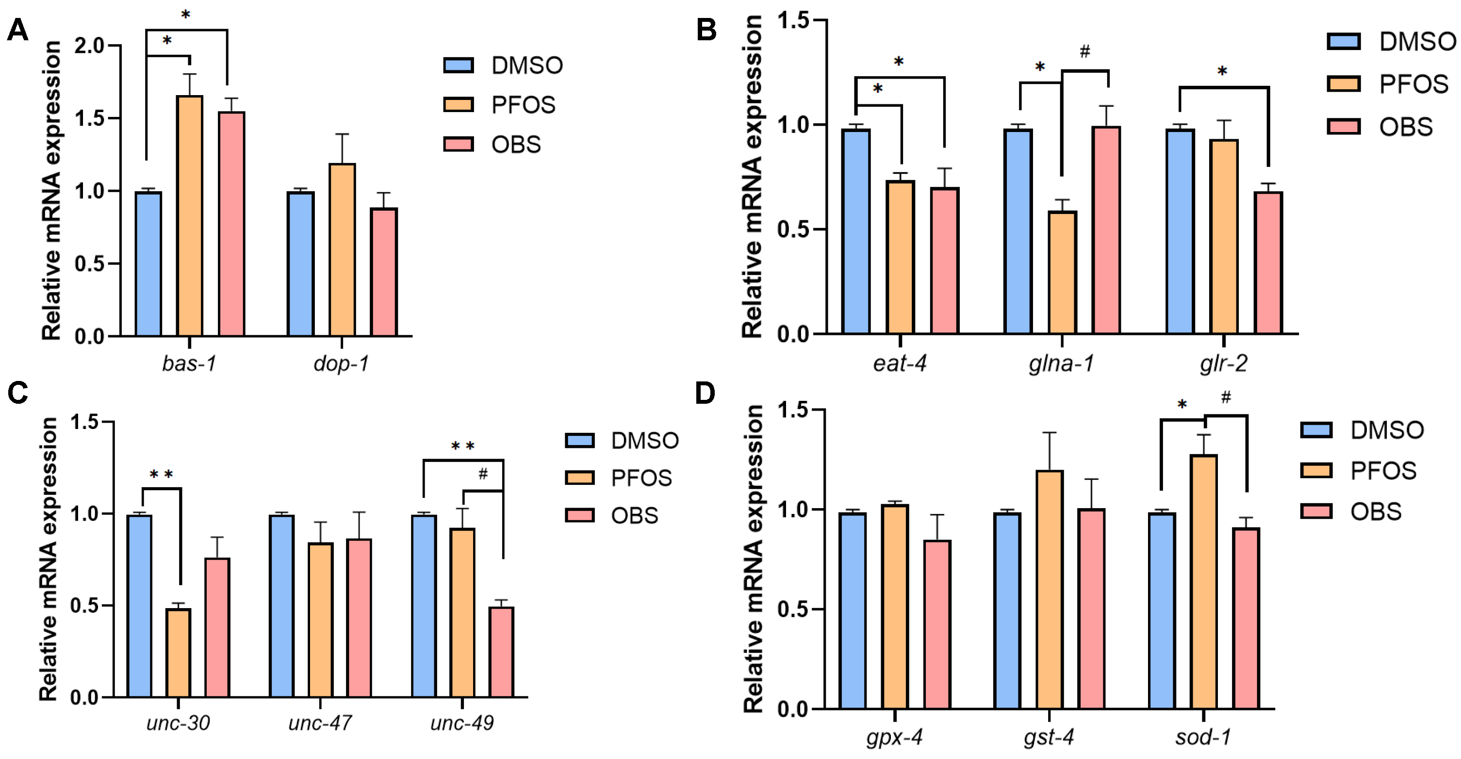
PFOS/OBS elevated bas-1 expression, but did not affect dop-1 expression in KU25 worms (Figure 7A), which were completely different from the decrease in dopamine neuron related genes in N2 worms. As for glutamate neuron related genes, OBS had no significant effects on glna-1 gene expression while OBS still resulted in the decrease in eat-4 and glr-2 in KU25 worms (Figure 7B). Among GABA neuron related genes, unc-30 and unc-47 did not show obvious changes but unc-49 was down-regulated by OBS in KU25 worms (Figure 7C). Similarly, OBS had no significant effects on ROS levels and antioxidant gene expression in KU25 worms compared to the control, although PFOS increased sod-1 expression level (Figure 7D). These results showed that pmk-1 mutation completely affected the alterations of dopamine neuron related genes and antioxidant genes when exposure to PFOS/OBS, while glutamate and GABA neuron-related genes showed different alterations in KU25 worms.

4. Discussion

PFOS is a widely used per-fluorinated compound known for its environmental persistence and neurotoxic effects. As a result, there has been a significant effort to develop and assess alternatives to PFOS such as OBS. The present study at the first time demonstrates that OBS significantly decreased pharyngeal pumping rate, head thrashes frequency and body bends frequency in C. elegans, indicating detrimental effects of OBS on neurobehavioral activity. OBS caused damage to neurons including dopaminergic, glutamatergic and GABAergic, and disrupted specific neurotransmitter systems. Additionally, OBS induced oxidative stress, modulating the antioxidant defense mechanisms in C elegans by influencing the p38 MAPK signaling pathway. OBS appears to have a more potent disruptive effect on neurobehaviors and key gene expression than PFOS, especially in pmk-1 mutant worms. These effects may account for the greater neurotoxicity exhibited by OBS in contrast to PFOS. These findings suggest the pivotal role of the p38 MAPK pathway in OBS neurotoxicity and highlight the importance of thoroughly assessing the safety of OBS as a substitute for PFOS.
The pharynx of C. elegans is a glutamatergic neuron-regulated neuromuscular pump that connects the oral cavity to the intestine and, thus, serves as a sensitive indicator of neurotoxicity [33]. Our results showed that both OBS and PFOS significantly inhibited the pharyngeal pumping rate in worms, with OBS exhibiting stronger inhibition. OBS and PFOS may disrupt glutamatergic synaptic transmission, thereby impairing pharyngeal neuromuscular control. Additionally, exposure to OBS and PFOS led to abnormal movement behaviors, evidenced by a decrease in frequency of head thrashing and body bending, consistent with established PFAS-induced motor neuron dysfunction. Cross-species validation confirms these effects: 32 µg/L OBS induces depression-like behavior, social deficits, and memory impairment in zebrafish [34], while 20 µM PFOS inhibits larval swimming [35]; similarly, developing zebrafish exposed to PFOS and OBS exhibited impaired motor behaviors [36]. In rodents, PFOS (10.75 mg/kg, 3 months) impairs spatial memory [36] and lactational exposure disrupts offspring motor coordination [37]. In C. elegans, 20 µM PFOS reduces forward movement, body bends, and head thrashing [38]. These findings demonstrate that OBS and PFOS elicit cross-species neurobehavioral toxicity by interfering with glutamatergic signaling, with OBS exhibiting more potent disruption of neuromuscular control.
The present study systematically assessed the neurotoxic effects and underlying mechanisms of OBS using various strains of C. elegans. BZ555 (dat-1p::GFP, dopaminergic neuron marker) strain expresses GFP under the control of the dat-1 promoter, which is specific to dopaminergic neurons [39]. Dopaminergic neurons are crucial for various behaviors and are often implicated in neurodegenerative diseases such as Parkinson’s disease [40]. DA1240 (eat-4::sGFP, glutamatergic neuron marker) strain expresses super GFP (sGFP) under the control of the eat-4 promoter, which is specific to glutamatergic neurons [41]. Glutamatergic neurons are involved in excitatory neurotransmission and play a key role in various cognitive functions [42]. EG1285 (unc-47p::GFP, GABAergic neuron marker) strain expresses GFP under the control of the unc-47 promoter, which is specific to GABAergic neurons [43]. GABAergic neurons are involved in inhibitory neurotransmission and are essential for maintaining balance in neural activity [44]. In terms of neuronal development, OBS (10–100 μM) and PFOS (100 μM) markedly reduced fluorescence intensity in dopamine, glutamatergic, and GABAergic neurons, suggesting induction of neurodegenerative changes. RT-qPCR analysis further confirmed widespread downregulation of dopamine (bas-1, dop-1), glutamate (eat-4, glna-1, glr-2), and GABA (unc-30, unc-47, unc-49)-related genes with exposure to OBS and PFOS, with OBS showing more pronounced inhibitory effects. It is reported that exposure to PFOS and OBS reduced numbers of subcutaneous dopaminergic neurons in developing zebrafish [37] and 32 µg/L OBS disrupted GABA and glutamate homeostasis [38]. These findings are consistent with our results, where both PFOS and OBS caused neuronal damage, reduced neurotransmitter levels, and consequently led to behavioral impairments.
Oxidative stress, characterized by excessive ROS accumulation, disrupts antioxidant defenses in C. elegans, leading to oxidative damage and neurotoxicity. The two antioxidant enzymes, SOD and GST, are critical for ROS neutralization. Our findings demonstrate that both OBS and PFOS significantly inhibit the expression of antioxidant genes (sod-3 and gst-4), impairing ROS clearance and exacerbating neuronal damage. This aligns with prior evidence: PFOS elevates lipid peroxidation and SOD activity while suppressing glutathione peroxidase activity [45], and 32 µg/L OBS induces mitochondrial dysfunction and ROS overproduction in zebrafish [46]. The resulting oxidative imbalance directly contributes to locomotor deficits, highlighting ROS-mediated neurotoxicity as a shared mechanism of PFAS-induced behavioral impairment across species.
Functional loss-of-function assays targeting the p38 MAPK pathway (via pmk-1 mutation) revealed its essential role in mediating OBS- and PFOS-induced neurotoxicity in C. elegans. Molecular docking demonstrated that both compounds directly interact with critical PMK-1 residues (e.g., Asp123, Ser165), impairing kinase activity and disrupting downstream stress responses and neuroprotective mechanisms. In pmk-1 mutant worms, the neurotoxic effects of OBS and PFOS were attenuated differently and markedly, confirming the regulatory role of p38 MAPK pathway. Consistently, RT-qPCR analysis showed that pmk-1 mutation abolished transcriptional responses in antioxidant (sod-3, gst-4) and neurotransmitter-related genes, confirming pmk-1 as a pivotal molecular target for neurotoxicity induced by PFOS and OBS in C. elegans. pmk-1 mutation completely affected the alterations of dopaminergic genes and antioxidant genes, while glutamatergic, and γ-aminobutyric acid related genes showed different alterations when exposure to PFOS/OBS. These findings suggest that the distinct mechanisms in neurotransmitter imbalance of PFOS/OBS via p38 MAPK pathway.
Approximately 60–80% of human disease genes have orthologs in C. elegans, enabling direct translation to disease mechanism research and robust cross-species mechanistic extrapolation [24]. Critically, mutation of pmk-1 (the ortholog of mammalian p38 MAPK) significantly alleviated the neurotoxic effects of OBS and PFOS in C. elegans, underscoring the conserved role of pmk-1 mediated pathway in regulating oxidative stress and neuroinflammation that implicated in human neurodegenerative disorders such as Alzheimer’s and Parkinson’s diseases [47]. Evolutionary conservation extends to the antioxidant enzymes and neurotransmitter systems, and various human homologs are implicated in neurological disorders (Table S2) [48,49,50,51,52,53,54,55,56,57,58,59]. For example, the dopamine-synthetic bas-1 (human DDC ortholog) links to Parkinson’s pathology [45]. The unc-47 gene, homologous to human GAD1, encodes a key enzyme for GABA synthesis, and its dysregulation is associated with neurodegenerative diseases, epilepsy and autism [34]. Notably, downregulation of eat-4 (vesicular glutamate transporter) may directly reduce glutamate release and impair neuromuscular control of pharyngeal pumping [46], whereas decreased unc-30 expression may disrupt GABAergic inhibition, potentially contributing to motor behavioral abnormalities [48]. These findings highlight C. elegans as a powerful translational model for dissecting PFAS neurotoxicity mechanisms, paving the way for targeted therapeutic interventions in humans.

5. Conclusions

This study systematically evaluates the neurotoxic mechanisms of PFOS and its alternative OBS in C. elegans, demonstrating that both compounds induce locomotor dysfunction, neuronal structural damage, and disruption of dopaminergic, glutamatergic, and GABAergic pathways. Loss-of-function analysis and molecular docking experiment confirm that the p38 MAPK pathway plays a critical role in mediating oxidative stress responses to PFOS/OBS, with OBS showing stronger binding affinity to PMK-1 than PFOS, suggesting a higher neurotoxic potential. By integrating oxidative stress, neurotransmitter dysregulation, and p38 MAPK pathway activation into a cohesive mechanistic framework, our findings highlight the necessity of cautious evaluation and regulation of PFOS alternatives, as their neurotoxic risks may be comparable to or even exceed those of PFOS, thereby emphasizing the importance of thorough safety assessments to protect environmental and public health.

Supplementary Materials

The following supporting information can be downloaded at: https://www.mdpi.com/article/10.3390/toxics13080662/s1, Table S1: RT-qPCR primer sequence. Table S2: Comparison of genetic homology between C. elegans and Homo sapiens.

Author Contributions

Conceptualization, D.X. and J.J.; methodology, J.J.; software, J.J.; validation, J.J. and Q.L.; formal analysis, B.Z.; investigation, J.J. and Q.L.; resources, L.Z.; data curation, B.Z.; writing—original draft preparation, J.J. and Q.L.; writing—review and editing, D.X.; supervision, D.X.; project administration, D.X.; funding acquisition, D.X. All authors have read and agreed to the published version of the manuscript.

Funding

This work was supported by the Fundamental Research Funds for the Central Universities (3132025171) and National Space Science Center (SCP03-01-02) of Chinese Academy of Sciences.

Institutional Review Board Statement

Our studies involving C. elegans do not require ethics approval, and therefore excluded from standard animal-ethics oversight.

Informed Consent Statement

Not applicable.

Data Availability Statement

Data will be made available on request.

Conflicts of Interest

The authors declare no conflicts of interest. The author asserts that they lack any apparent conflicting economic interests or personal affiliations that might potentially influence the work reported in this article.

Abbreviations

The following abbreviations are used in this manuscript:
OBSSodium p-perfluorous nonenoxybenzenesulfonate
PFOSPerfluorooctanesulfonic acid
PFASsPerfluoroalkyl and polyfluoroalkyl substances
C. elegansCaenorhabditis elegans
GFPGreen fluorescent protein
DADopamine
GluGlutamate
GABAGamma-aminobutyric acid
AchAcetylcholine
ADAlzheimer’s disease
PDParkinson’s disease
MAPKMitogen-activated protein kinase
ROSReactive oxygen species
E. coliEscherichia coli
NGMNematode growth medium
DMSODimethyl sulfoxide
SODSuperoxide dismutase
GSTGlutathione S-transferase

References

  1. Wang, S.; Yang, Q.; Chen, F.; Sun, J.; Luo, K.; Yao, F.; Wang, X.; Wang, D.; Li, X.; Zeng, G. Photocatalytic Degradation of Perfluorooctanoic Acid and Perfluorooctane Sulfonate in Water: A Critical Review. Chem. Eng. J. 2017, 328, 927–942. [Google Scholar] [CrossRef]
  2. Gaines, L.G.T. Historical and Current Usage of Per-and Polyfluoroalkyl Substances (PFAS): A Literature Review. Am. J. Ind. Med. 2023, 66, 353–378. [Google Scholar] [CrossRef]
  3. Vierke, L.; Staude, C.; Biegel-Engler, A.; Drost, W.; Schulte, C. Perfluorooctanoic Acid (PFOA)—Main Concerns and Regulatory Developments in Europe from an Environmental Point of View. Environ. Sci. Eur. 2012, 24, 16. [Google Scholar] [CrossRef]
  4. Tang, Z.W.; Shahul Hamid, F.; Yusoff, I.; Chan, V. A Review of PFAS Research in Asia and Occurrence of PFOA and PFOS in Groundwater, Surface Water and Coastal Water in Asia. Groundw. Sustain. Dev. 2023, 22, 100947. [Google Scholar] [CrossRef]
  5. Xiao, F.; Simcik, M.F.; Halbach, T.R.; Gulliver, J.S. Perfluorooctane Sulfonate (PFOS) and Perfluorooctanoate (PFOA) in Soils and Groundwater of a U.S. Metropolitan Area: Migration and Implications for Human Exposure. Water Res. 2015, 72, 64–74. [Google Scholar] [CrossRef] [PubMed]
  6. Wang, P.; Liu, D.; Yan, S.; Cui, J.; Liang, Y.; Ren, S. Adverse Effects of Perfluorooctane Sulfonate on the Liver and Relevant Mechanisms. Toxics 2022, 10, 265. [Google Scholar] [CrossRef]
  7. Wang, C.; Li, G.; Chen, S.; Li, R.; Wang, M.; Wang, X.; Ding, N.; Sun, Y. Mitigation of PFOA/PFOS Toxicity in Zebrafish (Danio rerio) by Oxidative Stress Modulation and Gut Microbial Metabolism through the Use of Aquatic Probiotic Lactobacillus Rhamnosus. Water Cycle 2025, 6, 71–81. [Google Scholar] [CrossRef]
  8. Liu, L.; Deng, S.; Bao, Y.; Huang, J.; Yu, G. Degradation of OBS (Sodium p-Perfluorous Nonenoxybenzenesulfonate) as a Novel Per-and Polyfluoroalkyl Substance by UV/Persulfate and UV/Sulfite: Fluorinated Intermediates and Treatability in Fluoroprotein Foam. Environ. Sci. Technol. 2022, 56, 6201–6211. [Google Scholar] [CrossRef]
  9. Wang, X.; Liu, Y.; Zhang, X.; Tu, W.; Wang, Q.; Liu, S.; Zhang, M.; Wu, Y.; Mai, B. Bioaccumulation, Tissue Distribution, and Maternal Transfer of Novel PFOS Alternatives (6:2 Cl-PFESA and OBS) in Wild Freshwater Fish from Poyang Lake, China. Chemosphere 2023, 336, 139253. [Google Scholar] [CrossRef]
  10. Xu, L.; Shi, Y.; Li, C.; Song, X.; Qin, Z.; Cao, D.; Cai, Y. Discovery of a Novel Polyfluoroalkyl Benzenesulfonic Acid around Oilfields in Northern China. Environ. Sci. Technol. 2017, 51, 14173–14181. [Google Scholar] [CrossRef]
  11. Huang, J.; Wang, Q.; Liu, S.; Zhang, M.; Liu, Y.; Sun, L.; Wu, Y.; Tu, W. Crosstalk between Histological Alterations, Oxidative Stress and Immune Aberrations of the Emerging PFOS Alternative OBS in Developing Zebrafish. Sci. Total Environ. 2021, 774, 145443. [Google Scholar] [CrossRef]
  12. Yang, D.; Li, X.; Dong, S.; Zhao, X.; Li, X.; Zhang, M.; Shi, Y.; Ding, G. Developmental Toxicity and Cardiotoxicity Induced by PFOS and Its Novel Alternative OBS in Early Life Stage of Zebrafish (Danio rerio). Water Air Soil Pollut. 2023, 234, 481. [Google Scholar] [CrossRef]
  13. Wang, C.; Weng, Y.; Tu, W.; Jin, C.; Jin, Y. Maternal Exposure to Sodium ρ-Perfluorous Nonenoxybenzene Sulfonate during Pregnancy and Lactation Disrupts Intestinal Barrier and May Cause Obstacles to the Nutrient Transport and Metabolism in F0 and F1 Generations of Mice. Sci. Total Environ. 2021, 794, 148775. [Google Scholar] [CrossRef]
  14. Ye, Y.; Liu, B.; Wang, Z.; Liu, L.; Zhang, Q.; Zhang, Q.; Jiang, W. Sodium P-Perfluorous Nonenoxybenzene Sulfonate Induces ROS-Mediated Necroptosis by Directly Targeting Catalase in HepG2 Cells. Sci. Total Environ. 2024, 910, 168446. [Google Scholar] [CrossRef] [PubMed]
  15. Zhong, X.; Li, J.; Lu, F.; Zhang, J.; Guo, L. Application of Zebrafish in the Study of the Gut Microbiome. Anim. Models Exp. Med. 2022, 5, 323–336. [Google Scholar] [CrossRef] [PubMed]
  16. Wang, C.; Zhang, Y.; Deng, M.; Wang, X.; Tu, W.; Fu, Z.; Jin, Y. Bioaccumulation in the Gut and Liver Causes Gut Barrier Dysfunction and Hepatic Metabolism Disorder in Mice after Exposure to Low Doses of OBS. Environ. Int. 2019, 129, 279–290. [Google Scholar] [CrossRef] [PubMed]
  17. Melnikov, K.; Kucharíková, S.; Bárdyová, Z.; Botek, N.; Kaiglová, A. Applications of a Powerful Model Organism Caenorhabditis elegans to Study the Neurotoxicity Induced by Heavy Metals and Pesticides. Physiol. Res. 2023, 72, 149–166. [Google Scholar] [CrossRef]
  18. Zhang, X.; Guo, L.; Luo, Y.; Xu, X.; Han, Y.; Chen, H.; Sun, H.; Xue, Y.; Ji, G. Neurotoxicity and Mechanism in Zebrafish Embryo Induced by Tetrabromobisphenol A Bis (2-Hydroxyethyl) Ether (TBBPA-DHEE) Exposure. Toxics 2025, 13, 76. [Google Scholar] [CrossRef]
  19. Bargmann, C.I. Neurobiology of the Caenorhabditis elegans Genome. Science 1998, 282, 2028–2033. [Google Scholar] [CrossRef]
  20. Long, N.P.; Kang, J.S.; Kim, H.M. Caenorhabditis elegans: A Model Organism in the Toxicity Assessment of Environmental Pollutants. Environ. Sci. Pollut. Res. 2023, 30, 39273–39287. [Google Scholar] [CrossRef]
  21. Wang, C.; Xia, C.; Zhu, Y.; Zhang, H. Innovative Fluorescent Probes for in Vivo Visualization of Biomolecules in Living Caenorhabditis elegans. Cytom. A 2021, 99, 560–574. [Google Scholar] [CrossRef]
  22. Keshet, A.; Mertenskötter, A.; Winter, S.A.; Brinkmann, V.; Dölling, R.; Paul, R.J. PMK-1 P38 MAPK Promotes Cadmium Stress Resistance, the Expression of SKN-1/Nrf and DAF-16 Target Genes, and Protein Biosynthesis in Caenorhabditis elegans. Mol. Genet. Genom. 2017, 292, 1341–1361. [Google Scholar] [CrossRef] [PubMed]
  23. Zhu, A.; Zheng, F.; Zhang, W.; Li, L.; Li, Y.; Hu, H.; Wu, Y.; Bao, W.; Li, G.; Wang, Q.; et al. Oxidation and Antioxidation of Natural Products in the Model Organism Caenorhabditis elegans. Antioxidants 2022, 11, 705. [Google Scholar] [CrossRef] [PubMed]
  24. Zhang, B.; Pan, C.; Feng, C.; Yan, C.; Yu, Y.; Chen, Z.; Guo, C.; Wang, X. Role of Mitochondrial Reactive Oxygen Species in Homeostasis Regulation. Redox Rep. 2022, 27, 45–52. [Google Scholar] [CrossRef] [PubMed]
  25. He, L.; He, T.; Farrar, S.; Ji, L.; Liu, T.; Ma, X. Antioxidants Maintain Cellular Redox Homeostasis by Elimination of Reactive Oxygen Species. Cell. Physiol. Biochem. 2017, 44, 532–553. [Google Scholar] [CrossRef]
  26. Sugawara, T.; Saraprug, D.; Sakamoto, K. Soy Sauce Increased the Oxidative Stress Tolerance of Nematode via P38 MAPK Pathway. Biosci. Biotechnol. Biochem. 2019, 83, 709–716. [Google Scholar] [CrossRef]
  27. Tsai, Y.; Lin, Y.-C.; Lee, Y.-H. Octopamine-MAPK-SKN-1 Signaling Suppresses Mating-Induced Oxidative Stress in Caenorhabditis elegans Gonads to Protect Fertility. iScience 2023, 26, 106162. [Google Scholar] [CrossRef]
  28. Lim, D.; Roh, J.; Eom, H.; Choi, J.; Hyun, J.; Choi, J. Oxidative Stress-related PMK-1 P38 MAPK Activation as a Mechanism for Toxicity of Silver Nanoparticles to Reproduction in the Nematode Caenorhabditis elegans. Environ. Toxicol. Chem. 2012, 31, 585–592. [Google Scholar] [CrossRef]
  29. Mertenskötter, A.; Keshet, A.; Gerke, P.; Paul, R.J. The P38 MAPK PMK-1 Shows Heat-Induced Nuclear Translocation, Supports Chaperone Expression, and Affects the Heat Tolerance of Caenorhabditis elegans. Cell Stress Chaperones 2013, 18, 293–306. [Google Scholar] [CrossRef]
  30. Soltanmohammadi, N.; Wang, S.; Schumacher, B. Somatic PMK-1/P38 Signaling Links Environmental Stress to Germ Cell Apoptosis and Heritable Euploidy. Nat. Commun. 2022, 13, 701. [Google Scholar] [CrossRef]
  31. Hou, M.; Jin, Q.; Na, G.; Cai, Y.; Shi, Y. Emissions, Isomer-Specific Environmental Behavior, and Transformation of OBS from One Major Fluorochemical Manufacturing Facility in China. Environ. Sci. Technol. 2022, 56, 8103–8113. [Google Scholar] [CrossRef] [PubMed]
  32. Sammi, S.R.; Foguth, R.M.; Nieves, C.S.; De Perre, C.; Wipf, P.; McMurray, C.T.; Lee, L.S.; Cannon, J.R. Perfluorooctane Sulfonate (PFOS) Produces Dopaminergic Neuropathology in Caenorhabditis elegans. Toxicol. Sci. 2019, 172, 417–434. [Google Scholar] [CrossRef] [PubMed]
  33. Albertson, D.G.; Thompson, J. The Pharynx of Caenorhabditis elegans. Philos. Trans. R. Soc. Lond. B Biol. Sci. 1976, 275, 299–325. [Google Scholar] [PubMed]
  34. Kalyn, M.; Lee, H.; Curry, J.; Tu, W.; Ekker, M.; Mennigen, J.A. Effects of PFOS, F-53B and OBS on Locomotor Behaviour, the Dopaminergic System and Mitochondrial Function in Developing Zebrafish (Danio Rerio). Environ. Pollut. 2023, 326, 121479. [Google Scholar] [CrossRef]
  35. Lu, Y.-S.; Chen, J.; He, X.-R.; Yang, S.-L.; Ma, B.-J.; Yu, J.; Qiu, J.; Qian, Y.-Z.; Xu, Y.-Y. Perfluorooctane Sulfonate (PFOS) and Benzo[a]Pyrene (BaP) Synergistically Induce Neurotoxicity in C6 Rat Glioma Cells via the Activation of Neurotransmitter and Cyp1a1-Mediated Steroid Hormone Synthesis Pathways. Food Chem. Toxicol. 2024, 193, 115058. [Google Scholar] [CrossRef]
  36. Sato, I.; Kawamoto, K.; Nishikawa, Y.; Tsuda, S.; Yoshida, M.; Yaegashi, K.; Saito, N.; Liu, W.; Jin, Y. Neurotoxicity of Perfluorooctane Sulfonate (PFOS) in Rats and Mice after Single Oral Exposure. J. Toxicol. Sci. 2009, 34, 569–574. [Google Scholar] [CrossRef]
  37. Long, Y.; Wang, Y.; Ji, G.; Yan, L.; Hu, F.; Gu, A. Neurotoxicity of Perfluorooctane Sulfonate to Hippocampal Cells in Adult Mice. PLoS ONE 2013, 8, e54176. [Google Scholar] [CrossRef]
  38. Ninomiya, A.; Mshaty, A.; Haijima, A.; Yajima, H.; Kokubo, M.; Khairinisa, M.A.; Ariyani, W.; Fujiwara, Y.; Ishii, S.; Hosoi, N.; et al. The Neurotoxic Effect of Lactational PFOS Exposure on Cerebellar Functional Development in Male Mice. Food Chem. Toxicol. 2022, 159, 112751. [Google Scholar] [CrossRef]
  39. Pu, P.; Le, W. Dopamine Neuron Degeneration Induced by MPP+ Is Independent of CED-4 Pathway in Caenorhabditis elegans. Cell Res. 2008, 18, 978–981. [Google Scholar] [CrossRef]
  40. Latif, S.; Jahangeer, M.; Razia, D.M.; Ashiq, M.; Ghaffar, A.; Akram, M.; El Allam, A.; Bouyahya, A.; Garipova, L.; Shariati, M.A. Dopamine in Parkinson’s Disease. Clin. Chim. Acta 2021, 522, 114–126. [Google Scholar] [CrossRef]
  41. Yu, Y.; Hua, X.; Chen, H.; Wang, Z.; Han, Y.; Chen, X.; Yang, Y.; Xiang, M. Glutamatergic Transmission Associated with Locomotion-Related Neurotoxicity to Lindane over Generations in Caenorhabditis elegans. Chemosphere 2022, 290, 133360. [Google Scholar] [CrossRef]
  42. Ke, T.; Santamaria, A.; Barbosa, F.; Rocha, J.B.T.; Skalny, A.V.; Tinkov, A.A.; Bowman, A.B.; Aschner, M. Developmental Methylmercury Exposure Induced and Age-Dependent Glutamatergic Neurotoxicity in Caenorhabditis elegans. Neurochem. Res. 2023, 48, 920–928. [Google Scholar] [CrossRef]
  43. Wu, Q.; Zhi, L.; Qu, Y.; Wang, D. Quantum Dots Increased Fat Storage in Intestine of Caenorhabditis elegans by Influencing Molecular Basis for Fatty Acid Metabolism. Nanomed. Nanotechnol. Biol. Med. 2016, 12, 1175–1184. [Google Scholar] [CrossRef]
  44. Zhang, X.; Ye, Y.; Sun, J.; Wang, J.-S.; Tang, L.; Xu, Y.; Ji, J.; Sun, X. Abnormal Neurotransmission of GABA and Serotonin in Caenorhabditis elegans Induced by Fumonisin B1. Environ. Pollut. 2022, 304, 119141. [Google Scholar] [CrossRef]
  45. Chen, N.; Li, J.; Li, D.; Yang, Y.; He, D. Chronic Exposure to Perfluorooctane Sulfonate Induces Behavior Defects and Neurotoxicity through Oxidative Damages, In Vivo and In Vitro. PLoS ONE 2014, 9, e113453. [Google Scholar] [CrossRef]
  46. Wang, K.; He, L.; Liu, X.; Wu, M. Sodium P-Perfluorinated Noneoxybenzen Sulfonate (OBS) Induced Neurotoxicity in Zebrafish through Mitochondrial Dysfunction. Chemosphere 2024, 362, 142651. [Google Scholar] [CrossRef]
  47. Pimentel, C.; Batista-Nascimento, L.; Rodrigues-Pousada, C.; Menezes, R.A. Oxidative Stress in Alzheimer’ s and Parkinson’ s Diseases: Insights from the Yeast Saccharomyces Cerevisiae. Oxid. Med. Cell. Longev. 2012, 2012, 132146. [Google Scholar] [CrossRef]
  48. Nass, R.; Blakely, R.D. The Caenorhabditis elegans Dopaminergic System: Opportunities for Insights into Dopamine Transport and Neurodegeneration. Annu. Rev. Pharmacol. Toxicol. 2003, 43, 521–544. [Google Scholar] [CrossRef]
  49. Xie, Y.; Kang, R.; Klionsky, D.J.; Tang, D. GPX4 in Cell Death, Autophagy, and Disease. Autophagy 2023, 19, 2621–2638. [Google Scholar] [CrossRef]
  50. Horspool, A.M.; Chang, H.C. Superoxide Dismutase SOD-1 Modulates Caenorhabditis elegans Pathogen Avoidance Behavior. Sci. Rep. 2017, 7, 45128. [Google Scholar] [CrossRef]
  51. Wong, C.-H.; Haque, A.; Chang, H.C. Superoxide Dismutase SOD-3 Regulates Redox Homeostasis in the Intestine. MicroPubl. Biol. 2023, 2023, 17912. [Google Scholar]
  52. Urbizo-Reyes, U.; Kim, K.-H.; Reddivari, L.; Anderson, J.M.; Liceaga, A.M. Oxidative Stress Protection by Canary Seed (Phalaris canariensis L.) Peptides in Caco-2 Cells and Caenorhabditis elegans. Nutrients 2022, 14, 2415. [Google Scholar] [CrossRef]
  53. Wang, C.; Vidal, B.; Sural, S.; Loer, C.; Aguilar, G.R.; Merritt, D.M.; Toker, I.A.; Vogt, M.C.; Cros, C.C.; Hobert, O. A Neurotransmitter Atlas of Caenorhabditis elegans Males and Hermaphrodites. Elife 2024, 13, RP95402. [Google Scholar] [CrossRef] [PubMed]
  54. Allen, A.T.; Maher, K.N.; Wani, K.A.; Betts, K.E.; Chase, D.L. Coexpressed D1-and D2-Like Dopamine Receptors Antagonistically Modulate Acetylcholine Release in Caenorhabditis elegans. Genetics 2011, 188, 579–590. [Google Scholar] [CrossRef] [PubMed]
  55. Leonelli, S.; Nkambeu, B.; Beaudry, F. Impaired EAT-4 Vesicular Glutamate Transporter Leads to Defective Nocifensive Response of Caenorhabditis elegans to Noxious Heat. Neurochem. Res. 2020, 45, 882–890. [Google Scholar] [CrossRef] [PubMed]
  56. Liang, Q.; Yang, H.; Zhang, Z.; Zheng, J.C.; Qin, Z. Loss of Mammalian Glutaminase Orthologs Impairs Sperm Function in Caenorhabditis elegans. iScience 2023, 26, 106206. [Google Scholar] [CrossRef]
  57. Choi, S.; Taylor, K.P.; Chatzigeorgiou, M.; Hu, Z.; Schafer, W.R.; Kaplan, J.M. Sensory Neurons Arouse Caenorhabditis elegans Locomotion via Both Glutamate and Neuropeptide Release. PLoS Genet. 2015, 11, e1005359. [Google Scholar] [CrossRef]
  58. Liu, S.; Alexander, K.D.; Francis, M.M. Neural Circuit Remodeling: Mechanistic Insights from Invertebrates. J. Dev. Biol. 2024, 12, 27. [Google Scholar] [CrossRef]
  59. Pohl, F.; Germann, A.L.; Mao, J.; Hou, S.; Bakare, B.; Kong Thoo Lin, P.; Yates, K.; Nonet, M.L.; Akk, G.; Kornfeld, K.; et al. UNC-49 Is a Redox-Sensitive GABAA Receptor That Regulates the Mitochondrial Unfolded Protein Response Cell Nonautonomously. Sci. Adv. 2023, 9, eadh2584. [Google Scholar] [CrossRef]
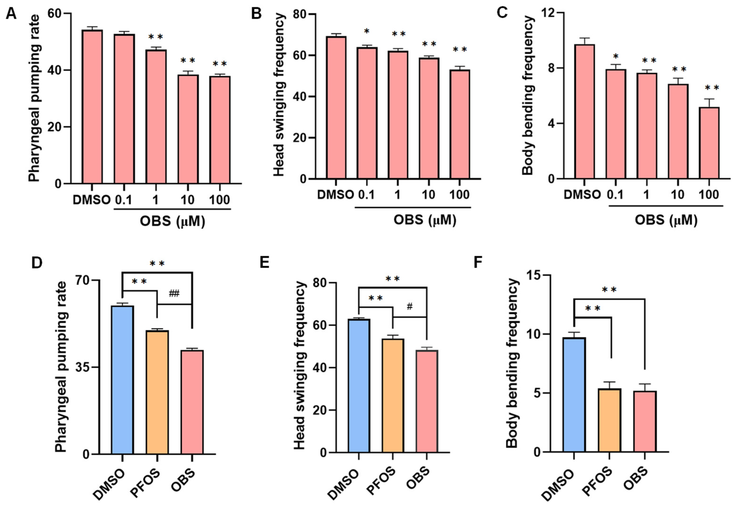
Figure 1. Effects of PFOS/OBS exposure on neurobehavioral activity in C. elegans. (AC) The worms were treated with different concentrations of OBS or DMSO control, following by the measure of (A) Pharyngeal pumping rate, (B) Head swinging frequency, (C) Body bending frequency. (DF) Alteration of neurobehavioral activity including (D) Pharyngeal pumping rate, (E) Head swinging frequency, and (F) Body bending frequency were compared when exposure to the same concentration (100 μM) of PFOS and OBS. * p < 0.05, ** p < 0.01 vs. DMSO. # p < 0.05, ## p < 0.01 vs. PFOS. n = 15.
Toxics 13 00662 g001
Figure 2. Effects of PFOS/OBS exposure on neuronal expression in transgenic C. elegans. (A,D,G) Representative fluorescence images were shown in transgenic C. elegans. (B,E,H) Relative fluorescent intensities were determined in transgenic C. elegans. Scale bar: 100 µm, n = 20. (C,F,I) Relative mRNA expression levels were examined by RT-qPCR. * p < 0.05, ** p < 0.01 vs. DMSO control; ## p < 0.01 vs. PFOS, n = 15.
Toxics 13 00662 g002aToxics 13 00662 g002b
Figure 3. Effects of PFOS/OBS exposure on ROS and antioxidant genes in C. elegans. (A) Representative fluorescence images in N2 worms labelled by H2DCFDA fluorescent probe. (B) Relative fluorescent intensities indicate ROS production. (C) Relative expression levels of antioxidant genes were detected by RT-qPCR. (D) Representative fluorescence images were shown in KN259 transgenic C elegans. (E) Relative fluorescent intensities were determined in transgenic C elegans. * p < 0.05, ** p < 0.01 vs. DMSO control; # p < 0.05 vs. PFOS, scale bar: 200 µm, n = 15.
Toxics 13 00662 g003
Figure 4. PFOS/OBS affect oxidative stress-neurotransmitter regulatory network. (A) PFOS exposure. (B) OBS exposure. Correlation between behaviors and neuronal development of C. elegans exposed to PFOS or OBS. The correlation coefficients (r and p value) were obtained from Pearson correlation analysis. A yellow color indicates a positive correlation, and a black color indicates a negative correlation. * p < 0.05, ** p < 0.01 *** p < 0.001.
Toxics 13 00662 g004
Figure 5. Binding potential of PFOS and OBS with PMK-1 protein. (A) Molecular docking between PFOS and PMK-1 protein. (B) Molecular docking between OBS and PMK-1 protein. (C,D) Relative expression levels of pmk-1 and skn-1 in N2 C elegans. * p < 0.05, ** p < 0.01 vs. DMSO, # p < 0.05 vs. PFOS; n = 15.
Toxics 13 00662 g005aToxics 13 00662 g005b
Figure 6. Effects of PFOS/OBS exposure on neurobehavioral activity in pmk-1 mutant. (A) Pharyngeal pumping rate. (B) Head swinging frequency. (C) Body bending frequency; * p < 0.05 vs. DMSO, # p < 0.05 vs. PFOS; n = 15.
Toxics 13 00662 g006
Figure 7. Effects of PFOS/OBS exposure on neurotransmission related genes and antioxidant genes in pmk-1 mutants. (AC) Relative mRNA expression levels of neuron related genes. (D) Relative mRNA expression levels of antioxidant genes. * p < 0.05, ** p < 0.01 vs. DMSO control; # p < 0.05 vs. PFOS; n = 15.
Toxics 13 00662 g007
Disclaimer/Publisher’s Note: The statements, opinions and data contained in all publications are solely those of the individual author(s) and contributor(s) and not of MDPI and/or the editor(s). MDPI and/or the editor(s) disclaim responsibility for any injury to people or property resulting from any ideas, methods, instructions or products referred to in the content.