Article

Exposure to 6-PPD Quinone Disrupts Adsorption and Catabolism of Leucine and Causes Mitochondrial Dysfunction in Caenorhabditis elegans

by
Wei Wang
1,2,
Yunhui Li
2,* and
Dayong Wang
1,3,*
1
Shenzhen Ruipuxun Academy for Stem Cell & Regenerative Medicine, Shenzhen 518122, China
2
School of Public Health, Southeast University, Nanjing 210009, China
3
Medical School, Southeast University, Nanjing 210009, China
*
Authors to whom correspondence should be addressed.
Toxics 2025, 13(7), 544; https://doi.org/10.3390/toxics13070544
Submission received: 3 June 2025 / Revised: 19 June 2025 / Accepted: 27 June 2025 / Published: 28 June 2025
(This article belongs to the Section Human Toxicology and Epidemiology)

Abstract

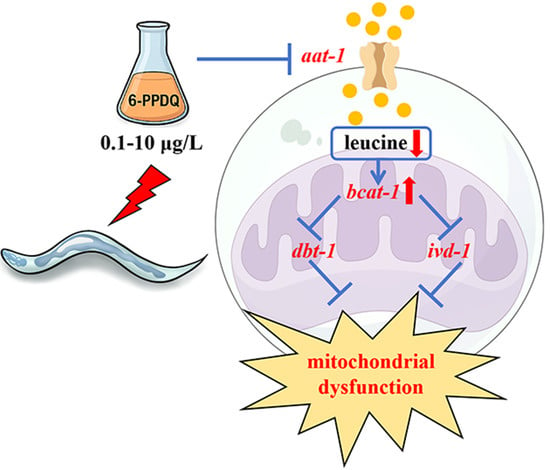
6-PPD quinone (6-PPDQ) is a derivative from 6-PPD, an antioxidant added in tires. Leucine is an important amino acid that needs to be obtained from the diet. In Caenorhabditis elegans, we examined the effect of 6-PPDQ exposure at environmentally relevant concentrations (ERCs) on the content of leucine and underlying mechanisms. In nematodes, 0.1–10 μg/L of 6-PPDQ decreased leucine content. The expression of the aat-1-encoding amino acid transmembrane transporter was decreased by 0.1–10 μg/L of 6-PPDQ, and leucine content was reduced by aat-1 RNAi. Meanwhile, the expression of bcat-1-encoding branched-chain amino acid transferase was increased by 0.1–10 μg/L of 6-PPDQ, and leucine content was increased by bcat-1 RNAi. Additionally, the expressions of dbt-1 and ivd-1 encoding two enzyme genes governing NADH and FADH2 generations were decreased by 0.1–10 μg/L of 6-PPDQ, and their expressions in 6-PPDQ exposed nematodes were increased by bcat-1 RNAi. After 6-PPDQ exposure, NADH content was reduced by dbt-1 RNAi, and FADH2 content was reduced by ivd-1 RNAi. Moreover, 6-PPDQ-induced mitochondrial dysfunction and other aspects of toxicity (such as intestinal ROS generation and lipofuscin accumulation, inhibited locomotion, and reduced brood size) were suppressed by bcat-1 RNAi and strengthened by dbt-1 and ivd-1 RNAi. The 6-PPDQ-induced toxicity and the decrease in dbt-1 and ivd-1 expressions could be inhibited by following leucine (5 mM) treatment. Our results demonstrate the important association of leucine adsorption and catabolism with 6-PPDQ toxicity induction.

Graphical Abstract

1. Introduction

N′-(1,3-Dimethylbutyl)-N′-phenyl-p-phenylenediamine (6-PPD) is widely applied in tires to delay their oxidation [1]. 6-PPDQ was initially observed to induce the lethality of salmon coho [2]. After reacting with ozone through different pathways, 6-PPDQ can be formed from 6-PPD [3]. In environments including water, soil, snow, sediment, and dust, 6-PPDQ can be detected [4,5,6,7,8]. Environmentally relevant concentrations (ERCs) of 6-PPDQ have been found in the range of ng/L to tens of μg/L [9,10,11,12]. Besides the induction of acute lethality [2,13], 6-PPDQ also causes toxicity on development, behavior, and reproduction and disruption in metabolisms and the intestinal microbial community in environmental animals [14,15,16,17]. In mammals, exposure to 6-PPDQ can further induce impairment on sperm quality [18], as well as the intestine [19] and the liver [20]. More recently, it was observed that mitochondrial dysfunction can be caused by 6-PPDQ exposure [21,22,23,24].
Caenorhabditis elegans has a small size and well-described genetic and molecular backgrounds. After exposure to pollutants, C. elegans can exhibit high sensitivity [25,26,27]. Due to these properties, it has been used to assess pollutant toxicity at ERCs [28,29,30]. In C. elegans, 6-PPDQ can result in multiple toxicities, such as neurotoxicity and intestinal toxicity [31,32]. Additionally, some metabolisms, including those of glycogen and glucose, are disrupted by 6-PPDQ exposure [33,34]. Moreover, 6-PPDQ exposure induces mitochondrial dysfunction, which is associated with both damage to complexes (such as complexes I and II) and suppression in mitochondrial UPR [35,36].
Leucine cannot be synthesized de novo [37]. Leucine plays important functions in modulating some physiological processes, including energy balance and protein synthesis [38]. Some solute carriers function to facilitate the transmembrane transport of amino acids from the intestinal lumen to intestinal cells [39,40], and C. elegans AAT-1 is the amino acid transmembrane transporter expressed in the intestine. Once inside the cells, leucine is catalyzed by branched-chain amino acid transferase (BCAT-1) into α-ketoisocaproate (Figure 1A) [41,42]. After this, NADH and FADH2 can be generated from the intermediates through the actions of dihydrolipoamide branched-chain transacylase E2 DBT-1 and isovaleryl-CoA dehydrogenase IVD-1 (Figure 1A) [42,43]. We assumed that exposure to 6-PPDQ may disrupt the adsorption and catabolism of leucine, which is associated with the induction of mitochondrial dysfunction. Thus, we first aimed to explore whether exposure to 6-PPDQ disrupts leucine adsorption and catabolism and its association with mitochondrial dysfunction in nematodes. Additionally, the pharmacological effect of leucine treatment against 6-PPDQ damage on mitochondrial function was determined. The results suggested an important link between disruption in leucine adsorption and catabolism and 6-PPDQ toxicity induction.

2. Materials and Methods

2.1. Animal Maintenance

Animals were normally cultured on nematode growth medium (NGM) plates fed with E. coli OP50 [44]. The used strain was wild-type N2. Eggs were prepared by treating hermaphrodite nematodes using an alkaline lysis solution [45] and were further cultured on new NGM plate to obtain the L1 larval population.

2.2. 6-PPDQ Exposure

The 6-PPDQ was purchased from Toronto Research Chemicals Co. Its working solutions (0.1, 1, and 10 μg/L) were selected based on reported ERCs of 6-PPDQ in the environment [3,11]. The L1 larvae were exposed to 6-PPDQ until they reached adult day-3 (approximately 6.5 days) [46]. The 6-PPDQ solutions were refreshed daily.

2.3. Leucine Content Assay

Approximately 0.2 g of nematodes were harvested from each group and homogenized on ice in a PBS buffer. The homogenate was centrifuged for 10 min at 12,000× g. The leucine content was measured using a leucine determination kit (Wuhan Mosher Biotechnology Co., Wuhan, China). Supernatants and an enzyme-labeled reagent were added to the enzyme-labeled plates to incubate at 37 °C for 1 h. Subsequently, 50 μL of chromogenic reagents A and B were added. The absorbance was measured at 450 nm. The experiments were repeated three times.

2.4. Transcriptional Expression

The total nematode RNAs were extracted from approximately 500 animals using Trizol, and their quality was evaluated using an A260/280 ratio. The total RNAs were reverse transcribed to obtain cDNA. Gene expression was determined in the StepOnePlus™ real-time PCR system (Applied Biosystems, Carlsbad, CA, USA) using the SYBR Green qRT-PCR master mix (Takara, Kusatsu, Japan) and analyzed using the comparative cycle threshold method. tba-1 was used as the reference gene [47]. The experiments were repeated three times. Information on the primers is available in Table S1.

2.5. NADH and FADH2 Contents

The NADH content was measured based on the kit instructions (Yfxbio Biotech, Nanjing, China). Approximately 0.2 g of nematodes were harvested from each group and homogenized in 400 μL of the extraction buffer. After centrifugation for 10 min at 12,000× g, the supernatant was incubated with hydrolyzed NAD+ at 60 °C, and working reagents were further added for absorbance measurement at 450 nm. The NADH concentrations were assessed based on a prepared standard curve. The FADH2 content was measured using a FADH2 ELISA kit (Shanghai Yansheng Biochemical Reagent Co., Shanghai, China). Supernatant and detection antibodies were added to the enzyme-labeled plates to incubate at 37 °C for 1 h. After this, substrate reagents A and B were added, gently mixed, and incubated at 37 °C for 15 min. The absorbance was measured at 450 nm. The experiments were repeated three times.

2.6. Mitochondrial Function

The endpoints of oxygen consumption rate and ATP content reflect the mitochondrial function [48]. Approximately 0.1 g of nematodes were collected and homogenized in pre-cooled lysate. Mitochondria were prepared using the differential centrifugation method. For the oxygen consumption assay, 100 μL of mitochondria were incubated together with 4 μL of the BBoxiProbe R01 oxygen fluorescence probe (Sangon, Shanghai, China). The absorbance was measured at 468 nm at 5 min intervals (for 30 min). For the ATP content assay, based on the ATP kit protocol (Sangon, Shanghai, China), the homogenate was centrifuged at 12,000× g for 5 min. The supernatants or standard liquids were used for the ATP content assay. The absorbance was measured at 340 nm. The ATP detection working solution was applied to evaluate the background ATP. The experiments were repeated three times.

2.7. Toxicity Assessments

Intestinal reactive oxygen species (ROS) generation and lipofuscin accumulation were used to reflect intestinal toxicity. To assess ROS generation, nematodes were labelled using 1 mM of CM-H2DCFDA for 3 h [49]. After washing using M9 buffer, the nematodes were observed at 510 nm of the emission filter and at 488 nm of the excitation wavelength. Intestinal ROS fluorescence signals were analyzed under upright fluorescence microscopy (AX10, Zeiss, Oberkochen, Germany). To assess lipofuscin accumulation, nematodes were analyzed under a DAPI filter using a fluorescence microscope [50]. The fluorescence intensity of ROS signals and lipofuscin accumulation were quantified in images (magnification, 20×) using the ImageJ V1.8.0.112. Fifty animals were examined per treatment. The experiments were repeated three times.
Head thrash and body bend reflect the locomotion behavior. Their frequencies were recorded in one minute or in 20 s intervals, respectively [51]. A change in the bending direction at the body mid-region is considered to be a head thrash. A change in the posterior bulb direction is considered to be a body bend. Fifty nematodes were analyzed per treatment. The experiments were repeated three times.
Brood size reflects the reproductive capacity. Brood size indicates the total number of offspring produced by each nematode, which was measured until the nematode ceased egg-laying [52]. Thirty nematodes were analyzed per treatment. The experiments were repeated three times.

2.8. RNA Interference (RNAi)

To perform the RNAi of certain genes, the nematodes were cultured on NGM plates fed with E. coli HT115 bacteria expressing corresponding double-stranded RNA [53]. The RNAi bacteria were prepared in a Luria broth containing 100 μg/mL of ampicillin and 100 μg/mL of tetracycline. The offspring of the nematodes cultured on RNAi plates were used for 6-PPDQ exposure. L4440 (empty vector) was used as the control [54]. RNAi efficiency is shown in Figure S1.

2.9. Pharmacological Treatment

Following 6-PPDQ exposure (10 μg/L) until adult day 3, the nematodes were washed with K buffer three times. After that, one subgroup was maintained on NGM medium, and another group was transferred into 5 mM leucine to be treated for 24 h at room temperature in darkness [55]. The experiments were conducted in triplicate.

2.10. Data Analysis

Data are presented the mean ± standard deviation (SD). Data were all continuous and passed the normality test and the homogeneity test of variance in SPSS 26.0. A one-way analysis of variance (ANOVA) was used to examine differences between different groups. For multiple factor comparison, two-way ANOVA analysis followed by a post-hoc test was employed. A p-value of < 0.01 (**) was considered to be statistically significant.

3. Results

3.1. 6-PPDQ Reduced Leucine Content

The leucine content in wild-type nematodes could be reduced by 0.1–10 μg/L of 6-PPDQ (Figure 1B). The reduction in leucine content was concentration-dependent in nematodes exposed to 0.1–10 μg/L of 6-PPDQ (Figure 1B). The leucine content was reduced by 30%, 39%, and 47%, respectively, after exposure to 0.1–10 μg/L of 6-PPDQ (Figure 1B).

3.2. Exposure to 6-PPDQ Inhibited Leucine Adsorption

After 0.1–10 μg/L 6-PPDQ exposure in wild-type nematodes, intestinal aat-1 expression was decreased (Figure 1C). Meanwhile, after aat-1 RNAi, the leucine content could be decreased by 54% in 6-PPDQ-exposed nematodes (Figure 1D). Therefore, leucine adsorption was inhibited after 6-PPDQ exposure.

3.3. 6-PPDQ Accelerated 6-PPDQ Catabolism

After 6-PPDQ exposure in wild-type nematodes, bcat-1 expression was further increased (Figure 2A). Meanwhile, after 6-PPDQ exposure, the leucine content was increased by bcat-1 RNAi (Figure 2B). Moreover, after 6-PPDQ exposure, we observed an increase in NADH and FADH2 contents in bcat-1 (RNAi) nematodes compared with the wild-type (L4440) (Figure 2C,D). That is, leucine catabolism could be enhanced by 6-PPDQ exposure, which was associated with a reduction in NADH and FADH2 contents.

3.4. 6-PPDQ Affected Expressions of dbt-1 and ivd-1 and Their Functions

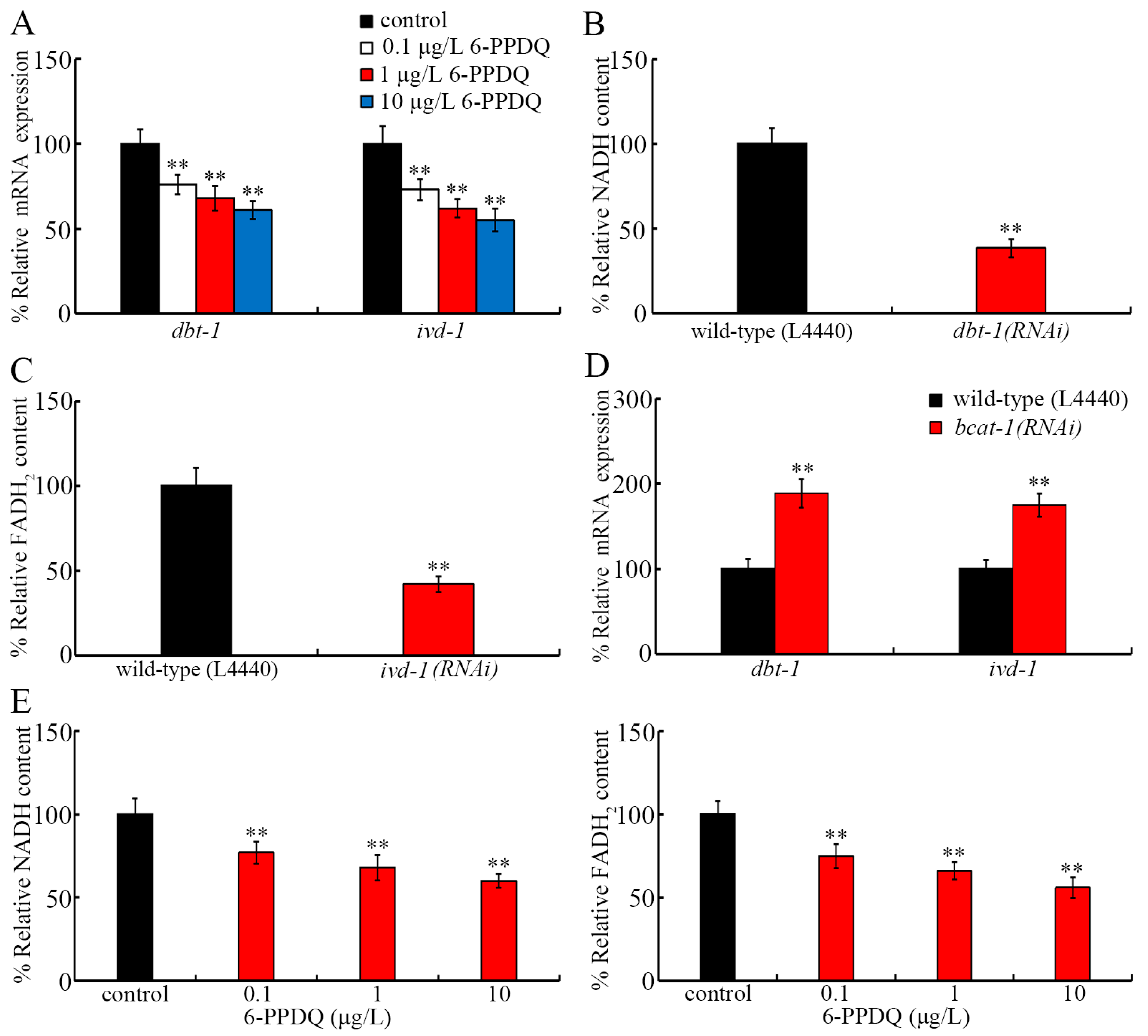
Considering the possible important role of DBT-1 and IVD-1 for the generation of NADH and FADH2 [42,43], we next investigated the effect of 6-PPDQ on the expressions of dbt-1 and ivd-1. The expressions of dbt-1 and ivd-1 were decreased by 0.1–10 μg/L of 6-PPDQ in wild-type nematodes (Figure 3A). After 6-PPDQ exposure, the NADH content could be reduced by RNAi of dbt-1 (Figure 3B), and the FADH2 content could be reduced by the RNAi of ivd-1 (Figure 3C). Moreover, after 6-PPDQ exposure, dbt-1 and ivd-1 expressions were increased by bcat-1 RNAi (Figure 3D). In 6-PPDQ-exposed wild-type nematodes, the NADH and FADH2 contents showed a tendency towards reduction (Figure 3E).

3.5. RNAi of bcat-1, dbt-1, and ivd-1 Affected 6-PPDQ-Induced Mitochondrial Dysfunction

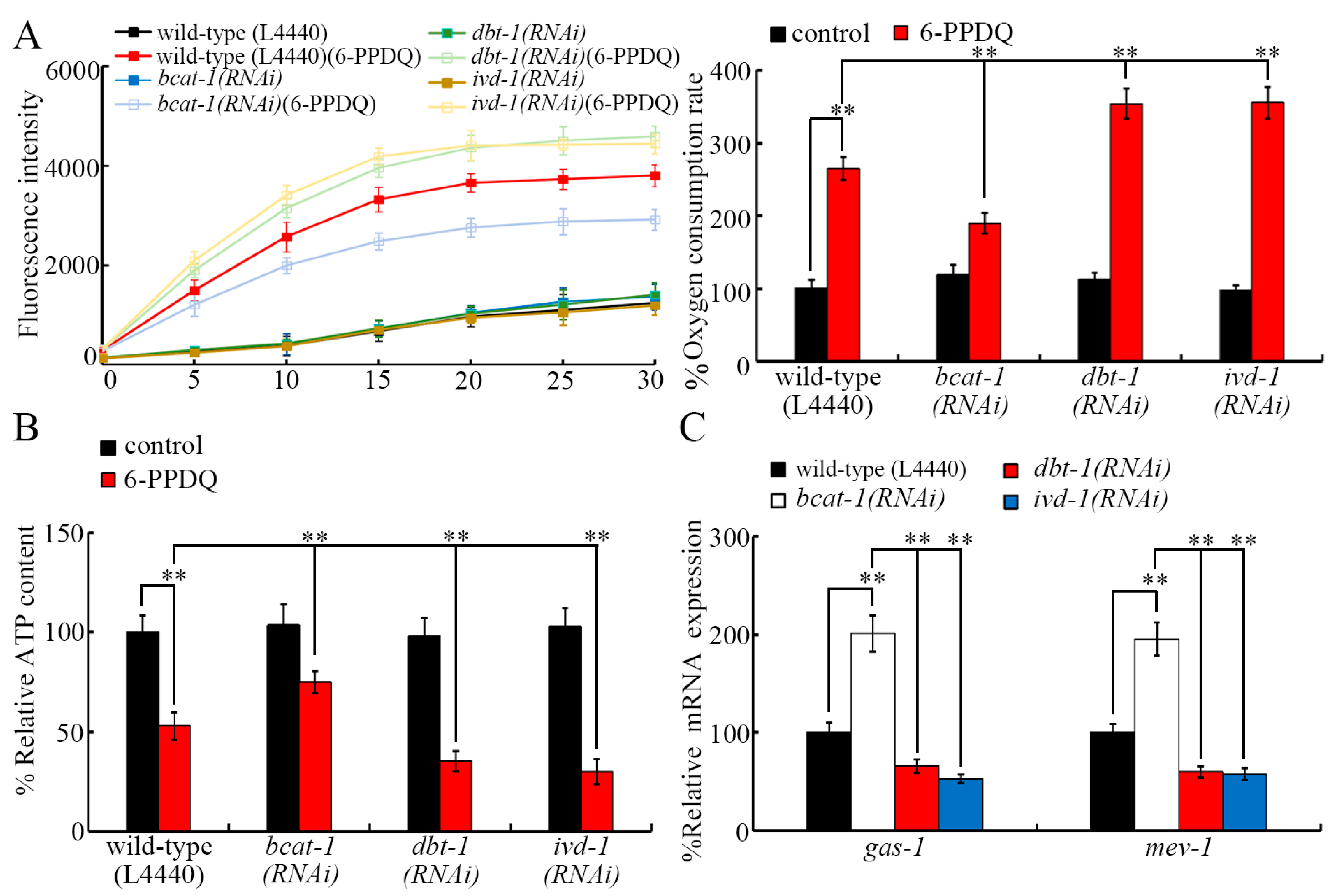
After 6-PPDQ exposure, mitochondrial dysfunction was induced [56]. The 6-PPDQ-induced increase in the oxygen consumption rate was inhibited by the RNAi of bcat-1 and was enhanced by the RNAi of dbt-1 and ivd-1 (Figure 4A). The 6-PPDQ-caused decrease in the ATP content was suppressed by the RNAi of bcat-1 and strengthened by the RNAi of dbt-1 and ivd-1 (Figure 4B). GAS-1 (complex I component) and MEV-1 (complex II component) were involved in controlling 6-PPDQ-induced mitochondrial dysfunction [35]. Moreover, after 6-PPDQ exposure, gas-1 and mev-1 expressions were increased by bcat-1 RNAi and decreased by dbt-1 and ivd-1 RNAi (Figure 4C).

3.6. RNAi of aat-1, bcat-1, dbt-1, and ivd-1 Affected Induction of 6-PPDQ Toxicity

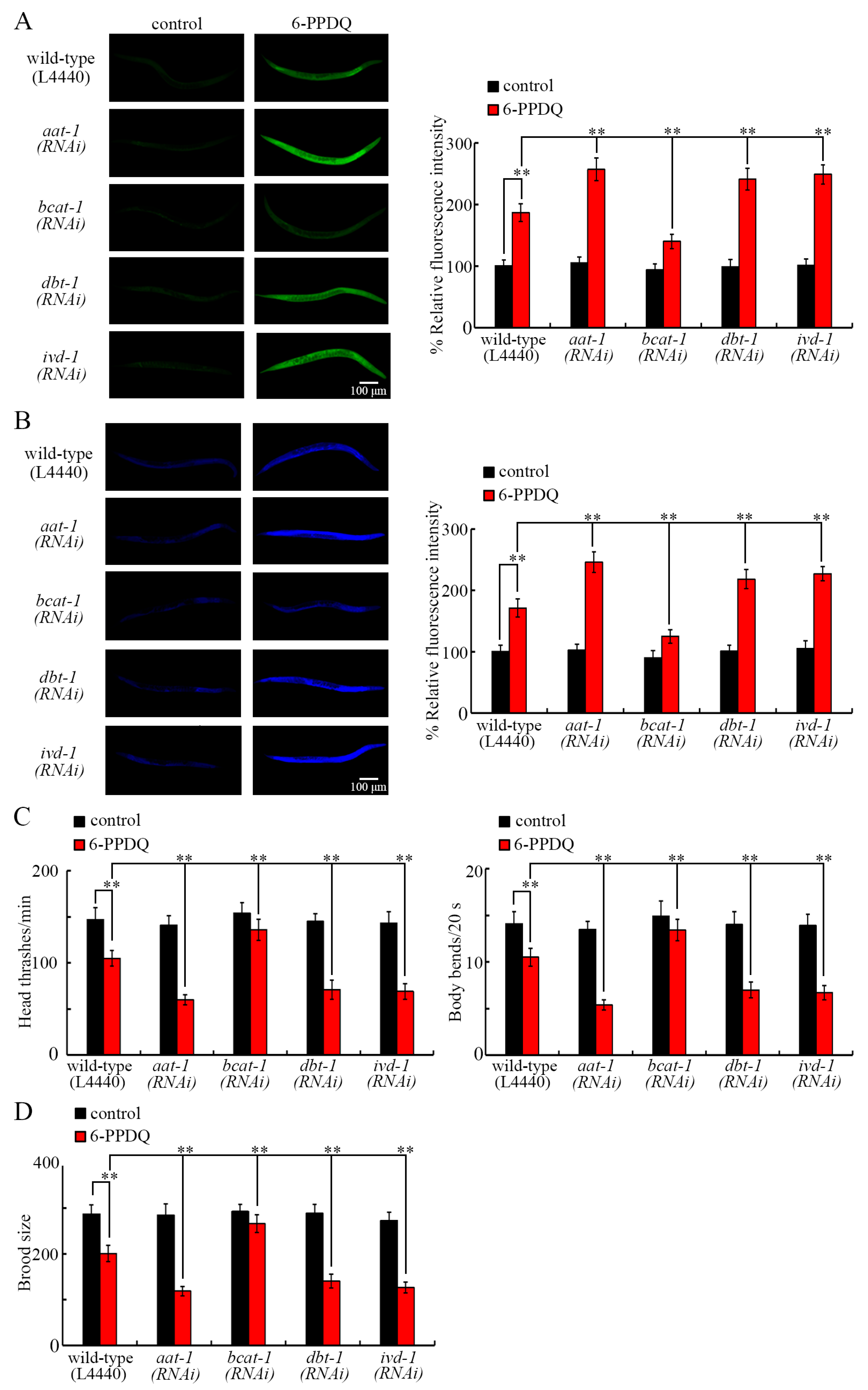
We further investigated the effect of aat-1, bcat-1, dbt-1, and ivd-1 RNAi on other 6-PPDQ-induced aspects of toxicity. Intestinal toxicity could be induced by 6-PPDQ, reflected by intestinal ROS generation and lipofuscin accumulation [50]. After 6-PPDQ exposure, induced intestinal ROS generation and lipofuscin accumulation were enhanced by the RNAi of aat-1, dbt-1, and ivd-1, and inhibited by the RNAi of bcat-1 (Figure 5A,B). Exposure to 6-PPDQ also caused neurotoxicity reflected by inhibited locomotion [50] and reproductive toxicity reflected by reduced brood size [32], which were strengthened by aat-1, dbt-1, and ivd-1 RNAi and suppressed by bcat-1 RNAi (Figure 5C,D).

3.7. Beneficial Effect of Leucine Treatment Against 6-PPDQ-Induced Mitochondrial Dysfunction

After exposure to 6-PPDQ (10 μg/L) in wild-type nematodes, a decrease in dbt-1 and ivd-1 expressions was inhibited by treatment with leucine (5 mM) (Figure 6A). Meanwhile, after exposure to 6-PPDQ (10 μg/L) in wild-type nematodes, a reduction in NADH and FADH2 contents was suppressed through treatment with leucine (5 mM) (Figure 6B,C). Moreover, 6-PPDQ (10 μg/L)-induced mitochondrial dysfunction was inhibited through treatment with leucine (5 mM) in wild-type nematodes (Figure 6D,E). Additionally, 6-PPDQ (10 μg/L)-induced decreases in the expressions of gas-1 and mev-1 were further suppressed through treatment with leucine (5 mM) in wild-type nematodes (Figure 6F).

3.8. Beneficial Effect of Leucine Treatment Against Other Aspects of 6-PPDQ Toxicity

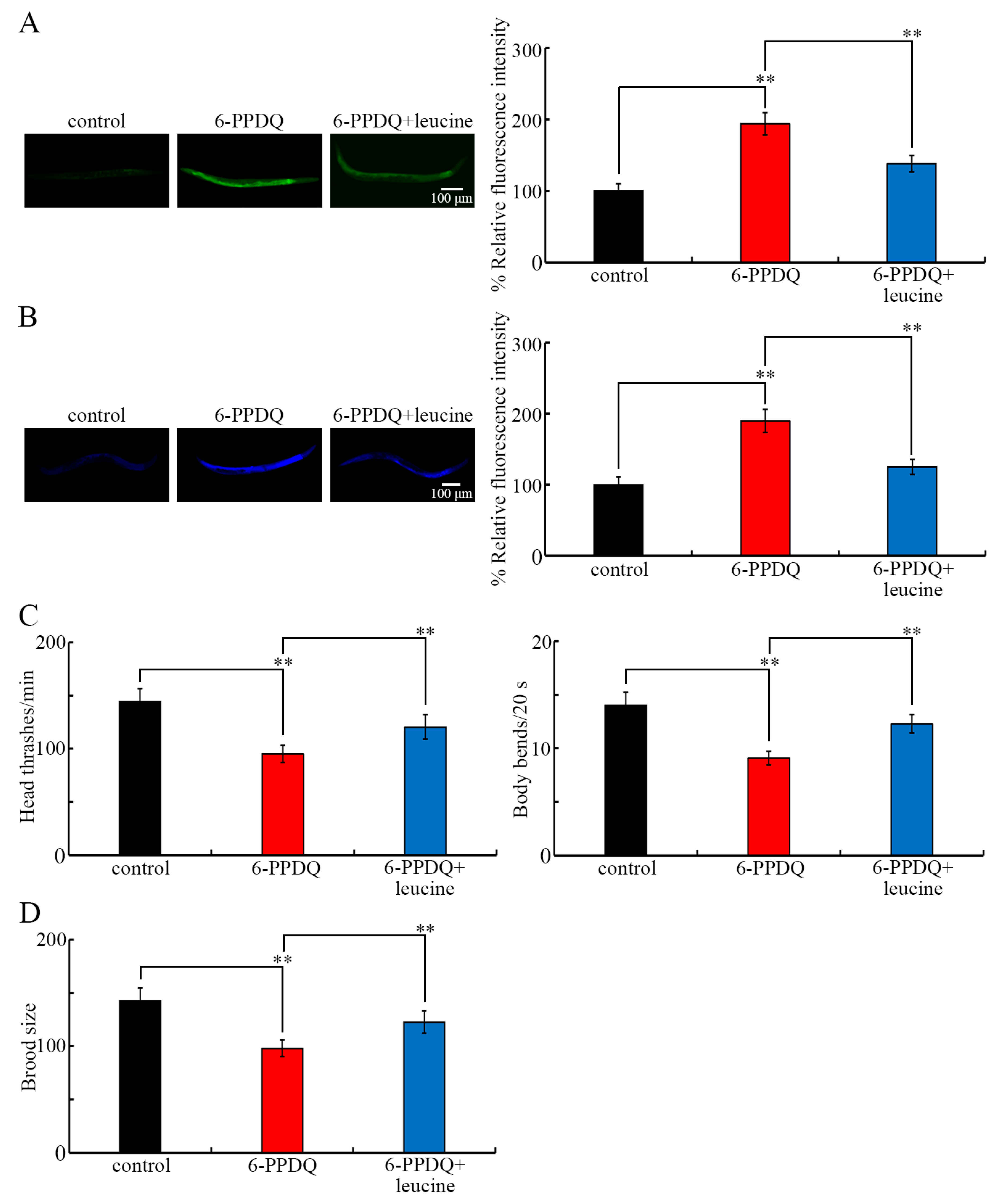
After exposure to 6-PPDQ (10 μg/L) in wild-type nematodes, induced intestinal toxicity, reflected by intestinal ROS generation and lipofuscin accumulation, could be inhibited through treatment with leucine (5 mM) (Figure 7A,B). 6-PPDQ (10 μg/L)-caused locomotion inhibition was suppressed through treatment with leucine (5 mM) (Figure 7C) in wild-type nematodes. Moreover, the 6-PPDQ (10 μg/L)-induced reduction in brood size was inhibited following treatment with leucine (5 mM) in wild-type nematodes (Figure 7D).

4. Discussion

Leucine plays important modulatory functions in organisms [57,58]. Largely due to this, leucine supplementation has been frequently suggested in clinical settings for the treatment of some diseases [59,60,61]. In the current study, we observed s reduction in leucine content after exposure to 6-PPDQ at ERCs (Figure 1B). Besides the leucine content, the glutamate content could also be reduced by 6-PPDQ at ERCs [62]. Moreover, after exposure to 6-PPDQ at ERCs, the accumulation of glycogen, glucose, and lipid could be observed [33,34,46]. The transgenerational accumulation of lipid and glucose could be observed after exposure to 6-PPDQ at ERCs at the parental generation [32,46]. Therefore, exposure to 6-PPDQ at ERCs potentially disrupts multiple metabolic processes in organisms.
One of the biochemical bases for the observed reduction in leucine content was the inhibition of leucine adsorption. 6-PPDQ at 0.1–10 μg/L decreased the expression of aat-1 encoding the amino acid transmembrane transporter (Figure 1C). In 6-PPDQ-exposed nematodes, the leucine content could be reduced by aat-1 RNAi (Figure 1D). That is, 6-PPDQ could reduce the leucine content partially by inhibiting leucine adsorption through the suppression of AAT-1 expression. In C. elegans, AAT-1 was identified as one of the amino acid transporters and was shown to have the function to facilitate amino acid transport [63].
Another biochemical basis for the observed reduction in leucine content was the acceleration of leucine catabolism. 6-PPDQ at 0.1–10 μg/L increased the expression of bcat-1 encoding the branched-chain amino acid transferase (Figure 2A). Meanwhile, in 6-PPDQ-exposed nematodes, the leucine content was increased by bcat-1 RNAi (Figure 2B). Therefore, 6-PPDQ could also reduce leucine content by accelerating leucine catabolism through the activation of BCAT-1. In nematodes, BCAT-1 governs branched-chain amino acid metabolism [41] and is involved in the control of several other biological processes, such as the reprograming of proteasomal degradation and longevity [64].
In 6-PPDQ-exposed nematodes, accompanied with an increase in bcat-1 expression, a decrease in expression of dbt-1 and ivd-1 encoding dihydrolipoamide branched-chain transacylase E2 and isovaleryl-CoA dehydrogenase was observed (Figure 2A), which suggests the inhibition in generation of NADH and FADH2 by 6-PPDQ through the influence on branched-chain amino acid metabolism, such as the leucine catabolism. Several lines of evidence support this. One is the observation of the reduction in NADH and FADH2 contents after exposure to 0.1–10 μg/L of 6-PPDQ (Figure 3E). Secondly, after 6-PPDQ exposure, the NADH content was reduced by the RNAi of dbt-1 (Figure 3B), and the FADH2 content was reduced by the RNAi of ivd-1 (Figure 3C). Thirdly, after 6-PPDQ exposure, NADH and FADH2 contents could be increased by the RNAi of bcat-1 (Figure 2C,D). Fourthly, in 6-PPDQ-exposed nematodes, expressions of dbt-1 and ivd-1 could be increased by the RNAi of bcat-1 (Figure 3D). Recently, we observed that 6-PPDQ could reduce NADH content by disrupting the citric acid cycle [48]. Therefore, 6-PPDQ at ERCs potentially reduced NADH and/or FADH2 contents by disrupting branched-chain amino acid metabolism and the citric acid cycle, simultaneously. dbt-1 was also identified as the response gene to arsenic exposure in nematodes [65].
In organisms, NADH and FADH2 are two starting points for the electron transport chain, and the activities of mitochondrial complexes I and II could be inhibited by 6-PPDQ [35]. Moreover, the alteration in bcat-1, dbt-1, and ivd-1 expressions was related to the induction of mitochondrial dysfunction in 6-PPDQ-exposed nematodes. Based on the analysis of endpoints of the oxygen consumption rate and ATP content reflecting mitochondrial function, the 6-PPDQ-induced increase in the oxygen consumption rate and the decrease in the ATP content could be inhibited by bcat-1 RNAi and enhanced by dbt-1 and ivd-1 RNAi (Figure 4A,B). Meanwhile, we further observed that 6-PPDQ caused the induction of intestinal ROS generation and lipofuscin accumulation, an inhibition in locomotion, and a reduction in brood size could be suppressed by bcat-1 RNAi and strengthened by the RNAi of aat-1, dbt-1, and ivd-1 (Figure 5A–D). Therefore, the RNAi of bcat-1 could cause resistance to 6-PPDQ toxicity, and the RNAi of aat-1, dbt-1, and/or ivd-1 could induce susceptibility to 6-PPDQ toxicity, including the induction of mitochondrial dysfunction. These observations suggested that BCAT-1 had a different function from that of DBT-1 and IVD-1 in modulating biological processes, including mitochondrial function in 6-PPDQ-exposed nematodes. In nematodes, expressions of gas-1 and mev-1 encoding components of complexes I and II were decreased by 6-PPDQ, and the RNAi of these two genes caused susceptibility to 6-PPDQ toxicity on mitochondrial function [35]. Moreover, in 6-PPDQ-exposed nematodes, expressions of gas-1 and mev-1 were increased by bcat-1 RNAi and decreased by dbt-1 and ivd-1 RNAi (Figure 4C). Additionally, in 6-PPDQ-exposed nematodes, expressions of gas-1 and mev-1 could also be decreased by the RNAi of cts-1, idh-2, dlst-1, and dld-1, which govern the citric acid cycle [48]. These observations suggest that both leucine catabolism-related enzyme genes and citric acid cycle-related enzyme genes could be involved in the control of mitochondrial function in 6-PPDQ-exposed nematodes by affecting the expressions of gas-1 and mev-1.
Pharmacological treatment with leucine further demonstrated the beneficial effect of leucine treatment against 6-PPDQ toxicity, including the induction of mitochondrial dysfunction. Several lines of evidence indicated the beneficial effect of leucine treatment against 6-PPDQ damage on mitochondrial function. Firstly, the 6-PPDQ-induced increase in the oxygen consumption rate and the decrease in ATP content could be inhibited by leucine treatment (Figure 6D,E). Secondly, the 6-PPDQ-caused reduction in NADH and FADH2 contents could be suppressed by leucine treatment (Figure 6B,C). Thirdly, the 6-PPDQ-induced decrease in gas-1 and mev-1 expressions could be inhibited by leucine treatment (Figure 6F). Moreover, the 6-PPDQ-caused decrease in dbt-1 and ivd-1 expressions could also be suppressed by leucine treatment (Figure 6A), which suggests that leucine treatment potentially increased gas-1 and mev-1 expressions and mitochondrial function by activating dbt-1 and ivd-1 expressions. Besides the beneficial effect on mitochondrial function, leucine treatment could further inhibit the induction of intestinal ROS generation and lipofuscin accumulation (Figure 7A,B), accelerate locomotion (Figure 7C), and increase brood size (Figure 7D) in 6-PPDQ-exposed nematodes, which suggests the multiple possible beneficial effects of leucine treatment. In nematodes, treatment with glutamate could inhibit 6-PPDQ by activating the corresponding receptors of GLR-1, GLR-2, and GLR-4 [62]. In zebrafish larvae, ghrelin, a growth hormone secretagogue receptor (GHSR) agonist, also showed neuroprotective function against 6-PPDQ toxicity [66]. That is, 6-PPDQ toxicity can be inhibited by different pharmacological treatments through different mechanisms.

5. Conclusions

Together, we observed a reduction in leucine content by 6-PPDQ at ERCs in nematodes. This leucine reduction in 6-PPDQ-exposed nematodes was associated with an inhibition in leucine adsorption, indicated by a decrease in aat-1 expression. Additionally, this leucine reduction in 6-PPDQ-exposed nematodes was related to an increase in leucine catabolism, indicated by an increase in bcat-1 expression. Additionally, in 6-PPDQ-exposed nematodes, expressions of dbt-1 and ivd-1 were decreased, which was linked to a reduction in NADH and FADH2 contents and mitochondrial dysfunction. Pharmacological treatment with leucine could inhibit the decrease in dbt-1 and ivd-1 expressions, the reduction in NADH and FADH2 contents, and the mitochondrial dysfunction caused by 6-PPDQ exposure. Our results provide new insight into the induction of mitochondrial dysfunction by 6-PPDQ exposure at ERCs in organisms.

Supplementary Materials

The following supporting information can be downloaded at https://www.mdpi.com/article/10.3390/toxics13070544/s1. Figure S1: RNAi efficiency of aat-1, bcat-1, ivd-1, and dbt-1. ** p < 0.01 vs wild-type (L4440); Table S1: Primer information for qRT-PCR.

Author Contributions

Investigation and writing of the draft, W.W.; supervision and review of the draft, Y.L. and D.W. All authors have read and agreed to the published version of the manuscript.

Funding

This study was supported by grants from the Guangdong Basic and Applied Basic Research Foundation (2024A1515011115 and 2025A1515010712) and the Shenzhen Science and Technology Program (JCYJ20220530163605011).

Institutional Review Board Statement

Not applicable.

Informed Consent Statement

Not applicable.

Data Availability Statement

The original data presented in this study are included in the article/Supplementary Materials; further inquiries can be directed to the corresponding author.

Conflicts of Interest

The authors declare that they have no known competing financial interests or personal relationships that could appear to have influenced the work reported in this paper.

References

  1. Zeng, L.; Li, Y.; Sun, Y.; Liu, L.Y.; Shen, M.; Du, B. Widespread occurrence and transport of p-phenylenediamines and their quinones in sediments across urban rivers, estuaries, coasts, and deep-sea regions. Environ. Sci. Technol. 2023, 57, 2393–2403. [Google Scholar] [CrossRef] [PubMed]
  2. Tian, Z.; Zhao, H.; Peter, K.T.; Gonzalez, M.; Wetzel, J.; Wu, C.; Hu, X.; Prat, J.; Mudrock, E.; Hettinger, R.; et al. A ubiquitous tire rubber-derived chemical induces acute mortality in coho salmon. Science 2021, 371, 185–189. [Google Scholar] [CrossRef]
  3. Hua, X.; Wang, D.-Y. Tire-rubber related pollutant 6-PPD quinone: A review of its transformation, environmental distribution, bioavailability, and toxicity. J. Hazard. Mater. 2023, 459, 132265. [Google Scholar] [CrossRef]
  4. Seiwert, B.; Nihemaiti, M.; Troussier, M.; Weyrauch, S.; Reemtsma, T. Abiotic oxidative transformation of 6-PPD and 6-PPD quinone from tires and occurrence of their products in snow from urban roads and in municipal wastewater. Water Res. 2022, 212, 118122. [Google Scholar] [CrossRef]
  5. Liang, Y.; Zhu, F.; Li, J.; Wan, X.; Ge, Y.; Liang, G.; Zhou, Y. P-phenylenediamine antioxidants and their quinone derivatives: A review of their environmental occurrence, accessibility, potential toxicity, and human exposure. Sci. Total Environ. 2024, 948, 174449. [Google Scholar] [CrossRef]
  6. Lane, R.F.; Smalling, K.L.; Bradley, P.M.; Greer, J.B.; Gordon, S.E.; Hansen, J.D.; Kolpin, D.W.; Spanjer, A.R.; Masoner, J.R. Tire-derived contaminants 6PPD and 6PPD-Q: Analysis, sample handling, and reconnaissance of United States stream exposures. Chemosphere 2024, 363, 142830. [Google Scholar] [CrossRef]
  7. Jaeger, A.; Monaghan, J.; Tomlin, H.; Atkinson, J.; Gill, C.G.; Krogh, E.T. Intensive spatiotemporal characterization of the tire wear toxin 6PPD quinone in urban waters. ACS ES T Water 2024, 4, 5566–5574. [Google Scholar] [CrossRef]
  8. Yi, J.; Ruan, J.; Yu, H.; Wu, B.; Zhao, J.; Wang, H.; Chen, R.; Yang, Q.; Chen, J.; Sun, D. Environmental fate, toxicity, and mitigation of 6PPD and 6PPD-Quinone: Current understanding and future directions. Environ. Pollut. 2025, 375, 126352. [Google Scholar] [CrossRef]
  9. Rauert, C.; Charlton, N.; Okoffo, E.D.; Stanton, R.S.; Agua, A.R.; Pirrung, M.C.; Thomas, K.V. Concentrations of tire additive chemicals and tire road wear particles in an Australian urban tributary. Environ. Sci. Technol. 2022, 56, 2421–2431. [Google Scholar] [CrossRef]
  10. Varshney, S.; Gora, A.H.; Siriyappagouder, P.; Kiron, V.; Olsvik, P.A. Toxicological effects of 6PPD and 6PPD quinone in zebrafish larvae. J. Hazard. Mater. 2022, 424, 127623. [Google Scholar] [CrossRef]
  11. Wan, X.; Liang, G.-Y.; Wang, D.-Y. Potential human health risk of the emerging environmental contaminant 6-PPD quinone. Sci. Total Environ. 2024, 949, 175057. [Google Scholar] [CrossRef] [PubMed]
  12. Black, G.P.; De Parsia, M.; Uychutin, M.; Lane, R.; Orlando, J.L.; Hladik, M.L. 6PPD-quinone in water from the San Francisco-San Joaquin Delta, California, 2018-2024. Environ. Monit. Assess. 2025, 197, 369. [Google Scholar] [CrossRef]
  13. Roberts, C.; Lin, J.; Kohlman, E.; Jain, N.; Amekor, M.; Alcaraz, A.J.; Hogan, N.; Hecker, M.; Brinkmann, M. Acute and subchronic toxicity of 6PPD-quinone to early life stage lake trout (Salvelinus namaycush). Environ. Sci. Technol. 2025, 59, 791–797. [Google Scholar] [CrossRef]
  14. Ricarte, M.; Prats, E.; Montemurro, N.; Bedrossiantz, J.; Bellot, M.; Gómez-Canela, C.; Raldúa, D. Environmental concentrations of tire rubber-derived 6PPD-quinone alter CNS function in zebrafish larvae. Sci. Total Environ. 2023, 896, 165240. [Google Scholar] [CrossRef]
  15. Liao, X.L.; Chen, Z.F.; Liu, Q.Y.; Zhou, J.M.; Cai, W.X.; Wang, Y.; Cai, Z. Tissue accumulation and biotransformation of 6PPD-quinone in adult zebrafish and its effects on the intestinal microbial community. Environ. Sci. Technol. 2024, 58, 10275–10286. [Google Scholar] [CrossRef]
  16. Liu, Z.; Feng, Y.; Sun, W.; Wang, B.; Shi, C.; Ran, R.; Zhang, Y.; Lu, L.; Zhang, H. Environmental concentrations of 6PPD and 6PPD-quinone induce hepatic lipid metabolism disorders in male black-spotted frogs. J. Hazard. Mater. 2024, 480, 136400. [Google Scholar] [CrossRef]
  17. Jiang, Y.; Wang, C.; Ma, L.; Gao, T.; Wāng, Y. Environmental profiles, hazard identification, and toxicological hallmarks of emerging tire rubber-related contaminants 6PPD and 6PPD-quinone. Environ. Int. 2024, 187, 108677. [Google Scholar] [CrossRef]
  18. Yao, K.; Kang, Q.; Liu, W.; Chen, D.; Wang, L.; Li, S. Chronic exposure to tire rubber-derived contaminant 6PPD-quinone impairs sperm quality and induces the damage of reproductive capacity in male mice. J. Hazard. Mater. 2024, 470, 134165. [Google Scholar] [CrossRef]
  19. Yang, Y.; Sun, N.; Lv, J.; Chen, H.; Wang, H.; Xu, J.; Hu, J.; Tao, L.; Fang, M.; Huang, Y. Environmentally realistic dose of tire-derived metabolite 6PPD-Q exposure causes intestinal jejunum and ileum damage in mice via cannabinoid receptor-activated inflammation. Sci. Total Environ. 2024, 918, 170679. [Google Scholar] [CrossRef]
  20. Wang, L.; Tang, W.; Sun, N.; Lv, J.; Hu, J.; Tao, L.; Zhang, C.; Wang, H.; Chen, L.; Xu, D.X.; et al. Low-dose tire wear chemical 6PPD-Q exposure elicit fatty liver via promoting fatty acid biosynthesis in ICR mice. J. Hazard. Mater. 2025, 489, 137574. [Google Scholar] [CrossRef]
  21. Di, S.; Xu, H.; Yu, Y.; Qi, P.; Wang, Z.; Liu, Z.; Zhao, H.; Jin, Y.; Wang, X. Environmentally relevant concentrations of S-6PPD-quinone caused more serious hepatotoxicity than r-enantiomer and racemate in Oncorhynchus mykiss. Environ. Sci. Technol. 2024, 58, 17617–17628. [Google Scholar] [CrossRef]
  22. Chen, H.; Xie, M.; Li, W.; Tan, L.; Cai, X.; Shen, M.; Li, R. Detection of 6-PPD and 6-PPDQ in airborne particulates and assessment of their toxicity in lung cells. Chemosphere 2024, 364, 143205. [Google Scholar] [CrossRef]
  23. Fang, J.; Wang, X.; Cao, G.; Wang, F.; Ru, Y.; Wang, B.; Zhang, Y.; Zhang, D.; Yan, J.; Xu, J.; et al. 6PPD-quinone exposure induces neuronal mitochondrial dysfunction to exacerbate Lewy neurites formation induced by alpha-synuclein preformed fibrils seeding. J. Hazard. Mater. 2024, 465, 133312. [Google Scholar] [CrossRef]
  24. Fang, C.; Di, S.; Yu, Y.; Qi, P.; Wang, X.; Jin, Y. 6PPD induced cardiac dysfunction in zebrafish associated with mitochondrial damage and inhibition of autophagy processes. J. Hazard. Mater. 2024, 471, 134357. [Google Scholar] [CrossRef]
  25. Chen, H.; Wang, C.; Li, H.; Ma, R.; Yu, Z.; Li, L.; Xiang, M.; Chen, X.; Hua, X.; Yu, Y. A review of toxicity induced by persistent organic pollutants (POPs) and endocrine-disrupting chemicals (EDCs) in the nematode Caenorhabditis elegans. J. Environ. Manag. 2019, 237, 519–525. [Google Scholar] [CrossRef]
  26. Li, H.; Zeng, L.; Wang, C.; Shi, C.; Li, Y.; Peng, Y.; Chen, H.; Zhang, J.; Cheng, B.; Chen, C.; et al. Review of the toxicity and potential molecular mechanisms of parental or successive exposure to environmental pollutants in the model organism Caenorhabditis elegans. Environ. Pollut. 2022, 311, 119927. [Google Scholar] [CrossRef]
  27. Yao, Y.; Zhang, T.; Tang, M. A critical review of advances in reproductive toxicity of common nanomaterials to Caenorhabditis elegans and influencing factors. Environ. Pollut. 2022, 306, 119270. [Google Scholar] [CrossRef]
  28. Liu, H.; Wu, Y.; Wang, Z. Long-term exposure to polystyrene nanoparticles at environmentally relevant concentration causes suppression in heme homeostasis signal associated with transgenerational toxicity induction in Caenorhabditis elegans. J. Hazard. Mater. 2023, 459, 132124. [Google Scholar] [CrossRef]
  29. Liu, Z.-Y.; Bian, Q.; Wang, D.-Y. 6-PPD quinone induces response of nuclear hormone receptors in the germline associated with formation of reproductive toxicity in Caenorhabditis elegans. J. Hazard. Mater. 2025, 495, 138815. [Google Scholar] [CrossRef]
  30. Yang, Y.; Li, M.; Zheng, J.; Zhang, D.; Ding, Y.; Yu, H.Q. Environmentally relevant exposure to tetrabromobisphenol A induces reproductive toxicity via regulating glucose-6-phosphate 1-dehydrogenase and sperm activation in Caenorhabditis elegans. Sci. Total Environ. 2024, 907, 167820. [Google Scholar] [CrossRef]
  31. Wang, Y.-X.; Wang, D.-Y. Transgenerational intestinal toxicity of 6-PPD quinone in causing ROS production, enhancement in intestinal permeability and suppression in innate immunity in C. elegans. Environ. Pollut. 2024, 363, 125208. [Google Scholar] [CrossRef]
  32. Liu, Z.-J.; Li, Y.-H.; Wang, D.-Y. Transgenerational response of glucose metabolism in Caenorhabditis elegans exposed to 6-PPD quinone. Chemosphere 2024, 367, 143653. [Google Scholar] [CrossRef]
  33. Wang, Y.-X.; Wang, D.-Y. Exposure to 6-PPD quinone enhances glycogen accumulation in Caenorhabditis elegans. Environ. Pollut. 2024, 359, 124600. [Google Scholar] [CrossRef]
  34. Liu, Z.-J.; Li, Y.-H.; Wang, D.-Y. Exposure to 6-PPD quinone disrupts glucose metabolism associated with lifespan reduction by affecting insulin and AMPK signals in Caenorhabditis elegans. Chemosphere 2024, 363, 142975. [Google Scholar] [CrossRef]
  35. Hua, X.; Wang, D.-Y. Environmentally relevant concentration of 6-PPD quinone inhibits two types of mitophagy to cause mitochondrial dysfunction and lifespan reduction in Caenorhabditis elegans. Environ. Sci. Process. Impacts 2025. [Google Scholar] [CrossRef]
  36. Hua, X.; Wang, D.-Y. 6-PPD quinone at environmentally relevant concentrations induced damage on longevity in C. elegans: Mechanistic insight from inhibition in mitochondrial UPR response. Sci. Total Environ. 2024, 954, 176275. [Google Scholar] [CrossRef]
  37. Nie, C.; He, T.; Zhang, W.; Zhang, G.; Ma, X. Branched chain amino acids: Beyond nutrition metabolism. Int. J. Mol. Sci. 2018, 19, 954. [Google Scholar] [CrossRef]
  38. Duan, Y.; Li, F.; Li, Y.; Tang, Y.; Kong, X.; Feng, Z.; Anthony, T.G.; Watford, M.; Hou, Y.; Wu, G.; et al. The role of leucine and its metabolites in protein and energy metabolism. Amino Acids 2016, 48, 41–51. [Google Scholar] [CrossRef]
  39. Rotoli, B.M.; Barilli, A.; Visigalli, R.; Ferrari, F.; Dall’Asta, V. y+LAT1 and y+LAT2 contribution to arginine uptake in different human cell models: Implications in the pathophysiology of Lysinuric Protein Intolerance. J. Cell Mol. Med. 2020, 24, 921–929. [Google Scholar] [CrossRef]
  40. Díaz-Pérez, F.; Radojkovic, C.; Aguilera, V.; Veas, C.; González, M.; Lamperti, L.; Escudero, C.; Aguayo, C. L-arginine transport and nitric oxide synthesis in human endothelial progenitor cells. J. Cardiovasc. Pharmacol. 2012, 60, 439–449. [Google Scholar] [CrossRef]
  41. Mansfeld, J.; Urban, N.; Priebe, S.; Groth, M.; Frahm, C.; Hartmann, N.; Gebauer, J.; Ravichandran, M.; Dommaschk, A.; Schmeisser, S.; et al. Branched-chain amino acid catabolism is a conserved regulator of physiological ageing. Nat. Commun. 2015, 6, 10043. [Google Scholar] [CrossRef]
  42. Ravanelli, S.; Li, Q.; Annibal, A.; Trifunovic, A.; Antebi, A.; Hoppe, T. Reprograming of proteasomal degradation by branched chain amino acid metabolism. Aging Cell 2022, 21, e13725. [Google Scholar] [CrossRef]
  43. Reifenberg, P.; Zimmer, A. Branched-chain amino acids: Physico-chemical properties, industrial synthesis and role in signaling, metabolism and energy production. Amino Acids 2024, 56, 51. [Google Scholar] [CrossRef]
  44. Brenner, S. The genetics of Caenorhabditis elegans. Genetics 1974, 77, 71–94. [Google Scholar] [CrossRef]
  45. Zhao, K.; Zhang, Y.; Liu, M.; Huang, Y.; Wang, S.; An, J.; Wang, Y.; Shang, Y. The joint effects of nanoplastics and TBBPA on neurodevelopmental toxicity in Caenorhabditis elegans. Toxicol. Res. 2023, 12, 76–85. [Google Scholar] [CrossRef]
  46. Wang, Y.-X.; Wu, J.-W.; Wang, D.-Y. 6-PPD quinone causes lipid accumulation across multiple generations differentially affected by metabolic sensors and components of COMPASS complex in Caenorhabditis elegans. Environ. Pollut. 2025, 366, 125539. [Google Scholar] [CrossRef]
  47. Song, M.-X.; Ruan, Q.-L.; Wang, D.-Y. Comparison of transgenerational toxicity on locomotion behavior and development of D-type motor neurons between pristine and amino modified nanoplastics at environmentally relevant doses in C. elegans. Toxics 2024, 12, 555. [Google Scholar] [CrossRef]
  48. Zheng, F.; Aschner, M.; Li, H. Evaluations of environmental pollutant-induced mitochondrial toxicity using Caenorhabditis elegans as a model system. Methods Mol. Biol. 2021, 2326, 33–46. [Google Scholar]
  49. Guo, X.; Li, Q.; Shi, J.; Shi, L.; Li, B.; Xu, A.; Zhao, G.; Wu, L. Perfluorooctane sulfonate exposure causes gonadal developmental toxicity in Caenorhabditis elegans through ROS-induced DNA damage. Chemosphere 2016, 155, 115–126. [Google Scholar] [CrossRef]
  50. Luo, X.; Tu, J.; Xu, H.; Xia, E.; Yao, S.; Han, Y.; Niu, C.; Chen, L. Biological effects of graphene oxide-induced fluorescent marker perturbation in Caenorhabditis elegans via food chain exposure. Ecotoxicol. Environ. Saf. 2025, 302, 118524. [Google Scholar] [CrossRef]
  51. Wu, J.-W.; Shao, Y.-T.; Hua, X.; Wang, D.-Y. Activated hedgehog and insulin ligands by decreased transcriptional factor DAF-16 mediate transgenerational nanoplastic toxicity in Caenorhabditis elegans. J. Hazard. Mater. 2024, 480, 135909. [Google Scholar] [CrossRef] [PubMed]
  52. Mo, A.; Liang, Y.; Cao, X.; Jiang, J.; Liu, Y.; Cao, X.; Qiu, Y.; He, D. Polymer chain extenders induce significant toxicity through DAF-16 and SKN-1 pathways in Caenorhabditis elegans: A comparative analysis. J. Hazard. Mater. 2024, 473, 134730. [Google Scholar] [CrossRef] [PubMed]
  53. Wei, J.; Zhang, Y.; Shi, W.; Lu, L.; Zhou, Q.; Pu, Y.; Yin, L. Copper exposure induces neurotoxicity through ferroptosis in C. elegans. Chem. Biol. Interact. 2025, 407, 111369. [Google Scholar] [CrossRef] [PubMed]
  54. Hua, X.; Zhang, L.; Wang, D.-Y. Nanoplastic at environmentally relevant concentrations activates germline mir-240-rab-5 signaling cascade to affect secreted ligands associated with transgenerational toxicity induction in C. elegans. Environ. Sci. Nano 2024, 11, 3615. [Google Scholar] [CrossRef]
  55. Edwards, C.; Canfield, J.; Copes, N.; Brito, A.; Rehan, M.; Lipps, D.; Brunquell, J.; Westerheide, S.D.; Bradshaw, P.C. Mechanisms of amino acid-mediated lifespan extension in Caenorhabditis elegans. BMC Genet. 2015, 16, 8. [Google Scholar] [CrossRef]
  56. Hua, X.; Wang, D.-Y. 6-PPD quinone causes alteration in ubiquinone-mediated complex III associated with toxicity on mitochondrial function and longevity in Caenorhabditis elegans. J. Environ. Chem. Engineer. 2025, 13, 116571. [Google Scholar] [CrossRef]
  57. Ananieva, E.A.; Powell, J.D.; Hutson, S.M. Leucine Metabolism in T Cell Activation: mTOR Signaling and Beyond. Adv. Nutr. 2016, 7, 798S–805S. [Google Scholar] [CrossRef]
  58. Souza, H.F.S.; Marsiccobetre, S.; Souza, R.O.O.; Luévano-Martínez, L.A.; Silber, A.M. Branched chain amino acids catabolism as a source of new drug targets in pathogenic protists. Exp. Parasitol. 2023, 249, 108499. [Google Scholar] [CrossRef]
  59. Ham, D.J.; Caldow, M.K.; Lynch, G.S.; Koopman, R. Leucine as a treatment for muscle wasting: A critical review. Clin. Nutr. 2014, 33, 937–945. [Google Scholar] [CrossRef]
  60. Beaudry, A.G.; Law, M.L. Leucine supplementation in cancer cachexia: Mechanisms and a review of the pre-clinical literature. Nutrients 2022, 14, 2824. [Google Scholar] [CrossRef]
  61. Chang, M.C.; Choo, Y.J. Effects of whey protein, leucine, and vitamin D supplementation in patients with sarcopenia: A systematic review and meta-analysis. Nutrients 2023, 15, 521. [Google Scholar] [CrossRef] [PubMed]
  62. Wang, W.; Li, Y.-H.; Wang, D.-Y. Long-term exposure to 6-PPD quinone inhibits glutamate synthesis and glutamate receptor function associated with its toxicity induction in Caenorhabditis elegans. Toxics 2025, 13, 434. [Google Scholar] [CrossRef] [PubMed]
  63. Veljkovic, E.; Stasiuk, S.; Skelly, P.J.; Shoemaker, C.B.; Verrey, F. Functional characterization of Caenorhabditis elegans heteromeric amino acid transporters. J. Biol. Chem. 2004, 279, 7655–7662. [Google Scholar] [CrossRef]
  64. Lesnik, C.; Kaletsky, R.; Ashraf, J.M.; Sohrabi, S.; Cota, V.; Sengupta, T.; Keyes, W.; Luo, S.; Murphy, C.T. Enhanced branched-chain amino acid metabolism improves age-related reproduction in C. elegans. Nat. Metab. 2024, 6, 724–740. [Google Scholar] [CrossRef]
  65. Zdraljevic, S.; Fox, B.W.; Strand, C.; Panda, O.; Tenjo, F.J.; Brady, S.C.; Crombie, T.A.; Doench, J.G.; Schroeder, F.C.; Andersen, E.C. Natural variation in C. elegans arsenic toxicity is explained by differences in branched chain amino acid metabolism. Elife 2019, 8, e40260. [Google Scholar] [CrossRef] [PubMed]
  66. Huang, Z.; Chen, C.; Guan, K.; Xu, S.; Chen, X.; Lin, Y.; Li, X.; Shan, Y. Protective role of ghrelin against 6PPD-quinone-induced neurotoxicity in zebrafish larvae (Danio rerio) via the GHSR pathway. Ecotoxicol. Environ. Saf. 2024, 285, 117031. [Google Scholar] [CrossRef]
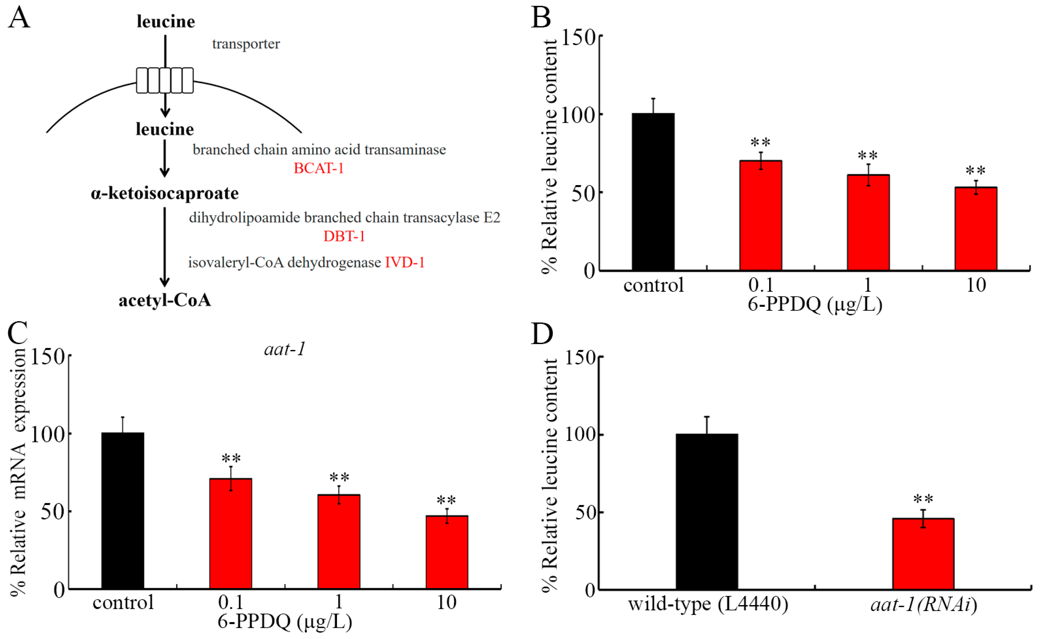
Figure 1. The effect of 6-PPDQ exposure on leucine content and adsorption. (A) A diagram showing leucine adsorption and catabolism in C. elegans. (B) The effect of 6-PPDQ exposure on leucine content. ** p < 0.01 vs. control. (C) The effect of 6-PPDQ exposure on intestinal aat-1 expression. ** p < 0.01 vs. control. Thirty intact intestines were isolated for qRT-PCR analysis. (D) The effect of RNAi of aat-1 on leucine content in 6-PPDQ-exposed nematodes. The exposure concentration of 6-PPDQ was 10 μg/L. ** p < 0.01 vs. wild-type (L4440).
Toxics 13 00544 g001
Figure 2. Effect of 6-PPDQ exposure on leucine catabolism. (A) Effect of 6-PPDQ exposure on bcat-1 expression. ** p < 0.01 vs. control. (B) Effect of RNAi of bcat-1 on leucine content in 6-PPDQ-exposed nematodes. Exposure concentration of 6-PPDQ was 10 μg/L. ** p < 0.01 vs. wild-type (L4440). (C) Effect of RNAi of bcat-1 on NADH content in 6-PPDQ-exposed nematodes. Exposure concentration of 6-PPDQ was 10 μg/L. ** p < 0.01 vs. wild-type (L4440). (D) Effect of RNAi of bcat-1 on FADH2 content in 6-PPDQ-exposed nematodes. Exposure concentration of 6-PPDQ was 10 μg/L. ** p < 0.01 vs. wild-type (L4440).
Toxics 13 00544 g002
Figure 3. Effect of 6-PPDQ exposure on expressions of dbt-1 and ivd-1 and their functions. (A) Effect of 6-PPDQ exposure on expressions of dbt-1 and ivd-1. ** p < 0.01 vs. control. (B) Effect of RNAi of dbt-1 on NADH content in 6-PPDQ-exposed nematodes. Exposure concentration of 6-PPDQ was 10 μg/L. ** p < 0.01 vs. wild-type (L4440). (C) Effect of RNAi of ivd-1 on FADH2 content in 6-PPDQ-exposed nematodes. Exposure concentration of 6-PPDQ was 10 μg/L. ** p < 0.01 vs. wild-type (L4440). (D) Effect of RNAi of bcat-1 on expressions of dbt-1 and ivd-1 in 6-PPDQ-exposed nematodes. Exposure concentration of 6-PPDQ was 10 μg/L. ** p < 0.01 vs. wild-type (L4440). (E) Effect of 6-PPDQ exposure on NADH and FADH2 contents. ** p < 0.01 vs. control.
Toxics 13 00544 g003
Figure 4. Effect of RNAi of bcat-1, dbt-1, and ivd-1 on oxygen consumption rate (A), ATP content (B), and expressions of gas-1 and mev-1 (C) in 6-PPDQ-exposed nematodes. Exposure concentration of 6-PPDQ was 10 μg/L. ** p < 0.01.
Toxics 13 00544 g004
Figure 5. Effect of RNAi of aat-1, bcat-1, dbt-1, and ivd-1 on 6-PPDQ toxicity in inducing intestinal ROS generation (A), causing intestinal lipofuscin accumulation (B), decreasing locomotion (C), and reducing brood size (D). Exposure concentration of 6-PPDQ was 10 μg/L. ** p < 0.01.
Toxics 13 00544 g005
Figure 6. Effect of treatment with leucine (5 mM) on toxicity of 6-PPDQ (10 μg/L) in affecting expressions of dbt-1 and ivd-1 (A), NADH content (B), FADH2 content (C), oxygen consumption rate (D), ATP content (E), and expressions of gas-1 and mev-1 (F). ** p < 0.01.
Toxics 13 00544 g006
Figure 7. Effect of treatment with leucine (5 mM) on toxicity of 6-PPDQ (10 μg/L) in inducing intestinal ROS generation (A), causing intestinal lipofuscin accumulation (B), decreasing locomotion (C), and reducing brood size (D). ** p < 0.01.
Toxics 13 00544 g007
Disclaimer/Publisher’s Note: The statements, opinions and data contained in all publications are solely those of the individual author(s) and contributor(s) and not of MDPI and/or the editor(s). MDPI and/or the editor(s) disclaim responsibility for any injury to people or property resulting from any ideas, methods, instructions or products referred to in the content.