Machine Learning for Predicting Human Drug-Induced Cardiotoxicity: A Scoping Review

 , ,
, ,
Abstract
1. Introduction
2. Methods
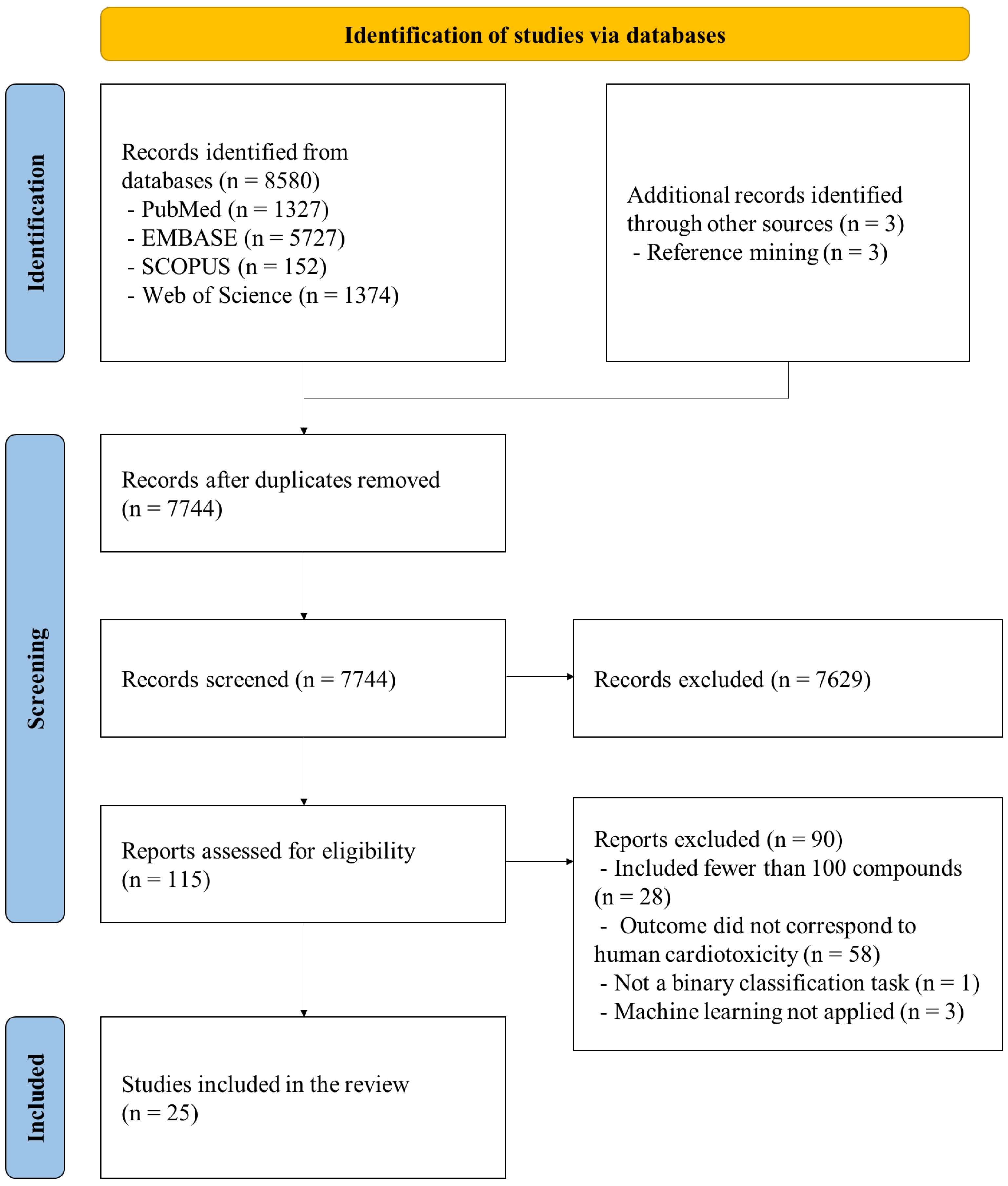
2.1. Study Selection
2.2. Data Extraction and Synthesis
2.3. Study Quality and Risk-of-Bias Assessment
3. Results
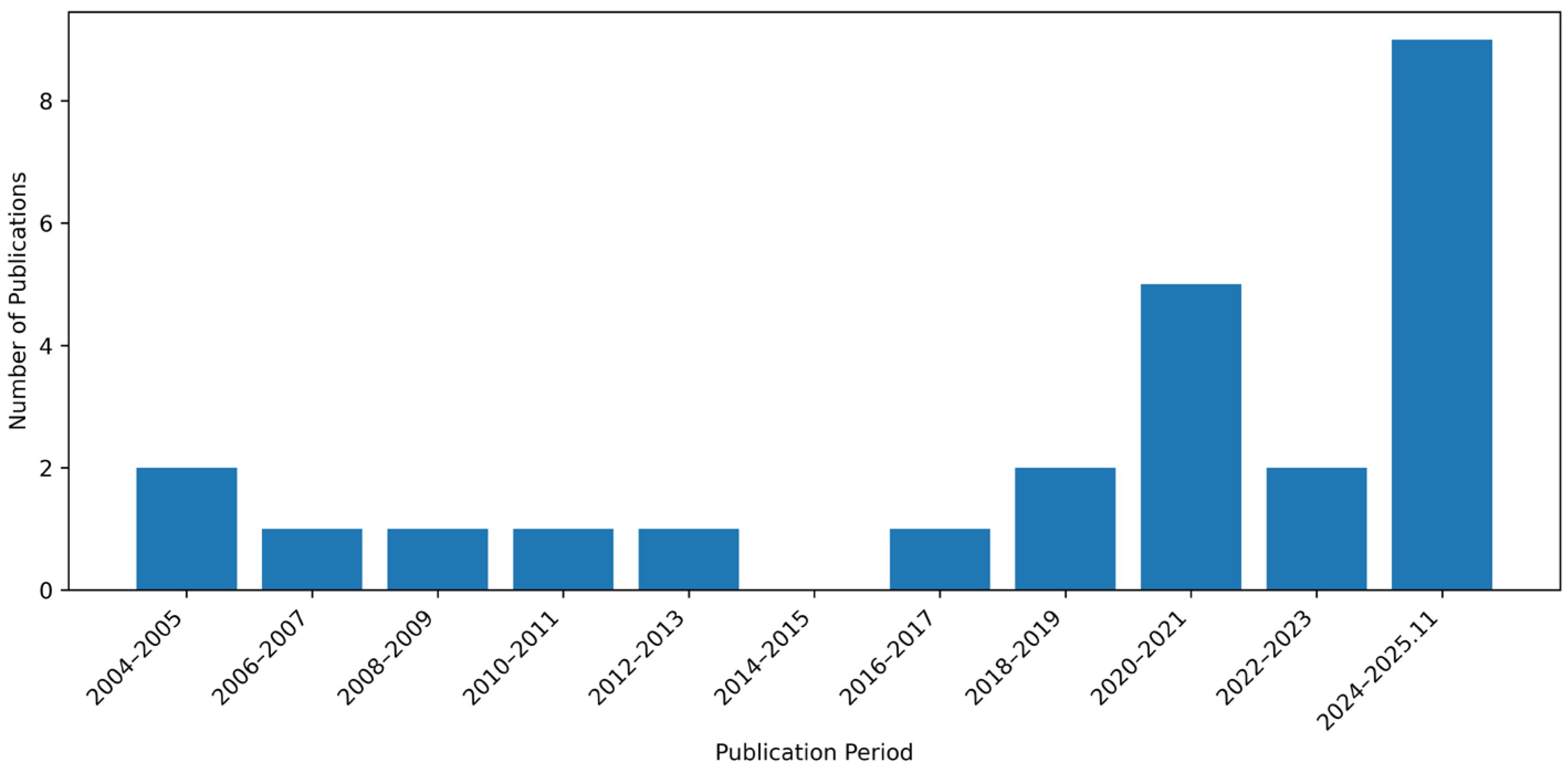
3.1. Summary of Selected Studies
3.2. Summary of Databases, Selected Features, and Algorithms
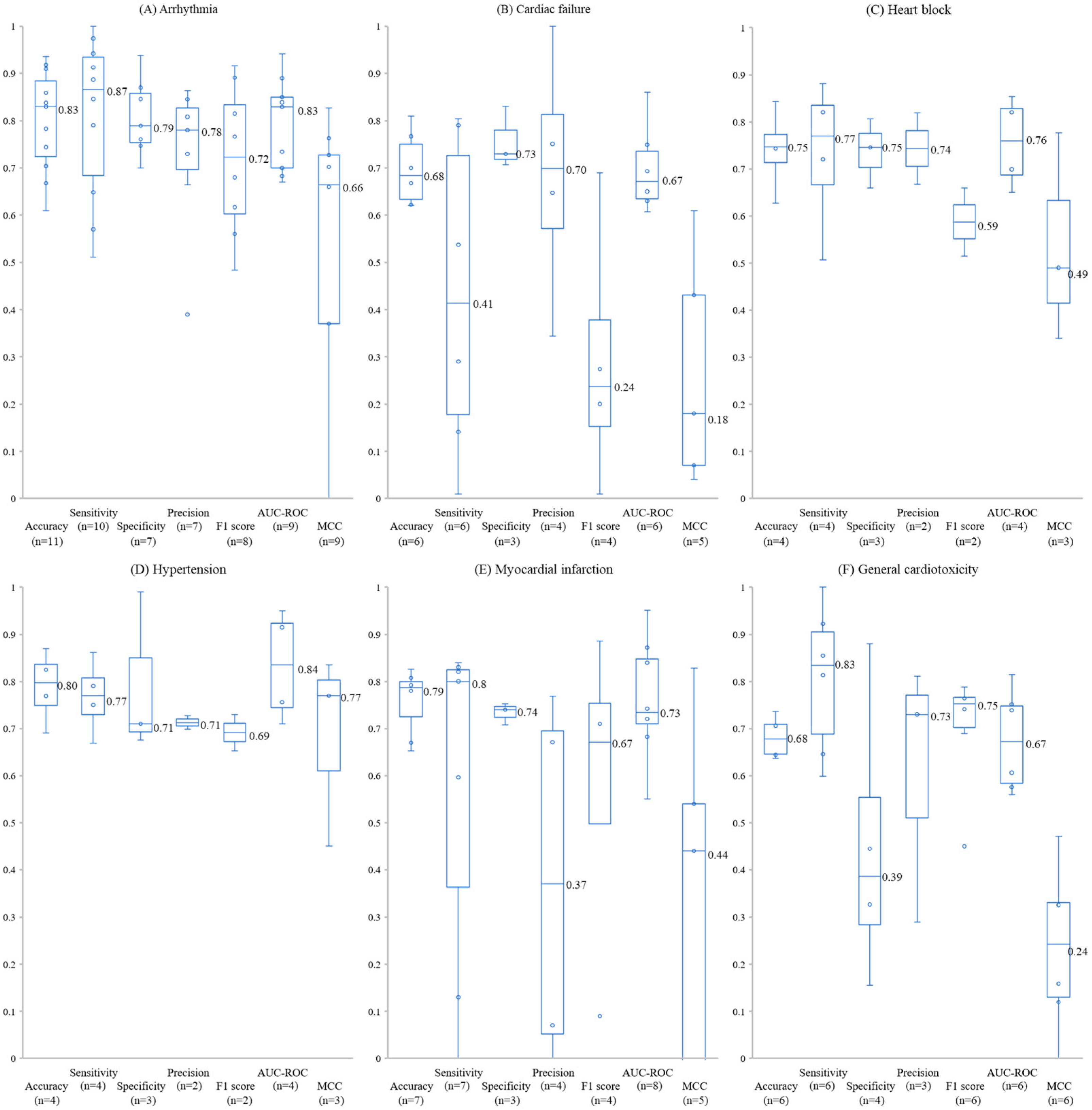
3.3. Performance Across Cardiotoxicity Outcomes
3.3.1. Arrhythmia
3.3.2. Cardiac Failure
3.3.3. Heart Block
3.3.4. Hypertension
3.3.5. Myocardial Infarction
3.3.6. General Cardiotoxicity
3.4. Study Quality
4. Discussion
5. Conclusions
Supplementary Materials
Author Contributions
Funding
Institutional Review Board Statement
Informed Consent Statement
Data Availability Statement
Conflicts of Interest
Abbreviations
| ML | Machine learning |
| hERG | human Ether-à-go-go-Related Gene |
| TdP | Torsades de Pointes |
| QSAR | Quantitative Structure-Activity Relationship |
| SVM | support vector machine |
| AUC-ROC | area under the receiver operating characteristic curve |
| TP | true positive |
| FP | false positive |
| TN | true negative |
| FN | false negative |
| XAI | explainable artificial intelligence |
| MCC | Matthews correlation coefficient |
| SIDER | Side Effect Resource |
| OMOP | Observational Medical Outcomes Partnership |
| LSER | linear solvation energy relationship |
| MOE | molecular operating environment |
| MACCS | Molecular Access System |
| ECFP | Extended Connectivity Fingerprints |
| RF | random forest |
| AGN | attention-based graph neural network |
| XGBoost | extreme gradient boosting |
| DICTrank | Drug-Induced Cardiotoxicity Rank |
| DIQTA | Drug-Induced QT Prolongation Atlas |
| EHRs | electronic health records |
References
- Ferri, N.; Siegl, P.; Corsini, A.; Herrmann, J.; Lerman, A.; Benghozi, R. Drug attrition during pre-clinical and clinical development: Understanding and managing drug-induced cardiotoxicity. Pharmacol. Ther. 2013, 138, 470–484. [Google Scholar] [CrossRef]
- Seal, S.; Spjuth, O.; Hosseini-Gerami, L.; Garcia-Ortegon, M.; Singh, S.; Bender, A.; Carpenter, A.E. Insights into Drug Cardiotoxicity from Biological and Chemical Data: The First Public Classifiers for FDA Drug-Induced Cardiotoxicity Rank. J. Chem. Inf. Model. 2024, 64, 1172–1186. [Google Scholar] [CrossRef]
- Onakpoya, I.J.; Heneghan, C.J.; Aronson, J.K. Post-marketing withdrawal of 462 medicinal products because of adverse drug reactions: A systematic review of the world literature. BMC Med. 2016, 14, 10. [Google Scholar] [CrossRef] [PubMed]
- Mamoshina, P.; Rodriguez, B.; Bueno-Orovio, A. Toward a broader view of mechanisms of drug cardiotoxicity. Cell Rep. Med. 2021, 2, 100216. [Google Scholar] [CrossRef]
- Yang, S.; Kar, S. Application of artificial intelligence and machine learning in early detection of adverse drug reactions (ADRs) and drug-induced toxicity. Artif. Intell. Chem. 2023, 1, 100011. [Google Scholar] [CrossRef]
- Zhang, L.; Tan, J.; Han, D.; Zhu, H. From machine learning to deep learning: Progress in machine intelligence for rational drug discovery. Drug Discov. Today 2017, 22, 1680–1685. [Google Scholar] [CrossRef] [PubMed]
- Jo, S.H.; Hong, H.K.; Jung, S.J.; Chong, S.H.; Yun, J.H.; Koh, Y.S.; Choe, H. Maprotiline block of the human ether-a-go-go-related gene (HERG) K+ channel. Arch. Pharm. Res. 2007, 30, 453–460. [Google Scholar] [CrossRef] [PubMed]
- Doddareddy, M.R.; Klaasse, E.C.; Shagufta; Ijzerman, A.P.; Bender, A. Prospective validation of a comprehensive in silico hERG model and its applications to commercial compound and drug databases. ChemMedChem 2010, 5, 716–729. [Google Scholar] [CrossRef]
- Ogura, K.; Sato, T.; Yuki, H.; Honma, T. Support Vector Machine model for hERG inhibitory activities based on the integrated hERG database using descriptor selection by NSGA-II. Sci. Rep. 2019, 9, 12220. [Google Scholar] [CrossRef]
- Tang, X.; Wang, Z.; Hu, S.; Zhou, B. Assessing Drug-Induced Mitochondrial Toxicity in Cardiomyocytes: Implications for Preclinical Cardiac Safety Evaluation. Pharmaceutics 2022, 14, 1313. [Google Scholar] [CrossRef]
- Li, S.; Xu, H.; Liu, F.; Ni, R.; Shi, Y.; Li, X. In silico prediction of drug-induced cardiotoxicity with ensemble machine learning and structural pattern recognition. Mol. Divers. 2025. [Google Scholar] [CrossRef]
- Tosca, E.M.; Bartolucci, R.; Magni, P.; Poggesi, I. Modeling approaches for reducing safety-related attrition in drug discovery and development: A review on myelotoxicity, immunotoxicity, cardiovascular toxicity, and liver toxicity. Expert Opin. Drug Discov. 2021, 16, 1365–1390. [Google Scholar] [CrossRef] [PubMed]
- Khairnar, S.I.; Kulkarni, Y.A.; Singh, K. Cardiotoxicity linked to anticancer agents and cardioprotective strategy. Arch. Pharm. Res. 2022, 45, 704–730. [Google Scholar] [CrossRef] [PubMed]
- Grafton, F.; Ho, J.; Ranjbarvaziri, S.; Farshidfar, F.; Budan, A.; Steltzer, S.; Maddah, M.; Loewke, K.E.; Green, K.; Patel, S.; et al. Deep learning detects cardiotoxicity in a high-content screen with induced pluripotent stem cell-derived cardiomyocytes. eLife 2021, 10, e68714. [Google Scholar] [CrossRef] [PubMed]
- van Hasselt, J.G.C.; Rahman, R.; Hansen, J.; Stern, A.; Shim, J.V.; Xiong, Y.; Pickard, A.; Jayaraman, G.; Hu, B.; Mahajan, M.; et al. Transcriptomic profiling of human cardiac cells predicts protein kinase inhibitor-associated cardiotoxicity. Nat. Commun. 2020, 11, 4809. [Google Scholar] [CrossRef]
- Yap, C.W.; Cai, C.Z.; Xue, Y.; Chen, Y.Z. Prediction of torsade-causing potential of drugs by support vector machine approach. Toxicol. Sci. 2004, 79, 170–177. [Google Scholar] [CrossRef]
- Xue, Y.; Li, Z.R.; Yap, C.W.; Sun, L.Z.; Chen, X.; Chen, Y.Z. Effect of molecular descriptor feature selection in support vector machine classification of pharmacokinetic and toxicological properties of chemical agents. J. Chem. Inf. Comput. Sci. 2004, 4, 1630–1638. [Google Scholar] [CrossRef]
- Bhavani, S.; Nagargadde, A.; Thawani, A.; Sridhar, V.; Chandra, N. Substructure-based support vector machine classifiers for prediction of adverse effects in diverse classes of drugs. J. Chem. Inf. Model. 2006, 46, 2478–2486. [Google Scholar] [CrossRef]
- Yang, S.Y.; Huang, Q.; Li, L.L.; Ma, C.Y.; Zhang, H.; Bai, R.; Teng, Q.Z.; Xiang, M.L.; Wei, Y.Q. An integrated scheme for feature selection and parameter setting in the support vector machine modeling and its application to the prediction of pharmacokinetic properties of drugs. Artif. Intell. Med. 2009, 46, 155–163. [Google Scholar] [CrossRef]
- Huang, L.C.; Wu, X.; Chen, J.Y. Predicting adverse side effects of drugs. BMC Genom. 2011, 12, S11. [Google Scholar] [CrossRef]
- He, Y.; Lim, S.W.; Yap, C.W. Determination of torsade-causing potential of drug candidates using one-class classification and ensemble modelling approaches. Curr. Drug Saf. 2012, 7, 298–308. [Google Scholar] [CrossRef] [PubMed]
- Wang, Z.; Clark, N.R.; Ma’ayan, A. Drug-induced adverse events prediction with the LINCS L1000 data. Bioinformatics 2016, 32, 2338–2345. [Google Scholar] [CrossRef] [PubMed]
- Cai, C.; Fang, J.; Guo, P.; Wang, Q.; Hong, H.; Moslehi, J.; Cheng, F. In Silico Pharmacoepidemiologic Evaluation of Drug-Induced Cardiovascular Complications Using Combined Classifiers. J. Chem. Inf. Model. 2018, 58, 943–956. [Google Scholar] [CrossRef] [PubMed]
- Dey, S.; Luo, H.; Fokoue, A.; Hu, J.; Zhang, P. Predicting adverse drug reactions through interpretable deep learning framework. BMC Bioinform. 2018, 19, 476. [Google Scholar] [CrossRef] [PubMed]
- Amano, Y.; Honda, H.; Sawada, R.; Nukada, Y.; Yamane, M.; Ikeda, N.; Morita, O.; Yamanishi, Y. In silico systems for predicting chemical-induced side effects using known and potential chemical protein interactions, enabling mechanism estimation. J. Toxicol. Sci. 2020, 45, 137–149. [Google Scholar] [CrossRef]
- Llopis-Lorente, J.; Gomis-Tena, J.; Cano, J.; Romero, L.; Saiz, J.; Trenor, B. In Silico Classifiers for the Assessment of Drug Proarrhythmicity. J. Chem. Inf. Model. 2020, 60, 5172–5187. [Google Scholar] [CrossRef]
- Mamoshina, P.; Bueno-Orovio, A.; Rodriguez, B. Dual Transcriptomic and Molecular Machine Learning Predicts all Major Clinical Forms of Drug Cardiotoxicity. Front. Pharmacol. 2020, 11, 639. [Google Scholar] [CrossRef]
- Joshi, P.; Vedhanayagam, M.; Ramesh, R. An Ensembled SVM Based Approach for Predicting Adverse Drug Reactions. Curr. Bioinform. 2021, 16, 422–432. [Google Scholar] [CrossRef]
- Kadioglu, O.; Klauck, S.M.; Fleischer, E.; Shan, L.; Efferth, T. Selection of safe artemisinin derivatives using a machine learning-based cardiotoxicity platform and in vitro and in vivo validation. Arch. Toxicol. 2021, 95, 2485–2495. [Google Scholar] [CrossRef]
- Iftkhar, S.; de Sa, A.G.C.; Velloso, J.P.L.; Aljarf, R.; Pires, D.E.V.; Ascher, D.B. cardioToxCSM: A Web Server for Predicting Cardiotoxicity of Small Molecules. J. Chem. Inf. Model. 2022, 62, 4827–4836. [Google Scholar] [CrossRef] [PubMed]
- Ye, L.; Ngan, D.K.; Xu, T.; Liu, Z.; Zhao, J.; Sakamuru, S.; Zhang, L.; Zhao, T.; Xia, M.; Simeonov, A. Prediction of drug-induced liver injury and cardiotoxicity using chemical structure and in vitro assay data. Toxicol. Appl. Pharmacol. 2022, 454, 116250. [Google Scholar] [CrossRef]
- Çelik, F.K.; Doğan, S.; Karaduman, G. Drug-induced torsadogenicity prediction model: An explainable machine learning-driven quantitative structure-toxicity relationship approach. Comput. Biol. Med. 2024, 182, 109209. [Google Scholar] [CrossRef]
- Lin, J.; He, Y.; Ru, C.; Long, W.; Li, M.; Wen, Z. Advancing Adverse Drug Reaction Prediction with Deep Chemical Language Model for Drug Safety Evaluation. Int. J. Mol. Sci. 2024, 25, 4516. [Google Scholar] [CrossRef] [PubMed]
- Vinh, T.; Nguyen, L.; Trinh, Q.H.; Nguyen-Vo, T.H.; Nguyen, B.P. Predicting Cardiotoxicity of Molecules Using Attention-Based Graph Neural Networks. J. Chem. Inf. Model. 2024, 64, 1816–1827. [Google Scholar] [CrossRef] [PubMed]
- He, Y.; Lv, X.; Long, W.; Zhai, S.; Li, M.; Wen, Z. ToxBERT: An explainable AI framework for enhancing prediction of adverse drug reactions and structural insights. J. Pharm. Anal. 2025, 15, 101387. [Google Scholar] [CrossRef]
- Li, T.; Qu, Y.; Chen, A.; Thakkar, S.; Li, D.; Tong, W. Beyond QSARs: Quantitative Knowledge-Activity Relationships (QKARs) for Enhanced Drug Toxicity Prediction. Toxicol. Sci. 2025, 208, 269–278. [Google Scholar] [CrossRef]
- Qu, Y.; Li, T.; Liu, Z.; Tong, W.; Li, D. DICTrank Is a Reliable Dataset for Cardiotoxicity Prediction Using Machine Learning Methods. Chem. Res. Toxicol. 2025, 38, 647–655. [Google Scholar] [CrossRef]
- Rouen, K.C.; Narang, K.; Han, Y.; Wang, D.; Jang, E.; Brunkow, S.; Yarov-Yarovoy, V.; MacKerell, A.D., Jr.; Vorobyov, I. Prediction of TdP Arrhythmia Risk Through Molecular Simulations of Conformation-specific Drug Interactions with the hERG K+, NaV1.5, and CaV1.2 Channels. bioRxiv 2025. [Google Scholar] [CrossRef]
- De Ponti, F.; Poluzzi, E.; Montanaro, N. Organising evidence on QT prolongation and occurrence of Torsades de Pointes with non-antiarrhythmic drugs: A call for consensus. Eur. J. Clin. Pharmacol. 2001, 57, 185–209. [Google Scholar] [CrossRef] [PubMed]
- Karim, A.; Lee, M.; Balle, T.; Sattar, A. CardioTox net: A robust predictor for hERG channel blockade based on deep learning meta-feature ensembles. J. Cheminform. 2021, 13, 60. [Google Scholar] [CrossRef]
- Munawar, S.; Windley, M.J.; Tse, E.G.; Todd, M.H.; Hill, A.P.; Vandenberg, J.I.; Jabeen, I. Experimentally Validated Pharmacoinformatics Approach to Predict hERG Inhibition Potential of New Chemical Entities. Front. Pharmacol. 2018, 9, 1035. [Google Scholar] [CrossRef] [PubMed]
- Kuhn, M.; Letunic, I.; Jensen, L.J.; Bork, P. The SIDER database of drugs and side effects. Nucleic Acids Res. 2016, 44, D1075–D1079. [Google Scholar] [CrossRef] [PubMed]
- Ryan, P.B.; Schuemie, M.J.; Welebob, E.; Duke, J.; Valentine, S.; Hartzema, A.G. Defining a reference set to support methodological research in drug safety. Drug Saf. 2013, 36, 33–47. [Google Scholar] [CrossRef]
- Qu, Y.; Li, T.; Liu, Z.; Li, D.; Tong, W. DICTrank: The largest reference list of 1318 human drugs ranked by risk of drug-induced cardiotoxicity using FDA labeling. Drug Discov. Today 2023, 28, 103770. [Google Scholar] [CrossRef]
- Klon, A.E. Machine learning algorithms for the prediction of hERG and CYP450 binding in drug development. Expert Opin. Drug Metab. Toxicol. 2010, 6, 821–833. [Google Scholar] [CrossRef]
- Li, S.; Xu, Z.; Guo, M.; Li, M.; Wen, Z. Drug-induced QT Prolongation Atlas (DIQTA) for enhancing cardiotoxicity management. Drug Discov. Today 2022, 27, 831–837. [Google Scholar] [CrossRef]
- Guo, W.; Liu, J.; Dong, F.; Song, M.; Li, Z.; Khan, M.K.H.; Patterson, T.; Hong, H. Review of machine learning and deep learning models for toxicity prediction. Exp. Biol. Med. 2023, 248, 1952–1973. [Google Scholar] [CrossRef]
- Falcón-Cano, G.; Morales-Helguera, A.; Lambert, H.; Cabrera-Pérez, M.; Molina, C. hERG toxicity prediction in early drug discovery using extreme gradient boosting and isometric stratified ensemble mapping. Sci. Rep. 2025, 15, 15585. [Google Scholar] [CrossRef]
- Hossain, D.; Al Abir, F.; Chen, J.Y. hERG-LTN: A New Paradigm in hERG Cardiotoxicity Assessment Using Neuro-Symbolic and Generative AI Embedding (MegaMolBART, Llama3.2, Gemini, DeepSeek) Approach. bioRxiv 2025. [Google Scholar] [CrossRef]
- Jing, Y.; Zhang, Y.; Zhao, G.; McGuire, T.; Zhao, J.; Gibbs, B.; Hou, G.; Feng, Z.; Xue, Y.; Xie, X.Q. GraphDeep-hERG: Graph Neural Network PharmacoAnalytics for Assessing hERG-Related Cardiotoxicity. Pharm. Res. 2025, 42, 579–591. [Google Scholar] [CrossRef] [PubMed]
- Mayr, A.; Klambauer, G.; Unterthiner, T.; Hochreiter, S. DeepTox: Toxicity prediction using deep learning. Front. Environ. Sci. 2016, 3, 80. [Google Scholar] [CrossRef]
- Cavasotto, C.N.; Scardino, V. Machine Learning Toxicity Prediction: Latest Advances by Toxicity End Point. ACS Omega 2022, 7, 47536–47546. [Google Scholar] [CrossRef] [PubMed]
- van Berlo, J.H.; Maillet, M.; Molkentin, J.D. Signaling effectors underlying pathologic growth and remodeling of the heart. J. Clin. Investig. 2013, 123, 37–45. [Google Scholar] [CrossRef]
- Hu, Q.; Li, J.; Li, X.; Zou, D.; Xu, T.; He, Z. Machine learning to predict adverse drug events based on electronic health records: A systematic review and meta-analysis. J. Int. Med. Res. 2024, 52, 3000605241302304. [Google Scholar] [CrossRef]
- International Conference on Harmonisation. The Non-Clinical Evaluation of the Potential for Delayed Ventricular Repolarization (QT Interval Prolongation) by Human Pharmaceuticals. 2005. Available online: https://www.ich.org/page/safety-guidelines (accessed on 10 December 2025).
- U.S. Food and Drug Administration. Artificial Intelligence-Enabled Device Software Functions: Lifecycle Management and Marketing Submission Recommendations. 2025. Available online: https://www.fda.gov/regulatory-information/search-fda-guidance-documents/artificial-intelligence-enabled-device-software-functions-lifecycle-management-and-marketing (accessed on 10 December 2025).
- Tricco, A.C.; Lillie, E.; Zarin, W.; O’Brien, K.K.; Colquhoun, H.; Levac, D.; Moher, D.; Peters, M.D.J.; Horsley, T.; Weeks, L.; et al. PRISMA Extension for Scoping Reviews (PRISMAScR): Checklist and Explanation. Ann. Intern. Med. 2018, 169, 467–473. [Google Scholar] [CrossRef] [PubMed]



| Source | Search Query | Date Range | Hits |
|---|---|---|---|
| PubMed | (“Cardiotoxicity”[Mesh] AND “Myocardial Infarction”[Mesh] OR “Heart Valves” [Mesh] OR “Atrial Fibrillation”[Mesh] OR “Arrhythmias, Cardiac” [Mesh] OR “Long QT Syndrome”[Mesh] OR “Torsades de Pointes”[Mesh] OR “Atrial Flutter”[Mesh]) AND (“Artificial Intelligence”[Mesh] OR artificial intelligence OR machine learning OR deep learning) AND (drug OR medication OR pharmaceutic *) | Up to 25 November 2025 | 1327 |
| Embase | #1. ‘cardiotoxicity’/exp OR cardiotoxicity #2. ‘heart infarction’/exp OR ‘heart infarction’ #3. ‘myocardial infarction’ #4. ‘heart valve’/exp OR ‘heart valve’ #5 ‘atrial fibrillation’/exp OR ‘atrial fibrillation’ #6. ‘heart arrhythmia’/exp OR ‘heart arrhythmia’ #7. ‘qt prolongation’/exp OR ‘qt prolongation’ #8. ‘torsade de pointes’/exp OR ‘torsade de pointes’ #9. ‘heart atrium flutter’/exp OR ‘heart atrium flutter’ #10. ‘atrial flutter’ #11. #1 OR #2 OR #3 OR #4 OR #5 OR #6 OR #7 OR #8 OR #9 OR #10 #12. ‘drug’/exp OR drug #13. Medication #14. pharmaceutic * #15. #12 OR #13 OR #14 #16. ‘artificial intelligence’/exp OR ‘artificial intelligence’ #17. ‘machine learning’/exp OR ‘machine learning’ #18. ‘deep learning’/exp OR ‘deep learning’ #19. #16 OR #17 OR #18 #20. #11 AND #15 AND #19 | Up to 25 November 2025 | 5727 |
| Web of Science | (TS = (Cardiotoxicity) OR TS = (Myocardial Infarction) OR TS = (Heart Valves) OR TS = (Atrial Fibrillation) OR TS = (arrhythmia) OR TS = (qt prolongation) OR TS = (Torsades de Pointes) OR TS = (Atrial Flutter)) AND (ALL = (artificial intelligence) OR ALL = (machine learning) OR ALL = (deep learning)) AND (ALL = (drug) OR ALL = (medication) OR ALL = (pharmaceutic *)) | Up to 25 November 2025 | 1374 |
| SCOPUS | ((TITLE-ABS-KEY(Cardiotoxicity)) OR (TITLE-ABS-KEY(Myocardial Infarction)) OR (TITLE-ABS-KEY(Heart Valves)) OR (TITLE-ABS-KEY (Atrial Fibrillation)) AND (TITLE-ABS-KEY(arrhythmia)) AND (TITLE-ABS-KEY(qt prolongation)) OR (TITLE-ABS-KEY(Torsades de Pointes)) OR (TITLE-ABS-KEY(Atrial Flutter))) AND ((ALL(artificial intelligence)) OR (ALL(machine learning)) OR (ALL(deep learning))) AND ((ALL(drug)) OR (ALL(medication)) OR (ALL(pharmaceutic *))) | Up to 25 November 2025 | 152 |
| Reference | Year | Cardiotoxicity Database | Included Features | Outcome | Algorithms | Summary | |
|---|---|---|---|---|---|---|---|
| Molecular Features | Other Features | ||||||
| [16] | 2004 | AZCERT, Micromedex *, Drug Information Handbook *, Meyler’s Side Effects of Drugs *, De ponti et al. [39] *, AHFS Drug Information * | LSER descriptors | Arrhythmia (TdP) | SVM PNN kNN C4.5 decision tree | SVM approach using LSER descriptors achieved prediction accuracies of 97.4% for TdP-causing agents and 84.6% for non-TdP-causing agents. | |
| [17] | 2004 | AZCERT, Micromedex, Drug Information Handbook, Meyler’s Side Effects of Drugs, AHFS Drug Information | Molecular descriptors | Arrhythmia (TdP) | SVM | The SVM model using recursive feature elimination achieved accuracies of 66.8% for TdP+ and 89.3% for TdP−. | |
| [18] | 2006 | Yap et al. [16] | Number of instances of substructures, Euclidean distances | Arrhythmia (TdP) | SVM | Combining weighted instances with the Euclidean distance measure improved prediction accuracy to 93.59%. | |
| [19] | 2009 | Xue et al. [17] | Molecular descriptors (PCLI-ENT online program) | Arrhythmia (TdP) | SVM | The SVM model using a genetic algorithm and the conjugate gradient method achieved accuracies of 64.7% for TdP+ and 92.8% for TdP−. | |
| [20] | 2011 | SIDER | Drug target expanding protein-protein interaction networks network | General cardiotoxicity | SVM LR | Incorporating protein-protein interaction networks and gene ontology annotations significantly improved SVM-based cardiotoxicity prediction, achieving a median AUC of 0.771, accuracy of 0.675, sensitivity of 0.632, and specificity of 0.789. | |
| [21] | 2012 | Micromedex, AZCERT | Molecular descriptors (PaDEL-descriptor version 2.7) | Arrhythmia (TdP) | OCSVM OCLOF OCPD Their ensemble model | A final ensemble model was constructed based on selected base models and it had sensitivity and specificity value of 78.4% and 90% respectively. | |
| [22] | 2016 | SIDER, Offsides, OMOP * | Molecular fingerprints (MACCS) | Gene ontology enrichment vector from gene expression (LINCS L1000 dataset) | Myocardial infraction | RF SVM L1-regularized LR NB kNN Extra tree | Extra tree classifiers show improved AUC-ROC, F1-score, and accuracy for acute myocardial infarction when combining gene ontology enrichment vectors with chemical structure features. Available at: http://maayanlab.net/SEP-L1000/ (accessed on 10 December 2025) |
| [23] | 2018 | Comparative Toxicogenomics Database, SIDER, MetaADEDB, Offsides * | 2D descriptors (MOE 2010 software), molecular fingerprints (MACCS, Estate, Pubchem, substructure fingerprint) | Arrhythmia, cardiac failure, heart block, hypertension, myocardial infarction | kNN LR RF SVM Their combined classifier | Combined classifiers had 87% success rate in predicting cardiotoxicity. | |
| [24] | 2018 | SIDER, OMOP * | Molecular fingerprint (ECFP6) | Myocardial infarction | L2-regularized LR | The ECFP6 circular fingerprint method outperformed the neural fingerprint method in predicting acute myocardial infarction. | |
| [25] | 2020 | SIDER | Known and newly estimated target proteins | General cardiotoxicity | L1-regularized LR | Chemical-protein interaction-base model performed better than or comparable to the chemical structure-based model. | |
| [26] | 2020 | CredibleMeds | Tx (drug concentration prolonging action potential by 10%), TqNet (net charge carried by ionic currents), Ttriang (triangulation of drug concentration over control) | Arrhythmia (TdP) | Decision tree | Combining Tx, TqNet, and Ttraing in a decision tree classifier improved prediction accuracy to 94.5%. Available at: https://riunet.upv.es/handle/10251/136919 (accessed on 10 December 2025) | |
| [27] | 2020 | SIDER | Molecular weight, partition coefficient, atomic polarizabilities, topological polar surface area, polar surface area expressed as a ratio to molecular size, Ghose-Crippen LogKow, molar refractivity, molecular fingerprints (Estate) | Transcriptional profiles of landmark genes | Arrhythmia, cardiac failure, myocardial disorders, general cardiotoxicity | RF GB CatBoost Elastic net | The chain of RF classifiers achieved the best performance, with an average AUC of 0.79 on validation and 0.66 on testing across all cardiotoxicity forms. |
| [28] | 2021 | SIDER, OMOP * | Atom-level features (type of atoms, degree of the atom, number of hydrogen atoms connected, valence of the atom), bond-level features (bond type, conjugation, ring presence) | Myocardial infarction | Ensemble SVM | Ensembled SVM based approach outperformed the competing method in predicting acute myocardial infraction, achieving a sensitivity of 0.84. | |
| [29] | 2021 | DrugBank | LogP, drug likeness, amines, ligand efficiency, alkyl-amines, aromatic nitrogens, basic nitrogens | Arrhythmia, cardiac failure, heart block, hypertension, myocardial infarction | RF SVM kNN NB AdaBoost | RF demonstrated optimal performance, achieving AUC scores above 0.830 across all five cardiotoxicity indications. | |
| [30] | 2022 | Karim et al. [40], Cai et al. [23], Munawar et al. [41] | General molecular descriptors from RDKit, graph-based signatureso model geometry and physicochemical properties, toxicophore matchings via substructure search, molecular fingerprints | Arrhythmia, cardiac failure, heart block, hypertension, myocardial infarction | RF GB XGBoost | The models achieved AUCs of up to 0.898 under 5-fold cross-validation and generalizable performance (up to 0.951) in the blind test sets. Available at: https://biosig.lab.uq.edu.au/cardiotoxcsm (accessed on 10 December 2025) | |
| [31] | 2022 | ChemIDplus, Pharmapendium®, CDER, NCTR, Enzo Life Sciences cardiotoxicity library, SIDER | Extended-connectivity fingerprints (ECFP4) | General cardiotoxicity | RF NB XGBoost SVM | Chemical structure-based models showed good predictive power for DICT (AUC-ROC = 0.83 ± 0.03), while Tox21 assay data performed only slightly better than random. | |
| [32] | 2024 | CredibleMeds | Molecular descriptors (PaDEL-descriptor) | Arrhythmia (TdP) | SVM XGBoost RF CatBoost | RF algorithm achieved the best overall performance across accuracy rate, sensitivity, MCC, and F1 score. | |
| [33] | 2024 | DIQTA | SMILES | Arrhythmia (QTc prolongation) | MoLFormer-XL-CNN | MoLFormer-XL-CNN model, using SMILES as input, outperformed conventional models in predicting DIQT. | |
| [2] | 2024 | DICTrank, SIDER | Mordred descriptors, ECFP4 fingerprints | Mechanism of action, CELLSCAPE target prediction dataset, cell painting, gene expression, gene ontology, Cmax | General cardiotoxicity | RF | Models based on physicochemical properties achieved high predictive accuracy (AUCPR = 0.93). Available at: https://broad.io/DICTrank_Predictor (accessed on 10 December 2025) |
| [34] | 2024 | Iftkhar et al. [30] | Atomic, degree, IsCharge, orbital hybridization, IsAromatic, Caln-Ingold-Prelog priority, chirality features | Arrhythmia, cardiac failure, heart block, hypertension, myocardial infarction | AGN | The simplified attention-based graph neural network outperformed traditional ML and advanced graph-based models. | |
| [35] | 2025 | DIQTA | SMILES | Arrhythmia (QTc prolongation) | BERT | ToxBERT achieved AUC-ROC of 0.839 for predicting drug-induced QT prolongation. | |
| [11] | 2025 | Cai et al. [23], Iftkhar et al. [30] | Molecular descriptors (Online Chemical Modeling Environment platform) | Arrhythmia, cardiac failure, heart block, hypertension, myocardial infarction | Ensemble model of 7 machine learning and 5 deep learning models | A total of 110 predictive models were constructed for each cardiotoxicity endpoint and the consensus models consistently outperformed individual models. Available at: https://ochem.eu/article/166881 (accessed on 10 December 2025) | |
| [36] | 2025 | DICTrank | Chemical descriptors (Mordred) | GPT-4o–generated pharmacology-and-toxicity summary embedding | General cardiotoxicity | LR kNN RF SVM XGBoost | Quantitative Knowledge & Structure-Activity Relationships model consistently outperformed QSAR model |
| [37] | 2025 | DICTrank | Molecular descriptors (Mold2 software) | General cardiotoxicity | LR kNN SVM RF XGBoost | LR and XGBoost achieved the best results with DICTrank. | |
| [38] | 2025 | CredibleMeds | Molecular descriptors | Drug interactions with the hERG, NaV1.5, CaV1.2 channels | Arrhythmia (TdP) | LR RF XGBoost Neural net | RF model using computed drug binding affinities for hERG and CaV1.2 channels achieved 94% overall model accuracy. |
| Database | Last Update | Evidence | Label | Noise/Limitation | ML Suitability |
|---|---|---|---|---|---|
| SIDER | 21 October 2015 | Drug labels | Positive label-only: drug–adverse event pairs explicitly reported in the labels. | Negative label: positive label-only. Update: outdated. Evidence reliability: text-extracted AEs may be ambiguous or noisy. Labeling variability introduces noise. | Useful for large-scale drug–AE association modeling. Freely available for download. |
| CredibleMeds | Updated regularly | Drug labels, literature, case reports | Positive label: drugs classified into Known, Possible, and Conditional risk of TdP based on expert review. Negative label: therapeutic options that are not included on the QT drug list. | Negative label: non-listed drugs are not guaranteed safe. Accessibility: limited scope (TdP) and login required. Evidence reliability: Relies on available literature and case reports (publication bias). | High-quality expert labels valuable for supervised classification of QT/TdP risk; limited size but strong signal. |
| DICTrank | November 2023 | Drug labels | Four-level label: drugs classified into Most-, Less-, Ambiguous-, or No-DICT-Concern based on FDA labeling sections and the severity of reported cardiotoxicity. Most-DICT-Concern: withdrawn drugs or those with BW-level or severe/moderate DICT in WP. Less-DICT-Concern: mild DICT in WP or DICT mentioned in AR/Overdosage. Ambiguous-DICT-Concern: DICT keywords present only in special clinical contexts. No-DICT-Concern: no DICT-related information in any labeling section. | Negative label: “No-DICT-Concern” only reflects absence in labeling, not true safety. Update: static snapshot. Evidence reliability: rule-based classification may misinterpret context. Labeling variability introduces noise. | Structured multi-level toxicity labels good for ordinal classification. Freely available for download. |
| Micromedex | Updated regularly | Drug labels, literature | Positive label-only: drug–adverse event pairs explicitly reported in the labels or literature. | Negative label: positive label-only. Accessibility: subscription-based Evidence reliability: mixed sources lead to variable consistency. Variable evidence depth. | Good for training models requiring clinically validated AE signals. |
| OMOP | October 2013 | Drug labels, literature | Positive label: drugs with MI-related evidence from labels and the literature, with no conflicting studies. Negative label: drugs without MI-related terms in labels, without supporting literature. | Negative label: absence of MI-related evidence does not ensure no risk. Update: outdated Evidence reliability: publication bias. | Positive/negative AE associations useful for binary classification and validation tasks. |
| Outcome | Model | Number of Compounds | Performance Metrics | Reference | |||||||||
|---|---|---|---|---|---|---|---|---|---|---|---|---|---|
| Total | Train | Test | External | Accuracy | Sensitivity | Specificity | Precision | F1 Score | AUC-ROC | MCC | |||
| Arrhythmia | AGN | 1496 | 1346 | 150 | - | [b] 0.632 [c] 0.744 | - | - | - | [b] 0.573 [c] 0.484 | [b] 0.692 [c] 0.850 | [b] 0.271 [c] 0.727 | [34] |
| BERT | 251 | - | - | - | [c] 0.783 | [c] 0.913 | [c] 0.700 | [c] 0.664 | [c] 0.766 | [c] 0.839 | [c] 0.664 | [35] | |
| CNN | 255 | - | - | - | [c] 0.859 | [c] 0.942 | [c] 0.747 | [c] 0.845 | [c] 0.891 | [c] 0.829 | [c] 0.702 | [33] | |
| Decision tree | 109 | 109 | - | - | [b] 0.945 | [b] 0.940 | [b] 0.950 | - | - | - | - | [26] | |
| Ensemble model | 260 | 260 | - | - | [a] 0.912 [b] 0.856 | [a] 0.889 [b] 0.784 | [a] 0.928 [b] 0.900 | - | - | [a] 0.932 [b] 0.825 | [a] 0.817 [b] 0.692 | [21] | |
| 1592 | 1450 | - | 142 | [b] 0.717 [d] 0.704 | [b] 0.712 [d] 0.648 | [b] 0.723 [d] 0.761 | [b] 0.720 [d] 0.730 | - | [b] 0.784 [d] 0.734 | - | [23] | ||
| 2169 | 1743 | 426 | - | [c] 0.830 | [c] 0.790 | [c] 0.870 | - | - | [c] 0.890 | [c] 0.660 | [11] | ||
| RF | 408 | 291 | 66 | 51 | [b] 0.735 [c] 0.668 [d] 0.610 | [b] 0.596 [c] 0.511 [d] 1.000 | - | [b] 0.860 [c] 0.780 [d] 0.390 | [b] 0.704 [c] 0.617 [d] 0.560 | [b] 0.880 [c] 0.683 [d] 0.700 | [b] 0.504 [c] 0.370 [d] 0 | [27] | |
| 1594 | 1451 | - | 143 | [a] 0.770 [d] 0.838 | [a] 0.775 [d] 0.887 | [a] 0.764 [d] 0.789 | [a] 0.767 [d] 0.808 | - | [a] 0.849 | - | [29] | ||
| 1568 | 1410 | 158 | - | - | [b] 0.630 [c] 0.570 | - | - | [b] 0.700 [c] 0.680 | [b] 0.710 [c] 0.670 | [b] 0.400 [c] 0.370 | [30] | ||
| 141 | 98 | 43 | - | [b] 0.837 | [b] 0.963 | [b] 0.625 | [b] 0.625 | [b] 0.758 | - | [b] 0.651 | [32] | ||
| 300 | - | - | - | [c] 0.918 | [c] 0.846 | [c] 0.938 | - | [c] 0.815 | [c] 0.941 | [c] 0.763 | [38] | ||
| SVM | 349 | 271 | - | 78 | [d] 0.910 | [d] 0.974 | [d] 0.846 | [d] 0.864 | [d] 0.916 | - | [d] 0.827 | [16] | |
| 361 | 361 | - | - | [b] 0.839 | [b] 0.668 | [b] 0.893 | - | - | - | [b] 0.560 | [17] | ||
| 349 | 271 | - | 78 | [d] 0.936 | - | - | - | - | - | - | [18] | ||
| 361 | 361 | - | - | [b] 0.861 | [b] 0.647 | [b] 0.928 | - | - | - | - | [19] | ||
| Cardiac Failure | AGN | 1096 | 986 | 110 | - | [b] 0.517 [c] 0.621 | - | - | - | [b] 0.187 [c] 0.274 | [b] 0.627 [c] 0.749 | [b] 0.049 [c] 0.431 | [34] |
| Ensemble model | 1170 | 630 | - | 540 | [b] 0.711 [d] 0.622 | [b] 0.686 [d] 0.537 | [b] 0.737 [d] 0.707 | [b] 0.722 [d] 0.647 | - | [b] 0.785 [d] 0.693 | - | [23] | |
| 1201 | 1066 | 135 | - | [c] 0.810 | [c] 0.790 | [c] 0.830 | - | - | [c] 0.860 | [c] 0.610 | [11] | ||
| RF | 408 | 291 | 66 | 51 | [b] 0.676 [c] 0.668 [d] 0.700 | [b] 0.484 [c] 0.141 [d] 0.010 | - | [b] 0.583 [c] 0.344 [d] 1.000 | [b] 0.529 [c] 0.200 [d] 0.010 | [b] 0.787 [c] 0.607 [d] 0.650 | [b] 0.288 [c] 0.040 [d] 0.070 | [27] | |
| 1164 | 626 | - | 538 | [a] 0.755 [d] 0.767 | [a] 0.768 [d] 0.804 | [a] 0.742 [d] 0.730 | [a] 0.752 [d] 0.751 | - | [a] 0.831 | - | [29] | ||
| XGBoost | 1153 | 1037 | 116 | - | - | [b] 0.350 [c] 0.290 | - | - | [b] 0.730 [c] 0.690 | [b] 0.610 [c] 0.630 | [b] 0.290 [c] 0.180 | [30] | |
| Heart Block | AGN | 883 | 794 | 89 | - | [b] 0.651 [c] 0.743 | - | - | - | [b] 0.656 [c] 0.515 | [b] 0.708 [c] 0.854 | [b] 0.306 [c] 0.777 | [34] |
| Ensemble model | 946 | 544 | - | 402 | [b] 0.767 [d] 0.627 | [b] 0.721 [d] 0.507 | [b] 0.813 [d] 0.746 | [b] 0.794 [d] 0.667 | - | [b] 0.842 [d] 0.699 | - | [23] | |
| 1488 | 1177 | 311 | - | [c] 0.750 | [c] 0.820 | [c] 0.660 | - | - | [c] 0.820 | [c] 0.490 | [11] | ||
| RF | 948 | 545 | - | 403 | [a] 0.781 [d] 0.843 | [a] 0.783 [d] 0.881 | [a] 0.779 [d] 0.806 | [a] 0.780 [d] 0.819 | - | [a] 0.869 | - | [29] | |
| 932 | 838 | 94 | - | - | [b] 0.750 [c] 0.720 | - | - | [b] 0.730 [c] 0.660 | [b] 0.720 [c] 0.650 | [b] 0.460 [c] 0.340 | [30] | ||
| Hypertension | AGN | 1311 | 1179 | 132 | - | [b] 0.622 [c] 0.825 | - | - | - | [b] 0.605 [c] 0.653 | [b] 0.676 [c] 0.915 | [b] 0.247 [c] 0.835 | [34] |
| Ensemble model | 1452 | 1162 | - | 290 | [b] 0.739 [d] 0.690 | [b] 0.750 [d] 0.669 | [b] 0.728 [d] 0.710 | [b] 0.734 [d] 0.698 | - | [b] 0.800 [d] 0.756 | - | [23] | |
| 2228 | 1787 | 441 | - | [c] 0.870 | [c] 0.790 | [c] 0.990 | - | - | [c] 0.950 | [c] 0.770 | [11] | ||
| RF | 1454 | 1163 | - | 291 | [a] 0.781 [d] 0.769 | [a] 0.795 [d] 0.862 | [a] 0.766 [d] 0.676 | [a] 0.773 [d] 0.727 | - | [a] 0.854 | - | [29] | |
| 1374 | 1236 | 138 | - | - | [b] 0.780 [c] 0.750 | - | - | [b] 0.720 [c] 0.730 | [b] 0.710 [c] 0.710 | [b] 0.450 [c] 0.450 | [30] | ||
| Myocardial Infarction | AGN | 773 | 695 | 78 | - | [b] 0.655 [c] 0.808 | - | - | - | [b] 0.646 [c] 0.633 | [b] 0.718 [c] 0.872 | [b] 0.315 [c] 0.828 | [34] |
| Ensemble model | 816 | 638 | - | 178 | [b] 0.727 [d] 0.652 | [b] 0.690 [d] 0.596 | [b] 0.765 [d] 0.708 | [b] 0.746 [d] 0.671 | - | [b] 0.790 [d] 0.742 | - | [23] | |
| 1602 | 1430 | - | 172 | - | [d] 0.840 | - | - | - | - | - | [28] | ||
| 1277 | 1048 | 229 | - | [c] 0.780 | [c] 0.800 | [c] 0.740 | - | - | [c] 0.840 | [c] 0.540 | [11] | ||
| Extra Tree | 20,413 | - | - | 60 | [d] 0.826 | - | - | - | [d] 0.886 | [d] 0.951 | - | [22] | |
| GB | 803 | 722 | 81 | - | - | [b] 0.690 [c] 0.830 | - | - | [b] 0.660 [c] 0.710 | [b] 0.690 [c] 0.720 | [b] 0.340 [c] 0.440 | [30] | |
| LR | 1602 | 1430 | - | 172 | - | - | - | - | - | [d] 0.682 | - | [24] | |
| RF | 818 | 639 | - | 179 | [a] 0.762 [d] 0.787 | [a] 0.765 [d] 0.820 | [a] 0.759 [d] 0.753 | [a] 0.760 [d] 0.768 | - | [a] 0.834 | - | [29] | |
| 408 | 291 | 66 | 51 | [b] 0.762 [c] 0.792 [d] 0.670 | [b] 0.315 [c] 0 [d] 0.130 | - | [b] 0.944 [c] 0 [d] 0.070 | [b] 0.472 [c] NaN [d] 0.090 | [b] 0.875 [c] 0.726 [d] 0.550 | [b] 0.457 [c] −0.077 [d] −0.090 | [27] | ||
| General cardiotoxicity | LR | 1320 | - | - | - | [b] 0.864 | [b] 0.490 | [b] 0.898 | - | - | [b] 0.668 | - | [25] |
| 924 | 621 | 303 | - | [b] 0.805 [c] 0.706 | [b] 0.860 [c] 0.855 | [b] 0.610 [c] 0.445 | [b] 0.888 [c] 0.730 | [b] 0.873 [c] 0.788 | [c] 0.751 | [b] 0.455 [c] 0.332 | [36] | ||
| 924 | 621 | 303 | - | [b] 0.725 [c] 0.637 | [b] 0.773 [c] 0.813 | [b] 0.554 [c] 0.327 | - | [b] 0.814 [c] 0.741 | [b] 0.721 [c] 0.576 | [b] 0.297 [c] 0.159 | [37] | ||
| NB | 646 | - | - | - | - | - | - | - | - | [b] 0.830 | - | [31] | |
| RF | 408 | 291 | 66 | 51 | [b] 0.608 [c] 0.649 [d] 0.710 | [b] 0.465 [c] 0.599 [d] 1.000 | - | [b] 0.626 [c] 0.811 [d] 0.290 | [b] 0.534 [c] 0.689 [d] 0.450 | [b] 0.627 [c] 0.739 [d] 0.560 | [b] 0.215 [c] 0.326 [d] 0 | [27] | |
| 1566 | 1476 | 90 | - | [c] 0.763 | [c] 0.646 | [c] 0.880 | - | [c] 0.764 | [b] 0.691 [c] 0.814 | [c] 0.471 | [2] | ||
| SVM | 877 | - | - | - | [b] 0.654 | [b] 0.800 | [b] 0.583 | - | - | [b] 0.736 | - | [20] | |
| XGBoost | 924 | 621 | 303 | - | [b] 0.796 [c] 0.644 | [b] 0.928 [c] 0.922 | [b] 0.328 [c] 0.155 | - | [b] 0.877 [c] 0.767 | [b] 0.782 [c] 0.606 | [b] 0.319 [c] 0.120 | [37] | |
| Reference | Provenance | Imbalance | External Validation | Confusion Matrix | Code Availability | Explainability |
|---|---|---|---|---|---|---|
| [16] | Low | High | Low | Low | High | High |
| [17] | Low | High | High | Low | High | Low |
| [18] | Low | High | Low | High | High | Low |
| [19] | Low | High | High | High | High | High |
| [20] | Low | Low | High | High | High | Low |
| [21] | Low | High | High | High | High | Low |
| [22] | Low | Low | Low | High | Low | Low |
| [23] | Low | High | Low | Low | High | High |
| [24] | Low | High | Low | High | High | Low |
| [25] | Low | Low | High | High | High | Low |
| [26] | Low | High | High | Low | Low | Low |
| [27] | Low | Low | Low | Low | High | Low |
| [28] | Low | Low | Low | High | High | Low |
| [29] | Moderate | High | Low | Low | High | High |
| [30] | Low | High | Moderate | High | Low | Low |
| [31] | Low | Low | High | High | Low | Low |
| [32] | Low | Low | High | Low | High | Low |
| [33] | Low | High | Moderate | High | Low | Low |
| [2] | Low | Low | Moderate | High | Low | Low |
| [34] | Low | High | Moderate | High | Low | High |
| [35] | Low | Low | Low | High | Low | Low |
| [11] | Low | Low | Moderate | High | Low | Low |
| [36] | Low | Low | Moderate | Low | Low | High |
| [37] | Low | High | Moderate | Low | Low | Low |
| [38] | Low | High | Moderate | High | Low | Low |
Disclaimer/Publisher’s Note: The statements, opinions and data contained in all publications are solely those of the individual author(s) and contributor(s) and not of MDPI and/or the editor(s). MDPI and/or the editor(s) disclaim responsibility for any injury to people or property resulting from any ideas, methods, instructions or products referred to in the content. |
© 2025 by the authors. Licensee MDPI, Basel, Switzerland. This article is an open access article distributed under the terms and conditions of the Creative Commons Attribution (CC BY) license (https://creativecommons.org/licenses/by/4.0/).
Share and Cite
Han, J.-Y.; Kim, M.J.; Kim, H.; Choi, K.; Ju, S.; Kim, M.G. Machine Learning for Predicting Human Drug-Induced Cardiotoxicity: A Scoping Review. Toxics 2025, 13, 1087. https://doi.org/10.3390/toxics13121087
Han J-Y, Kim MJ, Kim H, Choi K, Ju S, Kim MG. Machine Learning for Predicting Human Drug-Induced Cardiotoxicity: A Scoping Review. Toxics. 2025; 13(12):1087. https://doi.org/10.3390/toxics13121087
Chicago/Turabian StyleHan, Ja-Young, Min Jung Kim, Hyunwoo Kim, KeunOh Choi, Seongjin Ju, and Myeong Gyu Kim. 2025. "Machine Learning for Predicting Human Drug-Induced Cardiotoxicity: A Scoping Review" Toxics 13, no. 12: 1087. https://doi.org/10.3390/toxics13121087
APA StyleHan, J.-Y., Kim, M. J., Kim, H., Choi, K., Ju, S., & Kim, M. G. (2025). Machine Learning for Predicting Human Drug-Induced Cardiotoxicity: A Scoping Review. Toxics, 13(12), 1087. https://doi.org/10.3390/toxics13121087